Embed Size (px)
Citation preview

Grapevine leafroll-associated viruses and grapevine virus A inselectedVitis vinifera cultivars in northern Italy
R. CREDI and L. GIUNCHEDIIstituto di Patologia Vegetale, Universita` degli Studi, 40126 Bologna, Italy
The occurrence of grapevine leafroll-associated virus 1 (GLRaV-1), grapevine leafroll-associated virus 3(GLRaV-3) and grapevine virus A (GVA) was demonstrated in a viticultural region of northern Italy (Emilia-Romagna) using immunoelectron microscopy. Virus incidence was subsequently assessed using ELISA. Atotal of 60.6% of the 150 clone selections tested, from 18 localVitis vinifera cultivars, were found to beinfected. ELISA did not reveal the presence of grapevine leafroll-associated virus 2 (GLRaV-2) or grapevineleafroll-associated virus 5 (GLRaV-5). GLRaV-1, GLRaV-3 and GVA were found individually and invarious combinations. The most common findings were GLRaV-1 alone (25.3%) and associated with GVA(33%). Serological data confirmed that the majority (91%) of the clones known to be affected by grapevineleafroll (GLR), on its own or in association with rugose wood (RW), contained viruses. On the other hand,where the RW phenomenon was present on its own, only 40% of these clones were ELISA-positive. Theimplications for the biology of GLR and RW are discussed and the complex aetiology of these grapevinediseases is confirmed.
INTRODUCTION
Grapevine is susceptible to several graft-transmis-sible diseases caused by viruses and virus-likepathogenic agents. Amongst these diseases, grape-vine leafroll (GLR) and rugose wood (RW), whichrepresent a great economic threat to growers, havehigh incidences in all the viticultural regions of theworld. The causal agents of GLR and RW have notbeen clearly established. In recent years, however,different clostero-like particles (flexuous, filamen-tous), ranging in modal length from about 800 to2200 nm, have been found associated with sympto-matic vines (Martelli, 1993). GLR is apparentlyinduced by a complex of viral agents, known asgrapevine leafroll-associated viruses (GLRaVs).On the other hand, a key role in the developmentof RW abnormalities, particularly the Kober stemgrooving (KSG) syndrome, may be played by thepresence of grapevine virus A (GVA) (Hu et al.,1990; Zimmermannet al., 1990; Digiaroet al.,1994; Garauet al., 1994; Chevalieret al., 1995;Bosciaet al., 1995).
These disorders often show diagnostic symptomsin the field. However, the only reliable identifica-tion method is the use of woody indexing tests.Nevertheless, with the production of antibodiesagainst a pool of GLRaVs (Huet al., 1990;
Zimmermannet al., 1990) and GVA (Contiet al.,1980; Milneet al., 1984; Bosciaet al., 1992), it isnow possible to perform rapid and sensitiveserological tests such as immunosorbent electronmicroscopy (ISEM) and enzyme-linked immuno-sorbent assay (ELISA).
This paper reports the results of the use of someof these antisera with the ISEM and decorationtechniques. It also reports the results of ELISA usedon a larger scale to determine the incidence of theviruses, their relative frequency and to analyse thecorrelation between their presence and the GLR andRW conditions.
MATERIALS AND METHODS
Plant material
The grapevine samples used in this study were froma foundation vineyard set up by the Dipartimento diColture Arboree, University of Bologna, Italy. Thiscollection was established in 1980 with severalclones of most of the localVitis vinifera cultivarsgrown in Emilia-Romagna (north Italy), developedfrom a single mother vine selection (Table 1). Allthe original individual mother vines and thedaughter clonal lines were inspected for symptomexpression of virus and virus-like diseases for manyyears. Moreover, most of them were, or are
Plant Pathology(1996)45, 1110–1116
Accepted 1 July 1996.

Grapevine leafroll-associated viruses 1111
Tab
le1
Inci
denc
ean
dfr
eque
ncy
ofG
LRaV
-1,
GLR
aV-3
and
GV
Ain
fect
ions
incl
ones
ofV
.vi
nife
racu
ltiva
rsse
lect
ed
inth
eE
mili
a-R
omag
nare
gion
asse
ssed
byE
LIS
A
Num
ber
ofcl
ones
cont
aini
ngvi
ruse
sa
Num
ber
GLR
aV-1
+of
clon
esG
LRaV
-1+
GLR
aV-1
+G
LRaV
-3+
GLR
aV-3
+C
ultiv
arte
sted
GLR
aV-1
GLR
aV-3
GV
AG
LRaV
-3G
VA
GV
AG
VA
Tot
alcl
ones
infe
cted
Alb
ana
203
04
26
11
17A
lionz
a4
01
00
10
02
Bia
ncal
e10
20
02
10
05
Bru
gnol
a2
00
01
00
12
Can
ino
30
10
10
00
2C
arga
rello
40
10
00
00
1F
orta
na7
00
10
00
01
Gra
ppel
lo3
10
00
20
03
Lanc
ello
tta4
10
00
20
03
Lam
brus
co35
72
12
90
021
Mon
tuni
111
03
02
11
8M
osca
to4
00
00
30
03
Pig
nole
tto4
00
00
00
00
Rib
olla
40
00
21
00
3R
ossi
ola
21
00
00
00
1S
angi
oves
e18
32
00
33
213
Tre
bbia
noR
.12
32
00
00
05
Tre
bbia
noC
.3
10
00
00
01
Tot
als
150
23(2
5. 3%
)9
(9. 9
%)
9(9
. 9%
)10
(11%
)30
(33%
)5
(5. 5%
)5
(5. 5
%)
91(6
0. 6%
)
a Sam
ples
wer
era
ted
posi
tive
byE
LIS
Aif
abso
rban
ceva
lues
wer
eat
leas
tth
ree
times
the
aver
age
read
ing
ofhe
alth
ygr
apev
ine
extr
acts
.

currently, subject to extensive indexing usingherbaceous and woody indicator plants (Credi &Babini, 1987; unpublished data). Consequently,based on the field symptoms and on theexamination of indexing data from these grape-vine sources, they are known to be either GLRand/or RW-affected or free from these diseases.
Extraction and electron microscopyidentification of viruses from grapevine tissues
Grapevine leafroll-associated viruses and GVAwere extracted from infected grapevine tissueusing the method of Gugerli (1987). Proceduresfor negative staining of crude extracts, ISEMand decoration tests, were similar to thosedescribed elsewhere (Milne, 1984). Virus particleswere trapped for 10–30 min at room temperatureusing grids coated with an antiserum dilution of1:1000 and decorated with an antiserum dilutionof 1:50. All the preparations were finallynegatively stained in 2% aqueous uranyl acetatebefore observation with a Philips CM10 electronmicroscope.
Antisera and serological assays
A conventional double-antibody (DAS) ELISA(Clark & Adams, 1977) was used for GLRaV-1(Gugerli et al., 1984) and GLRaV-3 (Zeeet al.,1987) with coating and conjugate antibody pre-parations purchased from Sanofi Phyto-Diagnostics(Marnes-La-Coquette, France) and Bioreba AG(Basle, Switzerland). Biotin-DAS-ELISA wasused for GLRaV-2 (Huet al., 1990; Zimmermannet al., 1990; Bosciaet al., 1995) and GLRaV-5(Zimmermannet al., 1990) using kits also preparedby Sanofi Phyto-Diagnostics. The presence of GVA(Conti et al., 1980) in grapevine samples wasassayed with polyclonal and enzyme-conjugatedmonoclonal antibodies supplied by Dr D. Boscia(Dipartimento di Protezione delle Piante, Bari,Italy). The antibody-trapped antigen form ofELISA was used following a protocol developedby Bosciaet al. (1992).
For electron microscopy investigations, poly-clonal antisera to other GLRaV-1, GLRaV-3 andGVA isolates were included. The former wasobtained from Dr P. Gugerli (Station Fe´derale deRecherches Agronomiques de Changins, Nyon,Switzerland). For the second virus, one antiserumwas supplied by Dr D. Gonsalves (Department ofPlant Pathology, New York State AgriculturalResearch Station, Geneva NY, USA) and theother was produced locally. The third antiserum
was supplied by Dr M. Conti (Istituto di Fito-virologia Applicata del CNR, Turin, Italy).
ELISA tests were carried out with a fewmodifications, as described in literature (Clark &Adams, 1977; Gugerliet al., 1984; Rosciglione &Gugerli, 1986; Telizet al., 1987; Zimmermannet al., 1988) and in accordance with the manu-facturer’s instructions. Duplicate samples werecollected from each vine for two consecutiveyears. Leaf samples (13-mm-diameter discs takenfrom the main veins of the oldest nonsenescentbasal leaves) and/or bark scrapings of dormantcanes were collected, frozen in liquid nitrogen,ground with a pestle and mortar, mixed inextraction buffer (0.2-M Tris/HCl pH 8.2 contain-ing 0.8% NaCl, 2% PVP and 0.05% Tween 20)with a ratio of 1:10 (weight/vol.) and groundagain. The extracts were filtered through twolayers of muslin prior to adding 200-�L samplesto duplicate wells on micro-ELISA plates (Dyna-tech). Samples of virus-free grapevine tissues,from vines previously subjected to heat therapy,were also added to each plate as negative controls.Optical density (A 405 nm) was measured with anELISA reader. Absorbance values three timesgreater than those of the mean healthy controls(0.03) and equal to or greater than 0.10 wereconsidered as positive. Samples with an absorb-ance close to the positive–negative thresholdwere assayed again and/or checked by electronmicroscopy.
RESULTS
Virus detection by electron microscopy
Physical identification of the virus particles byelectron microscopy was performed on crudegrapevine extracts. ISEM with preparations madefrom tissues of symptomatic vines, subsequentlyfound to be ELISA-positive, always showedflexuous rod viral particles, exhibiting a distinctcross-banding structure (Fig. 1 a,c,e). On the otherhand, samples collected from asymptomatic vines,ELISA-negative for the viruses, revealed no similarparticles. Decoration tests with the antisera andhomologous antigens provided the definitive con-firmation of virion serological identity (Fig. 1 b,d,f).When viewing preparations from plants withmultiple infection (assessed by ELISA), stronglydecorated and undecorated particles were oftendetected, as expected. Moreover, virus particles, inpart decorated, were also observed in some cases(Fig. 1 g).
R. Credi & L. Giunchedi1112

Grapevine leafroll-associated viruses 1113
Fig. 1 Electron microscopy of virus particles. Micrographs of GLRaV-1 and GLRaV-3 (a, c), decorated by the homologousantisera, respectively (b, d). Bars = 100 nm. Undecorated (e) and decorated (f) GVA particle and a particle from a grapevineextract with multiple infection, partially decorated after treatment with GLRaV-1 antiserum (g). Bars = 100 nm. Thepreparations were stained with 2% uranyl acetate.
R. Credi & L. Giunchedi1114
Tab
le2
Cor
rela
tion
ofG
LRaV
-1,G
LRaV
-3an
dG
VA
infe
ctio
nsw
ithth
ele
afro
llan
dru
gose
woo
dco
nditi
ons,
prev
ious
lyas
cert
aine
din
the
sele
cted
V.v
inife
racl
ones
ofE
mili
a-R
omag
nacu
ltiva
rs
GLR
aV-1
+T
otal
Num
ber
GLR
aV–1
+G
LRaV
-1+
GLR
aV–3
+G
LRaV
-3+
clon
esD
isea
sest
atusa
ofcl
ones
GLR
aV-1
GLR
aV-3
GV
AG
LRaV
-3G
VA
GV
AG
VA
infe
cte
d
Leaf
roll
(GLR
)33
7(b)
41
68
13
30(9
1%)
Rug
ose
woo
d(R
W)
257
11
00
10
10(4
0%)
GLR
+R
W45
52
34
223
241
(91. 1%
)G
LRan
dR
Wne
gativ
e47
42
40
00
010
(21
. 3%
)T
otal
s15
023
99
1030
55
91(6
0. 6
%)
a San
itary
stat
eac
cord
ing
tofie
ldsy
mpt
oms
and/
orin
dexi
ngon
spec
ific
indi
cato
rpl
ants
.b N
umbe
rof
clon
espo
sitiv
efo
rvi
ruse
sou
tof
the
num
ber
ofcl
ones
with
orw
ithou
tdi
seas
esy
mpt
oms.

Incidence of viruses amongst grapevines
In this study, where grapevine samples from a totalof 150 selected clones were processed, GLRaV-2and GLRaV-5 were not found with ELISA. On thecontrary, an overall 60.6% of clones were infectedby GLRaV-1, GLRaV-3 and GVA, the onlyexception being the few selections of cv. Pignoletto.The incidence of viruses in the other cultivarselections varied; most had more than 50%infection. Single GLRaV-1 infections were detectedin 25.3%, GLRaV-3 in 9.9% and GVA in 9.9% ofthe clones. Multiple infections of these viruses werealso found. GLRaV-1 with GLRaV-3 was present in11%, GLRaV-1 plus GVA in 33%, GLRaV-3 withGVA in 5.5% and, finally, the combined presence ofthe three viruses was detected in 5.5% of thegrapevine sources tested (Table 1).
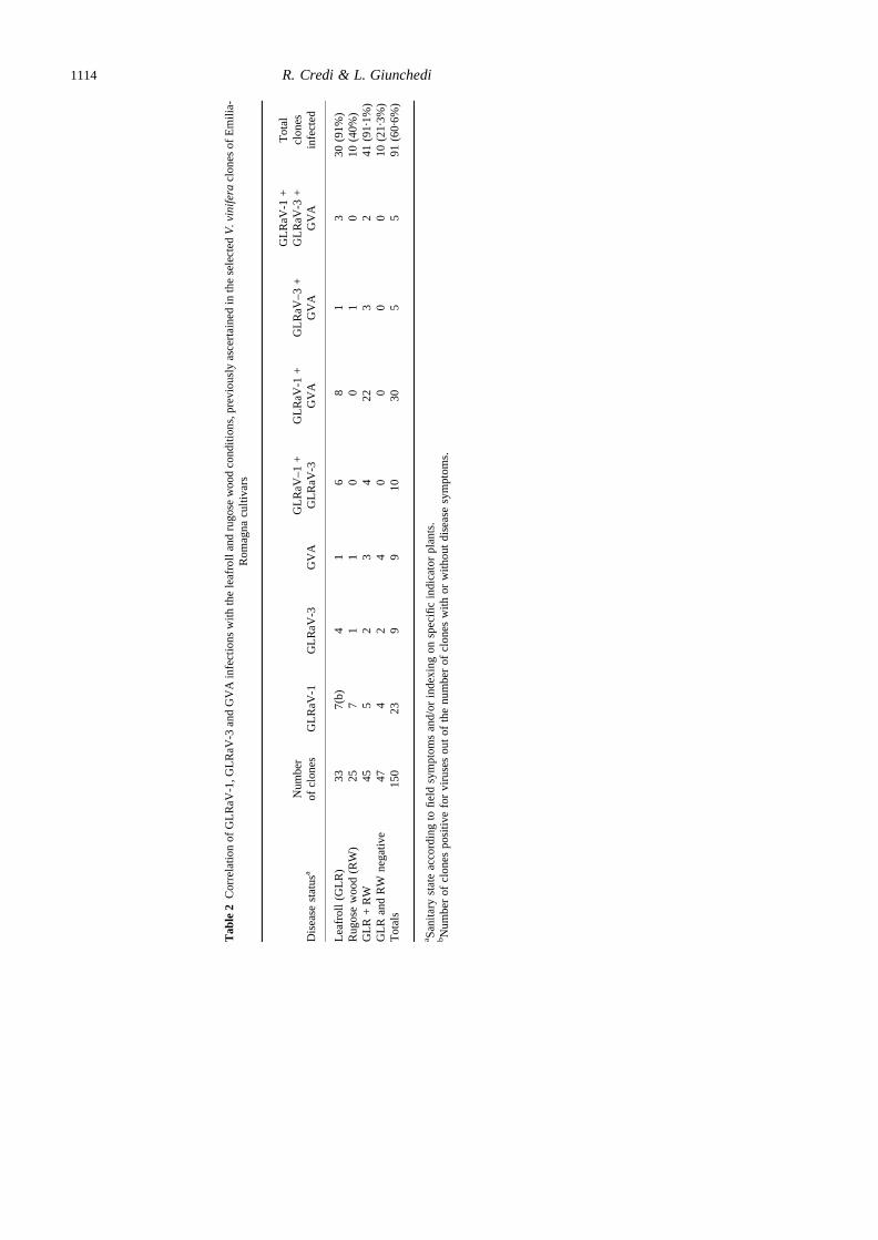
Correlation of virus infections with GLR andRW diseases
Table 2 shows the incidence of viruses in the cloneswhich were, or were not, affected by GLR and RW.The ELISA data revealed that 30 (91%) of the 33GLR-diseased clones were infected by viruses. Themost prevalent infection was that of GLRaV-1alone and in combination with GVA or GLRaV-3.Only 10 (40%) of the 25 RW-affected clonesrevealed viruses. Again the most prevalent one wasGLRaV-1. Forty-five clones were known to containmultiple GLR and RW diseases, and 41 of these(91.1%) were ELISA-positive, the majority con-taining the GLRaV-1 and GVA association. Finally,10 (21.3%) of the 47 clones rated free from thesediseases also appeared to contain viruses.
DISCUSSION
The occurrence of filamentous virus-like particles inclones of severalV. viniferacultivars, selected in aregion in north Italy, was first demonstrated byelectron microscopy observations. In ISEM decora-tion studies, they were coated by the GLRaV-1,GLRaV-3 or GVA antisera. Hybrid-like particleswere sometimes observed in grapevine tissueextracts, subsequently found to contain multipleviruses with ELISA. A similar phenomenon hasalready been reported by Milneet al. (1984) as apossible end-to-end aggregation of two different virusparticle serotypes.
The widespread presence of these viruses in thefoundation planting, and their incidence, wassubsequently demonstrated with ELISA. GLRaV-1 was the most frequent of the single-virus
infections. This virus combined with GVA wasalso the most prevalent of the mixed infections.These findings differed from those reported for thesouth of Italy by Digiaroet al. (1994) who, on thecontrary, found higher GLRaV-3 levels. GLRaV-2and GLRaV-5 were not detected, indicating thatthey do not commonly infect grapevine in thisviticultural region.
ELISA data also made it possible to analyse therelationship between the presence of GLRaV-1,GLRaV-3 and GVA with the GLR and RWconditions, previously determined on the basis offield symptoms and/or traditional woody indexing.In particular, the study confirmed the strongassociation (91%) of these viruses with GLR, aswell as multiple GLR and RW diseases. On theother hand, only 40% of the clones affected by RWon its own were ELISA-positive. Recently, theclose association between GVA and KSG wasreported (Garauet al., 1994; Chevalieret al., 1995).To explore the significance of these findings, morerefined woody indexing tests with our RW-affectedsources are needed to differentiate between thesyndromes now considered to be different compo-nents, or diseases, of the RW complex (Martelli,1993). Moreover, the possible occurrence of grape-vine virus B (GVB), reported by Bosciaet al.(1993), and a similar but serologically distinct onecalled grapevine virus C (GVC), reported byMonette & James (1991), must also be investigated.
In our survey, screening of grapevine sources,found to be negative for GLR and RW withbiological indexing, also revealed some virusinfections. One of the explanations of these positiveELISA reactions may be that woody indexing testsare not completely reliable as results can be affectedby various factors. On the other hand, the absenceof positive reactions in a few extracts of GLR-affected grapevines is not surprising, because otherclostero-like virus particles may be involved. Forexample, the possible occurrence of GLRaV-4 (Huet al., 1990) and GLRaV-6 (Bosciaet al., 1995)remains unknown as they were not investigated.
In conclusion, the study demonstrates the aetio-logic complexity of GLR and RW. From a practicalpoint of view, however, the use of the three antiserain ELISA seems to be reliable enough to detect thefirst disease, singly or in association with the secondone. For almost all the samples, the results ofELISA testing and indexing on woody indicatorswere almost identical. The opposite was true whenRW phenomena were found in the absence ofGLR. Research is needed to provide more informa-tion on the biology of these grapevine infectiousdiseases.
Grapevine leafroll-associated viruses 1115
ACKNOWLEDGEMENTS
The authors wish to thank Drs D. Boscia, M. Conti,D. Gonsalves and P. Gugerli for their kindassistance with antisera.
The research supported by the National ResearchCouncil of Italy, Special Project RAISA, Subpro-ject no. 2, Paper No. 2789.
REFERENCES
Boscia D, Aslouj E, Elicio V, Savino V, Castellano MA,Martelli GP, 1992. Production characterization and useof monoclonal antibodies to grapevine virus A.Archives of Virology127,185–94.
Boscia D, Greif C, Gugerli P, Martelli GP, Walter B,Gonsalves D, 1995. Nomenclature of grapevine leaf-roll-associated putative closteroviruses. Vitis 34, 171–5.
Boscia D, Savino V, Minafra A, Namba S, Elicio V,Castellano MA, Gonsalves D, Martelli GP, 1993.Properties of a filamentous virus isolated from grape-vines affected by corky bark. Archives of Virology130,109–20.
Chevalier S, Greif C, Clauzel JM, Walter B, Fritsch C,1995. Use of an immunocapture-polymerase chainreaction procedure for the detection of grapevine virusA in Kober stem grooving-infected grapevines. Journalof Phytopathology143,369–73.
Clark MF, Adams AN, 1977. Characteristics of themicroplate method of enzyme-linked immunosorbentassay for the detection of plant viruses. Journal ofGeneral Virology34, 475–83.
Conti M, Milne RG, Luisoni E, Boccardo G, 1980. Aclosterovirus from a stem-pitting-diseased grapevine.Phytopathology70, 394–9.
Credi R, Babini AR, 1987. Miglioramento sanitario dellavite ed incidenza di alcune malattie da virus e virus-simili nelle regioni dell’Emilia-Romagna e Piemonte.Comptes Rendus du IVe Symposium International sur laSelection Clonale de la Vigne,Nyon-Changins(Suisse), 1986. Recherche Agronomique en Suisse26,328–31.
Digiaro M, Popovic Bedzrob M, D’Onghia AM, Boscia D,Savino V, 1994. On the correlation between grapevinevirus A and rugose wood. Phytopathologia Mediterra-nea33, 187–93.
Garau R, Prota VA, Piredda R, Boscia D, Prota U, 1994.On the possible relationship between Kober stemgrooving and grapevine virus A. Vitis 33, 161–3.
Gugerli P, 1987. Grapevine leafroll disease: rapiddiagnosis by electron microscopy and serology.Comp-tes Rendus de IVe Symposium International sur laSelection Clonale du la Vigne, Nyon-Changins(Suisse), 1986. Recherche Agronomique en Suisse26,388–9.
Gugerli P, Brugger JJ, Bovey R, 1984. L’enroulement dela vigne: mise en e´vidence de particules virales etdeveloppement d’une me´thod immuno-enzymatiquepour le diagnostic rapide. Revue Suisse ViticultureArboriculture Horticulture16, 299–304.
Hu JS, Gonsalves D, Teliz D, 1990. Characterization ofclosterovirus-like particles associated with grapevineleafroll disease. Journal of Phytopathology128,1–14.
Martelli GP, ed., 1993.Graft-Transmissible Diseases ofGrapevines. Handbook for Detection and Diagnosis.Rome, Italy: FAO Publications Division.
Milne RG, 1984. Electron microscopy for the identifica-tion of plant virusesin vitro preparations. In: Mar-amorosch K, Koprowski H, eds.Methods in Virology.Orlando, Florida, USA: Academic Press, Vol. 7, 87–120.
Milne RG, Conti M, Lesemann DE, Stellmach G, Tanne E,Cohen J, 1984. Closterovirus-like particles of two typesassociated with diseased grapevines. Phytopatholo-gische Zeitschrift110,360–68.
Monette PL, James D, 1991. Detection of a closterovirus-like particle from a corky bark-affected grapevinecultivar. Vitis 30, 37–43.
Rosciglione B, Gugerli P, 1986. Maladies de l’enroule-ment et du bois strie´ de la vigne: analyse microscopiqueet serologique. Revue Suisse Viticulture ArboricultureHorticulture 18, 207–11.
Teliz D, Tanne E, Gonsalves D, Zee F, 1987. Fieldserological detection of viral antigens associated withgrapevine leafroll disease. Plant Disease71, 704–9.
Zee F, Gonsalves D, Goheen A, Kim KS, Pool R, Lee RF,1987. Cytopathology of leafroll-diseased grapevinesand the purification and serology of associatedclosterovirus-like particles. Phytopathology77, 1427–34.
Zimmermann D, Bass P, Legin R, Walter B, 1990.Characterization and serological detection of fourclosterovirus-like particles associated with leafrolldisease on grapevine. Journal of Phytopathology130,205–18.
Zimmermann D, Walter B, Le Gall O, 1988. Purifica-tion de particules virales associe´es a l’enroulementde la vigne et mise au point d’un protocole ELISApermettant leur de´tection. Agronomie8, 731–41.
R. Credi & L. Giunchedi1116


![Grape Entomology Update: Grapevine Leafroll and Grape ...•Treatments (2011 & 2012)-Spirotetramat [Movento] (Yes or No) •Experimental design-plot size: 12 vines X 4 rows = 48 vines](https://img.dokumen.tips/doc/110x75/6126bf982fdf005bb9434b19/grape-entomology-update-grapevine-leafroll-and-grape-atreatments-2011-.jpg)