Embed Size (px)
Citation preview

Graduate Theses, Dissertations, and Problem Reports
2016
Grainyhead-like-2 inhibits the co-activator p300, suppressing Grainyhead-like-2 inhibits the co-activator p300, suppressing
tubulogenesis and the epithelial-mesenchymal transition tubulogenesis and the epithelial-mesenchymal transition
Phillip M. Pifer
Follow this and additional works at: https://researchrepository.wvu.edu/etd
Recommended Citation Recommended Citation Pifer, Phillip M., "Grainyhead-like-2 inhibits the co-activator p300, suppressing tubulogenesis and the epithelial-mesenchymal transition" (2016). Graduate Theses, Dissertations, and Problem Reports. 6420. https://researchrepository.wvu.edu/etd/6420
This Dissertation is protected by copyright and/or related rights. It has been brought to you by the The Research Repository @ WVU with permission from the rights-holder(s). You are free to use this Dissertation in any way that is permitted by the copyright and related rights legislation that applies to your use. For other uses you must obtain permission from the rights-holder(s) directly, unless additional rights are indicated by a Creative Commons license in the record and/ or on the work itself. This Dissertation has been accepted for inclusion in WVU Graduate Theses, Dissertations, and Problem Reports collection by an authorized administrator of The Research Repository @ WVU. For more information, please contact [email protected].

Grainyhead-like-2 inhibits the co-activator p300, suppressing tubulogenesis and the
epithelial-mesenchymal transition
Phillip M. Pifer
Dissertation submitted to the School of Medicine at West Virginia University in partial
fulfillment of the requirements for the degree of
Doctor of Philosophy in Cancer Cell Biology
Karen Martin, PhD, Committee Chairperson
Michael Schaller, PhD
Peter Mathers, PhD
Peter Stoilov, PhD
David M. Smith, PhD
Steven M. Frisch, PhD, Mentor
Cancer Cell Biology Program
Morgantown, West Virginia
2016
Keywords: EMT, grainyhead-like-2, p300, CBP, histone acetyltransferase, MDCK, tubulogenesis
Copyright 2016 Phillip M. Pifer

Abstract Grainyhead-like-2 inhibits the co-activator p300, suppressing tubulogenesis and the
epithelial-mesenchymal transition
Phillip M. Pifer
Tumor progression and developmental morphogenesis require transient or stable
Epithelial-Mesenchymal transitions (EMT) to modulate epithelial junctional complexes allowing programmed migration, invasion, and anoikis resistance of cells. Grainyhead-like-2 (GRHL2), an epithelial master-regulatory transcription factor, suppresses EMT and causes a mesenchymal-epithelial transition to the default epithelial phenotype. This body of work investigates the role of GRHL2 in kidney cancer, in kidney development using a kidney specific GRHL2 knockout mouse model, and in tubulogenesis using Madin-Darby Canine Kidney (MDCK) cells. In kidney cancer, GRHL2 was not expressed in the normal kidney proximal tubule epithelial cells which are thought to be the progenitors of clear cell renal cell carcinomas. However, GRHL2 could function as biomarker to differentiate renal cell carcinoma subtypes. In kidney development, a large amount of evidence suggests Grhl2 is a critical regulator of this process. However, kidney specific Grhl2 knockout mice did not elucidate the role of Grhl2 in the developing kidney for technical reasons, and this area needs to be further investigated. In the MDCK model, GRHL2 was required for cystogenesis, but it suppressed Hepatocyte Growth Factor-induced tubulogenesis, a process requiring transient, partial EMT. Subsequent results demonstrated that GRHL2 suppressed tubulogenesis by inhibiting the histone acetyltransferase co-activator, p300, thereby preventing the induction of matrix metalloproteases and other p300-dependent genes required for tubulogenesis. A thirteen amino acid region (425-437aa) of GRHL2 was required and sufficient for the inhibition of p300 histone acetyltransferase activity, the suppression of tubulogenesis and the interference with EMT. These results demonstrate that p300 is required for EMT plasticity occurring in tubulogenesis or tumor progression, and that GRHL2 suppresses EMT in both contexts, through inhibition of p300.

iii
Dedication
This dissertation is dedicated to all my family members who have fought cancer, especially my grandfather Harry Higgs. I would also like to dedicate this work to my family and friends whose unwavering support allows me to pursue my passions. I would specifically like to thank my parents, Michael and Ann Pifer, my brother and his family, Joseph, Miranda, and Aven, and my amazing fiancé, Colleen Beatty. I would also like to thank my grandmother, Judy Higgs, for her careful reading of the dissertation.

iv
Acknowledgments
Steven Frisch for his unparalleled mentorship. His research career has been incredibly impactful, and his dedication to scientific advancement is unmatched. He demonstrates critical thinking skills, passion, and persistence that scientific research requires, and I have learned a wealth of knowledge from him.
Fred Minnear and the MD/PhD admissions committee for my acceptance into the program.
My dissertation committee including Karen Martin, Peter Mathers, Peter Stoilov, Mike Schaller, and David Smith for their unwavering support and valuable scientific insight throughout my pursuit of this degree.
Special thanks to Mike Lutz laying the foundation for my love of chemistry during my high school education.
Thanks to Jeffrey Petersen, who convinced me to become a Chemistry major during my first day of college, and whose mentorship encouraged me to pursue a career in both medicine and research.
Thanks to Justin Legleiter, who was an exceptional undergraduate research mentor and was the first person to encourage me into a MD/PhD dual doctoral degree.
I would like to thank my labmate Joshua Farris, for the constant companionship during the ups and downs of the last four years pursuing a Ph.D. degree. I would also like to thank previous lab members, Benjamin Cieply and Sun Hee Park, for their excellent training and advice.
Finally, I would like to thank the MD/PhD program for both financial support to attain these degrees as well as consistent mentorship from both faculty and students to guide me through the difficult stages of my training.

v
Table of Contents Title Page…..………………………………………………………………………………………i Abstract….………………………………………………………………………………………...ii Dedication………………………………………………………………………………………...iii Acknowledgements……………………………………………………………………………….iv Table of Contents….….....………………………………………………………………………...v List of Abbreviations..……………………………………………………………………………vi Chapter 1 - Introduction and Literature Review………..……………………………………...….1 Chapter 2 - Grainyhead-like-2 Inhibits the Co-activator p300, Suppressing Tubulogenesis and the
Epithelial-Mesenchymal Transition...……………………………………………………………60
Chapter 3 – GRHL2 in Kidney Cancer and Development..……………………………………138 Chapter 4 – Overall Discussion and Future Directions……………………….………………..171 Chapter 5 – Curriculum vitae...…………………………………………………………………181 Appendix AI: Cieply B., Riley P., Pifer PM., et al., Suppression of the Epithelial-Mesenchymal Transition by Grainyhead-like-2. Cancer Research 72(9); 2440-53. Appendix AII: Farris J., Pifer PM., et al., Grainyhead-like 2 Reverses the Metabolic Changes Induced by the Oncogenic Epithelial-mesenchymal Transition: Effects on Anoikis. Molecular Cancer Research (In Press)

vi
LIST OF ABBREVIATIONS
ac acetylation
acetyl-CoA acetyl-coenzyme A
AKT protein kinase B
ALL acute lymphoid leukemia
AP-1 activator protein 1
Axd Axial defect
Bd bromodomain
BMP Bone morphogenetic protein
CBP Creb-binding protein p300
ccRCC cell renal cell carcinoma
ChIP Chromatin Immunoprecipitation
Cldn3 claudin 3
Cldn4 claudin 4
CREB cAMP responsive element binding protein
CSC Cancer stem cells
DNA deoxyribonucleic acids
E1a adenovirus early region 1A protein
E-cadherin epithelial-cadherin
EDC epidermal differentiation complex
EGF epidermal growth factor
EID E1a-like inhibitor of differentiation protein
EMT epithelial-mesenchymal transition
ENU N-ethyl-N-nitrosourea
Ep-CAM epithelial cell adhesion molecule
ERK extracellular signal-regulated kinase
ESRP1/2 epithelial splicing regulatory proteins 1 and 2
Etv4 Ets variant 4 transcription factor

vii
Etv5 Ets variant 5 transcription factor
FBS fetal bovine serum
FGF fibroblast growth factor
FGFR2-IIIb Fibroblast growth factor receptor 2-IIIb
Foxj1 forkhead box J1
GDNF glial cell line-derived neurotrophic factor
GLUD1 glutamate dehydrogenase 1
Grh grainyhead
GRHL1 Grainyhead-like-1
GRHL2 Grainyhead-like-2
GRHL3 Grainyhead-like-3
H3 histone 3
H4 histone 4
HAT histone acetyltransferase domain
HBE Human bronchial epithelial
HDAC1 histone deacetylase 1
HDAC2 histone deacetylase 2
HGF hepatocyte growth factor
HIFα hypoxia-induced factor alpha
HIV human immunodeficiency virus
hTERT human telomerase reverse transcriptase
IBID IRF-3 Binding Domain
ISH In Situ Hybridization
K lysine
KIX kinase inducible domain
Krt5 keratin 5
LBP1a upstream binding protein 1
LBP-9 transcription factor CP2-like 1
MAPK mitogen-activated protein kinase
MDCK Madin-Darby Canine Kidney

viii
me methylation
me1 Monomethylation
me3 trimethylation
MET mesenchymal-epithelial transition
miMCD mouse inner medullary collecting duct
MLL mixed lineage leukemia
MMP matrix metalloproteases
MOZ monocytic leukemia zinc finger
MSP mesenchymal subpopulation
MUC5AC mucin 5AC, oligomeric mucus/gel-forming
MZT maternal to zygotic transition
Nkx2-1 NK2 homeobox 1
OVOL1 ovo-like transcriptional repressor 1
OVOL2 ovo-like transcriptional repressor 2
p300 E1A binding protein p300, Ep300, and KAT3B
p53 tumor protein p53
p63 tumor protein p63
PDGF platelet-derived growth factor
pEMT partial epithelial-mesenchymal transition
PHD plant homeodomain
PI3K phosphatidylinositol-3-kinases
PSG penicillin-Streptomycin-Glutamine
qPCR quantitative polymerase chain reaction
RET ret proto-oncogene
RING Really Interesting New Gene domain
RNA ribonucleic acid
RTS Rubinstein-Taybi syndrome
Scgb1a1 secretoglobin family 1A member 1
shRNA short hairpin RNA
Six1 SIX homeobox 1transcription factor

ix
Spint1 serine peptidase inhibitor Kunitz type 1
TFCP2 transcription factor CP2
TGF-β transforming growth factor beta
TIC tumor initiating cells
TJP1 tight junction protein 1
VHL Von Hippel-Lindau
ZEB1 Zinc finger E-box-binding homeobox 1

1
Chapter 1
Introduction and Literature Review

2
Introduction to the Epithelial-Mesenchymal Transition
Epithelial Cells
Epithelial cells enclose organs and glands forming a protective barrier by directly
interacting with neighboring cells. In two dimensional culture, epithelial cells form an intact
cobblestone monolayer with high transepithelial electrical resistance. In three dimensional
matrices such as collagen or matrigel, epithelial cells form spherical cysts that have a distinct cell
polarity. The basal side of the epithelial cell attaches to the extracellular matrix through the
basement membrane, and the apical side faces the lumen of the duct where it can secrete
molecules into the lumen and control the uptake of molecules. Polarity is essential for the
survival of epithelial cells as detachment from the extracellular matrix leads to anoikis. Anoikis
is programmed cell death caused by detachment from an extracellular matrix (Frisch and Francis,
1994). Epithelial cells form barriers through the interaction of specific epithelial proteins such as
epithelial-cadherin (E-cadherin), claudins, occludins, and desmogleins (Baum and Georgiou,
2011; Turksen and Troy, 2011). E-cadherin’s extracellular domain forms a homophilic
interaction with other E-cadherin molecules of adjacent cells, forming adherens junctions. The
cytosolic domain of E-cadherin is important for cortical actin assembly and the sequestration of
β-catenin (Qin et al., 2005; Tian et al., 2011). Claudins and occludins contribute to cell polarity
by forming tight junctions which restrict integral membrane protein movement to the apical and
basolateral membranes (Yeaman et al., 1999). In the cytosol, epithelial specific proteins, Rab25
and Ankyrin G, help ensure that junctional proteins are localized properly (Kizhatil et al., 2007a;
Kizhatil et al., 2007b). Two major characteristics of epithelial cells are cell-cell adhesions and
cell polarity.

3
Our laboratory has proposed that the epithelial cell is the default cell phenotype in both
development and cancer. Under this hypothesis, the epithelial phenotype is adopted
automatically by a cell in the absence of genomic/epigenetic alterations or inductive
differentiation signaling (Frisch, 1997). Developmentally, E-cadherin-expressing cells are the
first to emerge, at the late Two-cell stage; these eventually give rise to all differentiated cell
types of the adult organism (Hyafil et al., 1980; Vestweber and Kemler, 1985; Larue et al.,
1994). This idea has been invoked in recent studies from other laboratories to explain, for
example, the mesenchymal-epithelial transition (MET) occurring at metastatic sites (Thiery and
Sleeman, 2006; Polyak and Weinberg, 2009; Tam and Weinberg, 2013). The regulation of the
epithelial phenotype is important for understanding all of these processes.
Mesenchymal Cells
Mesenchymal cells lack many of the cell-cell adhesion molecules that epithelial cells
express, and therefore lack significant cell-cell adhesion and polarity (Taube et al., 2010).
However, mesenchymal cells express their own unique markers such as N-cadherin, fibronectin,
and vimentin which are not present or have low expression in epithelial cells. Mesenchymal
cells are able to migrate and invade as individual cells and can survive independent of an
extracellular matrix interaction. This property makes mesenchymal cells resistant to anoikis
(Frisch and Francis, 1994; Scheel et al., 2011; Frisch et al., 2013). Mesenchymal cells are
essential for the formation of the extracellular matrix and may function as mesenchymal stem
cells in some populations such as chondrocytes, osteoblasts, and adipocytes (Dominici et al.,
2006).

4
Physiological Epithelial-Mesenchymal Transition
Epithelial cells can transform into mesenchymal cells by a process referred to as the
epithelial-mesenchymal transition (EMT). EMT is associated with loss of cell-cell adhesion
molecules and cell polarity and gain of motility, anoikis resistance, and chemotherapeutic
resistance. EMT is classified into three different types: Type 1- Developmental EMT, Type 2-
Fibrosis and tissue regeneration, and Type 3- Cancer progression and metastasis (Kalluri and
Weinberg, 2009). During development, cells demonstrate significant plasticity by undergoing
EMT and the opposite process, mesenchymal-epithelial transitions (MET). In mice, the
depletion of Snail1 and Snail2, critical EMT driving transcription factors, leads to failed
gastrulation because the epithelial cells are unable to EMT thereby preventing the formation of
the primitive streak (Hudson et al., 2009). The reverse process, MET, is essential for neural tube
formation where epithelial cells are required for neural tube closure. Depletion of Grainyhead-
like-2, an epithelial regulating transcription factor, prevents neural tube closure in mice (Rifat et
al.; Werth et al., 2010; Pyrgaki et al., 2011). After neural tube closure, another sequence of
EMT and MET is responsible for the neural crest delamination and the formation of notochord
and somites (Thiery et al., 2009).
Type 2 EMT occurs in organ fibrosis, wound healing, and tissue regeneration. Wound
healing requires myofibroblasts for restructuring of the extracellular matrix and wound
contractility, and keratinocytes undergo a partial EMT to form an attached epithelial sheet over a
wound (Savagner et al., 2005; Thiery et al., 2009). Also, kidney epithelial cells may undergo an
EMT during kidney fibrosis (Liu, 2010).

5
EMT in Cancer
Type 3 EMT occurs in metastasis and cancer progression. EMT is a physiological
process that is hijacked by cancer to increase survival and invasion. Type 3 EMT gives a
survival advantage to cancer cells by increasing motility, invasiveness, anoikis resistance, and
chemotherapeutic resistance (Kalluri and Weinberg, 2009). EMT can contribute to a
chemotherapeutic resistant cancer sub-population leading to tumors that are refractory to
treatment (Woodward et al., 2007; Barr et al., 2008; Li et al., 2008). These characteristics allow
cancer cells to metastasize from the primary tumor site and form colonies in other parts of the
body (Kalluri and Weinberg, 2009). Metastasis is responsible for the majority of cancer related
deaths. Carcinomas, cancers of the epithelium, undergo multiple genetic hits leading to
uncontrolled proliferation during the progression from normal epithelium to an invasive
carcinoma. However, metastatic dissemination does not occur until invasive properties of the
cell have been increased which are usually conferred by type 3 EMT. Cells that have undergone
an EMT are known to upregulate matrix metalloproteases (MMP) which are responsible for the
degradation of the extracellular matrix allowing cancer cells to invade through the basement
membrane and disseminate (Ala-aho et al., 2002; Culhaci et al., 2004; Yuan et al., 2005; Park et
al., 2008a; Mehner et al., 2014). After the cancer cell invades through the basement membrane,
the cell can intravasate, be transported via the circulatory system to a distant site, extravasate,
and establish a distant metastasis (Kalluri and Weinberg, 2009). It should be noted that for
metastasis to occur, the cells must become anoikis resistance because this process requires
detachment from the extracellular matrix, which under normal physiological conditions this
would induce apoptosis in epithelial cells (Frisch et al., 2013).

6
Mechanistically, one of the most studied phenomena of the EMT is loss of E-cadherin
and its subsequent implications in cancer. Constitutive E-cadherin expression in cancer cell
lines can inhibit cancer’s invasive properties (Frixen et al., 1991), and E-cadherin expression has
been shown to inversely predict cancer metastasis (Dorudi et al., 1993; Younis et al., 2007). E-
cadherin could block the invasive traits by structurally interacting with other epithelial cells via
its homophilic interaction keeping the cells in close contact and preventing invasion. Also, E-
cadherin sequesters factors such as β-catenin (Wnt pathway), NRAGE (pro-apoptotic factor),
SMAD3 (TGF-β pathway), and YAP/TAZ (Hippo Signaling) on the cellular membrane
preventing pathway activation, and the loss of E-cadherin can cause the activation of these
respective pathways thereby promoting an EMT.
Cancer stem cells (CSC), also known as tumor initiating cells (TIC), have been shown to
recapitulate tumors in a variety of different cancers such as leukemia, lymphoma, breast, and
brain cancer (Bergsagel, 1967; Terpstra et al., 1996; Bonnet and Dick, 1997; Al-Hajj et al.,
2003; Gupta et al., 2009b). The epithelial-mesenchymal transition can cause cells to acquire
CSC properties such as the ability to form mammospheres, differentiate into two separate cell
populations, and generate tumors in animals that recapitulate the heterogeneity of tumor
histology (Mani et al., 2008). The clinical significance of CSC theory is that one cell can cause a
tumor recurrence suggesting the eradication of all tumorigenic cells is necessary for the
prevention of recurrence and metastasis. The CSC theory is still debated, and the percentage of
tumor that CSCs comprise is controversial (Gupta et al., 2009a). In breast cancer research, one
of the most commonly accepted CSC markers is the CD44High/CD24Low phenotype. In human
mammary luminal epithelial (HMLE) cells, normal epithelial cells (CD44low/CD24high) undergo
an EMT causing the cells to exhibit the CD44High/CD24Low mesenchymal subpopulation

7
phenotype. This conversion is associated with increased ability of the CD44High/CD24Low cells
to form tumors in mice (Mani et al., 2008), and this phenotype is supported by TFG-β and Wnt
autocrine and paracrine signaling (Scheel et al., 2011).
Regulation of EMT
Many EMT promoting factors have been discovered, but only a few EMT inhibitory
factors have been found. Growth factors derived from the mesenchymal stroma such as EGF,
HGF, PDGF, and TGF-β can promote an EMT (Kalluri and Weinberg, 2009; Xu et al., 2009).
The activation of multiple pathways such as RAS/MAPK/ERK, PI3K/AKT, β-catenin/LEF, and
TGF-β/SMAD3 (usually activated by the growth factors just described) lead to the upregulation
of EMT driving transcription factors (Kalluri and Weinberg, 2009; Sanchez-Tillo et al., 2012).
Two of these transcription factors, ZEB1 and Snail, are E-box binding transcription factors and
function by direct repression of the E-cadherin promoter (Cano et al., 2000; Grooteclaes et al.,
2003). Other EMT promoting transcription factors such as Twist, Goosecoid, and FOXC2
function more indirectly, usually upregulating ZEB1 or Snail (Thiery et al., 2009; Taube et al.,
2010). These mechanisms emphasize the importance of E-cadherin downregulation for EMT to
occur.
Inhibitors of EMT have remained fairly elusive. Bone morphogenetic protein-7 (BMP7),
a member of the TGF-β superfamily, can prevent EMT and cause MET by inhibiting TGF-β
signaling (Zeisberg et al., 2003). Other BMP family members are able to inhibit TGF-β signaling
and prevent EMT (Cieply et al., 2012). The mir-200 microRNA family is downregulated in
EMT and when constitutively expressed, the microRNA family can suppress EMT by repressing
ZEB1/2. Alternatively, ZEB1/2 are able to repress mir-200 microRNA expression thereby
forming a negative feedback loop (Gregory et al., 2008; Wellner et al., 2009; Gregory et al.,

8
2011). The splicing regulatory proteins, epithelial splicing regulatory proteins 1 and 2
(ESRP1/2), are responsible for epithelial specific alternative splicing events in the cell such as
FGFR2-IIIb, an epithelial specific FGFR2 isoform. ESRP1/2 shRNA depletion causes an EMT,
and ESRP1 overexpression causes MDA-MB-231 cells to re-express epithelial alternative
spliced isoforms (Warzecha et al., 2009; Warzecha et al., 2010). The first EMT inhibiting
transcription factor discovered was GRHL2 which will be discussed at length in subsequent
sections (Cieply et al., 2012). Another family of EMT inhibiting transcription factors is the
ovo-like transcription factor family, OVOL1 and OVOL2, which function by repressing the
ZEB1 promoter and upregulating ESRP1/2 (Roca et al., 2013).
Also, our previous work reported that adenovirus E1A establishes an epithelial
phenotype, suppresses EMT, enhances anoikis, suppresses MMP expression through AP-1
signaling, and acts as tumor suppressor (Frisch, 1991, 1994, 1996; Somasundaram et al., 1996;
Frisch, 2001; Frisch and Mymryk, 2002). One of the major functions of E1A is its modulation of
p300. A deletion of E1A n-terminus amino acids 2-36 prevents interaction with p300 and is
unable to transform cells (Stein et al., 1990). Of utmost importance in this work, E1A
demonstrates functional similarity with the GRHL2 which will be discussed subsequently.
Epigenetics and Post-translational modifications of histones
The blueprint of the human body is comprised of paired deoxyribonucleic acids, DNA,
with a double helix structure discovered by Watson and Crick in 1953 (Watson, 1953). The
DNA comprising one human genome is approximately 2 meter in length, but it fits inside almost
every cell in the human body (Dancy and Cole, 2015). DNA is organized on nucleosomes, the
fundamental subunit of chromatin, and is packaged into a cell’s nucleus being about 10 µm in
diameter. A nucleosome is comprised of 146 DNA base pairs bound to a histone octamer

9
containing duplicates of H2A, H2B, H3, and H4 (Dancy and Cole, 2015). Although, DNA
contains the genetic information for all genes, it is becoming readily apparent that epigenetic
regulation is essential for proper gene expression (Kiefer, 2007). Chromatin structure
conformation is regulated by histone modifications and DNA modification which are some of the
important factors that determine the “On/Off” status of the DNA/Chromatin complex. The “On”
state chromatin conformation means the chromatin is open and permissive for gene expression
machinery to bind and function. The “Off” state chromatin conformation means the chromatin is
closed and inaccessible to gene expression machinery (Dancy and Cole, 2015). Histone 3 (H3)
is highly regulated during this “On/Off” switch, and H3 is one of the proteins studied in the work
presented subsequently. Post-translational modifications of H3 occur on the n-terminus of the
protein, most frequently on lysine (K) residues. Two of the most important histone modification
are methylation and acetylation, but others modifications occur such as phosphorylation,
deimination, ADP ribosylation, ubiquitylation, and sumoylation (Bannister and Kouzarides,
2011). Methylation of H3K4 is a signature of transcriptional activation. Monomethylation
(me1) of H3K4 denotes enhancer activation and trimethylation (me3) of H3K4 denotes promoter
activation (Calo and Wysocka, 2013). H3K4 methylation is mediated by the SET1/MLL histone
methylase family (Yokoyama et al., 2004; Dou et al., 2005; Cho et al., 2007b). However, most
other H3 methylation marks such as H3K27me3 and H3K9me3 are repressive gene signature
marks (Kim and Kim, 2012). Acetylation of the H3 tail is a signature of transcriptional
activation, and frequently occurs on H3K27 and H3K9 (Creyghton et al., 2010; Karmodiya et
al., 2012). H3K27ac is mediated by the histone acetyltransferase, p300 (Kasper et al., 2010).
H3K4 and H3K27 distinguish the enhancer activation state. The H3K4me1 and H3K27me3
histone mark is a signature of a poised but not active enhancer, and H3K4me1 and H3K27ac is a

10
signature of an active enhancer (Creyghton et al., 2010). Histone modifications are highly
regulated to allow epigenetic control of gene expression.
Plasticity in EMT/MET
Although EMT has been shown to increase metastasis, recent emerging studies suggest
that EMT plasticity, the adaptability of a cell to change between an epithelial and a mesenchymal
phenotype, is more important than the permanent conversion to mesenchymal cells for forming
metastases. For example, when Twist1, an EMT promoting transcription factor, is transiently
expressed, it promotes metastasis. However, constitutive Twist1 expression prevents metastasis
formation (Schmidt et al., 2015). Other studies have demonstrated that a MET may enhance
metastatic colonization (Kalluri and Weinberg, 2009). Conceptually, tumor cells exist as
epithelial cells in the primary tumor site, when the primary site undergoes stress (hypoxia,
chemotherapy, or other selective pressures), the tumor cells undergo an EMT allowing for
anoikis resistance and increased invasion. The EMT tumor cells can then spread to a distant site
where the tumor cell undergoes a MET and forms a distant metastasis with epithelial
characteristics (Bissell et al., 2002; Jechlinger et al., 2002). The EMT-MET process is supported
by the fact that breast cancer metastases express E-cadherin and demonstrate other epithelial
characteristic (Kowalski et al., 2003). The EMT-MET process has two significant implications.
First, the prevention of either the EMT or the MET could prevent metastasis. Secondly, the
EMT cancer cells are relatively resistant to chemotherapeutic treatment making them very hard
to eradicate. The two clinical solutions to this issue would be to lock the tumor cells in the
epithelial state and eradicate using standard chemotherapeutic methods, or design cancer
treatments that specifically kill the EMT/CSC phenotype.

11
As more publications demonstrate the importance of both EMT and MET in the
formation of metastases, it becomes clear that cell plasticity between the two states may be the
most important factor contributing to metastatic disease. Both metastatic sites and tumor
recurrences are often resistant to first line chemotherapeutic treatment because of either
upregulation of compensatory signaling pathways or de novo mutations in the targeted pathway.
This process is associated with cell plasticity driven by epigenetic modification machinery. Cells
with this plasticity are able to survive stressors such as chemotherapy, oxidative stress, hypoxia,
nutrient deficiency, and matrix detachment. Drug-resistance, tumor recurrence, and disease
progression have been clearly linked to EMT and CSC (Creighton et al., 2009; Oliveras-Ferraros
et al., 2012; Mitra et al., 2015; Ye and Weinberg, 2015; Marcucci et al., 2016).
Importantly, both phenotypic and epigenetic plasticity contribute to recurrence and drug
resistance (Scheel and Weinberg, 2011; Singh et al., 2014; Ye and Weinberg, 2015; Doherty et
al., 2016). In breast cancer, increased levels of mesenchymal circulating tumor cells correlate
with cancer progression, and chemotherapy increases the shift to the mesenchymal phenotype
(Yu et al., 2013). Emerging evidence suggests EMT is driven by epigenetic re-programming
(Tam and Weinberg, 2013). For example, E-cadherin is epigenetically silenced by histone-
modifying enzymes leading to varying degrees of repression depending on the level of plasticity
needed. The histone methyltransferase Polycomb repressive complexes promote EMT by
catalyzing H3K27me3 of the E-cadherin promoter leading to transcriptional repression (Bracken
et al., 2006; Cao et al., 2008; Herranz et al., 2008). Histone acetylation is often is sign of
transcriptional activation, and during metastasis, Snail recruits histone deacetylases, HDAC1 and
HDAC2, to the E-cadherin promotor leading to silencing (Peinado et al., 2004; von Burstin et
al., 2009). Clinical trials using epigenetic therapies to target drug resistant solid tumors is

12
currently underway using de-acetylating and de-methylating agents (Brown et al., 2014). The
ZEB1 drug-resistant phenotype, an EMT-driven cancer cell subpopulation with increased
invasiveness and chemoresistance, has been reversed by using the HDAC1 inhibitor,
mocetinostat (Meidhof et al., 2015; Zhang et al., 2015). Furthermore, EMT transcription factors,
ZEB1 and Twist1, contribute to epigenetic modifications that occur in plasticity, the CSC
phenotype, and the drug resistant phenotype (Brabletz and Brabletz, 2010; Malouf et al., 2013).
Introduction to Grainyhead-like-2
Discovery of GRHL2
The grainyhead-like transcription factor family is named for grainyhead (Grh), the
Drosophila homolog. The Grh mutant causes defects in cuticle, trachea, and head formation
resulting in embryonic lethality. Abnormal head granular sclerites lead to a “grainyhead”
phenotype which is the namesake of the family (Nüsslein-Volhard et al.). Grh was discovered
for its regulation of the Ultrabithorax and dopa decarboxylase promoters (Bray et al., 1989;
Dynlacht et al., 1989). Grh interacts with the RNA polymerase II preinitiation complex for its
transcriptional activity (Dynlacht et al., 1991). Grh regulates trachea elongation, post-embryonic
neuroblasts, epidermal wound response, and the promoter of the Drosophila E-cadherin
homolog, shotgun (Hemphälä et al., 2003; Almeida and Bray, 2005; Mace et al., 2005).
Grhl1 and Grhl2 were discovered by using Grh protein sequence in a murine GenBank
protein search query (Wilanowski et al., 2002), and Grhl3 was discovered by querying similar
protein sequences to Grhl1 and Grhl2’s dimerization domain (Ting et al., 2003b). Human
GRHL2 has 94% homology with murine Grhl2. The grainyhead-like transcription factors are
part of the LSF/GRH transcription factor family. Phylogenetic analysis suggests two distinct

13
branches of this family, the GRH subfamily consists of GRHL1, GRHL2, and GRHL3, and the
LSF/CP2 subfamily consists of TFCP2, LBP1a, and LBP-9 transcription factors (Wilanowski et
al., 2002; Traylor-Knowles et al., 2010). The structure of GRHL1, 2, and 3 contain an N-
terminal transcriptional activation domain, a central DNA binding immunoglobulin fold domain
similar to p53, and a C-terminal dimerization domain which is predicted to have ubiquitin-like
folds (Kokoszynska et al., 2008). The GRHL proteins are able to homo- and hetero-dimerize
with each other, but they are not able to dimerize with other LSF/GRH transcription factors
(Wilanowski et al., 2002).
In murine studies, Grhl2 is observed at high expression levels in the primitive surface
ectoderm from E8.5 until E12.5 when the expression starts to decline until E15.5. At this time,
Grhl2 is only present in basal layer, stratum spinosum and stratum granulosum of the ectoderm.
During murine development, Grhl2 is present in kidney, lungs, olfactory epithelium, esophagus,
stomach, small and large intestine, urogenital tract, oral epithelium, and submandibular gland.
Interestingly, Grhl2 expression is inversely expressed in a spatial and temporal manner
compared to Grhl1 and Grhl3 in the developing lung and kidney (Auden et al., 2006).
Function of GRHL2 in epidermis
The role of GRHL2 has been intensively studied in the epidermis and associated skin
disorders. In keratinocytes, GRHL2 increases cell replicative life span by increasing telomerase
activity via upregulation of the hTERT promoter. GRHL2 knockdown decreases cell
proliferation, reduces epithelial thickness in three dimensional culture, and causes
hypermethylation of the hTERT promoter indicating that GRHL2 partially regulates hTERT
through an epigenetic mechanism (Chen et al., 2010b). Importantly, GRHL2 is required for the
upregulation of E-cadherin, claudin 4, and claudin 3, epithelial apical junction complex proteins

14
(Werth et al., 2010; Senga et al., 2012). Grhl2 binds the Cldn4 promoter and the second intronic
region of E-cadherin increasing promoter activity in both cases (Werth et al., 2010). Since
GRHL2 appears to be the master regulator of the epithelial phenotype, it is not surprising that
GRHL2 correlates with E-cadherin expression better than any other transcription factor (Werth et
al., 2010; Kohn et al., 2014), and Grhl2-/- mice show a reduction in E-cadherin and Cldn4
expression (Werth et al., 2010). GRHL2 also suppresses ZEB1 and upregulates mir-200c and
ESRP1/2 (Cieply et al., 2012; Cieply et al., 2013). Mir-200c is a microRNA associated with the
establishment of the epithelial phenotype by suppressing ZEB1 expression (Park et al., 2008b).
ESRP1 and ESRP2 are responsible for promoting epithelial specific RNA splicing of multiple
transcripts (Warzecha et al., 2009). In keratinocytes, GRHL2 and p63, a member of the p53
family, form a positive feedback loop which is necessary for proper epidermal development, and
the loss of p63 causes an EMT (Truong et al., 2006; Mehrazarin et al., 2015).
Both Grhl1-/- and Grhl3-/- mice show epidermal defects (Boglev et al., 2011), however,
embryonic lethality of Grhl2-/- mice at E11.5 precludes the examination of epidermal defects.
Grhl2-/+/ Grhl3-/+ mice have defects in epithelial wound repair. Furthermore, GRHL2-/+/ GRHL3-
/- mice have failure of eyelid closure and are born with open eyes. This suggests a defect in
epithelial cell migration in utero (Boglev et al., 2011).
GRHL2 prevents keratinocyte terminal differentiation by repressing the epidermal
differentiation complex (EDC). GRHL2 blocks the recruitment of Jmjd3, a histone demethylase,
to the EDC gene promoters (Chen et al., 2012). GRHL2 is also overexpressed in
hyperproliferative skin lesions seen in psoriatic tissue (Chen et al., 2012). A GRHL2 mutation
causes an autosomal-recessive ectodermal dysplasia syndrome in two consanguineous families
presenting with hypodontia, enamel hypoplasia, oral hyperpigmentation, nail dystrophy,

15
marginal palmoplantar keratodema, and dysphagia. Also, a fraction of the patients present with
sensorineural deafness. When the patient’s keratinocytes were cultured, the cells had decreased
epithelial features including defective epithelial adhesion junction complexes, altered cell
morphology, mis-localizition of GRHL2 in the cytoplasm, and decreased E-cadherin expression
(Petrof et al., 2014).
Function of GRHL2 in neural tube closure
Both Grhl2 and Grhl3 are involved in the neural tube closure. In the murine model,
primary neurulation occurs at three separate closure sites; hindbrain/cervical (closure 1),
forebrain/midbrain (closure 2), and rostral end of the brain (closure 3). For neural closure to
transpire, neural fold elevation must occur in conjunction with non-neural ectodermal-induced
formation of paired dorso-lateral hinge points (Shum and Copp, 1996). Grhl2-/- mice exhibit
failure of the rostral end of the forebrain closure (closure 3) leading to exencephaly and a split-
face malformation. Grhl2-/- mice fail to produce paired dorso-lateral hinges at which point
primary neurulation ceases and defects are observed (Rifat et al.), and these results were
confirmed in subsequent GRHL2 mouse models (Werth et al., 2010; Pyrgaki et al., 2011).
Grhl3-/- mice produce thoraco-lumbo-sacral spina bifida, but only exhibit exencephaly in 3% of
the mice (Ting et al., 2003a), and Grhl2 substitution into Grhl3 genome loci did not rescue the
spina bifida phenotype suggesting non-redundant functions of Grhl2 and Grhl3. Interestingly,
Grhl2-/-/Grhl3+/- mice displayed failed forebrain/midbrain closure (closure 2) causing fully
penetrant exencephaly, and there was no defect in hindbrain/cervical closure (closure 1). This
data suggest that closure 3 is dependent on Grhl2, closure 2 is dependent on total Grhl2 and
Grhl3 dose, and closure 1 is independent of Grhl genes (Rifat et al.). Axial defect, Axd, mice
present with spina bifida caused by the upregulation of GRHL2, and GRHL2 downregulation in

16
Axd mice prevented the spina bifida (Brouns et al., 2011). Since both the upregulation and
downregulation of GRHL2 causes neural tube defects, the expression levels of GRHL2 is critical
for proper development. An ENU-induced GRHL2 nonsense mutation results in the previous
published GRHL2 neural tube defects and previously unobserved defects;
thoracoabdominoschisis, failure of optic fissure closure, and heart and lung defects (Pyrgaki et
al., 2011). The GRHL2 nonsense mutation causes down-regulation of multiple epithelial genes
including E-cadherin, desmoglein 2, Ep-CAM, ESRP1, and CLDN4 (Pyrgaki et al., 2011). This
GRHL2 nonsense mutation causes the non-neural ectoderm to exhibit a mesenchymal phenotype
leading to the failure of cranial neural fold elevation ultimately causing the GRHL2-associated
neural tube defects. The Centers for Disease Control recommends all women with the possibility
of pregnancy to take folic acid to prevent spina bifida (CDC 1992). Interestingly, folic acid
supplementation increases spina bifida in Grhl2+/- mice (Marean et al., 2011). However, GRHL2
has never been implicated in folic acid metabolism. Since folic acid has been demonstrated to
cause epigenetic changes (Li et al., 2011a), it could be downregulating GRHL2 leading to the
increase in spina bifida incidence.
Function of GRHL2 in liver
GRHL2 promotes liver cholangiocytes, but not hepatocytes. Mature cholangiocyte cells
form larger cysts in three dimensional culture than liver progenitor cells. By comparing
transcription factors differentially regulated between hepatoblasts and cholangiocytes, GRHL2 is
upregulated in the mature cholangiocytes, and subsequently when expressed in liver progenitor
cells causes the formation of larger cysts by increasing luminal space. In two dimensional
culture, GRHL2 increases transepithelial electric resistance when expressed in the liver
progenitor cells (Senga et al., 2012). GRHL2 upregulates CLDN3 and CLDN4 which are

17
important for establishing epithelial cell barrier function. Expression of CLDN3, but not
CLDN4, is able to increase cyst lumen size in the liver progenitor cells (Senga et al., 2012).
Rab25, a vesicle transport protein, increases cyst size and regulates CLDN4 expression and
localization at cell-cell contacts and tight junctions. These results were the first to demonstrate
the role of GRHL2 on tight junctions and regulation of cell polarity, and its importance in
cholangiocytes (Tanimizu et al., 2013). During development, neonatal cholangiocytes are able
to differentiate into hepatocytes and express low levels of GRHL2. However, adult
cholangiocytes express high levels of GRHL2 and cannot differentiate into hepatocytes
(Tanimizu et al., 2013). The overexpression of GRHL2 in neonatal cholangiocytes prevents
hepatocyte differentiation, and reciprocally, the knockdown of GRHL2 in adult cholangiocytes
allows these cells to differentiate into hepatocytes (Tanimizu et al., 2013). Mir-122 correlates
with hepatocyte differentiation (Laudadio et al.), and GRHL2 binds and represses the Mir-122
promoter (Tanimizu et al., 2014). GRHL2 is responsible for maintaining the cholangiocyte
phenotype and preventing hepatocyte differentiation.
Function of GRHL2 in lung
Drosophila Grh expression finely regulates tracheal elongation and apical membrane
growth. Grh depletion results in increased apical membrane growth and longer trachea whereas
Grh overexpression results in decreased apical membrane growth and shorter trachea. FGF
signaling inhibits GRH during apical expansion and elongation (Hemphälä et al., 2003). In the
murine model, Grhl2 is expressed in luminal and basal p63+/ Krt5+ epithelial cells of the distal
lung epithelium. However, Grhl1 and Grhl3 are expressed in the proximal lung epithelium with
Grhl1 being expressed in MUC5AC+ goblet cells and Grhl3 being expressed in acetylated
tubulin ciliated cells (Varma et al., 2012; Gao et al., 2013). Grhl2 expression co-localizes with

18
Nkx2-1 in alveolar epithelial type II cells. Nkx2-1 is a homeodomain-containing transcription
factor essential for lung differentiation that regulates the E-cadherin promoter (Saito et al., 2009;
Das et al., 2011). Grhl2 depletion decreases Nkx2-1 expression, and Nkx2-1 knockdown
decreases Grhl2 expression. Conversely, Grhl2 binds the Nkx2-1 promoter, and Nkx2-1 binds
the Grhl2 intronic region forming a positive feedback loop (Varma et al., 2012). GRHL2
establishes cuboidal morphology of alveolar type II epithelial cells, and its depletion causes the
flattening of the cells which resemble alveolar type 1 epithelial cells (Varma et al., 2012).
Human bronchial epithelial (HBE) cells which express p63+/ Krt5+ basal lung cell markers
differentiate into mucociliary epithelium without losing GRHL2 expression when cultured on an
air-liquid interface. In HBE cells, a GRHL2 dominant negative protein lacking the
transactivation domain downregulates E-cadherin, TJP1, and CLDN4 and causes the loss of
transepithelial resistance (Gao et al., 2013). Mucociliary cells are comprised of Foxj1+
multiciliated cells, Scgb1a1+ secretory cells, and p63+/ Krt5+ basal cells, and Grhl2 is expressed
in all three subtypes. When Grhl2 is depleted in the progenitor Krt5+/p63+ basal cells, there is a
decrease in Foxj1+ multiciliated cells suggesting Grhl2 regulates the differentiation of
multiciliated cells via a Notch3-dependent pathway (Gao et al., 2015). Grhl2 may play a role in
idiopathic pulmonary fibrosis where it is downregulated, and lung epithelial cells may undergo
cell flattening contributing to lung fibrosis (Varma et al., 2014).
Function of GRHL2 in kidney and placental tubulogenesis
Grhl2 is present in developing kidney ureteric buds and collecting duct epithelia (Werth
et al., 2010; Aue et al., 2015). Grhl2-/- mice demonstrate decreased collecting duct luminal
expansion, and Grhl2 depletion in mouse inner medullary collecting duct (miMCD) cells
decreases luminal expansion in three dimensional culture and affects epithelial barrier formation

19
(Aue et al., 2015). Interestingly, Grhl2 binds and upregulates OVOL2, an epithelial transcription
factor (Roca et al., 2013), which is able to rescue the epithelial barrier and luminal expansion
defects in miMCD cells (Aue et al., 2015).
OVOL2 controls mammary tubulogenesis by regulating the epithelial-mesenchymal
transition in mammary terminal end buds (Watanabe et al., 2014), presumably downstream of
GRHL2 regulation. The depletion of OVOL2 in mammary epithelial cells causes an EMT,
depletes stem cells reserves, and reduces alveologenesis and milk production (Watanabe et al.,
2014). Similar to GRHL2, OVOL2 reverts MDA-MB-231 cells back to an epithelial phenotype
(Cieply et al., 2012; Watanabe et al., 2014).
GRHL2 also regulates placental branching morphogenesis where it is expressed in the
basal chorionic trophoblast cells (Walentin et al., 2015). GRHL2 knockdown leads to the loss of
basal chorionic trophoblast cell polarity and defects in labyrinth branching morphogenesis.
GRHL2 upregulates Spint1, serine peptidase inhibitor Kunitz type 1, which regulates placental
development (Szabo et al., 2006).
Function of GRHL2 in cancer
Our laboratory was the first to demonstrate that GRHL2 is a tumor suppressor in the
claudin–low breast cancer subtype (Cieply et al., 2012; Cieply et al., 2013). Multiple other
publications suggest GRHL2 can act as either a tumor suppressor or an oncogene. The
mechanism of GRHL2 on cancer will be presented here to clarify its effects.
GRHL2 expression is lost in claudin-low and basal B subtypes of breast cancer which
exhibit an aggressive EMT phenotype (Cieply et al., 2012). Constitutive GRHL2 expression
causes a MET in the MDA-MB-231 breast cancer cell line and the HMLE mesenchymal

20
subpopulation (MSP) cell line. In breast, ovarian, and colorectal cancers, GRHL2 and ZEB1
have been shown to form a negative feedback loop where GRHL2 either directly or indirectly
represses the ZEB1 promoter, and ZEB1 directly represses the GRHL2 promoter (Cieply et al.,
2013; Werner et al., 2013; Quan et al., 2014; Chung et al., 2016). Furthermore, GRHL2
prevents Six1, a developmental EMT homeoprotein transcription factor, from transactivating the
ZEB1 promoter (Cieply et al., 2013). GRHL2 inhibits TGF-β signaling by inhibiting SMAD
transcription (Cieply et al., 2012). GRHL2 also upregulates mir-200a, mir-200b, OVOL1/2 and
ESRP1/2 (see EMT for discussion) (Cieply et al., 2012; Chen et al., 2016). In anoikis resistant
MDA-MB-231 cells and HMLE MSP lines, GRHL2 restores anoikis sensitivity (Cieply et al.,
2012; Farris et al., 2016). The anoikis sensitizing effect of GRHL2 is partially regulated
through its downregulation of glutamate dehydrogenase 1 (GLUD1), a mitochondrial enzyme
that produces α-ketoglutarate. GLUD1 promotes tumorigenicity in an orthotopic cancer model
and correlates with advanced stages of breast and lung cancers (Jin et al., 2015). GRHL2
represses the GLUD1 promoter through a p300-dependent mechanism (Pifer et al., 2016).
Upon paclitaxol treatment, GRHL2 suppresses the emergence of the CSC population in
HMLE and MDA-MB-231 cell lines (Cieply et al., 2013). This suggests that GRHL2 locks cells
into an epithelial state preventing the emergence of CSCs thereby inhibiting cancer recurrence.
In a murine model of drug resistant recurrent tumors, GRHL2 is downregulated in recurrent
tumors compared to primary tumors (Cieply et al., 2012). In another study, epithelial SUM149
breast cancer cells were deprived of glutamine until a metabolically adaptable cell population
emerged that is resistant to multiple chemotherapeutics and downregulates GRHL2 (Singh et al.,
2014). The metabolically adaptable cell population exhibits reduction in histone activation

21
marks, H3K4 trimethylation and H3K14 acetylation, which have been previously published to
correlate with a drug resistant state (Sharma et al., 2010).
Werner et al. demonstrate a loss of GRHL2 at the invasive front of breast cancer tumor
samples, and GRHL2 inversely correlates with lymph node metastasis and higher tumor staging
(Werner et al., 2013). The study also anecdotally notes that basal cell carcinoma and non-
invasive bladder cancer have high GRHL2 expression and very rarely metastasize (Werner et al.,
2013). In ovarian cancer cells, GRHL2 depletion enhances invasion (Chung et al., 2016). This
information suggests how the loss of GRHL2 can lead to an EMT and increase in metastasis.
However, GRHL2 has also been reported to be associated with poor relapse-free survival
and metastasis in human breast cancer (Xiang et al., 2012; Yang et al., 2013b). In Xiang et al,
GRHL2 knockdown was confirmed to produce an EMT in human mammary epithelial cells.
However, GRHL2 overexpression in 4T1 murine mammary carcinoma cells causes an increase
in lung metastasis. One explanation for this observation is 4T1 cells with GRHL2 form larger
primary tumors thereby increasing tumor shedding. Interestingly, GRHL2 is downregulated in
the metastasis of the parental 4T1 cells arguing against GRHL2’s metastasis enhancing
properties (Xiang et al., 2012). In oral squamous cell carcinoma, GRHL2 binds and upregulated
the hTERT promoter increasing proliferation and promoting tumorigenicity (Kang et al., 2008;
Chen et al., 2010b). GRHL2 is amplified in hepatocellular carcinoma and predicts early
recurrence (Tanaka et al., 2008). In colorectal carcinomas, GRHL2 expression enhances
proliferation and correlates with larger tumor size and advanced staging (Quan et al., 2014; Quan
et al., 2015). In gastric cancer, an increase in GRHL2 copy number has been demonstrated
(Cheng et al., 2012). However, a subsequent study shows GRHL2 is downregulated in gastric

22
cancer and constitutive GRHL2 expression decreases proliferation and enhances apoptosis
(Xiang et al., 2013).
The role of GRHL2 appears to be dependent upon cancer type. However, it is very clear
that GRHL2 maintains or establishes the epithelial phenotype in these cancers. This is usually
associated with chemotherapeutic susceptibility (See EMT section), and therefore the GRHL2-
expressing cells should be more responsive to classical chemotherapeutic treatment.
p300
Discovery of p300/CBP
p300 and Creb binding protein (CBP) are highly homologous transcriptional co-activators
that interact with DNA-binding transcription factors on an extraordinary amount of promoters.
p300 (Ep300,KAT3B) and CBP (Crebbp, KAT3A) form the KAT3 histone acetyltransferase
family sharing five similar protein interaction domains discussed subsequently (Arany et al.,
1995; Lundblad et al., 1995). p300 was discovered from its interaction with E1a which
modulates P300 HAT activity and function (noted previously) (Eckner et al., 1994; Bannister et
al., 1995; Goodman and Smolik, 2000; Santer et al., 2011). p300-/- mice die at E9.5-11.5 from
defects in neurulation and heart development and decreased proliferation of mesenchymal cells,
erythroid cells, and colony forming cells (Yao et al.; Oike et al., 1999). CBP was discovered
from its association with CREB transcription factor (Chrivia et al., 1993). Because p300/CBP
are highly redundant, p300/CBP will be referred to as p300 unless specific situations are being
discussed where the proteins display different functions (Kalkhoven, 2004).

23
Structure of p300
p300 is a 300kDa protein with 2014 amino acids, and its various domains interact with
over 400 proteins (Bedford et al., 2010). The domains and structure of p300 are shown in figure
1. The nuclear receptor interaction domain (RID) is important for binding of nuclear hormone
receptors. p300 has three cysteine/histidine regions, CH1(TAZ1), CH2 (RING and PHD), and
CH3(ZZ and TAZ2) which each contain two zinc binding motifs with four alpha helices (De
Guzman et al., 2005a). CH1(TAZ1) and (CH3)TAZ2 regions are responsible for interaction
with multiple transcription factors such as HIF1, p73, twist, c-fos, and ets-1 (Frisch and
Mymryk, 2002; De Guzman et al., 2005b). The CH2 domain contains two functional domains,
PHD and RING. The plant homeodomain (PHD) domain is a cysteine-histidine-cysteine motif
found in many acetyltransferases (Bordoli et al., 2001). RING (Really Interesting New Gene)
domains are normally present in ubiquitin E3 ligases. However in p300, the RING domain
inhibits the active site of the p300’s histone acetyltransferase domain (HAT) forming an auto-
inhibitory loop (Delvecchio et al., 2013; Ringel and Wolberger, 2013). Interestingly, p300
mutations that cause cancer occur in this RING region (Delvecchio et al., 2013). The kinase
inducible domain (KIX) is a three helix bundle that interacts with CREB and other transcription
factors (Chrivia et al., 1993; Thakur et al., 2013). The bromodomain (Bd) is responsible for
binding acetyl-lysine residues on histones and transcription factors (Chen et al., 2010a). This
domain is responsible for forming a stable interaction with chromatin allowing transcriptional
activation to occur (Manning et al., 2001). The histone acetyltransferase region (HAT) is the
catalytic region for acetylation, and it will be discussed in greater detail in the p300 function
section. p300 C-terminal domain (1665aa-2414aa) can cause transcriptional transactivation, and
its effects are potentiated by the p300 HAT (Stiehl et al., 2007). The minimal domain for p300

24
C-terminal transactivation is 2000-2180 aa which includes the IRF-3 binding domain (IBID) also
known as the SRC1 interaction domain (SID) (2050-2096aa) (Lin et al., 2001; Matsuda et al.,
2004). The p300 IBID domain cooperates with SRC1, a p160/steroid receptor coactivator
family member, to activate transcription (Sheppard et al., 2001).
Functions of p300
p300 facilitates transcriptional activation by the acetylation of histone tails and/or
transcription factors via its intrinsic histone/lysine acetyltransferase activity
(HAT/KAT)(Ogryzko et al., 1996). Histone acetylation causes relaxation of chromatin structure
allowing increase accessibility for transcriptional machinery. In vitro, p300 is able to acetylate
all four core histones (Ogryzko et al., 1996). In p300-/- mice, histone 3 lysine 18 and 27
acetylation is lost demonstrating p300’s specificity for these substrates (Kasper et al., 2010; Jin
et al., 2011). p300 also acetylates transcription factors leading to regulation of their function.
p300 acetylates p53 in response to many stressors causing an increase in p53 DNA binding and
protein stability (Gu and Roeder, 1997; Knights et al., 2006). p300 acetylates β-catenin causing
nuclear localization and increases affinity for TCF4 (Lévy et al., 2004; Chocarro-Calvo et al.,
2013).
p300 may function as a signal integrator because of the dose limiting amount of p300 in
the cell, and the multitude of signaling pathways and cellular processes that require p300 for
transcriptional activation (Yao et al.,1998; Kamei et al., 1996; Vo and Goodman, 2001; Bedford
et al., 2010). This is further supported by the fact that a single mutation in CBP allele leads to
Rubinstein-Taybi syndrome (discussed later) suggesting the protein level of p300 is very
important (Petrif et al., 1995). Also, enhanceosomes, a group of proteins that bind and recruit
co-activators and basal transcription factors to promoters, in some instances contain multiple

25
transcription factors that bind p300 such as the IFN-β enhanceosomes (Merika et al., 1998; Yie
et al., 1999)
p300 has been demonstrated to be essential for the differentiation of muscle, erythroid
lineages, osteoblast, oligodendrocytes, and stem cells (Puri et al., 1997; Oike et al., 1999; Blobel,
2000; Polesskaya et al., 2001; Narayanan et al., 2004; Teo and Kahn, 2010; Zhang et al., 2016).
In a similar process to cell differentiation, activator proteins that utilize p300 and additional
coactivators that interact with p300 have been implicated in the EMT phenotype and tumor
progression (Matthews et al., 2007; Qin et al., 2009; Santer et al., 2011; Zhou et al., 2012;
Delvecchio et al., 2013; Ringel and Wolberger, 2013; Yang et al., 2013a; Xu et al., 2014; Cho et
al., 2015). The upregulation of matrix metalloproteases (MMPs) is associated with the EMT
phenotype and contributes to tumor invasion (Radisky and Radisky, 2010). Many MMPS are
regulated through p300 and the AP-1 transcription factor axis (Westermarck and Kahari, 1999;
Sun et al., 2004; Chou et al., 2006; Clark et al., 2008; Lee and Partridge, 2010; Santer et al.,
2011).
Although the role of p300 in the acetylation of histone has been well researched, the
effect of p300 on other histone modifications is emerging. Recent results demonstrate p300
interacts with the SET1 complex through a p53 mediated event to regulate H3K4 trimethylation
(Tang et al., 2013). The SET1/MLL family (SET1A, SET1B, SET1C, MLL1, MLL2, MLL3
and MLL4) is a group of histone methyltransferases responsible for H3K4 monomethylation and
trimethylation (Yokoyama et al., 2004; Dou et al., 2005; Cho et al., 2007a; Wu et al., 2008).
The SET1/MLL members are part of the Set1/MLL/COMPASS-like complex which include
Wdr5, Ash2, RbBp5, and other proteins depending upon which Set1/MLL family member is
present in the complex (Shilatifard, 2012). H3K4 trimethylation is a necessary, specific marker

26
for active gene transcription on eukaryotic promoters (Santos-Rosa et al., 2002; Ng et al., 2003).
In the p53/p300/SET1C interaction, p53 recruits p300 to chromatin, and then p300 acetylates H3
and binds the acetyl-lysine residues via p300’s bromodomain stabilizing the interaction. Both
p53 and p300 directly interact with SET1C and enhance the H3K4 trimethylation of the
promoter. The p53/p300/SET1C complex promotes H3 acetylation and methylation thereby
increasing transcription (Tang et al., 2013). Interestingly, a recent study has demonstrated p300
is a marker for super-enhancers that place it as a position to control cell state transitions (Witte et
al., 2015).
p300/CBP in disease and cancer
The loss of one allele of CBP and less frequently, p300, leads to Rubinstein-Taybi
syndrome (RTS) (Petrif et al., 1995). RTS occurs in about one in 100,000 live births from
mostly spontaneous mutations (Roelfsema et al., 2005). The causative mutation of RTS can be
located in multiple exons and consist of deletions, insertions, or point mutations in CBP/p300
(Roelfsema et al., 2005; Roelfsema and Peters, 2007; Viosca et al., 2010). RTS is characterized
by dysmorhphic facial features (low hairline, low set ears, arched eyebrows, slanted palpebral
fissures, arched palate, mild micrognathia. and dental anomalies), enlarged first finger, cervical
vertebral abnormalities, and RTS patients are at higher risk for sensorineural deafness, optic
nerve coloboma, enamel hypoplasia, and congenital heart and renal defects (Petrif et al., 1995).
Specifically relating to cancer, RTS patients are at increased risk of neuroblastoma,
medulloblastoma, leukemia and lymphoma (Siraganian et al., 1989; Skousen et al., 1996; de
Kort et al., 2014). More generally, p300 has been implicated and discussed as a therapeutic
target for HIV retroviral infections, platelet cell production, heart disease, and diabetes
(Gusterson et al., 2003; Zhou et al., 2004; Kauppi et al., 2008; Mujtaba and Zhou, 2011). In

27
these processes, p300 is vital for transcriptional regulation, and inhibition could have therapeutic
advantages.
p300 exhibits both tumor suppressor and oncogenic functions depending on the context it
is examined (Goodman and Smolik, 2000; Wang et al., 2013; Dancy and Cole, 2015). As
discussed previously, RTS patients have increased risk of malignancies during their life. Multiple
studies have shown the loss of heterozygosity of p300 and/or CBP in cancer cell lines and
ovarian, colon, and breast cancer samples (Muraoka et al., 1996; Bryan et al., 2002; Tillinghast
et al., 2003). p300 mutations have been demonstrated in ovarian, pancreatic, cervical, colorectal,
and oral squamous tumors or tumor cell lines (Iyer et al., 2001). Also, CBP loss of
heterozygosity has been shown to correlate with therapy resistance in acute lymphoblastic
leukemia (ALL) (Mullighan et al., 2011). On a molecular level, p300 interacts with and
promotes the function of known tumor suppressors, p53 and retinoblastoma protein (Gu and
Roeder, 1997; Grossman, 2001; Iyer et al., 2007).
On the other hand, p300 overexpression has also been correlated with cancer progression.
In nasopharyngeal carcinoma, hepatocellular carcinomas, and prostate cancer, increased p300
levels lead to poor prognosis and increased aggressiveness of the cancer (Debes et al., 2003; Li
et al., 2011b; Santer et al., 2011; Liao et al., 2012). In breast cancer, p300 is overexpressed in
invasive ductal carcinomas (Hudelist et al., 2003; Vleugel et al., 2006). In melanoma,
cytoplasmic localization instead of nuclear localization of p300 is associated with aggressiveness
and poor prognosis (Rotte et al., 2013). p300/CBP can also fuse with the mixed lineage
leukemia (MLL) and monocytic leukemia zinc finger (MOZ) genes to form an oncogenic fusion
protein and target p300 acetyltransferase activity to oncogenic genes in ALL (Borrow et al.,
1996; Ida et al., 1997; Taki et al., 1997). On a molecular level, p300 is involved in the

28
transcriptional machinery of multiple oncogenic proteins including AP-1 complex (c-jun and c-
fos), c-myb, and human T-cell leukemia virus (Arias et al., 1994; Bannister et al., 1995; Dai et
al., 1996; Zhao et al., 2011). As discussed previously, p300 is very context dependent in its role
in cancer. Because p300/CBP is a global transcriptional activator that can interact with both
oncogenic and tumor suppressors, these effects should not come as a surprise.
Inhibitors of p300 HAT Function
The HAT domain of p300 is responsible for the acetylation of proteins. The p300
acetyltransferase domain uses acetyl-CoA and a lysine containing protein (usually histones) to
form an acetyl-lysine-containing protein and coenzyme A with a sulfhydryl functional group
(Dancy and Cole, 2015). The acetylation step occurs in the HAT active site containing six
acidic residues, and the lysine residue of the protein to be acetylated is usually surrounded by
basic amino acids (Liu et al., 2008). The acetyltransferase reaction is predicted to occur through
a Theorell-Chance, hit-and-run catalytic, mechanism which involves no stable ternary complex
and transient peptide substrate association (Cleland, 1963; Liu et al., 2008).
Lys-CoA, a bisubstrate of acetyl-lysine and coenzyme A, was the first potent
acetyltransferase inhibitor identified and works by interacting with both individual substrate sites
of p300, simultaneously (Lau et al., 2000). Lys-CoA is highly specific for p300/CBP, but Lys-
CoA and its derivatives are not cell permeable making it ill-suited for therapeutics (Dancy and
Cole, 2015). Many natural chemicals have been suggested as acetyltransferase inhibitors.
Anacardic acid, a salicylic acid with an alkyl chain, is derived from geraniums, cashew shells,
and mango and has broad effects on all KAT families (Balasubramanyam et al., 2003; Ghizzoni
et al., 2012). Curcumin, two phenols connected by two carbonyls, is derived from turmeric and
has acetyltransferase inhibitor affects in the p300/CBP and MYST HAT families

29
(Balasubramanyam et al., 2004b). Garcinol, a polyisoprenylated benzophenone, is derived for
Garcinia indica and is a potent inhibitor of p300 and PCAF. However, Garcinol is cytotoxic, but
some of its derivatives show promise (Balasubramanyam et al., 2004a). Other natural HAT
inhibitor have been discovered, but they have broad specificity and only modest potency to all
the HAT families which chemical modifications have not improved (Varier et al., 2004; Choi et
al., 2009).
Since 2005, synthetic inhibitors for p300 HAT activity have been emerging.
Isothazolone analogs were demonstrated to inhibit acetyltransferases, but these chemicals
inhibited PCAF more potently than p300 (Stimson et al., 2005; Dekker et al., 2009). Quinolone
compounds also inhibit acetyltransferases and some of these analogs have modest potency for
CBP and p300 (Mai et al., 2006; Mai et al., 2009). The most promising compound, C646, has
three aromatic rings and a benzoic acid and is a potent inhibitor of p300/CBP with great
specificity over other HAT families (Bowers et al., 2010). C646 appears to compete for the
acetyl-CoA binding site and not the peptide substrate, and C646 has inhibitory effects on
acetylation in cells (Santer et al., 2011).
Although more than four hundred proteins interact with p300 and utilize it as a co-activator,
the inhibitory effect of GRHL2 on p300 is nearly unique, shared by only one other family of
proteins called the E1a-like inhibitor of differentiation (EID) proteins. EID1 inhibits p300 HAT
activity (MacLellan et al., 2000) and EID3 blocks the SRC-1/CBP interaction, while the
mechanism of EID2 is controversial (Ji et al., 2003; Miyake et al., 2003; Båvner et al., 2005). The
effects of EID proteins upon EMT or tubulogenesis have not yet been investigated to our
knowledge.

30
MDCK tubulogenesis
The opposing roles of GRHL2 in establishing the epithelial state vs. p300 in differentiation --
including EMT -- motivated us to investigate a potential role for GRHL2 in inhibiting p300
function. Kidney tubulogenesis is contingent upon temporal and spatial regulation of epithelial
cells in which GRHL2 plays a critical organizing role. Furthermore, GRHL2 is expressed in the
ureteric buds of the developing kidney (Schmidt-Ott et al., 2005; Aue et al., 2015; Walentin et
al., 2015). Madin-Darby Canine Kidney (MDCK) tubulogenesis in response to HGF models
some aspects of kidney collecting duct tubulogenesis in vivo and requires a transient, partial
EMT (pEMT) (Pollack et al., 1998; O'Brien et al., 2002; Pollack et al., 2004; Leroy and Mostov,
2007; Hellman et al., 2008; Jung et al., 2012; Zhang et al., 2014). Kidney development involves
multiple signaling pathways (GDNF, FGFs, BMP4, HGF, and Gremlin) with glial cell line-
derived neurotrophic factor (GDNF) and its receptor, RET, being the most important (Costantini
and Kopan, 2010). However, RET receptor is not expressed in MDCK cells. HGF causes
MDCK cells to undergo cell scattering in two-dimensional culture, and induces tubulogenesis of
MDCK cysts that form in a three-dimensional collagen gel (Wang et al., 1990, Pollack et al.,
2004). Induction of MMP1 and MMP13 by HGF through AP1/p300 complexes is required for
tubulogenesis in the MDCK model (Benbow and Brinckerhoff, 1997; Clark et al., 2008; Hellman
et al., 2008; Chacon-Heszele et al., 2014). Targeted knockout studies indicate a role for
HGF/Met signaling in kidney development, and HGF/Met signaling is a potent stimulator of
Wnt signaling, a crucial signaling pathway in kidney development, validating the in vivo
relevance of this approach (Monga et al., 2002; Bridgewater et al., 2008; Ishibe et al., 2009).

31
References
Al-Hajj, M., Wicha, M., Benito-Hernandez, A., Morrison, S., and Clarke, M. (2003). Prospective identification of tumorigenic breast cancer cells. Proc Natl Acad Sci U S A early edition, 1-6.
Ala-aho, R., Johansson, N., Baker, A.H., and Kähäri, V.-M. (2002). Expression of collagenase-3 (MMP-13) enhances invasion of human fibrosarcoma HT-1080 cells. Int J Cancer 97, 283-289.
Almeida, M.S., and Bray, S.J. (2005). Regulation of post-embryonic neuroblasts by Drosophila Grainyhead. Mech Dev 122, 1282-1293.
Arany, Z., Newsome, D., Oldread, E., Livingston, D.M., and Eckner, R. (1995). A family of transcriptional adaptor proteins targeted by the E1A oncoprotein. Nature 374, 81-84.
Arias, J., Alberts, A.S., Brindle, P., Claret, F.X., Smeal, T., Karin, M., Feramisco, J., and Montminy, M. (1994). Activation of cAMP and mitogen responsive genes relies on a common nuclear factor. Nature 370, 226-229.
Auden, A., Caddy, J., Wilanowski, T., Ting, S.B., Cunningham, J.M., and Jane, S.M. (2006). Spatial and temporal expression of the Grainyhead-like transcription factor family during murine development. Gene Expr Patterns 6, 964-970.
Aue, A., Hinze, C., Walentin, K., Ruffert, J., Yurtdas, Y., Werth, M., Chen, W., Rabien, A., Kilic, E., Schulzke, J.-D., Schumann, M., and Schmidt-Ott, K.M. (2015a). A Grainyhead-Like 2/Ovo-Like 2 Pathway Regulates Renal Epithelial Barrier Function and Lumen Expansion. JASN 26, 2704-2715.
Balasubramanyam, K., Altaf, M., Varier, R.A., Swaminathan, V., Ravindran, A., Sadhale, P.P., and Kundu, T.K. (2004a). Polyisoprenylated Benzophenone, Garcinol, a Natural Histone Acetyltransferase Inhibitor, Represses Chromatin Transcription and Alters Global Gene Expression. J Biol Chem 279, 33716-33726.
Balasubramanyam, K., Swaminathan, V., Ranganathan, A., and Kundu, T.K. (2003). Small Molecule Modulators of Histone Acetyltransferase p300. J Biol Chem 278, 19134-19140.
Balasubramanyam, K., Varier, R.A., Altaf, M., Swaminathan, V., Siddappa, N.B., Ranga, U., and Kundu, T.K. (2004b). Curcumin, a Novel p300/CREB-binding Protein-specific Inhibitor of Acetyltransferase, Represses the Acetylation of Histone/Nonhistone Proteins and Histone Acetyltransferase-dependent Chromatin Transcription. J Biol Chem 279, 51163-51171.

32
Bannister, A.J., and Kouzarides, T. (2011). Regulation of chromatin by histone modifications. Cell Res 21, 381-395.
Bannister, A.J., Oehler, T., Wilhelm, D., Angel, P., and Kouzarides, T. (1995). Stimulation of c-Jun activity by CBP: c-Jun residues Ser63/73 are required for CBP induced stimulation in vivo and CBP binding in vitro. Oncogene 11, 2509-2514.
Barr, S., Thomson, S., Buck, E., Russo, S., Petti, F., Sujka-Kwok, I., Eyzaguirre, A., Rosenfeld-Franklin, M., Gibson, N.W., Miglarese, M., Epstein, D., Iwata, K.K., and Haley, J.D. (2008). Bypassing cellular EGF receptor dependence through epithelial-to-mesenchymal-like transitions. Clin Exp Metastasis 25, 685-693.
Baum, B., and Georgiou, M. (2011). Dynamics of adherens junctions in epithelial establishment, maintenance, and remodeling. J Cell Biology 192, 907-917.
Båvner, A., Matthews, J., Sanyal, S., Gustafsson, J.-Å., and Treuter, E. (2005). EID3 is a novel EID family member and an inhibitor of CBP-dependent co-activation. Nucleic Acids Res 33, 3561-3569.
Bedford, D.C., Kasper, L.H., Fukuyama, T., and Brindle, P.K. (2010). Target gene context influences the transcriptional requirement for the KAT3 family of CBP and p300 histone acetyltransferases. Epigenetics 5, 9-15.
Benbow, U., and Brinckerhoff, C.E. (1997). The AP-1 site and MMP gene regulation: What is all the fuss about? Matrix Biology 15, 519-526.
Bergsagel, D.E. (1967). The Chronic Leukemias: A Review of Disease Manifestations and the Aims of Therapy. CMAJ 96, 1615-1620.
Bissell, M.J., Radisky, D.C., Rizki, A., Weaver, V.M., and Petersen, O.W. (2002). The organizing principle: microenvironmental influences in the normal and malignant breast. Differentiation; 70, 537-546.
Blobel, G.A. (2000). CREB-binding protein and p300: molecular integrators of hematopoietic transcription. Blood 95, 745-755.
Boglev, Y., Wilanowski, T., Caddy, J., Parekh, V., Auden, A., Darido, C., Hislop, N.R., Cangkrama, M., Ting, S.B., and Jane, S.M. (2011). The unique and cooperative roles of the

33
Grainy head-like transcription factors in epidermal development reflect unexpected target gene specificity. Dev Biol 349, 512-522.
Bonnet, D., and Dick, J.E. (1997). Human acute myeloid leukemia is organized as a hierarchy that originates from a primitive hematopoietic cell. Nat Med 3, 730-737.
Bordoli, L., Hüsser, S., Lüthi, U., Netsch, M., Osmani, H., and Eckner, R. (2001). Functional analysis of the p300 acetyltransferase domain: the PHD finger of p300 but not of CBP is dispensable for enzymatic activity. Nucleic Acids Res 29, 4462-4471.
Borrow, J., Stanton, V.P., Andresen, J.M., Becher, R., Behm, F.G., Chaganti, R.S.K., Civin, C.I., Disteche, C., Dube, I., Frischauf, A.M., Horsman, D., Mitelman, F., Volinia, S., Watmore, A.E., and Housman, D.E. (1996). The translocation t(8;16)(p11;p13) of acute myeloid leukaemia fuses a putative acetyltransferase to the CREB-binding protein. Nat Genet 14, 33-41.
Bowers, E.M., Yan, G., Mukherjee, C., Orry, A., Wang, L., Holbert, M.A., Crump, N.T., Hazzalin, C.A., Liszczak, G., Yuan, H., Larocca, C., Saldanha, S.A., Abagyan, R., Sun, Y., Meyers, D.J., Marmorstein, R., Mahadevan, L.C., Alani, R.M., and Cole, P.A. (2010). Virtual Ligand Screening of the p300/CBP Histone Acetyltransferase: Identification of a Selective Small Molecule Inhibitor. Chemistry & Biology 17, 471-482.
Brabletz, S., and Brabletz, T. (2010). The ZEB/miR‐200 feedback loop—a motor of cellular plasticity in development and cancer? EMBO reports 11, 670-677.
Bracken, A.P., Dietrich, N., Pasini, D., Hansen, K.H., and Helin, K. (2006). Genome-wide mapping of Polycomb target genes unravels their roles in cell fate transitions. Genes Dev. 20, 1123-1136.
Bray, S.J., Burke, B., Brown, N.H., and Hirsh, J. (1989). Embryonic expression pattern of a family of Drosophila proteins that interact with a central nervous system regulatory element. Genes Dev. 3, 1130-1145.
Bridgewater, D., Cox, B., Cain, J., Lau, A., Athaide, V., Gill, P.S., Kuure, S., Sainio, K., and Rosenblum, N.D. (2008). Canonical WNT/β-catenin signaling is required for ureteric branching. Dev Biol 317, 83-94.
Brouns, M.R., De Castro, S.C.P., Terwindt-Rouwenhorst, E.A., Massa, V., Hekking, J.W., Hirst, C.S., Savery, D., Munts, C., Partridge, D., Lamers, W., Köhler, E., van Straaten, H.W., Copp, A.J., and Greene, N.D.E. (2011). Over-expression of Grhl2 causes spina bifida in the Axial defects mutant mouse. Hum Mol Gen 20, 1536-1546.

34
Brown, R., Curry, E., Magnani, L., Wilhelm-Benartzi, C.S., and Borley, J. (2014). Poised epigenetic states and acquired drug resistance in cancer. Nat Rev Cancer 14, 747-753.
Bryan, E.J., Jokubaitis, V.J., Chamberlain, N.L., Baxter, S.W., Dawson, E., Choong, D.Y.H., and Campbell, I.G. (2002). Mutation analysis of EP300 in colon, breast and ovarian carcinomas. Int J Cancer 102, 137-141.
Calo, E., and Wysocka, J. (2013). Modification of Enhancer Chromatin: What, How, and Why? Molecular Cell 49, 825-837.
Cano, A., Perez-Moreno, M.A., Rodrigo, I., Locascio, A., Blanco, M.J., del Barrio, M.G., Portillo, F., and Nieto, M.A. (2000). The transcription factor snail controls epithelial-mesenchymal transitions by repressing E-cadherin expression. Nat Cell Biol 2, 76-83.
Cao, Q., Yu, J., Dhanasekaran, S.M., Kim, J.H., Mani, R.S., Tomlins, S.A., Mehra, R., Laxman, B., Cao, X., Yu, J., Kleer, C.G., Varambally, S., and Chinnaiyan, A.M. (2008). Repression of E-cadherin by the polycomb group protein EZH2 in cancer. Oncogene 27, 7274-7284.
Center for Disease control and Prevention (1992). Recommendations for the use of folic acid to reduce the number of cases of spina bifida and other neural tube defects. MMWR. Recommendations And Reports: Morbidity And Mortality Weekly Report. Recommendations And Reports / Centers For Disease Control 41, 1-7.
Chacon-Heszele, M.F., Zuo, X., Hellman, N.E., McKenna, S., Choi, S.Y., Huang, L., Tobias, J.W., Park, K.M., and Lipschutz, J.H. (2014). Novel MAPK-dependent and -independent tubulogenes identified via microarray analysis of 3D-cultured Madin-Darby canine kidney cells. JASN 306, F1047-F1058.
Chen, J., Ghazawi, F.M., and Li, Q. (2010a). Interplay of bromodomain and histone acetylation in the regulation of p300-dependent genes. Epigenetics 5, 509-515.
Chen, W., Dong, Q., Shin, K.-H., Kim, R.H., Oh, J.-E., Park, N.-H., and Kang, M.K. (2010b). Grainyhead-like 2 Enhances the Human Telomerase Reverse Transcriptase Gene Expression by Inhibiting DNA Methylation at the 5′-CpG Island in Normal Human Keratinocytes. J Biol Chem 285, 40852-40863.
Chen, W., Xiao Liu, Z., Oh, J.E., Shin, K.H., Kim, R.H., Jiang, M., Park, N.H., and Kang, M.K. (2012). Grainyhead-like 2 (GRHL2) inhibits keratinocyte differentiation through epigenetic mechanism. Cell Death & Disease 3, e450.

35
Chen, W., Yi, J.K., Shimane, T., Mehrazarin, S., Lin, Y.-L., Shin, K.-H., Kim, R.H., Park, N.-H., and Kang, M.K. (2016). Grainyhead-like 2 regulates epithelial plasticity and stemness in oral cancer cells. Carcinogenesis.
Cheng, L., Wang, P., Yang, S., Yang, Y., Zhang, Q., Zhang, W., Xiao, H., Gao, H., and Zhang, Q. (2012). Identification of genes with a correlation between copy number and expression in gastric cancer. BMC Medical Genomics 5, 14-14.
Cho, M.H., Park, J.H., Choi, H.J., Park, M.K., Won, H.Y., Park, Y.J., Lee, C.H., Oh, S.H., Song, Y.S., Kim, H.S., Oh, Y.H., Lee, J.Y., and Kong, G. (2015). DOT1L cooperates with the c-Myc-p300 complex to epigenetically derepress CDH1 transcription factors in breast cancer progression. Nat Commun 6, 7821.
Cho, Y.-W., Hong, T., Hong, S., Guo, H., Yu, H., Kim, D., Guszczynski, T., Dressler, G.R., Copeland, T.D., Kalkum, M., and Ge, K. (2007a). PTIP Associates with MLL3- and MLL4-containing Histone H3 Lysine 4 Methyltransferase Complex. J Biol Chem 282, 20395-20406.
Cho, Y.W., Hong, T., Hong, S., Guo, H., Yu, H., Kim, D., Guszczynski, T., Dressler, G.R., Copeland, T.D., Kalkum, M., and Ge, K. (2007b). PTIP associates with MLL3- and MLL4-containing histone H3 lysine 4 methyltransferase complex. J Biol Chem 282, 20395-20406.
Chocarro-Calvo, A., García-Martínez, Jose M., Ardila-González, S., De la Vieja, A., and García-Jiménez, C. (2013). Glucose-Induced β-Catenin Acetylation Enhances Wnt Signaling in Cancer. Molecular Cell 49, 474-486.
Choi, K.-C., Jung, M.G., Lee, Y.-H., Yoon, J.C., Kwon, S.H., Kang, H.-B., Kim, M.-J., Cha, J.-H., Kim, Y.J., Jun, W.J., Lee, J.M., and Yoon, H.-G. (2009). Epigallocatechin-3-Gallate, a Histone Acetyltransferase Inhibitor, Inhibits EBV-Induced B Lymphocyte Transformation via Suppression of RelA Acetylation. Cancer Res 69, 583-592.
Chou, Y.T., Wang, H., Chen, Y., Danielpour, D., and Yang, Y.C. (2006). Cited2 modulates TGF-[beta]-mediated upregulation of MMP9. Oncogene 25, 5547-5560.
Chrivia, J.C., Kwok, R.P.S., Lamb, N., Hagiwara, M., Montminy, M.R., and Goodman, R.H. (1993). Phosphorylated CREB binds specifically to the nuclear protein CBP. Nature 365, 855-859.
Chung, V.Y., Tan, T.Z., Tan, M., Wong, M.K., Kuay, K.T., Yang, Z., Ye, J., Muller, J., Koh, C.M., Guccione, E., Thiery, J.P., and Huang, R.Y.-J. (2016). GRHL2-miR-200-ZEB1 maintains

36
the epithelial status of ovarian cancer through transcriptional regulation and histone modification. Scientific Reports 6, 19943.
Cieply, B., Farris, J., Denvir, J., Ford, H.L., and Frisch, S.M. (2013). Epithelial-Mesenchymal Transition and Tumor Suppression Are Controlled by a Reciprocal Feedback Loop between ZEB1 and Grainyhead-like-2. Cancer Res 73, 6299-6309.
Cieply, B., Riley, P.t., Pifer, P.M., Widmeyer, J., Addison, J.B., Ivanov, A.V., Denvir, J., and Frisch, S.M. (2012). Suppression of the Epithelial-Mesenchymal Transition by Grainyhead-like-2. Cancer Res 72, 2440-2453.
Clark, I.M., Swingler, T.E., Sampieri, C.L., and Edwards, D.R. (2008). The regulation of matrix metalloproteinases and their inhibitors. Int J Biochem Cell 40, 1362-1378.
Cleland, W.W. (1963). The kinetics of enzyme-catalyzed reactions with two or more substrates or products. Biochim Biophys Acta 67, 188-196.
Costantini, F., and Kopan, R. (2010). Patterning a complex organ: branching morphogenesis and nephron segmentation in kidney development. Developmental cell 18, 698-712.
Creighton, C.J., Li, X., Landis, M., Dixon, J.M., Neumeister, V.M., Sjolund, A., Rimm, D.L., Wong, H., Rodriguez, A., Herschkowitz, J.I., Fan, C., Zhang, X., He, X., Pavlick, A., Gutierrez, M.C., Renshaw, L., Larionov, A.A., Faratian, D., Hilsenbeck, S.G., Perou, C.M., Lewis, M.T., Rosen, J.M., and Chang, J.C. (2009). Residual breast cancers after conventional therapy display mesenchymal as well as tumor-initiating features. Proc Natl Acad Sci U S A 106, 13820-13825.
Creyghton, M.P., Cheng, A.W., Welstead, G.G., Kooistra, T., Carey, B.W., Steine, E.J., Hanna, J., Lodato, M.A., Frampton, G.M., and Sharp, P.A. (2010a). Histone H3K27ac separates active from poised enhancers and predicts developmental state. Proc Natl Acad Sci U S A 107.
Culhaci, N., Metin, K., Copcu, E., and Dikicioglu, E. (2004). Elevated expression of MMP-13 and TIMP-1 in head and neck squamous cell carcinomas may reflect increased tumor invasiveness. BMC Cancer 4, 42-42.
Dai, P., Akimaru, H., Tanaka, Y., Hou, D.X., Yasukawa, T., Kanei-Ishii, C., Takahashi, T., and Ishii, S. (1996). CBP as a transcriptional coactivator of c-Myb. Genes Dev 10, 528-540.
Dancy, B.M., and Cole, P.A. (2015). Protein lysine acetylation by p300/CBP. Chemical Reviews 115, 2419-2452.

37
Das, A., Acharya, S., Gottipati, K.R., McKnight, J.B., Chandru, H., Alcorn, J.L., and Boggaram, V. (2011). Thyroid transcription factor-1 (TTF-1) gene: identification of ZBP-89, Sp1, and TTF-1 sites in the promoter and regulation by TNF-α in lung epithelial cells. Am J Physiol Lung Cell Mol Physiol 301, L427-L440.
De Guzman, R.N., Martinez-Yamout, M.A., Dyson, H.J., and Wright, P.E. (2005a). Structure and Function of the CBP/p300 TAZ Domains. In: Zinc Finger Proteins: From Atomic Contact to Cellular Function, AM J Physiol Lung Cell Mol, 114-120.
De Guzman, R.N., Wojciak, J.M., Martinez-Yamout, M.A., Dyson, H.J., and Wright, P.E. (2005b). CBP/p300 TAZ1 Domain Forms a Structured Scaffold for Ligand Binding. Biochemistry 44, 490-497.
de Kort, E., Conneman, N., and Diderich, K. (2014). A case of Rubinstein-Taybi syndrome and congenital neuroblastoma. Am. J. Med. Genet 164, 1332-1333.
Debes, J.D., Sebo, T.J., Lohse, C.M., Murphy, L.M., Haugen, D.A.L., and Tindall, D.J. (2003). p300 in prostate cancer proliferation and progression. Cancer Res63, 7638-7640.
Dekker, F.J., Ghizzoni, M., van der Meer, N., Wisastra, R., and Haisma, H.J. (2009). Inhibition of the PCAF histone acetyl transferase and cell proliferation by isothiazolones. Bioorg Med Chem Lett. 17, 460-466.
Delvecchio, M., Gaucher, J., Aguilar-Gurrieri, C., Ortega, E., and Panne, D. (2013). Structure of the p300 catalytic core and implications for chromatin targeting and HAT regulation. Nat Struct Mol Biol 20, 1040-1046.
Doherty, R.M., Smigiel, M.J., Junk, J.D., and Jackson, W.M. (2016). Cancer Stem Cell Plasticity Drives Therapeutic Resistance. Cancers 8.
Dominici, M., Le Blanc, K., Mueller, I., Slaper-Cortenbach, I., Marini, F., Krause, D., Deans, R., Keating, A., Prockop, D., and Horwitz, E. (2006). Minimal criteria for defining multipotent mesenchymal stromal cells. The International Society for Cellular Therapy position statement. Cytotherapy 8, 315-317.
Dorudi, S., Sheffield, J.P., Poulsom, R., Northover, J.M., and Hart, I.R. (1993). E-cadherin expression in colorectal cancer. An immunocytochemical and in situ hybridization study. Am J Pathol. 142, 981-986.

38
Dou, Y., Milne, T.A., Tackett, A.J., Smith, E.R., Fukuda, A., Wysocka, J., Allis, C.D., Chait, B.T., Hess, J.L., and Roeder, R.G. (2005). Physical Association and Coordinate Function of the H3 K4 Methyltransferase MLL1 and the H4 K16 Acetyltransferase MOF. Cell 121, 873-885.
Dynlacht, B.D., Attardi, L.D., Admon, A., Freeman, M., and Tjian, R. (1989). Functional analysis of NTF-1, a developmentally regulated Drosophila transcription factor that binds neuronal cis elements. Genes Dev 3, 1677-1688.
Dynlacht, B.D., Hoey, T., and Tjian, R. (1991). Isolation of coactivators associated with the TATA-binding protein that mediate transcriptional activation. Cell 66, 563-576.
Eckner, R., Arany, Z., Ewen, M., Sellers, W., and Livingston, D.M. (1994). The Adenovirus E1A-associated 300-kD Protein Exhibits Properties of a Transcriptional Coactivator and Belongs to an Evolutionarily Conserved Family. Cold Spring Harbor Symposia on Quantitative Biology 59, 85-95.
Farris, J.C., Pifer, P.M., Zheng, L., Gottlieb, E., Denvir, J., and Frisch, S.M. (2016). Grainyhead-like 2 Reverses the Metabolic Changes Induced by the Oncogenic Epithelial-mesenchymal Transition: Effects on Anoikis. Mol Cancer Res 14, 528-38
Frisch, S.M. (1991). Antioncogenic effect of adenovirus E1A in human tumor cells. Proc Natl Acad Sci U S A 88, 9077-9081.
Frisch, S.M. (1994). E1a induces the expression of epithelial characteristics. J Cell Biol 127, 1085-1096.
Frisch, S.M. (1996). Reversal of malignancy by the adenovirus E1a gene. Mutat Res 350, 261-266.
Frisch, S.M. (1997). The epithelial cell default-phenotype hypothesis and its implications for cancer. Bioessays 19, 705-709.
Frisch, S.M. (2001). Tumor suppression activity of adenovirus E1a protein: anoikis and the epithelial phenotype. Adv Cancer Res 80, 39-49.
Frisch, S.M., and Francis, H. (1994). Disruption of epithelial cell-matrix interactions induces apoptosis. J Cell Biol 124, 619-626.

39
Frisch, S.M., and Mymryk, J.S. (2002). Adenovirus-5 E1A: paradox and paradigm. Nat Rev Mol Cell Biol 3, 441-452.
Frisch, S.M., Schaller, M., and Cieply, B. (2013). Mechanisms that link the oncogenic epithelial-mesenchymal transition to suppression of anoikis. J Cell Sci 1, 21-29
Frixen, U.H., Behrens, J., Sachs, M., Eberle, G., Voss, B., Warda, A., Löchner, D., and Birchmeier, W. (1991). E-cadherin-mediated cell-cell adhesion prevents invasiveness of human carcinoma cells. J Cell Biol 113, 173-185.
Gao, X., Bali, A.S., Randell, S.H., and Hogan, B.L.M. (2015). GRHL2 coordinates regeneration of a polarized mucociliary epithelium from basal stem cells. J Cell Biol 211, 669-682.
Gao, X., Vockley, C.M., Pauli, F., Newberry, K.M., Xue, Y., Randell, S.H., Reddy, T.E., and Hogan, B.L. (2013). Evidence for multiple roles for grainyhead-like 2 in the establishment and maintenance of human mucociliary airway epithelium.[corrected]. Proc Natl Acad Sci U S A 110, 9356-9361.
Ghizzoni, M., Wu, J., Gao, T., Haisma, H.J., Dekker, F.J., and George Zheng, Y. (2012). 6-alkylsalicylates are selective Tip60 inhibitors and target the acetyl-CoA binding site. Eur J Med Chem 47, 337-344.
Goodman, R.H., and Smolik, S. (2000). CBP/p300 in cell growth, transformation, and development. Genes Dev 14, 1553-1577.
Gregory, P.A., Bert, A.G., Paterson, E.L., Barry, S.C., Tsykin, A., Farshid, G., Vadas, M.A., Khew-Goodall, Y., and Goodall, G.J. (2008). The miR-200 family and miR-205 regulate epithelial to mesenchymal transition by targeting ZEB1 and SIP1. Nat Cell Biol 10, 593-601.
Gregory, P.A., Bracken, C.P., Smith, E., Bert, A.G., Wright, J.A., Roslan, S., Morris, M., Wyatt, L., Farshid, G., Lim, Y.Y., Lindeman, G.J., Shannon, M.F., Drew, P.A., Khew-Goodall, Y., and Goodall, G.J. (2011). An autocrine TGF-beta/ZEB/miR-200 signaling network regulates establishment and maintenance of epithelial-mesenchymal transition. Mol Biol Cell 22, 1686-1698.
Grooteclaes, M., Deveraux, Q., Hildebrand, J., Zhang, Q., Goodman, R.H., and Frisch, S.M. (2003). C-terminal-binding protein corepresses epithelial and proapoptotic gene expression programs. Proc Natl Acad Sci U S A 100, 4568-4573.

40
Grossman, S.R. (2001). p300/CBP/p53 interaction and regulation of the p53 response. Eur J Med Chem 268, 2773-2778.
Gu, W., and Roeder, R.G. (1997). Activation of p53 Sequence-Specific DNA Binding by Acetylation of the p53 C-Terminal Domain. Cell 90, 595-606.
Gupta, P., Onder, T., Jiang, G., Tao, K., Kuperwasser, C., Weinberg, R., and Lander, E. (2009a). Identification of selective inhibitors of cancer stem cells by high-throughput screening. Cell 138.
Gupta, P.B., Chaffer, C.L., and Weinberg, R.A. (2009b). Cancer stem cells: mirage or reality? Nat Med 15, 1010-1012.
Gusterson, R.J., Jazrawi, E., Adcock, I.M., and Latchman, D.S. (2003). The transcriptional co-activators CREB-binding protein (CBP) and p300 play a critical role in cardiac hypertrophy that is dependent on their histone acetyltransferase activity. J Biol Chem 278, 6838-6847.
Hellman, N.E., Spector, J., Robinson, J., Zuo, X., Saunier, S., Antignac, C., Tobias, J.W., and Lipschutz, J.H. (2008). Matrix Metalloproteinase 13 (MMP13) and Tissue Inhibitor of Matrix Metalloproteinase 1 (TIMP1), Regulated by the MAPK Pathway, Are Both Necessary for Madin-Darby Canine Kidney Tubulogenesis. J Biol Chem 283, 4272-4282.
Hemphälä, J., Uv, A., Cantera, R., Bray, S., and Samakovlis, C. (2003). Grainy head controls apical membrane growth and tube elongation in response to Branchless/FGF signalling. Development 130, 249-258.
Herranz, N., Pasini, D., Díaz, V.M., Francí, C., Gutierrez, A., Dave, N., Escrivà, M., Hernandez-Muñoz, I., Di Croce, L., Helin, K., García de Herreros, A., and Peiró, S. (2008). Polycomb Complex 2 Is Required for E-cadherin Repression by the Snail1 Transcription Factor. Mol Biol Cell. 28, 4772-4781.
Hudelist, G., Czerwenka, K., Kubista, E., Marton, E., Pischinger, K., and Singer, C.F. (2003). Expression of Sex Steroid Receptors and their Co-Factors in Normal and Malignant Breast Tissue: AIB1 is a Carcinoma-Specific Co-Activator. Breast Cancer Res Treat 78, 193-204.
Hudson, L.G., Newkirk, K.M., Chandler, H.L., Choi, C., Fossey, S.L., Parent, A.E., and Kusewitt, D.F. (2009). Cutaneous wound reepithelialization is compromised in mice lacking functional Slug (Snai2). J Dermatol Sci. 56, 19-26.

41
Hyafil, F., Morello, D., Babinet, C., and Jacob, F. (1980). A cell surface glycoprotein involved in the compaction of embryonal carcinoma cells and cleavage stage embryos. Cell 21, 927-934.
Ida, K., Kitabayashi, I., Taki, T., Taniwaki, M., Noro, K., Yamamoto, M., Ohki, M., and Hayashi, Y. (1997). Adenoviral E1A-Associated Protein p300 Is Involved in Acute Myeloid Leukemia With t(11; 22)(q23; q13). Blood 90, 4699-4704.
Ishibe, S., Karihaloo, A., Ma, H., Zhang, J., Marlier, A., Mitobe, M., Togawa, A., Schmitt, R., Czyczk, J., Kashgarian, M., Geller, D.S., Thorgeirsson, S.S., and Cantley, L.G. (2009). Met and the epidermal growth factor receptor act cooperatively to regulate final nephron number and maintain collecting duct morphology. Development 136, 337-345.
Iyer, N.G., Ozdag, H., and Caldas, C. (2001). p300//CBP and cancer. Oncogene 23, 4225-4231.
Iyer, N.G., Xian, J., Chin, S.F., Bannister, A.J., Daigo, Y., Aparicio, S., Kouzarides, T., and Caldas, C. (2007). p300 is required for orderly G1/S transition in human cancer cells. Oncogene 26, 21-29.
Jechlinger, M., Grünert, S., and Beug, H. (2002). Mechanisms in epithelial plasticity and metastasis: insights from 3D cultures and expression profiling. J Mammary Gland Biol Neoplasia 7, 415-432.
Ji, A., Dao, D., Chen, J., and MacLellan, W.R. (2003). EID-2, a novel member of the EID family of p300-binding proteins inhibits transactivation by MyoD. Gene 318, 35-43.
Jin, L., Li, D., Alesi, Gina N., Fan, J., Kang, H.-B., Lu, Z., Boggon, Titus J., Jin, P., Yi, H., Wright, Elizabeth R., Duong, D., Seyfried, Nicholas T., Egnatchik, R., DeBerardinis, Ralph J., Magliocca, Kelly R., He, C., Arellano, Martha L., Khoury, Hanna J., Shin, Dong M., Khuri, Fadlo R., and Kang, S. (2015). Glutamate Dehydrogenase 1 Signals through Antioxidant Glutathione Peroxidase 1 to Regulate Redox Homeostasis and Tumor Growth. Cancer Cell 27, 257-270.
Jin, Q., Yu, L.-R., Wang, L., Zhang, Z., Kasper, L.H., Lee, J.-E., Wang, C., Brindle, P.K., Dent, S.Y.R., and Ge, K. (2011). Distinct roles of GCN5/PCAF-mediated H3K9ac and CBP/p300-mediated H3K18/27ac in nuclear receptor transactivation. The EMBO Journal 30, 249-262.
Jung, Y.S., Liu, X.-W., Chirco, R., Warner, R.B., Fridman, R., and Kim, H.-R.C. (2012). TIMP-1 Induces an EMT-Like Phenotypic Conversion in MDCK Cells Independent of Its MMP-Inhibitory Domain. PLoS ONE 7, e38773.

42
Kalkhoven, E. (2004). CBP and p300: HATs for different occasions. Biochemical Pharmacology 68, 1145-1155.
Kalluri, R., and Weinberg, R.A. (2009). The basics of epithelial-mesenchymal transition. J Clin Invest 119, 1420-1428.
Kamei, Y., Xu, L., Heinzel, T., Torchia, J., Kurokawa, R., Gloss, B., Lin, S.-C., Heyman, R.A., Rose, D.W., Glass, C.K., and Rosenfeld, M.G. (1996). A CBP Integrator Complex Mediates Transcriptional Activation and AP-1 Inhibition by Nuclear Receptors. Cell 85, 403-414.
Kang, X., Chen, W., Kim, R.H., Kang, M.K., and Park, N.H. (2008). Regulation of the hTERT promoter activity by MSH2, the hnRNPs K and D, and GRHL2 in human oral squamous cell carcinoma cells. Oncogene 28, 565-574.
Karmodiya, K., Krebs, A.R., Oulad-Abdelghani, M., Kimura, H., and Tora, L. (2012). H3K9 and H3K14 acetylation co-occur at many gene regulatory elements, while H3K14ac marks a subset of inactive inducible promoters in mouse embryonic stem cells. BMC Genomics 13, 1-18.
Kasper, L.H., Lerach, S., Wang, J., Wu, S., Jeevan, T., and Brindle, P.K. (2010). CBP/p300 double null cells reveal effect of coactivator level and diversity on CREB transactivation. The EMBO Journal 29, 3660-3672.
Kauppi, M., Murphy, J.M., de Graaf, C.A., Hyland, C.D., Greig, K.T., Metcalf, D., Hilton, A.A., Nicola, N.A., Kile, B.T., Hilton, D.J., and Alexander, W.S. (2008). Point mutation in the gene encoding p300 suppresses thrombocytopenia in Mpl-/- mice. Blood 112, 3148-3153.
Kiefer, J.C. (2007). Epigenetics in development. Dev Dyn 236, 1144-1156.
Kim, J., and Kim, H. (2012). Recruitment and Biological Consequences of Histone Modification of H3K27me3 and H3K9me3. ILAR Journal 53, 232-239.
Kizhatil, K., Davis, J.Q., Davis, L., Hoffman, J., Hogan, B.L., and Bennett, V. (2007a). Ankyrin-G is a molecular partner of E-cadherin in epithelial cells and early embryos. J Biol Chem 282, 26552-26561.
Kizhatil, K., Yoon, W., Mohler, P.J., Davis, L.H., Hoffman, J.A., and Bennett, V. (2007b). Ankyrin-G and beta2-spectrin collaborate in biogenesis of lateral membrane of human bronchial epithelial cells. J Biol Chem 282, 2029-2037.

43
Knights, C.D., Catania, J., Giovanni, S.D., Muratoglu, S., Perez, R., Swartzbeck, A., Quong, A.A., Zhang, X., Beerman, T., Pestell, R.G., and Avantaggiati, M.L. (2006). Distinct p53 acetylation cassettes differentially influence gene-expression patterns and cell fate. J Cell Biol 173, 533-544.
Kohn, K.W., Zeeberg, B.M., Reinhold, W.C., and Pommier, Y. (2014). Gene expression correlations in human cancer cell lines define molecular interaction networks for epithelial phenotype. PLoS One 9, e99269.
Kokoszynska, K., Ostrowski, J., Rychlewski, L., and Wyrwicz, L.S. (2008). The fold recognition of CP2 transcription factors gives new insights into the function and evolution of tumor suppressor protein p53. Cell Cycle 7, 2907-2915.
Kowalski, P.J., Rubin, M.A., and Kleer, C.G. (2003). E-cadherin expression in primary carcinomas of the breast and its distant metastases. Breast Cancer Res 5, R217-R222.
Larue, L., Ohsugi, M., Hirchenhain, J., and Kemler, R. (1994). E-cadherin null mutant embryos fail to form a trophectoderm epithelium. Proc Natl Acad Sci USA 91, 8263-8267.
Lau, O.D., Kundu, T.K., Soccio, R.E., Ait-Si-Ali, S., Khalil, E.M., Vassilev, A., Wolffe, A.P., Nakatani, Y., Roeder, R.G., and Cole, P.A. (2000). HATs off: Selective Synthetic Inhibitors of the Histone Acetyltransferases p300 and PCAF. Molecular Cell 5, 589-595.
Laudadio, I., Manfroid, I., Achouri, Y., Schmidt, D., Wilson, M.D., Cordi, S., Thorrez, L., Knoops, L., Jacquemin, P., Schuit, F., Pierreux, C.E., Odom, D.T., Peers, B., and Lemaigre, F.P. A Feedback Loop Between the Liver-Enriched Transcription Factor Network and Mir-122 Controls Hepatocyte Differentiation. Gastroenterology 142, 119-129.
Lee, M., and Partridge, N.C. (2010). Parathyroid Hormone Activation of Matrix Metalloproteinase-13 Transcription Requires the Histone Acetyltransferase Activity of p300 and PCAF and p300-dependent Acetylation of PCAF. J Biol Chem 285, 38014-38022.
Leroy, P., and Mostov, K.E. (2007). Slug Is Required for Cell Survival during Partial Epithelial-Mesenchymal Transition of HGF-induced Tubulogenesis. Mol Bio Cell 18, 1943-1952.
Lévy, L., Wei, Y., Labalette, C., Wu, Y., Renard, C.-A., Buendia, M.A., and Neuveut, C. (2004). Acetylation of β-Catenin by p300 Regulates β-Catenin-Tcf4 Interaction. Mol. Cell. Biol. 24, 3404-3414.

44
Li, C.C.Y., Cropley, J.E., Cowley, M.J., Preiss, T., Martin, D.I.K., and Suter, C.M. (2011a). A Sustained Dietary Change Increases Epigenetic Variation in Isogenic Mice. PLoS Genet 7, e1001380.
Li, M., Luo, R.-Z., Chen, J.-W., Cao, Y., Lu, J.-B., He, J.-H., Wu, Q.-L., and Cai, M.-Y. (2011b). High expression of transcriptional coactivator p300 correlates with aggressive features and poor prognosis of hepatocellular carcinoma. J. Transl. Med. 9, 1-11.
Li, X., Lewis, M.T., Huang, J., Gutierrez, C., Osborne, C.K., Wu, M.F., Hilsenbeck, S.G., Pavlick, A., Zhang, X., Chamness, G.C., Wong, H., Rosen, J., and Chang, J.C. (2008). Intrinsic resistance of tumorigenic breast cancer cells to chemotherapy. J Natl Cancer Inst 100, 672-679.
Liao, Z.-W., Zhou, T.-C., Tan, X.-J., Song, X.-L., Liu, Y., Shi, X.-Y., Huang, W.-J., Du, L.-L., Tu, B.-J., and Lin, X.-d. (2012). High expression of p300 is linked to aggressive features and poor prognosis of Nasopharyngeal Carcinoma. J. Transl. Med. 10, 1-8.
Lin, C.H., Hare, B.J., Wagner, G., Harrison, S.C., Maniatis, T., and Fraenkel, E. (2001). A Small Domain of CBP/p300 Binds Diverse Proteins: Solution Structure and Functional Studies. Mol Cell 8, 581-590.
Liu, X., Wang, L., Zhao, K., Thompson, P.R., Hwang, Y., Marmorstein, R., and Cole, P.A. (2008). The structural basis of protein acetylation by the p300/CBP transcriptional coactivator. Nature 451, 846-850.
Liu, Y. (2010). New Insights into Epithelial-Mesenchymal Transition in Kidney Fibrosis. J. AM. Soc. Nephrol. 21, 212-222.
Lundblad, J.R., Kwok, R.P.S., Laurance, M.E., Harter, M.L., and Goodman, R.H. (1995). Adenoviral ElA-associated protein p300 as a functional homologue of the transcriptional co-activator CBP. Nature 374, 85-88.
Mace, K.A., Pearson, J.C., and McGinnis, W. (2005). An Epidermal Barrier Wound Repair Pathway in Drosophila Is Mediated by grainy head. Science 308, 381-385.
MacLellan, W.R., Xiao, G., Abdellatif, M., and Schneider, M.D. (2000). A Novel Rb- and p300-Binding Protein Inhibits Transactivation by MyoD. Mol. Cell. Biol. 20, 8903-8915.

45
Mai, A., Rotili, D., Tarantino, D., Nebbioso, A., Castellano, S., Sbardella, G., Tini, M., and Altucci, L. (2009). Identification of 4-hydroxyquinolines inhibitors of p300/CBP histone acetyltransferases. Bioorg. Med. Chem. Lett. 19, 1132-1135.
Mai, A., Rotili, D., Tarantino, D., Ornaghi, P., Tosi, F., Vicidomini, C., Sbardella, G., Nebbioso, A., Miceli, M., Altucci, L., and Filetici, P. (2006). Small-Molecule Inhibitors of Histone Acetyltransferase Activity: Identification and Biological Properties. J. Med. Chem. 49, 6897-6907.
Malouf, G.G., Taube, J.H., Lu, Y., Roysarkar, T., Panjarian, S., Estecio, M.R., Jelinek, J., Yamazaki, J., Raynal, N.J.-M., Long, H., Tahara, T., Tinnirello, A., Ramachandran, P., Zhang, X.-Y., Liang, S., Mani, S.A., and Issa, J.-P.J. (2013). Architecture of epigenetic reprogramming following Twist1-mediated epithelial-mesenchymal transition. Genome Biology 14, 1-17.
Mani, S.A., Guo, W., Liao, M.J., Eaton, E.N., Ayyanan, A., Zhou, A.Y., Brooks, M., Reinhard, F., Zhang, C.C., Shipitsin, M., Campbell, L.L., Polyak, K., Brisken, C., Yang, J., and Weinberg, R.A. (2008). The epithelial-mesenchymal transition generates cells with properties of stem cells. Cell 133, 704-715.
Manning, E.T., Ikehara, T., Ito, T., Kadonaga, J.T., and Kraus, W.L. (2001). p300 Forms a Stable, Template-Committed Complex with Chromatin: Role for the Bromodomain. Mol. Cell. Biol. 21, 3876-3887.
Marcucci, F., Stassi, G., and De Maria, R. (2016). Epithelial-mesenchymal transition: a new target in anticancer drug discovery. Nat Rev Drug Discov 15, 311-325.
Marean, A., Graf, A., Zhang, Y., and Niswander, L. (2011). Folic acid supplementation can adversely affect murine neural tube closure and embryonic survival. Hum. Mol. Gen. 20, 3678-3683.
Matsuda, S., Harries, J.C., Viskaduraki, M., Troke, P.J.F., Kindle, K.B., Ryan, C., and Heery, D.M. (2004). A Conserved α-Helical Motif Mediates the Binding of Diverse Nuclear Proteins to the SRC1 Interaction Domain of CBP. J Biol. Chem. 279, 14055-14064.
Matthews, C.P., Colburn, N.H., and Young, M.R. (2007). AP-1 a target for cancer prevention. Curr Cancer Drug Targets 7, 317-324.
Mehner, C., Hockla, A., Miller, E., Ran, S., Radisky, D.C., and Radisky, E.S. (2014). Tumor cell-produced matrix metalloproteinase 9 (MMP-9) drives malignant progression and metastasis of basal-like triple negative breast cancer. Oncotarget 5, 2736-2749.

46
Mehrazarin, S., Chen, W., Oh, J.-E., Liu, Z.X., Kang, K.L., Yi, J.K., Kim, R.H., Shin, K.-H., Park, N.-H., and Kang, M.K. (2015). The p63 Gene Is Regulated by Grainyhead-like 2 (GRHL2) through Reciprocal Feedback and Determines the Epithelial Phenotype in Human Keratinocytes. J Biol. Chem. 290, 19999-20008.
Meidhof, S., Brabletz, S., Lehmann, W., Preca, B.T., Mock, K., Ruh, M., Schüler, J., Berthold, M., Weber, A., Burk, U., Lübbert, M., Puhr, M., Culig, Z., Wellner, U., Keck, T., Bronsert, P., Küsters, S., Hopt, U.T., Stemmler, M.P., and Brabletz, T. (2015). ZEB1‐associated drug resistance in cancer cells is reversed by the class I HDAC inhibitor mocetinostat. EMBO Molecular Medicine 7, 831-847.
Merika, M., Williams, A.J., Chen, G., Collins, T., and Thanos, D. (1998). Recruitment of CBP/p300 by the IFNB; Enhanceosome Is Required for Synergistic Activation of Transcription. Mol. Cell 1, 277-287.
Mitra, A., Mishra, L., and Li, S. (2015). EMT, CTCs and CSCs in tumor relapse and drug-resistance. Oncotarget 6, 10697-10711.
Miyake, S., Yanagisawa, Y., and Yuasa, Y. (2003). A Novel EID-1 Family Member, EID-2, Associates with Histone Deacetylases and Inhibits Muscle Differentiation. J Biol. Chem. 278, 17060-17065.
Monga, S.P.S., Mars, W.M., Pediaditakis, P., Bell, A., Mulé, K., Bowen, W.C., Wang, X., Zarnegar, R., and Michalopoulos, G.K. (2002). Hepatocyte Growth Factor Induces Wnt-independent Nuclear Translocation of β-Catenin after Met-β-Catenin Dissociation in Hepatocytes. Cancer Res 62, 2064-2071.
Mujtaba, S., and Zhou, M.-M. (2011). Anti-viral opportunities during transcriptional activation of latent HIV in the host chromatin. Methods 53, 97-101.
Mullighan, C.G., Zhang, J., Kasper, L.H., Lerach, S., Payne-Turner, D., Phillips, L.A., Heatley, S.L., Holmfeldt, L., Collins-Underwood, J.R., Ma, J., Buetow, K.H., Pui, C.-H., Baker, S.D., Brindle, P.K., and Downing, J.R. (2011). CREBBP mutations in relapsed acute lymphoblastic leukaemia. Nature 471, 235-239.
Muraoka, M., Konishi, M., Kikuchi-Yanoshita, R., Tanaka, K., Shitara, N., Chong, J.M., Iwama, T., and Miyaki, M. (1996). p300 gene alterations in colorectal and gastric carcinomas. Oncogene 12, 1565-1569.

47
Narayanan, K., Srinivas, R., Peterson, M.C., Ramachandran, A., Hao, J., Thimmapaya, B., Scherer, P.E., and George, A. (2004). Transcriptional Regulation of Dentin Matrix Protein 1 by JunB and p300 during Osteoblast Differentiation. J Biol. Chem. 279, 44294-44302.
Ng, H.H., Robert, F., Young, R.A., and Struhl, K. (2003). Targeted Recruitment of Set1 Histone Methylase by Elongating Pol II Provides a Localized Mark and Memory of Recent Transcriptional Activity. Mol. Cell 11, 709-719.
Nüsslein-Volhard, C., Wieschaus, E., and Kluding, H. Mutations affecting the pattern of the larval cuticle inDrosophila melanogaster. Wilhelm Roux's archives of developmental biology 193, 267-282.
O'Brien, L.E., Zegers, M.M.P., and Mostov, K.E. (2002). Building epithelial architecture: insights from three-dimensional culture models. Nat Rev Mol Cell Biol 3, 531-537.
Ogryzko, V.V., Schiltz, R.L., Russanova, V., Howard, B.H., and Nakatani, Y. (1996). The Transcriptional Coactivators p300 and CBP Are Histone Acetyltransferases. Cell 87, 953-959.
Oike, Y., Takakura, N., Hata, A., Kaname, T., Akizuki, M., Yamaguchi, Y., Yasue, H., Araki, K., Yamamura, K.-i., and Suda, T. (1999). Mice Homozygous for a Truncated Form of CREB-Binding Protein Exhibit Defects in Hematopoiesis and Vasculo-angiogenesis. Blood 93, 2771-2779.
Oliveras-Ferraros, C., Corominas-Faja, B., Cufí, S., Vazquez-Martin, A., Martin-Castillo, B., Iglesias, J.M., López-Bonet, E., Martin, Á.G., and Menendez, J.A. (2012). Epithelial-to-mesenchymal transition (EMT) confers primary resistance to trastuzumab (Herceptin). Cell Cycle 11, 4020-4032.
Park, D., Shim, E., Kim, Y., Kim, Y.M., Lee, H., Choe, J., Kang, D., Lee, Y.S., and Jeoung, D. (2008a). C-FLIP promotes the motility of cancer cells by activating FAK and ERK, and increasing MMP-9 expression. Mol Cells 25, 184-195.
Park, S.-M., Gaur, A.B., Lengyel, E., and Peter, M.E. (2008b). The miR-200 family determines the epithelial phenotype of cancer cells by targeting the E-cadherin repressors ZEB1 and ZEB2. Genes Dev. 22, 894-907.
Peinado, H., Ballestar, E., Esteller, M., and Cano, A. (2004). Snail Mediates E-Cadherin Repression by the Recruitment of the Sin3A/Histone Deacetylase 1 (HDAC1)/HDAC2 Complex. Mol. Cell. Biol. 24, 306-319.

48
Petrif, F., Giles, R.H., Dauwerse, H.G., Saris, J.J., Hennekam, R.C.M., Masuno, M., Tommerup, N., van Ommen, G.-J.B., Goodman, R.H., Peters, D.J.M., and Breuning, M.H. (1995). Rubinstein-Taybi syndrome caused by mutations in the transcriptional co-activator CBP. Nature 376, 348-351.
Petrof, G., Nanda, A., Howden, J., Takeichi, T., McMillan, James R., Aristodemou, S., Ozoemena, L., Liu, L., South, Andrew P., Pourreyron, C., Dafou, D., Proudfoot, Laura E., Al-Ajmi, H., Akiyama, M., McLean, W.H.I., Simpson, Michael A., Parsons, M., and McGrath, John A. (2014). Mutations in GRHL2 Result in an Autosomal-Recessive Ectodermal Dysplasia Syndrome. Am. J. Hum. Genet. 95, 308-314.
Pifer, P.M., Farris, J.C., Thomas, A.L., Stoilov, P., Denvir, J., Smith, D.M., and Frisch, S.M. (2016). Grainyhead-like-2 inhibits the coactivator p300, suppressing tubulogenesis and the epithelial-mesenchymal transition. Mol. Biol. Cell.
Polesskaya, A., Naguibneva, I., Fritsch, L., Duquet, A., Ait-Si-Ali, S., Robin, P., Vervisch, A., Pritchard, L.L., Cole, P., and Harel-Bellan, A. (2001). CBP/p300 and muscle differentiation: no HAT, no muscle. The EMBO Journal 20, 6816-6825.
Pollack, A.L., Apodaca, G., and Mostov, K.E. (2004). Hepatocyte growth factor induces MDCK cell morphogenesis without causing loss of tight junction functional integrity. Am. J. Physiol. 286, C482-C494.
Pollack, A.L., Runyan, R.B., and Mostov, K.E. (1998). Morphogenetic Mechanisms of Epithelial Tubulogenesis: MDCK Cell Polarity Is Transiently Rearranged without Loss of Cell–Cell Contact during Scatter Factor/Hepatocyte Growth Factor-Induced Tubulogenesis. Dev Biol 204, 64-79.
Polyak, K., and Weinberg, R.A. (2009). Transitions between epithelial and mesenchymal states: acquisition of malignant and stem cell traits. Nat Rev Cancer 9, 265-273.
Puri, P.L., Sartorelli, V., Yang, X.-J., Hamamori, Y., Ogryzko, V.V., Howard, B.H., Kedes, L., Wang, J.Y.J., Graessmann, A., Nakatani, Y., and Levrero, M. (1997). Differential Roles of p300 and PCAF Acetyltransferases in Muscle Differentiation. Mol. Cell 1, 35-45.
Pyrgaki, C., Liu, A., and Niswander, L. (2011a). Grainyhead-like 2 regulates neural tube closure and adhesion molecule expression during neural fold fusion. Dev Biol 353, 38-49.
Qin, L., Liu, Z., Chen, H., and Xu, J. (2009). The steroid receptor coactivator-1 regulates twist expression and promotes breast cancer metastasis. Cancer Res 69, 3819-3827.

49
Qin, Y., Capaldo, C., Gumbiner, B.M., and Macara, I.G. (2005). The mammalian Scribble polarity protein regulates epithelial cell adhesion and migration through E-cadherin. J Cell Biol 171, 1061-1071.
Quan, Y., Jin, R., Huang, A., Zhao, H., Feng, B., Zang, L., and Zheng, M. (2014). Downregulation of GRHL2 inhibits the proliferation of colorectal cancer cells by targeting ZEB1. Cancer Biol Ther. 15, 878-887.
Quan, Y., Xu, M., Cui, P., Ye, M., Zhuang, B., and Min, Z. (2015). Grainyhead-like 2 Promotes Tumor Growth and is Associated with Poor Prognosis in Colorectal Cancer. J. Cancer 6, 342-350.
Radisky, E.S., and Radisky, D.C. (2010). Matrix Metalloproteinase-Induced Epithelial-Mesenchymal Transition in Breast Cancer. J Mammary Gland Biol Neoplasia 15, 201-212.
Rifat, Y., Parekh, V., Wilanowski, T., Hislop, N.R., Auden, A., Ting, S.B., Cunningham, J.M., and Jane, S.M. Regional neural tube closure defined by the Grainy head-like transcription factors. Dev Biol 345, 237-245.
Ringel, A.E., and Wolberger, C. (2013). A new RING tossed into an old HAT. Structure 21, 1479-1481.
Roca, H., Hernandez, J., Weidner, S., McEachin, R.C., Fuller, D., Sud, S., Schumann, T., Wilkinson, J.E., Zaslavsky, A., Li, H., Maher, C.A., Daignault-Newton, S., Healy, P.N., and Pienta, K.J. (2013). Transcription Factors OVOL1 and OVOL2 Induce the Mesenchymal to Epithelial Transition in Human Cancer. PLoS ONE 8, e76773.
Roelfsema, J.H., and Peters, D.J.M. (2007). Rubinstein–Taybi syndrome: clinical and molecular overview. Expert Rev Mol Med. 9, 1-16.
Roelfsema, J.H., White, S.J., Ariyürek, Y., Bartholdi, D., Niedrist, D., Papadia, F., Bacino, C.A., den Dunnen, J.T., van Ommen, G.-J.B., Breuning, M.H., Hennekam, R.C., and Peters, D.J.M. (2005). Genetic Heterogeneity in Rubinstein-Taybi Syndrome: Mutations in Both the CBP and EP300 Genes Cause Disease. Am J Hum Genet. 76, 572-580.
Rotte, A., Bhandaru, M., Cheng, Y., Sjoestroem, C., Martinka, M., and Li, G. (2013). Decreased Expression of Nuclear p300 Is Associated with Disease Progression and Worse Prognosis of Melanoma Patients. PLoS ONE 8, e75405.

50
Saito, R.-A., Watabe, T., Horiguchi, K., Kohyama, T., Saitoh, M., Nagase, T., and Miyazono, K. (2009). Thyroid Transcription Factor-1 Inhibits Transforming Growth Factor-β–Mediated Epithelial-to-Mesenchymal Transition in Lung Adenocarcinoma Cells. Cancer Res 69, 2783-2791.
Sanchez-Tillo, E., de Barrios, O., Siles, L., Cuatrecasas, M., Castells, A., and Postigo, A. (2012). beta-catenin/TCF4 complex induces the epithelial-to-mesenchymal transition (EMT)-activator ZEB1 to regulate tumor invasiveness. Proc Natl Acad Sci U S A 108, 19204-19209.
Santer, F.R., Hoschele, P.P., Oh, S.J., Erb, H.H., Bouchal, J., Cavarretta, I.T., Parson, W., Meyers, D.J., Cole, P.A., and Culig, Z. (2011a). Inhibition of the acetyltransferases p300 and CBP reveals a targetable function for p300 in the survival and invasion pathways of prostate cancer cell lines. Mol Cancer Ther 10, 1644-1655.
Santos-Rosa, H., Schneider, R., Bannister, A.J., Sherriff, J., Bernstein, B.E., Emre, N.C.T., Schreiber, S.L., Mellor, J., and Kouzarides, T. (2002). Active genes are tri-methylated at K4 of histone H3. Nature 419, 407-411.
Savagner, P., Kusewitt, D.F., Carver, E.A., Magnino, F., Choi, C., Gridley, T., and Hudson, L.G. (2005). Developmental transcription factor slug is required for effective re-epithelialization by adult keratinocytes. J Cell Physiol 202, 858-866.
Scheel, C., Eaton, E.N., Li, S.H., Chaffer, C.L., Reinhardt, F., Kah, K.J., Bell, G., Guo, W., Rubin, J., Richardson, A.L., and Weinberg, R.A. (2011). Paracrine and autocrine signals induce and maintain mesenchymal and stem cell States in the breast. Cell 145, 926-940.
Scheel, C., and Weinberg, R.A. (2011). Phenotypic plasticity and epithelial-mesenchymal transitions in cancer and normal stem cells? Int J Cancer 129, 2310-2314.
Schmidt-Ott, K.M., Yang, J., Chen, X., Wang, H., Paragas, N., Mori, K., Li, J.-Y., Lu, B., Costantini, F., Schiffer, M., Bottinger, E., and Barasch, J. (2005). Novel Regulators of Kidney Development from the Tips of the Ureteric Bud. J. Am. Soc. Nephro.16, 1993-2002.
Schmidt, Johanna M., Panzilius, E., Bartsch, Harald S., Irmler, M., Beckers, J., Kari, V., Linnemann, Jelena R., Dragoi, D., Hirschi, B., Kloos, Uwe J., Sass, S., Theis, F., Kahlert, S., Johnsen, Steven A., Sotlar, K., and Scheel, Christina H. (2015). Stem-Cell-like Properties and Epithelial Plasticity Arise as Stable Traits after Transient Twist1 Activation. Cell Reports 10, 131-139.

51
Senga, K., Mostov, K.E., Mitaka, T., Miyajima, A., and Tanimizu, N. (2012). Grainyhead-like 2 regulates epithelial morphogenesis by establishing functional tight junctions through the organization of a molecular network among claudin3, claudin4, and Rab25. Mol. Biol. Cell 23, 2845-2855.
Sharma, S.V., Lee, D.Y., Li, B., Quinlan, M.P., Takahashi, F., Maheswaran, S., McDermott, U., Azizian, N., Zou, L., Fischbach, M.A., Wong, K.-K., Brandstetter, K., Wittner, B., Ramaswamy, S., Classon, M., and Settleman, J. (2010). A chromatin-mediated reversible drug tolerant state in cancer cell subpopulations. Cell 141, 69-80.
Sheppard, H.M., Harries, J.C., Hussain, S., Bevan, C., and Heery, D.M. (2001). Analysis of the Steroid Receptor Coactivator 1 (SRC1)-CREB Binding Protein Interaction Interface and Its Importance for the Function of SRC1. Mol. Biol. Cell 21, 39-50.
Shilatifard, A. (2012). The COMPASS Family of Histone H3K4 Methylases: Mechanisms of Regulation in Development and Disease Pathogenesis. Annu Rev Biochem. 81, 65-95.
Shum, A.S., and Copp, A.J. (1996). Regional differences in morphogenesis of the neuroepithelium suggest multiple mechanisms of spinal neurulation in the mouse. Anatomy And Embryology 194, 65-73.
Singh, B., Shamsnia, A., Raythatha, M.R., Milligan, R.D., Cady, A.M., Madan, S., and Lucci, A. (2014). Highly Adaptable Triple-Negative Breast Cancer Cells as a Functional Model for Testing Anticancer Agents. PLoS ONE 9, e109487.
Siraganian, P.A., Rubinstein, J.H., and Miller, R.W. (1989). Keloids and neoplasms in the Rubinstein-Taybi syndrome. Medical And Pediatric Oncology 17, 485-491.
Skousen, G.J., Wardinsky, T., and Chenaille, P. (1996). Medulloblastoma in patient with Rubinstein-Taybi syndrome. Am. J. Med. Genet 66, 367-367.
Somasundaram, K., Jayaraman, G., Williams, T., Moran, E., Frisch, S., and Thimmapaya, B. (1996). Repression of a matrix metalloprotease gene by E1A correlates with its ability to bind to cell type-specific transcription factor AP-2. Proc. Natl. Acad. Sci. USA 93, 3088-3093.
Stein, R.W., Corrigan, M., Yaciuk, P., Whelan, J., and Moran, E. (1990). Analysis of E1A-mediated growth regulation functions: binding of the 300-kilodalton cellular product correlates with E1A enhancer repression function and DNA synthesis-inducing activity. J Virol. 64, 4421-4427.

52
Stiehl, D.P., Fath, D.M., Liang, D., Jiang, Y., and Sang, N. (2007). Histone Deacetylase Inhibitors Synergize p300 Autoacetylation that Regulates Its Transactivation Activity and Complex Formation. Cancer Res 67, 2256-2264.
Stimson, L., Rowlands, M.G., Newbatt, Y.M., Smith, N.F., Raynaud, F.I., Rogers, P., Bavetsias, V., Gorsuch, S., Jarman, M., Bannister, A., Kouzarides, T., McDonald, E., Workman, P., and Aherne, G.W. (2005). Isothiazolones as inhibitors of PCAF and p300 histone acetyltransferase activity. Mol. Cancer Therap. 4, 1521-1532.
Sun, Y., Zeng, X.-R., Wenger, L., Firestein, G.S., and Cheung, H.S. (2004). p53 down-regulates matrix metalloproteinase-1 by targeting the communications between AP-1 and the basal transcription complex. J. Cell.Biochem. 92, 258-269.
Szabo, R., Molinolo, A., List, K., and Bugge, T.H. (2006). Matriptase inhibition by hepatocyte growth factor activator inhibitor-1 is essential for placental development. Oncogene 26, 1546-1556.
Taki, T., Sako, M., Tsuchida, M., and Hayashi, Y. (1997). The t(11;16)(q23;p13) Translocation in Myelodysplastic Syndrome Fuses the MLL Gene to the CBP Gene. Blood 89, 3945-3950.
Tam, W.L., and Weinberg, R.A. (2013). The epigenetics of epithelial-mesenchymal plasticity in cancer. Nat Med 19, 1438-1449.
Tanaka, Y., Kanai, F., Tada, M., Tateishi, R., Sanada, M., Nannya, Y., Ohta, M., Asaoka, Y., Seto, M., Shiina, S., Yoshida, H., Kawabe, T., Yokosuka, O., Ogawa, S., and Omata, M. (2008). Gain of GRHL2 is associated with early recurrence of hepatocellular carcinoma. J. Hepat.49, 746-757.
Tang, Z., Chen, W.-Y., Shimada, M., Nguyen, U.T.T., Kim, J., Sun, X.-J., Sengoku, T., McGinty, R.K., Fernandez, J.P., Muir, T.W., and Roeder, R.G. (2013). SET1 and p300 Act Synergistically, through Coupled Histone Modifications, in Transcriptional Activation by p53. Cell 154, 297-310.
Tanimizu, N., Kobayashi, S., Ichinohe, N., and Mitaka, T. (2014). Downregulation of miR122 by grainyhead-like 2 restricts the hepatocytic differentiation potential of adult liver progenitor cells. Development 141, 4448-4456.
Tanimizu, N., Nakamura, Y., Ichinohe, N., Mizuguchi, T., Hirata, K., and Mitaka, T. (2013). Hepatic biliary epithelial cells acquire epithelial integrity but lose plasticity to differentiate into hepatocytes in vitro during development. J. Cell Sci.126, 5239-5246.

53
Taube, J.H., Herschkowitz, J.I., Komurov, K., Zhou, A.Y., Gupta, S., Yang, J., Hartwell, K., Onder, T.T., Gupta, P.B., Evans, K.W., Hollier, B.G., Ram, P.T., Lander, E.S., Rosen, J.M., Weinberg, R.A., and Mani, S.A. (2010). Core epithelial-to-mesenchymal transition interactome gene-expression signature is associated with claudin-low and metaplastic breast cancer subtypes. Proc Natl Acad Sci U S A 107, 15449-15454.
Teo, J.-L., and Kahn, M. (2010). The Wnt signaling pathway in cellular proliferation and differentiation: A tale of two coactivators. Advanced Drug Delivery Reviews 62, 1149-1155.
Terpstra, W., Prins, A., Ploemacher, R.E., Wognum, B.W., Wagemaker, G., Lowenberg, B., and Wielenga, J.J. (1996). Long-term leukemia-initiating capacity of a CD34-subpopulation of acute myeloid leukemia. Blood 87, 2187-2194.
Thakur, J.K., Yadav, A., and Yadav, G. (2013). Molecular recognition by the KIX domain and its role in gene regulation. Nucleic Acids Res.
Thiery, J.P., Acloque, H., Huang, R.Y., and Nieto, M.A. (2009). Epithelial-mesenchymal transitions in development and disease. Cell 139, 871-890.
Thiery, J.P., and Sleeman, J.P. (2006). Complex networks orchestrate epithelial-mesenchymal transitions. Nat Rev Mol Cell Biol 7, 131-142.
Tian, X., Liu, Z., Niu, B., Zhang, J., Tan, T.K., Lee, S.R., Zhao, Y., Harris, D.C.H., and Zheng, G. (2011). E-Cadherin/β-Catenin Complex and the Epithelial Barrier. J Biomed Biotechnol. 2011, 567305.
Tillinghast, G.W., Partee, J., Albert, P., Kelley, J.M., Burtow, K.H., and Kelly, K. (2003). Analysis of genetic stability at the EP300 and CREBBP loci in a panel of cancer cell lines. Genes Chromosomes Cancer 37, 121-131.
Ting, S.B., Wilanowski, T., Auden, A., Hall, M., Voss, A.K., Thomas, T., Parekh, V., Cunningham, J.M., and Jane, S.M. (2003a). Inositol- and folate-resistant neural tube defects in mice lacking the epithelial-specific factor Grhl-3. Nat Med 9, 1513-1519.
Ting, S.B., Wilanowski, T., Cerruti, L., Zhao, L.L., Cunningham, J.M., and Jane, S.M. (2003b). The identification and characterization of human Sister-of-Mammalian Grainyhead (SOM) expands the grainyhead-like family of developmental transcription factors. Biochem J 370, 953-962.
54
Traylor-Knowles, N., Hansen, U., Dubuc, T.Q., Martindale, M.Q., Kaufman, L., and Finnerty, J.R. (2010). The evolutionary diversification of LSF and Grainyhead transcription factors preceded the radiation of basal animal lineages. BMC Evolutionary Biology 10, 1-13.
Truong, A.B., Kretz, M., Ridky, T.W., Kimmel, R., and Khavari, P.A. (2006). p63 regulates proliferation and differentiation of developmentally mature keratinocytes. Genes Dev 20, 3185-3197.
Turksen, K., and Troy, T.-C. (2011). Junctions gone bad: Claudins and loss of the barrier in cancer. Biochim. Biophys. Acta Reviews on Cancer 1816, 73-79.
Varier, R.A., Swaminathan, V., Balasubramanyam, K., and Kundu, T.K. (2004). Implications of small molecule activators and inhibitors of histone acetyltransferases in chromatin therapy. Biochem. Pharm. 68, 1215-1220.
Varma, S., Cao, Y., Tagne, J.-B., Lakshminarayanan, M., Li, J., Friedman, T.B., Morell, R.J., Warburton, D., Kotton, D.N., and Ramirez, M.I. (2012). The Transcription Factors Grainyhead-like 2 and NK2-Homeobox 1 Form a Regulatory Loop That Coordinates Lung Epithelial Cell Morphogenesis and Differentiation. J. Biol. Chem.287, 37282-37295.
Varma, S., Mahavadi, P., Sasikumar, S., Cushing, L., Hyland, T., Rosser, A.E., Riccardi, D., Lu, J., Kalin, T.V., Kalinichenko, V.V., Guenther, A., Ramirez, M.I., Pardo, A., Selman, M., and Warburton, D. (2014). Grainyhead-like 2 (GRHL2) distribution reveals novel pathophysiological differences between human idiopathic pulmonary fibrosis and mouse models of pulmonary fibrosis. Am. J. of Physiol. - Lung Cellular and Molecular Physiology 306, L405-L419.
Vestweber, D., and Kemler, R. (1985). Identification of a putative cell adhesion domain of uvomorulin. The EMBO Journal 4, 3393-3398.
Viosca, J., Lopez-Atalaya, J.P., Olivares, R., Eckner, R., and Barco, A. (2010). Syndromic features and mild cognitive impairment in mice with genetic reduction on p300 activity: Differential contribution of p300 and CBP to Rubinstein–Taybi syndrome etiology. Neurobiology of Disease 37, 186-194.
Vleugel, M.M., Shvarts, D., van der Wall, E., and van Diest, P.J. (2006). p300 and p53 levels determine activation of HIF-1 downstream targets in invasive breast cancer. Hum Pathol 37.
Vo, N., and Goodman, R.H. (2001). CREB-binding Protein and p300 in Transcriptional Regulation. J. Biol. Chem. 276, 13505-13508.

55
von Burstin, J., Eser, S., Paul, M.C., Seidler, B., Brandl, M., Messer, M., von Werder, A., Schmidt, A., Mages, J., Pagel, P., Schnieke, A., Schmid, R.M., Schneider, G., and Saur, D. (2009). E-Cadherin Regulates Metastasis of Pancreatic Cancer In Vivo and Is Suppressed by a SNAIL/HDAC1/HDAC2 Repressor Complex. Gastroenterology 137, 361-371.e365.
Walentin, K., Hinze, C., Werth, M., Haase, N., Varma, S., Morell, R., Aue, A., Pötschke, E., Warburton, D., Qiu, A., Barasch, J., Purfürst, B., Dieterich, C., Popova, E., Bader, M., Dechend, R., Staff, A.C., Yurtdas, Z.Y., Kilic, E., and Schmidt-Ott, K.M. (2015). A Grhl2-dependent gene network controls trophoblast branching morphogenesis. Development 142, 1125-1136.
Wang, A.Z., Ojakian, G.K., and Nelson, W.J. (1990). Steps in the morphogenesis of a polarized epithelium. I. Uncoupling the roles of cell-cell and cell-substratum contact in establishing plasma membrane polarity in multicellular epithelial (MDCK) cysts. J. Cell Sci. 95, 137-151.
Wang, F., Marshall, C.B., and Ikura, M. (2013). Transcriptional/epigenetic regulator CBP/p300 in tumorigenesis: structural and functional versatility in target recognition. Cell. Mol. Life Sci, 70, 3989-4008.
Warzecha, C.C., Jiang, P., Amirikian, K., Dittmar, K.A., Lu, H., Shen, S., Guo, W., Xing, Y., and Carstens, R.P. (2010). An ESRP-regulated splicing programme is abrogated during the epithelial-mesenchymal transition. The EMBO Journal 29, 3286-3300.
Warzecha, C.C., Sato, T.K., Nabet, B., Hogenesch, J.B., and Carstens, R.P. (2009). ESRP1 and ESRP2 Are Epithelial Cell-Type-Specific Regulators of FGFR2 Splicing. Mol. Cell 33, 591-601.
Watanabe, K., Villarreal-Ponce, A., Sun, P., Salmans, Michael L., Fallahi, M., Andersen, B., and Dai, X. (2014). Mammary Morphogenesis and Regeneration Require the Inhibition of EMT at Terminal End Buds by Ovol2 Transcriptional Repressor. Dev. Cell 29, 59-74.
Watson, J.D.a.C., F. H. C. . (1953). Molecular Structure of Nucleic acids Nature 4356.
Wellner, U., Schubert, J., Burk, U.C., Schmalhofer, O., Zhu, F., Sonntag, A., Waldvogel, B., Vannier, C., Darling, D., Hausen, A.z., Brunton, V.G., Morton, J., Sansom, O., Schuler, J., Stemmler, M.P., Herzberger, C., Hopt, U., Keck, T., Brabletz, S., and Brabletz, T. (2009). The EMT-activator ZEB1 promotes tumorigenicity by repressing stemness-inhibiting microRNAs. Nat Cell Biol 11, 1487-1495.
Werner, S., Frey, S., Riethdorf, S., Schulze, C., Alawi, M., Kling, L., Vafaizadeh, V., Sauter, G., Terracciano, L., Schumacher, U., Pantel, K., and Assmann, V. (2013). Dual roles of the transcription factor grainyhead-like 2 (GRHL2) in breast cancer. J Biol Chem 288, 22993-23008.

56
Werth, M., Walentin, K., Aue, A., Schönheit, J., Wuebken, A., Pode-Shakked, N., Vilianovitch, L., Erdmann, B., Dekel, B., Bader, M., Barasch, J., Rosenbauer, F., Luft, F.C., and Schmidt-Ott, K.M. (2010). The transcription factor grainyhead-like 2 regulates the molecular composition of the epithelial apical junctional complex. Development 137, 3835-3845.
Westermarck, J., and KÄHÄRi, V.-M. (1999). Regulation of matrix metalloproteinase expression in tumor invasion. The FASEB Journal 13, 781-792.
Wilanowski, T., Tuckfield, A., Cerruti, L., O'Connell, S., Saint, R., Parekh, V., Tao, J., Cunningham, J.M., and Jane, S.M. (2002). A highly conserved novel family of mammalian developmental transcription factors related to Drosophila grainyhead. Mech Dev 114, 37-50.
Witte, S., Bradley, A., Enright, A.J., and Muljo, S.A. (2015). High-density P300 enhancers control cell state transitions. BMC Genomics 16, 1-13.
Woodward, W.A., Chen, M.S., Behbod, F., Alfaro, M.P., Buchholz, T.A., and Rosen, J.M. (2007). WNT/beta-catenin mediates radiation resistance of mouse mammary progenitor cells. Proc Natl Acad Sci U S A 104, 618-623.
Wu, M., Wang, P.F., Lee, J.S., Martin-Brown, S., Florens, L., Washburn, M., and Shilatifard, A. (2008). Molecular Regulation of H3K4 Trimethylation by Wdr82, a Component of Human Set1/COMPASS. Mol. Cell. Biol. 28, 7337-7344.
Xiang, J., Fu, X., Ran, W., Chen, X., Hang, Z., Mao, H., and Wang, Z. (2013). Expression and role of grainyhead-like 2 in gastric cancer. Medical Oncology 30, 1-7.
Xiang, X., Deng, Z., Zhuang, X., Ju, S., Mu, J., Jiang, H., Zhang, L., Yan, J., Miller, D., and Zhang, H.-G. (2012). Grhl2 Determines the Epithelial Phenotype of Breast Cancers and Promotes Tumor Progression. PLoS ONE 7, e50781.
Xu, J., Lamouille, S., and Derynck, R. (2009). TGF-[beta]-induced epithelial to mesenchymal transition. Cell Res 19, 156-172.
Xu, Y., Hu, B., Qin, L., Zhao, L., Wang, Q., Wang, Q., Xu, Y., and Jiang, J. (2014). SRC-1 and Twist1 expression positively correlates with a poor prognosis in human breast cancer. Int J Biol Sci 10, 396-403.
Yang, H., Pinello, C.E., Luo, J., Li, D., Wang, Y., Zhao, L.Y., Jahn, S.C., Saldanha, S.A., Chase, P., Planck, J., Geary, K.R., Ma, H., Law, B.K., Roush, W.R., Hodder, P., and Liao, D. (2013a).

57
Small-molecule inhibitors of acetyltransferase p300 identified by high-throughput screening are potent anticancer agents. Mol Cancer Ther 12, 610-620.
Yang, X., Vasudevan, P., Parekh, V., Penev, A., and Cunningham, J.M. (2013b). Bridging Cancer Biology with the Clinic: Relative Expression of a GRHL2-Mediated Gene-Set Pair Predicts Breast Cancer Metastasis. PLoS ONE 8, e56195.
Yao, T.-P., Oh, S.P., Fuchs, M., Zhou, N.-D., Ch'ng, L.-E., Newsome, D., Bronson, R.T., Li, E., Livingston, D.M., and Eckner, R. Gene Dosage–Dependent Embryonic Development and Proliferation Defects in Mice Lacking the Transcriptional Integrator p300. Cell 93, 361-372.
Ye, X., and Weinberg, R.A. (2015). Epithelial–Mesenchymal Plasticity: A Central Regulator of Cancer Progression. Trends in Cell Biology 25, 675-686.
Yeaman, C., Grindstaff, K.K., Hansen, M.D.H., and Nelson, W.J. (1999). Cell polarity: Versatile scaffolds keep things in place. Current Biology 9, R515-R517.
Yie, J., Senger, K., and Thanos, D. (1999). Mechanism by which the IFN-β enhanceosome activates transcription. Proc. Natl. Acad. Sci. USA 96, 13108-13113.
Yokoyama, A., Wang, Z., Wysocka, J., Sanyal, M., Aufiero, D.J., Kitabayashi, I., Herr, W., and Cleary, M.L. (2004). Leukemia Proto-Oncoprotein MLL Forms a SET1-Like Histone Methyltransferase Complex with Menin To Regulate Hox Gene Expression. Mol. Cell. Biol. 24, 5639-5649.
Younis, L.K., El Sakka, H., and Haque, I. (2007). The Prognostic Value of E-cadherin Expression in Breast Cancer. International Journal of Health Sciences 1, 43-51.
Yu, M., Bardia, A., Wittner, B.S., Stott, S.L., Smas, M.E., Ting, D.T., Isakoff, S.J., Ciciliano, J.C., Wells, M.N., Shah, A.M., Concannon, K.F., Donaldson, M.C., Sequist, L.V., Brachtel, E., Sgroi, D., Baselga, J., Ramaswamy, S., Toner, M., Haber, D.A., and Maheswaran, S. (2013). Circulating Breast Tumor Cells Exhibit Dynamic Changes in Epithelial and Mesenchymal Composition. Science 339, 580-584.
Yuan, J., Dutton, C.M., and Scully, S.P. (2005). RNAi mediated MMP-1 silencing inhibits human chondrosarcoma invasion. J Orthop Res. 23, 1467-1474.

58
Zeisberg, M., Bottiglio, C., Kumar, N., Maeshima, Y., Strutz, F., Müller, G.A., and Kalluri, R. (2003). Bone morphogenic protein-7 inhibits progression of chronic renal fibrosis associated with two genetic mouse models. Am J Physiol Renal Physiol 285, F1060-F1067.
Zhang, L., He, X., Liu, L., Jiang, M., Zhao, C., Wang, H., He, D., Zheng, T., Zhou, X., Hassan, A., Ma, Z., Xin, M., Sun, Z., Lazar, Mitchell A., Goldman, Steven A., Olson, Eric N., and Lu, Q.R. (2016). Hdac3 Interaction with p300 Histone Acetyltransferase Regulates the Oligodendrocyte and Astrocyte Lineage Fate Switch. Dev. Cell 36, 316-330.
Zhang, P., Sun, Y., and Ma, L. (2015). ZEB1: At the crossroads of epithelial-mesenchymal transition, metastasis and therapy resistance. Cell Cycle 14, 481-487.
Zhang, Y., Yan, W., and Chen, X. (2014). P63 regulates tubular formation via epithelial-to-mesenchymal transition. Oncogene 33, 1548-1557.
Zhao, T., Satou, Y., Sugata, K., Miyazato, P., Green, P.L., Imamura, T., and Matsuoka, M. (2011). HTLV-1 bZIP factor enhances TGF-β signaling through p300 coactivator. Blood 118, 1865-1876.
Zhou, B., Liu, Y., Kahn, M., Ann, D.K., Han, A., Wang, H., Nguyen, C., Flodby, P., Zhong, Q., Krishnaveni, M.S., Liebler, J.M., Minoo, P., Crandall, E.D., and Borok, Z. (2012). Interactions between beta-catenin and transforming growth factor-beta signaling pathways mediate epithelial-mesenchymal transition and are dependent on the transcriptional co-activator cAMP-response element-binding protein (CREB)-binding protein (CBP). J Biol Chem 287, 7026-7038.
Zhou, X.Y., Shibusawa, N., Naik, K., Porras, D., Temple, K., Ou, H., Kaihara, K., Roe, M.W., Brady, M.J., and Wondisford, F.E. (2004). Insulin regulation of hepatic gluconeogenesis through phosphorylation of CREB-binding protein. Nature Medicine 10, 633-637.

59
Figure 1. p300 protein. A. Domains of p300 B. Ribbon and surface representations of
p300 core structures.
Figure 1
Reprinted by permission from Macmillan Publishers Ltd: [Nature Structural and Molecular
Biology], Structure of the p300 catalytic core and implications for chromatin targeting and HAT
regulation, 3856130394085, 2013

60
Chapter 2 Grainyhead-like-2 inhibits the co-activator p300, suppressing
tubulogenesis and the epithelial-mesenchymal transition
Published in: Molecular Biology of the Cell
Mol. Biol. Cell. 2016 Jun 1. doi: 10.1091/mbc.E16-04-0249

61
Grainyhead-like-2 inhibits the co-activator p300, suppressing
tubulogenesis and the epithelial-mesenchymal transition
Phillip M. Pifer1, Joshua C. Farris1, Alyssa L.Thomas1, Peter Stoilov2 James Denvir3, David M. Smith2 and Steven M. Frisch1,2*
1-Mary Babb Randolph Cancer Center
1 Medical Center Drive, Campus Box 9300
West Virginia University
Morgantown, WV 26506
2 -Department of Biochemistry
1 Medical Center Drive
West Virginia University
Morgantown, WV 26506
3-Department of Biochemistry and Microbiology
Marshall University
One John Marshall Drive
Huntington, West Virginia 25755
*Address for correspondence: Steven M. Frisch, Mary Babb Randolph Cancer Center, 1
Medical Center Drive, Room 2833, West Virginia University, Morgantown, WV 26506.
Email: [email protected]

62
Abstract
Developmental morphogenesis and tumor progression require a transient or stable
breakdown of epithelial junctional complexes to permit programmed migration, invasion and
anoikis-resistance, characteristics endowed by the Epithelial-Mesenchymal Transition. The
epithelial master-regulatory transcription factor Grainyhead-like 2 (GRHL2) suppresses and
reverses EMT, causing a mesenchymal-epithelial transition to the default epithelial phenotype.
Here, we investigated the role of GRHL2 in tubulogenesis of Madin-Darby Canine Kidney
(MDCK) cells, a process requiring transient, partial EMT. GRHL2 was required for
cystogenesis, but it suppressed tubulogenesis in response to Hepatocyte Growth Factor (HGF).
Surprisingly, GRHL2 suppressed this process by inhibiting the histone acetyltransferase co-
activator, p300, preventing the induction of matrix metalloproteases and other p300-dependent
genes required for tubulogenesis. A thirteen amino acid region of GRHL2 was necessary and
sufficient for the inhibition of p300, the suppression of tubulogenesis and the interference with
EMT. The results demonstrate that p300 is required for the partial or complete EMT occurring
in tubulogenesis or tumor progression, and that GRHL2 suppresses EMT in both contexts,
through inhibition of p300.

63
INTRODUCTION
p300 is a co-activator of transcription that interacts with at least four hundred DNA-
factors to activate transcription from thousands of enhancers/promoters, mostly contingent upon
its intrinsic histone acetyltransferase (HAT) activity. In this context, it is viewed as an
“integrator” factor that serves as a nexus among multiple signaling pathways and programs of
gene expression (Yao et al.; Kamei et al., 1996; Vo and Goodman, 2001; Bedford et al., 2010).
Accordingly, p300/CBP null mice die at E9.5-11.5 with massive deficits of mesenchymal cells
and erythroid cells, as well as defects in neurulation and heart development (Yao et al.; Oike et
al., 1999). p300 is essential for the differentiation of muscle, erythroid lineages, osteoblast,
oligodendrocytes, and stem cells (Puri et al., 1997; Oike et al., 1999; Blobel, 2000; Polesskaya et
al., 2001; Narayanan et al., 2004; Teo and Kahn, 2010; Zhang et al., 2016). A recent study has
demonstrated p300 is a marker for super-enhancers, positioning it to control cell state transitions
(Witte et al., 2015).
Grainyhead-like 2 (GRHL2) is one of the three genes comprising a family of mammalian
transcription factors related to Drosophila Grainyhead, the first zygotically encoded transcription
factor expressed during the maternal to zygotic transition (the MZT) (Harrison et al.). GRHL2 is
important for epithelial barrier assembly in, for example, the morphogenesis of kidney collecting
ducts, placenta, lung alveoli and the mammary gland ductal system (through OVOL2, a GRHL2
target gene) (Gao et al., 2013; Watanabe et al., 2014; Aue et al., 2015; Walentin et al., 2015).
Grainyhead factors are also critical for diverse developmental events that involve the formation
of epithelial barriers, including epidermal assembly, neural crest formation, and, in the adult,
wound-healing (Wilanowski et al., 2002; Gustavsson et al., 2008; Wang and Samakovlis, 2012;
Mlacki et al., 2015). GRHL2 activates the expression of multiple genes encoding epithelial cell

64
adhesion molecules (e.g., E-cadherin, desmosomal proteins, tight junction proteins), and due to
its widespread expression, may be generally important for enforcing an epithelial phenotype
(Rifat et al.; Senga et al.; Wilanowski et al., 2002; Ting et al., 2003; Ting et al., 2005; Auden et
al., 2006; Werth et al., 2010; Boglev et al., 2011; Cieply et al., 2012; Tanimizu and Mitaka,
2013; Aue et al., 2015). Correspondingly, GRHL2 expression plays an important role in
suppressing the oncogenic EMT, thereby functioning as a tumor suppressor that enhances
anoikis-sensitivity (Cieply et al., 2012; Cieply et al., 2013; Mlacki et al., 2015); in fact, the gene
most tightly correlated with E-cadherin expression in human tumors was GRHL2 (Kohn et al.,
2014).
The opposing roles of GRHL2 in establishing the epithelial state vs. p300 in differentiation -
- including EMT -- motivated us to investigate a potential role for GRHL2 in inhibiting p300
function. Kidney tubulogenesis is contingent upon temporal and spatial regulation of epithelial
cells, in which GRHL2, which is expressed in the ureteric buds of the developing kidney plays a
critical organizing role (Schmidt-Ott et al., 2005; Aue et al., 2015; Walentin et al., 2015).
Madin-Darby Canine Kidney (MDCK) tubulogenesis in response to hepatocyte growth factor
(HGF) models some aspects of kidney collecting duct tubulogenesis in vivo and requires a
transient, partial EMT (pEMT) (Pollack et al., 1998; O'Brien et al., 2002; Pollack et al., 2004;
Leroy and Mostov, 2007; Hellman et al., 2008; Jung et al., 2012; Zhang et al., 2014). HGF
causes MDCK cells to undergo cell scattering in two-dimensional culture, and induces
tubulogenesis of MDCK cysts that form in a three-dimensional collagen gel (Wang et al.,
1990,1991 #4686; Pollack et al., 2004). Induction of MMP1 and MMP13 by HGF through
AP1/p300 complexes is required for tubulogenesis in the MDCK model (Benbow and
Brinckerhoff, 1997; Clark et al., 2008; Hellman et al., 2008; Chacon-Heszele et al., 2014).

65
Targeted knockout studies indicate a role for HGF/Met in kidney development, and, relatedly,
HGF/Met signaling is a potent stimulator of Wnt signaling, a crucial signaling pathway in this
process, validating the in vivo relevance of this approach (Monga et al., 2002; Bridgewater et al.,
2008; Ishibe et al., 2009).
Herein, we report that GRHL2 promoted cystogenesis but suppressed tubulogenesis. GRHL2
protein interacted functionally with p300, inhibiting its HAT activity and transcriptional
activation of target genes, including matrix metalloproteases. A small (thirteen amino acid)
sequence of GRHL2 was important for inhibition of p300, suppression of tubulogenesis and the
reversal of EMT. These results mechanistically position GRHL2, an enforcer of the epithelial
default phenotype, as an antagonist of p300, a co-activator of differentiation-specific genes, with
important ramifications for developmental and tumor biology.
RESULTS
GRHL2 suppresses cell scattering and tubulogenesis
In light of the epithelial programming role of GRHL2, we tested the effect of HGF on
GRHL2 levels in MDCK cells. Interestingly, we found that HGF down-regulated the expression
of endogenous GRHL2 protein levels at early time points (figure 1A). When constitutively
expressed in MDCK cells, GRHL2 significantly inhibited HGF-induced cell scattering compared
to vector control cells (figure 1B). Conversely, MDCK cells with GRHL2 shRNA knockdown
demonstrated enhanced HGF-induced cell scattering; retention of E-cadherin expression showed
that EMT did not occur in response to the knockdown of GRHL2 alone (figure 1C and see video

66
in supplemental figure S1). GRHL2 had only modest effects on the phosphorylation status of
two established HGF/Met signaling mediators, Erk and Akt (figure S2).
We then examined the effects of GRHL2 on HGF-induced tubulogenesis in the three-
dimensional collagen model. GRHL2 expression caused the formation of larger cysts compared
to vector control cells, consistent with previous reports in hepatic bile duct cells (figure 1D)
(Tanimizu and Mitaka, 2013). GRHL2 significantly suppressed tubulogenesis/branching
morphogenesis following HGF treatment (figure 1D); a similar effect of murine Grhl2 was
observed in mouse inner medullary collecting duct cell line (figure S3A), indicating the
important role of GRHL2 down-regulation in tubulogenesis. Conversely, the stable knockdown
of GRHL2 prevented cystogenesis (figure S3B), presumably by down-regulating epithelial
adhesion molecules (Senga et al.; Werth et al., 2010). Transient knockdown of GRHL2
following cyst formation using a doxycycline-induced shRNA vector, however, clearly indicated
that the loss of GRHL2 promoted tubulogenesis (figure S4A). There was no effect of the
GRHL2 shRNA on cell proliferation (figure S4B). These results indicated that GRHL2
suppressed HGF-activated cell scattering and tubulogenesis.
GRHL2 suppresses the expression of matrix metalloproteases
To identify GRHL2 target genes responsible for the attenuation of cellular responses to
HGF/Met signaling, RNA-Seq was used to compare MDCK cells with constitutive GRHL2
expression vs. GRHL2 depletion, either with no treatment or with HGF induction (24 hours).
This experimental scheme allowed for two distinct comparisons: genes regulated by HGF in the
absence vs. presence of GRHL2, and genes regulated by GRHL2 with vs. without HGF
induction. GRHL2 down-regulated several known matrix metalloproteases (MMP) family
members (Table 1, confirmed by qPCR in figure 2A). In light of the established importance of

67
specific MMPs for tubulogenesis in the MDCK and mIMCD-3 cell culture models (Hotary et al.,
2000; Jorda et al., 2005; Hellman et al., 2008; Chacon-Heszele et al., 2014), we verified their
importance for branching tubulogenesis in embryonic kidneys cultured ex-vivo using the
generalized MMP inhibitor, batimastat (figure S5). The down-regulation of MMPs by GRHL2
could contribute significantly to the tubulogenesis-suppressing effect of constitutive GRHL2
expression, although additional contributions from EMT/MET-related GRHL2 genes are likely.
To investigate the transcriptional mechanisms of MMP gene regulation by GRHL2,
MMP1 and MMP14 promoters were assayed as luciferase reporter constructs by cotransfection
in HT1080 fibrosarcoma cells, in which the endogenous MMPs are expressed constitutively
(Tang and Hemler, 2004; Nonaka et al., 2005). GRHL2 was found to suppress MMP1 and
MMP14 promoters using adenovirus E1a protein, a known repressor of MMP genes, as a positive
control (figure 2B). These results demonstrated that GRHL2 repressed MMP1 and MMP14
promoters; however, examination of the promoter sequences utilized in our reporters constructs
did not indicate the presence of GRHL2 DNA binding consensus sites (Wilanowski et al., 2002;
Gao et al., 2013; Aue et al., 2015; Walentin et al., 2015), suggesting a more subtle mechanism
for repression not involving direct DNA binding.
GRHL2 inhibits p300 function
The AP-1 transcription factor family is important in the regulation of most MMP genes
(Clark et al., 2008). GRHL2 significantly suppressed AP-1 activity on minimal reporter
constructs in HT1080 and 293 cells (figure 3A). GRHL2 did not appear to affect the expression
of FOS or JUN family members following HGF induction (figure S6).

68
In light of the functional similarities between adenovirus E1a and GRHL2 (see
Discussion), we compared a published list of genes that E1a down-regulated through p300
interaction (Ferrari et al., 2014) against our list of GRHL2-downregulated genes; this latter list
was derived from the RNA-seq analysis that we performed on GRHL2-expressing human
mesenchymal subpopulation cells (MSP), reported in (Farris et al., 2016) (as comparison of
canine vs. human gene lists proved uninformative for technical reasons). About 43.5% of the
genes that E1a repressed through p300 were also repressed by GRHL2 (figure 3B). Downstream
Effector Analysis/Ingenuity Pathway analysis revealed that the expression of GRHL2 suppressed
the regulation of numerous p300-associated genes that were up-regulated or down-regulated by
HGF in the absence of GRHL2 expression (figure 3C and figure S7). These results suggested
that GRHL2 could potentially inhibit p300 function.
To investigate this, the effect of GRHL2 upon the activity of a GAL4-DNA binding
domain-p300 fusion protein was assayed by transient transfection using a GAL4-responsive
luciferase promoter, an established method for testing p300 coactivator function without
confounding effects from primary DNA binding activator proteins (Snowden et al., 2000).
Adenovirus E1a protein repressed the AP-1 and GAL4/GAL4-p300 reporter activities, but a
p300-non-binding mutant of E1a (Ferrari et al., 2014) failed to do so (figure S8), validating the
p300-dependence of transcription in these assays. GRHL2 inhibited the co-activator function of
p300 in this assay (figure 3D). As additional negative controls, GRHL2 did not affect the ability
of a p300-independent GAL4-activator fusion protein (VP16-GAL4; (Kutluay et al., 2009)) to
activate the same reporter; another transcription factor unrelated to GRHL2, c-Myc, failed to
inhibit GAL4-p300 function in this assay (figure S9). These results indicated that GRHL2 was a

69
potent suppressor of p300 function. The highly related GRHL1 and GRHL3 proteins were found
to have similar effects in the reporter assay (figure S10).
GRHL2 inhibits the histone acetyltransferase (HAT) activity of p300
The HAT domain of p300 acetylates numerous substrates, including lysines 18 and 27 in
the N-terminal tail of histone 3 (H3) (Ogryzko et al., 1996; Kasper et al., 2010; Jin et al., 2011).
Using recombinant proteins, GRHL2 inhibited acetylation of H3K27 by p300 in a dose-
dependent manner in vitro (figure 4A). Similar results were obtained using the p300 HAT
domain alone (amino acids1283-1673) rather than full-length p300 (figure S11). Recombinant
GRHL1 and GRHL3 proteins also inhibited the HAT activity of p300 (figure S12).
To confirm these in vitro effects in cells, we analyzed histone acetylation by CHIP-qPCR
using the human cell line, HT1080 (as Encode reference data reporting histone acetylation are
available for the human genome.) Consistent with the in vitro results, the acetylation of H3K27
was inhibited in HT1080 cells expressing GRHL2 constitutively, specifically on promoters that
GRHL2 repressed, including MMP1, MMP14, MMP2, and ZEB1; acetylation of GRHL2-
upregulated gene promoter-associated H3K27 was either not affected (CDH1, ESRP1) or up-
regulated (RAB25) (figure 4B). Unlike the effect of E1a on p300, there was neither an effect of
GRHL2 on global H3 acetylation nor global chromatin condensation, assayed by Western
blotting (figure 4B) or by immunofluorescent localization of a LacI-mCherry-GRHL2 protein
(Ferrari et al., 2014), respectively (figure S13).
GRHL2 interacted with p300 by the criteria of co-immunoprecipitation of transiently co-
transfected genes, or of endogenous proteins (figure 4C). The recombinant proteins interacted

70
only weakly, however (figure S14) suggesting that the interaction is transient or requires post-
translational modifications absent from our recombinant proteins.
GRHL2 inhibits the C-terminal transactivation domain of P300
Multiple domains of p300 can co-activate transcription independently. One of these is
the C-terminal domain (1665aa-2414aa) which interacts with the p160 family of co-activators
(Stiehl et al., 2007). Previous reports suggested that the activity of the C-terminal domain is
stimulated by auto-acetylation, catalyzed by the p300 HAT domain (Stiehl et al., 2007). In light
of the HAT-inhibitory effect of GRHL2 described above, we hypothesized that GRHL2 inhibited
the C-terminal domain through this mechanism. We confirmed that transcriptional activation by
the C-terminal domain, assayed as a GAL4-C-term fusion protein, was stimulated by a co-
transfected p300 HAT domain, and that the latter domain had no detectable ability to activate
transcription independently (figure 5A). GRHL2 inhibited the enhanced transactivation by the
p300 C-terminal domain in the presence of co-transfected p300 HAT (figure 5B). Embedded
within the C-terminal domain is a small sub-domain called the IRF-3 Binding Domain (IBID;
amino acids 2050-2096) that mediates the interaction with the p160 co-activator family,
including the family member Steroid Receptor Coactivator-1 (SRC-1) (Sheppard et al., 2001).
While neither the IBID domain of p300 nor SRC-1 alone activated transcription efficiently, the
combination of both activated transcription, as reported previously (figure 5C) (Sheppard et al.,
2001). GRHL2 potently inhibited transcription that was driven by the complex of p300-IBID
and SRC-1 (figure 5D). Interestingly, mutation of the two lysines (K2086 and K2091) of the
IBID domain that are acetylated in vivo abrogated the ability of the p300-IBID/SRC1 complex to
activate transcription (figure 5E). These results indicate that the transcriptional activation by the
p300 C-terminus -- which is supported by SRC-1 and abrogated by GRHL2 – depends upon

71
acetylation of at least these (and probably other) lysines by p300 HAT, or perhaps other GRHL2-
sensitive HATs that remain to be determined.
A small region of GRHL2 inhibits p300 and is critical for suppressing tubulogenesis and EMT
To identify the region of GRHL2 responsible for p300 inhibition, whole domains of
GRHL2 were deleted initially (figure 6A) and assayed for inhibition of HAT activity and
transcriptional activation by GAL4-p300. In light of the lack of GRHL2 binding sites in most
GRHL2-repressed promoters (data not shown), the surprising result was that the DNA binding
domain (amino acids 245-494) was critical for inhibition (figures S15, S16). GST-GRHL2
protein sub-fragments derived from the DNA binding domain were assayed for HAT inhibition
(figure 6B). The region 325-475aa, and within that, the thirteen amino acid sequence 425-437aa,
inhibited HAT activity potently (figure 6C), and a synthetic peptide (aa420-442) was also able to
inhibit p300 HAT activity (figure S15C). The 425-437 fragment (IRDEERKQNRKKG) is an α-
helix conserved amongst GRHL1, GRHL2, and GRHL3 (Kokoszynska et al., 2008). A GRHL2
∆425-437 construct (with a synthetic nuclear localization signal) was unable to repress an AP-1-
promoter, GAL4-P300 transactivation (assayed on a GAL4-Luc reporter), the MMP1 promoter
or the MMP14 promoter (figure 6D). We recently reported that GRHL2 repressed the expression
of the glutamate dehydrogenase-1 gene (GLUD1), with important consequences for metabolism
and anoikis (Farris et al., 2016). Interestingly, GRHL2 also repressed this promoter in a manner
that required amino acids 425 to 437 (figure S17). GRHL2 ∆425-437 was still able to interact
with p300 in a co-transfection assay, however (figure S18).
We hypothesized that the inhibition of p300 function could provide a mechanism for
some aspects of transcriptional reprogramming by GRHL2. Consistent with the inability of the

72
GRHL2 ∆425-437 to repress the AP1-dependent promoters MMP1 and MMP14 (figure 6C), this
mutant was unable to suppress HGF-induced tubulogenesis in MDCK cells (figure 7A).
Moreover, wild-type GRHL2 reverses EMT in Mesenchymal Subpopulation (MSP) cells, a
cancer stem cell-like subpopulation derived from the mammary epithelial cell line HMLE
(Cieply et al., 2012; Cieply et al., 2013). The GRHL2 ∆425-437 mutant failed to reverse EMT,
by the criteria of cell morphology as well as EMT marker protein and mRNA expression (figure
7B). These results indicated that the inhibition of p300 by GRHL2 contributes significantly to
transcriptional reprogramming involved in both tubulogenesis and EMT (figure 8).
DISCUSSION
Previously, we proposed that the epithelial cell is the default phenotype from both
developmental and cancer biologic points of view. Under this hypothesis, the epithelial
phenotype is adopted automatically by a cell in the absence of genomic/epigenetic alterations or
inductive differentiation signaling (Frisch, 1997). Consistent with this idea, E-cadherin-
expressing cells are the first to emerge at the late two cell stage; these eventually give rise to all
differentiated cell types of the adult organism (Hyafil et al., 1980; Vestweber and Kemler, 1985;
Fleming et al.,1989; Larue et al., 1994). The adenovirus-5 E1a protein, which interacts with and
negatively modulates p300, enforces an epithelial phenotype, suppresses EMT, enhances anoikis,
suppresses MMP expression, and exerts a ubiquitously acting tumor suppression effect in human
tumor cells (Frisch and Mymryk, 2002).
The default idea has been invoked in recent studies from other laboratories, to explain,
for example, the mesenchymal-epithelial transition (MET) occurring at metastatic sites (Thiery
and Sleeman, 2006; Polyak and Weinberg, 2009; Tam and Weinberg, 2013). The default
hypothesis could be explained molecularly if critical co-activators such as p300 were required

73
for differentiation-specific but not epithelial-specific gene expression (Frisch, 1997).
Accordingly, we reported previously that GRHL2, shown here to antagonize the co-activator
p300, inhibits and reverses the oncogenic EMT, suppressing tumor initiation, drug-resistance and
anoikis-resistance (Cieply et al., 2012; Cieply et al., 2013).
The novel observation that GRHL2 antagonizes p300-dependent pathways in the context
of tubulogenesis provides a potentially unifying mechanism for both the developmental and
oncologic effects of GRHL2. GRHL2 probably regulates tubulogenesis by additional, p300-
independent mechanisms. For example, cells which fail to down-regulate GRHL2 appropriately
may fail to down-regulate cell adhesion molecules in response to morphogenic factors (Werth et
al., 2010; Senga et al., 2012). For example, E-cadherin relocalizes from cell junctions to diffuse
membrane pattern in protrusive cell extensions during tubulogenesis; this relocalization might be
envisioned to require GRHL2 down-regulation and subsequent loss of junctional complex
components in these specific cells (Pollack et al., 1998).
p300 contributes to tubulogenesis and cancer-related EMT in multiple important ways
providing a mechanism for GRHL2, via inhibition of p300, to suppress these processes. With
regard to cancer, transcription factors that utilize p300/CBP and additional coactivators that
interact with p300/CBP have been implicated in the oncogenic EMT, tumor drug-resistance and
tumor recurrence (Matthews et al., 2007; Qin et al., 2009; Santer et al., 2011; Zhou et al., 2012;
Delvecchio et al., 2013; Ringel and Wolberger, 2013; Yang et al., 2013; Xu et al., 2014; Cho et
al., 2015). The upregulation of matrix metalloproteases (MMPs) associated with the oncogenic
EMT requires AP-1, Ets and other p300-dependent factors (Westermarck et al., 1999; Sun et al.,
2004; Chou et al., 2006; Clark et al., 2008; Lee and Partridge, 2010; Santer et al., 2011).

74
With regard to tubulogenesis, the p300-dependent Ets family transcription factors Etv4
and Etv5, are key transcriptional regulators of this developmental process (Yang et al., 1998;
Jayaraman et al., 1999; Goel and Janknecht, 2003; Foulds et al., 2004; Lu et al., 2009; Oh et al.,
2012; Wollenick et al., 2012). In addition, the family of p300-dependent TCF/β-catenin
transcription complexes is a central hub for Wnt signaling, crucial for nephron induction (Li et
al., 2007; Bridgewater et al., 2008; Miller and McCrea, 2010; Ma et al., 2012).
p300 activates transcription through an array of mechanisms; acetylation of histones,
acetylation of transcription factors, the binding of the p300 bromodomain to acetylated histones to
promote transcriptional activity, and recruitment of histone methyltransferases (e.g., Set1b
complex) (Mizzen and Allis; Ogryzko et al., 1996; Gu and Roeder, 1997; Ito et al., 2001; Wolf et
al., 2002; Stiehl et al., 2007; Chen et al., 2010; Tang et al., 2013). By inhibiting the p300 HAT
activity, GRHL2 could enforce an inactive state on p300 in at least two ways. First, p300 HAT
activity is substantially increased by auto-acetylation of an autoinhibitory loop on the HAT
domain, which GRHL2 would suppress, stably inhibiting the enzyme (Thompson et al., 2004).
Secondly, the p300 HAT domain potentiates p300 C-terminal domain (1665aa-2414aa)
transactivation (Stiehl et al., 2007), which GRHL2 blocked. The minimal domain for p300 C-
terminal transactivation is amino acids 2000-2180 which include the IRF-3 binding domain (IBID)
(Lin et al., 2001; Matsuda et al., 2004). The p300 IBID domain cooperates with SRC1, a
p160/steroid receptor coactivator family member, to activate transcription (Sheppard et al., 2001).
By inhibiting the HAT activity of p300, GRHL2 suppressed co-activation by the p300-IBID
complex, which was highly dependent upon the two known acetylation sites, K2086 and K2091
for activity. Interestingly, SRC1-p300 has been shown previously to induce TWIST expression
and EMT in breast cancer (Qin et al., 2009), and GRHL2 is a potent EMT suppressor.

75
Although more than four hundred DNA binding proteins interact with p300 and utilize it
as a co-activator, the inhibitory effect of GRHL2 on p300 is nearly unique, shared by only one
other family of proteins called the E1a-like inhibitor of differentiation (EID) proteins. EID1
inhibits p300 HAT activity (MacLellan et al., 2000) and EID3 blocks the SRC-1/CBP interaction,
while the mechanism of EID2 is controversial (Ji et al., 2003; Miyake et al., 2003; Båvner et al.,
2005). The effects of EID proteins upon EMT or tubulogenesis have not yet been investigated to
our knowledge.
GRHL2 regulates intracellular metabolism and this could indirectly affect p300 by altering
the levels of the co-factors for p300 HAT, acetyl-CoA or crotonyl-CoA (Li and Li, 2015; Farris et
al., 2016). This may provide an additional, more indirect mechanism for GRHL2 to affect p300
function.
In summary, p300 and related co-activators are crucial for cells to differentiate. Under the
default-phenotype hypothesis, this can be extended to critical roles in EMT, in contexts such as
tubulogenesis and oncogenesis. The inhibition of p300 by GRHL2 provides a mechanism for
GRHL2 to enforce the default epithelial phenotype.
MATERIALS AND METHODS
Cell lines
293 HEK cells without T antigen (Doug Black, UCLA, Los Angeles), MDCK cells (Clone M8
described previously in Frisch et al. (Frisch and Francis, 1994), and HT1080 cells (ATCC) were
cultured in advanced Dulbecco’s Modified Eagle’s Medium (DMEM) (Gibco) with 10% fetal
bovine serum (Hyclone) and 1x penicillin-streptomycin-glutamine (PSG) (Invitrogen). miMCD-

76
3 cells (ATCC) cultured in advanced DMEM:Ham’s F12 (Gibco) with 10% fetal bovine serum
and 1x PSG. HMLE and HMLE MSP cells were described previously (Cieply et al., 2013).
Generation of stable cell lines by retroviral transduction and lentiviral transduction
Full-length GRHL2/pMXS-IRES-Puro and retroviral packaging protocol were described
previously (Farris et al., 2016). GRHL2 ∆425-437 was subcloned into pMXS-IRES-Puro from
GRHL2 ∆425-437-NLS-3xFLAG/pCDNA3.1+. GRHL2 shRNA/pGipZ and scramble control
shRNA/pGipz were obtained from Open Biosystems and packaged as previously described
(Cieply et al., 2012). The shRNA construct in pTRIPZ was made by subcloning a MluI-XbaI
fragment from pGIPZ.
Cell Scattering Assays
MDCK cells were plated on collagen coated 6 well dishes to give 25% cell confluency on HGF
induction. Cells were treated with 60ng/ml recombinant human HGF (RD Systems) in DMEM
media or fresh DMEM media for the time periods indicated. The cells were imaged and then
harvested for protein or RNA analysis. Images were obtained using a Zeiss Axiovert 200M,
Axiocam MRM camera, Phase 20x/0.55, RT, Axiovision Rel. 4.8 software. Cell scattering time
lapse movies were taken every 15 minutes over a 24 hour period. Time lapse images were
obtained using a Nikon Eclipse TE2000-E with Photometrics CoolSNAP HQ2 Monochrome
CCD with Phase 40x/0.75 objective using Bioptechs Delta T4 Culture Dish Heater, Prior
ProScan II Encoded Motorized Stage, and OKO LABS Digital Stage Top Incubator (37 degrees
and 5% CO2). For each data point, four 40x images were stitched together using 10% overlap
with background correction in NIS-Elements AR Nikon.

77
Western Blotting
Protein lysates were incubated at 95 degrees for 5 minutes in 1X SDS lysis buffer (125mM Tris-
HCl, pH 6.8, 0.04% SDS, 0.024% bromophenol blue, 5% β-mercapatoethanol, and 20%
glycerol). SDS-Page was run in 4%-20% gradient Novex Tris-Glycine gel (Invitrogen).
Proteins were electrophoretically transferred to a polyvinylidene difluoride membrane
(Immobilon) in Tris-Glycine buffer with 5% methanol. Membranes were blocked for 1 hour in
1xTBS+ 5% nonfat milk. Primary and secondary antibodies were incubated in 1xTBS+5%
nonfat milk+ 0.1% Tween-20. Primary antibodies were incubated for 2 hour/RT or overnight/4
degrees at 1:1000 dilution. Primary antibodies used were: GRHL2, rb (Sigma); β-actin, ms
(Thermo-pierce); Fibronectin, ms (BD Biosciences); Vimentin, ms ((Santa Cruz Bio-tech
[SCBT]); β-actin, ms (Sigma); E-cadherin, ms (BD Biosciences); β-catenin, ms (BD
Biosciences); GAPDH, ms (Origene); acetyl-lysine, rb (Millipore); H3K18AC, rb (Cell
Signaling [CS]); H3K27AC, rb (CS); total H3, rb (CS); GST, ms (Thermo-pierce); p300,rb
(SCBT sc-584); p300, rb (SCBT sc-585); FLAG, ms (Sigma); GAL4, rb (SCBT). Goat anti-
rabbit or anti-mouse horseradish peroxidase conjugated secondary antibodies (Bio-rad) were
used at 1:3000 dilution and incubated for 1hour/RT. Western blot membranes were developed
using ECL-West Pico (Pierce) and analyzed using standard chemiluminescence techniques.
MDCK Tubulogenesis assay
MDCK Tubulogenesis assay was performed as previously described (Elia and Lippincott-
Schwartz, 2001). Briefly, collagen matrix was made by mixing 10x MEM (Invitrogen), 100x PSG,
23.5 g/L NaHCO3 (Invitrogen), sterile water, and HEPES (Invitrogen) to working concentrations.
Vitrogen Bovine Collagen (Advanced Biomatrix 5005-b) was added to a final concentration of 2

78
mg/mL. NaHCO3 was used to bring the solution to pH 7.3 via pH strip indicator (Elia and
Lippincott-Schwartz, 2001). In a 24-well plate, 100 µl of working collagen solution was placed
in a 1.0 micron pore cell culture insert (Falcon) and allowed to solidify for 3 hours in tissue culture
incubator. Sub-confluent MDCK cells were trypsinized and counted using Countess Cell counting
machine (Invitrogen). Cells were pipetted into the working collagen solution to give a final
concentration of 5,000 cells/ml. Then, 250µl of cell/collagen solution was pipetted onto the pre-
solidified collagen matrix in the cell culture insert. The cell-collagen matrix was allowed to
solidify for 2 hours at 37 degrees and 5% CO2 and then 250 µl and 500 µl of media were place on
cell-collagen matrix cell culture insert and in the 24 well, respectively. Media were changed every
3 days. After 6 days, 60 ng/ml recombinant human HGF was added to indicated wells, and
branching morphogenesis was monitored by microscopy. MDCK cyst staining was performed as
previously described (Elia and Lippincott-Schwartz, 2001), with the only modification being the
substitution of 7mg/mL fish skin gelatin for Triton X-100 in permeabilization buffer. Cysts were
stained with Alexa Fluor 488 Phalloidin (Molecular Probes) and Hoescht 33258 (Molecular
Probes). Representative 2 day post-HGF images were obtained using a Zeiss 710 Confocal with
Airyscan, C-Apochromat 40x/1.2 W Corr, room temperature, AxioObserver.Z1 software, and
Diode 405 and Argon 488 lasers. Images were processed in Adobe Photoshop. Tubulogenesis
was observed by day 2 post-HGF addition and quantified on day 5 post-HGF addition.
RNA-Seq Assay
MDCK+GRHL2/pMXS and MDCK+shGRHL2/Gipz cells were plated in collagen-coated
60mm2 dishes to obtained 25% confluency upon HGF treatment. Cells were treated with either
60ng/ml HGF in DMEM complete or DMEM complete media alone for 24 hours; RNA was
isolated from all four treatment conditions in triplicate using the RNeasy Plus Kit (Qiagen) and

79
quantified using Nandrop (Fischer). Raw data were aligned to the CanFam3 reference genome
using TopHat2 (Kim et al., 2013). For each sample and each gene, the number of reads
generated from that sample and mapping to that gene were counted using RSamtools. Genes
were as defined in the ensembl database v82 (Cunningham et al., 2015). The resulting count
table was analyzed using DESeq (Anders and Huber, 2010), performing two independent
comparisons: samples with HGF compared to those without, with GRHL2 overexpressed, and,
samples with HGF compared to those without, both expressing GRHL2 shRNA. Genes were
considered differentially expressed if they showed a false discovery rate of 0.05 or less and a
fold change of 1.5 or higher in either direction. Genes found to be regulated by HGF with
shRNA against GRHL2, but not found to be regulated by HGF with GRHL2 overexpressed were
uploaded to Ingenuity Pathway Analysis (Qiagen), where a Core Analysis was performed using
default settings.
Quantitative reverse transcription PCR analysis
MDCK parent, GRHL2/pMXS, and shGRHL2/pGipZ cells were plated on collagen-coated
60mm2 dishes and treated as indicated. Cells were harvested using RNAeasy Plus mini kit
(Qiagen), analyzed using Nanodrop, and converted to cDNA using SuperScript III First-Strand
Synthesis SuperMix (Invitrogen) with oligo dT primers and 1ug of RNA. cDNA was analyzed
using SYBR Green PCR Master Mix on an Applied Biosystems 7500 Real Time PCR System.
Values reported were determined using the delta delta CT method using canine CBX1 as internal
control. Primers were as follows: CBX1-F (AAGTATTGCAAGGCCACCCA), CBX-R
(TTTTCCCAATCAGGCCCCAA), GRHL2-F (TTCCTCCCCAGGAGAGATGG), GRHL2-R
(GGCCCAACACACGGATAAGA), MMP1-F (TGACTGGAAAGGTCGACACG), MMP1-R
(GACCTTGGTGAAGGTCAGGG), MMP3-F (CTGACCCTAGCGCTCTGATG), MMP3-R

80
(GTCCTGAGGGATTTTCGCCA ), MMP13-F (GGACTTCCCAGGGATTGGTG), MMP13-R
(ATGACACGGACAATCCGCTT), MMP14-F (CCCAGAGGAGGGCATGTTTT), MMP14-R
(GACTCCCCACCCTTCCAATG), FOS-F (GGGAGGACCTTATCTGTGCG), FOS-R
(ACACACTCCATGCGTTTTGC), FOSL1-F (ACACCCTCCCTGACTCCTTT), FOSL1-R
(CTGAAGAGGGCATGGGATTA), FOSB-F (GGAGAAGAGAAGGGTTCGCC), FOSB-R
(CACAAACTCCAGACGCTCCT), JUN-F (GACCTTCTACGACGATGCCC), JUN-R
(TTCTTGGGGCACAGGAACTG), JUNB-F (GTGAAGACACTCAAGGCCGA), and JUNB-R
(AGACTGGGGGAATGTCCGTA).
Luciferase Reporter Assays
HT1080 or 293 NOT cells were transiently transfected using Lipofectamine 2000 (Invitrogen) at
a 1µg DNA: 3µl Lipofectamine ratio. After 20 minute incubation in 300 µl of Opti-MEM
(Invitrogen), a total of 1.0 µg of DNA was transfected into a 12 well with DMEM media and re-
feed 4 hours later with DMEM media. After 28-36 hours post-transfection, the cells were
washed twice with cold PBS, lysed in 200 µl of 1x Cell Culture Lysis Buffer (Promega), frozen
for at least 1 hour at -80 degrees, thawed on ice, and centrifuged at 13,200rpm. The supernatants
were assayed for luciferase activity (Promega) and β-galactosidase activity as internal control
(2x β-galactosidase assay reagent, 200mmol/l sodium phosphate, pH 7.3, 2mmol/L MgCl2,
100mmol/L 2-mercaptoethanol, and 1.33mg/mL o-nitrophenylgalactoside). MMP1 promoter
(-1200 to +60) and MMP14 promoter (-1114 to -242) luciferase reporter plasmids were
generated by subcloning the promoter from genomic DNA using PCR into the pGL3-basic
luciferase reporter plasmid. Primer sequences were: MMP1prom-F
(TTATTACTCGAGCCCTCCCTCTGATGCCTCTGA), MMP1prom-R
(TATATAAAGCTTCTCCAATATCCCAGCTAGGAA), MMP14prom-f

81
(TTATTACTCGAGCCCTTTCTCACGGTCAGCTT), MMP14prom-R
(TATATAAAGCTTTCTCGCTCTCTCCTCTGGTC). GRHL2/pcDNA3.1, TK-LacZ and
E1a/pCDNA3.1 plasmids were described previously (Grooteclaes and Frisch, 2000; Cieply et al.,
2012). E1A p300 binding mutant was a generous gift from Arnold Berk (UCLA, Los Angeles,
California) subcloned into pCDNA3.1+. GRHL2 ∆425-437 was generated by cloning GRHL2
1-424aa and GRHL2 438-625aa fragments and ligating together with Bglll and then ligating into
pCDNA3.1+ using BamHI and Sal1. Primers used were GRHL2-1-424-F
(TATATAGGATCCATGTCACAAGAGTCGGACAA), GRHL2-1-424-R
(ATATAAAGATCTTTTTCTTTCTGCTCCTTTGT), GRHL2-438-625-F
(TAAATTAGATCTAAAGGCCAGGCCTCCCAAAC), GRHL2-438-625-R
(TTATATGTCGACCTAGATTTCCATGAGCGTGA). pGL4.44[luc2P/AP1 RE/Hygro]
(Promega) was used to assay for AP-1 responsive promoter activity and control plasmid was
pGL4.27 (Promega). Phorhol 12-myristate 13-acetate (PMA) induction (10ng/mL)
(ThermoFisher) was used for assaying AP-1 activity in tranfected 293 cells. Full length p300-
GAL4/VR1012 and EV-GAL4/VR1012 were generous gifts from Neil Perkins (Newcastle
University, UK); they were assayed on the GAL4 responsive reporter pG5-Luc (Promega).
GAL4- p300 HAT (1283-1674aa), GAL4- p300C-terminus (1665-2414aa), and GAL4- p300
IBID (2050-2096) were generated by subcloning PCR fragments (MluI-NotI) into pBIND
(Promega). Primer sequences were: p300 c-terminal-F
(TATATAACGCGTATCGCTTTGTCTACACCTGCAAT), p300 c-terminal-R
(TTTAAAGCGGCCGCCTAGTGTATGTCTAGTGTAC), p300 HAT-F
(TATATAACGCGTATAGGAAAGAAAATAAGTTTTCT), p300 HAT-R
(TTTAAAGCGGCCGCCTAGTCCTGGCTCTGCGTGTGCAG), p300 IBID-F

82
(TATATAACGCGTATGCCTTACAAAACCTTTTGCG), and p300 IBID-R
(TTTAAAGCGGCCGCCTAATTAGAGTTGGCATACTTGG). To generate the p300-IBID
Mutant (K2086A and K2091A), the p300 IBID lysine nucleotide sequences were mutated to
alanine and flanking Mlu1 and Not1 restriction enzyme were added to the 5’ and 3’ ends,
respectively. The sequence was then generated as a gBlock gene fragment (IDT). The p300
mutant sequence was 5’ CACAACCAGG ATTGGGCCAG GTAGGTATCA GCCCACTCGC
ACCAGGCACT GTGTCTCAAC AAGCCTTACA AAACCTTTTG CGGACTCTCA
GGTCTCCCAG CTCTCCCCTG CAGCAGCAAC AGGTGCTTAG TATCCTTCAC
GCCAACCCCC AGCTGTTGGC TGCATTCATC GCGCAGCGGG CTGCCGCGTA
TGCCAACTCT AATCCACAAC 3”. The IBID mutant fragment was digested and ligated into
pBIND vector. HA-SRC1/pACTAG was a generous gift from Timothy Beischlag (Simon Fraser
University, Canada).
Immunoprecipitation
GRHL2-STAP contained full-length human GRHL2 fused to a C-terminal myc-Streptavidin
Binding Peptide-S-Tag sequence derived by ligating a synthetic oligonucleotide containing the
S-tag to a PCR product containing the myc-SBP tags from the pCeMM-CTAP vector described
(Burckstummer et al., 2006). 293 NOT cells were plated on collagen coated 6 well dishes and
transfected using 1µg of empty vector/pCDNA3.1+ or GRHL2-S-TAP/pCDNA3.1+ and 1ug of
empty vector/VR1012 or p300/VR1012) with 6ul of lipofectamine (Invitrogen) in 300ul of Opti-
MEM (Invitrogen). Cells were incubated for 4-6 hours with DMEM media, and then refed with
DMEM media. After 28 hours post-transfection, cells were lysed in 600µl of cold p300 Lysis
Buffer (25mM Tris-HCl pH8, 75mM NaCl, 0.5mM EDTA, 10% glycerol, 0.1% NP40, and
protease inhibitor tablet (Roche)). Sample lysates were passed through 1ml syringe with 27g

83
needle 3 times and precleared in 4 degree microcentrifuge at 13,200rpm for 10 minutes. Total
lysate samples were obtained, mixed 1:1 with 2X SDS Lysis buffer (250mM Tris-HCl, pH 6.8,
0.08% SDS, 0.048% bromophenol blue, 10% β-mercapatoethanol, 40% glycerol) and heated for
5 minutes at 95 degrees. Lysates were precipitated for 2 hours with 40ul of a 50% of S protein-
agarose (EMD Millipore) which had been pre-equilibrated with p300 lysis buffer containing
10mg/ml BSA. Immunoprecipitation lysates were washed three times with p300 lysis buffer,
diluted with 2X SDS lysis buffer, and heated for 5 minutes at 95 degrees. Lysates were
processed using previously described western blotting methods. For endogenous p300 and
ectopic GRHL2 co-immunoprecipitation, MSP cells with empty vector/pMXS or GRHL2/pMXS
were used in the experiment. For endogenous p300 and endogenous GRHL2 co-
immunoprecipitation, parental HMLE cells were used in the experiment. The co-
immunoprecipitation method was identical between the different cell lines. For each cell line, 2
100mm dishes were grown to 80% confluency, washed twice with cold PBS, lysed in 1mL of
p300 lysis Buffer with protease inhibitors, and passed through 27g needle three times. After
lysates were precleared at 13,200rp, 10µg of p300 antibody (SCBT, SC-584) or rabbit IgG
(Jackson) was added to lysates and incubated overnight on 4 degree wheel. Pre-equilbrated
Protein A-Sepharose (45µl) (GE Healthcare) was added and incubated for 90 minutes. Samples
were washed 3 times in p300 lysis buffer, added 45ul of 2X SDS lysis buffer, and heated at 95
degrees for 5 minutes. Lysates were processed using previously described western blotting
methods.
Histone Acetylation Assays
Histone acetylation reactions contained indicated amounts of recombinant baculovirus flag-
tagged p300 (37.5ng) (Active Motif) or baculovirus p300 HAT (1283-1673) (Sigma), 250ng of

84
H3 (Cayman), 100 µM acetyl-CoA (MP Biomedical), and indicated amounts of prescission
cleaved GRHL2, GST-GRHL2 or GST alone in 30µl of p300 acetylation buffer (50mM Tris-
HCl pH 8.0, 1mM DTT, 0.1mM EDTA, 10% glycerol and protease inhibitor). Acetylation
reactions were incubated for 15 minutes at 30 degrees on heat block. Reactions were diluted
with 2x SDS Lysis buffer and incubated for 5 minutes at 95 degrees. Lysates were processed
using previously described western blotting methods.
Recombinant protein purification
Full length GRHL2 was subcloned into pGEX-6P-3 vector (GE Healthcare) and transformed in
to BL21 bacteria (Invitrogen). GRHL2 fragments were subcloned into pGEX-6p-3 using
BamH1 and Sal1. A colony was selected and grown in 20mL of LB+amp overnight at 37
degrees. The following day, 10 mL of culture was inoculated into 500 mL of LB+amp and
incubated for approximately 2 hours at 37 degrees with shaking until OD~ 0.6 was obtained.
IPTG was added to final concentration of 0.1mM and incubated overnight on shaker at room
temperature. The bacteria were spun in chilled centrifuge for 10 minutes at 5000rpm. Bacterial
pellet was resuspended in 10 mL of BL21 lysis buffer (1xPBS, 1% Triton-100, 1mM DTT, 0.4%
lysozyme and protease inhibitor tablet), incubated for 10 minutes, and sonicated twice for 30
seconds. The suspension was cleared at 13,000 rpm for 10 minutes. To the supernatant, 150µL
of 50% glutathione-sepharose beads (GE Healthcare) was added and incubated for 2 hours with
rotation at 4 degrees. Glutathione-sepharose beads were washed three times with glutathione
washing buffer (1xPBS, 1mM DTT and 0.1% Triton-X) and eluted for 30 minutes with
glutathione elution buffer (100mM Tris pH 8, 150mM NaCl, 20mM Glutathione, and protease
inhibitors). Alternatively, glutathione-sepharose beads with GRHL2 protein attached were
washed 3 times in PreScission cleavage buffer (50mM Tris-HCl, 150mM NaCl, 1mM EDTA,

85
1mM DTT, pH 7.0). The GST-GRHL2 on glutathione-sepharose beads was cleaved by adding
80ul of Prescission Protease (GE Healthcare) per mL of PreScission cleavage buffer at 5 degrees
overnight. The sample was spun at 2,000rpm and the supernatant with the GST cleaved GRHL2
was obtained. Proteins were dialyzed against p300 acetylation buffer overnight at 4 degrees
using 20,000 MWCO Slide-A-Lyzer MINI Dialysis Unit (Thermo-Scientific). Protein was
analyzed on SDS-PAGE by Coomassie Blue staining and quantified by comparison of
Coomassie staining to a BSA standard and/or a BCA assay. Synthetic peptides were purchased
from ThermoFischer Scientific with n-terminus acetyl group and c-terminus amidation: GRHL2
420-442aa (GAERKIRDEERKQNRKKGKGQAS) and scramble (IDEKNKGRERQRK)
Chromatin Immunoprecipitation (ChIP)
For ChIP assay, HT1080 cell lines expressing empty vector/pMXS or GRHL2/pMXS cells were
used, and chromatin generated as described previously with the following modifications (Kumar
et al.): (i) Immunopreciptation was performed with 3ug of H3K27-Ac antibody (Cell Signaling)
or rabbit IgG (Jackson) to 3.3x10⁶ cell-equivalents in ChIP RIPA buffer. (ii) Protein A-sepharose
was used instead of FLAG-agarose beads. (iii) qPCRs were performed using 2X SYBR green
master mix (Applied Biosystems; 10 uL), 0.08 uL of 100 micromolar primer, 1 uL ChIP DNA in
a 20 uL reaction. Primer sequences were as follows: MMP1-1-F
(AACCTCAGAGAACCCCGAAG), MMP1-1-R (TACTAACACTGCGCACCTGA) MMP1-2-
F (CAGAGTGGGGCATGAGTAGG), MMP1-2-R (TGTCTTCGGTACTGGTGACC),
MMP14-F (AAGTAAGTGAGCTTCCCGGC), MMP14-R (GGAGTTCGCCCCAGTTGTAA),
MMP2-F (TCGCCCATCATCAAGTTCCC), MMP2-R (CCCCCAAGCTGTTTACCGAA),
ZEB1-1-F (GCGAGGCGTGGGACTGATGG), ZEB1-1-R (AAAGTTGGAGGCTCGGCGGC),
ZEB1-2-F (CTGCACGGCGATGACCGCT), ZEB1-2-R (TTCCGCTTGCCAGCAGCCTC),

86
CDH1-F (ACTCCAGGCTAGAGGGTCACC), CHD1-R
(CCGCAAGCTCACAGCTTTGCAGTTCC), ESRP1-F (GGGAGCTTGGTCAAGTCAAC),
ESRP1-R (TCTTAAATCGGGCCACGCAG), RAB25-F (AGGTCCTGTCCCTTTTTCGC),
RAB25-R (TTGGGGGTAAGGGGACTTCT), GAPDH-F (ATGGTTGCCACTGGGGATCT),
and GAPDH-R (TGCCAAAGCCTAGGGGAAGA). Results were normalized to GAPDH
values.
Ex Vivo Kidney Culture
All experiments involving mice were approved by WVU IACUC. Hoxb7creEGFP transgenic
mice and ex vivo kidney culture method were described previously (Zhao et al., 2004). We
removed embryonic kidney at E13.5 and the kidneys were cultured in 1uM batimastat (Tocris) or
vector in culture media for 48 hours. Images were taken of GFP ureteric buds on Olympus
MVX10 Macro Zoom equipped with an ORCA-Flash 4.0 camera, using 2x objective and 4x
zoom 4x, FITC, and using CellSen Imaging Software. Ureteric buds were counted for
quantification.
Confocal microscopy of transfected RRE.1 cells
RRE.1 cells, wt e1a-NLS-LacI-mCherry, and empty vector NLS-LacI-mCherry were
generous gift from Arnold Berk (UCLA, Los Angeles) and experiment was performed as
previously described (Verschure et al., 2005; Ferrari et al., 2014). GRHL2 was subcloned into
empty vector NLS-LacI-mCherry using Sal1. Images were taken on Zeiss Axiolmager Z1
microscope LSM 510 Confocal, 63x/0.75 LDPlan-neofluar, DAPI and Rhodamine, and Zeiss
Zen Software. Images were quantified using mean area on Image J.

87
Statistical Analysis
Error bars in graphs represent standard deviation. P-values were calculated using a
Student’s two-tailed t-test.
ACKNOWLEDGMENTS
The authors would like to thank Dr. Neil Perkins (University of Newcastle, UK), Dr.
Arnold Berk (UCLA) and Dr. Stephen Jane (Monash University, Australia) for constructs and
technical advice, Dr. Kathy Brundage for flow cytometry, Dr. Karen Martin and Dr. Amanda
Ammer for imaging expertise, Dr. Ryan Percifield for assistance in RNA library construction,
Dr. Neil Infante for bioinformatics support, Dr. Peter Mathers, Sarah McLaughlin and Emily
Ellis for animal management, Dr. Paolo Fagone for protein preparation, Dr. Carlton Bates and
Dr. Kenneth Walker for kidney development techniques, and Dr. Mary Davis for Ingenuity
Pathway Analysis. The work was supported by a grant from the Mary Kay Foundation and a
grant from the National Institute Of General Medical Sciences, U54GM104942. Dr. James
Denvir was supported in part by the West Virginia IDeA Network for Biomedical Research
Excellence (NIH/NIGMS P20GM103434). The following NIH grants supported the flow
cytometry facility: GM103488/RR032138; RR020866;OD016165;GM103434. AMIF: “Small
animal imaging and image analysis were performed in the West Virginia University Animal
Models & Imaging Facility, which has been supported by the Mary Babb Randolph Cancer
Center and NIH grants P20 RR016440, P30 GM103488 and S10 RR026378.” MIF: “Imaging
experiments and image analysis were performed in the West Virginia University Microscope
Imaging Facility, which has been supported by the Mary Babb Randolph Cancer Center and NIH
grants P20 RR016440, P30 GM103488 and P20 GM103434.”

88
Table 1.
Genes regulated by GRHL2
Gene ID Fold Change
(shGRHL2+HGF/GRHL2+HGF)
Significance
MMP-1 31.7 Interstitial collagenase, necessary for MDCK
tubulogenesis, p300-dependent expression
MMP-9 5.8 Gelatinase B, p300-dependent cancer
invasiveness, upregulated in EMT
MMP-13 15.1 Collagenase 3, Necessary for MDCK
tubulogenesis, p300-dependent expression
MMP-14 5.82 Membrane-type matrix metalloprotease, involved
in EMT regulation and invasiveness phenotype
MMP-19 3.5 Increases invasion in multiple cancers
MMP-24 9.6
Table 1- HGF-induced MMPs expression in absence of GRHL2 compared to the presence of GRHL2

89
References
Anders, S., and Huber, W. (2010). Differential expression analysis for sequence count data.
Genome Biol 11, R106.
Auden, A., Caddy, J., Wilanowski, T., Ting, S.B., Cunningham, J.M., and Jane, S.M. (2006).
Spatial and temporal expression of the Grainyhead-like transcription factor family during murine
development. Gene Expr Patterns 6, 964-970.
Aue, A., Hinze, C., Walentin, K., Ruffert, J., Yurtdas, Y., Werth, M., Chen, W., Rabien, A.,
Kilic, E., Schulzke, J.-D., Schumann, M., and Schmidt-Ott, K.M. (2015). A Grainyhead-Like
2/Ovo-Like 2 Pathway Regulates Renal Epithelial Barrier Function and Lumen Expansion. J Am
Soc Nephrol 26, 2704-2715.
Båvner, A., Matthews, J., Sanyal, S., Gustafsson, J.-Å., and Treuter, E. (2005). EID3 is a novel
EID family member and an inhibitor of CBP-dependent co-activation. Nucleic Acids Res 33,
3561-3569.
Bedford, D.C., Kasper, L.H., Fukuyama, T., and Brindle, P.K. (2010). Target gene context
influences the transcriptional requirement for the KAT3 family of CBP and p300 histone
acetyltransferases. Epigenetics 5, 9-15.
Benbow, U., and Brinckerhoff, C.E. (1997). The AP-1 site and MMP gene regulation: What is all
the fuss about? Matrix Biol 15, 519-526.
Blobel, G.A. (2000). CREB-binding protein and p300: molecular integrators of hematopoietic
transcription. Blood 95, 745-755.
Boglev, Y., Wilanowski, T., Caddy, J., Parekh, V., Auden, A., Darido, C., Hislop, N.R.,
Cangkrama, M., Ting, S.B., and Jane, S.M. (2011). The unique and cooperative roles of the
Grainy head-like transcription factors in epidermal development reflect unexpected target gene
specificity. Dev Biol 349, 512-522.

90
Bridgewater, D., Cox, B., Cain, J., Lau, A., Athaide, V., Gill, P.S., Kuure, S., Sainio, K., and
Rosenblum, N.D. (2008). Canonical WNT/β-catenin signaling is required for ureteric branching.
Dev Biol 317, 83-94.
Burckstummer, T., Bennett, K.L., Preradovic, A., Schutze, G., Hantschel, O., Superti-Furga, G.,
and Bauch, A. (2006). An efficient tandem affinity purification procedure for interaction
proteomics in mammalian cells. Nat Methods 3, 1013-1019.
Chacon-Heszele, M.F., Zuo, X., Hellman, N.E., McKenna, S., Choi, S.Y., Huang, L., Tobias,
J.W., Park, K.M., and Lipschutz, J.H. (2014). Novel MAPK-dependent and -independent
tubulogenes identified via microarray analysis of 3D-cultured Madin-Darby canine kidney cells.
Am J Physiol Renal Physiol 306, F1047-F1058.
Chen, J., Ghazawi, F.M., and Li, Q. (2010). Interplay of bromodomain and histone acetylation in
the regulation of p300-dependent genes. Epigenetics 5, 509-515.
Cho, M.H., Park, J.H., Choi, H.J., Park, M.K., Won, H.Y., Park, Y.J., Lee, C.H., Oh, S.H., Song,
Y.S., Kim, H.S., Oh, Y.H., Lee, J.Y., and Kong, G. (2015). DOT1L cooperates with the c-Myc-
p300 complex to epigenetically derepress CDH1 transcription factors in breast cancer
progression. Nat Commun 6, 7821.
Chou, Y.T., Wang, H., Chen, Y., Danielpour, D., and Yang, Y.C. (2006). Cited2 modulates
TGF-[beta]-mediated upregulation of MMP9. Oncogene 25, 5547-5560.
Cieply, B., Farris, J., Denvir, J., Ford, H.L., and Frisch, S.M. (2013). Epithelial-Mesenchymal
Transition and Tumor Suppression Are Controlled by a Reciprocal Feedback Loop between
ZEB1 and Grainyhead-like-2. Cancer Res 73, 6299-6309.

91
Cieply, B., Riley, P.t., Pifer, P.M., Widmeyer, J., Addison, J.B., Ivanov, A.V., Denvir, J., and
Frisch, S.M. (2012). Suppression of the Epithelial-Mesenchymal Transition by Grainyhead-like-
2. Cancer Res 72, 2440-2453.
Clark, I.M., Swingler, T.E., Sampieri, C.L., and Edwards, D.R. (2008). The regulation of matrix
metalloproteinases and their inhibitors. Int J Biochem Cell Biol.40, 1362-1378.
Cunningham, F., Amode, M.R., Barrell, D., Beal, K., Billis, K., Brent, S., Carvalho-Silva, D.,
Clapham, P., Coates, G., Fitzgerald, S., Gil, L., Girón, C.G., Gordon, L., Hourlier, T., Hunt, S.E.,
Janacek, S.H., Johnson, N., Juettemann, T., Kähäri, A.K., Keenan, S., Martin, F.J., Maurel, T.,
McLaren, W., Murphy, D.N., Nag, R., Overduin, B., Parker, A., Patricio, M., Perry, E.,
Pignatelli, M., Riat, H.S., Sheppard, D., Taylor, K., Thormann, A., Vullo, A., Wilder, S.P.,
Zadissa, A., Aken, B.L., Birney, E., Harrow, J., Kinsella, R., Muffato, M., Ruffier, M., Searle,
S.M.J., Spudich, G., Trevanion, S.J., Yates, A., Zerbino, D.R., and Flicek, P. (2015). Ensembl
2015. Nucleic Acids Res 43, D662-D669.
Delvecchio, M., Gaucher, J., Aguilar-Gurrieri, C., Ortega, E., and Panne, D. (2013). Structure of
the p300 catalytic core and implications for chromatin targeting and HAT regulation. Nat Struct
Mol Biol 20, 1040-1046.
Elia, N., and Lippincott-Schwartz, J. (2009). Culturing MDCK Cells in Three Dimensions for
Analyzing Intracellular Dynamics. In: Curr Protoc Cell Biol: John Wiley & Sons, Inc.
Farris, J.C., Pifer, P.M., Zheng, L., Gottlieb, E., Denvir, J., and Frisch, S.M. (2016). Grainyhead-
like 2 Reverses the Metabolic Changes Induced by the Oncogenic Epithelial-mesenchymal
Transition: Effects on Anoikis. Mol Cancer Res
Ferrari, R., Gou, D., Jawdekar, G., Johnson, S.A., Nava, M., Su, T., Yousef, A.F., Zemke, N.R.,
Pellegrini, M., Kurdistani, S.K., and Berk, A.J. (2014). Adenovirus Small E1A Employs the
Lysine Acetylases p300/CBP and Tumor Suppressor Rb to Repress Select Host Genes and
Promote Productive Virus Infection. Cell Host & Microbe 16, 663-676.

92
Foulds, C.E., Nelson, M.L., Blaszczak, A.G., and Graves, B.J. (2004). Ras/Mitogen-Activated
Protein Kinase Signaling Activates Ets-1 and Ets-2 by CBP/p300 Recruitment. Mol Cell Biol 24,
10954-10964.
Fleming, T. , McConnell, J., Johnson, MH., and Stevenson BR. (1989). Development of tight
junctions de novo in the mouse early embryo: control of assembly of the tight junction-specific
protein, ZO-1. J Cell Biol 108, 1407-1418.
Frisch, S.M. (1997). The epithelial cell default-phenotype hypothesis and its implications for
cancer. Bioessays 19, 705-709.
Frisch, S.M., and Francis, H. (1994). Disruption of epithelial cell-matrix interactions induces
apoptosis. J Cell Biol 124, 619-626.
Frisch, S.M., and Mymryk, J.S. (2002). Adenovirus-5 E1A: paradox and paradigm. Nat Rev Mol
Cell Biol 3, 441-452.
Gao, X., Vockley, C.M., Pauli, F., Newberry, K.M., Xue, Y., Randell, S.H., Reddy, T.E., and
Hogan, B.L. (2013). Evidence for multiple roles for grainyhead-like 2 in the establishment and
maintenance of human mucociliary airway epithelium.[corrected]. Proc Natl Acad Sci U S A
110, 9356-9361.
Goel, A., and Janknecht, R. (2003). Acetylation-Mediated Transcriptional Activation of the ETS
Protein ER81 by p300, P/CAF, and HER2/Neu. Molecular and Cellular Biology 23, 6243-6254.
Grooteclaes, M.L., and Frisch, S.M. (2000). Evidence for a function of CtBP in epithelial gene
regulation and anoikis. Oncogene 19, 3823-3828.
Gu, W., and Roeder, R.G. (1997). Activation of p53 Sequence-Specific DNA Binding by
Acetylation of the p53 C-Terminal Domain. Cell 90, 595-606.
93
Gustavsson, P., Copp, A.J., and Greene, N.D. (2008). Grainyhead genes and mammalian neural
tube closure. Birth Defects Res A Clin Mol Teratol 82, 728-735.
Harrison, M.M., Botchan, M.R., and Cline, T.W. (2010). Grainyhead and Zelda compete for
binding to the promoters of the earliest-expressed Drosophila genes. Dev Biol 345, 248-255.
Hellman, N.E., Spector, J., Robinson, J., Zuo, X., Saunier, S., Antignac, C., Tobias, J.W., and
Lipschutz, J.H. (2008). Matrix Metalloproteinase 13 (MMP13) and Tissue Inhibitor of Matrix
Metalloproteinase 1 (TIMP1), Regulated by the MAPK Pathway, Are Both Necessary for
Madin-Darby Canine Kidney Tubulogenesis. J Biol Chem 283, 4272-4282.
Hotary, K., Allen, E., Punturieri, A., Yana, I., and Weiss, S.J. (2000). Regulation of Cell
Invasion and Morphogenesis in a Three-Dimensional Type I Collagen Matrix by Membrane-
Type Matrix Metalloproteinases 1, 2, and 3. J Cell Biol 149, 1309-1323.
Hyafil, F., Morello, D., Babinet, C., and Jacob, F. (1980). A cell surface glycoprotein involved in
the compaction of embryonal carcinoma cells and cleavage stage embryos. Cell 21, 927-934.
Ishibe, S., Karihaloo, A., Ma, H., Zhang, J., Marlier, A., Mitobe, M., Togawa, A., Schmitt, R.,
Czyczk, J., Kashgarian, M., Geller, D.S., Thorgeirsson, S.S., and Cantley, L.G. (2009). Met and
the epidermal growth factor receptor act cooperatively to regulate final nephron number and
maintain collecting duct morphology. Development 136, 337-345.
Ito, A., Lai, C.-H., Zhao, X., Saito, S.i., Hamilton, M.H., Appella, E., and Yao, T.-P. (2001).
p300/CBP-mediated p53 acetylation is commonly induced by p53-activating agents and inhibited
by MDM2. EMBO J. 20, 1331-1340.
Jayaraman, G., Srinivas, R., Duggan, C., Ferreira, E., Swaminathan, S., Somasundaram, K.,
Williams, J., Hauser, C., Kurkinen, M., Dhar, R., Weitzman, S., Buttice, G., and Thimmapaya,
B. (1999). p300/cAMP-responsive Element-binding Protein Interactions with Ets-1 and Ets-2 in

94
the Transcriptional Activation of the Human Stromelysin Promoter. J Biol Chem 274, 17342-
17352.
Ji, A., Dao, D., Chen, J., and MacLellan, W.R. (2003). EID-2, a novel member of the EID family
of p300-binding proteins inhibits transactivation by MyoD. Gene 318, 35-43.
Jin, Q., Yu, L.-R., Wang, L., Zhang, Z., Kasper, L.H., Lee, J.-E., Wang, C., Brindle, P.K., Dent,
S.Y.R., and Ge, K. (2011). Distinct roles of GCN5/PCAF-mediated H3K9ac and CBP/p300-
mediated H3K18/27ac in nuclear receptor transactivation. EMBO J. 30, 249-262.
Jorda, M., Olmeda, D., Vinyals, A., Valero, E., Cubillo, E., Llorens, A., Cano, A., and Fabra, A.
(2005). Upregulation of MMP-9 in MDCK epithelial cell line in response to expression of the
Snail transcription factor. J Cell Sci 118, 3371-3385.
Jung, Y.S., Liu, X.-W., Chirco, R., Warner, R.B., Fridman, R., and Kim, H.-R.C. (2012). TIMP-
1 Induces an EMT-Like Phenotypic Conversion in MDCK Cells Independent of Its MMP-
Inhibitory Domain. PLoS ONE 7, e38773.
Kamei, Y., Xu, L., Heinzel, T., Torchia, J., Kurokawa, R., Gloss, B., Lin, S.-C., Heyman, R.A.,
Rose, D.W., Glass, C.K., and Rosenfeld, M.G. (1996). A CBP Integrator Complex Mediates
Transcriptional Activation and AP-1 Inhibition by Nuclear Receptors. Cell 85, 403-414.
Kasper, L.H., Lerach, S., Wang, J., Wu, S., Jeevan, T., and Brindle, P.K. (2010). CBP/p300
double null cells reveal effect of coactivator level and diversity on CREB transactivation. EMBO
J. 29, 3660-3672.
Kim, D., Pertea, G., Trapnell, C., Pimentel, H., Kelley, R., and Salzberg, S.L. (2013). TopHat2:
accurate alignment of transcriptomes in the presence of insertions, deletions and gene fusions.
Genome Biol 14.

95
Kohn, K.W., Zeeberg, B.M., Reinhold, W.C., and Pommier, Y. (2014). Gene expression
correlations in human cancer cell lines define molecular interaction networks for epithelial
phenotype. PLoS One 9, e99269.
Kokoszynska, K., Ostrowski, J., Rychlewski, L., and Wyrwicz, L.S. (2008). The fold recognition
of CP2 transcription factors gives new insights into the function and evolution of tumor
suppressor protein p53. Cell Cycle 7, 2907-2915.
Kumar, S., Park, S.H., Cieply, B., Schupp, J., Killiam, E., Zhang, F., Rimm, D.L., and Frisch,
S.M. A pathway for the control of anoikis sensitivity by E-cadherin and epithelial-to-
mesenchymal transition. Mol Cell Biol 31, 4036-4051.
Kutluay, S.B., DeVos, S.L., Klomp, J.E., and Triezenberg, S.J. (2009). Transcriptional
Coactivators Are Not Required for Herpes Simplex Virus Type 1 Immediate-Early Gene
Expression In Vitro. J Virol. 83, 3436-3449.
Larue, L., Ohsugi, M., Hirchenhain, J., and Kemler, R. (1994). E-cadherin null mutant embryos
fail to form a trophectoderm epithelium. Proc Natl Acad Sci USA. 91, 8263-8267.
Lee, M., and Partridge, N.C. (2010). Parathyroid Hormone Activation of Matrix
Metalloproteinase-13 Transcription Requires the Histone Acetyltransferase Activity of p300 and
PCAF and p300-dependent Acetylation of PCAF. J Biol Chem 285, 38014-38022.
Leroy, P., and Mostov, K.E. (2007). Slug Is Required for Cell Survival during Partial Epithelial-
Mesenchymal Transition of HGF-induced Tubulogenesis. Mol Biol Cell 18, 1943-1952.
Li, J., Sutter, C., Parker, D.S., Blauwkamp, T., Fang, M., and Cadigan, K.M. (2007). CBP/p300
are bimodal regulators of Wnt signaling. EMBO J. 26, 2284-2294.
Li, L., and Li, W. (2015). Epithelial–mesenchymal transition in human cancer: Comprehensive
reprogramming of metabolism, epigenetics, and differentiation. PharmacolTher. 150, 33-46.

96
Lin, C.H., Hare, B.J., Wagner, G., Harrison, S.C., Maniatis, T., and Fraenkel, E. (2001). A Small
Domain of CBP/p300 Binds Diverse Proteins: Solution Structure and Functional Studies. Mol
Cell 8, 581-590.
Lu, B.C., Cebrian, C., Chi, X., Kuure, S., Kuo, R., Bates, C.M., Arber, S., Hassell, J., MacNeil,
L., Hoshi, M., Jain, S., Asai, N., Takahashi, M., Schmidt-Ott, K.M., Barasch, J., D'Agati, V., and
Costantini, F. (2009). Etv4 and Etv5 are required downstream of GDNF and Ret for kidney
branching morphogenesis. Nat Genet 41, 1295-1302.
Ma, H., Guo, M., Shan, B., and Xia, Z. (2012). Targeted functional analysis of p300 coactivator
in Wnt/β-catenin signaling pathway using phosphoproteomic and biochemical approaches. J
Proteomics 75, 2601-2610.
MacLellan, W.R., Xiao, G., Abdellatif, M., and Schneider, M.D. (2000). A Novel Rb- and p300-
Binding Protein Inhibits Transactivation by MyoD. Mol Cell Biol. 20, 8903-8915.
Matsuda, S., Harries, J.C., Viskaduraki, M., Troke, P.J.F., Kindle, K.B., Ryan, C., and Heery,
D.M. (2004). A Conserved α-Helical Motif Mediates the Binding of Diverse Nuclear Proteins to
the SRC1 Interaction Domain of CBP. J. Biol Chem 279, 14055-14064.
Matthews, C.P., Colburn, N.H., and Young, M.R. (2007). AP-1 a target for cancer prevention.
Curr Cancer Drug Targets 7, 317-324.
Miller, R.K., and McCrea, P.D. (2010). Wnt to Build a Tube: Contributions of Wnt signaling to
epithelial tubulogenesis. Dev Dyn. 239, 77-93.
Miyake, S., Yanagisawa, Y., and Yuasa, Y. (2003). A Novel EID-1 Family Member, EID-2,
Associates with Histone Deacetylases and Inhibits Muscle Differentiation. J. Biol Chem. 278,
17060-17065.

97
Mizzen, A.C., and Allis, D.C. Linking histone acetylation to transcriptional regulation. Cell Mol
Life Sci. 54, 6-20.
Mlacki, M., Kikulska, A., Krzywinska, E., Pawlak, M., and Wilanowski, T. (2015). Recent
discoveries concerning the involvement of transcription factors from the Grainyhead-like family
in cancer. Exp Biol Med (Maywood) 240, 1396-1401.
Monga, S.P.S., Mars, W.M., Pediaditakis, P., Bell, A., Mulé, K., Bowen, W.C., Wang, X.,
Zarnegar, R., and Michalopoulos, G.K. (2002). Hepatocyte Growth Factor Induces Wnt-
independent Nuclear Translocation of β-Catenin after Met-β-Catenin Dissociation in
Hepatocytes. Cancer Res 62, 2064-2071.
Narayanan, K., Srinivas, R., Peterson, M.C., Ramachandran, A., Hao, J., Thimmapaya, B.,
Scherer, P.E., and George, A. (2004). Transcriptional Regulation of Dentin Matrix Protein 1 by
JunB and p300 during Osteoblast Differentiation. J Biol Chem. 279, 44294-44302.
Nonaka, T., Nishibashi, K., Itoh, Y., Yana, I., and Seiki, M. (2005). Competitive disruption of
the tumor-promoting function of membrane type 1 matrix metalloproteinase/matrix
metalloproteinase-14 in vivo. Mol Cancer Ther 4, 1157-1166.
O'Brien, L.E., Zegers, M.M.P., and Mostov, K.E. (2002). Building epithelial architecture:
insights from three-dimensional culture models. Nat Rev Mol Cell Biol 3, 531-537.
Ogryzko, V.V., Schiltz, R.L., Russanova, V., Howard, B.H., and Nakatani, Y. (1996). The
Transcriptional Coactivators p300 and CBP Are Histone Acetyltransferases. Cell 87, 953-959.
Oh, S., Shin, S., and Janknecht, R. (2012). ETV1, 4 and 5: An Oncogenic Subfamily of ETS
Transcription Factors. Biochim Biophys Acta. 1826, 1-12.
Oike, Y., Takakura, N., Hata, A., Kaname, T., Akizuki, M., Yamaguchi, Y., Yasue, H., Araki,
K., Yamamura, K.-i., and Suda, T. (1999). Mice Homozygous for a Truncated Form of CREB-

98
Binding Protein Exhibit Defects in Hematopoiesis and Vasculo-angiogenesis. Blood 93, 2771-
2779.
Polesskaya, A., Naguibneva, I., Fritsch, L., Duquet, A., Ait-Si-Ali, S., Robin, P., Vervisch, A.,
Pritchard, L.L., Cole, P., and Harel-Bellan, A. (2001). CBP/p300 and muscle differentiation: no
HAT, no muscle. EMBO J. 20, 6816-6825.
Pollack, A.L., Apodaca, G., and Mostov, K.E. (2004). Hepatocyte growth factor induces MDCK
cell morphogenesis without causing loss of tight junction functional integrity. Am J Physiol Cell
Physiol 286, C482-C494.
Pollack, A.L., Runyan, R.B., and Mostov, K.E. (1998). Morphogenetic Mechanisms of Epithelial
Tubulogenesis: MDCK Cell Polarity Is Transiently Rearranged without Loss of Cell–Cell
Contact during Scatter Factor/Hepatocyte Growth Factor-Induced Tubulogenesis. Dev Biol/ 204,
64-79.
Polyak, K., and Weinberg, R.A. (2009). Transitions between epithelial and mesenchymal states:
acquisition of malignant and stem cell traits. Nat Rev Cancer 9, 265-273.
Puri, P.L., Sartorelli, V., Yang, X.-J., Hamamori, Y., Ogryzko, V.V., Howard, B.H., Kedes, L.,
Wang, J.Y.J., Graessmann, A., Nakatani, Y., and Levrero, M. (1997). Differential Roles of p300
and PCAF Acetyltransferases in Muscle Differentiation. Mol Cell 1, 35-45.
Qin, L., Liu, Z., Chen, H., and Xu, J. (2009). The steroid receptor coactivator-1 regulates twist
expression and promotes breast cancer metastasis. Cancer Res 69, 3819-3827.
Rifat, Y., Parekh, V., Wilanowski, T., Hislop, N.R., Auden, A., Ting, S.B., Cunningham, J.M.,
and Jane, S.M. Regional neural tube closure defined by the Grainy head-like transcription
factors. Dev Biol 345, 237-245.

99
Ringel, A.E., and Wolberger, C. (2013). A new RING tossed into an old HAT. Structure 21,
1479-1481.
Santer, F.R., Hoschele, P.P., Oh, S.J., Erb, H.H., Bouchal, J., Cavarretta, I.T., Parson, W.,
Meyers, D.J., Cole, P.A., and Culig, Z. (2011). Inhibition of the acetyltransferases p300 and CBP
reveals a targetable function for p300 in the survival and invasion pathways of prostate cancer
cell lines. Mol Cancer Ther 10, 1644-1655.
Schmidt-Ott, K.M., Yang, J., Chen, X., Wang, H., Paragas, N., Mori, K., Li, J.-Y., Lu, B.,
Costantini, F., Schiffer, M., Bottinger, E., and Barasch, J. (2005). Novel Regulators of Kidney
Development from the Tips of the Ureteric Bud. J Am Soc Nephrol 16, 1993-2002.
Senga, K., Mostov, K.E., Mitaka, T., Miyajima, A., and Tanimizu, N. Grainyhead-like 2
regulates epithelial morphogenesis by establishing functional tight junctions through the
organization of a molecular network among claudin3, claudin4, and Rab25. Mol Biol Cell 23,
2845-2855.
Sheppard, H.M., Harries, J.C., Hussain, S., Bevan, C., and Heery, D.M. (2001). Analysis of the
Steroid Receptor Coactivator 1 (SRC1)-CREB Binding Protein Interaction Interface and Its
Importance for the Function of SRC1. Mol Cell Biol 21, 39-50.
Snowden, A.W., Anderson, L.A., Webster, G.A., and Perkins, N.D. (2000). A Novel
Transcriptional Repression Domain Mediates p21WAF1/CIP1 Induction of p300
Transactivation. Mol Cell Biol 20, 2676-2686.
Stiehl, D.P., Fath, D.M., Liang, D., Jiang, Y., and Sang, N. (2007). Histone Deacetylase
Inhibitors Synergize p300 Autoacetylation that Regulates Its Transactivation Activity and
Complex Formation. Cancer Res.67, 2256-2264.

100
Sun, Y., Zeng, X.-R., Wenger, L., Firestein, G.S., and Cheung, H.S. (2004). p53 down-regulates
matrix metalloproteinase-1 by targeting the communications between AP-1 and the basal
transcription complex. J Cell Biochem 92, 258-269.
Tam, W.L., and Weinberg, R.A. (2013). The epigenetics of epithelial-mesenchymal plasticity in
cancer. Nat Med 19, 1438-1449.
Tang, W., and Hemler, M.E. (2004). Caveolin-1 Regulates Matrix Metalloproteinases-1
Induction and CD147/EMMPRIN Cell Surface Clustering. J. Biol Chem. 279, 11112-11118.
Tang, Z., Chen, W.-Y., Shimada, M., Nguyen, Uyen T.T., Kim, J., Sun, X.-J., Sengoku, T.,
McGinty, Robert K., Fernandez, Joseph P., Muir, Tom W., and Roeder, Robert G. (2013). SET1
and p300 Act Synergistically, through Coupled Histone Modifications, in Transcriptional
Activation by p53. Cell 154, 297-310.
Tanimizu, N., and Mitaka, T. (2013). Role of grainyhead-like 2 in the formation of functional
tight junctions. Tissue Barriers 1, e23495.
Teo, J.-L., and Kahn, M. (2010). The Wnt signaling pathway in cellular proliferation and
differentiation: A tale of two coactivators. Advanced Drug Delivery Reviews 62, 1149-1155.
Thiery, J.P., and Sleeman, J.P. (2006). Complex networks orchestrate epithelial-mesenchymal
transitions. Nat Rev Mol Cell Biol 7, 131-142.
Thompson, P.R., Wang, D., Wang, L., Fulco, M., Pediconi, N., Zhang, D., An, W., Ge, Q.,
Roeder, R.G., Wong, J., Levrero, M., Sartorelli, V., Cotter, R.J., and Cole, P.A. (2004).
Regulation of the p300 HAT domain via a novel activation loop. Nat Struct Mol Biol 11, 308-
315.

101
Ting, S.B., Caddy, J., Hislop, N., Wilanowski, T., Auden, A., Zhao, L.L., Ellis, S., Kaur, P.,
Uchida, Y., Holleran, W.M., Elias, P.M., Cunningham, J.M., and Jane, S.M. (2005). A homolog
of Drosophila grainy head is essential for epidermal integrity in mice. Science 308, 411-413.
Ting, S.B., Wilanowski, T., Cerruti, L., Zhao, L.L., Cunningham, J.M., and Jane, S.M. (2003).
The identification and characterization of human Sister-of-Mammalian Grainyhead (SOM)
expands the grainyhead-like family of developmental transcription factors. Biochem J 370, 953-
962.
Verschure, P.J., van der Kraan, I., de Leeuw, W., van der Vlag, J., Carpenter, A.E., Belmont,
A.S., and van Driel, R. (2005). In Vivo HP1 Targeting Causes Large-Scale Chromatin
Condensation and Enhanced Histone Lysine Methylation. Mol Cell Biol 25, 4552-4564.
Vestweber, D., and Kemler, R. (1985). Identification of a putative cell adhesion domain of
uvomorulin. EMBO J. 4, 3393-3398.
Vo, N., and Goodman, R.H. (2001). CREB-binding Protein and p300 in Transcriptional
Regulation. J Biol Chem 276, 13505-13508.
Walentin, K., Hinze, C., Werth, M., Haase, N., Varma, S., Morell, R., Aue, A., Pötschke, E.,
Warburton, D., Qiu, A., Barasch, J., Purfürst, B., Dieterich, C., Popova, E., Bader, M., Dechend,
R., Staff, A.C., Yurtdas, Z.Y., Kilic, E., and Schmidt-Ott, K.M. (2015). A Grhl2-dependent gene
network controls trophoblast branching morphogenesis. Development 142, 1125-1136.
Wang, A.Z., Ojakian, G.K., and Nelson, W.J. (1990). Steps in the morphogenesis of a polarized
epithelium. I. Uncoupling the roles of cell-cell and cell-substratum contact in establishing plasma
membrane polarity in multicellular epithelial (MDCK) cysts. J Cell Sci 95, 137-151.
Wang, S., and Samakovlis, C. (2012). Grainy head and its target genes in epithelial
morphogenesis and wound healing. Curr Top Dev Biol 98, 35-63.

102
Watanabe, K., Villarreal-Ponce, A., Sun, P., Salmans, Michael L., Fallahi, M., Andersen, B., and
Dai, X. (2014). Mammary Morphogenesis and Regeneration Require the Inhibition of EMT at
Terminal End Buds by Ovol2 Transcriptional Repressor. Dev Cell 29, 59-74.
Werth, M., Walentin, K., Aue, A., Schönheit, J., Wuebken, A., Pode-Shakked, N., Vilianovitch,
L., Erdmann, B., Dekel, B., Bader, M., Barasch, J., Rosenbauer, F., Luft, F.C., and Schmidt-Ott,
K.M. (2010). The transcription factor grainyhead-like 2 regulates the molecular composition of
the epithelial apical junctional complex. Development 137, 3835-3845.
Westermarck, J., and KÄHÄRi, V.-M. (1999). Regulation of matrix metalloproteinase
expression in tumor invasion. The FASEB Journal 13, 781-792.
Wilanowski, T., Tuckfield, A., Cerruti, L., O'Connell, S., Saint, R., Parekh, V., Tao, J.,
Cunningham, J.M., and Jane, S.M. (2002). A highly conserved novel family of mammalian
developmental transcription factors related to Drosophila grainyhead. Mech Dev 114, 37-50.
Witte, S., Bradley, A., Enright, A.J., and Muljo, S.A. (2015). High-density P300 enhancers
control cell state transitions. BMC Genomics 16, 1-13.
Wolf, D., Rodova, M., Miska, E.A., Calvet, J.P., and Kouzarides, T. (2002). Acetylation of β-
Catenin by CREB-binding Protein (CBP). J Bioll Chem 277, 25562-25567.
Wollenick, K., Hu, J., Kristiansen, G., Schraml, P., Rehrauer, H., Berchner-Pfannschmidt, U.,
Fandrey, J., Wenger, R.H., and Stiehl, D.P. (2012). Synthetic transactivation screening reveals
ETV4 as broad coactivator of hypoxia-inducible factor signaling. Nucleic Acids Res 40, 1928-
1943.
Xu, Y., Hu, B., Qin, L., Zhao, L., Wang, Q., Wang, Q., Xu, Y., and Jiang, J. (2014). SRC-1 and
Twist1 expression positively correlates with a poor prognosis in human breast cancer. Int J Biol
Sci 10, 396-403.

103
Yang, C., Shapiro, L.H., Rivera, M., Kumar, A., and Brindle, P.K. (1998). A Role for CREB
Binding Protein and p300 Transcriptional Coactivators in Ets-1 Transactivation Functions. Mol
Cell Biol 18, 2218-2229.
Yang, H., Pinello, C.E., Luo, J., Li, D., Wang, Y., Zhao, L.Y., Jahn, S.C., Saldanha, S.A., Chase,
P., Planck, J., Geary, K.R., Ma, H., Law, B.K., Roush, W.R., Hodder, P., and Liao, D. (2013).
Small-molecule inhibitors of acetyltransferase p300 identified by high-throughput screening are
potent anticancer agents. Mol Cancer Ther 12, 610-620.
Yao, T.-P., Oh, S.P., Fuchs, M., Zhou, N.-D., Ch'ng, L.-E., Newsome, D., Bronson, R.T., Li, E.,
Livingston, D.M., and Eckner, R. Gene Dosage (2013);Dependent Embryonic Development and
Proliferation Defects in Mice Lacking the Transcriptional Integrator p300. Cell 93, 361-372.
Zhang, L., He, X., Liu, L., Jiang, M., Zhao, C., Wang, H., He, D., Zheng, T., Zhou, X., Hassan,
A., Ma, Z., Xin, M., Sun, Z., Lazar, Mitchell A., Goldman, Steven A., Olson, Eric N., and Lu,
Q.R. (2016). Hdac3 Interaction with p300 Histone Acetyltransferase Regulates the
Oligodendrocyte and Astrocyte Lineage Fate Switch. Dev Cell 36, 316-330.
Zhang, Y., Yan, W., and Chen, X. (2014). P63 regulates tubular formation via epithelial-to-
mesenchymal transition. Oncogene 33, 1548-1557.
Zhao, H., Kegg, H., Grady, S., Truong, H.-T., Robinson, M.L., Baum, M., and Bates, C.M.
(2004). Role of fibroblast growth factor receptors 1 and 2 in the ureteric bud. Dev Biol 276, 403-
415.
Zhou, B., Liu, Y., Kahn, M., Ann, D.K., Han, A., Wang, H., Nguyen, C., Flodby, P., Zhong, Q.,
Krishnaveni, M.S., Liebler, J.M., Minoo, P., Crandall, E.D., and Borok, Z. (2012). Interactions
between beta-catenin and transforming growth factor-beta signaling pathways mediate epithelial-
mesenchymal transition and are dependent on the transcriptional co-activator cAMP-response
element-binding protein (CREB)-binding protein (CBP). J Biol Chem 287, 7026-7038.

104
Figure Legends
Figure 1. GRHL2 suppresses HGF-induced cell scattering and tubulogenesis, and is down-
regulated by HGF. A. HGF induction downregulates endogenous GRHL2 protein. Western blot
and densitometric quantitation of HGF treatment time course in MDCK cells are shown. HGF
induction downregulates endogenous GRHL2 mRNA in MDCK cells; qPCR results expressed as
ratios compared to vector control. B. Constitutive GRHL2 expression in MDCK cells suppresses
HGF-induced cell scattering; images represent representative morphologies of cells treated for
42 hours. C. GRHL2 knockdown in enhanced HGF-induced cell scattering (16 hours), but
MDCK shGRHL2 cells did not undergo an EMT phenotypic change when EMT markers were
examined via western blotting. D. Constitutive GRHL2 expression in MDCK cells prevents
tubulogenesis (blue-nuclei and green-actin). Scale bar equals 20 microns. Graph represents
quantification of percentage of cysts that demonstrated tubulogenesis.
Figure 2. GRHL2 suppresses the induction of MMP genes and MMP promoters by HGF.
A. GRHL2 suppresses the induction of MMP genes (qPCR analysis; EN: endogenous GRHL2,
CE: constitutively expressed GRHL2, KD: GRHL2 shRNA knockdown). B. GRHL2 suppresses
the induction of MMP promoters by HGF. HT1080 cells were cotransfected with either MMP1
or MMP14 promoter-luciferase reporter constructs and GRHL2, E1A, or empty expression
vectors. Values represent relative luciferase activity normalized to TK-β-galactosidase control.
Figure 3. GRHL2 suppresses AP-1 and p300 function. A. GRHL2 suppresses AP-1 function.
HT1080 and 293T cells were cotransfected with AP-1 response element- luciferase reporter
construct and GRHL2 expression vector or empty vector. Phorbol 12-myristate 13-acetate
(PMA) was used to induce AP-1 signaling in 293T cells. Values represent relative luciferase
activity normalized to TK-β-galactosidase control. B. Venn diagram comparing GRHL2-

105
repressed genes (Farris et al., 2016) vs. p300 target genes identified via E1a (Ferrari et al., 2014).
C. GRHL2 suppresses the p300 pathway. p300 effector and target genes induced by HGF in
MDCK cells with GRHL2 shRNA vs. cells with constitutive GRHL2 determined by IPA
analysis quantitation of the number of unregulated, HGF up-regulated or HGF down-regulated
genes for both cell lines is shown in the graph. The p300 interactome diagram on which this is
based is shown in figure S7. D. GRHL2 suppresses p300 function. GAL4-minimal promoter-
luciferase activation by a co-transfected GAL4-p300 was assayed in HT1080 cells in the
presence or absence of cotransfected GRHL2 expression vector; values represent relative
luciferase activity normalized to TK-β-galactosidase control.
Figure 4. GRHL2 suppresses the HAT activity of p300. A. In vitro HAT assays with
recombinant H3, p300 and GRHL2 proteins. Coomassie stains to assess the quality of
recombinant proteins used throughout this study are shown in figure S19. B. GRHL2 inhibits the
acetylation of H3 on GRHL2-repressed but not GRHL2-induced promoters in vivo (ChIP assay).
Crosslinked chromatin from HT1080+vector HT-1080+GRHL2 cells immunoprecipitated with
H3K27-Ac or rabbit IgG; the indicated promoters were assayed for H3K27Ac by qPCR using the
primers in Materials and Methods. * indicates p values of < 0.05. GRHL2 does not affect global
histone H3K18-Ac or H3K27-Ac (western blotting of total histones). C. GRHL2 interacts with
p300. (left panel): co-transfection of indicated expression constructs, followed by co-
immunoprecipation/western blotting; (middle panel): co-immunoprecipitation of retrovirally
expressed S-tagged GRHL2 with endogenous p300; (right panel): co-immunoprecipitation of
endogenous GRHL2 and endogenous P300.

106
Figure 5. GRHL2 inhibits the C-terminal transactivation domain of p300. A. The p300 HAT
domain stimulates transactivation by the p300 C-terminus (co-transfection in HT1080 cells). B.
GRHL2 inhibits transactivation by the p300 C-terminus. HT1080 cells were cotransfected with
GAL4 response element luciferase reporter in the presence of the indicated expression
constructs. C. The IBID domain of p300 and the co-activator SRC-1 synergize to activate
transcription. HT1080 cells were cotransfected with GAL4 response element luciferase reporter I
in the presence of the indicated expression constructs. D. GRHL2 inhibits transactivation by the
IBID-SRC-1 complex. HT1080 cells were cotransfected with GAL4 response element luciferase
reporter in the presence of the indicated expression constructs. E. Transactivation by the IBID-
SRC-1 complex is contingent upon lysines 2086 and 2091. HT1080 cells were cotransfected
with GAL4 response element luciferase reporter in the presence of the indicated expression
constructs. (a-e):Values represent relative luciferase activity normalized to TK-β-galactosidase
control. F. Schematic of p300 domains. KIX- CREB-binding domain, Bd- bromodomain, HAT -
histone acetyltransferase domain, and IBID- IRF-3 binding domain.
Figure 6. A small region within the DNA binding domain of GRHL2, amino acids 425-437,
inhibits p300. A. Schematic of GRHL2 domains. TAD- transactivation domain , DBD- DNA
binding domain, and DD- dimerization domain. B. HAT assays using the indicated GRHL2
fragments, assayed as GST-fusion proteins. C. GRHL2 aa425-437 inhibits p300 HAT activity.
The indicated fragments derived from GRHL2 were assayed as GST-fusions (left panels) or as
recombinant peptides (right panel) for inhibition of HAT activity. D. GRHL2 amino acids 425-
437 are required for inhibition of an AP-1 reporter, GAL4 reporter in conjunction with GAL4-
p300, MMP1 reporter or MMP14 reporter. Values represent relative luciferase activity
normalized to TK-β-galactosidase control.

107
Figure 7. The p300-inhibitory domain of GRHL2 is important for the suppression of
tubulogenesis and reversion of EMT. A MDCK cells expressing GRHL2 wild-type or GRHL2
∆425-437 were assayed for tubulogenesis (blue-nuclei and green-actin). Scale bar equals 20
microns.; quantitation is shown in the middle panel and Western blot confirmation of protein
levels in the right panel. B. MSP cells expressing GRHL2 wild-type or GRHL2 ∆425-437 were
assayed for reversion of EMT, by western blotting for EMT markers (left panel), cell
morphology (middle panel), or qPCR (right panel).
Figure 8. GRHL2 suppresses tubulogenesis and EMT by inhibition of p300.

108
Figure 1

109
Figure 2

110
Figure 3

111
Figure 4

112
Figure 5

113
Figure 6

114
Figure 7

115
Figure 8

116
Supplemental Figure Legends
Figure S1. GRHL2 shRNA accelerates HGF-induced cell scattering (videomicroscopy).
Cell scattering time lapse movies were taken every 15 minutes over a 24 hour period. Time
lapse images were obtained using a Nikon Eclipse TE2000-E with Photometrics CoolSNAP HQ2
Monochrome CCD with Phase 40x/0.75 objective. (left panel): MDCK + vector; (right panel):
MDCK+GRHL2 shRNA
Figure S2. GRHL2 did not affect Erk or Akt signaling downstream of HGF/Met. Lysates
from HGF induction time course using the indicated cell lines was analyzed by western blotting
using the indicated antibodies. Densitometry data represent ratios of phospho-Akt or phospho-
ERK to the corresponding total protein levels.
Figure S3. Effect of GRHL2 on cyst formation. A. Constitutive GRHL2 expression in
mIMCD-3 cells prevents tubulogenesis. B. Knockdown of GRHL2 prevents cyst formation in
MDCK. (left panel) GRHL2 knockdown by western blotting. (middle panel) Quantification of
cysts per well. (right panel) Representative images from MDCK collagen cysts
Figure S4. Knockdown of GRHL2 following cyst formation (inducible shRNA) promotes
tubulogenesis. A. MDCK cells with doxycycline inducible GRHL2 shRNA were induced with
HGF in the presence or absence of doxycycline to induce GRHL2 shRNA. Cyst extensions were
quantified 24 hours after HGF induction. Quantification indicates the number of cysts that did
not have extensions at 24 hours. B. GRHL2 does not alter proliferation in MDCK cells. The
indicated cell lines were plated at equal density and counted in triplicate wells at three days post-
plating.

117
Figure S5. MMPs are important for kidney tubulogenesis. E13.5 ex vivo mouse kidney
cultures were grown for 48 hour with batimastat or solvent control and imaged. Ureteric buds
were counted for quantification.
Figure S6. GRHL2 did not affect FOS or JUN family members. FOS, FOSL1, FOSB, JUN,
and JUNB mRNA expression levels were determined in the indicated cell lines at one or four
hours post-HGF induction via qPCR.
Figure S7. GRHL2 suppresses the p300 pathway. p300 effector and target genes induced by
HGF in MDCK cells with GRHL2 shRNA vs. cells with constitutive GRHL2 determined by IPA
analysis quantitation of the number of unregulated, HGF up-regulated or HGF down-regulated
(red=up-regulated, green=down-regulated, and grey=unregulated)
Figure S8. Validation of p300-dependence of reporter assays using WT vs. p300-non-
binding mutant of E1A. HT1080 cells were cotransfected with the indicated reporters and
expression constructs. Values represent relative luciferase activity normalized to TK-β-
galactosidase control.
Figure S9. Further validation of reporter assays: A. c-Myc did not inhibit GAL4-p300 pG5
assay. B. GRHL2 did not inhibit GAL4-VP16 activation of GAL4- responsive element.
HT1080 cells were transfected with the indicated reporters and expression constructs. Values
represent relative luciferase activity with GRHL2 present compared to empty vector alone
normalized to TK-β-galactosidase control.
Figure S10. GRHL1 and GRHL3 inhibit transactivation by p300. HT1080 cells were
cotransfected with the indicated reporters and expression constructs. Values represent relative
luciferase activity normalized to TK-β-galactosidase control.

118
Figure S11. GRHL2 suppresses activity of the HAT domain (alone) of p300. Recombinant
GRHL2 inhibits recombinant p300 HAT (1283-1673) acetylation of H3K27 in a dose dependent
manner in the H3 acetylation assay. Coomassie stains to assess the quality of recombinant
proteins are shown in figure S19.
Figure S12. GRHL1 and GRHL3 inhibit p300 HAT activity. Recombinant GRHL1 and
GRHL3 inhibit p300’s acetylation of H3K27 in H3 acetylation assay. Coomassie stains to assess
the quality of recombinant proteins are shown in figure S19.
Figure S13. GRHL2 did not cause global changes in heterochromatin condensation like
E1A. (left panel) GRHL2-lacI-mCherry, E1a-lacI-mCherry or lacI-mCherry expression
constructs were transfected into RRE cells and confocal microscopic images of chromatin
condensation were quantitated.
Figure S14. Recombinant GRHL2 and p300 pulldown interaction. Recombinant GRHL2
and p300 were assayed for interaction by p300 IP/GRHL2 western blot.
Figure S15. Mapping of GRHL2 domains involved in inhibiting HAT activity in vitro. A.
Recombinant GST-GRHL2 fragments were generated without the GRHL2 transactivation
domain (136-625), DNA binding domain (∆245-494), or dimerization domain (1-520) and
assayed for inhibition of p300 HAT activity. B. Recombinant GST-GRHL2 fragments were
generated within the GRHL2 transactivation and assayed for inhibition of p300 HAT activity.
Coomassie stains to assess the quality of recombinant proteins are shown in figure S19. C.
Synthetic peptide (aa420-442) and scramble peptide were assayed for inhibition of p300 HAT
activity. (right panel) Quantification of H3K27-Ac levels compared to no peptide control.

119
Figure S16. Mapping of GRHL2 domains involved in inhibiting transactivation by GAL4-
p300 in reporter assays. HT1080 cells were cotransfected with the indicated reporters and
expression constructs. Values represent relative luciferase activity normalized to TK-β-
galactosidase control.
Figure S17. GRHL2 aa 425-437 important for repressing GLUD1 promoter through p300.
HT1080 cells were cotransfected with the indicated reporters and expression constructs. Values
represent relative luciferase activity normalized to TK-β-galactosidase control.
Figure S18. GRHL2 ∆425-437 interacts with p300 in a co-transfection assay. p300 and wt
GRHL2 or GRHL2 ∆425-437 expression vectors were transfected into 293 cells. Lysates were
immunoprecipitated with p300 antibody and probed for GRHL2 interaction.
Figure S19. Confirmation of recombinant proteins (Coomassie blue staining). A.
Prescission cleaved GRHL2 for figure 4A H3 acetylation assay. B. GST-GRHL1 and GST-
GRHL3 for figure S12 H3 acetylation assay. C and D. GST-GRHL2 fragments for figure S15
H3 acetylation assay. E. GST-GRHL2 fragments for figure 6A H3 acetylation assay. F. GST-
GRHL2 fragments for figure 6B H3 acetylation assay.

120
Figure S2

121
Figure S3

122
Figure S4

123
Figure S5

124
Figure S6

125
Figure S7

126
Figure S8

127
Figure S9

128
Figure S10

129
Figure S11

130
Figure S12

131
Figure S13

132
Figure S14

133
Figure S15
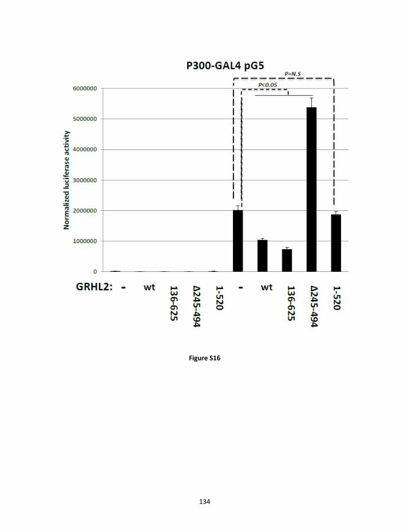
134
Figure S16

135
Figure S17

136
Figure S18

137
Figure S19

138
Chapter 3
The role of GRHL2 in Kidney Cancer and Kidney
Development
The following represents unpublished data.

139
Introduction
Kidney Cancer
As discussed in Chapter 1, GRHL2 suppresses EMT, causes MET, and is important in the
developing kidney (Werth et al.; Cieply et al., 2012; Aue et al., 2015). Previously published
microarray data (GEO website, GSE15641) shows that GRHL2 is significantly downregulated in
clear cell renal cell carcinomas (ccRCC) (figure 1). ccRCC is associated with an EMT
phenotype, consisting of gene markers resembling adipogenic and pluripotent mesenchymal stem
cell-like differentiated states (Tun et al., 2010). ccRCC overexpresses mesenchymal markers,
vimentin and N-cadherin (Tun et al., 2010). Also, TGF-β signaling, an EMT driver, is
upregulated in ccRCC (Boström et al., 2013). The most common mutation in ccRCC is an
inactivating mutation of Von Hippel-Lindau (VHL) (Clark, 2009). Under normoxic conditions,
VHL and its E3 ligase complex ubiquitinates hypoxia-induced factor alpha (HIFα) leading to
HIFα’s degradation (An and Rettig, 2005). When VHL is mutated in ccRCC, HIFα accumulates
and trans-activates the EMT transcription factors, Twist and Snail (Banumathy and Cairns,
2010). In a breast cancer microarray (GEO website, GSE24202), Twist and Snail are able to
downregulate GRHL2 suggesting a mechanism of GRHL2 downregulation in ccRCC (figure 1).
These observations led to the hypothesis that GRHL2 would function as a tumor suppressor in
ccRCC cell lines and revert the EMT phenotype.
Kidney development
Kidney development is an intricate process involving morphogenesis, multiple signaling
pathways, and cell-cell interactions. This developmental process occurs via the interaction of the
ureteric bud and the metanephric mesenchyme. Growth factors originating from the metanephric

140
mesenchyme include glial cell derived growth factor (GDNF) and hepatocyte growth factor
(HGF). These factors activate their cognate receptors on the ureteric epithelium – RET and
MET, respectively (Costantini, 2010). Inductive signaling from the metanephric mesenchyme
results in elongation and branching of the numerous ureteric bud tips. At this point, the ureteric
bud induces the differentiation of the metanephric mesenchyme into the glomerulus. The
metanephric mesenchyme goes on to form the glomerulus, proximal and distal convoluted
tubules, and loop of henle. The ureteric bud is derived from the epithelial Wolffian duct and
expresses GRHL2 (Werth et al., 2010; Aue et al., 2015). The ureteric bud goes on to form the
kidney collecting ducts, calyces, renal pelvis, and ureter. In the adult kidney, GRHL2 is
expressed in the collecting duct. As previously noted, GRHL2 is essential for programming the
epithelial cell phenotype, partially through activation of various cell-cell adhesion genes,
including E-cadherin, claudins, and desmosmal genes (Werth et al., 2010). Therefore, GRHL2
expression leads to tight epithelial cell-cell connections and may be responsible for collecting
duct integrity. Also, GRHL2 suppresses the EMT and promotes the reciprocal process, MET
(Cieply et al., 2012). Both of these processes are required for kidney development (Faa et al.,
2012). Cells undergoing EMT demonstrate increased migration and invasion (Kalluri and
Weinberg, 2009). Because GDNF and HGF increase migration, GRHL2 is expected to
antagonize these signaling pathways (Uehara and Kitamura, 1992; Tang et al., 1998).
Reciprocally, GRHL2 would be inhibited in ureteric bud tip cells allowing the cells to bifurcate
into tubules and/or invade in response to GDNF and HGF. To support this hypothesis, ESRP1-/-
knockout mice increase kidney trifurcations (Russ Carstens, personal communications). ESRP1
is a downstream target of GRHL2 and is responsible for some of the epithelial regulatory effects
associated with GRHL2 expression (Cieply et al., 2012). The role of GRHL2 as a master

141
epithelial programming transcription factor places it in a position to influence all of these
processes and signaling pathways. The investigation of GRHL2 in kidney development will
elucidate its effect on ureteric bud formation, inductive signaling pathways, ureteric bud tip
development, and glomerulus maturation. Furthermore, the examination of GRHL2 and
HGF/MET signaling will have implication not only in kidney development, but also cancer
progression.
Results/Discussion
Kidney cancer
To investigate the role of GRHL2 in ccRCC, 786-0, a clear cell renal cell carcinoma cell
line lacking functional VHL protein, was utilized (Williams et al., 1978; Jiang et al., 2003).
Constitutively expressed GRHL2 in 786-0 cells caused a mesenchymal-epithelial transition
based on cell morphology (figure 2A, B). 786-0 cells undergo branching morphogenesis in three
dimensional matrigel culture upon HGF induction (Peruzzi et al., 2006; Lee et al., 2008), and
GRHL2 prevented HGF-induced branching morphogenesis (figure 2C). Interestingly, this effect
led us to examine GRHL2 in MDCK tubulogenesis which resulted in discoveries explored in
Chapter 2. Constitutively expressed wild type VHL causes a MET in 786-0 cell (Peruzzi et al.,
2006). Since both VHL and GRHL2 reverted ccRCC cells, the cells were examined for anoikis
sensitivity which is normally associated with the epithelial phenotype (Frisch and Francis, 1994).
Indeed, VHL and GRHL2 restored anoikis sensitivity to 786-0 cells (figure 2D). These results
suggested that GRHL2 was a bona fide tumor suppressor in the context of ccRCC since it caused
a MET, restored anoikis sensitivity, and decreased invasion.

142
The next step was to demonstrate that GRHL2 depletion in normal kidney epithelium
increased EMT, anoikis resistance, and invasion. It is a widely held view that ccRCC is derived
from the proximal tubule of the kidney (Büttner et al., 2015). Human kidney proximal tubular
epithelial cell lines, HKC-8 and HK-2, were used to perform GRHL2 shRNA depletion
experiments. Surprisingly, when HKC-8 and 786-0 cells were compared to HMLE cells using
qPCR, there was no detectable level of GRHL2 mRNA in the kidney cells (figure 3A), even
though HKC-8 cells are classified as proximal tubular epithelial cells. Next, a panel of kidney
cell lines were tested for GRHL2 expression with HMLE cells used as a positive control (figure
3B). HKC-8 cells displayed a protein band that migrated significantly higher than normal human
GRHL2. The protein band was resistant to three GRHL2 shRNAs, suggesting the band was not
GRHL2 when examined in conjunction with the qPCR data (figure 3A,C). Both HK-2 cells and
miMCD cells did not express GRHL2 (figure 3B). However, Madin-Darby canine kidney cells
(MDCK) expressed high levels of GRHL2, and MDCK cells are derived from canine collecting
duct cells (Gekle et al., 1994). The developmental process of the kidney clearly explains why
HKC-8 and HK-2 cells do not express GRHL2, but MDCK cells do (See Chapter introduction
for full description). During kidney development, the ureteric epithelium (positive for E-
cadherin and GRHL2) interacts with the metanephric mesenchyme (negative for E-cadherin and
GRHL2) to form the adult kidney. The ureteric epithelium give rises to collecting duct cells
from which the MDCK cell line was derived, thereby explaining the GRHL2 positivity of
MDCK cell. However, HKC-8 and HK-2 cells were derived from the proximal tubule cells
originating from the metanephric mesenchyme and do not express GRHL2.
Therefore, we were constitutively expressing GRHL2 in ccRCC cell lines that never
expressed GRHL2 under physiological conditions, making the clinical relevance of GRHL2 as

143
tumor suppressor in ccRCC questionable. It should be noted that the initial flaw in the
experiment arises in the published microarray data comparing normal kidney tissue and ccRCC.
When heterogeneous tissue samples are collected, genes present in only one cell population may
appear highly regulated. In this specific case, kidney collecting ducts express high levels of
GRHL2, but the other mesenchymal originating kidney cells have no detectable GRHL2 levels.
Therefore when ccRCC expands, the collecting duct cells are not amplified, causing the
microarray to appear as if GRHL2 was downregulated.
Even though our results suggest that GRHL2 is not a tumor suppressor in ccRCC, our
results do have significant implication in the diagnosis of ccRCC. GRHL2 regulates ESRP1/2
(See Chapter 1 EMT section) which is responsible for the alternative splicing of fibroblast
growth factor receptor 2 (FGFR2) isoforms (Warzecha et al., 2009). The mesenchymal FGFR2
isoform IIIc (FGFR2-IIIc) is only expressed in metanepheric mesenchyme derived cells whereas
the epithelial FGFR2 isoform IIIb (FGFR2-IIIB) is only expressed on ureteric epithelial cells.
GRHL2 upregulates FGFR2-IIIb presumably by its regulation of ESRP1 and subsequent FGFR2
splicing (figure 4). In a study with significant clinical implications, Zhao et al. examined 470
ccRCC specimens and 68 normal kidney specimens for alternative splicing events (Zhao et al.,
2013). In this cohort, ~90% of ccRCC expressed FGFR2-IIIc compared to the normal kidney
tissue expressing FGFR2-IIIb. The 10% of ccRCC cells exhibiting the FGFR2-IIIb isoform were
associated with higher patient survival, lower tumor grade, and decreased tumor size. More
importantly, when these FGFR2-IIIb isoform positive tumors were examined by a pathologist,
the tumors were re-classified as chromophobe renal cell carcinoma or papillary renal cell
carcinomas. FGFR2-IIIb isoform, ESRP1, and GRHL2 all correlated with these less invasive
renal cell carcinomas (Zhao et al., 2013). The data suggest that GRHL2 expressing cells do not

144
become ccRCC, and GRHL2 could be useful as a biomarker for predicting clinical kidney
carcinoma subtypes in undiagnosed renal cell carcinoma.
Kidney Development
An RNA-Seq from MDCK cells with GRHL2 constitutively expressed or depleted in the
presence or absence of HGF was previously performed (Chapter 2). Interestingly, several genes
with known or implied significance in kidney development, whose HGF regulation was
modulated by GRHL2, were identified (Table 1,2). These results, consistent with the MDCK
data (Chapter 2), suggested that GRHL2 could play a significant role in kidney development.
Because both GDNF/RET and HGF/MET signaling pathways are able to induce
scattering (Uehara and Kitamura, 1992; Tang et al., 1998), GDNF and HGF were investigated
for their ability to downregulate or inactivate GRHL2. When MDCK with constitutive RET
expression were treated with HGF and/or GDNF, GRHL2 protein was downregulated (figure
5A). Also, GDNF and HGF were both able to downregulate the GRHL2 promoter in promoter-
luciferase reporter constructs (figure 5B).
Since HGF/MET and GDNF/RET pathways play an important role in kidney development
and regulate GRHL2, the effect of GRHL2 on kidney development was examined in the murine
model. Since GRHL2-/- mice die at E11.5 from neural tube defects precluding them from kidney
examination, GRHL2+/- mice were examined for kidney abnormalities. GRHL2+/- mice survived
to adulthood with no apparent differences in weight, pregnancy, feeding, urine output, or serum
BUN levels. Our preliminary data suggested that GRHL2+/- mice have significantly less GRHL2
protein compared to their GRHL2+/+ littermates (figure 6A). Because ureteric epithelium stability
and ureteric tip bifurcation may be affected by depletion of GRHL2, the number of nephrons

145
generated in the adult kidney between GRHL2+/+ and GRHL2+/- mice was examined. P30
GRHL2+/+ and GRHL2+/- kidneys were paraffin-embedded, sectioned, H&E stained, and examined
for glomeruli/mm2. There was a 10-30% decrease in glomeruli numbers in GRHL2+/- kidneys
compared to GRHL2+/+ kidneys (figure 6B). In GRHL2+/+ and GRHL2+/- mice, a ViewRNA ISH
Tissue 2-plex assay system was used to examine RET and GRHL2 localization in paraffin sections
of E15.5 kidneys (figure 7). RET staining was localized to ureteric tips, and GRHL2 was present
in all the ureteric epithelium. Both GRHL2 and RET co-localized in the ureteric tips, and no
difference in GRHL2 RNA levels was detected between GRHL2+/+ and GRHL2+/- mice. These
results were promising, but needed to be further examined in an ureteric specific GRHL2-/- mouse
line.
GRHL2Flox/Flox mice (from collaborator Stephan Jane, Monash University) were crossed
with HOXB7promoter-cre-EGFP mice (from collaborator Carlton Bates), a promoter with ureteric
epithelial specific expression (Zhao et al., 2004). These mice were used to examine the role of
GRHL2 in ureteric bud formation and ureteric tip branching. Embryonic kidneys were examined
at E18.5 or E13.5 with 48 hour ex vivo culture for ureteric tips quantification (figure 8).
Surprisingly, there was no difference between ureteric tips in control and GRHL2UB-/- mice. Also,
there was no renal agenesis observed in D30 GRHL2UB-/- mice which was expected (data not
shown).
After these unexpected results, the GRHL2flox/flox model was examined to ensure GRHL2
was deleted in the ureteric bud. The Jane lab GRHL2flox/flox model had placed the two lox P sites
around exon 2 to create a GRHL2 frameshift mutation rendering the protein nonfunctional.
However, after detailed analysis of murine Grhl2 mRNA by Peter Stoilov, it was discovered that
Grhl2 mRNA has ATG start codons in exons 1, 2, and 4. GRHL2 exon 4 ATG start codon is the

146
strongest match to a Kozak sequence, and GRHL2 exon 1 and 2 ATG start codons were not good
matches, suggesting the start sequence could be leaky and skipped in preference for the exon 4
ATG start codon. Therefore, when the Jane GRHL2flox/flox mice excises exon 2, the exon 1 open
reading frame is only 48 nucleotides which can cause either reinitiation of translation downstream
at exon 4 or entirely bypass exon 1 and start translation at exon 4. The initiation of GRHL2 at
exon 4 would result in a 57 kDA Grhl2 protein with intact DNA binding and dimerization domains.
To examine if this phenomenon was occurring in the GRHL2flox/flox mice, kidneys were harvested,
and primary cells were generated. After the kidney epithelial cells were sorted with Dolichos
Biflorus Agglutinin (DBA), cells were infected with adenovirus-Cre initiating the deletion of exon
2. GRHL2 western blotting clearly shows the creation of a ~57kDA GRHL2 isoform upon adeno-
Cre addition that corresponds to initiation from the exon 4 start codon (figure 9). This data, in
conjunction with the phenotype observed in the GRHL2UB-/- mice, suggested that a functional
GRHL2 protein was being produced, and our GRHL2UB-/- mice were not useful for examining the
role of GRHL2 in kidney development.
During the period of time this research was occurring, Schmidt-Ott et. al. published
“Grainyhead-Like 2/Ovo-Like 2 Pathway Regulates Renal Epithelial Barrier Function and
Lumen Expansion”(Aue et al., 2015). In this Schmidt-Ott study, it was demonstrated that global
Grhl2-/- mice result in decreased collecting duct luminal expansion at E10.5 (Supported by
Chapter 2), and Grhl2 depletion in miMCD cells decreases luminal expansion in three
dimensional culture and affected epithelial barrier formation (Aue et al., 2015). Also, Grhl2
binds and upregulates OVOL2, an epithelial transcription factor (Roca et al., 2013), which is
able to rescue the epithelial barrier and luminal expansion defects in the miMCD cells.
Subsequently, in an abstract, the Schmidt-Ott laboratory saw renal agenesis in 6.8% of

147
ureteric bud specific GRHL2-/- mouse (excising GRHL2 exon 4) and a reduction in kidney
size, but these results have not been published to date. The Schimdt-Ott laboratory used
the Hoxb7cre+ mouse generated by McMahon et. al. (Yu et al., 2002) which has lower
penetrance than the Hoxb7cre+EGFP mouse generated by Bates et. al. (Zhao et al., 2004). If
the Schmidt-Ott GRHL2flox/flox mouse was crossed with the Bates Hoxb7cre+EGFP mouse, the
role of GRHL2 in kidney development would be better understood, and possibly a more severe
kidney phenotype would be observed.
Material and Methods
Cell lines
786-0 and 786+VHL cells (Donald Bottaro, NCI Bethesda, MD) were cultured in RPMI-
1640 (Invitrogen) with 10% fetal bovine serum (FBS) (Hyclone) and 1x penicillin-streptomycin-
glutamine (PSG) (Invitrogen). Human proximal tubular epithelial cell line (HKC clone 8)
(Youhua Liu, University of Pittsburgh) and miMCD-3 cells (ATCC) were cultured in advanced
DMEM:Ham’s F12 (Gibco) with 10% FBS and 1x PSG. MDCK cells (Clone M8 described
previously in Frisch et al. (Frisch and Francis, 1994) and MDCK+c-RET (Greg Dressler,
University of Michigan, Ann Arbor, Michigan) were cultured in advanced Dulbecco’s Modified
Eagle’s Medium (DMEM) (Gibco) with 10% FBS (Hyclone) and 1x PSG (Invitrogen). Human
proximal tubule kidney cells (HK-2) (ATCC) were maintained in Keratinocyte Serum Free
Medium (Invitrogen) with 0.05mg/ml bovine pituitary extract, 5ng/ml human epidermal growth
factor, and 1x PSG (Invitrogen). HMLE culture methods were described previously (Cieply et
al., 2013).

148
Generation of stable cell lines by retroviral transduction and lentiviral transduction
Full-length GRHL2/pMIG, GRHL2/pMXS and GRHL2 shRNA/GIPz were described previously
(Cieply et al., 2012; Farris et al., 2016).
Western Blotting
Western blotting was performed as described in chapter 2. Primary antibodies used were:
GRHL2, rb (Sigma); β-actin, ms (Thermo-pierce); and GAPDH, ms (Origene).
Cell Imaging Assays
786-0 cells images were obtained using a Zeiss Axiovert 200M, Axiocam MRM camera,
Phase 20x/0.55, RT, Axiovision Rel. 4.8 software.
Matrigel branching morphogenesis assay
Matrigel was thawed overnight at 4˚C. Undiluted matrigel (40 µl) was added to 8 well chamber
slides and allowed to solidify for 15 minutes. 786-0 cells were trypsinized, counted and
resuspended at 25,000 cells/ml. The cell solution was mixed 1:1 with DMEM Complete with 4%
matrigel solution with or without 320ng/ml HGF, and 400ul of this mixed solution was pipetted
on top of undiluted matrigel. The cells were refed every four days with DMEM complete with 4%
matrigel with or without HGF. Branching morphogenesis images were obtained using a Zeiss
Axiovert 200M, Axiocam MRM camera, Phase 20x/0.55, RT, Axiovision Rel. 4.8 software.
Anoiks Assay
Anoikis assay was performed as previously described (Cieply et al., 2012).

149
Quantitative reverse transcription PCR analysis
HMLE, HKC, and 786-0 cell were plated on 60mm2 dishes. Cells were harvested
using RNAeasy Plus mini kit (Qiagen), analyzed using Nanodrop, and converted to cDNA using
SuperScript III First-Strand Synthesis SuperMix (Invitrogen) with oligo dT primers and 1ug of
RNA. cDNA was analyzed using SYBR Green PCR Master Mix on an Applied Biosystems
7500 Real Time PCR System. Values reported were determined using the delta delta Ct method
using human GAPDH as internal control. Primers used: GRHL2-F
(GCGCCTATCTCAAAGACGAC), GRHL2-R (GGCTTCCAGGGTGTACTGAA), GAPDH-F
(TGCCAAAGCCTAGGGGAAGA), GAPDH-F (ATGGTTGCCACTGGGGATCT),
RNA-Seq Assay
RNA-Seq was previously described in Chapter 2 material and methods.
GDNF and HGF Cell Scattering Assays
MDCK+c-RET cells were plated on collagen coated 6 well dishes to give 25% cell
confluency on HGF and/or GDNF induction. Cells were treated with 60ng/ml recombinant
human HGF (RD Systems) and/or 50ng/ml GDNF (RD Systems) in DMEM media for the 32
hours. The cells were harvested for protein. Western blotting was performed as previously
described in chapter 2.
Luciferase Reporter Assays
MDCK+c-RET were transiently transfected using Lipofectamine 2000 (Invitrogen) at a
1ug DNA: 3ul Lipofectamine ratio. After 20 minute incubation in 300 µl of Opti-MEM
(Invitrogen), a total of 1.0ug of DNA was transfected into a 12 well with DMEM media and

150
refeed 4 hours later with DMEM contain 60ng/ml recombinant human HGF (RD Systems)
and/or 50ng/ml GDNF (RD Systems). After 28-36 hours post-transfection, the cells were
washed twice with cold PBS, lysed in 200 µl of 1x Cell Culture Lysis Buffer (Promega), frozen
for at least 1 hour at -80 degrees, thawed on ice, and centrifuged at 13,200rpm. The supernatants
were assayed for luciferase activity (Promega) and β-galactosidase activity as internal control (2x
𝛽𝛽-galactosidase assay reagent, 200mmol/l sodium phosphate, pH 7.3, 2mmol/L MgCl2,
100mmol/L 2-mercaptoethanol, and 1.33mg/mL o-nitrophenylgalactoside). GRHL2 promoter
luciferase reporter plasmids were described previously (Cieply et al., 2013).
FGFR2 splicing assay
MDCK parental, MDCK+GRHL2/pMXS, and MDCK+shGRHL2/pGIPz cDNA was prepared as
previously described (Chapter 2). The cDNA was amplified using 4ul of GoTaq Flexi buffer,
0.40 ul of 10mM PCR nucleotides, 0.25ul of GoTaq DNA polymerase, 1ul of template cDNA,
13.95ul of sterile water, and 0.20ul of forward and reverse primers. Primers were as follows:
FGFR2-sp-F (6-FAM CGTACAAAGAACGGGGAGCA) and FGFR2-sp-R
(GCTGAAGTCTGGCTTCTTGG). PCR products were digested with EcorV and Ava1. The
AvaI site was present in FGFR2-IIIb but not FGFR2-IIIc. The fragments were run on a 4%
polyacrylamide gel with ROX as internal ladder. Quantification was performed comparing
FGFR2-IIIc to FGFR2-IIIb bands.
RET and GRHL2 ISH Staining and H&E staining
Kidneys were harvested from mice according to our WVU ACUC protocol. Kidneys were
submerged in 10% formalin, paraffin embedded, and sectioned by the WVU Pathology Laboratory
for Translational Medicine. To perform the section RNA in situs, ViewRNA ISH Tissue 2-plex

151
assay (Affymetrix Panomics) was used. RNA probes for murine GRHL2 and RET were generated
and used to evaluated their co-localization. The protocol was followed as described in user manual.
Briefly, sections were deparaffinized, treated in pre-treatment solution, treated with proteinase K,
hybridized for 3 hours in a humidified 40° tissue culture incubator without CO2 with specific
probes for mouse c-RET and GRHL2 designed by Panomic QuantiGENE. The bound probes were
amplified using Pre-amp and Amp molecules then the labeled probes with fast red and fast blue
substrate to produce red dots (RET) and green dots (GRHL2). Images were obtained using WVU
MIF Olympus histology 20x microscope.
For H&E sections, P30 GRHL2+/+ and GRHL2+/- kidneys were harvested, prepared, and
H&E stained by WVU Pathology Laboratory for Translational Medicine. Images were obtained
using Olympus AX70 Provis microscope equipped with an Optronics MicroFire color CCD
camera Using a 20x/0.70 UPlanApo objective.
Ex Vivo Kidney Culture
All experiments involving mice were approved by WVU IACUC. Hoxb7creEGFP
transgenic mice were a gift from Carlton Bates (Children’s Hospital of Pittsburgh of UPMC,
Pittsburgh, PA) and were described previously (Zhao et al., 2004). GRHL2flox/flox and GRHL2+/-
mice were a gift from Stephen Jane (Monash University), and of the C57BL6 inbred background.
Mice of sexual maturity were timed bred to allow for embryo harvest at the specific
developmental time points E13.5, E15.5, and E18.5. Coitus was verified by checking for
vaginal plugs the following morning after timed mating. The embryos of pregnant mice were
considered E0.5 at noon the day of vaginal plug detection. Embryo’s kidneys were collected as
described in Barak et al (Barak and Boyle, 2011). Briefly, a pregnant mouse was euthanized by
cervical dislocation as approved in our IACUC protocol by highly trained laboratory personnel.

152
Cervical dislocation was necessary to allow for harvest of kidneys before degradation of oxygen
sensitive molecules occurred as well as to ensure survival of embryonic kidneys for ex vivo
culturing. Next, the abdomen was washed with ethanol and an incision was made in the midline,
cutting through the viscera and peritoneum. Subsequently, the uterine horn was dissected, and
the embryos were extracted from the uterus. The embryos are separated from the yolk sac and
placenta. The embryo’s head was removed for PCR genotyping. An incision was made midline
on the abdomen of the embryo, and the superficial organs were removed. The kidneys were
removed and processed according the experiment being performed. Ex vivo kidney culture
method was performed as previously described at indicated times (Zhao et al., 2004). For
MMP inhibitor experiment, ex vivo embryonic kidney at E13.5 were cultured in 1uM batimastat
(Tocris) or vector in culture media for 48 hours. Images were taken of GFP ureteric buds on
Olympus MVX10 Macro Zoom, ORCA-Flash 4.0 Monochrome camera, Objective 2x, Zoom 4x,
FITC, and using CellSen Imaging Software. Ureteric buds were counted for quantification.
GRHL2flox/flox primary kidney cells and AdenoCre infection
Kidneys were harvested from GRHL2flox/flox mice as described above, digested with
Collagenase/Dispase and then trypsin. Cells were plated on collagen 6 well dishes and allowed
to grow for 48 hours. Cells were then stained for Dolichos Biflorus Agglutinin (DBA)-GFP
(Vector Laboratories) to indicate collecting duct epithelial cells and flow sorted for GFP
positivity. DBA-GFP positive and DBA-GFP negative cells plated on collagen coated 12 wells
at 25,000 cells per well (Reichert et al., 2013). Ad5-CMV-CRE-eGFP (University of Iowa,
Gene Transfer Vector core) was added at Multiplicity of Infection of 10 and 50 in DMEM+2%
FBS to each 12 well and protein lysates were made at 48 and 72 hours, and processed using
previously described western blotting techniques

153
References
An, J., and Rettig, M.B. (2005). Mechanism of von Hippel-Lindau Protein-Mediated Suppression of Nuclear Factor kappa B Activity. Mol. Cell. Biol. 25, 7546-7556.
Aue, A., Hinze, C., Walentin, K., Ruffert, J., Yurtdas, Y., Werth, M., Chen, W., Rabien, A., Kilic, E., Schulzke, J.-D., Schumann, M., and Schmidt-Ott, K.M. (2015). A Grainyhead-Like 2/Ovo-Like 2 Pathway Regulates Renal Epithelial Barrier Function and Lumen Expansion. J Am Soc Nephrol. 26, 2704-2715.
Banumathy, G., and Cairns, P. (2010). Signaling pathways in renal cell carcinoma. Cancer Biol. Therapy 10, 658-664.
Barak, H., and Boyle, S.C. (2011). Organ Culture and Immunostaining of Mouse Embryonic Kidneys. Cold Spring Harbor Protocols 2011, pdb.prot5558.
Barak, H., Huh, S.-H., Chen, S., Jeanpierre, C., Martinovic, J., Parisot, M., Bole-Feysot, C., Nitschké, P., Salomon, R., Antignac, C., Ornitz, David M., and Kopan, R. (2012). FGF9 and FGF20 Maintain the Stemness of Nephron Progenitors in Mice and Man. Dev. Cell 22, 1191-1207.
Basta, J.M., Robbins, L., Kiefer, S.M., Dorsett, D., and Rauchman, M. (2014). Sall1 balances self-renewal and differentiation of renal progenitor cells. Development 141, 1047-1058.
Boström, A.-K., Lindgren, D., Johansson, M.E., and Axelson, H. (2013). Effects of TGF-β signaling in clear cell renal cell carcinoma cells. Biochem Biophys Res Commun. 435, 126-133.
Büttner, F., Winter, S., Rausch, S., Reustle, A., Kruck, S., Junker, K., Stenzl, A., Agaimy, A., Hartmann, A., Bedke, J., Schwab, M., and Schaeffeler, E. (2015). Survival Prediction of Clear Cell Renal Cell Carcinoma Based on Gene Expression Similarity to the Proximal Tubule of the Nephron. European Urology 68, 1016-1020.
Chacon-Heszele, M.F., Zuo, X., Hellman, N.E., McKenna, S., Choi, S.Y., Huang, L., Tobias, J.W., Park, K.M., and Lipschutz, J.H. (2014). Novel MAPK-dependent and -independent tubulogenes identified via microarray analysis of 3D-cultured Madin-Darby canine kidney cells. Am. J. Physio. - Renal Physiology 306, F1047-F1058.

154
Cieply, B., Farris, J., Denvir, J., Ford, H.L., and Frisch, S.M. (2013). Epithelial-Mesenchymal Transition and Tumor Suppression Are Controlled by a Reciprocal Feedback Loop between ZEB1 and Grainyhead-like-2. Cancer Res 73, 6299-6309.
Cieply, B., Riley, P.t., Pifer, P.M., Widmeyer, J., Addison, J.B., Ivanov, A.V., Denvir, J., and Frisch, S.M. (2012). Suppression of the Epithelial-Mesenchymal Transition by Grainyhead-like-2. Cancer Res 72, 2440-2453.
Clark, P.E. (2009). The role of VHL in clear-cell renal cell carcinoma and its relation to targeted therapy. Kidney international 76, 939-945.
Costantini, F. (2010). GDNF/Ret signaling and renal branching morphogenesis. Organogenesis 6, 252-262.
Faa, G., Gerosa, C., Fanni, D., Monga, G., Zaffanello, M., Van Eyken, P., and Fanos, V. (2012). Morphogenesis and molecular mechanisms involved in human kidney development. J. Cell. Physiol. 227, 1257-1268.
Farris, J.C., Pifer, P.M., Zheng, L., Gottlieb, E., Denvir, J., and Frisch, S.M. (2016). Grainyhead-like 2 Reverses the Metabolic Changes Induced by the Oncogenic Epithelial-mesenchymal Transition: Effects on Anoikis. Mol. Cancer Res. 14, 528-38
Frisch, S.M., and Francis, H. (1994). Disruption of epithelial cell-matrix interactions induces apoptosis. J Cell Biol 124, 619-626.
Gekle, M., Wünsch, S., Oberleithner, H., and Silbernagl, S. (1994). Characterization of two MDCK-cell subtypes as a model system to study principal cell and intercalated cell properties. Pflügers Archiv: European Journal Of Physiology 428, 157-162.
Hellman, N.E., Spector, J., Robinson, J., Zuo, X., Saunier, S., Antignac, C., Tobias, J.W., and Lipschutz, J.H. (2008). Matrix Metalloproteinase 13 (MMP13) and Tissue Inhibitor of Matrix Metalloproteinase 1 (TIMP1), Regulated by the MAPK Pathway, Are Both Necessary for Madin-Darby Canine Kidney Tubulogenesis. J Biol. Chem. 283, 4272-4282.
Ji, J., Li, Q., Xie, Y., Zhang, X., Cui, S., Shi, S., and Chen, X. (2012). Overexpression of Robo2 causes defects in the recruitment of metanephric mesenchymal cells and ureteric bud branching morphogenesis. Biochem Biophys Res Commun 421, 494-500.

155
Jiang, Y., Zhang, W., Kondo, K., Klco, J.M., St. Martin, T.B., Dufault, M.R., Madden, S.L., Kaelin, W.G., and Nacht, M. (2003). Gene Expression Profiling in a Renal Cell Carcinoma Cell Line: Dissecting VHL and Hypoxia-Dependent Pathways. Mol. Cancer Res. 1, 453-462.
Kalluri, R., and Weinberg, R.A. (2009). The basics of epithelial-mesenchymal transition. J Clin Invest 119, 1420-1428.
Lee, S.J., Lattouf, J.-B., Xanthopoulos, J., Linehan, W.M., Bottaro, D.P., and Vasselli, J.R. (2008). Von Hippel-Lindau Tumor Suppressor Gene Loss in Renal Cell Carcinoma Promotes Oncogenic Epidermal Growth Factor Receptor Signaling via Akt-1 and MEK1. European urology 54, 845-853.
Li, Z., Fei, T., Zhang, J., Zhu, G., Wang, L., Lu, D., Chi, X., Teng, Y., Hou, N., Yang, X., Zhang, H., Han, J.-Dong J., and Chen, Y.-G. BMP4 Signaling Acts via Dual-Specificity Phosphatase 9 to Control ERK Activity in Mouse Embryonic Stem Cells. Cell Stem Cell 10, 171-182.
Majumdar, A., Vainio, S., Kispert, A., McMahon, J., and McMahon, A.P. (2003). Wnt11 and Ret/Gdnf pathways cooperate in regulating ureteric branching during metanephric kidney development. Development 130, 3175-3185.
Marom, K., Fainsod, A., and Steinbeisser, H. (1999). Patterning of the mesoderm involves several threshold responses to BMP-4 and Xwnt-8. Mechanisms of Development 87, 33-44.
Michos, O., Gonçalves, A., Lopez-Rios, J., Tiecke, E., Naillat, F., Beier, K., Galli, A., Vainio, S., and Zeller, R. (2007). Reduction of BMP4 activity by gremlin 1 enables ureteric bud outgrowth and GDNF/WNT11 feedback signalling during kidney branching morphogenesis. Development 134, 2397-2405.
Peruzzi, B., Athauda, G., and Bottaro, D.P. (2006). The von Hippel–Lindau tumor suppressor gene product represses oncogenic β-catenin signaling in renal carcinoma cells. Proc Natl Acad Sci USA 103, 14531-14536.
Piper, M., Georgas, K., Yamada, T., and Little, M. (2000). Expression of the vertebrate Slit Gene family and their putative receptors, the Robo genes, in the developing murine kidney. Mechanisms of Development 94, 213-217.
Reichert, M., Takano, S., Heeg, S., Bakir, B., Botta, G.P., and Rustgi, A.K. (2013). Isolation, culture and genetic manipulation of mouse pancreatic ductal cells. Nature protocols 8, 1354-1365.

156
Roca, H., Hernandez, J., Weidner, S., McEachin, R.C., Fuller, D., Sud, S., Schumann, T., Wilkinson, J.E., Zaslavsky, A., Li, H., Maher, C.A., Daignault-Newton, S., Healy, P.N., and Pienta, K.J. (2013). Transcription Factors OVOL1 and OVOL2 Induce the Mesenchymal to Epithelial Transition in Human Cancer. PLoS ONE 8, e76773.
Tang, M.-J., Worley, D., Sanicola, M., and Dressler, G.R. (1998). The RET–Glial Cell-derived Neurotrophic Factor (GDNF) Pathway Stimulates Migration and Chemoattraction of Epithelial Cells. J Cell Biol. 142, 1337-1345.
Tun, H.W., Marlow, L.A., von Roemeling, C.A., Cooper, S.J., Kreinest, P., Wu, K., Luxon, B.A., Sinha, M., Anastasiadis, P.Z., and Copland, J.A. (2010). Pathway Signature and Cellular Differentiation in Clear Cell Renal Cell Carcinoma. PLoS ONE 5, e10696.
Uehara, Y., and Kitamura, N. (1992). Expression of a human hepatocyte growth factor/scatter factor cDNA in MDCK epithelial cells influences cell morphology, motility, and anchorage-independent growth. J. Cell Biol. 117, 889-894.
Warzecha, C.C., Sato, T.K., Nabet, B., Hogenesch, J.B., and Carstens, R.P. (2009). ESRP1 and ESRP2 Are Epithelial Cell-Type-Specific Regulators of FGFR2 Splicing. Mol. Cell 33, 591-601.
Werth, M., Walentin, K., Aue, A., Schonheit, J., Wuebken, A., Pode-Shakked, N., Vilianovitch, L., Erdmann, B., Dekel, B., Bader, M., Barasch, J., Rosenbauer, F., Luft, F.C., and Schmidt-Ott, K.M. The transcription factor grainyhead-like 2 regulates the molecular composition of the epithelial apical junctional complex. Development 137, 3835-3845.
Werth, M., Walentin, K., Aue, A., Schönheit, J., Wuebken, A., Pode-Shakked, N., Vilianovitch, L., Erdmann, B., Dekel, B., Bader, M., Barasch, J., Rosenbauer, F., Luft, F.C., and Schmidt-Ott, K.M. (2010). The transcription factor grainyhead-like 2 regulates the molecular composition of the epithelial apical junctional complex. Development 137, 3835-3845.
Williams, R.D., Elliott, A.Y., Stein, N., and Fraley, E.E. (1978). In vitro cultivation of human renal cell cancer. II. Characterization of cell lines. In Vitro 14, 779-786.
Yu, J., Carroll, T.J., and McMahon, A.P. (2002). Sonic hedgehog regulates proliferation and differentiation of mesenchymal cells in the mouse metanephric kidney. Development 129, 5301-5312.
Zhao, H., Kegg, H., Grady, S., Truong, H.-T., Robinson, M.L., Baum, M., and Bates, C.M. (2004). Role of fibroblast growth factor receptors 1 and 2 in the ureteric bud. Dev. Biology 276, 403-415.

157
Zhao, Q., Caballero, O.L., Davis, I.D., Jonasch, E., Tamboli, P., Yung, W.K.A., Weinstein, J.N., Kenna Shaw for the, T.r.n., Strausberg, R.L., and Yao, J. (2013). Tumor-Specific Isoform Switch of the Fibroblast Growth Factor Receptor 2 Underlies the Mesenchymal and Malignant Phenotypes of Clear Cell Renal Cell Carcinomas. Clin. Cancer Res. 19, 2460-2472.

158
Table 1. Gene regulated by HGF and GRHL2 via RNA-Seq
Genes regulated by HGF in the absence of GRHL2
Gene ID Fold Change
(shGRHL2+hgf/shGRHL2)
Significance in kidney development
FGF9 -23.6 Maintains stemness of nephron progenitors (Barak et al., 2012)
DUSP9 -22.6 Controls Erk activity in mouse embryonic stem cells (Li et al.)
SLIT2 -9.8 Restricts kidney induction to a single site (Ji et al., 2012)
Wnt11 -7.6 Cooperates with GDNF/RET in regulating ureteric branching (Majumdar et
al., 2003)
Wnt8a 6.4 Interacts with the BMP-4 pathway (Marom et al., 1999)

159
Figure legends
Table 2. Gene regulated by HGF and GRHL2 via RNA-Seq
Genes regulated by GRHL2
Gene ID Fold Change
(shGRHL2/GRHL2)
Significance in kidney development
MMP-13 15.1 Necessary for MDCK tubulogenesis
(Hellman et al., 2008)
MMP-1 5.0 Necessary for MDCK tubulogenesis
(Chacon-Heszele et al., 2014)
BMP-4 3.5 Inhibits ureteric branching morphogenesis
(Michos et al., 2007)
TIMP1 3.3 Necessary for MDCK tubulogenesis
(Hellman et al., 2008)
Robo1 2.1 Expressed in immature glomeruli (Piper et
al., 2000)
FGFR2IIIC 1.9 Predominate FGFR2 isoform in the
metanephric mesenchyme (Zhao et al., 2004)
SALL1 1.7 Influences self-renewal and differentiation of
renal progenitor cells (Basta et al., 2014)
GFRα -2.7 RET ligand-binding co-receptor Ɨ (38)

160
Figure 1. GRHL2 was downregulated in clear cell renal cell carcinoma. A. GRHL2,
GATA3, and Snai1 mRNA expression from GEO website, GSE15641. B. Proposed mechanism
of GRHL2 downregulation in ccRCC.
Figure 2. GRHL2 caused 786-0 ccRCC cells to undergo MET, prevented invasion, and
increased anoikis. A. Western blot confirming GRHL2 expression in 786-0 cells. B. GRHL2
promoted an epithelial phenotype in 786-0 cells. C. GRHL2 suppressed HGF-induced invasion
in 786-0 cell in three-dimensional matrigel culture. D. VHL and GRHL2 increased anoikis in
786-0 as measured by cellular DNA fragmentation assay. Values represent relative DNA
fragmentation between samples.
Figure 3. GRHL2 is not expressed in normal proximal tubule human kidney cell lines. A.
Quantitative PCR for GRHL2 mRNA in HMLE, HKC-8, and 786-0 cell lines. B. Cell line panel
for GRHL2 protein across multiple cell lines. (See discussion in results). C. HKC-8 western
band was resistant to GRHL2 shRNA knockdown via western blotting.
Figure 4. GRHL2 regulated FGFR2 isoform 3b exon inclusion. MDCK parent, GRHL2, and
shGRHL2 cDNAs were used to amplify FGFR2 3 splicing region. Differential digest was
performed to determine the presence of FGFR2 3b (epithelial) and FGFR2 3c (mesenchymal)
exon. Quantification was performed by comparing FGFR2 3c fragment to FGFR2 3b fragment
Figure 5. GRHL2 was down-regulated by HGF and GDNF. A. GDNF and HGF downregulated
GRHL2. MDCK+RET cells were treated with vehicle, 60ng/ml HGF and/or 50ng/ml GDNF for
32 hours (Western blot). B. GDNF and HGF downregulated induction of GRHL2 promoters.
MDCK+RET cells were cotransfected with GRHL2 promoter-luciferase reporter constructs.
Values represent relative luciferase activity normalized to TK-β-galactosidase control.

161
Figure 6. GRHL2+/- mice showed lower number of glomeruli. A. Representative western blot
of GRHL2+/+ and GRHL2+/- kidney GRHL2 protein levels. B. Day 30 GRHL2+/+ and
GRHL2+/- kidney sections were compared via glomerulis/mm2 (n=5). GRHL2+/+ mice had a
9.7±2.6 per mm2 whereas GRHL2+/- mice had 6.4±1.3 per mm2. p value < 0.01. C.
Representative images of GRHL2+/+ and GRHL2+/- glomeruli in H&E sections imaged with
200.75 Fluar an Axiovert 200M microscope.
Figure 7. RET and GRHL2 co-localization in GRHL2+/+ and GRHL2+/- mice. Representative
images of RET (Red), GRHL2 (Green), and merged co-localization using a ViewRNA ISH
Tissue 2-plex assay system. Images were obtained using Zeiss Violet Confocal, 20x.
Figure 8. Effect of ureteric epithelial specific GRHL2-/- on ureteric tip count. A. Control
and GRHL2UB-/- kidneys were harvested at E13.5 and cultured for 48 hours. B. Control and
GRHL2UB-/- kidneys were harvested at E18.5. Both A,B ureteric tips were counted and
normalized to area (mm2). Images were obtained using Olympus MVX10 Macro Zoom, ORCA-
Flash 4.0 Monochrome camera, Objective 2x, Zoom 4x, FITC, and using CellSen Imaging
Software.
Figure 9. GRHL2flox/flox mice generated a 57 kDA active GRHL2 isoform. A. Schematic of
Jane GRHL2flox/flox mouse model. B. Predicted GRHL2 isoform size depending on ATG Start
Codon. C. GRHL2flox/flox primary kidney cells treated with adenoviral-Cre. DBA is an epithelial
marker used for flow sorting of primary kidney cells.

162
Figure 1

163
Figure 2

164
Figure 3

165
Figure 4

166
Figure 5

167
Figure 6

168
Figure 7

169
Figure 8

170
Figure 9

171
Chapter 4
Conclusion and Future Directions

172
Conclusions
The present work examined the role of GRHL2 in an array of different systems and
circumstances. GRHL2 was not expressed in the normal kidney proximal tubule, and therefore
highly unlikely to regulate clear cell renal cell carcinoma tumor progression. However, GRHL2
could be used as a biomarker to distinguish between different renal cell carcinoma subtypes.
GRHL2 was involved in multiple signaling pathways and processes that are crucial in kidney
development, but further investigation using a ureteric epithelium specific GRHL2flox/flox mouse
is required to understand the role of GRHL2 in the developing kidney.
In the MDCK model, GRHL2 was critical for cystogenesis but suppressed tubulogenesis.
For the first time, GRHL2 was shown to interact with the histone acetyltransferase co-activator
protein, p300, and inhibit its HAT activity thereby preventing its transcriptional activation of
target genes. These findings place GRHL2 among a rare group of proteins that can inhibit p300
HAT activity. The p300 HAT inhibitory domain of GRHL2 was mapped to amino acids 425-
437 with the following sequence: IRDEERKQNRKKG. These 13 amino acids were required for
GRHL2 to suppress tubulogenesis and cause a mesenchymal-epithelial transition. Since p300 is
critical for cells to differentiate, GRHL2’s inhibition of p300 HAT activity provides a
mechanism for GRHL2 to enforce the default epithelial phenotype. This work mechanistically
positions GRHL2, a master regulator of the default epithelial phenotype, to antagonize p300, a
co-activator required for cell differentiation, having vast implications in both developmental and
cancer contexts.

173
Future Directions
Because the ureteric epithelium GRHL2flox/flox model our laboratory generated was not
informative, there are many unanswered questions left about GRHL2 and kidney development.
Previous results from our laboratory and from Schmidt-Ott et. al. show that GRHL2 depletion in
global GRHL2-/- mice, miMCD cells, and MDCK cells affect kidney tubulogenesis and lumen
size, highly suggestive of an important role in kidney development (Werth et al., 2010; Aue et
al., 2015). The GRHL2flox/flox mice generated by Aue et. al. (which deletes exon 4) could be
used in cooperation with the Zhao et. al. HOXB7creEGFP mice to elucidate the role of GRHL2
in kidney development (Zhao et al., 2004; Walentin et al., 2015). The expected outcome of
these experiments would be renal agenesis because of the role of GRHL2 in establishing
epithelial phenotype and cell polarity. GRHL2 would be a tool for examining the importance of
the ureteric bud in kidney development. From a clinical perspective, GRHL2 mutations could be
examined for causing renal agenesis or renal duplication anomalies.
As one of the main focuses of our laboratory is breast cancer, it would be highly
informative to examine the effects of GRHL2 on mammary development. OVOL2, a GRHL2
regulated epithelial transcription factor, is known to affect mammary morphogenesis. When
OVOL2 is depleted in the mammary glands, the cells undergo an EMT and decrease milk
production and alveologenesis (Roca et al., 2013). At the very least, GRHL2 would be expected
to have a similar effect as OVOL2 on mammary development. The loss of GRHL2 in these cells
would most likely cause a mesenchymal phenotype. Also, GRHL2 may cause a more severe
phenotype than OVOL2 since it regulates a vast number of genes other than OVOL2 in RNA-
Seq (Farris et al., 2016). If the GRHL2 phenotype was more severe, a rescue of OVOL2 would
be highly informative demonstrating alternative pathways not regulated through the

174
GRHL2/OVOL2 axis. These results would significantly expand the understanding of GRHL2 in
organogenesis. This examination might provide novel GRHL2 interaction partners that occur
during the developmental process with potential influences on breast cancer progression.
Our laboratory proposed the default epithelial phenotype hypothesis for both
development and cancer differentiation. Under this hypothesis, the epithelial phenotype is
adopted automatically by a cell in the absence of genomic/epigenetic alterations or inductive
differentiation signaling (Frisch, 1997). Consistent with this idea, E-cadherin-expressing cells are
the first to emerge at the late two cell stage; these eventually give rise to all differentiated cell
types of the adult organism (Hyafil et al., 1980; Vestweber and Kemler, 1985; Fleming et al.,
1989; Larue et al., 1994). Furthermore, GRH, the Drosophila GRHL2 homolog, is one of the
first proteins expressed in the pre-cellular blastoderm zygote. GRH is responsible for the
regulation of the maternal-to-zygotic transition, the process where a zygote degrades maternal
mRNA and starts transcribing its own mRNA (Harrison et al., 2010).
In recent studies, other laboratories have suggested the default epithelial phenotype
hypothesis may explain the mesenchymal-epithelial transition (MET) that occurs at metastatic
sites (Thiery and Sleeman, 2006; Polyak and Weinberg, 2009; Tam and Weinberg, 2013). Our
results demonstrating the interaction between p300 and GRHL2 provide another piece of support
for this hypothesis. The critical role of p300 in the differentiation of muscle, erythroid lineages,
osteoblasts, oligodendrocytes, and stem cells has been clearly established. The role of p300 in
differentiation demonstrates the importance of epigenetic modifications in this process (Puri et
al., 1997; Oike et al., 1999; Blobel, 2000; Polesskaya et al., 2001; Narayanan et al., 2004; Teo
and Kahn, 2010; Zhang et al., 2016). The ability of GRHL2 to inhibit p300 can explain the
default epithelial phenotype hypothesis since critical co-activators such as p300 are required for

175
differentiation-specific but not epithelial-specific gene expression (Frisch, 1997). These findings
could also extend to critical roles in EMT, in contexts such as tubulogenesis and oncogenesis. In
summary, p300 and related co-activators are crucial for cells to differentiate. Under the default-
phenotype hypothesis, the inhibition of p300 by GRHL2 provides a mechanism for GRHL2 to
enforce the default epithelial phenotype.
As described in Chapter 1, Roeder et. al. demonstrated that p300 interacts with the SET1
complex through a p53 mediated event to regulate H3K4 trimethylation at activated promoters
(Tang et al., 2013). The SET1/MLL histone methyltransferase family is comprised of SET1A,
SET1B, SET1C MLL1, MLL2, MLL3 and MLL4 (Yokoyama et al., 2004; Dou et al., 2005; Cho
et al., 2007; Wu et al., 2008). MLL1 has also been shown to interact with CBP (Ernst et al.,
2001). Also noted in Chapter 1, EMT is highly dependent on epigenetic regulation. In our
laboratory, GRHL2 was found to interact with MLL3/4 by a yeast two hybrid screen, and these
results were confirmed by MLL3/GRHL2 co-immunoprecipitation (data not shown). These
findings have interesting implications. GRHL2 interacts with two histone modifying factors,
p300 and MLL3/4 which collaborate for gene activation (Tang et al., 2013). Our laboratory has
formed a collaboration with Robert Roeder (Rockefeller University) to examine these
interactions. Theoretically, GRHL2 could be recruiting these proteins to specific promoters,
inhibiting the histone modifying factors, and/or modulating their enzymatic activities. This
would link GRHL2 directly to epigenetic methylation and acetylation events, and provide
another mechanism of how GRHL2 establishes and maintains the epithelial phenotype.
The result of our research with the most potential impact is the inhibition of p300 HAT
domain by GRHL2. p300 HAT inhibitors are currently an intense area of research with
implications in cancer, diabetes, HIV infections, heart disease, and platelet cell production

176
(Chapter 1) (Dancy and Cole, 2015). p300 HAT inhibitors have not been investigated in clinical
trials to date (Dancy and Cole, 2015). Two of the major issues facing p300 HAT inhibitors are
lack of specificity when compared to other histone acetyltransferase families and/or cell
impermeability making the chemicals difficult to use in vivo conditions. GRHL2 appears to
potently inhibit the p300 HAT domain, but more kinetic experiments need to be completed to
determine the half maximal inhibitory concentration. The GRHL2 13aa peptide could be
targeted for cellular uptake by attaching the peptide to a peptide/protein transduction domain
(Lönn and Dowdy, 2015), and its effect on cellular acetylation would be examined.
Furthermore, the GRHL2 peptide could be mutated to find the critical residues for p300
inhibition. These critical residues could be used to model the GRHL2 and p300 interaction
providing more information about the HAT domain and targetable sites that may have previously
gone undiscovered. The ability of GRHL2 to inhibit CBP or other histone acetyltransferase
families would be informative and demonstrate the generalizability of GRHL2’s HAT inhibition.
Our laboratory has previously published that activation of both the TGF-β and Wnt
pathways are able to downregulate GRHL2 (Cieply et al., 2012), and our present work has
demonstrated that HGF pathway is able to downregulate GRHL2. All of these pathways are
involved in tumor progression, and clinical trials are underway with selective inhibitors of these
pathways in different cancers (Blagodatski et al., 2014; Neuzillet et al., 2015; Zhang et al.,
2015). The examination of these inhibitors in the context of GRHL2 could provide a mechanism
to pharmacologically upregulate GRHL2 and maintain the epithelial phenotype thereby
preventing cancer metastasis.
In summary, GRHL2 is critical for establishing the epithelial phenotype, and more
research is required to understand its complex biological role.

177
References
Aue, A., Hinze, C., Walentin, K., Ruffert, J., Yurtdas, Y., Werth, M., Chen, W., Rabien, A., Kilic, E., Schulzke, J.-D., Schumann, M., and Schmidt-Ott, K.M. (2015). A Grainyhead-Like 2/Ovo-Like 2 Pathway Regulates Renal Epithelial Barrier Function and Lumen Expansion. J Am Soc Nephrol. 26, 2704-2715.
Blagodatski, A., Poteryaev, D., and Katanaev, V.L. (2014). Targeting the Wnt pathways for therapies. Mol. Cell. Therapies 2, 1-15.
Blobel, G.A. (2000). CREB-binding protein and p300: molecular integrators of hematopoietic transcription. Blood 95, 745-755.
Cho, Y.-W., Hong, T., Hong, S., Guo, H., Yu, H., Kim, D., Guszczynski, T., Dressler, G.R., Copeland, T.D., Kalkum, M., and Ge, K. (2007). PTIP Associates with MLL3- and MLL4-containing Histone H3 Lysine 4 Methyltransferase Complex. J. Biol. Chem. 282, 20395-20406.
Cieply, B., Riley, P.t., Pifer, P.M., Widmeyer, J., Addison, J.B., Ivanov, A.V., Denvir, J., and Frisch, S.M. (2012). Suppression of the Epithelial-Mesenchymal Transition by Grainyhead-like-2. Cancer Res 72, 2440-2453.
Dancy, B.M., and Cole, P.A. (2015). Protein lysine acetylation by p300/CBP. Chemical Reviews 115, 2419-2452.
Dou, Y., Milne, T.A., Tackett, A.J., Smith, E.R., Fukuda, A., Wysocka, J., Allis, C.D., Chait, B.T., Hess, J.L., and Roeder, R.G. (2005). Physical Association and Coordinate Function of the H3 K4 Methyltransferase MLL1 and the H4 K16 Acetyltransferase MOF. Cell 121, 873-885.
Ernst, P., Wang, J., Huang, M., Goodman, R.H., and Korsmeyer, S.J. (2001). MLL and CREB Bind Cooperatively to the Nuclear Coactivator CREB-Binding Protein. Mol. Cell. Biol. 21, 2249-2258.
Farris, J.C., Pifer, P.M., Zheng, L., Gottlieb, E., Denvir, J., and Frisch, S.M. (2016). Grainyhead-like 2 Reverses the Metabolic Changes Induced by the Oncogenic Epithelial-mesenchymal Transition: Effects on Anoikis. Mol. Cancer Res.
Fleming, T.P., McConnell, J., Johnson, M.H., and Stevenson, B.R. (1989). Development of tight junctions de novo in the mouse early embryo: control of assembly of the tight junction-specific protein, ZO-1. J. Cell Biol. 108, 1407-1418.

178
Frisch, S.M. (1997). The epithelial cell default-phenotype hypothesis and its implications for cancer. Bioessays 19, 705-709.
Harrison, M.M., Botchan, M.R., and Cline, T.W. (2010). Grainyhead and Zelda compete for binding to the promoters of the earliest-expressed Drosophila genes. Dev Biol 345, 248-255.
Hyafil, F., Morello, D., Babinet, C., and Jacob, F. (1980). A cell surface glycoprotein involved in the compaction of embryonal carcinoma cells and cleavage stage embryos. Cell 21, 927-934.
Larue, L., Ohsugi, M., Hirchenhain, J., and Kemler, R. (1994). E-cadherin null mutant embryos fail to form a trophectoderm epithelium. Proc Natl Acad Sci USA 91, 8263-8267.
Lönn, P., and Dowdy, S.F. (2015). Cationic PTD/CPP-mediated macromolecular delivery: charging into the cell. Expert Opin Drug Deliv. 12, 1627-1636.
Narayanan, K., Srinivas, R., Peterson, M.C., Ramachandran, A., Hao, J., Thimmapaya, B., Scherer, P.E., and George, A. (2004). Transcriptional Regulation of Dentin Matrix Protein 1 by JunB and p300 during Osteoblast Differentiation. J. Biol. Chem. 279, 44294-44302.
Neuzillet, C., Tijeras-Raballand, A., Cohen, R., Cros, J., Faivre, S., Raymond, E., and de Gramont, A. (2015). Targeting the TGFβ pathway for cancer therapy. Pharmacol. Ther. 147, 22-31.
Oike, Y., Takakura, N., Hata, A., Kaname, T., Akizuki, M., Yamaguchi, Y., Yasue, H., Araki, K., Yamamura, K.-i., and Suda, T. (1999). Mice Homozygous for a Truncated Form of CREB-Binding Protein Exhibit Defects in Hematopoiesis and Vasculo-angiogenesis. Blood 93, 2771-2779.
Polesskaya, A., Naguibneva, I., Fritsch, L., Duquet, A., Ait-Si-Ali, S., Robin, P., Vervisch, A., Pritchard, L.L., Cole, P., and Harel-Bellan, A. (2001). CBP/p300 and muscle differentiation: no HAT, no muscle. EMBO J. 20, 6816-6825.
Polyak, K., and Weinberg, R.A. (2009). Transitions between epithelial and mesenchymal states: acquisition of malignant and stem cell traits. Nat Rev Cancer 9, 265-273.
Puri, P.L., Sartorelli, V., Yang, X.-J., Hamamori, Y., Ogryzko, V.V., Howard, B.H., Kedes, L., Wang, J.Y.J., Graessmann, A., Nakatani, Y., and Levrero, M. (1997). Differential Roles of p300 and PCAF Acetyltransferases in Muscle Differentiation. Mol. Cell 1, 35-45.

179
Roca, H., Hernandez, J., Weidner, S., McEachin, R.C., Fuller, D., Sud, S., Schumann, T., Wilkinson, J.E., Zaslavsky, A., Li, H., Maher, C.A., Daignault-Newton, S., Healy, P.N., and Pienta, K.J. (2013). Transcription Factors OVOL1 and OVOL2 Induce the Mesenchymal to Epithelial Transition in Human Cancer. PLoS ONE 8, e76773.
Tam, W.L., and Weinberg, R.A. (2013). The epigenetics of epithelial-mesenchymal plasticity in cancer. Nat Med 19, 1438-1449.
Tang, Z., Chen, W.-Y., Shimada, M., Nguyen, U.T.T., Kim, J., Sun, X.-J., Sengoku, T., McGinty, R.K., Fernandez, J.P., Muir, T.W., and Roeder, R.G. (2013). SET1 and p300 Act Synergistically, through Coupled Histone Modifications, in Transcriptional Activation by p53. Cell 154, 297-310.
Teo, J.-L., and Kahn, M. (2010). The Wnt signaling pathway in cellular proliferation and differentiation: A tale of two coactivators. Adv. Drug Deliv. Rev. 62, 1149-1155.
Thiery, J.P., and Sleeman, J.P. (2006). Complex networks orchestrate epithelial-mesenchymal transitions. Nat Rev Mol Cell Biol 7, 131-142.
Vestweber, D., and Kemler, R. (1985). Identification of a putative cell adhesion domain of uvomorulin. EMBO J. 4, 3393-3398.
Walentin, K., Hinze, C., Werth, M., Haase, N., Varma, S., Morell, R., Aue, A., Pötschke, E., Warburton, D., Qiu, A., Barasch, J., Purfürst, B., Dieterich, C., Popova, E., Bader, M., Dechend, R., Staff, A.C., Yurtdas, Z.Y., Kilic, E., and Schmidt-Ott, K.M. (2015). A Grhl2-dependent gene network controls trophoblast branching morphogenesis. Development 142, 1125-1136.
Werth, M., Walentin, K., Aue, A., Schönheit, J., Wuebken, A., Pode-Shakked, N., Vilianovitch, L., Erdmann, B., Dekel, B., Bader, M., Barasch, J., Rosenbauer, F., Luft, F.C., and Schmidt-Ott, K.M. (2010). The transcription factor grainyhead-like 2 regulates the molecular composition of the epithelial apical junctional complex. Development 137, 3835-3845.
Wu, M., Wang, P.F., Lee, J.S., Martin-Brown, S., Florens, L., Washburn, M., and Shilatifard, A. (2008). Molecular Regulation of H3K4 Trimethylation by Wdr82, a Component of Human Set1/COMPASS. Mol. Cell. Biol. 28, 7337-7344.
Yokoyama, A., Wang, Z., Wysocka, J., Sanyal, M., Aufiero, D.J., Kitabayashi, I., Herr, W., and Cleary, M.L. (2004). Leukemia Proto-Oncoprotein MLL Forms a SET1-Like Histone Methyltransferase Complex with Menin To Regulate Hox Gene Expression. Mol. Cell. Biol. 24, 5639-5649.

180
Zhang, L., He, X., Liu, L., Jiang, M., Zhao, C., Wang, H., He, D., Zheng, T., Zhou, X., Hassan, A., Ma, Z., Xin, M., Sun, Z., Lazar, Mitchell A., Goldman, Steven A., Olson, Eric N., and Lu, Q.R. (2016). Hdac3 Interaction with p300 Histone Acetyltransferase Regulates the Oligodendrocyte and Astrocyte Lineage Fate Switch. Dev. Cell 36, 316-330.
Zhang, Y., Jain, R., and Zhu, M. (2015). Recent Progress and Advances in HGF/MET-Targeted Therapeutic Agents for Cancer Treatment. Biomedicines 3, 149.
Zhao, H., Kegg, H., Grady, S., Truong, H.-T., Robinson, M.L., Baum, M., and Bates, C.M. (2004). Role of fibroblast growth factor receptors 1 and 2 in the ureteric bud. Dev. Biology 276, 403-415.

181
Chapter 5
Curriculum Vitae

182
Phillip M. Pifer 22 REGENCY CT, MORGANTOWN, WV 26505
PHONE: (304) 834-6143 EMAIL: [email protected]
EDUCATION
Ph.D. M.D.-Ph.D. Dual degree scholar: Aug 2010-May 2018 Anticipated degree date: Ph.D.: May 2018 Time in Ph.D.: Aug 2010-June 2016
West Virginia University, Morgantown, WV Department of Biochemistry, Program in Cancer Cell Biology, School of Medicine Dissertation Title: Grainyhead-like-2 inhibits the coactivator p300, suppressing tubulogenesis and the epithelial-mesenchymal transition. Dissertation Advisor: Dr. Steven M. Frisch, Ph.D. Dissertation Defense Date: 06/24/2016
M.D. M.D.-Ph.D. Dual degree scholar: Aug 2010- May 2018 Anticipated degree date: M.D.: May 2018
West Virginia University, Morgantown, WV School of Medicine
B.S.
Chemistry: Aug 2006- May 2010 Honors: Summa Cum Laude
West Virginia University, Morgantown, WV Department of Chemistry, Eberly School of Arts & Sciences
RESEARCH EXPERIECNES
MD/PhD Trainee, Dept. of Biochemistry, WVU 2011 – Present
• Advisor: Steven M. Frisch, PhD. “Grainyhead-like-2 inhibits the coactivator p300, suppressing tubulogenesis and the epithelial-mesenchymal transition”
Visiting Graduate Researcher, Children’s Hospital of Pittsburgh, Dept. of Nephrology 2014 • Advisor: Carlton M. Bates, MD. Laboratory methods of the developing kidney.
Undergraduate Researcher, Dept. of Chemistry, WVU 2009-2010
• Advisor: Justin Legleiter, PhD. “Point mutations in Aβ result in the formation of distinct polymorphic aggregates in the presence of lipid bilayers.”

183
PEER-REVIEWED PUBLICATIONS Pifer, P. M., Farris, J. C., Thomas, A. L., Stoilov, P., Denvir, J., Smith, D. M., & Frisch, S. M. (2016). Grainyhead-
like-2 inhibits the coactivator p300, suppressing tubulogenesis and the epithelial-mesenchymal transition. Molecular Biology of the Cell.
Farris, J. C., Pifer, P. M., Zheng, L., Gottlieb, E., Denvir, J., & Frisch, S. M. (2016). Grainyhead-like 2 Reverses the
Metabolic Changes Induced by the Oncogenic Epithelial-mesenchymal Transition: Effects on Anoikis. Molecular Cancer Research.
Cieply, B., Riley, P. t., Pifer, P. M., Widmeyer, J., Addison, J. B., Ivanov, A. V., . . . Frisch, S. M. (2012).
Suppression of the Epithelial-Mesenchymal Transition by Grainyhead-like-2. Cancer Res, 72(9), 2440-2453.
Pifer, P. M., Yates, E. A., & Legleiter, J. (2011). Point mutations in Aβ result in the formation of distinct polymorphic aggregates in the presence of lipid bilayers. PLoS One, 6(1).
Kumar, B., Pifer, P. M., Giovengo, A., & Legleiter, J. (2010). The effect of set point ratio and surface Young’s modulus on maximum tapping forces in fluid tapping mode atomic force microscopy. Journal of Applied Physics, 107(4), 044508.
TECHNIQUES Cell and Molecular Biology: Tissue cell culture, three dimensional cell culture, PCR Primer Design, Molecular Cloning Techniques (Including sub-cloning, site-directed mutagenesis, and plasmid preparation), RNA isolation (from primary tissues and cells), DNA purification, qRT-PCR, RNA-Seq preparation, cell transfection, mammalian cell retroviral and lentiviral transduction, luciferase reporter assays, immunoblotting (western blot), immunofluorescent staining, histone acetylation assays, bacterial and mammalian protein purification, Ingenuity Pathway Analysis examination, immunoprecipitation, chromatin immunoprecipitation, Anoikis assays (caspase 3/7 activation assays, cell death ELISA, and cell viability assays), and confocal microscopy for MDCK Cysts. Animal and Kidney models: Timed breeding, embryonic kidney harvesting, ex vivo kidney culture, kidney imaging, colony maintenance, mouse genotyping, folic acid kidney injury model, murine models of mammary development, tumor burden scoring of mice, H&E Staining, and RNA in situ hybridization assays. ABSTRACTS, PRESENTATIONS, and CONFERENCES Abstracts Pifer, P.M., Cieply, B., Frisch, S.M. The role of GRHL-1 and GRHL-2 in clear cell renal cell carcinoma. – Van Liere Research Convocation- West Virginia University, Morgantown, WV (2013) Pifer, P.M., and Legleiter, J. The effect of mutant A-beta amyloid peptide aggregation on the stability of model lipid bilayers. - 54th Annual meeting of Biophysical Society February 20-24 (2010); WVNANO Initiative Research Symposium (2009); WVU American Chemical Society Student Affiliates Chemical Symposium (2009) Presentations Pifer, PM., Farris, JC. The Physician-Scientist career track: How to be a successful MD-PhD candidate- (May 31st, 2016) – WVU Cancer Institute undergraduate research program invited speaker

184
Pifer, PM. Advancements in Melanoma- PD1 inhibitors. (Jan. 21st, 2016) – WVU Cancer Cell Biology Journal Club Pifer, PM. Role of grainyhead-like 2 in the establishment of the epithelial phenotype (Jun. 24st, 2015) – WVU Genomics Group invited speaker Pifer, PM. The MD-PhD Track: A Brief Overview. (Jun. 8st, 2015) – IDeA Network of Biomedical Research Excellence (INBRE) invited speaker Pifer, PM, Frisch SM. GRHL2 in Kidney Tubulogenesis. (Nov. 16, 2014) Department of Biochemistry– Cancer Cell Biology Seminar. Pifer, PM, Frisch SM. GRHL2 in Kidney Tubulogenesis. (Apr. 28, 2014) Department of Biochemistry – Cancer Cell Biology Dissertation Proposal. Pifer, PM. Sharma SB, Farrugia MK. The MD-PhD Track: A Brief Overview. (Feb. 27st, 2014) – WVU American Chemical Society Student Affiliates invited speaker Pifer, PM, Frisch SM. The role of GRHL1 and GRHL2 in clear cell renal cell carcinoma. (Jan. 30, 2013) Department of Biochemistry – Cancer Cell Biology Seminar. Pifer, PM, Sharma SB, Farrugia MK, Pilkerton CK, Turner RC. The Physician-Scientist career track: How to be a successful MD-PhD candidate – (2011) WVU chapter of the American Physician-Scientist Association (APSA) sponsored talk
Conferences
American Physician Scientist Association Regional Meeting, (Oct 2015) SUNY UPSTATE Medical University, Syracuse, NY,
Tackling Translational Challenges, 14th Annual World Preclinical Congress Conference, (Jun 2015) Focus on Novel Preclinical Models in Oncology and Targeting Histone Acetylation, Boston, MA
54th Annual Meeting of Biophysical Society, (Feb 2010) San Francisco, CA,
HONORS & AWARDS Featured Researcher, WVU Microscope Imaging Facility Newsletter 2013
• My research images were featured in the November issue Class Honors (Top 15%), West Virginia University School of Medicine 2010-2011, 2011-2012
• Awarded in Human Function, Public Health, Physical Diagnosis & Clinical Integration 1, Human Structure, Neurobiology, Pathology, Microbiology, Pharmacology, and Physical Diagnosis & Clinical Integration 2B
Early Decision Scholar, West Virginia University School of Medicine 2010
• Early acceptance with priority scheduling

185
West Virginia University Medical Scholarship, West Virginia University School of Medicine 2010 • Awarded based on merit and financial need; ½ tuition waiver
Most Outstanding Senior, West Virginia University 2010
• One of thirty-five WVU graduating seniors of West Virginia University by the WVU Foundation
Phi Beta Kappa Society, West Virginia University Chapter 2010
• Recognizes excellence in liberal arts and sciences
Most Outstanding Chemistry Student, WVU College of Arts and Sciences 2010 • Only one student per major chosen
Phi Lambda Epsilon Member, West Virginia University 2009–2010
• WVU Chemistry Honorary Eberly College of Arts and Sciences Certification of Achievement Recipient, WVU 2007-2010
• Undergraduate and Graduate Division West Virginia PROMISE Scholarship Recipient, West Virginia University 2006 – 2010 WVU Presidential Award for Excellence in Scholarship, West Virginia University 2006 - 2010
• 4.0 GPA for four semesters
Outstanding Junior Chemistry Major, ACS Northern WV Section 2009 1st Place in WVNano Initiative Research Symposium, West Virginia University 2009
• Undergraduate Division
1st Place in WVU ACS Student Affiliates Chemical Symposium, WVU 2009 • Undergraduate and Graduate Division
LEADERSHIP EXPERIENCE President of WVU American Physician Scientist Association Chapter 2011-2015, 2015-2016
• Member from 2011-present, President from 2015-20016 Pre-Health Advisor Workshop, WVU School of Medicine 2014
• Panelist President/Member of WVU Neurology Specialty Group 2011-2012, 2010-2011
• Member from 2010-2011, President from 2011-2012 President/ Member of WVU American Chemical Society Student Affiliates 2007-2010
• Secretary for 2008-2009, President from 2009-2010 Coordinator of Chemistry Learning Center 2007-2010
• Volunteer teaching Chemistry 115 and 116 and coordinating tutors Officer in WVU Undergraduate Mortar Board Honorary 2009 - 2010
• Active leader in service events, social events and academic endeavors

186
COMMITTEES MD/PhD Student Admissions Committee 2012-2013, 2015-2016
• Interviewed perspective incoming MD/PhD candidates Cancer Cell Biology Faculty Meeting Student Representative 2015-2016
• Advocate for student issues Eberly College Outstanding Teacher Award Committee Member 2009-2010
• Advocate for student issues
TEACHING EXPERIENCE Mentorship of Undergraduate Researchers, West Virginia University School of Medicine 2012-2016
• Mentor of multiple undergraduate Biology Honor Thesis students Peer Advisor for MS1s, WVU School of Medicine 2011-2012
• Mentor to MS1 WVU medical students Head Tutor for Academic Resource Center, West Virginia University 2008-2010
• Chemistry tutor/teacher for WVU students Tutor for Chemistry Learning Center, West Virginia University Chemistry Department 2007-2010
• Chemistry tutor/teacher for WVU students Teaching Assistant, West Virginia University Chemistry Department 2008
• Teaching Assistant for Honors Chemistry class WORK EXPERIENCE Pifers’ Service Center, Mineral Wells, WV 2003 - 2010
• Light mechanic, cashier, CDL wrecker driver Tutor at Academic Resource Center, Morgantown, WV 2008-2010
• Tutor from 2008-2009, Head tutor from 2009-2010 Undergraduate Researcher, WVU Honors Sure, Morgantown, WV 2009
• Researching using Atomic Force Microscopy Teaching Assistant in WVU Chemistry Department, Morgantown, WV 2008
• Teaching assistant for honors CHEM 117

187
VOLUNTEER ACTIVITES
Community Service • Bartlett House: Volunteer on Wednesday evenings in the kitchen to prepare dinner for residents of
this transitional housing in Morgantown, WV • The MD-PhD Track Presentations for multiple undergraduate programs • WVU Fly-fishing Club, Get Trashed Event- picking up trash on Decker’s Creek • SAFE Clinic: participant; student and faculty run free health clinic • Class Trip to New Orleans, LA: Service work with Habitat for Humanity in the Lower Nine • Coordinator of the Chemistry Learning Center
*Over 200 hours logged in undergraduate; 140+ hours in medical school PERSONAL INTERESTS & HOBBIES Fly-fishing, traveling, backpacking, reading, and scuba diving

Appendix AI
Cieply B., Riley P., Pifer PM., et al., Suppression of the Epithelial-Mesenchymal Transition by
Grainyhead-like-2. Cancer Research 72(9); 2440-53.

Tumor and Stem Cell Biology
Suppression of the Epithelial–Mesenchymal Transition byGrainyhead-like-2
BenjaminCieply1, Philip Riley IV1, PhillipM.Pifer1, JosephWidmeyer1, JosephB. Addison1, AlexeyV. Ivanov1,2,James Denvir3, and Steven M. Frisch1,2
AbstractGrainyhead genes are involved in wound healing and developmental neural tube closure. In light of the high
degree of similarity between the epithelial–mesenchymal transitions (EMT) occurring in wound-healingprocesses and the cancer stem cell–like compartment of tumors, including TGF-b dependence, we investigatedthe role of the Grainyhead gene, Grainyhead-like-2 (GRHL2) in oncogenic EMT. GRHL2 was downregulatedspecifically in the claudin-low subclass breast tumors and in basal-B subclass breast cancer cell lines. GRHL2suppressed TGF-b–induced, Twist-induced or spontaneous EMT, enhanced anoikis sensitivity, and suppressedmammosphere generation in mammary epithelial cells. These effects were mediated in part by suppression ofZEB1 expression via direct repression of the ZEB1 promoter. GRHL2 also inhibited Smad-mediated transcriptionand it upregulated mir-200b/c as well as the TGF-b receptor antagonist, BMP2. Finally, ectopic expression ofGRHL2 in MDA-MB-231 breast cancer cells triggered an MET and restored sensitivity to anoikis. Taken together,ourfindings define amajor role for GRHL2 in the suppression of oncogenic EMT in breast cancer cells.Cancer Res;72(9); 2440–53. �2012 AACR.
IntroductionThe oncogenic epithelial–mesenchymal transition (EMT) is
a transcriptional reprogramming event that endows restrictedsubclasses of tumor cells with enhanced metastatic and stemcell–like properties. These properties include increased migra-tion/invasion, chemo- and radiation resistance, anoikis resis-tance, and extraordinary tumor initiation frequency, a cancerstem cell–like capability (1, 2). In breast cancer, 2 subclasses oftumors, "claudin-low" and "metaplastic", exhibit frank EMT-like gene expression signatures. These are among the mostaggressive and least treatment-responsive tumors (3).
In general, TGF-b signaling can either promote or suppresstumorigenicity and progression (4, 5). It can suppress tumorsthrough Smad-mediated induction of cyclin-dependent kinaseinhibitor genes such as p15, and inactivating mutations in thispathway occur in certain tumors. In other contexts, however,TGF-b can promote tumor progression by supporting onco-genic EMT induction, through both transcriptional and non-transcriptional mechanisms. For example, in claudin-low andmetaplastic mammary tumors—as well as in the stem cell–likeCD44high/CD24low tumor cell subpopulation fractionated frombreast tumors of other subclasses—TGF-b pathway compo-
nents are strikingly upregulated and, indeed, TGF-b receptorkinase inhibitors partially revert the EMT-related gene expres-sion profile (3, 6). This indicates a protumorigenic role of theTGF-b pathway in this context.
Cell culture models confirm these conclusions. An exten-sively characterized SV40 large T/hTERT-immortalized mam-mary epithelial cell line, HMLE, exhibits spontaneous EMT in asubpopulation of CD44high/CD24low cells (7). This EMT isaccompanied by upregulation of autocrine TGF-b signaling,mainly through downregulation of antagonists such as BMP2/4. Reversion to an epithelial phenotype could be achieved byinhibition of this signaling pathway. Moreover, even Twist-induced EMT in this system was partially dependent uponautocrine TGF-b signaling, showing its functional significancein multiple contexts.
The similarities between oncogenic and wound-healing–related EMT, including TGF-b dependence, have been docu-mented (8–10). In this light, we hypothesized that perhapsother "dedicated" wound-healing genesmight prove importantin oncogenic EMT. Grainyhead family genes have been shownto play an important role in wound healing, epidermal integ-rity, and the mechanistically related process of embryonicneural tube closure (11–14). Relevant, albeit limited, Grainy-head family target genes identified so far include E-cadherin,claudin-4, desmoglein-1, transglutaminase-1, rho-GEF19, sev-eral Zelda-targeted genes expressed during the maternal–zygotic transition (MZT) in Drosophila, and telomerase (11,12, 14–17). With regard to cancer, Grainyhead-like-2 (GRHL2)gene amplification has been noted in several tumor types,including breast cancer, and suppressed death receptorexpression, conferring resistance to apoptosis mediated bythe corresponding ligands, indicating that GRHL2 was a
Authors' Affiliations: 1Mary Babb Randolph Cancer Center, 2Departmentof Biochemistry, West Virginia University, Morgantown; and 3Departmentof Biochemistry and Microbiology, Marshall University, Huntington, WestVirginia
Corresponding Author: Steven M. Frisch, West Virginia University, POBox 9300, Morgantown WV 26506. Phone: 304-293-2980; Fax: 304-293-4667; E-mail: [email protected]
doi: 10.1158/0008-5472.CAN-11-4038
�2012 American Association for Cancer Research.
CancerResearch
Cancer Res; 72(9) May 1, 20122440
on May 29, 2016. © 2012 American Association for Cancer Research. cancerres.aacrjournals.org Downloaded from
Published OnlineFirst February 29, 2012; DOI: 10.1158/0008-5472.CAN-11-4038

potential oncogene (18). On the other hand, GRHL3 wasrecently shown to suppress squamous cell carcinoma, due toits activation of PTEN expression (19).Inspired by its role in wound healing, we hypothesized and
report here thatGRHL2 suppressed EMTmediated by theTGF-b signaling pathway. Consistent with this effect, GRHL2 wasdownregulated specifically in EMT-dependent mammarytumors (claudin low, metaplastic) and cell lines (basal-B).ZEB1 was found to be required for EMT and was a directtarget for repression by GRHL2. GRHL2 also enhanced anoikissensitivity. These data suggest an EMT-suppressive function ofGRHL2 that is downregulated in the context of TGF-b/EMT-driven tumor types.
Materials and MethodsCell linesHMLE, HMLEþ Twist-ER, and HMLEþ Ras cells (HMLER)
were generous contributions from R. Weinberg (The White-head Institute, Cambridge, MA). HMLE and HMLEþ Twist-ERwere grown in a 1:1 mixture of Mammary Epithelial GrowthMedium (MEGM; Lonza) and [Dulbecco's Modified Eagle'sMedium (DMEM): Ham's F-12 (Gibco) þ 5% horse serum þ1� penicillin-streptomycin-glutamine (PSG)þ 10mg/mL insu-lin, 10 ng/mL EGF, 0.5 mg/mL hydrocortisone], where indicat-ed, 4-hydroxytamixofen (10 ng/mL) was added to the HMLEþTwist-ER cells to activate the Twist-ER protein. HMLER cellswere grown in MEGM. MDA-MB-231LN were provided by E.Pugacheva (West Virginia University) and were grown inadvanced DMEM (Invitrogen) þ 10% FBS þ 1� penicillin–streptomycin–glutamine (PSG).
Generation of stable cell lines by retroviral transductionHuman GRHL2 was amplified from a template purchased
fromOpen Biosystems (MHS4426-99625903) and subcloned bystandard molecular biology methods into the pMIG or MSCV-IRES-puro retrovirus (Addgene; XhoI site). Retroviruses werepackaged and amplified in GP2þ 293T cells by transfection of4.5 mg of retroviral plasmid and 2.5 mg of pCMV-VSV-G per 60mm2 dish of cells, with Mirus TransIT reagent. Viral super-natants were collected 48 hours later, filtered through 0.45-micron filters (Whatman) and 0.6 mL of supernatant was usedto infect 1 well of a 6-well dish of target cells by centrifugationat 1,400 rpm for 1 hour followed by 6 hours to overnightincubation. Infected cells were either selected for puromycin(2 mg/mL for HMLE, 0.5 mg/mL for MDA-MB-231) or flowsorted for GFP, followed by Western blot analysis to verifyexpression.
Generation of stable cell knockdown cell lines bylentiviral transduction and transient knockdown(siRNA)GRHL2, ZEB1, and scrambled control short hairpin RNA
(shRNA) were purchased from Open Biosystems in the pGIPZvector. (catalog numbers: shGRHL2a: RHS4430-99291384;shGRHL2b: RHS4430-99614394 and shZEB1a: V3LHS_356186;shZEB1b: 3 V3LHS_356187; the latter were also subcloned intovector pTRIPz using Mlu1/Xho fragments). SiZEB1 Smartpool
was from Dharmacon catalog number (L-006564-01-0005) andwas transfected transiently using Lipofectamine RNAi-max(Invitrogen).
Mammosphere assayMammospheres were seeded at 1� 104 cells per well of a 6-
well Ultra Low Cluster Plate (Corning) and grown for 7 to 10days in the appropriate growth media þ 0.5% methylcellulose(MC). Wells were fed every third day with 1 mL media þ MC.Total mammospheres per well were counted and the sizecutoff value was set at 150 mm in diameter; the same cellswere plated at 2 � 105 cells per well of a 6-well plate and thenumber of cells was counted each day for 4 days, to measurenormal growth rate. Error bars represent the SD of replicates.
Anoikis assaysCells were dissociated using TrypLE Express (Invitrogen),
counted and afixed amount 1� 105 to 2� 105 cells perwell of a6-well Ultra-Low Attachment Cluster Dish (Costar) were sus-pended in normal growth media þ 0.5% MC for the indicatedfor 6 to 24 hours to induce anoikis. For cell death ELISA (CDE)analysis of apoptotic DNA fragmentation, the cells were col-lected in 3 volumes of media and then spun down at 1,500 rpmfor 3 minutes. The pellet was then washed with D-PBS (Invi-trogen) transferred to a microfuge tube, pelleted at 7,000 rpmfor 15 seconds, and lysed in PBS þ 0.5% Triton X-100 þ 10mmol/L EDTA (note: the lysis buffer with the Cell Death ELISAkit from Roche was found to lyse cells inadequately). Lysateswere incubated on ice for 15 minutes with occasional mixingand then centrifuged at 13,000 rpm for 12 minutes. The super-natants were subjected to the CDE according to the manualprovided with the kit (Roche). Alternatively, percentage of celldeath was determined by a Trypan blue exclusion assay,wherein cells were suspended in same manner but collectedand resuspended in Accumax (Innovative Cell Technology) toensure a single-cell suspension. After brief incubation (2–3minutes), Trypan blue was added to this solution and thepercentage of dead cells were immediately counted on ahemocytometer. All samples were analyzed in either duplicateor triplicate, and time-zero cell death values were subtractedfrom the data presented here.
Three-dimensional cultureThree-dimensional (3D) Matrigel culture methods were
adapted from (20). To summarize, 2.5 � 103 cells per wellwere seeded onto 8-well chamber slides (Labtek) where 45 mLMatrigel (Trevigen) had been evenly distributed. The cells wereoverlaid with the appropriate growthmedia (see above)þ 2.5%Matrigel. After 6 days in culture 3D migration/invasion wasquantified by counting the number of structures that hadformed protrusions versus those that grew as lobular struc-tures defined by their contact with matrix (see images forclarification). At least 200 structures were counted per exper-iment; error bars represent the SD across 3 samples.
Microarray methodsRNA isolated by RNeasy Plus kit (Qiagen) was quantified
using Nanodrop (Fisher Scientific). The RNA quality was check
GRHL2 Suppresses EMT
www.aacrjournals.org Cancer Res; 72(9) May 1, 2012 2441
on May 29, 2016. © 2012 American Association for Cancer Research. cancerres.aacrjournals.org Downloaded from
Published OnlineFirst February 29, 2012; DOI: 10.1158/0008-5472.CAN-11-4038

on Bioanalyzer (Agilent). Two hundred fifty nanograms of eachRNA sample with an RNA integrity number (RIN) value greaterthan 7 was processed by the Ambion WT Expression Kitaccording to the manufacturer's instructions. The typical yieldfrom the reactionwas 6 to 9micrograms of cDNA. The requiredamount of cDNA (5.5 mg) was processed for fragmentation andbiotin labeling by the GeneChip WT Terminal Labeling Kit(Affymetrix). The efficiency of fragmentation reaction waschecked via Agilent Bioanalyzer. The entire reaction of frag-mented and biotin-labeled cDNA (50 mL) with added hybrid-ization controls was hybridized to the human GeneChip 1.0 STExon Arrays (Affymetrix) at 45�C for 17 hours in GeneChipHybridization Oven 640 (Affymetrix). Human GeneChip 1.0 STExon Arrays were stained with FS 450_0001 protocol in Affy-metrix GeneChip Fluidics Station 450. Briefly, biotin-labeledcDNA was reacted using 2 rounds of washes with a solutioncontaining a streptavidin-phycoerythrin complex, with anintermediate treatment of biotin-labeled antistreptadvidinantibody to amplify the signal. Phycoerythrin labeling wasdetected within the Affymetrix GeneChip Scanner 3000 7Gplus using 532 nm light and detected by a photomultiplier tube.Expression Console software (Affymetrix) was used to checkquality controls of hybridized chips. All chips that passedquality controls were RMA normalized with Expression Con-sole software. The microarray data were deposited into theNCBI GEO database as accession number GSE36081.
To examine the extent to which GRHL2 affects the pro-pensity to undergo epithelial–mesenchymal transition(EMT), we compared the relative expression of genes in anidentified EMT signature (21) to the relative expression ofgenes in cells with constitutive GRHL2 expression. Specif-ically, we obtained the expression profile of the 251 CoreEMT signature genes from Supplementary Table S1 (21) andcomputed the mean log ratio of the relative expression. Werestricted the genes to those that appeared on our arrayplatform and computed the Pearson correlation coefficientof those genes to the log expression ratio of GRHL2-regu-lated genes compared with the control.
Reporter assaysHMLE were transiently transfected using Lipofectamine
2000 (Invitrogen) at a 1 mg DNA:2 mL Lipofectamine ratio. Atotal of 1.5mgDNAperwell of a 12-well platewas themaximumamount of DNA found to be tolerable. Transfection mixtureswere incubated for 20 minutes in 200 mL Opti-MEM (Invitro-gen) and then added to the cells in normal growth medialacking antibiotics. Cells were incubated for 4 hours then refedwith normal growth media. Lysates were made by washing thecells once with PBS then lysing in 1� Cell Culture Lysis Buffer(Promega). Lysates were centrifuged at 13,000 rpm for 10minutes, and the supernatants were assayed for luciferase andb-galactosidase activity as internal control. Luciferase assayreagent was obtained from Promega and the b-galactosidase2� assay reagent was 200 mmol/L sodium phosphate (pH 7.3),2 mmol/L MgCl2, 100 mmol/L 2-mercaptoethanol, and 1.33mg/mL o-nitrophenylgalactoside. The Smad reporter con-struct 3TP-Lux was from Addgene. The ZEB1 promoter-lucif-erase construct in pGL3 (22) was kindly provided by Antonio
Garcia de Herreros (Universitat Pompeu Fabra, Barcelona,Spain). CMV-LacZ or TK-LacZ were used as internal controls.The GRHL2 clone was purchased from Open biosystems,catalog no MHS4426-99625903, and the coding sequence wascloned into the XhoI site of pcDNA3.1þ. Subfragments of theZEB1 promoter were generated and cloned into pGL3-pro-moter (MluI-BglII) using the following primers: fragment 1:ZPfr1-f: Ttaat ACGCGT CCTTAAGGTCCTGCACGGCG, ZPfr1-r: tatat agatct AAGTTCCGCTTGCCAGCAGC; fragment 2:ZPfr2-f: TtaatACGCGT CTAGCCTCTCTTTCAATCCA, ZPfr2-r: tatatagatctTCCGCCCCCCGCACCCCGGGGC; fragment 3:ZPfr3-f: TtaatACGCGTCACGCGAGGCGTGGGACTGA, ZPfr3-r: tatat agatct GGATGCCGGGAAACCGTAGG; fragment 4:ZPfr4-f: Ttaat ACGCGT GCCTCCCTCTCCCCACCACA,ZPfr4-r: tatat agatct ACCGCACCTGGTTTACGACA; fragment5: ZPfr5-f: TtaatACGCGT GCGGACCGGGTGTGGGAGGC,ZPfr5-r: tatat agatct TACCTGACCCGCGCAGCCCG; fragment6: ZPfr6-f: Ttaat ACGCGT GCCTCTCTCCGGTCGCCGCG,ZPfr6-r: tatatagatct GGGGCAGGGAGGGATCTGGC, fragment7: ZPfr7-f: Ttaat ACGCGT GGGCAGCCGCGGCGGGTGTG,ZPfr7-r: tatat agatct ACCGTGGGCACTGCTGAATT.
The primers used to mutate the fragment 4 construct(using Stratagene Quickchange II XL kit) were: Mut-f,gtaaagccgggagtgtcgtcccacaggtgcggtagatctgcg; and Mut-r,cgcagatctaccgcacctgtgggacgacactcccggctttac.
ImmunofluorescenceFor Smad2 localization, TGF-b (5 ng/mL) was added for 6
hours and the coverslips were fixed with 4% paraformaldehyde(PFA) in PBS for 10 minutes. PFA was quenched with 100mmol/L glycine in PBS. Cells were permeabilized with 0.2%Triton X-100 in PBS at 4 degrees for 10 minutes, washed twicewith PBS, and blocked for 1 hour in: PBS þ 10% goat serum þ0.1% Tween-20þ 0.1% BSA. Primary and secondary antibodieswere diluted in blocking buffer. Primary antibodies were asfollows: SMAD2; Cell Signaling, rb, 1:200. Secondary: rb Alexa555 (red); Molecular Probes, 1:1,000. Mounting media: ProlongGold w/DAPI (Invitrogen).
For E-cadherin, vimentin, and GRHL2 the cells were fixed in100% methanol at �20�C for at least 1 hour. They were thenwashed twice with PBS and blocked as earlier. E-cadherin, ms(BD Biosciences), 1:200; Vimentin, rb (Cell Signalling), 1:200;GRHL2, rb (Sigma), 1:200. Secondaries used were anti-mouseAlexa 555 (red) or anti-rabbit Alexa 488 (green) or A555 (red);Molecular Probes, diluted 1:1,000. Coverslips were mounted inProlong Gold as above. Images were produced with the Axio-vert 200M microscope, AxioCam MRM camera, and AxioVision 4.3.1 software (Zeiss).
Chromatin immunoprecipitationA total of five 100-mm2 dishes of 4-hydroxytamoxifen
(4-OHT)–induced HMLE þ Twist-ER (þGRHL2, in someexperiments) were each fixed in 1.2 mL 10% electron micros-copy grade PFA for 10 minutes. Following quenching withglycine, chromatin immunoprecipitation (ChIP) was con-ducted exactly as described previously (23) with the fol-lowing antibodies, 3.3 mg each: GRHL2 (Sigma); HistoneH3 (Cell Signaling), or nonimmune rabbit IgG (Pierce).
Cieply et al.
Cancer Res; 72(9) May 1, 2012 Cancer Research2442
on May 29, 2016. © 2012 American Association for Cancer Research. cancerres.aacrjournals.org Downloaded from
Published OnlineFirst February 29, 2012; DOI: 10.1158/0008-5472.CAN-11-4038

ChIP-derived DNA was analyzed by PCR using the fol-lowing primer sets: ZEBf6, GCGAGGCGTGGGACTGAT-GG; ZEBr6, AAAGTTGGAGGCTCGGCGGC; ZEBf10, CTGCA-CGGCGATGACCGCT; ZEBr10, TTCCGCTTGCCAGCAGCC-TC; GAPDH-f, ATGGTTGCCACTGGGGATCT; and GAPDH-r,TGCCAAAGCCTAGGGGAAGA. For quantitative PCR analy-sis, ChIP DNAs were analyzed using 2� Power-Sybr GreenMaster Mix (Applied Biosystems) and signals were calculat-ed relative to input DNA using the "DDCt" method; a cor-rection of 1.5� was applied to adjust for the higher efficiencyof glyceraldehyde-3-phosphate dehydrogenase (GAPDH)amplification compared with ZEB1 amplification obtainedin a control reaction using lysates from GRHL2-null cells.
The primer set used was F8, GCCGCCGAGCCTCCAACTTT;and R8, TGCTAGGGACCGGGCGGTTT.
Western blottingSDS-PAGE was conducted using 4% to 20% gradient Tris
Glycine gels (Invitrogen). Proteins were immobilized by elec-tophoretically transferring them to a polyvinylidene difluoridefilter (Immobilon) in 5% MeOH containing Tris-Glycine trans-fer buffer. Filters were blocked in PBS þ 5% nonfat milk,primary antibodies were incubated in PBS þ 0.1% Tween-20þ 5% nonfatmilk, secondary antibodies were incubated in PBSþ 0.1% Tween-20 þ 5% milkþ 0.01% SDS. Primary antibodieswere typically incubated for 2 hours to overnight, secondary
A B
GRHL2-
CD44S-
Akt-
N-cadherin-
E-cadherin-
MSPHMLE
−1.5
−1
−0.5
0
0.5
1
1.5
2
2.5
3
3.5
HC
C1
00
7
BT-2
0
HC
C1
14
3
HC
C1
87
HC
C1
56
9
HC
C1
93
7
HC
C1
95
4
HC
C2
15
7
HC
C3
15
3
HC
C7
0
MD
A-M
B-4
68
SU
M-1
90
PT
SU
M-2
25C
WN
BT-5
49
HB
L-1
00
HC
C1
50
0
HC
C3
8
Hs
57
8T
MC
F1
0a
MC
F1
2a
MD
A-M
B-1
57
MD
A-M
B-2
31
MD
A-M
B-4
35
MD
A-M
B-4
36
SU
M-1
31
5M
02
SU
M-1
49
PT
SU
M-1
59
PT
60
0M
PE
AU
56
5
BT-4
74
BT-4
83
CA
MA-1
HC
C-1
42
8
HC
C-2
02
HC
C-2
18
5
LY-2
MC
F-7
MD
A-M
B-1
34-V
I
MD
A-M
B-1
75-V
II
MD
A-M
B-3
61
MD
A-M
B-4
15
MD
A-M
B-4
53
SK-B
R-3
SU
M-1
85
PE
SU
M-4
4P
E
SU
M-5
2P
E
T-4
7D
UA
CC-8
12
ZR-7
5-1
ZR-7
5-3
0
ZR-7
5B
Lo
g2 e
xp
ressio
n v
alu
e (
GR
HL
2)
Basal A Basal B Luminal
FC = 3.63
P < 4.93 x 10⁻¹¹
FC = 3.51
P < 10–16
C
D
Ba
sa
l
Lu
min
al
No
rma
l
HE
R2
CL
DN-lo
w
Lo
g2 e
xp
res
ss
ion
(G
RH
L2
)
FC = 2.7
P < 1.5 x 10⁻⁶ FC = 2.6
P < 2.7 x 10⁻⁶
FC = 1.8
P < 0.06
E
CD44hi
CD24lowother
Lo
g2 e
xp
resssio
n
(GR
HL
2)
TumorMSP
Lo
g2 e
xp
resssio
n
(GR
HL
2)
FC = 8.8
P < 0.0017
FC = 2.3 x 10⁵
P < 0.003
N-cad-
E-cad-
CD44S-
GRHL2-
Akt-
5 8 14 18 25 d4-OHT:
Figure 1. GRHL2 is downregulated in cells and tumors that have undergone EMT. A, Twist downregulates GRHL2. HMLE cells with tamoxifen-inducible Twist(Twist-ER) were induced with 4-hydroxytamoxifen (4-OHT) for the indicated periods of time and analyzed for epithelial, mesenchymal, and tumor-initiatingcell (CD44) marker genes. B, GRHL2 is downregulated in the MSP cells relative to parental HMLE cells. C, GRHL2 is downregulated specifically in "basal-B"subclass of breast cancer cell lines characterized by Neve and colleagues (42). D, GRHL2 is downregulated specifically in the "claudin-low" subclassof breast cancer characterized by Hennessy and colleagues (3). E, GRHL2 is downregulated specifically in the tumor-initiating/MSP characterized byCreighton and colleagues (25).
GRHL2 Suppresses EMT
www.aacrjournals.org Cancer Res; 72(9) May 1, 2012 2443
on May 29, 2016. © 2012 American Association for Cancer Research. cancerres.aacrjournals.org Downloaded from
Published OnlineFirst February 29, 2012; DOI: 10.1158/0008-5472.CAN-11-4038

antibodies were incubated for 1 hour. Primaries used were:E-cadherin, ms (BD Biosciences); Vimentin, ms [Santa CruzBio-Tech (SCBT)]; N-cadherin, ms (BD Biosciences); CD44(HCAM), ms (SCBT); ESRP1/2, ms (Contributed by Russ Car-stens, University of Pennsylvania, Philadelphia, PA); Actin, ms
(Millipore); Akt, rb or ms (Cell Signaling); GRHL2, rb (Sigma);Zeb1, rb (Sigma) or rb (Cell Signaling); Ankyrin-G, rb (S.M.F.custom generated; ref. 23); total-Smad2/3,ms (BDBiosciences);phospho-Smad2/3, rb (Cell Signaling); NF2 (merlin), rb (SCBT).Secondary antibodies for chemiluminescence were either anti-
A B
C
0102030405060
% c
ell
de
ath
0
0.1
0.2
0.3
0.4
0.5
0.6
0.7
DN
A f
rag
men
tati
on
HM
LE
Cell line:MSP+
vec
MSP+
GRHL2 HM
LE
MSP+
vec
MSP+
GRHL2
Time: 24 h6 h 0
200
400
600
800
1,000
1,200
1,400
Ma
mm
os
ph
ere
s/w
ell
Vector
GRHL2
Vecto
r
GRHL2
MSP+GRHL2MSP+vector
100
1,000
10,000
43210
Lo
g c
ell
nu
mb
er
Vector
GRHL2
(growth rate)(mammosphere formation)(anoikis)
GRHL2DAPI E-cadherin Merge
Vector
GRHL2
MSP+vecHMLE MSP+GRHL2
Vector
+GRHL2
Cell c
ou
nt
Cell c
ou
nt
CD44-PE (FL)
Zeb 1-E-Cadherin -N-Cadherin-
CD44S -GRHL2 -
Vim-Akt -
HM
LE
MS
P+
Vec
MS
P+
GR
HL
2
Figure 2. GRHL2 suppresses EMT. A, ectopic expression of GRHL2 suppresses the CD44 expression in the spontaneously occurring MSP of HMLE cells.HMLE þ Twist-ER cells (without 4-OHT) infected with GRHL2 or empty retroviral vector (pMIG) were analyzed for CD44 expression by FACS. B, GRHL2reverts MSP cells to an epithelial phenotype. The CD44high MSP of HMLE, obtained by flow sorting of HMLE cells, were infected with empty vector(vec) or GRHL2 and then (top) stained for the indicated proteins by immunofluorescence or (bottom) probed for epithelial andmesenchymal marker genes byWestern blotting. C, GRHL2 expression in MSP cells restores anoikis sensitivity (left, top graph represents a DNA fragmentation ELISA assay; bottom graphrepresents a Trypan blue permeability assay) and reduces mammosphere (middle), without affecting growth rate (right).
Cieply et al.
Cancer Res; 72(9) May 1, 2012 Cancer Research2444
on May 29, 2016. © 2012 American Association for Cancer Research. cancerres.aacrjournals.org Downloaded from
Published OnlineFirst February 29, 2012; DOI: 10.1158/0008-5472.CAN-11-4038

mouse or anti-rabbit, conjugated to horseradish peroxidase(Bio-Rad) and were used at 1:3,000 dilution; Western blotanalysis were developed using (ECL-West Pico; Pierce). Fluo-rescently tagged secondary antibodies were mouse IRDye800CW or rabbit IRDye 680LT (Li-Cor) used at 10:000 anddetected using the Odyssey Infrared Imaging System (Li-Cor).Fluorescent images were converted to gray scale.
Other bioinformatics methodsWe obtained GRHL2 expression data from NCBI Gene
Expression Omnibus (accession number GSE10885; ref. 3). Wecompared the log expression of GRHL2 among the tumor typesbasal, claudin, Her2, luminal, and normal. We carried out aTukey HSD test to determine which comparisons among thesegroups showed statistically significant differences; family-wise95% confidence intervals for the log expression were plotted.Statistical analyses were conducted with R version 2.13.1(www.r-project.org).
ResultsLoss of GRHL2 expression is associated with amesenchymal phenotypeOn the basis of the specific expression of GRHL2 in mouse
epithelia (12–14, 24), we investigated its potential regulationduring EMT. Using HMLE cells that express a Twist-ERfusion—a previously characterized model of inducible EMT(21)—we analyzed the levels of GRHL2 over a time course ofTwist induction by tamoxifen and found that, indeed, GRHL2protein was downregulated during EMT with kinetics similarto the loss of E-cadherin and gain of N-cadherin (Fig. 1A).
Consistent with this finding, spontaneous, stable EMT in asubpopulation of HMLE cells ("mesenchymal subpopulation")obtained by sorting for CD44high phenotype, described previ-ously (7), also displayed low GRHL2 expression (Fig. 1B).
Breast cancer cell lines have been classified by gene expres-sion profiles. One particular subclass, basal-B, was character-ized by amesenchymal profile. GRHL2was dramatically down-regulated, specifically in this subclass (Fig. 1C). Analogously,the "claudin-low" subclass of mammary tumor samples—alsocharacterized by a mesenchymal gene expression profile andpoor prognosis—expressed substantially lower levels ofGRHL2 than other subclasses, which, in fact, showed a modestupregulation, comparedwith normal breast tissue (Fig. 1D, andsee Discussion). GRHL2 was also downregulated dramaticallyin a different tumor type characterized by EMT, clear cell renalcarcinoma (Supplementary Fig. S1). Moreover, chemoresistantsubpopulations of primary breast tumor cells obtained afterchemotherapy of patients by sorting for CD44high/CD24low
marker expression or mammosphere generation (25)expressed decreased levels of GRHL2 as well (Fig. 1E).
These data suggested that GRHL2 loss is a widespreadcharacteristic of both primary and cultured tumor cells thathave undergone EMT and acquired a tumor-initiating pheno-type, informed the hypothesis that GRHL2 downregulationwasfunctionally important for EMT.
GRHL2 is an EMT suppressorThe spontaneously occurring, CD44highCD24low mesenchy-
mal subpopulation (MSP) cells within the heterogeneousHMLE cell line are characterized by EMT, attributed to
D
N-cadherin-
Vimentin-GRHL2-
E-cadherin-
Actin-
Zeb1-
10 d50– + – +– +
-
GRHL2:
GRHL2Vector
E
231+vec 231+GRHL2
E-cadherin-
N-cadherin-
Zeb1-
CD44S-
Vimentin-
Merlin-
GRHL2-
Actin-
GRHL2vec
00.050.1
0.150.2
0.250.3
0.350.4
0.45
GRHL2Vec
DN
A f
rag
me
nta
tio
n
100
1,000
10,000
43210
Lo
g c
ell
nu
mb
er
Vector
GRHL2
Vector
E-cadherinGRHL2DAPI Merge
GRHL2
Figure 2. (Continued) D, GRHL2 suppresses Twist-induced EMT. HMLE þ Twist-ER expressing either empty vector or GRHL2 were treated with 4-OHT(to activate the Twist transgene) for 7 days and photographed to record morphology (top). Bottom, time course of changes in epithelial and mesenchymalmarker genes after induction of the Twist gene with 4-OHT, in cells with or without ectopic GRHL2 expression. E, GRHL2 suppresses EMT and anoikisresistance in MDA-MB-231LN cells. MDA-MB-231LN expressing either empty vector or GRHL2 were analyzed for morphology (phase contrast microscopy,top left), E-cadherin expression and localization (immunofluorescence, top right), expression of epithelial and mesenchymal markers (Western blotting,bottom left), anoikis sensitivity (DNA fragmentation ELISA, middle), and adherent growth rate (bottom right). DAPI, 40,6-diamidino-2-phenylindole.
GRHL2 Suppresses EMT
www.aacrjournals.org Cancer Res; 72(9) May 1, 2012 2445
on May 29, 2016. © 2012 American Association for Cancer Research. cancerres.aacrjournals.org Downloaded from
Published OnlineFirst February 29, 2012; DOI: 10.1158/0008-5472.CAN-11-4038

autocrine TGF-b signaling pathway activation (7). Infectionof HMLE cells with a GRHL2 expression construct and selec-tion for the infected cells using a GFP marker caused thedisappearance of the CD44high subpopulation (Fig. 2A) within a
few days after infection, suggesting a conversion effect ratherthan selective growth (as shown later).
To further characterize this phenomenon, we isolatedMSP cells from the HMLE cell line by flow sorting and
A
GRHL2 shRNA-a
GRHL2 shRNA-b
– +TGF-β:
shRNA:scrambled
days
ZEB1-
E-cadherin-
Vimentin-
CD44-
GRHL2-
N-cadherin-
GSGSGSshRNA:
TGF-β: 1060
Akt- 0
200
400
600
800
1,000
1,200
1,400
Ma
mm
os
ph
ere
sp
er
we
ll
shSCR
shGRHL2
B
0
0.1
0.2
0.3
0.4
0.5
0.6
DN
A f
rag
me
nta
tio
n
– + – + – +
GRHL2-ashRNA: scrambled GRHL2-b
TGF-β:
C
0
100,000
200,000
300,000
400,000
500,000
600,000
700,000
800,000
Lu
c/β
-gal
shSCR
shGRHL2
TGF-β: 102.00.50102.00.50
GRHL2Vector
p-Smad2-
Smad2-
ng/mL
GRHL2
shRNA:
scrambled
TGF-β: – +
Smad2
DAPI
0
2
4
6
8
24 h0 h 24 h0 h
Re
lati
ve
exp
ressio
n
Re
lati
ve
exp
ressio
nCTGF shSCR
shGRHL2
0
5
10
15
20ZEB1 shSCR
shGRHL2
– +
GRHL2-a
GRHL2-b
TGF-β:
scr
E-cadherin
Figure 3. GRHL2 suppresses TGF-b–induced EMT. A, GRHL2 knockdown permits HMLE cells to undergo TGF-b–induced EMT. HMLE þ Twist-ER cells(no 4-OHT) with 2 distinct GRHL2 shRNAs or a scrambled control shRNA were exposed to TGF-b for 2 weeks before analysis for cell morphology (top left),E-cadherin expression/localization (immunofluorescence, top right) or mammosphere generation (graph, lower right). Epithelial and mesenchymal markerswere also analyzed at the indicated times of TGF-b treatment (Western blot, bottom left). B, GRHL2 suppresses the ability of TGF-b to confer anoikisresistance. The cell lines described earlier were assayed for anoikis by DNA fragmentation after 2-week incubation with or without TGF-b. C, GRHL2 inhibitsSmad-mediated transcription. Left, effect of stable GRHL2 knockdown on activity of a Smad-responsive reporter construct (3TP-lux) was determined byluciferase assays of transiently transfected HMLEþ Twist-ER cells (no 4-OHT) expressing either scrambled or GRHL2a shRNA that were treated with TGF-bfor 16 hours before lysis; values are luciferase activity normalized to an internal cotransfected b-galactosidase control. Bottom, effect of stable GRHL2knockdown on induction of TGF-b/Smad target genes, CTGF and ZEB1, by exogenous TGF-b, determined by qRT-PCR. Top, no effect of GRHL2 onphosphorylation ofSmad2/3.MSPcells expressingempty vector orGRHL2were treatedwith the indicated concentrationsof TGF-b for 24hours andanalyzedfor total and p-Smad2/3. Right, no effect of GRHL2 on nuclear translocation of Smad2. The GRHL2 knockdown or control cells described in part (A) weretreated with TGF-b for 6 hours and analyzed for Smad2 localization by immunofluorescence.
Cieply et al.
Cancer Res; 72(9) May 1, 2012 Cancer Research2446
on May 29, 2016. © 2012 American Association for Cancer Research. cancerres.aacrjournals.org Downloaded from
Published OnlineFirst February 29, 2012; DOI: 10.1158/0008-5472.CAN-11-4038

infected these with the GRHL2 retrovirus. On the basis of E-cadherin immunofluorescence and Western blotting forepithelial and mesenchymal markers, GRHL2 reverted MSPback to an epithelial phenotype (Fig. 2B). Anoikis resistanceand the ability to form mammospheres are key character-istics associated with EMT in HMLE cells. GRHL2 expressionin the MSP cells restored anoikis sensitivity and reducedmammosphere formation dramatically, without affectingadherent cell growth (Fig. 2C).These data indicated that GRHL2 reverted the sponta-
neous EMT and accompanying tumor-initiating cell char-acteristics of MSP cells. To test the effect of GRHL2 inother scenarios of EMT, we expressed it constitutively inHMLE þ Twist-ER cells and in the prototypical EMT-like/triple-negative breast cancer cell line, MDA-MB-231, where itcaused dramatic reversion of EMT and anoikis resistance inboth cases (Fig. 2D and E), indicating a surprisingly broadspecificity for this effect.
GRHL2 suppresses TGF-b–induced EMTMSP cells and Twist-expressing HMLE cells rely on auto-
crine TGF-b signaling for their maintenance of mesenchymaland tumor-initiating properties (7), suggesting that GRHL2could be suppressing EMT through this common pathway. Weconfirmed that Twist-mediated EMT and acquisition of anoi-
kis resistance were TGF-b–dependent by using LY364947, aspecific inhibitor of the TGF-bR1 kinase activity (Supplemen-tary Fig. S2). Because this inhibitor mimicked the effects ofectopic GRHL2 in some respects, we tested the effect of GRHL2on TGF-b–induced EMT.
TGF-b alone was previously reported to be insufficient forEMT induction in HMLE, a process requiring activation ofmultiple pathways (7). When GRHL2 was partially depleted by2 distinct shRNAs, TGF-b alone induced EMT more efficientlythan in cells with control shRNA (Fig. 3A). GRHL2 knockdownfacilitated several activities of TGF-b: induction of a mesen-chymal morphology, downregulation of epithelial-specificgenes (E-cadherin, ESRP1 and ankyrin-G—an epithelial cyto-skeletal protein that regulates anoikis; ref. 23), and upregula-tion of vimentin as well as the tumor-initiating cell marker,CD44S; surprisingly, TGF-b induced N-cadherin partly inde-pendently of GRHL2 expression. Coincident with this facilita-tion of EMT, GRHL2 knockdown also permitted TGF-b toconfer a mammosphere-generating, anoikis-resistant state(Fig. 3A and B). GRHL2 also suppressed another feature ofEMT, the formation of large protrusive structures during thegrowth of colonies in 3DMatrigel culture, indicative of invasivepotential (Supplementary Fig. S3).
These results suggested that signaling downstream of theTGF-b receptor might be suppressed by GRHL2. We focused
0
5
10
15
20
25
30
GRHL2-a GRHL2-bVector
% c
ell
de
ath
GRHL2-a GRHL2-bshRNA: scrambledD E
Caspase-8-
E-cadherin-
Vimentin-
GRHL2-
N-cadherin-
ZEB1-
CD44-
Ank-G-
ESRP1-
shRNA: sc
r
GR
HL
2-a
GR
HL
2-b
0
0.2
0.4
0.6
0.8
1
1.2
TGF-β2
Exp
resssio
n u
nit
s
Vector
GRHL2
0
1
2
3
4
5
6
7
8
9
BMP2
Exp
ressio
n u
nit
s
Vector
GRHL2
0
0.2
0.4
0.6
0.8
1
1.2
ZEB1
Exp
resssio
n u
nit
s
Vector
GRHL2
0
20
40
60
80
100
120
140
ESRP1
Exp
ressio
n u
nit
s
Vector
GRHL2
Figure 3. (Continued) D,GRHL2 knockdown inducesEMT inHMLEcellswithRas (HMLER).HMLERexpressing 2distinct GRHL2 shRNAsor scrambled shRNAcontrol were imagedwith phase contrastmicroscopy (top), immunoblotted for epithelial andmesenchymalmarkers (right), or assayed for anoikis (Trypan blueexclusion, left). E, GRHL2 upregulates BMP2. RT-PCR was carried out on RNAs from HMLE þ Twist-ER cell lines with or without constitutive GRHL2expression (induced with 4-OHT), using the indicated primer sets.
GRHL2 Suppresses EMT
www.aacrjournals.org Cancer Res; 72(9) May 1, 2012 2447
on May 29, 2016. © 2012 American Association for Cancer Research. cancerres.aacrjournals.org Downloaded from
Published OnlineFirst February 29, 2012; DOI: 10.1158/0008-5472.CAN-11-4038

on Smad-mediated transcription, as it plays an important,although not exclusive role in the response to exogenousTGF-b–mediated EMT (4). Using a well-characterized reporterassay (3TP-lux; ref. 4), GRHL2 knockdown stimulated TGF-b/Smad-mediated transcription by approximately 4.6� relativeto control cells (Fig. 3C). Correspondingly, GRHL2 knockdownpromoted the induction of the TGF-b/Smad target genes,CTGF (26), and ZEB1 (27), by exogenous TGF-b. Surprisingly,there was no discernable effect of GRHL2 on either the phos-phorylation or nuclear translocation of Smad2, suggesting thatother mechanisms of inhibition were operative (see Discus-sion). These results indicated that GRHL2 inhibited Smad-mediated transcription in response to exogenous TGF-b.
In HMLE cells expressing low levels of activated K-ras,(HMLER), GRHL2 knockdown sufficed to induce EMT—thatis, even without exogenous TGF-b based on the criteria usedearlier (Fig. 3D). This EMT was clearly dependent upon auto-crine TGF-b signaling, in that LY364947 reversed it (Supple-mentary Fig. S4). The TGF-b signaling antagonists BMP2 and -4were previously shown to be downregulated in MSP cellsrelative to normal HMLE, which promoted autocrine TGF-bsignaling (7). Interestingly, BMP2 expression was activated byGRHL2 (Fig. 3E), consistent with the idea that GRHL2 sup-pressed not only TGF-b signaling in response to exogenousligands but also (by comparison with reported data on thesame cell lines; ref. 7) autocrine signaling.
GRHL2 represses ZEB-1 expressionSuppression of EMT by GRHL2 could occur by a diversity of
mechanisms. To elucidate one or more of these in an unbiasedmanner, we conducted a microarray-based gene expressionprofiling comparing the HMLE-Twist cells with or withoutGRHL2 expression. This analysis revealed that genes regulatedby GRHL2 (GEO database accession number GSE36081) cor-related negatively (R ¼ �0.7508) with genes regulated duringEMT (by multiple transcription factors) in the HMLE system,validating the EMT-suppressive effect of GRHL2 (Fig. 4A). Thegenes regulated by GRHL2 included markers of epithelialversus mesenchymal phenotypes, several of which were ZEB1target genes; transcription factors implicated in the control ofEMT were also noted.
Interestingly, one of themajor downregulated GRHL2 targetgenes was the E-cadherin repressor/EMT inducer ZEB1, asshown by RT-PCR (Fig. 3E) and Western blotting in MSP cells(Fig. 2B), MDA-MB-231 cells (Fig. 2E), HMLEþ shGRHL2 cellswith TGF-b (Fig. 3A), HMLER þ shGRHL2 cells (Fig. 3D) andHMLE þ Twist-ER cells (Fig. 2D). Functional consequences ofZEB1 downregulation (28) such as the upregulation of mir-200b/c (Fig. 4B) and ESRP1 (Fig. 3E) were also evident. Thedownregulation of ZEB1 by GRHL2 was investigated further asa potential mechanism for suppression of EMT.
To determine whether the ZEB1 gene could be a direct targetfor GRHL2, we cotransfected the previously characterized ZEB1promoter (22) together with GRHL2 into the MSP cells (whichexpress low endogenous GRHL2). This revealed a markedrepression of the ZEB1 promoter by GRHL2, as did the converseexperiment, transfection of the ZEB1 promoter into cells with orwithout knockdown of endogenous GRHL2 (Fig. 4C).
Inspection of the approximately 1 kb of promoter sequencethat was GRHL2 responsive revealed several potential bindingsites for Grainyhead proteins. We tested approximately 200-bpnested fragments of the ZEB1 upstream region, in the context of
R = –0.7508
Mani exp
Gra
inyh
ead
exp
–4 –2 0 2 4 6
6
4
2
0
–2
A
B
0.0
0.2
0.4
0.6
0.8
1.0
1.2
Rela
tive e
xp
ress
ion
mir-200c
shRNA: scr GRHL2 scr GRHL2
HMLE HMLER
0.0
0.5
1.0
1.5
2.0
Rela
tive e
xp
res
sio
n
mir-200b
shRNA: scr GRHL2 scr GRHL2
HMLE HMLER
Figure 4. GRHL2 represses the ZEB1 gene. A, HMLE þ Twist-ERexpressing either empty vector or GRHL2 were induced with 4-OHT for17 days; 4 days following removal, RNAs were isolated and compared bymicroarray profiling. Gene changes due to GRHL2 were compared withthose due to Twist in the same cell line; a regression plot of thiscomparison is shown. B, GRHL2 upregulates mir-200b/c. RNAs fromHMLE or HMLER cells expressing GRHL2 or control shRNAs werecompared for the indicated mir-200 transcripts by RT-PCR.
Cieply et al.
Cancer Res; 72(9) May 1, 2012 Cancer Research2448
on May 29, 2016. © 2012 American Association for Cancer Research. cancerres.aacrjournals.org Downloaded from
Published OnlineFirst February 29, 2012; DOI: 10.1158/0008-5472.CAN-11-4038

an SV40 promoter, for repression by GRHL2, and identifiedone fragment (fragment 4) that was highly repressed. Thisfragment contained a consensus GRHL2-binding site and alsocarried a strong enhancer; the repression by GRHL2 wascompletely eliminated by a 4-base mutation of this consensussite (Fig. 4D). To determine whether the ZEB1 promoter was adirect target for repression by GRHL2, ChIP analysis wasconducted, showing a strong enrichment of PCR signal usingGRHL2 antibody, with respect to nonimmune IgG or a primerset (using GRHL2 antibody) representing an unrelated regionof the genome (Fig. 4E). These results indicated that GRHL2repressed ZEB1 expression and interacted directly with theZEB1 promoter.
Suppression of ZEB1 is critical for the suppression ofEMT by GRHL2ZEB1 plays a critical role in EMT in response to vari-
ous stimuli including TGF-b (29–33), informing the hypo-
thesis that GRHL2 suppressed EMT, at least in part, byrepressing ZEB1 expression. To test this, ZEB1 wasexpressed ectopically, using a doxycycline-inducible pro-moter, in the HMLE þ Twist-ER þ GRHL2 cells. By thecriteria of morphology, expression of epithelial and mes-enchymal markers, and anoikis resistance, ZEB1 restoredEMT that had previously been blocked by GRHL2 expres-sion (Fig. 5A). Analogous effects of ZEB1 expression werealso observed in MSP cells that had been reverted to anepithelial phenotype by stable GRHL2 expression (Supple-mentary Fig. S5). Conversely, in the HMLE cells whereGRHL2 knockdown predisposed the cells toward TGF-b–induced EMT, ZEB1 knockdown blocked this induction(Fig. 5B). Similarly, EMT that was induced by GRHL2knockdown in HMLER cells was reversed by ZEB1 knock-down (Supplementary Fig. S6). These results indicatedthat the repression of ZEB1 was a key mechanism bywhich GRHL2 suppressed EMT.
C E
D
0
5,000
10,000
15,000
20,000
25,000
30,000
35,000
40,000
45,000
No
rma
lize
d lu
cif
era
se
ac
tiv
ity
scr shRNA
GRHL2 shRNA
0
10,000
20,000
30,000
40,000
50,000
60,000
70,000
0 20 ng 50 ng 100 ng
No
rma
lize
d lu
cif
era
se
ac
tiv
ity
0
100,000
200,000
300,000
400,000
500,000
600,000
Vector
zeb1-1
zeb1-2
zeb1-3
zeb1-4
zeb1-5
zeb1-6
No
rma
lize
d l
uc
ac
tiv
ity Vector
-257:GCCTCCCTCTCCCCACCACACCTGAGGAAAACTTTTCCCTCGCCCCTCAA
-207:TTCAAATTCAGCAGTGCCCACGGTTGCCGCAAACCGCCCGGTCCCTAGCA
-157:ACAAGGTTCCGGCCGTAGAGCGAGAGCCTCTAGGTGTAAGGAAGGTGATG
-107:TCGTAAAGCCGGGAGTGTCGTAAACCAGGTGCGGT
0
0.5
1
1.5
2
2.5
3
3.5
4
100 ng201002010020
Fo
ld r
ep
res
sio
n b
y G
RH
L2
Vector mutWT
CCCA(mutant)
0
0.05
0.1
0.15
0.2
0.25
0.3
0.35
0.4
0.45
GRHL2 IgG
% in
pu
t
ZEB1 primers
GAPDH primers
Antibody: GRHL2
H3
Primers: ZEB6 ZEB10 GAPDH
GRHL2
Figure 4. (Continued) C, GRHL2 represses the ZEB1promoter. Left, MSP cellswere cotransfectedwith a ZEB1promoter-luciferase reporter construct, with theindicated input amounts of GRHL2 expression vector or equal amounts of empty vector. Values represent the relative luciferase activity normalized to aninternalb-galactosidasecontrol. Right,HMLEþTwist-ERcells (no4-OHT)with stableGRHL2knockdownor control (scrambled) knockdownwereassayed forluciferase activity after transient transfection of the ZEB1 promoter. D, identification of a ZEB1 promoter fragment that is GRHL2 sensitive and contains aGRHL2 consensus binding site. Left, fragments (�200 bp) spanning the 1 kb ZEB1 promoter were assayed for transcriptional activity in the presence orabsence of cotransfectedGRHL2. The fragment 1 to 4 sequence and predictedGRHL2-binding site are shown. Right, fragment 4 containingwild-type (WT) ormutant (mut) versions of the predicted GRHL2-binding site were assayed for repression by cotransfected GRHL2. E, GRHL2 protein interacts with the ZEB1promoter. Chromatin from HMLE þ Twist-ER þ GRHL2 (top) or MDA-MB-231 þ GRHL2 (bottom) was immunoprecipitated with GRHL2, histone H3, ornonimmune antibody and analyzed by gel-based PCR or quantitative PCR, using the indicated ChIP primers.
GRHL2 Suppresses EMT
www.aacrjournals.org Cancer Res; 72(9) May 1, 2012 2449
on May 29, 2016. © 2012 American Association for Cancer Research. cancerres.aacrjournals.org Downloaded from
Published OnlineFirst February 29, 2012; DOI: 10.1158/0008-5472.CAN-11-4038

DiscussionMammalian GRHL2 is a transcription factor that plays
important role in epidermal junctions, in part due to activationof target genes including claudin-4 and E-cadherin. Consistentwith this role, the Drosophila Grainyhead gene is among thefirst transcription factors used in the MZT during embryonicdevelopment (16), and the 3mammalian Grainyhead genes arecritical for embryonic and adultwound healing (11–14). In lightof the fact that wound healing is orchestrated in part by TGF-b
signaling (9), the suppressive effect of GRHL2 on this pathwaysuggests that GRHL2may contribute to the resolution phase ofwound healing, wherein transient EMT-like cell conversions inkeratinocytes are instructed to reverse. The suppressive effectof GRHL2 on oncogenic EMTmay be understood by analogy tothis function, given the similarities between the 2 contexts ofEMT (8).
The significance of mammalian Grainyhead proteins incancer is emerging. GRHL3 was recently shown to function
+vector
Vector
GRHL2
GRHL2 + ZEB1ZEB1
+GRHL2
4-OHT: – + +
ZEB1: –– +
E-cadherin0
0.1
0.2
0.3
0.4
0.5
0.6
0.7
0.8D
NA
fra
gm
en
tati
on
(O
D 4
05
)
(–) ZEB1Twist Twist + ZEB1
Vector
A
B
GRHL2ZEB1-
Ank-G-
CD44-
Vimentin-
GRHL2-
N-cadherin-
Akt-
E-cadherin-
Dox (ZEB1):
GRHL2:
–– ++
– + – +
+TGF-β
ZEB1conshRNA:
-TGF-β
E-cadherin DAPI
Zeb1-
E-cadherin-
Ank-G-
N-cadherin-
CD44S-
ESRP1-
Vimentin-
GAPDH-
siRNA: con ZEBZEBcon
TGF-β: – +
shRNA: ZEB1con
Figure 5. Suppression of ZEB1 isimportant for the suppression ofEMT by GRHL2. A, ZEB1 restoresEMT capability and anoikisresistance in cells that expressGRHL2 constitutively. HMLE þTwist-ER with or withoutconstitutive GRHL2 expressionand with empty vector or ectopicZEB1 expression vector weretreated with 4-OHT and thenanalyzed for anoikis (DNAfragmentation ELISA, top left);E-cadherin expression/localization(immunofluorescence, top right),cell morphology (phase contrast,bottom left) and expression ofepithelial and mesenchymalmarkers (Western blot, bottomright). B, ZEB1 knockdownprevents TGF-b–induced EMT inGRHL2 knockdown cells. HMLE þTwist-ER (without 4-OHT)expressing shGRHL2a weretransfected with eithernontargeting siRNA or ZEB1 siRNAbefore incubation with or withoutTGF-b. Cells were analyzed forepithelial and mesenchymalmarkers (Western blot, top),morphology (phase contrast,bottom left), or E-cadherinexpression/localization(immunofluorescence, bottomright). Control, con.
Cieply et al.
Cancer Res; 72(9) May 1, 2012 Cancer Research2450
on May 29, 2016. © 2012 American Association for Cancer Research. cancerres.aacrjournals.org Downloaded from
Published OnlineFirst February 29, 2012; DOI: 10.1158/0008-5472.CAN-11-4038

as a tumor suppressor in squamous cell carcinoma, acting, atleast in part, as a direct activator of PTEN expression (19);EMT-related issues were not examined, however. The GRHL2gene shows frequent amplification in unclassified breast tumorsamples, and has been proposed as a potential oncogene inbreast cancer, due, in part, to its suppression of death receptorexpression (18). Consistent with this, modest upregulation ofGRHL2mRNAwas observed in luminal A, B, andHER2-positivetumor types (Fig. 1D; although it is unclear whether this is anartifactual result of expansion of the epithelial cell compart-ment relative to normal mammary gland during tumor out-growth). In contrast, our results show that GRHL2 is down-regulated in EMT models and EMT-driven tumor subclasses,and that it suppresses TGF-b–induced ZEB1 expression; inlight of the established protumorigenic potential of ZEB1, thisresult predicts that GRHL2 will specifically suppress EMT-liketumors (34, 35).These results can be reconciled in light of the diametrically
opposed, context-dependent effects of TGF-b: growth arrestand tumor suppression in certain tumors versus tumor pro-motion in others (4). In breast cancer, fewer than 10% ofpatients have tumor types (claudin low, metaplastic) in whichEMT/TGF-b contributes critically to tumor progression,whereas in the majority of tumors (other basal, luminal A,B, and HER2-positive subclasses)—which most transgenicmouse models emulate—TGF-b is tumor suppressive (3, 36).By targeting the TGF-b pathway, GRHL2 is predicted usually toact as an oncogene (i.e., in the most common subclasses ofbreast cancer) or, less frequently, as a tumor suppressor gene(i.e., in EMT-like subclasses).The results here indicate that GRHL2 interferes with the
response to TGF-b by at least 2 mechanisms, interference withSmad2/3–mediated transcriptional activation and directrepression of the ZEB1 promoter (diagrammed in Fig. 6).Consistent with previous observations in other systems(28, 37), ZEB1 was required for EMT in response to Twist,TGF-b, and spontaneous conversion. GRHL2 also upregulatedmir-200b/c, consistent with a critical role of the establishedZEB1/mir-200 feed-forward regulatory loop in EMT (28). Theprecise mechanism by which GRHL2 represses the ZEB1promoter may relate to Grainyhead proteins' ability to represstranscription, by recruiting polycomb repression complexcomponents or by interfering with the binding of a transacti-vator (16, 38).The mechanism by which GRHL2 inhibits Smad-mediated
transcription is unresolved at present. Previous work hasshown that ZEB1 protein can bind to the Smad2/3 complex,enhancing transactivation (39); our preliminary observationsindicated that this mechanism did not apply in our system.Smad2/3 nuclear versus cytoplasmic localization is regulatedby phosphorylation as well as signaling from the Crumbspolarity complex through Hippo pathway components (26).These mechanisms were, however, unlikely to explain thesuppression by GRHL2, because Smad phosphorylation andnuclear translocation were not apparently affected. Othernuclear proteins that affect Smad2/3 transactivation, such asTGIF, Ski, and Sno (4), remain to be tested in the context ofGRHL2.
TGF-b–induced EMT is a highly restricted phenomenon incell culture models, occurring in only a small number ofepithelial cell lines (40). In fact, we observed that the mousemammary epithelial cell line NMuMg, commonly used to studythis phenomenon, has undetectable GRHL2 expression, where-as other mouse mammary lines that are unresponsive doexpress GRHL2 (Supplementary Fig. S7). These results areconsistent with the previous finding that additional factorsfrom the tumor microenvironment confer TGF-b responsive-ness upon HMLE cells, suggesting that one or more of thesefactors could function by downregulating GRHL2 (7). Moregenerally, the GRHL2 expression profile in breast cancersamples and cell lines indicate that GRHL2 is a general barrierto EMT. Accordingly, GRHL2 preventedTGF-b from conferringanoikis resistance, mammosphere formation (a tumor-initiat-ing cell behavior), and invasive growth in 3D culture, predictinga tumor-suppressive effect in this context.
These results also suggest that GRHL2 may be a usefulbiomarker for tumors predicted to respond to TGF-b receptorinhibitory drugs currently in clinical trials (41): GRHL2-nulltumors, being susceptible to the tumor-promoting effects ofTGF-b, are predicted to respond specifically to this class ofdrugs, an approach that could improve their efficacysubstantially.
Disclosure of Potential Conflicts of InterestNo potential conflicts of interest were declared.
Authors' ContributionsConception and design: B. Cieply, A.V. Ivanov, S.M. FrischDevelopment of methodology: B. Cieply, A.V. Ivanov, S.M. Frisch
Smads
ZEB1Other target genes
EMT and anoikis resistance
GRHL2
Twist, Snail,
NF-κB, others
TGF-β+
TGF-βR1/2
Other micro
environmental
factors
Figure 6. Summary of the proposed model. EMT is induced by thecombination of TGF-b with other microenvironmental factors andrequires the activation of ZEB1 plus other target genes.Microenvironmental factors (Wnt,NF-kBagonists?) upregulateTwist andSnail genes, which downregulate GRHL2. This downregulation alleviatesthe repression of the ZEB1 promoter, permitting TGF-b (partly, throughTwist and Snail themselves) to activate ZEB1 expression. Thedownregulation of GRHL2 also enhances Smad-mediatedtransactivation of TGF-b arget genes, which, together with ZEB1, induceEMT and anoikis resistance.
GRHL2 Suppresses EMT
www.aacrjournals.org Cancer Res; 72(9) May 1, 2012 2451
on May 29, 2016. © 2012 American Association for Cancer Research. cancerres.aacrjournals.org Downloaded from
Published OnlineFirst February 29, 2012; DOI: 10.1158/0008-5472.CAN-11-4038

Acquisition of data (provided animals, acquired and managed patients,provided facilities, etc.): B. Cieply, P. Riley, P.M. Pifer, J. Widmeyer, J. Addison,S.M. FrischAnalysis and interpretation of data (e.g., statistical analysis, biostatistics,computational analysis): B. Cieply, P.M. Pifer, J. Addison, A.V. Ivanov,J. Denvir, S.M. FrischWriting, review, and/or revision of the manuscript: B. Cieply, P. Riley,P.M. Pifer, J. Denvir, S.M. FrischAdministrative, technical, or material support (i.e., reporting or orga-nizing data, constructing databases): B. Cieply, P. Riley, S.M. FrischStudy supervision: B. Cieply, S.M. Frisch
AcknowledgmentsThe authors thank Kathy Brundage (flow cytometry), Kimmi Alonge, and the
laboratories of Peter Stoilov, Mike Ruppert, Laura Gibson, Antonio Garcia deHerreros, Antonio Postigo, Richard Myers, and Kai-Schmidt-Ott for help, advice,
and reagents and Wioletta Szeszel-Fedorowicz for carrying out the microarrayexperiment.
Grant SupportS.M. Frisch was supported by NIH grant R01CA123359. The flow cytometry
core facility (Mary Babb Randolph Cancer Center) was supported by NIH grantsRR020866 and P20 RR16440. J. Denvir was supported in part by theWest VirginiaIDEA Network of Biomedical Research Excellence (INBRE) P20 RR 016477-12(Rankin, Gary, PI). A.V. Ivanov was supported by NIH grant P20 RR16440,American Cancer Society grant 122300-IRG-09-061-04-IRG, and Susan G. Komenfor the Cure grant KG110350.
The costs of publication of this article were defrayed in part by the payment ofpage charges. This article must therefore be hereby marked advertisement inaccordance with 18 U.S.C. Section 1734 solely to indicate this fact.
Received December 19, 2011; revised February 6, 2012; accepted February 10,2012; published OnlineFirst February 29, 2012.
References1. Polyak K, Weinberg RA. Transitions between epithelial and mesen-
chymal states: acquisition of malignant and stem cell traits. Nat RevCancer 2009;9:265–73.
2. Thiery JP, Acloque H, Huang RY, Nieto MA. Epithelial-mesenchymaltransitions in development and disease. Cell 2009;139:871–90.
3. Hennessy BT, Gonzalez-Angulo AM, Stemke-Hale K, Gilcrease MZ,Krishnamurthy S, Lee JS, et al. Characterization of a naturallyoccurring breast cancer subset enriched in epithelial-to-mesenchy-mal transition and stem cell characteristics. Cancer Res 2009;69:4116–24.
4. Massague J. TGFbeta in Cancer. Cell 2008;134:215–30.5. Taylor MA, Parvani JG, Schiemann WP. The pathophysiology of
epithelial-mesenchymal transition induced by transforming growthfactor-beta in normal and malignant mammary epithelial cells. JMammary Gland Biol Neoplasia 2010;15:169–90.
6. Shipitsin M, Campbell LL, Argani P, Weremowicz S, Bloushtain-Qim-ron N, Yao J, et al. Molecular definition of breast tumor heterogeneity.Cancer Cell 2007;11:259–73.
7. Scheel C, Eaton EN, Li SH, Chaffer CL, Reinhardt F, Kah KJ, et al.Paracrine and autocrine signals induce and maintain mesenchymaland stem cell states in the breast. Cell 2011;145:926–40.
8. Schafer M, Werner S. Cancer as an overhealing wound: an oldhypothesis revisited. Nat Rev Mol Cell Biol 2008;9:628–38.
9. Margadant C, Sonnenberg A. Integrin-TGF-beta crosstalk in fibrosis,cancer and wound healing. EMBO Rep 2010;11:97–105.
10. Kalluri R. EMT: when epithelial cells decide to become mesenchymal-like cells. J Clin Invest 2009;119:1417–9.
11. Ting SB, Caddy J, Hislop N, Wilanowski T, Auden A, Zhao LL, et al. Ahomolog of Drosophila grainy head is essential for epidermal integrityin mice. Science 2005;308:411–3.
12. Boglev Y, Wilanowski T, Caddy J, Parekh V, Auden A, Darido C, et al.The unique and cooperative roles of the Grainy head-like transcriptionfactors in epidermal development reflect unexpected target genespecificity. Dev Biol 2011;349:512–22.
13. Pyrgaki C, Liu A, Niswander L. Grainyhead-like 2 regulates neural tubeclosure and adhesion molecule expression during neural fold fusion.Dev Biol 2011;353:38–49.
14. Werth M, Walentin K, Aue A, Sch€onheit J, Wuebken A, Pode-ShakkedN, et al. The transcription factor grainyhead-like 2 regulates themolecular composition of the epithelial apical junctional complex.Development 2010;137:3835–45.
15. Wilanowski T, Caddy J, Ting SB, Hislop NR, Cerruti L, Auden A, et al.Perturbed desmosomal cadherin expression in grainy head-like 1-nullmice. EMBO J 2008;27:886–97.
16. Harrison MM, Botchan MR, Cline TW. Grainyhead and Zelda competefor binding to the promoters of the earliest-expressed Drosophilagenes. Dev Biol 2010;345:248–55.
17. ChenW, DongQ, Shin KH, KimRH, Oh JE, Park NH, et al. Grainyhead-like 2 enhances the human telomerase reverse transcriptase geneexpression by inhibiting DNA methylation at the 50-CpG island innormal human keratinocytes. J Biol Chem 2010;285:40852–63.
18. DompeN,RiversCS, Li L,CordesS, SchwickartM,PunnooseEA, et al.A whole-genome RNAi screen identifies an 8q22 gene cluster thatinhibits death receptor-mediated apoptosis. Proc Natl Acad Sci U S A2011;108:E943–51.
19. DaridoC,GeorgySR,Wilanowski T, Dworkin S, Auden A, ZhaoQ, et al.Targeting of the Tumor Suppressor GRHL3 by a miR-21-DependentProto-Oncogenic Network Results in PTEN Loss and Tumorigenesis.Cancer Cell 2011;20:635–48.
20. Debnath J, Walker SJ, Brugge JS. Akt activation disrupts mammaryacinar architecture and enhances proliferation in anmTOR-dependentmanner. J Cell Biol 2003;163:315–26.
21. Mani SA, Guo W, Liao MJ, Eaton EN, Ayyanan A, Zhou AY, et al. Theepithelial-mesenchymal transition generates cells with properties ofstem cells. Cell 2008;133:704–15.
22. Dave N, Guaita-Esteruelas S, Gutarra S, Frias �A, Beltran M, Peir�o S,et al. Functional cooperation betweenSnail1 and twist in the regulationof ZEB1 expression during epithelial to mesenchymal transition. J BiolChem 2011;286:12024–32.
23. Kumar S, Park SH, Cieply B, Schupp J, Killiam E, Zhang F, et al. Apathway for the control of anoikis sensitivity by E-cadherin andepithelial-to-mesenchymal transition. Mol Cell Biol 2011;31:4036–51.
24. Rifat Y, Parekh V, Wilanowski T, Hislop NR, Auden A, Ting SB, et al.Regional neural tube closure defined by the Grainy head-like tran-scription factors. Dev Biol 2010;345:237–45.
25. Creighton CJ, Li X, Landis M, Dixon JM, Neumeister VM, Sjolund A,et al. Residual breast cancers after conventional therapy displaymesenchymal as well as tumor-initiating features. Proc Natl Acad SciU S A 2009;106:13820–5.
26. Varelas X, Samavarchi-Tehrani P, Narimatsu M, Weiss A, Cockburn K,Larsen BG, et al. The Crumbs complex couples cell density sensing toHippo-dependent control of the TGF-beta-SMAD pathway. Dev Cell2010;19:831–44.
27. Gregory PA, Bracken CP, Smith E, Bert AG, Wright JA, Roslan S, et al.An autocrine TGF-beta/ZEB/miR-200 signaling network regulatesestablishment and maintenance of epithelial-mesenchymal transition.Mol Biol Cell 2011;22:1686–98.
28. Brabletz S, Brabletz T. The ZEB/miR-200 feedback loop–a motor ofcellular plasticity in development and cancer? EMBO Rep 2010;11:670–7.
29. Guaita S, Puig I, Franci C, GarridoM,Dominguez D, Batlle E, et al. Snailinduction of epithelial to mesenchymal transition in tumor cells isaccompanied byMUC1 repression and ZEB1 expression. J Biol Chem2002;277:39209–16.
30. Taube JH, Herschkowitz JI, Komurov K, Zhou AY, Gupta S, Yang J,et al. Core epithelial-to-mesenchymal transition interactome gene-expression signature is associated with claudin-low and metaplas-tic breast cancer subtypes. Proc Natl Acad Sci U S A 2010;107:15449–54.
31. Smit MA, Peeper DS. Zeb1 is required for TrkB-induced epithelial-mesenchymal transition, anoikis resistance and metastasis. Onco-gene 2011;30:3735–44.
Cieply et al.
Cancer Res; 72(9) May 1, 2012 Cancer Research2452
on May 29, 2016. © 2012 American Association for Cancer Research. cancerres.aacrjournals.org Downloaded from
Published OnlineFirst February 29, 2012; DOI: 10.1158/0008-5472.CAN-11-4038

32. Aigner K, Dampier B, Descovich L, Mikula M, Sultan A, Schreiber M,et al. The transcription factor ZEB1 (deltaEF1) promotes tumour celldedifferentiation by repressing master regulators of epithelial polarity.Oncogene 2007;26:6979–88.
33. Grooteclaes ML, Frisch SM. Evidence for a function of CtBP inepithelial gene regulation and anoikis. Oncogene 2000;19:3823–8.
34. Spaderna S, Schmalhofer O, Wahlbuhl M, Dimmler A, Bauer K, SultanA, et al. The transcriptional repressor ZEB1 promotes metastasis andloss of cell polarity in cancer. Cancer Res 2008;68:537–44.
35. Wellner U, Schubert J, Burk UC, Schmalhofer O, Zhu F, Sonntag A,et al. The EMT-activator ZEB1 promotes tumorigenicity by repressingstemness-inhibiting microRNAs. Nat Cell Biol 2009;11:1487–95.
36. Herschkowitz JI, Simin K,Weigman VJ,Mikaelian I, Usary J, Hu Z, et al.Identification of conserved gene expression features between murinemammary carcinomamodels and human breast tumors. Genome Biol2007;8:R76.
37. Schmalhofer O, Brabletz S, Brabletz T. E-cadherin, beta-catenin, andZEB1 in malignant progression of cancer. Cancer Metastasis Rev2009;28:151–66.
38. Tuckfield A, Clouston DR, Wilanowski TM, Zhao LL, CunninghamJM, Jane SM. Binding of the RING polycomb proteins to speci-fic target genes in complex with the grainyhead-like familyof developmental transcription factors. Mol Cell Biol 2002;22:1936–46.
39. Postigo AA, Depp JL, Taylor JJ, Kroll KL. Regulation of Smad signalingthrough a differential recruitment of coactivators and corepressors byZEB proteins. EMBO J 2003;22:2453–62.
40. Brown KA, Aakre ME, Gorska AE, Price JO, Eltom SE, Pietenpol JA,et al. Induction by transforming growth factor-beta1 of epithelial tomesenchymal transition is a rare event in vitro. Breast Cancer Res2004;6:R215–31.
41. Ganapathy V, Ge R, Grazioli A, Xie W, Banach-Petrosky W, Kang Y,et al. Targeting the Transforming Growth Factor-beta pathwayinhibits human basal-like breast cancer metastasis. Mol Cancer2010;9:122.
42. Neve RM, Chin K, Fridlyand J, Yeh J, Baehner FL, Fevr T, et al. Acollectionof breast cancer cell lines for the studyof functionally distinctcancer subtypes. Cancer Cell 2006;10:515–27.
GRHL2 Suppresses EMT
www.aacrjournals.org Cancer Res; 72(9) May 1, 2012 2453
on May 29, 2016. © 2012 American Association for Cancer Research. cancerres.aacrjournals.org Downloaded from
Published OnlineFirst February 29, 2012; DOI: 10.1158/0008-5472.CAN-11-4038

2012;72:2440-2453. Published OnlineFirst February 29, 2012.Cancer Res Benjamin Cieply, Philip Riley IV, Phillip M. Pifer, et al. Grainyhead-like-2
Mesenchymal Transition by−Suppression of the Epithelial
Updated version
10.1158/0008-5472.CAN-11-4038doi:
Access the most recent version of this article at:
Material
Supplementary
http://cancerres.aacrjournals.org/content/suppl/2012/02/29/0008-5472.CAN-11-4038.DC1.html
Access the most recent supplemental material at:
Cited articles
http://cancerres.aacrjournals.org/content/72/9/2440.full.html#ref-list-1
This article cites 42 articles, 15 of which you can access for free at:
Citing articles
http://cancerres.aacrjournals.org/content/72/9/2440.full.html#related-urls
This article has been cited by 24 HighWire-hosted articles. Access the articles at:
E-mail alerts related to this article or journal.Sign up to receive free email-alerts
Subscriptions
Reprints and
To order reprints of this article or to subscribe to the journal, contact the AACR Publications Department at
Permissions
To request permission to re-use all or part of this article, contact the AACR Publications Department at
on May 29, 2016. © 2012 American Association for Cancer Research. cancerres.aacrjournals.org Downloaded from
Published OnlineFirst February 29, 2012; DOI: 10.1158/0008-5472.CAN-11-4038

Appendix AII
Farris J., Pifer PM., et al., Grainyhead-like 2 Reverses the Metabolic Changes Induced by the
Oncogenic Epithelial-mesenchymal Transition: Effects on Anoikis. Molecular Cancer Research
(In Press)

1
Grainyhead-like 2 Reverses the Metabolic Changes Induced by the Oncogenic Epithelial-mesenchymal Transition: Effects on Anoikis Joshua C. Farris1, Phillip M. Pifer1, Liang Zheng3, Eyal Gottlieb3, James Denvir4, and Steven M. Frisch1,2* 1-Mary Babb Randolph Cancer Center 1 Medical Center Drive, Campus Box 9300 West Virginia University Morgantown, WV 26506 2-Department of Biochemistry 1 Medical Center Drive West Virginia University Morgantown, WV 26506
3-Beatson Institute for Cancer Research Switchback Road Glasgow, UK 4-Department of Biochemistry and Microbiology Marshall University Huntington, West Virginia *Address for correspondence: Steven M. Frisch, Mary Babb Randolph Cancer Center, 1 Medical Center Drive, Room 2833, West Virginia University, Morgantown, WV 26506. Email: [email protected] The authors have no conflict of interest to disclose.
on May 29, 2016. © 2016 American Association for Cancer Research. mcr.aacrjournals.org Downloaded from
Author manuscripts have been peer reviewed and accepted for publication but have not yet been edited. Author Manuscript Published OnlineFirst on April 15, 2016; DOI: 10.1158/1541-7786.MCR-16-0050

2
Abstract
Resistance to anoikis is a pre-requisite for tumor metastasis. The epithelial-to-mesenchymal transition (EMT) allows tumor cells to evade anoikis. The wound healing regulatory transcription factor Grainyhead-like 2 (GRHL2) suppresses/reverses EMT, accompanied by suppression of the cancer stem cell (CSC) phenotype and by re-sensitization to anoikis. Here, the effects of GRHL2 upon intracellular metabolism in the context of reversion of the EMT/CSC phenotype, with a view toward understanding how these effects promote anoikis sensitivity were investigated. EMT enhanced mitochondrial oxidative metabolism. While this was accompanied by higher accumulation of superoxide, the overall level of Reactive Oxygen Species (ROS) declined, due to decreased hydrogen peroxide. Glutamate Dehydrogenase 1 (GLUD1) expression increased in EMT, and this increase, via the product α-ketoglutarate (α-KG), was important for suppressing hydrogen peroxide and protecting against anoikis. GRHL2 suppressed GLUD1 gene expression, decreased α-KG, increased ROS and sensitized cells to anoikis. Implications: These results demonstrate a mechanistic role for GRHL2 in promoting anoikis through metabolic alterations.
on May 29, 2016. © 2016 American Association for Cancer Research. mcr.aacrjournals.org Downloaded from
Author manuscripts have been peer reviewed and accepted for publication but have not yet been edited. Author Manuscript Published OnlineFirst on April 15, 2016; DOI: 10.1158/1541-7786.MCR-16-0050

3
Introduction When deprived of connections to their extracellular matrix, normal epithelial cells trigger a process of programmed cell death referred to as anoikis (1). The ability to metastasize depends critically upon a cancer cell’s ability to evade anoikis (2-4). The transcriptional reprogramming event known as the Epithelial to Mesenchymal transition (EMT) confers anoikis resistance and ultimately, increased metastatic potential (5). The increased phenotypic plasticity underlying transitions between epithelial and mesenchymal states is thought to be epigenetically driven (6-10). One additional manifestation of this increased plasticity, aside from EMT, is that cancer stem cell subpopulations emerge, that, like cells resulting from EMT, are resistant to anoikis; these contribute crucially to both metastasis and disease recurrence. The wound healing regulatory transcription factor, Grainyhead-like 2 (GRHL2), plays an indispensable role in the maintenance of the epithelial phenotype and branching morphogenesis (11-14). Previously, we reported that GRHL2 suppresses the oncogenic EMT, i.e., promotes MET (15-17). The loss of GRHL2 expression is associated with aggressive, metastatic breast tumor types that contain an unusually large fraction of EMT-like subpopulations, and in the cancer stem cell-like subpopulation of tumors resulting from EMT and/or drug resistance. Interestingly, the constitutive expression of GRHL2 invariably led also to anoikis-sensitivity (15, 16). The mechanism underlying this effect was unclear, however. Reactive oxygen species (ROS) are ubiquitously important in apoptosis through mechanisms such as direct mitochondrial peroxidation of cardiolipin, a lipid which sequesters cytochrome c, as well as direct inactivation of the anti-apoptotic Bcl-2 (18). Accordingly, ROS contributes to anoikis as well as the non-apoptotic cell death of detached cells accompanying ATP loss (19, 20). It is unclear at present, however, whether changes in ROS levels occur in EMT or cancer stem cell transitions, and, if so, what drives these changes. The HMLE cell line is an immortalized mammary epithelial cell line that contains a CD44hi/CD24low subpopulation referred to as the Mesenchymal Sub-Population (MSP) that co-expresses EMT and cancer stem cell phenotypes (21, 22). Previously, we reported that MSP cells have low GRHL2 expression, and are resistant to anoikis (15, 16). Forced expression of GRHL2 in MSP suppressed their cancer stem cell-like as well as EMT phenotypes and sensitized them to anoikis. The possibility that GRHL2 achieves the latter effect by altering intracellular metabolism has not been explored.
on May 29, 2016. © 2016 American Association for Cancer Research. mcr.aacrjournals.org Downloaded from
Author manuscripts have been peer reviewed and accepted for publication but have not yet been edited. Author Manuscript Published OnlineFirst on April 15, 2016; DOI: 10.1158/1541-7786.MCR-16-0050

4
Glutaminolysis is a critical metabolic pathway on which tumors rely as a major alternate carbon source to glucose, with significant ramifications for cell proliferation, metabolic adaptability and cell survival (23-25). Following deamination of glutamine to produce glutamate, glutamate dehydrogenase-1 (GLUD1 or GDH1) generates the Krebs cycle intermediate α-ketoglutarate (α-KG), which is converted by the Krebs cycle to fumarate, an important cofactor for glutathione peroxidase enzymes. Accordingly, α-KG is an important protective factor against oxidative stress, and GLUD1 is over-expressed in breast and lung carcinomas (26, 27). In this paper, we report that, in reversing EMT, GRHL2 suppresses GLUD1 expression, elevating H2O2 ROS levels and promoting anoikis-sensitivity. GRHL2 also reversed the cancer stem cell-like shift to oxidative phosphorylation-based ATP production and cell survival. These results inform a novel connection between EMT, metabolic pathways, ROS and anoikis-sensitivity, regulated by GRHL2.
Materials and Methods
Cell lines HMLE, and HMLE+Twist-ER cells and generously provided by R. Weinberg (The Whitehead Institute, Cambridge, MA); MCF10A neoT cells were provided by F. Miller (Karmanos Cancer Center). HMLE and HMLE+Twist-ER cells were maintained in Advanced Dulbecco’s Modified Eagle’s Medium (DMEM): Ham’s F-12 (Gibco) + 5% horse serum + 1X penicillin-streptomycin-glutamine (PSG) + 10 μg/mL insulin, 10 ng/mL EGF, 0.5 μg/mL hydrocortisone. MCF10A neoT cells were maintained in the same media as HMLE cells with the addition of 0.1 μg/mL cholera toxin. If indicated, HMLE+Twist-ER cells were growth in the presence of 4-hydroxytamoxifen (10 ng/mL) in order to activate the ER inducible Twist construct. MSP cells were obtained from HMLE cells by sorting for CD44-APC high population/CD24-Cy5.5 LOW. HMLE cells were sorted according to CD24-Cy5.5 high and CD44-APC low. Generation of stable cell lines by retroviral transduction The template for Human GRHL2 was purchased from Open Biosystems (MHS4426-99625903). GRHL2 was subcloned by standard molecular biology protocols into the pMXS-IRES-puro retroviral vector in frame with the C-terminal 3X-FLAG tag (vector contribution of Russ Carstens, University of Pennsylvania). Retroviral packaging and amplification was done in GP2+293T cells by transfection of 4.5 μg of retroviral plasmid and 2.5 μg of pCMV-VSV-G on
on May 29, 2016. © 2016 American Association for Cancer Research. mcr.aacrjournals.org Downloaded from
Author manuscripts have been peer reviewed and accepted for publication but have not yet been edited. Author Manuscript Published OnlineFirst on April 15, 2016; DOI: 10.1158/1541-7786.MCR-16-0050

5
collagen coated 60 mm2 dishes that were pre-coated for >1 hr with 50 μg/mL collagen using Lipofectamine 2000 (Invitrogen). Plates were refed 4-6 hours following transfection and viral supernatants were harvested approximately 36 hours following refeed. Viral stocks were filtered through 0.45 μm filters (Whatman) and 1 mL of supernatant was used to infect 1 well of target cells. This was followed by 1,400 RPM centrifugation for 1 hour at room temperature followed by 6 hour or overnight incubation. Infected cells were passaged to 100 mm dishes, and incubated for 48 hours following infection. Cells were then selected for puromycin (2 μg/mL for HMLE or MCF10A neoT cells). Generation of stable cell knockdown cell lines by lentiviral transduction Lentiviral GLUD1 short hairpin RNA (shRNA) was purchased from Open Biosystems in the pLKO vector. ShRNA #1 was Open Biosystems catalogue number RHS3979-201758959 and shRNA#2 was RHS3979-201758962. pLKO scramble control vector was generously contributed by the laboratory of Dr. Scott Weed (WVU). Lentiviral constructs were packaged and amplified in 293T cells by transfection of 3.5 μg shRNA vector, 2.3 μg sPAX2, and 1.2 μg CMV-VSV-G on 60 mm2 dishes that were pre-coated for >1 hr with 50 μg/mL collagen using Lipofectamine 2000. GRHL2 shRNA was described previously and was shown to duplicate the biologic effects of transfected siRNAs (15). Western Blotting SDS-PAGE was conducted using 4-20% gradient Tris-Glycine gels (Invitrogen). Proteins were electrophoretically transferred to polyvinylidine difluoride filters (Immobilon) in 5% methanol Tris-Glycine transfer buffer containing 5% methanol. PBS + 0.1% Tween-20 + 5% nonfat milk were used for blocking filters, primary antibodies were incubated in PBS+0.1% Tween-20 + 5% nonfat milk. Primary antibodies were typically incubated between 2 hours at room temperature or overnight at 4 degrees C. Primaries used were: E-cadherin, ms (BD Biosciences), CD44 (HCAM) ms [Santa Cruz Biotech (SCBT)]; GRHL2, rb (Sigma), Akt or pAkt, rb (Cell Signaling); GLUD1, rb (Abcam 168352), α-tubulin, Rb (Cell Signaling), Fibronectin, ms (BD Biosciences), GAPDH, ms (Origene), β-actin, ms (Thermo-pierce), SOD2, Rb (Cell Signaling), HIF1α, ms (BD Biosciences), p110, ms (BD Biosciences), TOMM20, ms (BD Biosciences), VDAC1, Rb (Cell Signaling). Secondary antibodies for chemiluminescence were either anti-mouse or anti-rabbit, conjugated to horseradish peroxidase (HRP) enzyme (Bio-Rad). Secondary antibodies (Biorad) were used at 1:3000 dilution
on May 29, 2016. © 2016 American Association for Cancer Research. mcr.aacrjournals.org Downloaded from
Author manuscripts have been peer reviewed and accepted for publication but have not yet been edited. Author Manuscript Published OnlineFirst on April 15, 2016; DOI: 10.1158/1541-7786.MCR-16-0050

6
and filters were incubated for approximately 1 hour at room temperature. Western blots were developed via ECL Super Signal West Pico (Thermo-Pierce).
Anoikis Assays For anoikis assays, caspase activation was measured using the Caspase-Glo 3/7 assay kit (Promega). Cells were dissociated using TrypLE Express (Invitrogen) and a fixed number of cells (1.5 x 10⁵ cells) were placed per 6 well poly-(2-hydroxyethyl methacrylate) (poly-HEMA) coated low attachment plates in 2.0 ml of normal growth medium + 0.5% methylcellulose for the indicated time. Aliquots of cells were mixed 1:1 with caspase glo reagent at indicated time points and assayed for luminescence utilizing a Wallac Envison Perkin Elmer plate reader according to manufacturer’s instructions. Where indicated, time-zero cell death values were subtracted from the data presented to normalize for small loading variation.
ROS Assays For ROS assay, cells were seeded as above and attached for 48 hours. Cells were detached by trypsinization, and equal numbers of cells were placed in a 15 mL centrifuge tube. Cells were assayed immediately by flow cytometry after staining for 15 min at 37 degrees with 1 uM CM-H2DCFDA (Invitrogen) protected from light. If indicated, 24 hr ROS was examined after plating cells in low attachment polyHEMA (2 mg/mL) coated 100 mm dishes in 5 mL of 0.5% methylcellulose to prevent excess cell clumping. Cells were harvested by diluting well with 10 mL DME/F12 + 10% Horse Serum containing media. Cells were centrifuged at 2000 RPM for 2 minutes. Pellets were washed 2X with HBSS and stained in 1 uM CM-H2DCFDA as indicated previously. In some cases, Amplex Red (Invitrogen) assays were used to measure ROS in order to compare attached and suspended conditions and were used according to manufacturer’s instructions. For measurement of general cellular superoxide, cells were processed as above and stained with 5 uM dihydroethidium (DHE) (Invitrogen) for 15 minutes at 37 degrees C protected from light followed by flow cytometric analysis at 605 nm fluorescence. Mitochondrial specific superoxide was measured using mitoSOX (Invitrogen) dye by staining cells with 5 uM mitoSOX solution for 10 minutes at 37 degrees C protected from light. Samples were analyzed by flow cytometry at 580 nm fluorescence using a BD LSRFortessa Cell analyzer. ATP Assays For the measurement of ATP in attached cells, Cell Titer Glo (Promega) reagent was added to 96 well plates. Luminescence was read after 10 minute incubation at room temperature. Values
on May 29, 2016. © 2016 American Association for Cancer Research. mcr.aacrjournals.org Downloaded from
Author manuscripts have been peer reviewed and accepted for publication but have not yet been edited. Author Manuscript Published OnlineFirst on April 15, 2016; DOI: 10.1158/1541-7786.MCR-16-0050

7
were normalized based on BCA protein assay (Pierce). To measure ATP in detached conditions, cells were detached and placed in 6 well poly-HEMA dishes. Aliquots were mixed 1:1 with Cell Titer Glo reagent. Values were normalized based on BCA protein assay (Pierce). JC1 Assay Cells were seeded on poly-d-lysine coated MatTek dishes. Cells were incubated in 2 ug/mL JC1 (5’,6,6’-tetrachloro-1,1’,3,3’-tetraethylbenzimidazolylcarbocyanine iodide) (Invitrogen) at 37 degrees for 30 minutes protected from light. After incubation, dye was washed off of cells and replaced with normal growth medium. Cells were analyzed using an Axiovert 100M LSM510 Zeiss Confocal microscope. Cells were excited with an argon-ion laser source at 488 nm. JC1 monomer emission was observed at 529 nm and J-aggregate emission was observed at 590 nm. Images were acquired using 20X/0.75 Plan-Neofluar objective. Software used was Zeiss AIM software, version 3.2. Oxygen Consumption and Extracellular Acidification Rate The oxygen consumption rates (OCR) and extracellular acidification rate (ECAR) of adherent cells were measured utilizing the Seahorse XF Extracellular Flux Analyzer (Seahorse Biosciences Inc., North Billerica, MA). This method of analysis allows for real time monitoring of extracellular acidification rates and oxygen consumption based on two fluorimetric probes inserted into the small volume well. Equal numbers of cells (7500 cells per well) were seeded in a defined small volume in Seahorse XF-96 well plates. The following day, wells were refed with fresh HEPES buffered media approximately 1 hour prior to assay. In short, the sensor cartridge was loaded with metabolic inhibitors to target specific components of the electron transport chain at specific times, while monitoring pH change as well as oxygen consumption. The metabolic inhibitors used are oligomycin (ATP synthase inhibitor), followed by the protonophore FCCP (an uncoupler of oxidative phosphorylation), and finally a combination of rotenone (complex I inhibitor) and antimycin A (complex III inhibitor). Basal OCR and ECAR measurement during time course treatment with the inhibitors listed above allow for calculations of basal OCR, maximum mitochondrial respiratory capacity, OCR due to proton leak, ATP production, spare respiratory capacity, and non-mitochondrial derived OCR. Fatty acid oxidation study was done by treating cells overnight prior to assay with substrate limited media (DME/F12 media + 1% horse serum, 0.5 mM carnitine hydrochloride [pH adjusted], 1X PSG, 0.5 mM D-(+)-Glucose, 5 mM HEPES buffer). On day of assay, substrate limited media was replaced with FAO medium 45 minutes prior to the start
on May 29, 2016. © 2016 American Association for Cancer Research. mcr.aacrjournals.org Downloaded from
Author manuscripts have been peer reviewed and accepted for publication but have not yet been edited. Author Manuscript Published OnlineFirst on April 15, 2016; DOI: 10.1158/1541-7786.MCR-16-0050

8
of the assay (Homemade Krebs Henseleit Buffer modified with CaCl2: To 500 mL sterile water the following was added: 111 mM NaCl, 5.7 mM KCl, 1.25 mM CaCl2, 2.0 mM MgSO4, 1.2 mM Na2HPO4; This buffer was supplemented with 2.5 mM glucose, 0.5 mM carnitine, 5 mM HEPES, pH adjusted to 7.4 with NaOH). 15 minutes prior to start of assay, 40 μM Etomoxir was added to the appropriate wells. At the beginning of the assay, either palmitate:BSA or BSA control was added to appropriate wells. Mitostress test was the conducted according to manufacturer’s instructions. For glycolytic stress test, cells are deprived of glucose for approximately 1 hour prior to the beginning of the assay. ECAR is measured and glucose is re-introduced to the media. The increase in ECAR is attributed to glucose conversion to pyruvate through glycolysis. Oligomycin is then added to shunt all metabolic function to glycolysis, which results in a large increase in ECAR indicating the glycolytic reserve. Following addition of 2-deoxy-glucose (competitive inhibitor of hexokinase), the non-glucose derived OCR can be calculated.
Flow Cytometry Sorting
Trypsinized cells (1 x 10⁶) were stained with APC tagged mouse anti-human CD44 (BD Biosciences 560890), and PerCP-Cy5.5 tagged mouse anti-human CD24 (BD Biosciences 561647) in 100 uL of flow sorting buffer (1X PBS, 2.5 mmol/L EDTA, 10 mmol/L HEPES, and 2% horse serum) on ice protected from light for 30 minutes. Following staining, cells were washed with 1X HBSS and resuspended in flow sorting buffer. Cells were analyzed and sorted using a FACS-Aria.
Cell Culture Antioxidant Treatments For suppression of reactive oxygen species, the following antioxidants were used. Where indicated, HMLE cells were treated with N-acetyl cysteine (NAC) at the concentration indicated for 1 hr prior to assay. Reduced glutathione (GSH) was added to HMLE cells after pH correction to 7.4 and incubated for approximately 1-2 hours depending on experiment at the indicated concentration. 6-hydroxy-2,5,7,8-tetramethylchroman-2-carboxylic acid (Trolox) was added to cells at the indicated concentration for overnight pretreatment of HMLE or HMLE+shGLUD1 cells. HMLE or HMLE+shGLUD1 cells were treated with cell permeable di-methyl-α-ketoglutarate (Sigma) which was pH corrected to 7.4. Cells were treated at the indicated concentration between overnight and 2 hour pre-treatment.
RNA Sequencing
on May 29, 2016. © 2016 American Association for Cancer Research. mcr.aacrjournals.org Downloaded from
Author manuscripts have been peer reviewed and accepted for publication but have not yet been edited. Author Manuscript Published OnlineFirst on April 15, 2016; DOI: 10.1158/1541-7786.MCR-16-0050

9
RNA was isolated in triplicate from both MSP cells expressing vector alone or stably expressing GRHL2. For RNA isolation, the RNeasy Plus Kit (Qiagen) was used and was quantified using Nanodrop (Fisher Scientific). The RNA quality check was performed on Bioanalyzer (Agilent). RNA quality check and libraries builds were done by the WVU genomics core. RNA samples were then sent to the University of Illinois Core Facility for 100BP single end data RNA sequencing. FastQC was used to visualize quality of sequencing. After it was determined that no filtering was needed, the reads were mapped to hg38 using STAR. The data was then quantified with featureCounts with the following flag set: —ignore dup—primary. This count data was used as input into DEseq2 to produce a list of differentially expressed genes with some visualizations also included. The list of genes with FPKM was also acquired and was produced from the count data with a simple custom script. Kinetic α-KG assay HMLE, MSP, or MSP+GRHL2 cells were seeded in 6 well dishes in duplicate wells and allowed to attach for 24 hours. Wells were washed 2X with PBS, followed by lysis in 300 uL RIPA buffer (50 mM Tris pH 7.4, 150 mM NaCl, 1% Triton-X100, 1% Sodium Deoxycholate, 0.1% sodium dodecyl sulfate, + pierce miniprotease inhibitor table per 10 mL lysis buffer). Lysates were scraped into microfuge tubes and cleared in cold centrifuge at 4ºC at full speed for 10 minutes. Supernatants were saved and assayed in 100 uL reaction buffer (100 mM KPO4 pH 7.2, 10 mM NH4Cl, 5 mM MgCl2, and 0.15 mM NADH added immediately prior to assay). 10 uL of sample were added to 100 uL reaction buffer. Immediately prior to the beginning of the assay, 0.7 uL (0.5 units) of L-Glutamate Dehydrogenase enzyme (bovine liver, sigma G2626) was added to each of the wells using a multi-channel pipettor. Positive control samples were done to ensure linear range of α-KG concentration in samples. OD was read kinetically at 37 ºC at 340 nm reading the sample every 10-20 seconds depending on the number of samples in the assay. Depending on the level of α-KG in the sample, the reaction would proceed more quickly or more slowly. The slope of the line created by monitoring the decline in NADH absorbance as it was used up in the reaction was calculated and indicated the relative concentration of α-KG. Calculated slopes were normalized to protein in the samples as determined quantitatively by BCA assay (Pierce). Extraction of Cells for LC-MS Analysis All solvents used were high quality HPLC gradient grade and sterile milliQ grade water was used. Cells were plated in triplicate per condition in 6 well dishes. Cells were seeded for at least 24
on May 29, 2016. © 2016 American Association for Cancer Research. mcr.aacrjournals.org Downloaded from
Author manuscripts have been peer reviewed and accepted for publication but have not yet been edited. Author Manuscript Published OnlineFirst on April 15, 2016; DOI: 10.1158/1541-7786.MCR-16-0050

10
hours prior to extraction. Wells were washed 3X with PBS. Extract solution was made up of 50% methanol, 30% acetonitrile, and 20% milliQ water. Extraction buffer was pre-chilled in a dry ice ethanol bath. Eppendorf tubes were pre-chilled in a dry ice/ethanol bath placed in a metal heating block for maximum cooling. Intracellular extracts were made by adding 500 uL of pre-chilled extraction buffer to wells and placing plate on a shaker in the cold room for 10 minutes. The solution was then transferred into pre-chilled Eppendorf tubes. Samples were centrifuged at 0 ºC at full speed for 10 minutes. Supernatants were transferred to HPLC vials and stored at -80 ºC. For LC-MS method, Column was the sequant Zic-pHilic (150 mm × 2.1 mm i.d. 5 μm) with the guard column
(20 mm × 2.1 mm i.d. 5 μm) from HiChrom, Reading, UK. Mobile phase A: 20 mM ammonium carbonate
plus 0.1% ammonia hydroxide in water. Mobile phase B: acetonitrile. The flow rate was kept at
100 μl min−1 and gradient as follow: 0 min 80% of B, 30 min 20% of B, 31 min 80% of B, 45 min 80% of B.
The mass spectrometer (Thermo Exactive Orbitrap) was operated in a polarity switching mode.
Immunofluorescence Cells were plated on chamber slides (LabTek154526) using ~0.5 ml medium per chamber. Wells were pre-coated with sterile 0.5 mg/ml poly-lysine or 20 ug/ml collagen for ~1 hour, and washed off prior to plating. Cells were washed in HBSS or D-PBS, and fixed in EM grade 4% formaldehyde in 1X PBS for 20 minutes at room temperature. Cells were washed 2X with PBS. Samples were then permeabilized in 0.5% TX100 in PBS at 4 degrees for 10minutes. Permeabilization solution was washed 2X with PBS. Cells were incubated in 100mM glycine in PBS for 10 minutes to quench the formaldehyde. Cells were blocked for one hour in PBS+10% goat serum+0.1% Tween-20+0.1%BSA for 1 hour. Primary antibody was diluted in blocking solution, and incubated 1 hr/RT or overnight/4 degrees (TOMM20, ms; BD Biosciences). Primary antibody was typically diluted 1:250. After probing, cells were washed with PBS+0.1% Tween-20, 3X. Secondary antibody (Anti-mouse Alex Fluor 555; Invitrogen) was diluted in blocking solution, and incubated 1-2 hr/RT (typically 1:1000 dilution). Cells were washed again with PBS+0.1% Tween-20, 3X. Samples mounted in ProLong Gold+DAPI.
Statistical Analysis Error bars in graphs represent SDs. P-values were calculated using a Student’s two-tailed t-test.
Results
on May 29, 2016. © 2016 American Association for Cancer Research. mcr.aacrjournals.org Downloaded from
Author manuscripts have been peer reviewed and accepted for publication but have not yet been edited. Author Manuscript Published OnlineFirst on April 15, 2016; DOI: 10.1158/1541-7786.MCR-16-0050

11
EMT shifts metabolism from glycolysis to oxidative phosphorylation, which is reversed by GRHL2. MSP and TGF-β-induced MCF10aneoT cells result from EMT that requires GRHL2 down-regulation (15, 16, 28), accompanied by a CD44highCD24low marker profile (figure 1A). Re-expression of GRHL2 sensitized these cell lines to anoikis significantly, as assessed by a caspase activation assay (figure 1B, 1C, figure S1A). Conversely, the stable knockdown of GRHL2 using shRNA conferred anoikis-resistance in HMLE+Twist-ER cells as well as MCF10A neoT cells (figure 1D, S1B). We first endeavored to characterize the effect of EMT and, conversely, the effect of GRHL2-induced MET on metabolism in our system. We found that the ATP level in attached MSP cells was elevated significantly compared to HMLE cells. When cells were suspended, ATP level declined significantly in HMLE cells, but was maintained in MSP cells, even at twenty-four hours of suspension (Figure 2A). GRHL2 reduced the level of ATP in both suspended and attached MSP cells (figure 2B). In light of the higher efficiency of mitochondrial oxidative phosphorylation compared to glycolysis in the production of ATP, these results suggested that EMT perhaps caused a shift to the former. MSP cells had a higher mitochondrial membrane potential (ΔΨ), reflecting increased oxidative phosphorylation. Conversely, GRHL2 overexpression resulted in decreased ΔΨ in MSP cells (Figure 2C). These results, and the marked ATP differences, indicated that EMT shifted cells toward an increased utilization of mitochondrial oxidative phosphorylation to increase basal ATP levels and sustain ATP even in suspended cells. In order to test the effect of EMT or GRHL2-induced MET on regulation of oxidative metabolism specifically, we compared oxygen consumption rate in HMLE, MSP, and MSP+GRHL2 cells using the Seahorse mitochondrial stress test. The Seahorse XF assay system measures extracellular acidification rate (ECAR) as well as oxygen consumption rate (OCR) in order to determine fluxes in metabolic parameters that occur upon addition of various inhibitors and stimulators of the electron transport chain. Our results indicated that MSP cells had greater ATP, maximum respiration, as well as spare oxidative capacity relative to HMLE cells, and re-expression of GRHL2 reversed these effects (figures 3A,B). The increased mitochondrial activity of MSP and reversal by GRHL2 were not limited to cells utilizing glucose and glutamine as carbon sources, as similar effects were seen with the fatty acid carnitine as the sole carbon source (figures S4A,S4B). Moreover, HMLE cells had higher glycolytic activity than MSP cells (figure S2A). Stably depleting
on May 29, 2016. © 2016 American Association for Cancer Research. mcr.aacrjournals.org Downloaded from
Author manuscripts have been peer reviewed and accepted for publication but have not yet been edited. Author Manuscript Published OnlineFirst on April 15, 2016; DOI: 10.1158/1541-7786.MCR-16-0050

12
GRHL2 in both HMLE+Twist-ER and MCF10A neoT cells partially reversed the mesenchymal metabolic phenotype, increasing ATP and maximum respiration (figure S2B, S2C, S2D). To determine whether the increased OCR observed was due to elevated mitochondrial number, cell lines were stained for a mitochondrial structural protein, TOMM20. Surprisingly, we found that MSP cells had lower mitochondrial staining than HMLE cells (figure S2E, S2F). This may indicate that HMLE cells partially compensate for low mitochondrial activity by up-regulating mitochondrial biogenesis. Taken together, these results confirmed that mesenchymal cells enhanced oxidative phosphorylation, and partially suppressed glycolysis, both of which are counteracted by GRHL2. Confirming this observation, we found that treatment with 2-deoxyglucose (2-DG) resulted in greater decrease in ATP level in HMLE cells (58% of no treatment control) and MSP+GRHL2 cells (59.1% of no treatment control) than in MSP cells (72.1% of no treatment control) (figure S3A). GLUD1 depletion did not directly affect ATP levels, as reported (29). The loss of GLUD1 did, however, result in a decrease in mitochondrial membrane potential (figure S3B).
EMT suppresses the accumulation of ROS; reversal by GRHL2
To identify genes regulated by GRHL2 underlying its metabolic effects, we conducted RNA-seq analysis. Surprisingly, we did not find that major ETC components or Krebs cycle metabolic enzymes were significantly affected by GRHL2 expression, with the possible exception of pyruvate dehydrogenase kinase 4 (PDK4), but its levels were highly sensitive to the initial conditions of each individual experiment, precluding definitive conclusions as to its regulation (data not shown). GRHL2 did, however, regulate several genes that participate in reactive oxygen species (ROS) regulation (Table 1). Interestingly, GRHL2 expression did not significantly affect expression of the major ROS regulatory enzymes superoxide dismutase or catalase (Table 1). We compared endogenous ROS levels in HMLE, MSP, and MSP+GRHL2 cells. We found that the MSP cells display a significant reduction in ROS by fluorigenic reporter assay (CM-H2DCFDA) in both 0 hour and 24 hour suspended conditions, and that GRHL2 increased ROS to the approximate level detected in HMLE cells (figure 4A). Inducing EMT in the basal mammary epithelial cell line, MCF10A neoT, with TGF-β treatment resulted in decreased ROS; further, overexpression of GRHL2 elevated ROS level (figure S5A). Conversely, shRNA depletion of GRHL2 suppressed ROS levels both in HMLE+Twist-ER cells and MCF10A neoT cells (figures 4C, S5D). To identify the species of ROS present, we analyzed cells with a variety of fluorometric dyes with varying species selectivities. CM-H2DCFDA staining is a general ROS indicator that responds on May 29, 2016. © 2016 American Association for Cancer Research. mcr.aacrjournals.org Downloaded from
Author manuscripts have been peer reviewed and accepted for publication but have not yet been edited. Author Manuscript Published OnlineFirst on April 15, 2016; DOI: 10.1158/1541-7786.MCR-16-0050

13
well to hydrogen peroxide (H2O2). In order to verify that this was the predominant species, we stained cells with MitoSOX red, dihydroethidium (DHE), and Amplex red-hydrogen peroxide reporter. Amplex Red, which reports hydrogen peroxide specifically, indicated ROS trends similar to those obtained by CM-H2DCFDA staining in both attached and suspended conditions (figure 4B). MitoSOX red and DHE staining report mitochondrial and generalized superoxide, respectively. These probes showed that HMLE cells had lower mitochondrial and general superoxide (O2.-) levels than MSP cells and that overexpression of GRHL2 resulted in a decreased O2.- level (figure S5B, S5C). Mitochondrial electron transport is the major source of cellular superoxide (30). In this light, our superoxide data correlated well with our data on mitochondrial oxidative phosphorylation. These data indicated clearly that mitochondria are not the major source of higher ROS in normal epithelial cells or GRHL2-reverted epithelial cells, as this ROS was hydrogen peroxide, not superoxide, and mitochondria were more active in mesenchymal cells, which had lower overall ROS. We further sought to examine the role played by ROS in contributing to anoikis sensitivity. HMLE cells were treated with two common antioxidant compounds including reduced glutathione and the vitamin E derivative, Trolox. We found that these compounds reduced ROS level in cells and significantly reduced both ROS and sensitivity to anoikis (Figure 4D). In figure S5A, we show that MCF10a neoT cells experience a large ROS burst between 0 and 24 hours. Speculatively, this burst may occur much earlier in HMLE cells, resulting in little difference between 0 and 24 hours. It is also plausible that in HMLE cells at 24 hours, enough cell death has occurred to obscure the ROS burst. These findings support the crucial role of ROS in triggering anoikis. We have previously reported that CD44S, which is upregulated in MSP cells, contributes to anoikis resistance (31). CD44 was shown elsewhere to decrease ROS by shifting metabolism from oxidative phosphorylation toward glycolysis and pentose phosphate pathways, thereby increasing reduced glutathione levels (32). Consistent with this, the stable knockdown of CD44 resulted in increased ROS, and increased sensitivity to anoikis (figure S7A, S7B). Our metabolic findings were inconsistent with the mechanism proposed in (32) because we observed a shift toward oxidative phosphorylation in MSP cells (figure 3) and because reduced glutathione levels in HMLE and MSP cells were similar (figure S8). Interestingly, neutralizing cellular ROS in HMLE cells with the scavenger N-acetyl cysteine (NAC) resulted in a significant increase in ΔΨ (Figure 4E). Neutralizing ROS in MSP+GRHL2 cells replicated this effect (figure S6A), indicating that the low-ROS cellular environment could play a role in sustaining the elevated mitochondrial function that we observed in mesenchymal cells (figure 2). on May 29, 2016. © 2016 American Association for Cancer Research. mcr.aacrjournals.org Downloaded from
Author manuscripts have been peer reviewed and accepted for publication but have not yet been edited. Author Manuscript Published OnlineFirst on April 15, 2016; DOI: 10.1158/1541-7786.MCR-16-0050

14
EMT up-regulates glutamate dehydrogenase-1; reversal by GRHL2 Our RNA-seq data indicated that GRHL2 down-regulated several genes that have well established effects on ROS in other systems (Table 1). Although NQO1 knockdown potentiated anoikis in non-small cell cancer cells (33), manipulation of the levels of NCF2/p67PHOX, ALOX15, FAT4 and NQO1 produced no significant change in ROS in our cells (data not shown). GRHL2 also down-regulated Glutamate Dehydrogenase 1 (GDH1 or GLUD1), an enzyme implicated in the suppression of ROS (27). By western blotting, we found that GLUD1 protein was upregulated in MSP cells relative to HMLE cells, and downregulated in MSP by GRHL2 expression, in agreement with our RNA-seq data (figure 5A). Similarly, EMT that was generated by transient activation of Twist-ER protein also up-regulated GLUD1, which was prevented by GRHL2 (figure 5B); similar effects were found in TGF-β-induced MCF10a neoT cells (Figure S8). Conversely, GLUD1 expression increased following depletion of GRHL2 in both HMLE+Twist-ER and MCF10A neoT cells (figure 5C), indicating that GRHL2 is an important repressor of GLUD1. Depletion of GLUD1 was previously shown to reduce α-ketoglutarate (α-KG) levels, indicating that GLUD1, through glutaminolysis, is a major source of α-KG (27). Interestingly, we found that MSP cells have an elevated α-KG:succinate ratio relative to HMLE cells and that GRHL2 decreased α-KG, using both enzymatic and liquid chromatography/mass spectrometric assays, consistent with the expression levels of GLUD1 (Figure 5D, 5E, 5F).
GLUD1 suppresses ROS and anoikis Next, we tested the effects of altered GLUD1 and α-KG levels on ROS and anoikis. The stable knockdown of GLUD1 enzyme using shRNA transduction resulted in both an increase in ROS level as well as increased sensitivity to anoikis (figure 6A). A similar effect of GLUD1 knockdown was observed in HMLE+Twist-ER cells (figure S8C). To confirm that the GLUD1 effect on ROS was mediated by α-KG, we tested the effect of α-KG supplementation using the cell-permeable di-methyl-α-ketoglutarate derivative in GLUD1-depleted cells. This resulted in a dose dependent protection from anoikis as well as a reduction in ROS in HMLE cells (figure 6B). Moreover, the excess anoikis observed in HMLE cells as well as on May 29, 2016. © 2016 American Association for Cancer Research. mcr.aacrjournals.org Downloaded from
Author manuscripts have been peer reviewed and accepted for publication but have not yet been edited. Author Manuscript Published OnlineFirst on April 15, 2016; DOI: 10.1158/1541-7786.MCR-16-0050

15
MSP+GRHL2 cells was suppressed by α-KG replacement, demonstrating that the loss of GLUD1 promoted anoikis through ROS (figure 6C, 6D, S10). Neutralizing ROS with antioxidants or α-KG partially protected HMLE+shGLUD1 cells against anoikis as well (figure 6E, 6F). These data indicate the critical role of GLUD1 up-regulation during EMT, resulting in protection from anoikis by suppressing ROS, and the converse role of GRHL2 in promoting anoikis through GLUD1 down-regulation (summarized in figure 7).
Discussion The role and mechanism of the Warburg-like shift to aerobic glycolysis in tumor cells has received extensive investigation, but this effect is now known to be highly context dependent. In fact, recent studies indicate that cancer stem cells (related, in some instances, to EMT) develop an enhanced capacity for and dependence on oxidative phosphorylation ((34),see (35) for an excellent review). Moreover, oxidative phosphorylation can contribute to tumor metastasis (36, 37). This is thought, counter-intuitively, to have advantages for slow-cycling cancer stem cells, such as better maintenance of ATP in nutrient-poor tumor environments due to the higher efficiency of the TCA/ETC pathway (using fatty acids or glutamine as carbon sources); moreover, the tumor microenvironment is rarely, if ever, hypoxic enough to inhibit oxidative phosphorylation (35). Our metabolic results comparing MSP vs. HMLE cells are highly consistent with this new understanding, and they show that GRHL2 reverts MSP cells back to an epithelial type metabolism (higher glycolysis, lower oxidative phosphorylation), accompanying re-sensitization to anoikis. Metabolic changes due to oncogenic transformation, EMT or transition to cancer stem cells have been reported extensively, with unique effects in each system (36, 38-41). In this paper, we provide a mechanism by which EMT/cancer stem cell transition lowers ROS so at protect cells against anoikis. Under this model, GRHL2 reverses EMT, suppressing mitochondrial oxidative function by repression of the critical enzyme regulating glutaminolysis, GLUD1, resulting in loss of glutamine utilization, increased ROS level, ultimately shifting metabolism resulting in anoikis sensitivity (Figure 6). There may be multiple mechanisms of ROS neutralization downstream of α-ketoglutarate. First, it can act synergistically with other compounds (e.g., ascorbic acid) as a co-antioxidant (42). Secondly, the reverse GLUD1 reaction yields glutamate, contributing to glutathione synthesis. Thirdly, α-ketoglutarate is a co-factor for both histone demethylase and DNA demethylase enzymes. Changes in the level of this co-factor could affect gene expression epigenetically, as observed in
on May 29, 2016. © 2016 American Association for Cancer Research. mcr.aacrjournals.org Downloaded from
Author manuscripts have been peer reviewed and accepted for publication but have not yet been edited. Author Manuscript Published OnlineFirst on April 15, 2016; DOI: 10.1158/1541-7786.MCR-16-0050

16
other settings (43), with effects on metabolism and ROS. Finally, a recent report shows that up-regulated GLUD1 in tumor cells led to increases in α-ketoglutarate, increasing fumarate, in turn, through Krebs cycle enzymes. This contributed to the neutralization of cellular ROS, because fumarate activated glutathione peroxidase activity. GLUD1 up-regulation was, accordingly, shown to be pro-tumorigenic, and this effect was mediated by decreased ROS levels (27). In renal cell carcinoma, fumarate hydratase (FH) deficiency was increased fumarate to millimolar levels, contributing to oxidative stress rather than alleviating it (44). However, the major ROS in that study was superoxide rather than hydrogen peroxide, and GRHL2 did not affect FH level in our cells (data not shown). We speculate that the contradictory effects of fumarate in FH-deficient vs. GLUD1-over-expressing tumor cells may be reconciled by these considerations. Interestingly, autophagy, which may play positive or negative roles in anoikis and non-apoptotic cell death after detachment, is regulated by glutaminolysis as well, through mTORC complexes (25). Speculatively, GLUD1/ α-ketoglutarate alterations due to EMT or GRHL2 may affect cell survival through these additional pathways. The upregulation of GLUD1 would have multiple advantages for EMT/CSC cells, including increased use of glutamine or glutamate as alternative carbon sources in glucose-poor environments, protection against ROS, and the increased ETC/oxidative phosphorylation capability afforded by less ROS-induced mitochondrial damage. Interestingly, the aspartate/glutamate transporter SLC1A6 was down-regulated dramatically by EMT, and, conversely, up-regulated by GRHL2 (table 1). Recently, SLC1A6 was found to act primarily as a glutamate exporter (45). EMT/CSC cells are predicted, therefore, to have increased accumulation of intracellular glutamate, contributing to the pathway that we have emphasized here. Acknowledgments The authors thank Dr. Stephanie Rellick and the laboratory of Dr. James Simpkins for assistance with Seahorse techniques, Dr. Kathy Brundage for flow cytometry, Drs. John Hollander and John LeMasters for technical advice concerning mitochondrial metabolism, Tyler Calkins for early contributions to the project, Aniello Infante and Ryan Percifield for assistance in RNA library construction and analysis of RNA seq data, Dr. Zachary Schafer for providing constructs and technical advice, Dr. Michael Ochs for significant guidance with bioinformatics analysis, and Dr. Amanda Ammer for expertise in Confocal image acquisition. The work was supported by a grant from the Mary Kay Foundation and a grant from the National Institute Of General Medical Sciences,
on May 29, 2016. © 2016 American Association for Cancer Research. mcr.aacrjournals.org Downloaded from
Author manuscripts have been peer reviewed and accepted for publication but have not yet been edited. Author Manuscript Published OnlineFirst on April 15, 2016; DOI: 10.1158/1541-7786.MCR-16-0050

17
U54GM104942. The following NIH grants supported the flow cytometry facility: GM103488/RR032138; RR020866;OD016165;GM103434.
References 1. Frisch SM, Francis H. Disruption of epithelial cell-matrix interactions induces apoptosis. J Cell Biol. 1994;124:619-26. 2. Chiarugi P, Giannoni E. Anoikis: a necessary death program for anchorage-dependent cells. Biochem Pharmacol. 2008;76:1352-64. 3. Paoli P, Giannoni E, Chiarugi P. Anoikis molecular pathways and its role in cancer progression. Biochim Biophys Acta. 2013;1833:3481-98. 4. Buchheit CL, Weigel KJ, Schafer ZT. Cancer cell survival during detachment from the ECM: multiple barriers to tumour progression. Nat Rev Cancer. 2014. 5. Frisch SM, Schaller M, Cieply B. Mechanisms that link the oncogenic epithelial-mesenchymal transition to suppression of anoikis. J Cell Sci. 2013;126:21-9. 6. Malouf GG, Taube JH, Lu Y, Roysarkar T, Panjarian S, Estecio MR, et al. Architecture of epigenetic reprogramming following Twist1-mediated epithelial-mesenchymal transition. Genome biology. 2013;14:R144. 7. Sharma SV, Lee DY, Li B, Quinlan MP, Takahashi F, Maheswaran S, et al. A chromatin-mediated reversible drug-tolerant state in cancer cell subpopulations. Cell. 2010;141:69-80. 8. Singh B, Shamsnia A, Raythatha MR, Milligan RD, Cady AM, Madan S, et al. Highly adaptable triple-negative breast cancer cells as a functional model for testing anticancer agents. PloS one. 2014;9:e109487. 9. Javaid S, Zhang J, Anderssen E, Black JC, Wittner BS, Tajima K, et al. Dynamic chromatin modification sustains epithelial-mesenchymal transition following inducible expression of Snail-1. Cell reports. 2013;5:1679-89. 10. McDonald OG, Wu H, Timp W, Doi A, Feinberg AP. Genome-scale epigenetic reprogramming during epithelial-to-mesenchymal transition. Nature structural & molecular biology. 2011;18:867-74. 11. Walentin K, Hinze C, Werth M, Haase N, Varma S, Morell R, et al. A Grhl2-dependent gene network controls trophoblast branching morphogenesis. Development. 2015;142:1125-36. 12. Senga K, Mostov KE, Mitaka T, Miyajima A, Tanimizu N. Grainyhead-like 2 regulates epithelial morphogenesis by establishing functional tight junctions through the organization of a molecular network among claudin3, claudin4, and Rab25. Mol Biol Cell.23:2845-55. 13. Werth M, Walentin K, Aue A, Schonheit J, Wuebken A, Pode-Shakked N, et al. The transcription factor grainyhead-like 2 regulates the molecular composition of the epithelial apical junctional complex. Development.137:3835-45. 14. Gao X, Vockley CM, Pauli F, Newberry KM, Xue Y, Randell SH, et al. Evidence for multiple roles for grainyhead-like 2 in the establishment and maintenance of human mucociliary airway epithelium.[corrected]. Proceedings of the National Academy of Sciences of the United States of America. 2013;110:9356-61. 15. Cieply B, Riley Pt, Pifer PM, Widmeyer J, Addison JB, Ivanov AV, et al. Suppression of the Epithelial-Mesenchymal Transition by Grainyhead-like-2. Cancer research. 2012;72:2440-53. 16. Cieply B, Farris J, Denvir J, Ford HL, Frisch SM. Epithelial-Mesenchymal Transition and Tumor Suppression Are Controlled by a Reciprocal Feedback Loop between ZEB1 and Grainyhead-like-2. Cancer research. 2013;73:6299-309.
on May 29, 2016. © 2016 American Association for Cancer Research. mcr.aacrjournals.org Downloaded from
Author manuscripts have been peer reviewed and accepted for publication but have not yet been edited. Author Manuscript Published OnlineFirst on April 15, 2016; DOI: 10.1158/1541-7786.MCR-16-0050

18
17. Mlacki M, Kikulska A, Krzywinska E, Pawlak M, Wilanowski T. Recent discoveries concerning the involvement of transcription factors from the Grainyhead-like family in cancer. Experimental biology and medicine. 2015;240:1396-401. 18. Huttemann M, Pecina P, Rainbolt M, Sanderson TH, Kagan VE, Samavati L, et al. The multiple functions of cytochrome c and their regulation in life and death decisions of the mammalian cell: From respiration to apoptosis. Mitochondrion. 2011;11:369-81. 19. Schafer ZT, Grassian AR, Song L, Jiang Z, Gerhart-Hines Z, Irie HY, et al. Antioxidant and oncogene rescue of metabolic defects caused by loss of matrix attachment. Nature. 2009;461:109-13. 20. Kamarajugadda S, Stemboroski L, Cai Q, Simpson NE, Nayak S, Tan M, et al. Glucose oxidation modulates anoikis and tumor metastasis. Molecular and cellular biology. 2012;32:1893-907. 21. Scheel C, Eaton EN, Li SH, Chaffer CL, Reinhardt F, Kah KJ, et al. Paracrine and autocrine signals induce and maintain mesenchymal and stem cell States in the breast. Cell. 2011;145:926-40. 22. Mani SA, Guo W, Liao MJ, Eaton EN, Ayyanan A, Zhou AY, et al. The epithelial-mesenchymal transition generates cells with properties of stem cells. Cell. 2008;133:704-15. 23. DeBerardinis RJ, Mancuso A, Daikhin E, Nissim I, Yudkoff M, Wehrli S, et al. Beyond aerobic glycolysis: transformed cells can engage in glutamine metabolism that exceeds the requirement for protein and nucleotide synthesis. Proceedings of the National Academy of Sciences of the United States of America. 2007;104:19345-50. 24. Lu W, Pelicano H, Huang P. Cancer metabolism: is glutamine sweeter than glucose? Cancer cell. 2010;18:199-200. 25. Villar VH, Merhi F, Djavaheri-Mergny M, Duran RV. Glutaminolysis and autophagy in cancer. Autophagy. 2015;11:1198-208. 26. Friday E, Oliver R, 3rd, Welbourne T, Turturro F. Glutaminolysis and glycolysis regulation by troglitazone in breast cancer cells: Relationship to mitochondrial membrane potential. Journal of cellular physiology. 2011;226:511-9. 27. Jin L, Li D, Alesi GN, Fan J, Kang HB, Lu Z, et al. Glutamate dehydrogenase 1 signals through antioxidant glutathione peroxidase 1 to regulate redox homeostasis and tumor growth. Cancer cell. 2015;27:257-70. 28. Werner S, Frey S, Riethdorf S, Schulze C, Alawi M, Kling L, et al. Dual roles of the transcription factor grainyhead-like 2 (GRHL2) in breast cancer. The Journal of biological chemistry. 2013;288:22993-3008. 29. Kang SW, Lee S, Lee EK. ROS and energy metabolism in cancer cells: alliance for fast growth. Arch Pharm Res. 2015;38:338-45. 30. Mailloux RJ, Harper ME. Mitochondrial proticity and ROS signaling: lessons from the uncoupling proteins. Trends in endocrinology and metabolism: TEM. 2012;23:451-8. 31. Cieply B, Koontz C, Frisch SM. CD44S-hyaluronan interactions protect cells resulting from EMT against anoikis. Matrix biology : journal of the International Society for Matrix Biology. 2015;48:55-65. 32. Tamada M, Nagano O, Tateyama S, Ohmura M, Yae T, Ishimoto T, et al. Modulation of glucose metabolism by CD44 contributes to antioxidant status and drug resistance in cancer cells. Cancer research. 2012;72:1438-48. 33. Madajewski B, Boatman MA, Chakrabarti G, Boothman DA, Bey EA. Depleting Tumor-NQO1 Potentiates Anoikis and Inhibits Growth of NSCLC. Molecular cancer research : MCR. 2016;14:14-25. 34. Gordon N, Skinner AM, Pommier RF, Schillace RV, O'Neill S, Peckham JL, et al. Gene expression signatures of breast cancer stem and progenitor cells do not exhibit features of Warburg metabolism. Stem cell research & therapy. 2015;6:157.
on May 29, 2016. © 2016 American Association for Cancer Research. mcr.aacrjournals.org Downloaded from
Author manuscripts have been peer reviewed and accepted for publication but have not yet been edited. Author Manuscript Published OnlineFirst on April 15, 2016; DOI: 10.1158/1541-7786.MCR-16-0050

19
35. Viale A, Corti D, Draetta GF. Tumors and Mitochondrial Respiration: A Neglected Connection. Cancer research. 2015. 36. LeBleu VS, O'Connell JT, Gonzalez Herrera KN, Wikman H, Pantel K, Haigis MC, et al. PGC-1alpha mediates mitochondrial biogenesis and oxidative phosphorylation in cancer cells to promote metastasis. Nature cell biology. 2014;16:992-1003, 1-15. 37. Amoedo ND, Rodrigues MF, Rumjanek FD. Mitochondria: are mitochondria accessory to metastasis? Int J Biochem Cell Biol. 2014;51:53-7. 38. Cuyas E, Corominas-Faja B, Menendez JA. The nutritional phenome of EMT-induced cancer stem-like cells. Oncotarget. 2014;5:3970-82. 39. Dong C, Yuan T, Wu Y, Wang Y, Fan TW, Miriyala S, et al. Loss of FBP1 by Snail-mediated repression provides metabolic advantages in basal-like breast cancer. Cancer cell. 2013;23:316-31. 40. Vlashi E, Lagadec C, Vergnes L, Reue K, Frohnen P, Chan M, et al. Metabolic differences in breast cancer stem cells and differentiated progeny. Breast cancer research and treatment. 2014;146:525-34. 41. Birsoy K, Possemato R, Lorbeer FK, Bayraktar EC, Thiru P, Yucel B, et al. Metabolic determinants of cancer cell sensitivity to glucose limitation and biguanides. Nature. 2014;508:108-12. 42. Kang YH, Park SH, Lee YJ, Kang JS, Kang IJ, Shin HK, et al. Antioxidant alpha-keto-carboxylate pyruvate protects low-density lipoprotein and atherogenic macrophages. Free radical research. 2002;36:905-14. 43. Carey BW, Finley LW, Cross JR, Allis CD, Thompson CB. Intracellular alpha-ketoglutarate maintains the pluripotency of embryonic stem cells. Nature. 2015;518:413-6. 44. Zheng L, Cardaci S, Jerby L, MacKenzie ED, Sciacovelli M, Johnson TI, et al. Fumarate induces redox-dependent senescence by modifying glutathione metabolism. Nature communications. 2015;6:6001. 45. Beaudin S, Welsh J. 1,25-Dihydroxyvitamin D induces the glutamate transporter SLC1A1 and alters glutamate handling in non-transformed mammary cells. Molecular and cellular endocrinology. 2016. Figure Legends Figure 1. MSP cells are an anoikis resistant subpopulation of HMLE cells which have
undergone EMT and lost GRHL2 expression. A. (upper panel): HMLE cells were sorted for CD44 and CD24 by FACS (insert shows up-regulated CD44S in MSP cells); B: Resulting sub-populations were assayed for anoikis sensitivity by caspase 3/7 activation assay; C. MSP cells in which GRHL2 was re-expressed were assayed for anoikis compared to vector control (24 hour time point with zero time subtracted). (lower panel): confirmation of GRHL2 expression by western blotting. D. HMLE+Twist-ER cells depleted of GRHL2 by shRNA transduction were assayed for anoikis; the confirmation of GRHL2 knockdown is shown as part of figure 5C.
on May 29, 2016. © 2016 American Association for Cancer Research. mcr.aacrjournals.org Downloaded from
Author manuscripts have been peer reviewed and accepted for publication but have not yet been edited. Author Manuscript Published OnlineFirst on April 15, 2016; DOI: 10.1158/1541-7786.MCR-16-0050

20
Figure 2. ATP levels are elevated in mesenchymal relative to epithelial cells due to increased
mitochondrial membrane potential (ΔΨ) A. HMLE and MSP cells were analyzed for ATP level under attached or suspended (16 h, 24 h) conditions. (The line indicates that the attached level cannot be directly compared to the suspended levels due to proliferation in attached condition). B. GRHL2 suppresses ATP level in MSP cells in both attached and suspended conditions. C. ΔΨ is elevated in MSP cells, which is reduced by GRHL2 overexpression (JC1 fluorescent probe; green=monomers to indicate free JC1, orange=aggregates of JC1 proportional to ΔΨ); (right panel): Image J quantification and ratio calculation of raw intensity pixel density for single phase red and green images. Figure 3. Shift from glycolysis to oxidative phosphorylation accompanies EMT; reversal by
GRHL2 A. MSP cells have higher ATP production, maximum respiration, and higher spare capacity for oxidative phosphorylation than HMLE; B. Reversal by GRHL2 Figure 4. Epithelial cells have higher ROS than mesenchymal cells, and the excess ROS
promotes anoikis. A. HMLE cells have higher ROS compared to MSP cells, which is elevated by GRHL2 expression in MSP cells: CM-H2DCFDA fluorescence at the indicated times of suspension. B. Hydrogen peroxide is the major regulated ROS type (Amplex Red assay); C. shRNA depletion of GRHL2 decreases ROS in HMLE+Twist-ER cells. D. Antioxidant compounds protect against anoikis. MSP+GRHL2 cells were pre-treated overnight with reduced glutathione (GSH, 5 mM) and Trolox (100 uM) followed by ROS and anoikis assay; E. Suppression of ROS resulted in increased JC1 aggregate formation and elevated ΔΨ. HMLE cells were treated with N-acetyl cysteine (NAC) at 0.25 and 0.5 mM for 1 hr before staining with JC-1 fluorimetric dye. (right panel): Image J quantification and ratio calculation of raw intensity pixel density of single phase red and green images. FIGURE 5. GRHL2 regulates Glutamate dehydrogenase-1 and α-ketoglutarate levels. A, GLUD1 is downregulated by GRHL2 in MSP cells (upper panel: RNA-seq data lower panel: western blot); B. GLUD1 expression is upregulated during EMT and is downregulated by GRHL2 in HMLE-Twist cells; C. shRNA depletion of GRHL2 results in increased GLUD1 protein. D. α-KG is higher in MSP cells relative to HMLE cells, and GRHL2 overexpression decreases it (enzymatic assay measuring loss of NADH absorbance); E. α-KG:succinate ratio is higher in MSP than in HMLE or
on May 29, 2016. © 2016 American Association for Cancer Research. mcr.aacrjournals.org Downloaded from
Author manuscripts have been peer reviewed and accepted for publication but have not yet been edited. Author Manuscript Published OnlineFirst on April 15, 2016; DOI: 10.1158/1541-7786.MCR-16-0050

21
MSP+GRHL2 cells (mass spectrometric assay). F. α-KG:succinate ratio is decreased by GRHL2 in HMLE cells induced to undergo EMT by Twist-ER activation (mass spectrometric assay). FIGURE 6. GLUD1 and α-KG regulate anoikis through ROS. A. (upper panel): Western blot analysis of HMLE+shGLUD1 cells. (lower panel): GLUD1 knockdown elevates ROS level; (B) GLUD1 knockdown promotes anoikis as assessed by CM-H2DCFDA assay and caspase 3/7 activation assay (respectively); C. Dimethyl-α-KG lowers ROS; (D) Dimethyl-α-KG protects HMLE cells against anoikis; E. DM-αKG and Trolox lower ROS level; (F) DM-αKG and Trolox protect GLUD1 knockdown cells against anoikis.
FIGURE 7. GRHL2 suppresses EMT and results in the loss of GLUD1. This decreases α-KG, suppressing oxidative metabolism, increasing ROS, and sensitizing cells to anoikis. Table 1. Effect of GRHL2 on expression of ROS-regulatory genes Gene ID Fold Change
(MSP+GRHL2/
MSP+vec control)
Significance in ROS or Metabolism
ALOX15
ALOXE3
ALOX12P2
+236.0
+21.0
+18.3
Can reportedly generate lipid peroxide radicals from
arachidonic acid substrates
SLC1A6 +165.7 Glutamate/Aspartate transporter which may alter α-KGDH
activity resulting in H2O2 radicals and reduce its TCA activity
NCF2 (p67-
PHOX) +23.4
Cytoplasmic subunit of NADPH-Oxidase (NOX) Complex
FAT4 -19.6
Human homologue of Drosophila Fat. Potentially contains Ftmito
fragment which binds complex I and stabilizes it to reduce
ROS/increase mitochondrial activity
SOD1 -1.4 Soluble superoxide dismutase enzyme involved in cytoplasmic
O2.- detoxification. Not significantly by GRHL2 overexpression
on May 29, 2016. © 2016 American Association for Cancer Research. mcr.aacrjournals.org Downloaded from
Author manuscripts have been peer reviewed and accepted for publication but have not yet been edited. Author Manuscript Published OnlineFirst on April 15, 2016; DOI: 10.1158/1541-7786.MCR-16-0050

22
SOD2 +1.2
Mitochondrial isoform of superoxide dismutase involved in
conversion of mitochondrial O2.- into diffusible H2O2. Not
significantly altered by GRHL2 overexpression
SOD3 NA Extracellular superoxide dismutase
Cat -1.3
Catalase enzyme involved in detoxification of H2O2 ROS species.
Appears to be slightly regulated by EMT, but not significantly
altered by GRHL2 overexpression
NQO1 -1.9 ROS detoxifying enzyme which reduces quinines to
hydroquinones – major Nrf2 target gene
GLUD1 -2.5
Reported be the primary source of cellular regulation of α-KG
which feeds to TCA cycle to increase Fumarate and stabilize
GPx1
on May 29, 2016. © 2016 American Association for Cancer Research. mcr.aacrjournals.org Downloaded from
Author manuscripts have been peer reviewed and accepted for publication but have not yet been edited. Author Manuscript Published OnlineFirst on April 15, 2016; DOI: 10.1158/1541-7786.MCR-16-0050

CD44
CD24
-101
102
103
104
105
-102
100
102
103
104
105
MSP
HMLE CD44S- CD44E-
HMLE MSP
tubulin-
Figure 1
A.
0100002000030000400005000060000700008000090000
0 hr 24 hr
casp
ase
act
ivit
y
HMLE MSPp =0.0002
B.
GAPDH-
GRHL2-
+GR
HL2
+ve
c
MSP
HM
LE
MSP
C.
0
2000
4000
6000
8000
10000
12000
14000
16000
24 hr
casp
ase
act
ivit
y
+vec +GRHL2
0
200
400
600
800
1000
1200
1400
1600ca
spas
e a
ctiv
ity
shGRHL2: - +
D. p =0.009
p=0.007
HMLE+Twist-ER Cells
on May 29, 2016. © 2016 American Association for Cancer Research. mcr.aacrjournals.org Downloaded from
Author manuscripts have been peer reviewed and accepted for publication but have not yet been edited. Author Manuscript Published OnlineFirst on April 15, 2016; DOI: 10.1158/1541-7786.MCR-16-0050

A.
Figure 2
0
500000
1000000
1500000
2000000
ATP
(R
LU)
MSP
p =0.01 p =0.01
HM
LE
Ve
c
GR
HL2
0
500000
1000000
1500000
2000000
2500000
ATP
(R
LU)
HMLE MSP
p=0.04
p=0.03 p=0.02
Att
16
hr
24
hr
B.
MSP
HM
LE
Ve
c
GR
HL2
0
200000
400000
600000
800000
1000000
1200000
1400000
ATP
(R
LU)
p=0.02 p=0.04
+vec HMLE +GRHL2 MSP C.
0
1
2
3
4
5
JC
1 (
red
:gre
en
)
p=0.0001 p=0.0007
MSP HMLE Vec GRHL2
Attached Suspended
on May 29, 2016. © 2016 American Association for Cancer Research. mcr.aacrjournals.org Downloaded from
Author manuscripts have been peer reviewed and accepted for publication but have not yet been edited. Author Manuscript Published OnlineFirst on April 15, 2016; DOI: 10.1158/1541-7786.MCR-16-0050

Figure 3
0.00
20.00
40.00
60.00
80.00
100.00
120.00
140.00
HMLE MSP
OC
R (
pm
ol/
min
)
ATP Production
0.00
50.00
100.00
150.00
200.00
250.00
300.00
HMLE MSP
OC
R (
pm
ol/
min
)
Maximum Respiration
0.00
20.00
40.00
60.00
80.00
100.00
120.00
140.00
160.00
HMLE MSP
OC
R (
pm
ol/
min
)
Spare Capacity p=0.002 p=6.9x10⁻⁶ p=2.3x10⁻⁵
A.
0
20
40
60
80
100
120
+ vec +GRHL2
OC
R (
pm
ol/
min
)
ATP production
0
50
100
150
200
250
300
+ vec +GRHL2
OC
R (
pm
ol/
min
) Maximum Respiration
0
20
40
60
80
100
120
140
+ vec +GRHL2
OC
R (
pm
ol/
min
)
Spare Capacity p=5.9x10⁻¹⁴ p= 4.7x10⁻¹³
p=1.1x10⁻⁹
B.
on May 29, 2016. © 2016 American Association for Cancer Research. mcr.aacrjournals.org Downloaded from
Author manuscripts have been peer reviewed and accepted for publication but have not yet been edited. Author Manuscript Published OnlineFirst on April 15, 2016; DOI: 10.1158/1541-7786.MCR-16-0050

Figure 4
0
20
40
60
80
100
120
140
160
180
200
0 hr 24 hr
RO
S
HMLE MSP+vec MSP+GRHL2
p=0.003
p=4.1x10⁻⁵
p=0.01
p=0.005
0
200
400
600
800
1000
1200
1400
RO
S
HM
LE
Ve
c
GR
HL2
attached
p=1.9x10⁻⁵
p=0.0002
MSP
suspended
01002003004005006007008009001000
RO
S
p=0.0003
p=4.6x10⁻⁵
HM
LE
Ve
c
GR
HL2
MSP
B.
0
100
200
300
400
500
600
700
RO
S
TL
GSH -
MSP+GRHL2 MSP
+Vec
p=0.001
p=0.002
0
1000
2000
3000
4000
5000
6000
7000
8000
9000
Cas
pas
e A
ct.
GSH TL -
MSP+GRHL2
p=0.009 p=0.003
D. E.
0.25 mM mM
0.5 mM
NAC: 0
0
0.2
0.4
0.6
0.8
1
1.2
1.4
1.6
1.8
2
Ctr 0.25 0.5
JC
1 R
ed
:Gre
en
p=0.02
p=0.0006
+NAC (mM)
HMLE+Twist-ER
0
200
400
600
800
1000
1200
1400
RO
S
C.
scr shGRHL2
p =0.01 A.
on May 29, 2016. © 2016 American Association for Cancer Research. mcr.aacrjournals.org Downloaded from
Author manuscripts have been peer reviewed and accepted for publication but have not yet been edited. Author Manuscript Published OnlineFirst on April 15, 2016; DOI: 10.1158/1541-7786.MCR-16-0050

GLUD1
β-actin
-4OHT +4OHT
+Vec +GRHL2 +Vec +GRHL2
GRHL2
E-cad
GLUD1
E-cad
ZEB1
b-actin
scr shGRHL2
GRHL2
MCF10A-neoT HMLE+Twist-ER
scr shGRHL2
-5
-4.5
-4
-3.5
-3
-2.5
-2
-1.5
-1
-0.5
0+vec +GRHL2 R
ate
of
NA
DH
Oxi
dat
ion
(Sl
op
e)
Figure 5
GLUD1
β-actin
HMLE +Vec +GRHL2 MSP
0
20
40
60
80
100
120p=1.3x10⁻⁵
+vec +GRHL2
R
NA
-Se
q C
ou
nts
(N
orm
aliz
ed
FP
KM
)
HMLE
p =0.0007 p =0.006
A.
0
5
10
15
20
25
30
35
40
α-K
eto
glu
tara
te :
succ
inat
e
rati
o (
Mas
s Sp
ec)
+vec +GRHL2 HMLE
0
0.1
0.2
0.3
0.4
0.5
0.6p=0.0002
α-K
eto
glu
tara
te :
succ
inat
e
rati
o (
Mas
s Sp
ec)
+vec +GRHL2
p=8.6x10⁻⁶
p=8.6x10⁻⁶
HMLE+Twist-ER
B. C.
D.
E. F.
on May 29, 2016. © 2016 American Association for Cancer Research. mcr.aacrjournals.org Downloaded from
Author manuscripts have been peer reviewed and accepted for publication but have not yet been edited. Author Manuscript Published OnlineFirst on April 15, 2016; DOI: 10.1158/1541-7786.MCR-16-0050

Figure 6
+scr #1 #2
shGLUD1
GLUD1
β-actin
0
100
200
300
400
500
600
RO
S
p=0.0003
p=0.0005
+scr #1 #2 shGLUD1
0
2000
4000
6000
8000
10000
12000
Cas
pas
e A
ct. n.s.
p=0.003
+scr #1 #2
shGLUD1
200
250
300
350
400
450
RO
S
p=0.04
DM-aKG: (mM)
0 1 5 10
DM-aKG: (mM)
0
2000
4000
6000
8000
Cas
pas
e A
ct.
p=0.007 p=0.007
0 5 10 0
1000
2000
3000
4000
5000
6000
Cas
pas
e A
ct.
p=0.005 p=0.03
A. B. C.
D. E. F.
0
200
400
600
800
1000
RO
S
p=0.02
p=0.001
scr
shGLUD1
NT +TL +DM-aKG scr
shGLUD1
NT +TL +DM-aKG
on May 29, 2016. © 2016 American Association for Cancer Research. mcr.aacrjournals.org Downloaded from
Author manuscripts have been peer reviewed and accepted for publication but have not yet been edited. Author Manuscript Published OnlineFirst on April 15, 2016; DOI: 10.1158/1541-7786.MCR-16-0050

GRHL2
GLUD1 EMT α-KG
Oxidative Phosphorylation
ROS
Anoikis
Figure 7
on May 29, 2016. © 2016 American Association for Cancer Research. mcr.aacrjournals.org Downloaded from
Author manuscripts have been peer reviewed and accepted for publication but have not yet been edited. Author Manuscript Published OnlineFirst on April 15, 2016; DOI: 10.1158/1541-7786.MCR-16-0050

Published OnlineFirst April 15, 2016.Mol Cancer Res Joshua C. Farris, Phillip M. Pifer, Liang Zheng, et al. Anoikisthe Oncogenic Epithelial-mesenchymal Transition: Effects on Grainyhead-like 2 Reverses the Metabolic Changes Induced by
Updated version
10.1158/1541-7786.MCR-16-0050doi:
Access the most recent version of this article at:
Material
Supplementary
http://mcr.aacrjournals.org/content/suppl/2016/05/13/1541-7786.MCR-16-0050.DC1.html
Access the most recent supplemental material at:
Manuscript
Authoredited. Author manuscripts have been peer reviewed and accepted for publication but have not yet been
E-mail alerts related to this article or journal.Sign up to receive free email-alerts
Subscriptions
Reprints and
To order reprints of this article or to subscribe to the journal, contact the AACR Publications
Permissions
To request permission to re-use all or part of this article, contact the AACR Publications
on May 29, 2016. © 2016 American Association for Cancer Research. mcr.aacrjournals.org Downloaded from
Author manuscripts have been peer reviewed and accepted for publication but have not yet been edited. Author Manuscript Published OnlineFirst on April 15, 2016; DOI: 10.1158/1541-7786.MCR-16-0050