Embed Size (px)
Citation preview
Gout and Hyperuricemia: Perceptions and Realities
1
Gout and Hyperuricemia:Perceptions and Realities
Paul P. Doghramji, MD, FAAFP
Family Practice Physician
Collegeville Family Practice & Pottstown Medical Specialists, Inc.
Medical Director of Health Services, Ursinus College – Collegeville, PA
Attending Family Practice Physician, Pottstown Memorial Medical Center – Pottstown, PA
Brian Koffman, MDCM, DCFP, DABFM, MS Ed
Chief Medical Officer, CLL Society (CLLSociety.org)
Retired Clinical Professor
Department of Family Medicine
Keck School of Medicine, USC Family Practice
Learning Objectives
▪ Describe the prevalence and pathophysiology of gout
▪ Discuss gout comorbidities, and the long-term
consequences of untreated gout
▪ Diagnose and manage all stages of hyperuricemia
and gout
Gout and Hyperuricemia: Perceptions and Realities
2
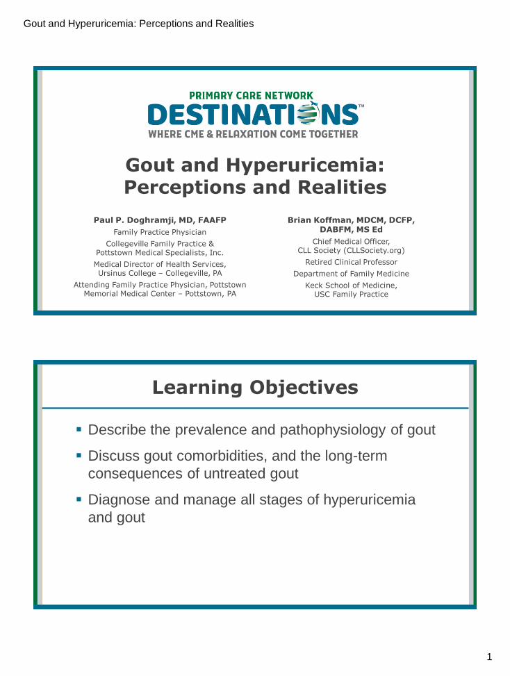
Gout Is a Urate Crystal Deposition Disease
▪ Most common cause of inflammatory arthritis in adults,1
affecting 8.3 million patients in the US2
▪ Caused by hyperuricemia, primarily due to inefficient excretion of
uric acid
▪ Crystal deposition in joints and soft tissues can lead to ▪ Inflammation3
▪ Acute gout flares3–5
▪ Tophi5
▪ Long-term bone and joint damage4
▪ Associated with increased risk of renal comorbidities6
▪ Mounting evidence that gout and crystal deposition can be
associated with cardiovascular and metabolic diseases; however,
causality has not been proven
▪ If untreated, ~70% of patients progress to tophaceous gout within
20 years7,8
Images courtesy of Dr. Fernando Perez-Ruiz, Cruces University Hospital, Barakaldo, Spain.
1. Doghramji PP, et al. Postgrad Med 2012;124:98–109. 2. Zhu Y, et al. Arthritis Rheum 2011;63:3136–41. 3. Schumacher R. Cleve Clin J Med 2008;75:S2-S4.
4. Mandell BF, et al. Cleve Clin J Med 2008;75:S5–S8. 5. Taylor JW, Grainger R. Chapter 9. In: Terkeltaub R (ed). Gout and Other Crystal Arthropathies (First edition). Philadelphia, PA: Elsevier Saunders, 2012. 6. Zhu Y, et
al. Am J Med 2012;125:679–87. 7. Terkeltaub R, Edwards NL. Chapter 3. In: Terkeltaub R, Edwards NL (eds). Gout: Diagnosis and Management of Gouty Arthritis and Hyperuricemia (Third edition). Oklahoma: Professional
Communications, Inc, 2013. 8. Gutman AB. Arthritis Rheum 1973;16:431–45.
X-ray of hand with tophaceous gout showing bone erosion of index finger (white arrow)
Tophaceous gout of the hand
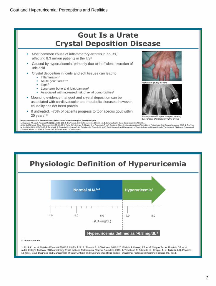
Physiologic Definition of Hyperuricemia
sUA=serum urate.
1. Rock KL, et al. Nat Rev Rheumatol 2013;9:13‒23. 2. So A, Thorens B. J Clin Invest 2010;120:1791‒9. 3. Keenan RT, et al. Chapter 94. In: Firestein GS, et al.
(eds). Kelley's Textbook of Rheumatology (Ninth edition). Philadelphia: Elsevier Saunders, 2013. 4. Terkeltaub R, Edwards NL. Chapter 1. In: Terkeltaub R, Edwards
NL (eds). Gout: Diagnosis and Management of Gouty Arthritis and Hyperuricemia (Third edition). Oklahoma: Professional Communications, Inc, 2013.
Normal sUA1–3 Hyperuricemia4
4.0 5.0 6.0 7.0 8.0
Hyperuricemia defined as >6.8 mg/dL4
sUA (mg/dL)
Gout and Hyperuricemia: Perceptions and Realities
3
Managing Hyperuricemia
▪ Uric acid-lowering drugs are not indicated for the management of asymptomatic hyperuricemia, but don’t ignore it
▪ Treat associated conditions
▪ Hypertension
▪ Hyperlipidemia
▪ Metabolic syndrome
▪ Dietary modifications are essential
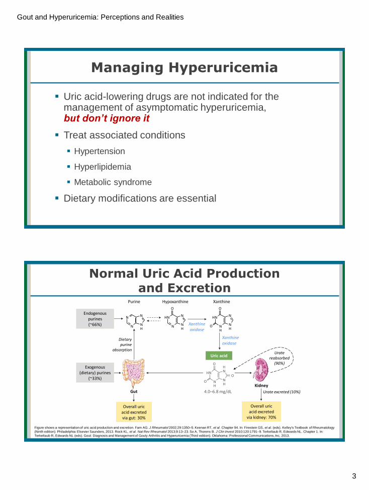
Normal Uric Acid Production and Excretion
Figure shows a representation of uric acid production and excretion. Fam AG. J Rheumatol 2002;29:1350‒5. Keenan RT, et al. Chapter 94. In: Firestein GS, et al. (eds). Kelley's Textbook of Rheumatology
(Ninth edition). Philadelphia: Elsevier Saunders, 2013. Rock KL, et al. Nat Rev Rheumatol 2013;9:13‒23. So A, Thorens B. J Clin Invest 2010;120:1791‒9. Terkeltaub R, Edwards NL. Chapter 1. In:
Terkeltaub R, Edwards NL (eds). Gout: Diagnosis and Management of Gouty Arthritis and Hyperuricemia (Third edition). Oklahoma: Professional Communications, Inc, 2013.
NH
NH
NHN
O
O
Uric acid
Purine Hypoxanthine Xanthine
Xanthine oxidase
Xanthine oxidase
Overall uricacid excretedvia gut: 30%
Urate reabsorbed
(90%)
Kidney
Gut 4.0–6.8 mg/dL
Exogenous(dietary) purines
(~33%)
Endogenouspurines(~66%)
Dietary purine
absorption
Urate excreted (10%)
Overall uricacid excreted
via kidney: 70%
NH
N
NHN
O
NH
N
NN
NH
NH
HNHN
O
O
O
Gout and Hyperuricemia: Perceptions and Realities
4
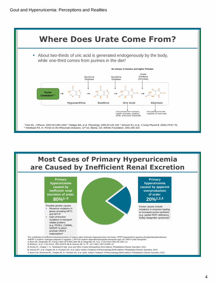
Where Does Urate Come From?
▪ About two-thirds of uric acid is generated endogenously by the body,
while one-third comes from purines in the diet1
No Uricase in Humans and Higher Primates
1 Fam AG. J Rheum. 2002;29:1350-1355; 2 Hediger MA, et al. Physiology. 2005;20:125-133; 3 Johnson RJ, et al. J Comp Physiol B. 2009;179:67-76; 4 Terkeltaub RA. In: Primer on the Rheumatic Diseases. 12th ed. Atlanta, GA: Arthritis Foundation; 2001:305-324.
Purine Catabolism2-4
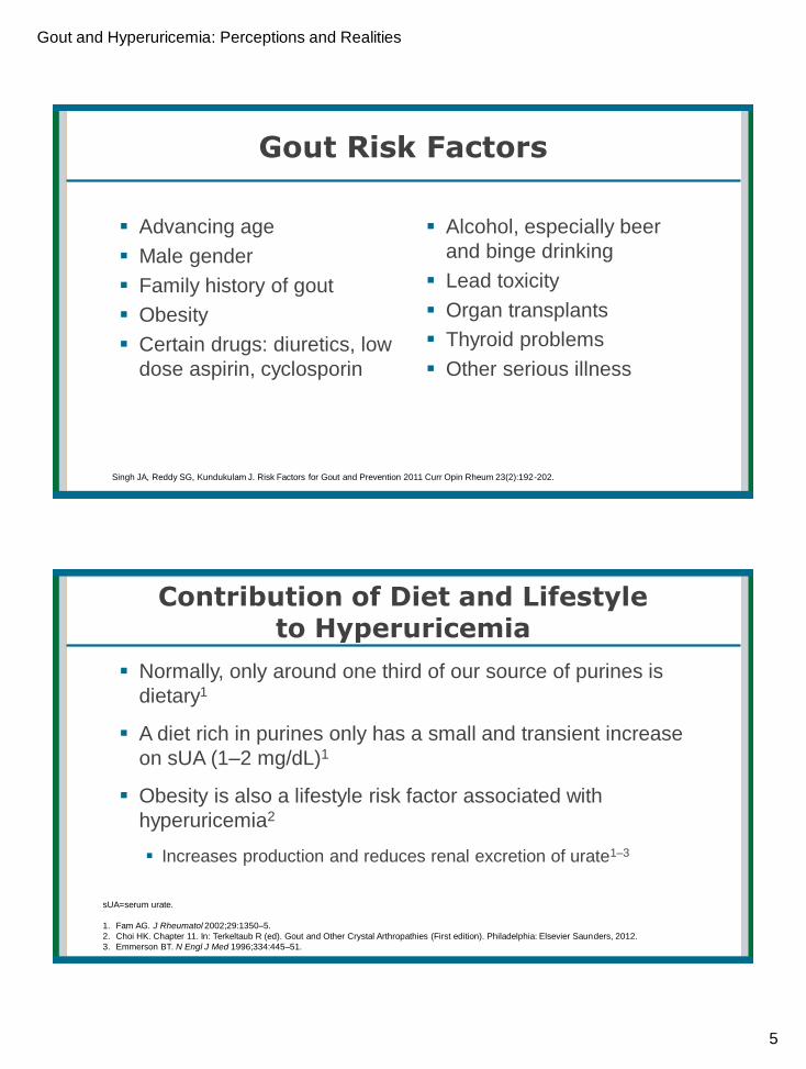
Most Cases of Primary Hyperuricemiaare Caused by Inefficient Renal Excretion
The contribution of inefficient intestinal excretion of urate to rates of primary hyperuricemia is not known. HPRT=hypoxanthine-guanine phosphoribosyltransferase;
NHERF-1=sodium‒hydrogen antiporter 3 regulator 1; NPT1/4=sodium-dependent phosphate transporter type 1/4; URAT=urate transporter.
1. Boss GR, Seegmiller JE. N Engl J Med 1979;300:1459–68. 2. Seegmiller JE, et al. J Clin Invest 1961;40:1304–14.
3. Bishop C, et al. J Clin Invest. 1951;30:879–88. 4. Gutman AB, Yu TF. Am J Med. 1957;23:600–22.
5. Riches PL. Chapter 7. In: Terkeltaub R (ed). Gout and Other Crystal Arthropathies (First edition). Philadelphia: Elsevier Saunders, 2012.
6. Keenan RT, et al. Chapter 94. In: Firestein GS, et al. (eds). Kelley's Textbook of Rheumatology (Ninth edition). Philadelphia: Elsevier Saunders, 2013.
7. Burns CM, Wortmann RL. Chapter 95. In: Firestein GS, et al. (eds). Kelley's Textbook of Rheumatology (Ninth edition). Philadelphia: Elsevier Saunders, 2013.
Possible genetic causes:
▪ Missense mutations in
genes encoding NPT1
and NPT45
▪ Gain-of-function
mutations in transport-
related proteins
(e.g. PDZK1, CARMIL,
NHERF-1) which
promote URAT1
reabsorption6
Primary
hyperuricemia
caused by
inefficient renal
excretion of urate:
80%1–3
Known causes include
mutations in enzymes leading
to increased purine synthesis
(e.g. partial HPRT deficiency;
Kelley-Seegmiller syndrome)7
Primary
hyperuricemia
caused by apparent
overproduction
of urate:
20%1,2,4
Gout and Hyperuricemia: Perceptions and Realities
5
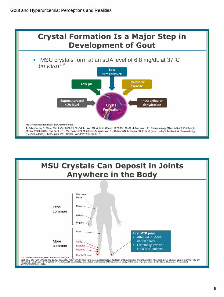
Gout Risk Factors
▪ Advancing age
▪ Male gender
▪ Family history of gout
▪ Obesity
▪ Certain drugs: diuretics, low
dose aspirin, cyclosporin
▪ Alcohol, especially beer
and binge drinking
▪ Lead toxicity
▪ Organ transplants
▪ Thyroid problems
▪ Other serious illness
Singh JA, Reddy SG, Kundukulam J. Risk Factors for Gout and Prevention 2011 Curr Opin Rheum 23(2):192-202.
Contribution of Diet and Lifestyle to Hyperuricemia
▪ Normally, only around one third of our source of purines is
dietary1
▪ A diet rich in purines only has a small and transient increase
on sUA (1–2 mg/dL)1
▪ Obesity is also a lifestyle risk factor associated with
hyperuricemia2
▪ Increases production and reduces renal excretion of urate1–3
sUA=serum urate.
1. Fam AG. J Rheumatol 2002;29:1350‒5.
2. Choi HK. Chapter 11. In: Terkeltaub R (ed). Gout and Other Crystal Arthropathies (First edition). Philadelphia: Elsevier Saunders, 2012.
3. Emmerson BT. N Engl J Med 1996;334:445‒51.
Gout and Hyperuricemia: Perceptions and Realities
6
Crystal Formation
Supersaturated sUA level
Low pH
Low temperature
Trauma or exercise
Intra-articular dehydration
Crystal Formation Is a Major Step in Development of Gout
▪ MSU crystals form at an sUA level of 6.8 mg/dL at 37°C (in vitro)1–5
MSU=monosodium urate; sUA=serum urate.
1. Schumacher R. Cleve Clin J Med 2008;75:S2–S4. 2. Loeb JN. Arthritis Rheum 1972;15:189–92. 3. McLean L. In: Rheumatology (Third edition). Edinburgh:
Mosby; 2003:1903–18. 4. Scott JT. J Clin Path 1978;31:205–13. 5. Wortmann RL, Kelley WN. In: Harris ED Jr, et al. (eds). Kelley’s Textbook of Rheumatology
(Seventh edition). Philadelphia, PA: Elsevier Saunders; 2005:1402–29.
MSU Crystals Can Deposit in Joints Anywhere in the Body
MSU=monosodium urate; MTP=metatarsophalangeal.
Scott JT. J Clin Path 1978;31:205–13. Wortmann RL, Kelley WN. In: Harris ED Jr, et al. (eds) Kelley’s Textbook of Rheumatology (Seventh edition). Philadelphia, PA: Elsevier Saunders; 2005:1402–29. Terkeltaub R, Edwards NL. Chapter 3. In: Terkeltaub R, Edwards NL (eds). Gout: Diagnosis and Management of Gouty Arthritis and Hyperuricemia (Third edition). Oklahoma: Professional Communications, Inc, 2013.
Less
common
Olecranonbursa
Elbow
Fingers
Knee
Subtalar
Wrists
First MTP joint
Midfoot
AnkleMore
common
First MTP joint
▪ Affected in ~50%
of first flares
▪ Eventually involved
in 90% of patients
Gout and Hyperuricemia: Perceptions and Realities
7
Prior to Any Clinical Manifestations of Gout, MSU Crystal Deposits Can Build Up1,2
MSU=monosodium urate.
1. Terkeltaub R, Edwards NL. Chapter 3. In: Terkeltaub R, Edwards NL (eds). Gout: Diagnosis and Management of Gouty Arthritis and Hyperuricemia (Third edition). Oklahoma: Professional Communications, Inc, 2013. 2. Pineda C, et al. Arthritis Res Ther 2011;13:R4. 3. Nicolaou S, et al. AJR Am J Roentgenol 2012;199(5 Suppl):S78–S86.
DECT scan showing MSU crystal deposition (green)
surrounding the right first MTP and along left
midfoot in a patient with asymptomatic
hyperuricemia3
DECT scan showing extensive MSU crystal deposition
(green) in a patient with gout3
Gout Patient Asymptomatic Hyperuricemia
In Addition to Joints, MSU Crystals May Deposit in Soft Tissues, Such as Kidney and Eye
MSU=monosodium urate.
1. Terkeltaub R, Edwards NL. Chapter 3. In: Terkeltaub R, Edwards NL (eds). Gout: Diagnosis and Management of Gouty Arthritis and Hyperuricemia (Third edition). Oklahoma: Professional Communications, Inc, 2013. 2. Taylor JW, Grainger R. Chapter 9. In: Terkeltaub R (ed). Gout and Other Crystal Arthropathies (First edition). Philadelphia, PA: Elsevier Saunders, 2012. 3. Bernard B, et al. Arthritis Rheum 2006;DOI 10.1002/art.21722.
Chronic urate nephropathy with presence of tophaceous
deposits (red arrow) in the medulla of the kidney1,2
Ophthalmologic examination revealing multiple chalky
deposits on the corneal stroma of a patient with long-
standing untreated gout (arrow). Polarized light microscopy
examination of a scraping from a deposit revealed typical
MSU crystals3
Conjunctival gouty tophi have
been reported2
Chronic urate nephropathy
occurs with the deposition of
MSU crystals in the kidney1
Gout and Hyperuricemia: Perceptions and Realities
8
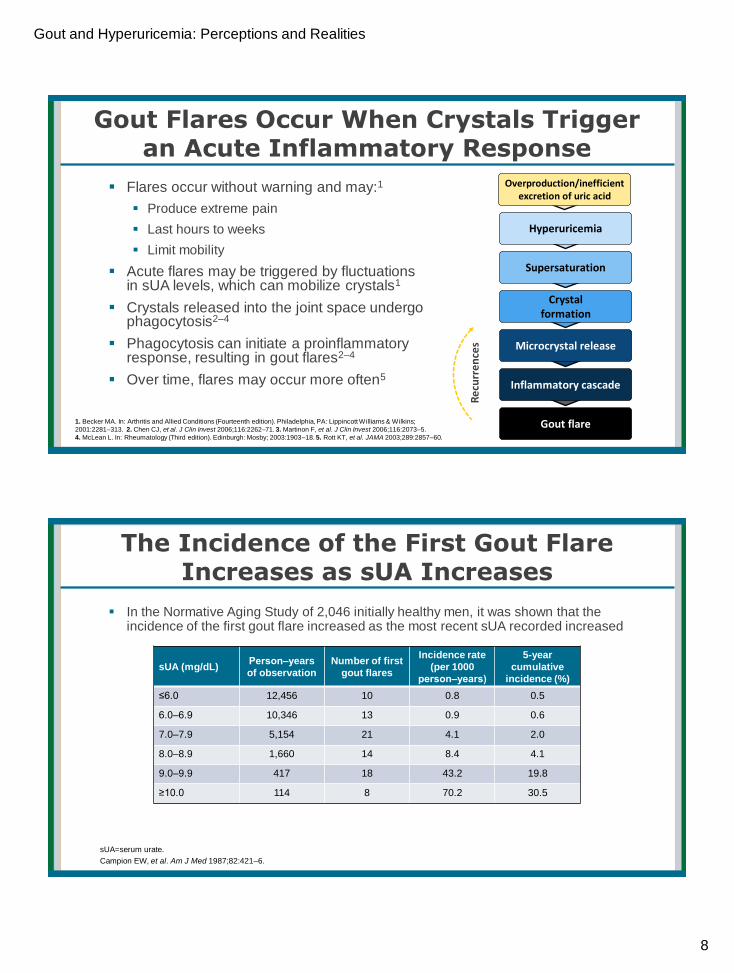
Gout Flares Occur When Crystals Trigger an Acute Inflammatory Response
▪ Flares occur without warning and may:1
▪ Produce extreme pain
▪ Last hours to weeks
▪ Limit mobility
▪ Acute flares may be triggered by fluctuations in sUA levels, which can mobilize crystals1
▪ Crystals released into the joint space undergo phagocytosis2–4
▪ Phagocytosis can initiate a proinflammatory response, resulting in gout flares2–4
▪ Over time, flares may occur more often5
1. Becker MA. In: Arthritis and Allied Conditions (Fourteenth edition). Philadelphia, PA: Lippincott Williams & Wilkins;
2001:2281–313. 2. Chen CJ, et al. J Clin Invest 2006;116:2262–71. 3. Martinon F, et al. J Clin Invest 2006;116:2073–5.
4. McLean L. In: Rheumatology (Third edition). Edinburgh: Mosby; 2003:1903–18. 5. Rott KT, et al. JAMA 2003;289:2857–60.
Supersaturation
Crystal formation
Microcrystal release
Inflammatory cascade
Gout flare
Hyperuricemia
Re
curr
en
ces
Overproduction/inefficient excretion of uric acid
The Incidence of the First Gout Flare Increases as sUA Increases
▪ In the Normative Aging Study of 2,046 initially healthy men, it was shown that the incidence of the first gout flare increased as the most recent sUA recorded increased
sUA=serum urate.
Campion EW, et al. Am J Med 1987;82:421‒6.
sUA (mg/dL)Person‒years
of observation
Number of first
gout flares
Incidence rate
(per 1000
person‒years)
5-year
cumulative
incidence (%)
≤6.0 12,456 10 0.8 0.5
6.0‒6.9 10,346 13 0.9 0.6
7.0–7.9 5,154 21 4.1 2.0
8.0–8.9 1,660 14 8.4 4.1
9.0–9.9 417 18 43.2 19.8
≥10.0 114 8 70.2 30.5
Gout and Hyperuricemia: Perceptions and Realities
9
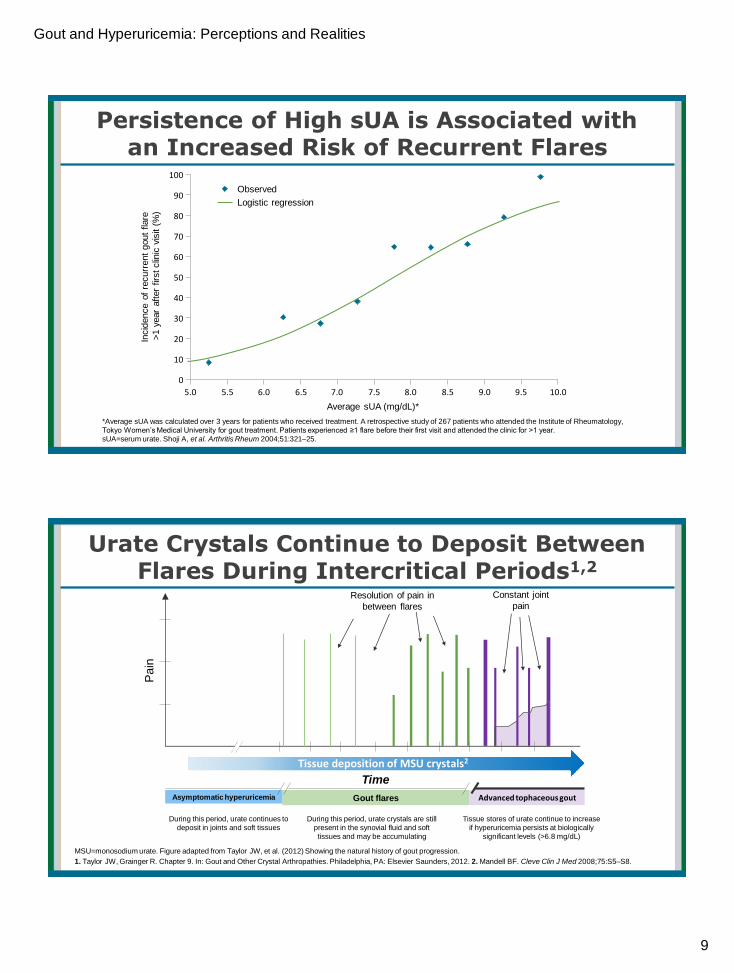
Persistence of High sUA is Associated withan Increased Risk of Recurrent Flares
*Average sUA was calculated over 3 years for patients who received treatment. A retrospective study of 267 patients who attended the Institute of Rheumatology,
Tokyo Women’s Medical University for gout treatment. Patients experienced ≥1 flare before their first visit and attended the clinic for >1 year.
sUA=serum urate. Shoji A, et al. Arthritis Rheum 2004;51:321–25.
Incid
ence o
f re
curr
ent
gout
flare
>1 y
ear
aft
er
firs
t clin
ic v
isit (
%)
Average sUA (mg/dL)*
100
90
80
70
60
50
40
30
20
10
0
5.0
Observed
Logistic regression
5.5 6.0 6.5 7.0 7.5 8.0 8.5 9.0 9.5 10.0
Urate Crystals Continue to Deposit Between Flares During Intercritical Periods1,2
MSU=monosodium urate. Figure adapted from Taylor JW, et al. (2012) Showing the natural history of gout progression.
1. Taylor JW, Grainger R. Chapter 9. In: Gout and Other Crystal Arthropathies. Philadelphia, PA: Elsevier Saunders, 2012. 2. Mandell BF. Cleve Clin J Med 2008;75:S5–S8.
Resolution of pain in
between flares
Gout flaresAsymptomatic hyperuricemia Advanced tophaceous gout
Time
Pain
Constant joint
pain
Tissue deposition of MSU crystals2
During this period, urate continues to
deposit in joints and soft tissues
During this period, urate crystals are still
present in the synovial fluid and soft
tissues and may be accumulating
Tissue stores of urate continue to increase
if hyperuricemia persists at biologically
significant levels (>6.8 mg/dL)

Gout and Hyperuricemia: Perceptions and Realities
10
25%
38% 42
% 48%
2%
6%
13%
10%
2% 3%
6%
13%
29%
47%
61%
71%
0%
10%
20%
30%
40%
50%
60%
70%
80%
90%
100%
1–5 (n=392)
6–10 (n=316)
11–15(n=204)
16–20(n=142)
Pati
ents
Years after initial flare without ULT
Minimal tophi
Moderate tophi
Extensive tophi
Total tophi
Development of Tophi Increases with Time and Severity of Hyperuricemia
A retrospective analysis of 1165 patients with gout before appropriate drug therapy. sUA=serum urate; ULT=urate-lowering therapy.
Gutman AB. Arthritis Rheum 1973;16:431‒45.19
9%
27%
42% 45
%
0% 2%
7%
17%
0%
10%
20%
30%
40%
50%
60%
70%
80%
90%
100%
<7.1–8.0(n=180)
8.1–9.0(n=272)
9.1–10.0(n=346)
>10(n=491)
Pati
ents
Mean sUA (mg/dL)
Minimal–moderate tophi
Extensive tophi
Development of Tophi can Lead to Joint Damage, Bone Erosion and Progressive Disability1–4
Image s courtesy of Dr Fernando Perez-Ruiz, Rheumatology Department, Cruces University Hospital, Barakaldo, Spain
Taylor WJ, Grainger R. Chapter 9. In: Terkeltaub R (ed). Gout and Other Crystal Arthropathies (First Edition). Philadelphia: Elsevier Saunders, 2012.
Burns CM, Wortmann RL. Chapter 95. In: Firestein GS, et al. (eds). Kelley's Textbook of Rheumatology (Ninth edition). Philadelphia: Elsevier Saunders, 2013.
Dalbeth N, Doyle AJ. Best Pract Res Clin Rheumatol 2012;26:823–38. Desai MA, et al. Radiographics 2011;31:1365‒75.
X-ray of hand with tophaceous gout showing
bone erosion of index finger (white arrow)
Tophaceous gout of the hand showing
numerous subcutaneous tophi
Gout and Hyperuricemia: Perceptions and Realities
11
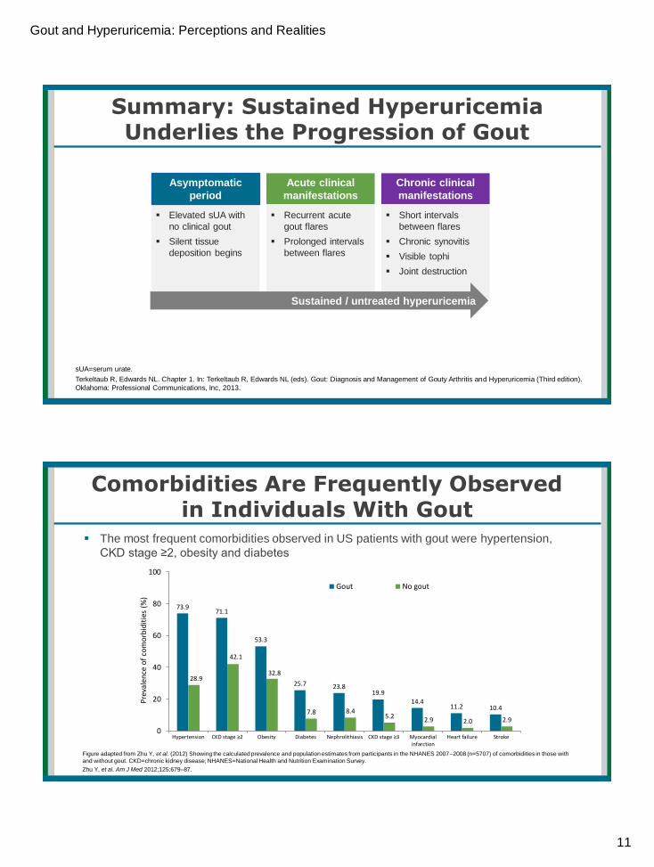
Summary: Sustained Hyperuricemia Underlies the Progression of Gout
sUA=serum urate.
Terkeltaub R, Edwards NL. Chapter 1. In: Terkeltaub R, Edwards NL (eds). Gout: Diagnosis and Management of Gouty Arthritis and Hyperuricemia (Third edition).
Oklahoma: Professional Communications, Inc, 2013.
▪ Elevated sUA with
no clinical gout
▪ Silent tissue
deposition begins
Asymptomatic
period
▪ Recurrent acute
gout flares
▪ Prolonged intervals
between flares
Acute clinical
manifestations
▪ Short intervals
between flares
▪ Chronic synovitis
▪ Visible tophi
▪ Joint destruction
Chronic clinical
manifestations
Sustained / untreated hyperuricemia
Comorbidities Are Frequently Observed in Individuals With Gout
▪ The most frequent comorbidities observed in US patients with gout were hypertension,
CKD stage ≥2, obesity and diabetes
Figure adapted from Zhu Y, et al. (2012) Showing the calculated prevalence and population estimates from participants in the NHANES 2007–2008 (n=5707) of comorbidities in those with
and without gout. CKD=chronic kidney disease; NHANES=National Health and Nutrition Examination Survey.
Zhu Y, et al. Am J Med 2012;125:679–87.
73.971.1
53.3
25.7 23.819.9
14.411.2 10.4
28.9
42.1
32.8
7.8 8.45.2
2.9 2.0 2.9
0
20
40
60
80
100
Hypertension CKD stage ≥2 Obesity Diabetes Nephrolithiasis CKD stage ≥3 Myocardialinfarction
Heart failure Stroke
Pre
vale
nce
of
com
orb
idit
ies
(%)
Gout No gout
Gout and Hyperuricemia: Perceptions and Realities
12
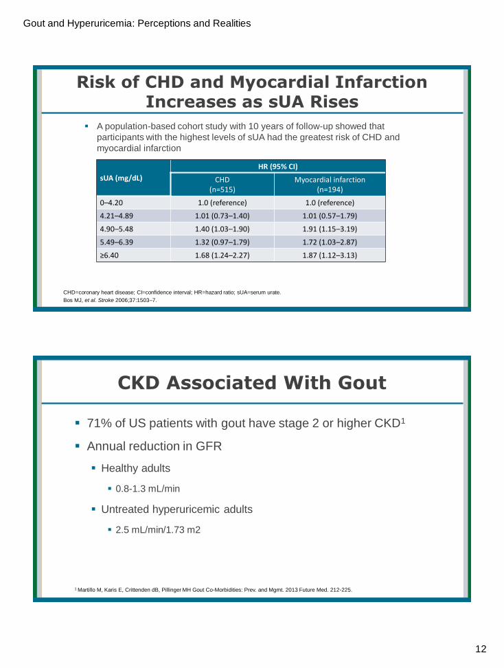
Risk of CHD and Myocardial Infarction Increases as sUA Rises
▪ A population-based cohort study with 10 years of follow-up showed that
participants with the highest levels of sUA had the greatest risk of CHD and
myocardial infarction
CHD=coronary heart disease; CI=confidence interval; HR=hazard ratio; sUA=serum urate.
Bos MJ, et al. Stroke 2006;37:1503–7.
sUA (mg/dL)
HR (95% CI)
CHD(n=515)
Myocardial infarction(n=194)
0–4.20 1.0 (reference) 1.0 (reference)
4.21–4.89 1.01 (0.73–1.40) 1.01 (0.57–1.79)
4.90–5.48 1.40 (1.03–1.90) 1.91 (1.15–3.19)
5.49–6.39 1.32 (0.97–1.79) 1.72 (1.03–2.87)
≥6.40 1.68 (1.24–2.27) 1.87 (1.12–3.13)
CKD Associated With Gout
▪ 71% of US patients with gout have stage 2 or higher CKD1
▪ Annual reduction in GFR
▪ Healthy adults
▪ 0.8-1.3 mL/min
▪ Untreated hyperuricemic adults
▪ 2.5 mL/min/1.73 m2
1 Martillo M, Karis E, Crittenden dB, Pillinger MH Gout Co-Morbidities: Prev. and Mgmt. 2013 Future Med. 212-225.
Gout and Hyperuricemia: Perceptions and Realities
13
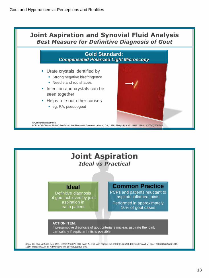
Joint Aspiration and Synovial Fluid AnalysisBest Measure for Definitive Diagnosis of Gout
▪ Urate crystals identified by
▪ Strong negative birefringence
▪ Needle and rod shapes
▪ Infection and crystals can be
seen together
▪ Helps rule out other causes
▪ eg, RA, pseudogout
RA, rheumatoid arthritis.
ACR. ACR Clinical Slide Collection on the Rheumatic Diseases. Atlanta, GA; 1998; Phelps P, et al. JAMA. 1968;12;203(7):508-512.
Gold Standard: Compensated Polarized Light Microscopy
Joint AspirationIdeal vs Practical
Segal JB, et al. Arthritis Care Res. 1999;12(6):376-380; Swan A, et al. Ann Rheum Dis. 2002;61(6):493-498; Underwood M. BMJ. 2006;332(7553):1315-
1319; Wallace SL, et al. Arthritis Rheum. 1977;20(3):895-900.
IdealDefinitive diagnosis
of gout achieved by joint aspiration in each patient
Common PracticePCPs and patients reluctant to
aspirate inflamed joints
Performed in approximately 10% of gout cases
ACTION ITEM:
If presumptive diagnosis of gout criteria is unclear, aspirate the joint,
particularly if septic arthritis is possible
Gout and Hyperuricemia: Perceptions and Realities
14
CT, computed tomography; MRI, magnetic resonance imaging.
Schlesinger N. Am J Manag Care. 2005;11(suppl 15):S443-S450.
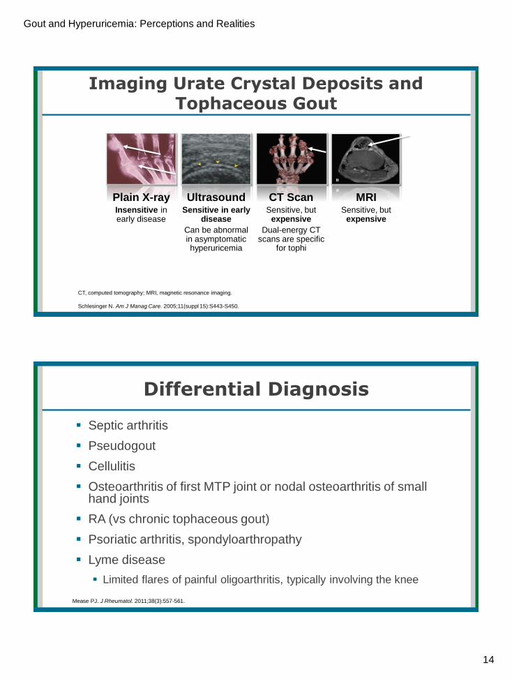
Imaging Urate Crystal Deposits and Tophaceous Gout
Plain X-ray Insensitive in early disease
Ultrasound Sensitive in early
disease
Can be abnormal in asymptomatic hyperuricemia
CT ScanSensitive, but
expensive
Dual-energy CT scans are specific
for tophi
MRI Sensitive, but
expensive
Differential Diagnosis
▪ Septic arthritis
▪ Pseudogout
▪ Cellulitis
▪ Osteoarthritis of first MTP joint or nodal osteoarthritis of small hand joints
▪ RA (vs chronic tophaceous gout)
▪ Psoriatic arthritis, spondyloarthropathy
▪ Lyme disease
▪ Limited flares of painful oligoarthritis, typically involving the knee
Mease PJ. J Rheumatol. 2011;38(3):557-561.
Gout and Hyperuricemia: Perceptions and Realities
15
Treating Gout Flares: Key Points
▪ When you start therapy is more important than which agent you
use (ACR recommends ≤ 24 hours)
▪ Select agent considering patient comorbidities
▪ The sooner treatment is initiated after symptoms begin, the faster it
will work
▪ When taken at the earliest hint of a flare, attacks may be aborted with
one dose
Wortmann PL, et al. Gout and hyperuricemia. In: Harris ED Jr, et al, eds. Kelly’s Textbook of Rheumatology. 7th ed. Philadelphia, PA: WB Saunders
Company; 2005:1402-1429.
COX-2, cyclooxygenase-2; NSAID, nonsteroidal anti-inflammatory drug.
Khanna D, et al. Arthritis Care Res. 2012;64(10):1447-1461; Peterson DM. J Pain Palliat Care Pharmacother. 2010;24(4):402-404;
Terkeltaub RA, et al. Arthritis Rheum. 2010;62:1060-1068.
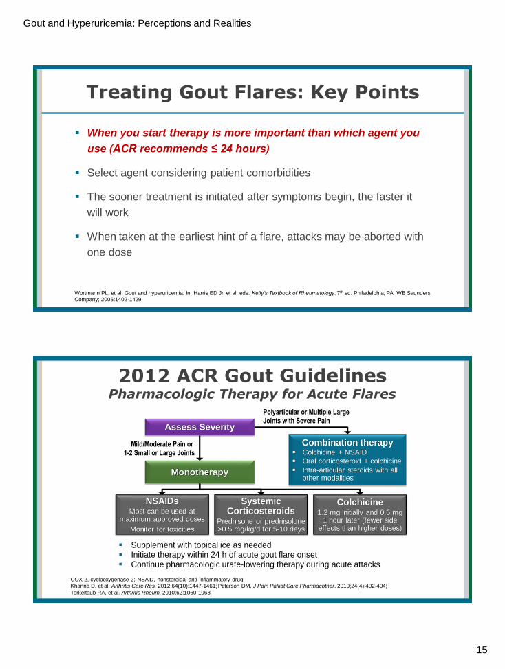
2012 ACR Gout GuidelinesPharmacologic Therapy for Acute Flares
▪ Supplement with topical ice as needed
▪ Initiate therapy within 24 h of acute gout flare onset
▪ Continue pharmacologic urate-lowering therapy during acute attacks
Combination therapy▪ Colchicine + NSAID
▪ Oral corticosteroid + colchicine
▪ Intra-articular steroids with all other modalities
Assess Severity
Monotherapy
NSAIDsMost can be used at
maximum approved doses
Monitor for toxicities
Systemic Corticosteroids
Prednisone or prednisolone >0.5 mg/kg/d for 5-10 days
Colchicine1.2 mg initially and 0.6 mg
1 hour later (fewer side effects than higher doses)
Polyarticular or Multiple Large
Joints with Severe Pain
Mild/Moderate Pain or
1-2 Small or Large Joints
Gout and Hyperuricemia: Perceptions and Realities
16
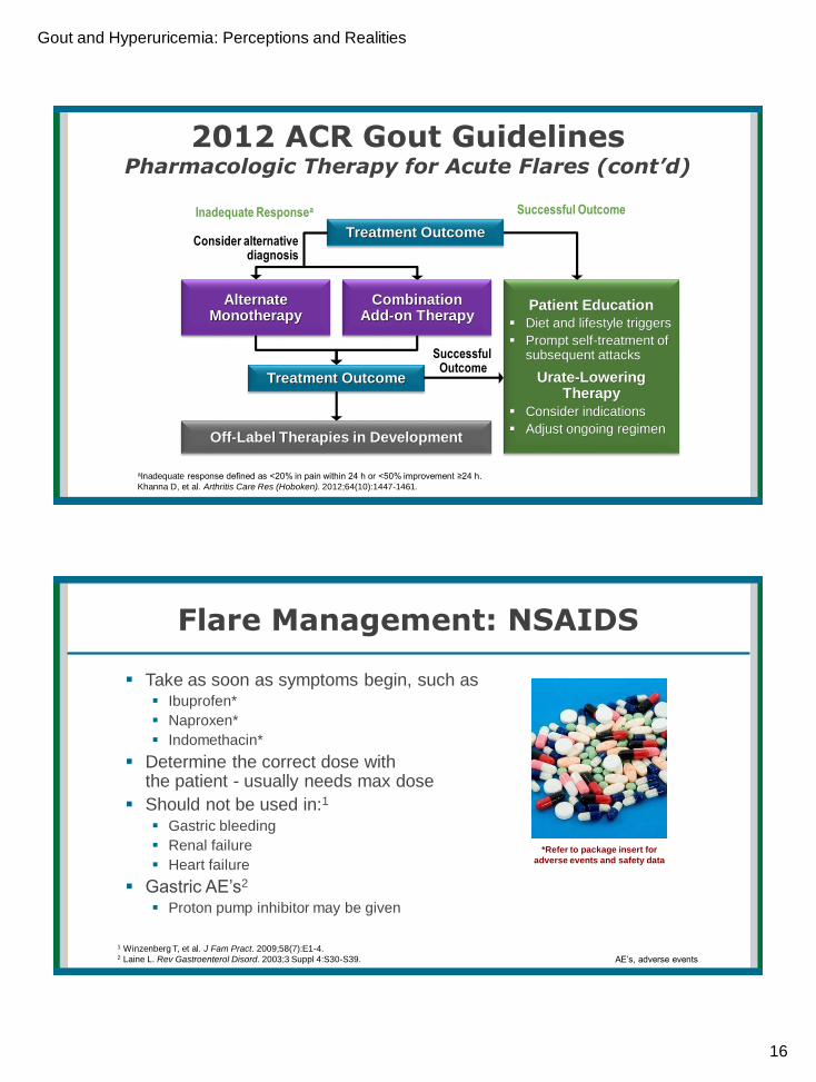
aInadequate response defined as <20% in pain within 24 h or <50% improvement ≥24 h.
Khanna D, et al. Arthritis Care Res (Hoboken). 2012;64(10):1447-1461.
2012 ACR Gout GuidelinesPharmacologic Therapy for Acute Flares (cont’d)
Patient Education
▪ Diet and lifestyle triggers
▪ Prompt self-treatment of subsequent attacks
Urate-Lowering Therapy
▪ Consider indications
▪ Adjust ongoing regimen
Treatment Outcome
Alternate Monotherapy
Off-Label Therapies in Development
Successful Outcome
Combination Add-on Therapy
Consider alternative diagnosis
Treatment Outcome
Inadequate Response
Successful Outcome
Inadequate Responsea
Flare Management: NSAIDS
▪ Take as soon as symptoms begin, such as▪ Ibuprofen*
▪ Naproxen*
▪ Indomethacin*
▪ Determine the correct dose with the patient - usually needs max dose
▪ Should not be used in:1
▪ Gastric bleeding
▪ Renal failure
▪ Heart failure
▪ Gastric AE’s2
▪ Proton pump inhibitor may be given
1 Winzenberg T, et al. J Fam Pract. 2009;58(7):E1-4. 2 Laine L. Rev Gastroenterol Disord. 2003;3 Suppl 4:S30-S39.
*Refer to package insert for
adverse events and safety data
AE’s, adverse events
Gout and Hyperuricemia: Perceptions and Realities
17
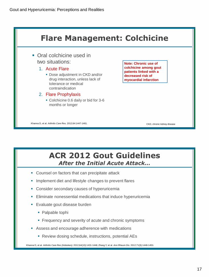
Flare Management: Colchicine
▪ Oral colchicine used in
two situations:
1. Acute Flare
▪ Dose adjustment in CKD and/or
drug interaction, unless lack of
tolerance or medical
contraindication
2. Flare Prophylaxis
▪ Colchicine 0.6 daily or bid for 3-6
months or longer
Khanna D, et al. Arthritis Care Res. 2012;64:1447-1461. CKD, chronic kidney disease
Note: Chronic use of
colchicine among gout
patients linked with a
decreased risk of
myocardial infarction
ACR 2012 Gout Guidelines After the Initial Acute Attack…
▪ Counsel on factors that can precipitate attack
▪ Implement diet and lifestyle changes to prevent flares
▪ Consider secondary causes of hyperuricemia
▪ Eliminate nonessential medications that induce hyperuricemia
▪ Evaluate gout disease burden
▪ Palpable tophi
▪ Frequency and severity of acute and chronic symptoms
▪ Assess and encourage adherence with medications
▪ Review dosing schedule, instructions, potential AEs
Khanna D, et al. Arthritis Care Res (Hoboken). 2012;64(10):1431-1446; Zhang Y, et al. Ann Rheum Dis. 2012;71(9):1448-1453.
Gout and Hyperuricemia: Perceptions and Realities
18
Khanna D, et al. Arthritis Care Res (Hoboken). 2012;64(10):1431-1446.
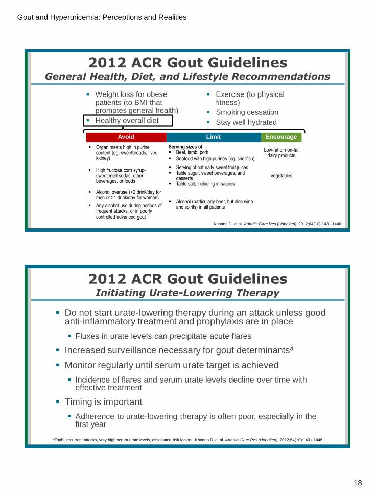
2012 ACR Gout GuidelinesGeneral Health, Diet, and Lifestyle Recommendations
▪ Weight loss for obese patients (to BMI that promotes general health)
▪ Healthy overall diet
▪ Exercise (to physical fitness)
▪ Smoking cessation
▪ Stay well hydrated
Avoid Limit Encourage
▪ Organ meats high in purinecontent (eg, sweetbreads, liver, kidney)
Serving sizes of▪ Beef, lamb, pork
▪ Seafood with high purines (eg, shellfish)
Low-fat or non-fat dairy products
▪ High fructose corn syrup-sweetened sodas, other beverages, or foods
▪ Serving of naturally sweet fruit juices▪ Table sugar, sweet beverages, and
desserts▪ Table salt, including in sauces
Vegetables
▪ Alcohol overuse (>2 drink/day for men or >1 drink/day for women)
▪ Any alcohol use during periods of frequent attacks, or in poorly controlled advanced gout
▪ Alcohol (particularly beer, but also wine and spirits) in all patients
aTophi, recurrent attacks, very high serum urate levels, associated risk factors. Khanna D, et al. Arthritis Care Res (Hoboken). 2012;64(10):1431-1446.
2012 ACR Gout GuidelinesInitiating Urate-Lowering Therapy
▪ Do not start urate-lowering therapy during an attack unless good anti-inflammatory treatment and prophylaxis are in place
▪ Fluxes in urate levels can precipitate acute flares
▪ Increased surveillance necessary for gout determinantsa
▪ Monitor regularly until serum urate target is achieved
▪ Incidence of flares and serum urate levels decline over time with effective treatment
▪ Timing is important
▪ Adherence to urate-lowering therapy is often poor, especially in the first year
Gout and Hyperuricemia: Perceptions and Realities
19
Khanna D, et al. Arthritis Care Res (Hoboken). 2012;64(10):1431-1446.
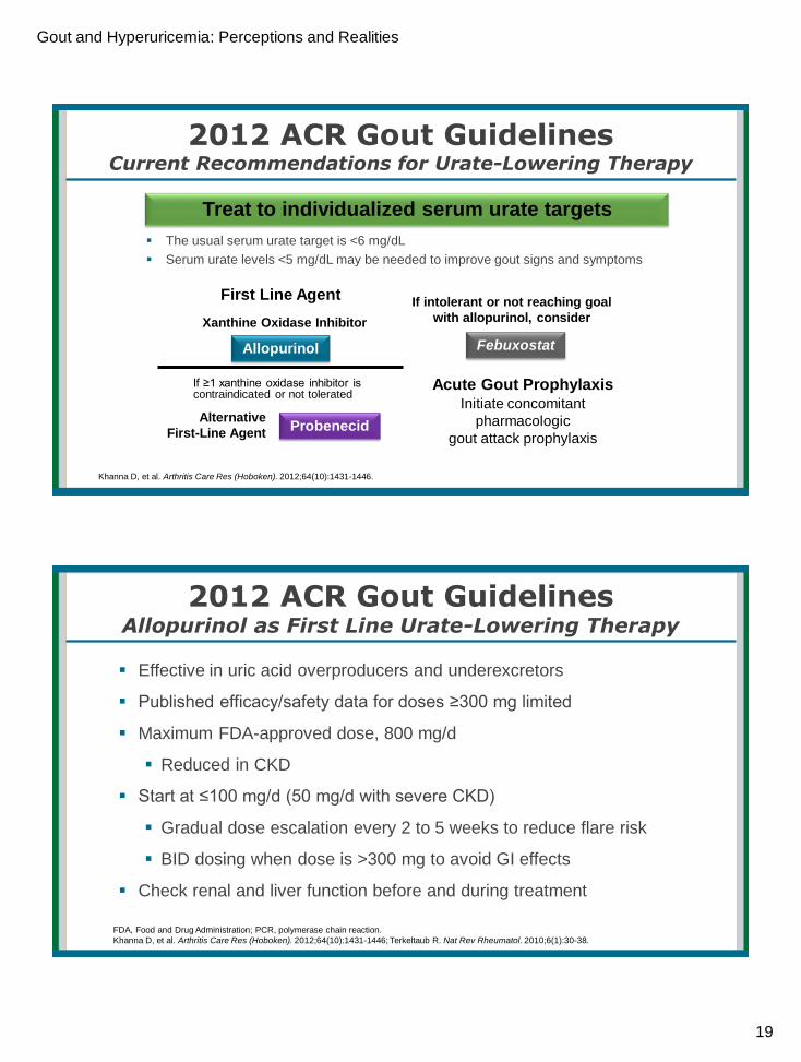
2012 ACR Gout GuidelinesCurrent Recommendations for Urate-Lowering Therapy
▪ The usual serum urate target is <6 mg/dL
▪ Serum urate levels <5 mg/dL may be needed to improve gout signs and symptoms
Treat to individualized serum urate targets
First Line Agent
Acute Gout ProphylaxisInitiate concomitant
pharmacologic
gout attack prophylaxis
Xanthine Oxidase Inhibitor
Allopurinol
Alternative
First-Line AgentProbenecid
If ≥1 xanthine oxidase inhibitor is contraindicated or not tolerated
If intolerant or not reaching goal
with allopurinol, consider
Febuxostat
FDA, Food and Drug Administration; PCR, polymerase chain reaction.
Khanna D, et al. Arthritis Care Res (Hoboken). 2012;64(10):1431-1446; Terkeltaub R. Nat Rev Rheumatol. 2010;6(1):30-38.
2012 ACR Gout GuidelinesAllopurinol as First Line Urate-Lowering Therapy
▪ Effective in uric acid overproducers and underexcretors
▪ Published efficacy/safety data for doses ≥300 mg limited
▪ Maximum FDA-approved dose, 800 mg/d
▪ Reduced in CKD
▪ Start at ≤100 mg/d (50 mg/d with severe CKD)
▪ Gradual dose escalation every 2 to 5 weeks to reduce flare risk
▪ BID dosing when dose is >300 mg to avoid GI effects
▪ Check renal and liver function before and during treatment
Gout and Hyperuricemia: Perceptions and Realities
20
CNS, central nervous system; PCR, polymerase chain reaction.
Chao J, et al. Curr Rheumatol Rep. 2009;11:135-140; Hande KR, et al. Am J Med. 1984;76(1):47-56; Hung SI, et al. Proc Natl Acad Sci USA.
2005;102(11):4134-4139; Riedel AA, et al. J Rheumatol. 2004;31:1575-1581.
Allopurinol Limitations as Urate-Lowering Therapy
▪ Intolerance in 5%-10% (eg, liver function, GI, CNS)
▪ Pruritic rash in ~2%
▪ Major allopurinol hypersensitivity syndrome
▪ Incidence 0.1%-0.4% (~25% mortality)
▪ Usually in first months of therapy
▪ More common with CKD, thiazides, HLA-B*5801
▪ Consider pharmacogenetic HLA-B*5801 PCR testing in high-risk populations (Han Chinese, Thai, Koreans)
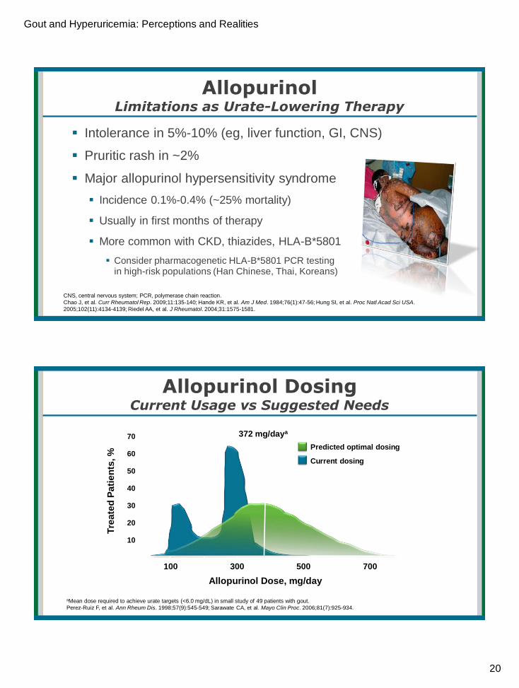
Allopurinol DosingCurrent Usage vs Suggested Needs
aMean dose required to achieve urate targets (<6.0 mg/dL) in small study of 49 patients with gout.
Perez-Ruiz F, et al. Ann Rheum Dis. 1998;57(9):545-549; Sarawate CA, et al. Mayo Clin Proc. 2006;81(7):925-934.
Tre
ate
d P
ati
en
ts, %
10
20
30
40
50
60
70
100 300 500 700
Allopurinol Dose, mg/day
Current dosing
Predicted optimal dosing
372 mg/daya
Gout and Hyperuricemia: Perceptions and Realities
21
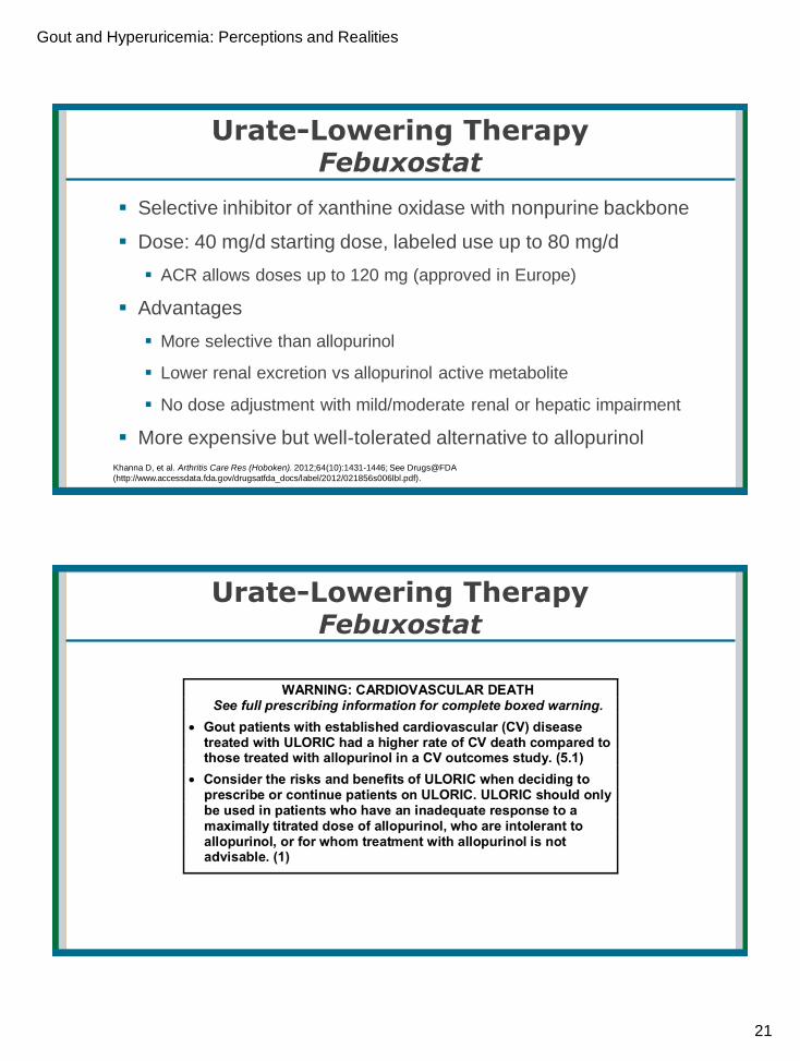
Urate-Lowering Therapy Febuxostat
▪ Selective inhibitor of xanthine oxidase with nonpurine backbone
▪ Dose: 40 mg/d starting dose, labeled use up to 80 mg/d
▪ ACR allows doses up to 120 mg (approved in Europe)
▪ Advantages
▪ More selective than allopurinol
▪ Lower renal excretion vs allopurinol active metabolite
▪ No dose adjustment with mild/moderate renal or hepatic impairment
▪ More expensive but well-tolerated alternative to allopurinol
Khanna D, et al. Arthritis Care Res (Hoboken). 2012;64(10):1431-1446; See Drugs@FDA
(http://www.accessdata.fda.gov/drugsatfda_docs/label/2012/021856s006lbl.pdf).
Urate-Lowering Therapy Febuxostat
Gout and Hyperuricemia: Perceptions and Realities
22
Urate-Lowering TherapyUricosurics
▪ Increase urate excretion by inhibiting urate reabsorption in the kidney
▪ Probenecid − only FDA-approved uricosuric
▪ Alternative in those intolerant to xanthine oxidase inhibitors
▪ Not recommended in persons with history of urolithiasis
▪ Increases urolithiasis risk, especially with acidic urine pH
▪ Not recommended if CrCl <50 mL/min
▪ Losartan, atorvastatin, and fenofibrate
▪ Less potent
▪ Off label
CrCl, creatinine clearance.
Khanna D, et al. Arthritis Care Res. 2012;64(10):1431-1446; Terkeltaub RA. Arthritis Res Ther. 2009;11:236; Terkeltaub RA, et al. Joint Bone Spine.
2009;76:444-446; Terkeltaub RA. N Engl J Med. 2003;349:1647-1655.
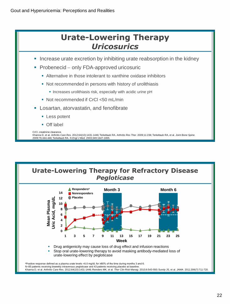
Urate-Lowering Therapy for Refractory DiseasePegloticase
▪ Drug antigenicity may cause loss of drug effect and infusion reactions
▪ Stop oral urate-lowering therapy to avoid masking antibody-mediated loss of urate-lowering effect by pegloticase
aPositive response defined as a plasma urate levels <6.0 mg/dL for ≥80% of the time during months 3 and 6.
N=85 patients receiving biweekly intravenous pegloticase and 43 patients receiving placebo at baseline.
Khanna D, et al. Arthritis Care Res. 2012;64(10):1431-1446; Reinders MK, et al. Ther Clin Risk Manag. 2010;6:543-550; Sundy JS, et al. JAMA. 2011;306(7):711-720.
Me
an
Pla
sm
a
Uri
c A
cid
, m
g/d
L
Week
14
12
10
8
6
4
2
0
1 3 5 7 9 11 13 15 17 19 21 23 25
Month 3 Month 6
Placebo
Nonresponders
Respondersa
Gout and Hyperuricemia: Perceptions and Realities
23
Urate-Lowering TherapyEmerging Options in Development
▪ Lesinurad (only available in Europe)
▪ Selective uric acid reabsorption inhibitor that inhibits URAT1 and OAT4 transporters in the renal tubule
▪ Available in 200 mg dose, only as add on to allopurinol or febuxistat
▪ Approved but as of 2/1/19 no longer commercially available in the US
▪ Ulodesine
▪ Purine nucleoside phosphorylase inhibitor
▪ Blocks production of uric acid higher in the purine catabolic pathway than xanthine oxidase inhibition
▪ Arhalofenate
▪ Dual-acting anti-inflammatory and urate-lowering therapy
▪ Oral IL-1β inhibitor combined with uricosuric effects
Edwards NL, So A. Rheum Dis Clin North Am. 2014;40(2):375-387; Fleischmann R, et al. Rheumatology (Oxford). 2014;53(12):2167-2174.
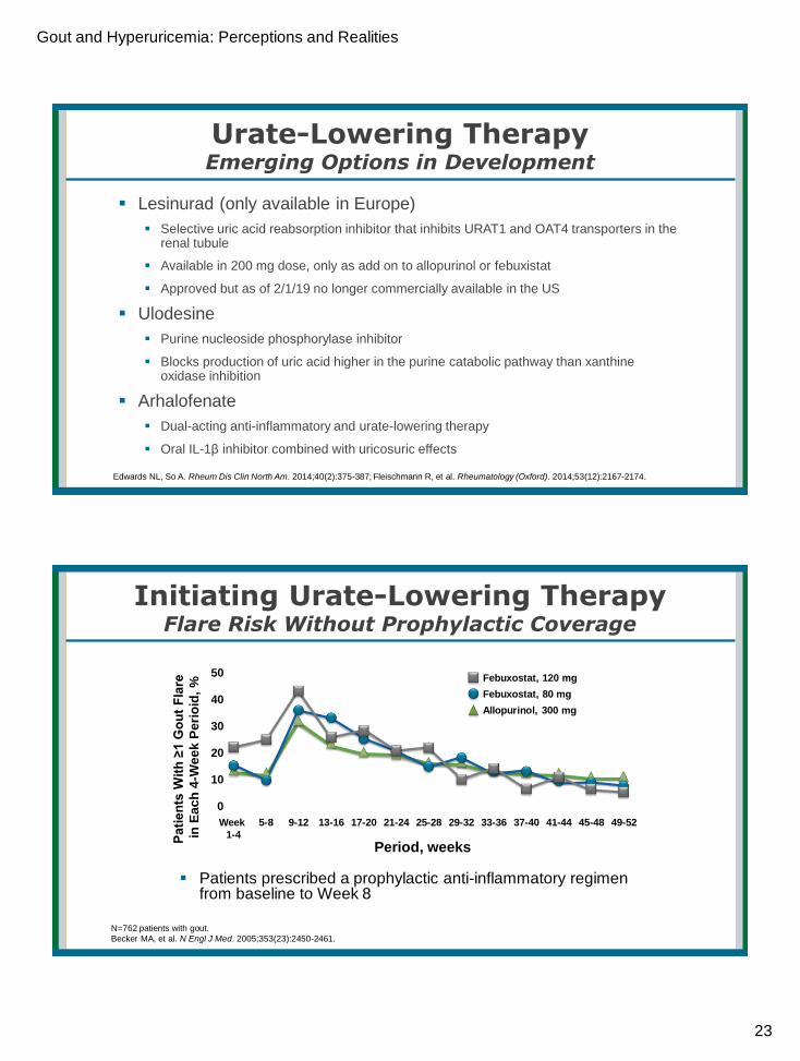
Initiating Urate-Lowering TherapyFlare Risk Without Prophylactic Coverage
▪ Patients prescribed a prophylactic anti-inflammatory regimen from baseline to Week 8
N=762 patients with gout.
Becker MA, et al. N Engl J Med. 2005;353(23):2450-2461.
Pati
en
ts W
ith
≥1 G
ou
t F
lare
in E
ach
4-W
eek P
eri
oid
, %
Period, weeks
50
40
30
20
10
0
Week
1-4
5-8 9-12 13-16 17-20 21-24 25-28 29-32 33-36 37-40 41-44 45-48 49-52
Febuxostat, 80 mg
Febuxostat, 120 mg
Allopurinol, 300 mg
Gout and Hyperuricemia: Perceptions and Realities
24
Gout Flare Risk Increases With ULT
▪ Expect gout flares with all ULT strategies, especially in first 6 months of treatment
▪ Remain on ULT during flares
▪ Flares indicate effective ULT due to tophus remodeling
▪ Manage flares
▪ Initiate prophylaxis 1-2 weeks before starting ULT
▪ Important: unexpected flares decrease compliance with ULT - educate patient!
Becker MA, et al. N Eng J Med. 2005;353:2450-2461.
PPI, proton pump inhibitor.
Khanna D, et al. Arthritis Care Res. 2012;64(10):1447-1461.
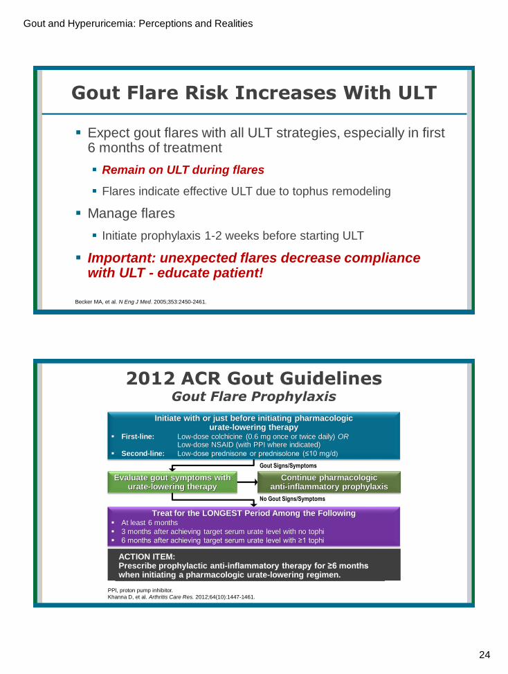
2012 ACR Gout GuidelinesGout Flare Prophylaxis
Initiate with or just before initiating pharmacologic urate-lowering therapy
▪ First-line: Low-dose colchicine (0.6 mg once or twice daily) ORLow-dose NSAID (with PPI where indicated)
▪ Second-line: Low-dose prednisone or prednisolone (≤10 mg/d)
Evaluate gout symptoms with urate-lowering therapy
Treat for the LONGEST Period Among the Following▪ At least 6 months
▪ 3 months after achieving target serum urate level with no tophi
▪ 6 months after achieving target serum urate level with ≥1 tophi
No Gout Signs/Symptoms
Continue pharmacologic anti-inflammatory prophylaxis
Gout Signs/Symptoms
ACTION ITEM:Prescribe prophylactic anti-inflammatory therapy for ≥6 months when initiating a pharmacologic urate-lowering regimen.
Gout and Hyperuricemia: Perceptions and Realities
25
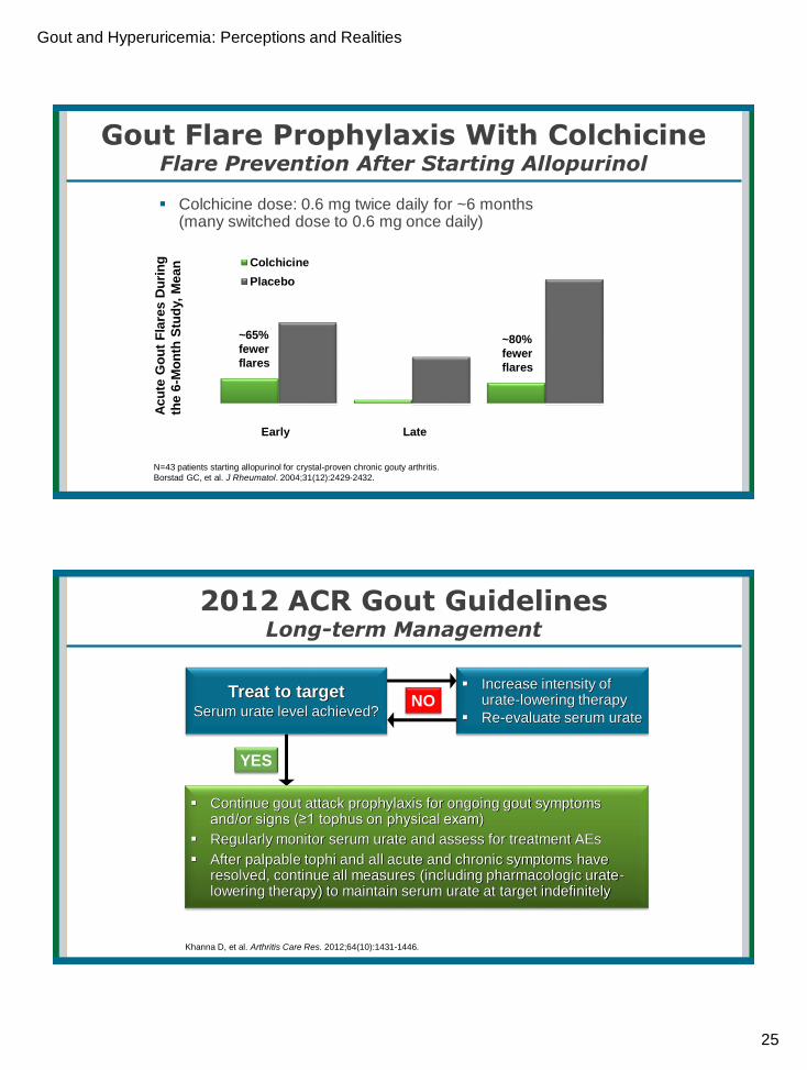
Gout Flare Prophylaxis With ColchicineFlare Prevention After Starting Allopurinol
▪ Colchicine dose: 0.6 mg twice daily for ~6 months (many switched dose to 0.6 mg once daily)
N=43 patients starting allopurinol for crystal-proven chronic gouty arthritis.
Borstad GC, et al. J Rheumatol. 2004;31(12):2429-2432.
0.0
0.5
1.0
1.5
2.0
2.5
3.0
3.5
Baseline to Month 3 Month 4 to 6 Overall
Colchicine
Placebo
Acu
te G
ou
t F
lare
s D
uri
ng
the 6
-Mo
nth
Stu
dy,
Me
an
Early
~80%
fewer
flares
~65%
fewer
flares
Late
Khanna D, et al. Arthritis Care Res. 2012;64(10):1431-1446.
2012 ACR Gout GuidelinesLong-term Management
▪ Increase intensity of urate-lowering therapy
▪ Re-evaluate serum urateNO
▪ Continue gout attack prophylaxis for ongoing gout symptoms and/or signs (≥1 tophus on physical exam)
▪ Regularly monitor serum urate and assess for treatment AEs
▪ After palpable tophi and all acute and chronic symptoms have resolved, continue all measures (including pharmacologic urate-lowering therapy) to maintain serum urate at target indefinitely
YES
Treat to targetSerum urate level achieved?
Gout and Hyperuricemia: Perceptions and Realities
26
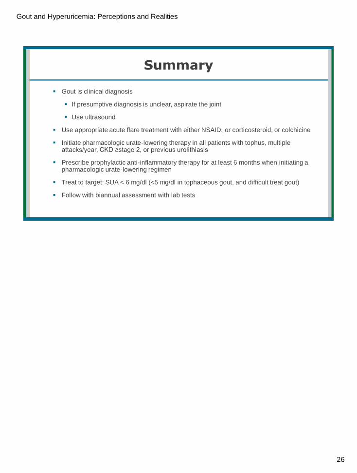
Summary
▪ Gout is clinical diagnosis
▪ If presumptive diagnosis is unclear, aspirate the joint
▪ Use ultrasound
▪ Use appropriate acute flare treatment with either NSAID, or corticosteroid, or colchicine
▪ Initiate pharmacologic urate-lowering therapy in all patients with tophus, multiple attacks/year, CKD ≥stage 2, or previous urolithiasis
▪ Prescribe prophylactic anti-inflammatory therapy for at least 6 months when initiating a pharmacologic urate-lowering regimen
▪ Treat to target: SUA < 6 mg/dl (<5 mg/dl in tophaceous gout, and difficult treat gout)
▪ Follow with biannual assessment with lab tests