Embed Size (px)
Citation preview
"Science Stays True Here" Biological and Chemical Research, Volume 2014, 52-65 | Science Signpost Publishing
Gonad Development and Histology in Bagrus Bayad
Cultured in Outdoor Earthen Ponds
Tsadu S.M.*, Lamai S.L., Yisa T.A. And Ibrahim S.U. Department of Water Resources, Aquaculture and Fisheries Technology, School of Agriculture and Agricultural Technology, Federal University of
Technology PMB 65 Minna Nigeria.
Received: September 03, 2014 / Accepted: September 27, 2014 / Published: November 25, 2014
Abstract: Bagrus bayad (Daget) are found in Nigerian freshwaters in Rivers Niger, Benue, Kaduna and Lake Chad. This feral species were observed to have high growth rate and other aquaculture qualities. 200 live juveniles (100 males and 100 females) were maintained in 5m2 outdoor earthen ponds for 12 months. The objective was to study some aspects of their biology particularly the gonad development and histology, growth and survival with the aim of introducing the species into aquaculture. The fish were checked regularly at 4 weeks intervals for gonad development and maturity stages. One male and one female were dissected each time and gonads used for histological studies. Gonad developmental stages were classified according to description by Bruton (1979). Cross sections through the ovaries and testis showed gonads in stages II, III, IV and V. Photomicrographs of all the stages observed were made and described. Growth and development continued in the fish under culture and it was concluded that Bagrus bayad could be introduced into aquaculture. Keywords: Bagrus bayad, gonad, histology, aquaculture.
1. Introduction
Bagrus bayad (Daget), a member of Bagridae family are commercially important species found in Nigeria freshwaters especially in Rivers Niger, Kaduna, Benue, Lake Chad and other inland waters (Holden and Reed, 1972; Olaosebikan and Raji …). It has high growth rate but has not been cultured due to limited information on their biology, breeding and survival under captivity. Many catfishes do not readily breed under captivity because they are not able to exhibit their natural spawning behavior in artificial ponds (Lamai, 1993) and this constitutes a limiting factor to their culture and mass production. High production capacity of members of Bagridaefamily in the wild have prompted many attempts by fisheries scientists to culture them. Chrysichthys nigrodigitatus have been cultured along with Tilapia melanoplura and carp in Buguma, Nigeria, with encouraging results (Badarch et al 1972). Bagrus docmachave been cultured with Tilapia in in Uganda and being carnivorous, they were found to be effective in controlling Tilapia population. They were also found to breed freely in ponds. Auchenoglanis occidentalis and Chrysichthys have been cultured in Togo (Bardach et al, 1972). According to Bruton (1979), during the non breeding period in Clarias gariepinus the gonadotropin content of the pituitary becomes low, steroid production from gonad reduces and gametogenesis ceases. This is where artificial induced breeding becomes useful. This involves inducing the fish to develop suitable internal and external spawning conditions. Fish can be made to spawn by simulating natural environmental conditions in ponds (Davy and Choinard, 1981). This can stimulate the natural process of breeding.
Corresponding author:
Tsadu Shaba Mohammed, Associate Professor/Ph.D., Federal University of Technology Minna, Research Field: Aquaculture and
Fisheries Biology. E-mail: [email protected].

Gonad Development and Histology in Bagrus Bayad Cultured in Outdoor Earthen Ponds 53
Maintaining fish in captivity allows the gonad developmental stages and maturation process to be studied through regular checking of gonad materials and also by conducting gonad histology. This is the study of the microscopic structure of tissues and cells. Through this, the structure and function of tissues or cell components can be interpreted (Drury et al, 1976). Berlinsky and Jackson (1995) studied the annual reproductive cycle of White bass, Morone chrysops and carried out gonadal histology to determine oocyte developmental stages. Mylonas et al, (1995) studied induced spawning of wild American shad Alosa sapidisima and conducted its gonad histology to study the ovarian biopsy. In this study, wild samples of Bagrus bayad were maintained in captivity in outdoor earthen ponds to study their gonad developmental stages, maturation and histology of the various stages. The objective was to determine the minimum time and body size at maturity and ability to reproduce under culture.
2. Materials and Method
Two outdoor earthen ponds 5 m2 each were prepared and filled with water from rain water runoff and supplemented with borehole water. Two hundred (200) live juveniles of Bagrus bayad, size ranging from 130 – 150 g Total body weight (TBW) and 21 – 25 cm Standard Length (SL) were obtained from fishermen from Shiroro Lake Minna and transported in open plastic containers to the fish hatchery of Federal University of Technology Minna. They were acclimatized for 24 hours and disinfected with salt bath. The males and females were separated and their initial mean TBW and SL were determined. They were then stocked separately in the outdoor ponds. Initial mortalities for the first two weeks were replaced until the stock stabilized. The initial dead samples were used to study the initial gonad sizes and stages of development. The fish were reared in the ponds for 12 months during which the growth in terms of increase in body length and weight was determined monthly. Specific growth rates and mean weight and stadard length gains were calculated. Length / weight relationship and regression analysis were calculated for the male and female samples. Mortality and survival were monitored daily and percentage cumulative mortality and survival was calculated at the end of the culture period. The fish were fed 40 % crude protein commercial diet at 5 % body weight per day. The feed was divided into two and offered twice (morning and evening) per day.
The fish were regularly checked monthly for gonad development, maturation and ovulation or spermiation. This was done by random sampling of 15 females and 15 males each time for examination. Males were inspected for rigid and reddish infusion of the papilla and genital orifice. Females were inspected for maturity and ovulation by checking the genital orifice for reddish infusion, distention of the belly and release of eggs when gentle pressure was applied to the abdomen. Where eggs were not released from some large samples that show signs of maturity, egg catheterization was done with 2 mm diameter egg catheter to determine the stage of egg ripeness. Standard length (SL) and Total body weight (TBW) of the samples examined were recorded and one sample each of male and female were dissected to remove the gonads for determination of developmental stage and for histological study. Fish of higher body sizes than the previous ones were always selected for dissection to obtain gonads of different sizes and body weight. Gonad weight, fecundity and developmental stages were determined for the samples dissected. Classification of gonad developmental stages was done according to the description by Bruton (1979). A total of 24 gonads comprising 12 ovaries and 12 testes were processed into slides for histological analysis. The organs were dissected out, rinsed in tap water and fixed in formosaline solution. The tissues were then processed into slides according to Carleton standard histological technique revised by Drury et al (1976). The slides were viewed under compound microscope at x150 magnification. Photomicrographs were taken with Olympus P.A. 35 Camera fitted to the microscope. Water quality parameters were measured monthly throughout the study period.
Gonad Development and Histology in Bagrus Bayad Cultured in Outdoor Earthen Ponds 54
Mean + standard deviation (SD) were determined according to Steel and Torrie (1980) for initial and final body weight, standard length, mean weight and standard length gains, mean specific growth rate (SGR) and mean fecundity. Length / weight relationship of the male and female samples were determined by regression analysis using cricket graph package.
3. Results
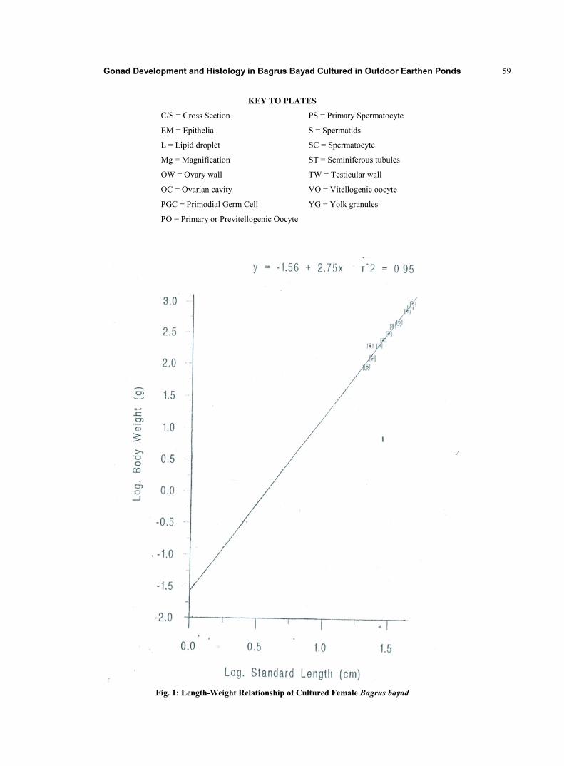
Plate I shows B. bayad external features with its extraordinarily long maxillary barbells and a large adipose fin. Mean values of growth parameters for the male and female Bagrus bayad cultured in outdoor earthen ponds are presented in Table 2. Males achieved higher growth than the females as their mean weight and standard length gains were higher. Figs. 1 and 2 shows the log regression and correlation coefficient of the relationship between total body weight and standard length of the male and female samples. The relationship showed high correlation which was also indicated by the regression equations. Males – Y = 1.56 x 2.75; r2 0.95, n = 15. Females – Y = 1.63 x 2.81; r2 0.91, n = 15. Table 3, shows gonad weight, fecundity and gonad developmental stages of some female samples. Size of fish that showed signs of well developed gonads with visible and countable eggs ranged from 483 g – 1.75 kg TBW and the gonads were between II and V developmental stages. Results of water quality parameters monitored during the culture period are presented in Table 4. Cumulative mortality for males during culture was 18 out of 100 giving 82 (82%) survival while mortality for females was 27 out of 100, giving 73 (73%) survival. The testes are long, slender, unconstructed, unlobed and have serrated outer edge which are observable even at early stage. Immature testis appears whitish and transparent while mature ones are creamy white and opaque. The ovaries are elongated, round, unlobed, tapering at both ends and covered with smooth membrane. Immature ovaries are whitish and transparent while mature ones appear yellowish-red with visible yellowish eggs. The gonads end with duct which unit to form urogenital duct.
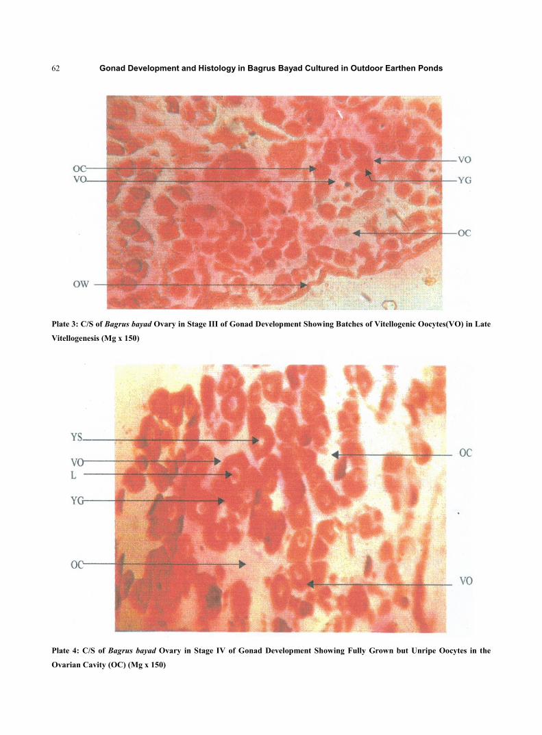
Plates 2 – 9 show the gonads histological analysis. Plate 2 shows a cross section of ovary in stage II gonad development. The fish TBW was 250.51 g and SL was 25.50 cm. Gonad weight (GW) was 1.90 g. It consists of batches of secondary oocytes or Vitellogenic oocytes (VO) in early vitellogenic stage. Eggs were visible in batches surrounded by follicular wall or egg membrane (EM). Plate 3 shows a cross section of ovary in stage III gonad development. It consists of batches of secondary oocytes in late vitellogenic stage. The fish TBW was 400 g and SL was 26.00 cm and the gonad weighed 21.00 g. Plate 4 shows a cross section of ovary in stage IV gonad development. It consists of fully grown but unripe vitellogenic oocytes (VO). The egg membrane or follicular wall has degenerated leaving eggs in the ovarian cavity (OC). The egg yolk granules (YG) with lipid droplet (LP) are visible. The fish TBW was 600 g and SL was 32.50 cm and gonad weighed 25.34 g. Plate 5 shows a cross section of ovary in stage V of gonad development. It consists of fully grown but unripe vitellogenic oocytes (VO) in loose batches. The egg follicular membrane has degenerated. The fish TBW was 1.07 kg, SL was 45.00 cm and gonad weighed 32.05 g. Plate 6 shows a cross section of Testis in stage II of gonad development. It consists of numerous seminiferous tubules (ST) containing primary spermatocytes (PS), premeotic or Primodial Germ Cell (PGC) and Testicular wall (TW). The fish TBW was 450.71 g, SL was 28.50 cm and the gonad weighed o.24 g. Plate 7 shows a cross section of Testis in stage III of gonad development. It consists of batches of secondary spermatocysts (SC) in the lumen of seminiferous tubules (ST). The fish TBW was 700 g, SL was 35.50 cm and gonad weighed 1.9 g. Plate 8 shows a cross section of testis in stage IV of gonad development consisting of secondary spermatocyst (SC) in the lumen of seminiferous tubules (ST). The gonad has attained increase in size which is characteristic of this stage. The fish TBW was 800 g, SL was 42.50 cm and gonad weighed 2.5 g. Plate 9 shows a cross section of testis in stage V of gonad development consisting of loose batches of
Gonad Development and Histology in Bagrus Bayad Cultured in Outdoor Earthen Ponds 55
secondary spermatocysts (SC) in the lumen of seminiferous tubules (ST). The testis and sperms are fully grown but not ripe. The fish TBW was 1.25 kg, SL was 50.25 cm and gonad weighed 4.10 g.
4. Discussion
Bagrus bayad (Daget) is the only freshwater species in Nigerian waters that possess visible vertical lateral lines in addition to the horizontal ones. It is also the only species with extraordinarily long maxillary barbells (Reeds et al, 1967). Lewis (1974) observed that males of this spcies grow bigger than the females. In this study it was also observed that males had higher mean body weight and standard length than the females. Regression analysis value for males was r2 = 0.95 while females had r2 = 0.91. Bruton (1979) and Lamai (1993) classified gonad development in Clarias gariepinus into eight stages (Table 1). Their description of the stages is applicable to Bagrus bayad except the body size at maturity. It was observed in this study that Bagrus bayad does not reach spawning stage until they are about 1 kg and above in total body weight. Imevbore (1970) in his study of sex ratio and fecundity of fishes in River Niger, encountered the smallest mature but unripe Bagrus bayad female at about 450 g TBW and 26.50 cm SL and mature but unripe male at about 750 g TBW and 41.50 cm SL. The species do not show pre-spawning morphology of distended abdomen due to increased gonad size as commonly observed in other fish species. Lack of pre-spawning morphology had also been observed by Mylonas et al (1995) in American shad Alosa sapidisima.
Gonad histology showed that the gonads were between stage II – V developmental stages which according to Purdon (1993) are within growth phase of gonad development. This is marked by meiotic division, increase in size and increase in cytoplasmic content and incorporation of vitellogenin into the oocytes. After incorporation vitelloginin the vitellogenic oocytes (VO) then develops into mature eggs.
5. Conclusion
In this study Bagrus bayad survived under culture in outdoor earthen ponds for the one year culture period. Survival rate was high especially among those not wounded during capture and handling. The species although carnivorous in habit accepted artificial diet under culture indicating that they can be cultured and fed formulated diet. There was significant increase in body size during captivity indicating that growth continued in the fish. It was also observed that gonad development continued in the fish under captivity. There were no signs of gonad regression. Although breeding was not achieved during captivity the fish showed signs of maturity such as reddish infusion of genital pore in females and rigidness and reddish infusion of papilla in males. From the results of this study, Bagrus bayad could be recommended for aquaculture.
References
[1] Bardach, K.H., John, H.R. and William, O.M. (1972). Aquaculture: The Farming and Husbandry of Freshwater and Marine
Organisms. John Wiley and Sons Inc. p 244-255.
[2] Berlinsky, D. L. and Jackson, L. F. (1995). The annual reproductive cycle of the white Bass Morone chrysops. Journal of world
aquaculture society. 26 (3) 252-260.
[3] Bruton, M. N. (1979). The breeding biology and early development of Clarias gariepinus in Lake Sibaya South Africa with a
review of breeding in species of the Subgenus Clarias (Pisces: Clariidae). Trans. 2001, Society. London, 35: 1-45.
[4] Davy, F. B. and Choinard, A. (1981). Induced fish breeding in South East Asia. Workshop Report, Singapore 25th-28th
November 1980. IDRC 48: 12-211.

Gonad Development and Histology in Bagrus Bayad Cultured in Outdoor Earthen Ponds 56
[5] Drury, R. A. B., Wallington, E. A. and Cameron, S. R. (1976). Carleton’s Histological Techniques. 4th Ed. Oxford University
Press. pp. 1-7.
[6] Holden, M. and Reed, W. (1972). West African Freshwater Fish. West African Nature Hand Book. Longmans 45pp.
[7] Imevbore, A. M. A. (1970). Some Preliminary observations on the sex Ratios and Fecundity of the fish in the River Niger. In S.
A. Visser (Ed). Kainji, A Nigerian man-made Lake. Kainji Lake Studies Vol.1. Ecology. Nigerian Institute of Social and
Economic Research (Pub), 87-98.
[8] Lamai, S .L. (1993) Aspects of the applied biology of Clarias gariepinus, aquacultural techniques and Deldrin toxicity. PhD
Thesis. Dept. of Pure and Applied Zoology, University of Reading, UK. 204 pp.
[9] Lewis, D. S. C. (1974) An illustrated key to the fishes of Lake Kainji, Nigeria. 67 pp.
[10] Mylonas, C.C., Tabata, Y., Langer, R. and Zohar, Y. (1995). Preparation and evaluation of Polyanhydride microshperes
containing gonadotropin- releasing hormone (GnRH) for inducing ovulation and spermiation in fish. Journal of controlled
release 35: 23-34.
[11] Olaosebikan, B.D. and Raji, A. (1998) Field guide to Nigerian freshwater fishes. Decency Printers and Stationaries LtD. Ilorin.
43 pp.
[12] Purdon, C.E. (1993) Genetics and fish breeding. 1st Ed. Fish and Fisheries Series No. 8 Chapman and Hall, London p 79-80.
[13] Reed, W., Buchard, J., Hopson, A. J., Jennes, J. and Yaro, I. (1967). Fish and Fisheries of Northern Nigeria. Ministry of
Agriculture, Northern Nigeria, Gaskiya Corp. LtD, Zaria. 226 pp.
[14] Steel, R. G. D. and Torrie, J. H. (1980). Principles and procedures of statistics-a biometric approach. 3rd Ed. McGraw Hill
U.S.A. 85 pp.
Gonad Development and Histology in Bagrus Bayad Cultured in Outdoor Earthen Ponds 57
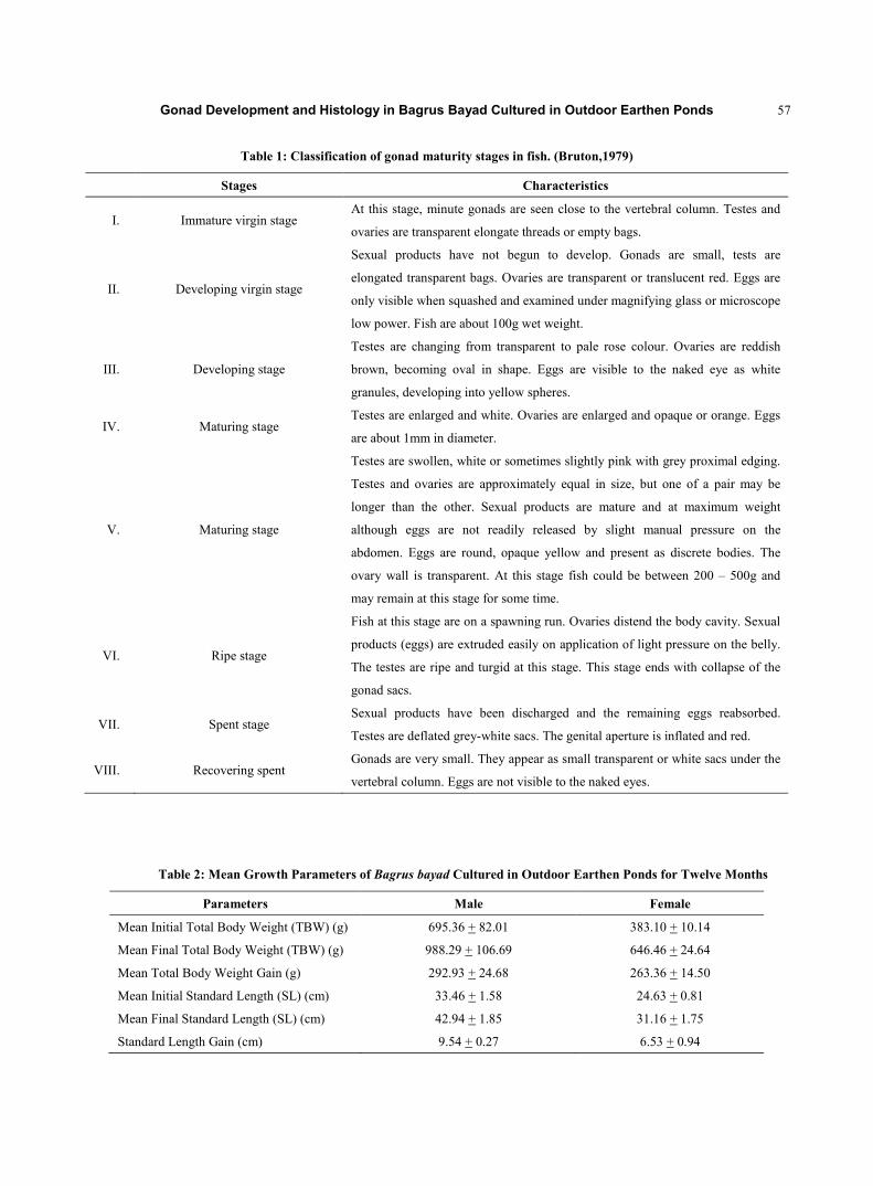
Table 1: Classification of gonad maturity stages in fish. (Bruton,1979)
Stages Characteristics
I. Immature virgin stage At this stage, minute gonads are seen close to the vertebral column. Testes and
ovaries are transparent elongate threads or empty bags.
II. Developing virgin stage
Sexual products have not begun to develop. Gonads are small, tests are
elongated transparent bags. Ovaries are transparent or translucent red. Eggs are
only visible when squashed and examined under magnifying glass or microscope
low power. Fish are about 100g wet weight.
III. Developing stage
Testes are changing from transparent to pale rose colour. Ovaries are reddish
brown, becoming oval in shape. Eggs are visible to the naked eye as white
granules, developing into yellow spheres.
IV. Maturing stage Testes are enlarged and white. Ovaries are enlarged and opaque or orange. Eggs
are about 1mm in diameter.
V. Maturing stage
Testes are swollen, white or sometimes slightly pink with grey proximal edging.
Testes and ovaries are approximately equal in size, but one of a pair may be
longer than the other. Sexual products are mature and at maximum weight
although eggs are not readily released by slight manual pressure on the
abdomen. Eggs are round, opaque yellow and present as discrete bodies. The
ovary wall is transparent. At this stage fish could be between 200 – 500g and
may remain at this stage for some time.
VI. Ripe stage
Fish at this stage are on a spawning run. Ovaries distend the body cavity. Sexual
products (eggs) are extruded easily on application of light pressure on the belly.
The testes are ripe and turgid at this stage. This stage ends with collapse of the
gonad sacs.
VII. Spent stage Sexual products have been discharged and the remaining eggs reabsorbed.
Testes are deflated grey-white sacs. The genital aperture is inflated and red.
VIII. Recovering spent Gonads are very small. They appear as small transparent or white sacs under the
vertebral column. Eggs are not visible to the naked eyes.
Table 2: Mean Growth Parameters of Bagrus bayad Cultured in Outdoor Earthen Ponds for Twelve Months
Parameters Male Female
Mean Initial Total Body Weight (TBW) (g) 695.36 + 82.01 383.10 + 10.14
Mean Final Total Body Weight (TBW) (g) 988.29 + 106.69 646.46 + 24.64
Mean Total Body Weight Gain (g) 292.93 + 24.68 263.36 + 14.50
Mean Initial Standard Length (SL) (cm) 33.46 + 1.58 24.63 + 0.81
Mean Final Standard Length (SL) (cm) 42.94 + 1.85 31.16 + 1.75
Standard Length Gain (cm) 9.54 + 0.27 6.53 + 0.94

Gonad Development and Histology in Bagrus Bayad Cultured in Outdoor Earthen Ponds 58
Table 3: Gonad Weight, Fecundity and Maturity Stage of Some Cultured Bagrus bayad
S/No TBW (g) S L (cm) G W (g) Fecundity Maturity Stage*
1 483.53 32.00 4.02 6464.16 III
2 412.69 31.00 2.77 3988.80 II
3 1.33kg 54.50 13.84 22836.00 IV
4 1.72kg 41.00 13.25 21438.50 V
5 1.38kg 56.50 33.32 60642.40 IV
6 1.28kg 46.50 11.92 18952.80 IV
7 470.00 33.00 4.12 4559.04 III
8 1.10kg 43.50 8.80 15276.80 III
9 1.20kg 45.50 9.60 16896.00 IV
10 845.73 42.00 4.92 8560.80 III
11 483.83 32.00 4.00 6800.00 III
12 1.75kg 58.00 33.64 63924.56 V
TBW =Total Body Weight. SL = Standard Length. * = According to Bruton (1979)
Table 4: Mean Water Quality Parameters Measured From the Ponds During Culture
Parameters
Ponds Mean Dissolved
Oxygen (mgL-1) Mean Temp. (oC) Mean pH
Mean Water
Depth (cm)
Mean Sechi Disc
Visibility (cm)
Mean Conductivity
(moh/s)
Male Pond 4.43 + 1.11 28.09 + 2.09 7.35+ 0.47 55.23+ 0.25 18.22+2.75 1244.64 + 1.70
Female Pond 4.55 + 0.72 28.16 + 1.20 7.09+ 0.42 56.12+ 0.34 18.21+2.66 1264.26 + 1.76
Gonad Development and Histology in Bagrus Bayad Cultured in Outdoor Earthen Ponds 59
KEY TO PLATES
C/S = Cross Section
EM = Epithelia
L = Lipid droplet
Mg = Magnification
OW = Ovary wall
OC = Ovarian cavity
PGC = Primodial Germ Cell
PO = Primary or Previtellogenic Oocyte
PS = Primary Spermatocyte
S = Spermatids
SC = Spermatocyte
ST = Seminiferous tubules
TW = Testicular wall
VO = Vitellogenic oocyte
YG = Yolk granules
Fig. 1: Length-Weight Relationship of Cultured Female Bagrus bayad

Gonad Development and Histology in Bagrus Bayad Cultured in Outdoor Earthen Ponds 60
Fig. 2: Length- Weight Relationship of Cultured Male Bagrus bayad

Gonad Development and Histology in Bagrus Bayad Cultured in Outdoor Earthen Ponds 61
Plate 1: Dorsolateral View of Bagrus bayad (Daget) Showing some External DistinguishingFeatures
Plate 2: Cross Section (C/S) of Bagrus bayad in Stage II Gonad Development Showing Batches of Early Vitelogenic Oocytes
(VO) (Mg x 150)
Gonad Development and Histology in Bagrus Bayad Cultured in Outdoor Earthen Ponds 62
Plate 3: C/S of Bagrus bayad Ovary in Stage III of Gonad Development Showing Batches of Vitellogenic Oocytes(VO) in Late
Vitellogenesis (Mg x 150)
Plate 4: C/S of Bagrus bayad Ovary in Stage IV of Gonad Development Showing Fully Grown but Unripe Oocytes in the
Ovarian Cavity (OC) (Mg x 150)

Gonad Development and Histology in Bagrus Bayad Cultured in Outdoor Earthen Ponds 63
Plate 6: C/S of Bagrus bayad Testis in Stage II of Gonad Development Showing Seminiferous Tubules (ST) Containing
Primary Spermatocystes (PS) x150
PS
ST

Gonad Development and Histology in Bagrus Bayad Cultured in Outdoor Earthen Ponds 64
Plate 7: C/S of Bagrus bayad Testis in Stage III of Gonad Development Showing Secondary Spermatocytes (SC) in the Lumen
of Seminiferous Tubules (T) (Mg x 150)
Plate 8: C/S of Bagrus bayad Testis in Stage III of Gonad Development Showing Secondary Spermatocytes (SC) in the Lumen
of Seminiferous Tubules (ST) x 150
ST
SC
SC
LC

Gonad Development and Histology in Bagrus Bayad Cultured in Outdoor Earthen Ponds 65
Plate 9: C/S of Bagrus bayad Testis in Stage V of Gonad Development Showing Secondary Spermatocytes (SC) in the Lumen
of Seminiferous Tubules (ST) (Mg x 15
SC
ST
SC