Embed Size (px)
Citation preview

Glycoconjugate storage & pathogenesis in an in vitro cellular model of
Sandhoff disease
Stephanie Boomkamp

Sandhoff disease
Loss motor skills
Seizures
Hearing and vision loss
Mental retardation
Paralysis
Cherry red macular spot in retina
Enlarged organs
Greatly reduced lifespan
Disease severity and onset dependent on residual β-hexosaminidase activity.

GSL metabolic pathway & ß-hexosaminidase
NeuAcα(2-3)Galβ(1-4)GlcCer
GalNAcβ(1-4)
GM2
Galβ(1-4)GlcCer
NeuAcα(2-3)
GM3
GalNAcβ(1-4)
Galβ(1-4)GlcCer
Galβ(1-4)GlcCer
GA2
LacCer

Glycoprotein degradation & ß-hexosaminidase
β-hexosaminidase
GlcNAc Man
Man
ManGlcNAc
R
R
GlcNAc GlcNAc
β-hexosaminidase
Chitobiase
R = NeuAcα(2-3/6)Galβ(1-4)
Asn
Bidirectional degradation in the lysosome
Deficient ß-hexosaminidase termination and accumulation of glycan.

Project aims
Development of an in vitro cellular model of Sandhoff disease
Characterization and cellular localization storage products
Disease pathology
Validation of substrate reduction therapeutics NB-DNJ and NB-DGJ: effects on storage levels and disease pathology.

murine RAW macrophages
0-50 µM SR1 30 days
Culture cells
OS GSL
2-AA
MS
Enzyme digests
NP-HPLC
+N
AcHN OH
OH
Experimental procedures
IC50
Cell line PNP-GlcNAc (µM) PNP-GalNAc (µM)
RAW 1.05 ± 0.10 2.89 ± 0.40
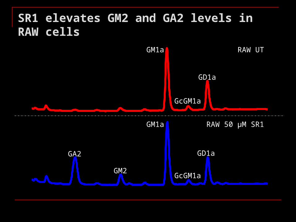
SR1 elevates GM2 and GA2 levels in RAW cells
RAW UT
RAW 50 µM SR1
GA2
GM2
GM1a
GcGM1a
GD1a
GM1a
GcGM1a
GD1a

mV
RAW UT
RAW 50 µM SR1
SR1 elevates GlcNAc-terminating OS levels in RAW cells
David Harvey

Stored GSL are localized in the lysosome - GlcNAc-OS in light fractions
7 8 9 10 RAW UT
RAW SR1
LAMP1
~ 44kDa
Cell fraction
1
23
4
5678
91011
12
GA2 GM2 GM1a fraction
density
123456789101112
density
fraction

Stored GSL are localized in the lysosome - GlcNAc-OS in light fractions
1 2 3 4 5 6 7 8 9 10 11 12
OS GSL
Robin Antrobus
Density
Protein markers
Novel cellular compartment OS?

Disease pathology
Wada R., Tifft C.J., Proia R.L. (2000) PNAS 20:10954-10959.
GSL accumulation
Neuronal damage/death
Microglial phagocytosis
Microglial activation/expansion
Production neurotoxic mediators
+MIP-1, TNF-, TGF-ß1, IL-1

SR1 down-regulates cytokine expression
RAW UT RAW SR1
IL-1
IL-6
TNF-
Stimulation with LPS: similar intracellular signalling events presence of feedback control loop in SR1-treated RAW cells
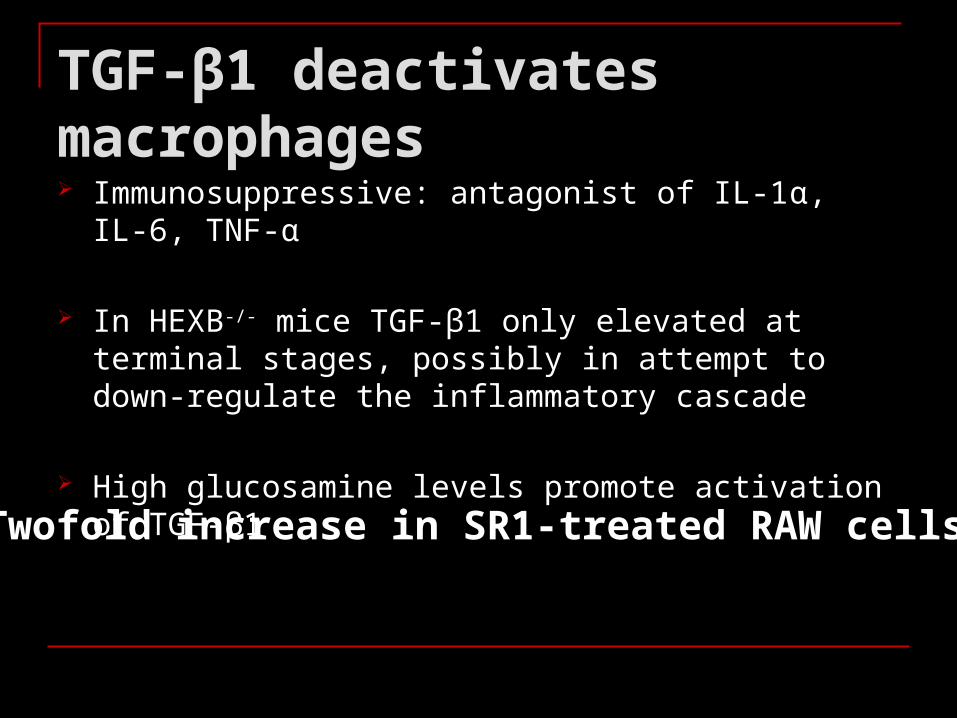
TGF-β1 deactivates macrophages
Immunosuppressive: antagonist of IL-1α, IL-6, TNF-α
In HEXB-/- mice TGF-β1 only elevated at terminal stages, possibly in attempt to down-regulate the inflammatory cascade
High glucosamine levels promote activation of TGF-β1
Twofold increase in SR1-treated RAW cells

Summary
SR1 induces storage of GM2, GA2 and GlcNAc-OS in RAW cells as seen in Sandhoff patients/mice
GM2 and GA2 are localized in the lysosome, whereas OS are present in light, buoyant cell compartment(s)
SR1 triggers an immunosuppressive response due to a twofold increase in TGF-ß1.

Therapy
Bone marrow transplantation (BMT) Enzyme replacement therapy (ERT)
Chaperone-mediated therapy (CMT) Substrate reduction therapy (SRT)
Best thus far is SRT & BMT or combination of therapies with use of anti-inflammatory drugs.
Inaccessibility by BBB
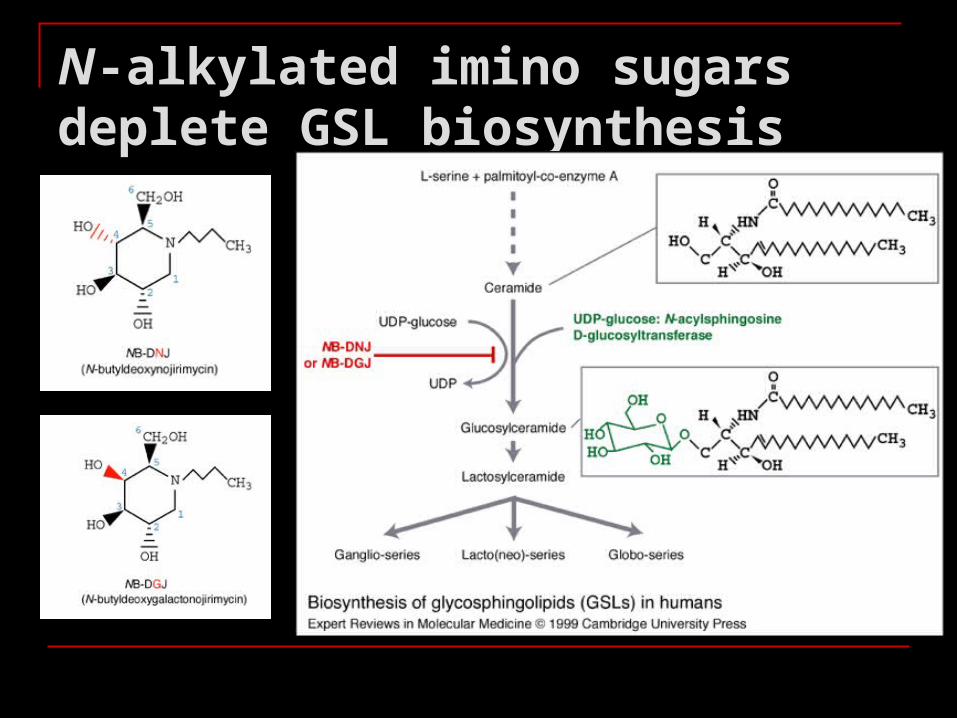
N-alkylated imino sugars deplete GSL biosynthesis
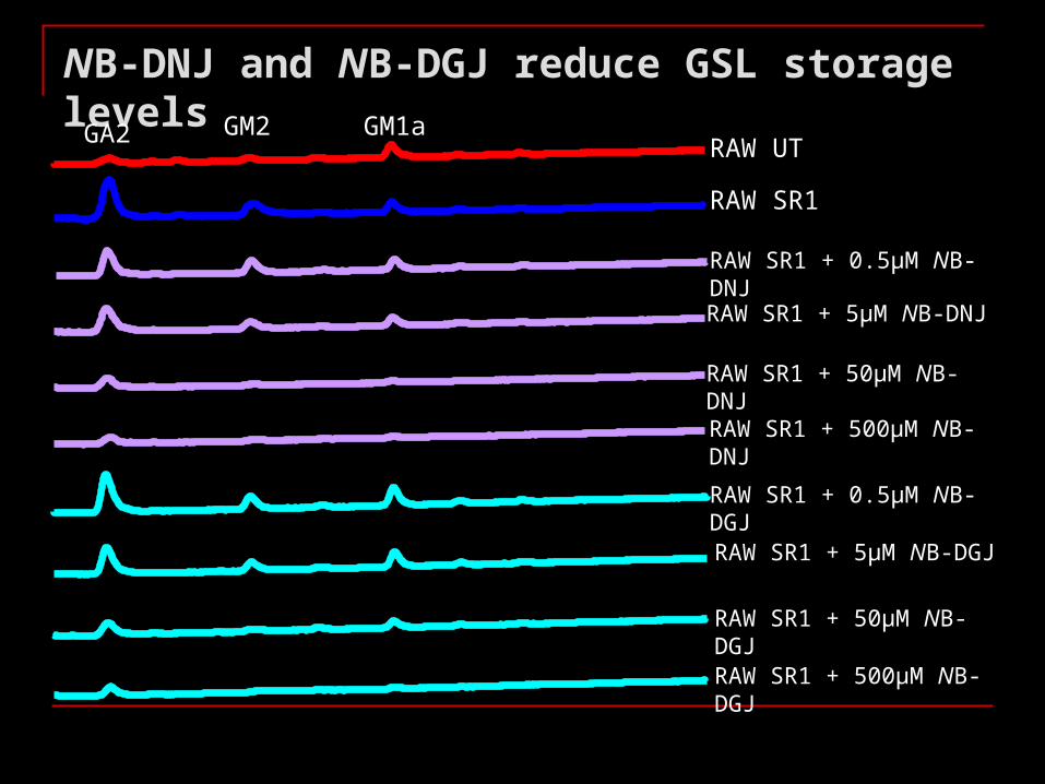
NB-DNJ and NB-DGJ reduce GSL storage levels
GA2 GM2 GM1aRAW UT
RAW SR1
RAW SR1 + 0.5µM NB-DNJ
RAW SR1 + 5µM NB-DNJ
RAW SR1 + 50µM NB-DNJ
RAW SR1 + 500µM NB-DNJ
RAW SR1 + 0.5µM NB-DGJ
RAW SR1 + 5µM NB-DGJ
RAW SR1 + 50µM NB-DGJ
RAW SR1 + 500µM NB-DGJ

< 500 µM NB-DNJ and NB-DGJ do not reduce GlcNAc-OS levels
RAW UT
RAW SR1
RAW SR1 + 0.5µM NB-DNJ
RAW SR1 + 5µM NB-DNJ
RAW SR1 + 50µM NB-DNJ
RAW SR1 + 500µM NB-DNJ
RAW SR1 + 0.5µM NB-DGJ
RAW SR1 + 5µM NB-DGJ
RAW SR1 + 50µM NB-DGJ
RAW SR1 + 500µM NB-DGJ

Iminosugars normalize inflammatory response
Iminosugar Concentration (µM) Effect
NB-DNJ 5 Normalization
50 + MCP1
500
500 µM NB-DNJ
500 µM NB-DGJ
NB-DGJ 5 Normalization
50
500 + MCP1

Iminosugars reduce TGF-β1 levels
0
10
20
30
40
50
60
70
80
0 100 200 300 400 500
uM iminosugar
TG
FB
1/u
g p
rote
in
NB-DNJ
NB-DGJ
UT RAW

Summary
NB-DNJ and NB-DGJ reduce GSL storage levels but do not affect GlcNAc-OS levels at achievable therapeutic concentrations
At concentrations ≥ 50 µM increase MCP1, due to: Overall reduction in GSL levels? Reduction in OS storage? Glucosylated/galactosylated OS?
5 µM NB-DNJ or NB-DGJ potentially induces a non-pathological phenotype (Jeyakumar et al 1999).
GSL, not OS, play a role in the disease pathology of Sandhoff disease
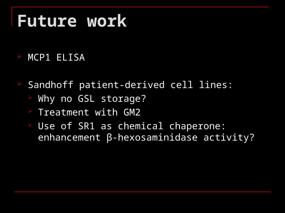
Future work
MCP1 ELISA
Sandhoff patient-derived cell lines: Why no GSL storage? Treatment with GM2 Use of SR1 as chemical chaperone: enhancement β-
hexosaminidase activity?

Acknowledgments
Terry Butters Raymond Dwek
David Neville David Harvey Robin Antrobus
OGBI


![TUESDAY, SEPTEMBER 22 - PMF..., Nikola Cindro, Katarina Leko, Vladislav Tomišić Synthesis of Phenanthridine-Based Calix[4]arene Glycoconjugate and Physicochemical Characterization](https://img.dokumen.tips/doc/110x75/60db49f2187ef168b51bfb07/tuesday-september-22-pmf-nikola-cindro-katarina-leko-vladislav-tomii.jpg)