Embed Size (px)
Citation preview
INTRODUCTION
Solitary or multiple lipomas can occur almost anywherein the trunk, extremities, mediastinum, pelvis, and retroperi-toneum, but they rarely originate in the intestinal mesentery(1-4). A search of the English literature has revealed less than30 documented cases. In the literature review, mesentericlipomas do not cause any intestinal symptoms in most casesas they usually allow the passage of intestinal contents (1, 5),and those causing abdominal pain make up only a small per-centage (2, 6). Mesenteric lipomas may cause abdominal painby complete intestinal obstruction as a result of torsion orvolvulus (3, 7-9) or partial intestinal obstruction associatedwith compression of the intestine.
In the present report, we describe a case of a giant mesen-teric lipoma causing abdominal pain by entrapment of an ilealloop and review the cases with mesenteric lipomas causingabdominal pain.
CASE REPORT
A 29-yr-old woman presented with colicky lower abdom-inal pain. Her abdominal pain had developed 3 yr before inter-mittently, but it had worsened over the past 6 months. She
was unable to sleep well due to her abdominal pain, whichaggravated after eating meals. She also experienced episodesof abdominal distension, constipation, and urinary frequen-cy or urgency. Other symptoms, as well as her past medicalhistory and family history, were otherwise unremarkable.
The patient had a height of 164 cm and weight of 59.6kg, for a body mass index of 22.2. Physical examination re-vealed a slightly distended abdomen, but the remainder ofthe examination was unremarkable. Routine blood tests, in-cluding renal function and urine analysis, were normal.
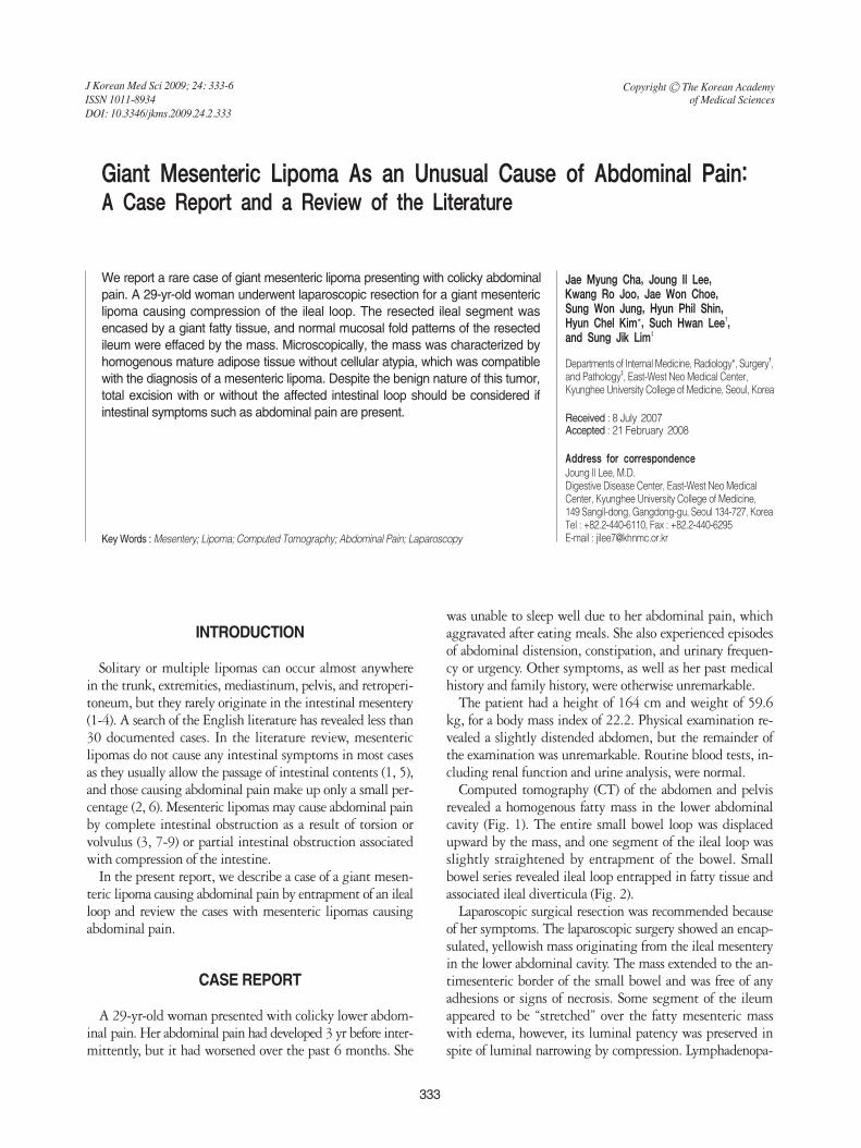
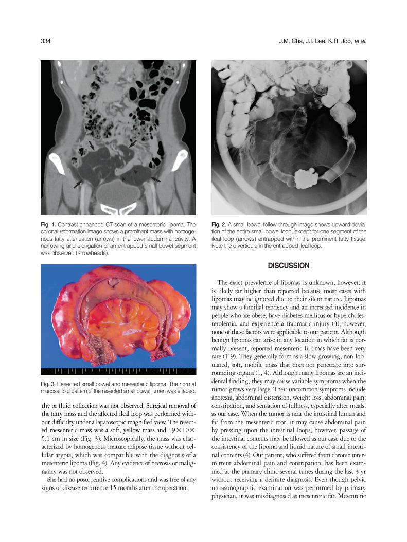
Computed tomography (CT) of the abdomen and pelvisrevealed a homogenous fatty mass in the lower abdominalcavity (Fig. 1). The entire small bowel loop was displacedupward by the mass, and one segment of the ileal loop wasslightly straightened by entrapment of the bowel. Smallbowel series revealed ileal loop entrapped in fatty tissue andassociated ileal diverticula (Fig. 2).
Laparoscopic surgical resection was recommended becauseof her symptoms. The laparoscopic surgery showed an encap-sulated, yellowish mass originating from the ileal mesenteryin the lower abdominal cavity. The mass extended to the an-timesenteric border of the small bowel and was free of anyadhesions or signs of necrosis. Some segment of the ileumappeared to be ‘‘stretched’’ over the fatty mesenteric masswith edema, however, its luminal patency was preserved inspite of luminal narrowing by compression. Lymphadenopa-
333
Jae Myung Cha, Joung Il Lee, Kwang Ro Joo, Jae Won Choe, Sung Won Jung, Hyun Phil Shin, Hyun Chel Kim*, Such Hwan Lee�, and Sung Jik Lim�
Departments of Internal Medicine, Radiology*, Surgery�,and Pathology�, East-West Neo Medical Center, Kyunghee University College of Medicine, Seoul, Korea
Address for correspondenceJoung Il Lee, M.D.Digestive Disease Center, East-West Neo MedicalCenter, Kyunghee University College of Medicine, 149 Sangil-dong, Gangdong-gu, Seoul 134-727, KoreaTel : +82.2-440-6110, Fax : +82.2-440-6295 E-mail : [email protected]
J Korean Med Sci 2009; 24: 333-6ISSN 1011-8934DOI: 10.3346/jkms.2009.24.2.333
Copyright � The Korean Academyof Medical Sciences
Giant Mesenteric Lipoma As an Unusual Cause of Abdominal Pain: A Case Report and a Review of the Literature
We report a rare case of giant mesenteric lipoma presenting with colicky abdominalpain. A 29-yr-old woman underwent laparoscopic resection for a giant mesentericlipoma causing compression of the ileal loop. The resected ileal segment wasencased by a giant fatty tissue, and normal mucosal fold patterns of the resectedileum were effaced by the mass. Microscopically, the mass was characterized byhomogenous mature adipose tissue without cellular atypia, which was compatiblewith the diagnosis of a mesenteric lipoma. Despite the benign nature of this tumor,total excision with or without the affected intestinal loop should be considered ifintestinal symptoms such as abdominal pain are present.
Key Words : Mesentery; Lipoma; Computed Tomography; Abdominal Pain; Laparoscopy
Received : 8 July 2007Accepted : 21 February 2008
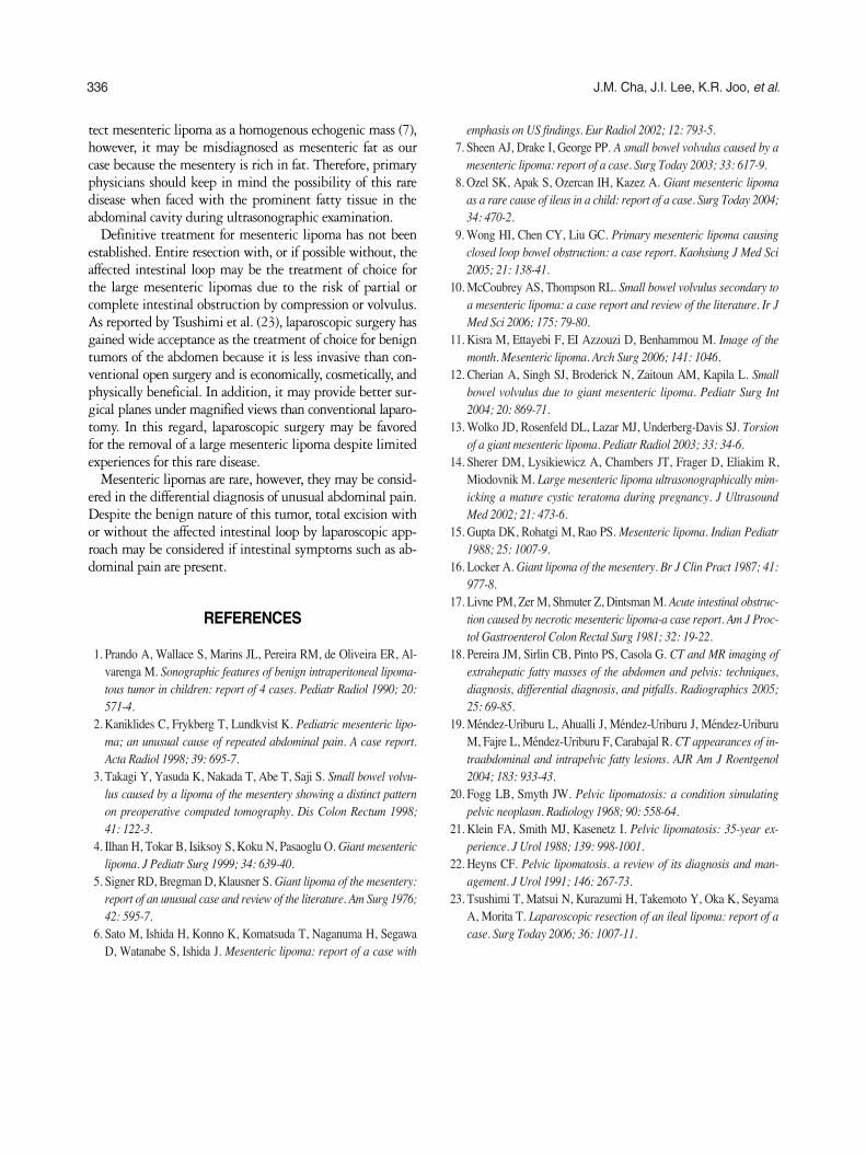
thy or fluid collection was not observed. Surgical removal ofthe fatty mass and the affected ileal loop was performed with-out difficulty under a laparoscopic magnified view. The resect-ed mesenteric mass was a soft, yellow mass and 19×10×5.1 cm in size (Fig. 3). Microscopically, the mass was char-acterized by homogenous mature adipose tissue without cel-lular atypia, which was compatible with the diagnosis of amesenteric lipoma (Fig. 4). Any evidence of necrosis or malig-nancy was not observed.
She had no postoperative complications and was free of anysigns of disease recurrence 15 months after the operation.
DISCUSSION
The exact prevalence of lipomas is unknown, however, itis likely far higher than reported because most cases withlipomas may be ignored due to their silent nature. Lipomasmay show a familial tendency and an increased incidence inpeople who are obese, have diabetes mellitus or hypercholes-terolemia, and experience a traumatic injury (4); however,none of these factors were applicable to our patient. Althoughbenign lipomas can arise in any location in which fat is nor-mally present, reported mesenteric lipomas have been veryrare (1-9). They generally form as a slow-growing, non-lob-ulated, soft, mobile mass that does not penetrate into sur-rounding organs (1, 4). Although many lipomas are an inci-dental finding, they may cause variable symptoms when thetumor grows very large. Their uncommon symptoms includeanorexia, abdominal distension, weight loss, abdominal pain,constipation, and sensation of fullness, especially after meals,as our case. When the tumor is near the intestinal lumen andfar from the mesenteric root, it may cause abdominal painby pressing upon the intestinal loops, however, passage ofthe intestinal contents may be allowed as our case due to theconsistency of the lipoma and liquid nature of small intesti-nal contents (4). Our patient, who suffered from chronic inter-mittent abdominal pain and constipation, has been exam-ined at the primary clinic several times during the last 3 yrwithout receiving a definite diagnosis. Even though pelvicultrasonographic examination was performed by primaryphysician, it was misdiagnosed as mesenteric fat. Mesenteric
334 J.M. Cha, J.I. Lee, K.R. Joo, et al.
Fig. 1. Contrast-enhanced CT scan of a mesenteric lipoma. Thecoronal reformation image shows a prominent mass with homoge-nous fatty attenuation (arrows) in the lower abdominal cavity. Anarrowing and elongation of an entrapped small bowel segmentwas observed (arrowheads).
Fig. 2. A small bowel follow-through image shows upward devia-tion of the entire small bowel loop, except for one segment of theileal loop (arrows) entrapped within the prominent fatty tissue.Note the diverticula in the entrapped ileal loop.
Fig. 3. Resected small bowel and mesenteric lipoma. The normalmucosal fold pattern of the resected small bowel lumen was effaced.

lipomas may pose diagnostic difficulties due to absence ofabnormal laboratory findings and its vague non-specific symp-toms as our case. Therefore, physicians should be aware ofthe various manifestations of mesenteric lipomas and avoidinappropriate managements.
We reviewed reports of mesenteric lipomas identified usinga computerized search of Pubmed (articles in English bet-ween 1965 and 2007), and thirteen cases of which causingabdominal pain (Table 1). The incidence of this tumor is equalfor both sexes, and seven of the cases have been reported inchildren (2, 8, 11-13, 15). All tumors are located in the me-sentery of the small intestine, mostly the ileum, as our case.The main sequelae of mesenteric lipoma are complete intesti-nal obstruction as a result of torsion or volvulus (3, 7, 9-13)or partial intestinal obstruction due to compression of intes-tine by giant lipomas (2, 8, 14-16). Abdominal pain mayalso be related to altered peristalsis secondary to adhered smallbowel loops. In terms of size, mesenteric lipomas have a ten-dency to produce symptoms dependent on their size. The
size of the mesenteric lipoma in our patient was one of thelargest reported in the literature.
Generally, the size of mesenteric lipomas with obstructionby compression of intestinal loop (mean=21 cm) was largerthan those of mesenteric lipomas with obstruction causedby torsion or volvulus (mean=13 cm).
Radiological examination suggests the possible diagnosis,and the best definitive diagnostic procedure is CT, whichdemonstrates a proliferation of homogenous adipose tissuein the lower abdominal cavity (18, 19). The CT number read-ing provides information about the nature of the lesion, andthe density of the fatty mass with attenuation values between-80 and -120 HU, which is identical to that of subcutaneousfat, may lead to its diagnosis (19). When a large mesentericlipoma extends lower in the abdominal or even pelvic cavitydue to its gravity as our case, CT findings of mesenteric lipo-ma may mimic pelvic lipomatosis, which is a rare conditiondefined as a nonmalignant overgrowth of normal fat in theperivesical and perirectal spaces (20-22). Ultrasound can de-
A Case Report of Giant Mesenteric Lipoma 335
Fig. 4. Microscopic findings of the mesenteric lipoma. (A) The tumor revealed mature fat cells proliferating in the subserosal layer (H&E; origi-nal magnification, ×20). (B) Mature adipocytes are relatively uniform in size and lack cytologic atypia (H&E; original magnification, ×200).
A B
Year Author Sex/age Intestine Size (cm) Condition
2006 McCoubrey et al. (10) M/40 SI* 16×8×5 Volvulus2006 Kisra et al. (11) M/14 Ileum 12.4×6.8×11 Volvulus2005 Wong et al. (9) F/45 Ileum 6.5×4.0×1.2 Volvulus2004 Cherian et al. (12) F/14 Jejunum 16×15×7.5 Volvulus2004 Ozel et al. (8) F/7 Ileum 15×13×7 Compression2003 Sheen et al. (7) M/31 Ileum 10×9.5×5 Volvulus2003 Volko et al. (13) M/9 Ileum 15×10 Torsion2002 Sherer et al. (14) F/25 Ileum 12×10×8 Compression1998 Kaniklides et al. (2) M/11 SI* 29×22×5 Compression1998 Takagi et al. (3) M/20 SI* 5.5 Volvulus1988 Gupta et al. (15) F/2 Ileum 15 Compression1987 Locker et al. (16) M/53 SI* 32×32×10 Compression1981 Livne et al. (17) F/76 Ileum 9×6×6 Adhesion
The present case Ileum 19×10×5.1 Compression
Table 1. Summary of cases with mesenteric lipomas causing abdominal pain reported in the English literature
M, male; F, female; *SI, small intestine-exact location was not mentioned.
tect mesenteric lipoma as a homogenous echogenic mass (7),however, it may be misdiagnosed as mesenteric fat as ourcase because the mesentery is rich in fat. Therefore, primaryphysicians should keep in mind the possibility of this raredisease when faced with the prominent fatty tissue in theabdominal cavity during ultrasonographic examination.
Definitive treatment for mesenteric lipoma has not beenestablished. Entire resection with, or if possible without, theaffected intestinal loop may be the treatment of choice forthe large mesenteric lipomas due to the risk of partial orcomplete intestinal obstruction by compression or volvulus.As reported by Tsushimi et al. (23), laparoscopic surgery hasgained wide acceptance as the treatment of choice for benigntumors of the abdomen because it is less invasive than con-ventional open surgery and is economically, cosmetically, andphysically beneficial. In addition, it may provide better sur-gical planes under magnified views than conventional laparo-tomy. In this regard, laparoscopic surgery may be favoredfor the removal of a large mesenteric lipoma despite limitedexperiences for this rare disease.
Mesenteric lipomas are rare, however, they may be consid-ered in the differential diagnosis of unusual abdominal pain.Despite the benign nature of this tumor, total excision withor without the affected intestinal loop by laparoscopic app-roach may be considered if intestinal symptoms such as ab-dominal pain are present.
REFERENCES
1. Prando A, Wallace S, Marins JL, Pereira RM, de Oliveira ER, Al-varenga M. Sonographic features of benign intraperitoneal lipoma-tous tumor in children: report of 4 cases. Pediatr Radiol 1990; 20:571-4.
2. Kaniklides C, Frykberg T, Lundkvist K. Pediatric mesenteric lipo-ma; an unusual cause of repeated abdominal pain. A case report.Acta Radiol 1998; 39: 695-7.
3. Takagi Y, Yasuda K, Nakada T, Abe T, Saji S. Small bowel volvu-lus caused by a lipoma of the mesentery showing a distinct patternon preoperative computed tomography. Dis Colon Rectum 1998;41: 122-3.
4. Ilhan H, Tokar B, Isiksoy S, Koku N, Pasaoglu O. Giant mesentericlipoma. J Pediatr Surg 1999; 34: 639-40.
5. Signer RD, Bregman D, Klausner S. Giant lipoma of the mesentery:report of an unusual case and review of the literature. Am Surg 1976;42: 595-7.
6. Sato M, Ishida H, Konno K, Komatsuda T, Naganuma H, SegawaD, Watanabe S, Ishida J. Mesenteric lipoma: report of a case with
emphasis on US findings. Eur Radiol 2002; 12: 793-5. 7. Sheen AJ, Drake I, George PP. A small bowel volvulus caused by a
mesenteric lipoma: report of a case. Surg Today 2003; 33: 617-9.8. Ozel SK, Apak S, Ozercan IH, Kazez A. Giant mesenteric lipoma
as a rare cause of ileus in a child: report of a case. Surg Today 2004;34: 470-2.
9. Wong HI, Chen CY, Liu GC. Primary mesenteric lipoma causingclosed loop bowel obstruction: a case report. Kaohsiung J Med Sci2005; 21: 138-41.
10. McCoubrey AS, Thompson RL. Small bowel volvulus secondary toa mesenteric lipoma: a case report and review of the literature. Ir JMed Sci 2006; 175: 79-80.
11. Kisra M, Ettayebi F, EI Azzouzi D, Benhammou M. Image of themonth. Mesenteric lipoma. Arch Surg 2006; 141: 1046.
12. Cherian A, Singh SJ, Broderick N, Zaitoun AM, Kapila L. Smallbowel volvulus due to giant mesenteric lipoma. Pediatr Surg Int2004; 20: 869-71.
13. Wolko JD, Rosenfeld DL, Lazar MJ, Underberg-Davis SJ. Torsionof a giant mesenteric lipoma. Pediatr Radiol 2003; 33: 34-6.
14. Sherer DM, Lysikiewicz A, Chambers JT, Frager D, Eliakim R,Miodovnik M. Large mesenteric lipoma ultrasonographically mim-icking a mature cystic teratoma during pregnancy. J UltrasoundMed 2002; 21: 473-6.
15. Gupta DK, Rohatgi M, Rao PS. Mesenteric lipoma. Indian Pediatr1988; 25: 1007-9.
16. Locker A. Giant lipoma of the mesentery. Br J Clin Pract 1987; 41:977-8.
17. Livne PM, Zer M, Shmuter Z, Dintsman M. Acute intestinal obstruc-tion caused by necrotic mesenteric lipoma-a case report. Am J Proc-tol Gastroenterol Colon Rectal Surg 1981; 32: 19-22.
18. Pereira JM, Sirlin CB, Pinto PS, Casola G. CT and MR imaging ofextrahepatic fatty masses of the abdomen and pelvis: techniques,diagnosis, differential diagnosis, and pitfalls. Radiographics 2005;25: 69-85.
19. Me@ndez-Uriburu L, Ahualli J, Me@ndez-Uriburu J, Me@ndez-UriburuM, Fajre L, Me@ndez-Uriburu F, Carabajal R. CT appearances of in-traabdominal and intrapelvic fatty lesions. AJR Am J Roentgenol2004; 183: 933-43.
20. Fogg LB, Smyth JW. Pelvic lipomatosis: a condition simulatingpelvic neoplasm. Radiology 1968; 90: 558-64.
21. Klein FA, Smith MJ, Kasenetz I. Pelvic lipomatosis: 35-year ex-perience. J Urol 1988; 139: 998-1001.
22. Heyns CF. Pelvic lipomatosis. a review of its diagnosis and man-agement. J Urol 1991; 146: 267-73.
23. Tsushimi T, Matsui N, Kurazumi H, Takemoto Y, Oka K, SeyamaA, Morita T. Laparoscopic resection of an ileal lipoma: report of acase. Surg Today 2006; 36: 1007-11.
336 J.M. Cha, J.I. Lee, K.R. Joo, et al.