Embed Size (px)
Citation preview

Giant Mesenteric Cystic Lymphangioma Originating from the Lesser Omentum in the Abdominal Cavity
Byung Hee Kang, Hoon Hur, Yong-Sik Joung, Do Kyung Kim1, Young Bae Kim1, Chang Wook Ahn, Sang-Uk Han, and Yong Kwan Cho
Departments of Surgery, 1Pathology, Ajou University School of Medicine, Suwon, Korea
A 48 year old woman was diagnosed with a huge cystic mass in her abdominal cavity. She complained of significant abdominal dis-comfort due to the mass. The abdominal computed tomography revealed a giant multi-lobulated mass, measuring 26×12 cm in size, adjacent to the lesser curvature of the stomach. In the operation field, the mass was found to originate from the lesser omentum, includ-ing the right and left gastric vessels and the vagus nerves, and to invade the lesser curvature of the stomach. For curative resection, dis-tal subtotal gastrectomy with mass excision followed by gastroduodenostomy were performed. This mass was pathologically diagnosed to be a mesenteric cystic lymphangioma; in fact, the largest ever reported. The patient had no complications during the postoperative period and was discharged from the hospital on the seventh day after surgery.
Key Words: Lymphangioma; Gastrectomy; Omentum
Case ReportJ Gastric Cancer 2011;11(4):243-247 http://dx.doi.org/10.5230/jgc.2011.11.4.243
Correspondence to: Hoon Hur
Department of Surgery, Ajou University School of Medicine, San 5 Woncheon-dong, Yeongtong-gu, Suwon 442-721, KoreaTel: +82-31-219-4384, Fax: +82-31-219-5755E-mail: [email protected] December 8, 2011Revised December 14, 2011Accepted December 14, 2011
Copyrights © 2011 by The Korean Gastric Cancer Association www.jgc-online.org
This is an open-access article distributed under the terms of the Creative Commons Attribution Non-Commercial License (http://creativecommons.org/licenses/by-nc/3.0) which permits unrestricted noncommercial use, distribution, and reproduction in any medium, provided the original work is properly cited.
Introduction
While cystic lymphangiomas are often diagnosed in the head
and neck and the axilla of infants, this tumor is notably rare in
the abdominal cavity.(1,2) Because of the low incidence and vague
symptoms of lymphangiomas in the abdominal cavity, they are
often unexpectedly diagnosed as large size multi-cystic tumors in
the operative field of other diseases.(3) Most lymphangiomas in
the abdominal cavity originate from the mesentery in which most
lymphatic channels are included. Therefore, the lymphangiomas
in the abdominal cavity were previously called “mesenteric cystic
lymphangiomas (MCLs)”. To date, various sizes and locations of
MCLs have been reported, and intestinal resection is rarely required
to pathologically diagnose and cure them.(4,5)
The greater and lesser omentums, which are attached in the
stomach, are typical sites of MCLs in the intraabdominal cavity.
MCLs near the stomach can be found as submucosal tumors in the
gastrofiberscope, and large lymphangiomas can induce mass effect
symptoms. Although most MCLs in the omentum can be removed
by omental excision, gastrectomy may be required, depending on
the tumors’ locations and characteristics. To the best our knowl-
edge, three cases of MCL from the omentum near the stomach
have been reported, but the sizes of these tumors were below 20
cm.
Recently, we diagnosed a giant MCL of over 20 cm in size that
originated from the lesser omentum in the abdominal cavity. This
tumor was completely resected by gastrectomy.
Case Report
A 48 year old woman who was diagnosed with a huge intraab-
dominal cystic mass was transferred to our department. She had
been diagnosed with breast cancer two months earlier and had un-

Kang BH, et al.
244
dergone partial mastectomy with sentinel lymph node biopsy. After
surgery, adjuvant chemotherapy of three cycles was administered.
She had recently developed intermittent abdominal pain. Under
physical examination, a diffuse soft mass was palpable with mild
tenderness, but the margin of the mass was not clear. Abdominal
computed tomography (CT) for evaluating the abdominal pain and
mass revealed a giant multi-lobulated mass, measuring 26×12 cm
in size, which was adjacent to the lesser curvature of the stomach
(Fig. 1). However, the origin of this mass was not determined by
CT. The gastrofiberscopic findings revealed a large bulging lesion
with intact overlying mucosa on the posterior wall and lesser cur-
vature side of the stomach (Fig. 2). All blood laboratory data values
were within normal ranges. Based on the physical examination and
the imaging study, we decided to perform exploratory laparotomy
after obtaining informed consent from the patient.
After the upper midline incision, a large multi-cystic mass
originating from the lesser omentum of the stomach was detected.
A portion of the mass adhered to the omentum; this adhesion was
easily dissected, but the lesser omentum, including the right and
left gastric vessels and the vagus nerves, were fully involved in the
mass. The mass appeared to invade the lesser curvature of the gas-
tric antrum. We decided to perform distal gastrectomy with mass
excision for curative resection.
First, the gastrohepatic and gastrocolic ligaments were dissected
to mobilize the stomach and the mass, and the right gastric ves-
sels were resected. The right gastroepiploic vessels were resected,
and the first portion of the duodenum was resected. The posterior
wall of the stomach was exposed, and the left gastric vessels were
subsequently resected. Because the mass involved the trunk of both
vagus nerves near the abdominal esophagogastric junction, truncal
vagotomy and dissection of the lesser omentum from the upper
portion of the lesser curvature were performed. Finally, the distal
portion of the stomach was resected, and the mass with the distal
stomach was extracted without any lymphatic leakage. The gastro-
duodenostomy was performed with a 29 mm circular stapler.
The specimen was 22×10×5 cm in size, and its cross-section
Fig. 1. Findings of the computed tomography analysis. (A) A multi-lobulated cystic mass was observed to extend into the pelvic cavity. (B) This mass originated from the lesser omen-tum, including the truncal vagus.
Fig. 2. Gastrofi berscopic fi ndings showing the narrowing of the stom-ach lumen by the extrinsic compression of the lesser curvature. How-ever, the mucosa was intact.
Cystic Lymphangioma from Lesser Omentum
245
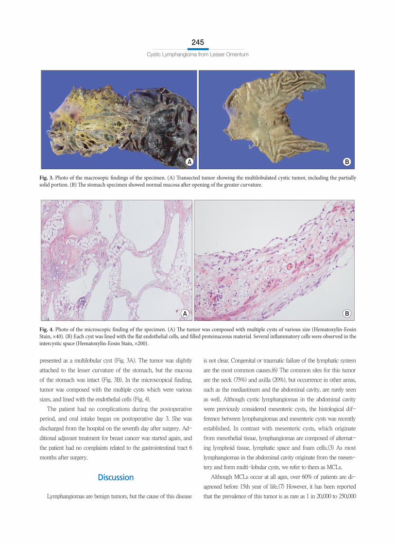
presented as a multilobular cyst (Fig. 3A). The tumor was slightly
attached to the lesser curvature of the stomach, but the mucosa
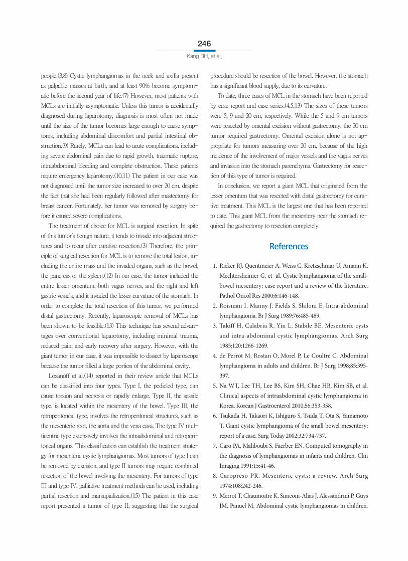
of the stomach was intact (Fig. 3B). In the microscopical finding,
tumor was composed with the multiple cysts which were various
sizes, and lined with the endothelial cells (Fig. 4).
The patient had no complications during the postoperative
period, and oral intake began on postoperative day 3. She was
discharged from the hospital on the seventh day after surgery. Ad-
ditional adjuvant treatment for breast cancer was started again, and
the patient had no complaints related to the gastrointestinal tract 6
months after surgery.
Discussion
Lymphangiomas are benign tumors, but the cause of this disease
is not clear. Congenital or traumatic failure of the lymphatic system
are the most common causes.(6) The common sites for this tumor
are the neck (75%) and axilla (20%), but occurrence in other areas,
such as the mediastinum and the abdominal cavity, are rarely seen
as well. Although cystic lymphangiomas in the abdominal cavity
were previously considered mesenteric cysts, the histological dif-
ference between lymphangiomas and mesenteric cysts was recently
established. In contrast with mesenteric cysts, which originate
from mesothelial tissue, lymphangiomas are composed of alternat-
ing lymphoid tissue, lymphatic space and foam cells.(3) As most
lymphangiomas in the abdominal cavity originate from the mesen-
tery and form multi-lobular cysts, we refer to them as MCLs.
Although MCLs occur at all ages, over 60% of patients are di-
agnosed before 15th year of life.(7) However, it has been reported
that the prevalence of this tumor is as rare as 1 in 20,000 to 250,000
Fig. 3. Photo of the macrosopic fi ndings of the specimen. (A) Transected tumor showing the multilobulated cystic tumor, including the partially solid portion. (B) Th e s tomach specimen showed normal mucosa aft er opening of the greater curvature.
Fig. 4. Photo of the microscopic fi nding of the specimen. (A) Th e tumor was composed with multiple cysts of various size (Hematoxylin-Eosin Stain, ×40). (B) Each cyst was lined with the fl at endothelial cells, and fi lled proteinaceous material. Several infl ammatory cells were observed in the intercystic space (Hematoxylin-Eosin Stain, ×200).
Kang BH, et al.
246
people.(3,8) Cystic lymphangiomas in the neck and axilla present
as palpable masses at birth, and at least 90% become symptom-
atic before the second year of life.(7) However, most patients with
MCLs are initially asymptomatic. Unless this tumor is accidentally
diagnosed during laparotomy, diagnosis is most often not made
until the size of the tumor becomes large enough to cause symp-
toms, including abdominal discomfort and partial intestinal ob-
struction.(9) Rarely, MCLs can lead to acute complications, includ-
ing severe abdominal pain due to rapid growth, traumatic rupture,
intraabdominal bleeding and complete obstruction. These patients
require emergency laparotomy.(10,11) The patient in our case was
not diagnosed until the tumor size increased to over 20 cm, despite
the fact that she had been regularly followed after mastectomy for
breast cancer. Fortunately, her tumor was removed by surgery be-
fore it caused severe complications.
The treatment of choice for MCL is surgical resection. In spite
of this tumor’s benign nature, it tends to invade into adjacent struc-
tures and to recur after curative resection.(3) Therefore, the prin-
ciple of surgical resection for MCL is to remove the total lesion, in-
cluding the entire mass and the invaded organs, such as the bowel,
the pancreas or the spleen.(12) In our case, the tumor included the
entire lesser omentum, both vagus nerves, and the right and left
gastric vessels, and it invaded the lesser curvature of the stomach. In
order to complete the total resection of this tumor, we performed
distal gastrectomy. Recently, laparoscopic removal of MCLs has
been shown to be feasible.(13) This technique has several advan-
tages over conventional laparotomy, including minimal trauma,
reduced pain, and early recovery after surgery. However, with the
giant tumor in our case, it was impossible to dissect by laparoscope
because the tumor filled a large portion of the abdominal cavity.
Losanoff et al.(14) reported in their review article that MCLs
can be classified into four types. Type I, the pedicled type, can
cause torsion and necrosis or rapidly enlarge. Type II, the sessile
type, is located within the mesentery of the bowel. Type III, the
retroperitoneal type, involves the retroperitoneal structures, such as
the mesenteric root, the aorta and the vena cava. The type IV mul-
ticentric type extensively involves the intraabdominal and retroperi-
toneal organs. This classification can establish the treatment strate-
gy for mesenteric cystic lymphangiomas. Most tumors of type I can
be removed by excision, and type II tumors may require combined
resection of the bowel involving the mesentery. For tumors of type
III and type IV, palliative treatment methods can be used, including
partial resection and marsupialization.(15) The patient in this case
report presented a tumor of type II, suggesting that the surgical
procedure should be resection of the bowel. However, the stomach
has a significant blood supply, due to its curvature.
To date, three cases of MCL in the stomach have been reported
by case report and case series.(4,5,13) The sizes of these tumors
were 5, 9 and 20 cm, respectively. While the 5 and 9 cm tumors
were resected by omental excision without gastrectomy, the 20 cm
tumor required gastrectomy. Omental excision alone is not ap-
propriate for tumors measuring over 20 cm, because of the high
incidence of the involvement of major vessels and the vagus nerves
and invasion into the stomach parenchyma. Gastrectomy for resec-
tion of this type of tumor is required.
In conclusion, we report a giant MCL that originated from the
lesser omentum that was resected with distal gastrectomy for cura-
tive treatment. This MCL is the largest one that has been reported
to date. This giant MCL from the mesentery near the stomach re-
quired the gastrectomy to resection completely.
References
1. Rieker RJ, Quentmeier A, Weiss C, Kretzschmar U, Amann K, Mechtersheimer G, et al. Cystic lymphangioma of the small-bowel mesentery: case report and a review of the literature. Pathol Oncol Res 2000;6:146-148.
2. Roisman I, Manny J, Fields S, Shiloni E. Intra-abdominal lymphangioma. Br J Surg 1989;76:485-489.
3. Takiff H, Calabria R, Yin L, Stabile BE. Mesenteric cysts and intra-abdominal cystic lymphangiomas. Arch Surg 1985;120:1266-1269.
4. de Perrot M, Rostan O, Morel P, Le Coultre C. Abdominal lymphangioma in adults and children. Br J Surg 1998;85:395-397.
5. Na WT, Lee TH, Lee BS, Kim SH, Chae HB, Kim SB, et al. Clinical aspects of intraabdominal cystic lymphangioma in Korea. Korean J Gastroenterol 2010;56:353-358.
6. Tsukada H, Takaori K, Ishiguro S, Tsuda T, Ota S, Yamamoto T. Giant cystic lymphangioma of the small bowel mesentery: report of a case. Surg Today 2002;32:734-737.
7. Caro PA, Mahboubi S, Faerber EN. Computed tomography in the diagnosis of lymphangiomas in infants and children. Clin Imaging 1991;15:41-46.
8. Caropreso PR. Mesenteric cysts: a review. Arch Surg 1974;108:242-246.
9. Merrot T, Chaumoitre K, Simeoni-Alias J, Alessandrini P, Guys JM, Panuel M. Abdominal cystic lymphangiomas in children.
Cystic Lymphangioma from Lesser Omentum
247
Clinical, diagnostic and therapeutic aspects: apropos of 21 cases. Ann Chir 1999;53:494-499.
10. Noundou PM, Michel G, Santiago M. Mesenteric cystic lymphangioma associated with necrosis of the Bauhin’s valvula in children. J Chir (Paris) 1993;130:87-89.
11. Vlazakis SS, Gardikis S, Sanidas E, Vlachakis I, Charissis G. Rupture of mesenteric cyst aft er blunt abdominal trauma. Eur J Surg 2000;166:262-264.
12. Stopinski J, Stephan S, Staib I. Intra-abdominal cystic lymph-angioma and mesenteric cysts as a cause of abdominal discom-
fort. Langenbecks Arch Chir 1994;379:182-187.13. Kenney B, Smith B, Bensoussan AL. Laparoscopic excision
of a cystic lymphangioma. J Laparoendosc Surg 1996;6 Suppl 1:S99-101.
14. Losanoff JE, Richman BW, El-Sherif A, Rider KD, Jones JW. Mesenteric cystic lymphangioma. J Am Coll Surg 2003;196:598-603.
15. Hebra A, Brown MF, McGeehin KM, Ross AJ 3rd. Mesenteric, omental, and retroperitoneal cysts in children: a clinical study of 22 cases. South Med J 1993;86:173-176.