Embed Size (px)
Citation preview
Giant Cell Arteritis
Case History
This is the case of C.S., a 51-year-old Portuguese known female patient who presented in February 1994 with blurring of vision and temporal headaches.
She claims that for the past few weeks she has been feeling very sluggish. She has had poor vision previously however she noted a further decrease in her visual acuity. She has more difficulty recognizing objects during the past few days. In addition she has been experiencing a new onset temporal headache which has been nagging her ever so effectively. Past ocular history
Cataracts, OD
Band keratopathy, OD
H/o Uveitis, OU
H/o Retinal vasculitis, OU
H/o angle closure glaucoma
S/p yag iridotomy and cyclophotocoagulation Past medical history
Hypertension
Migraine headaches
Lupus suspect (ANA 1:128 speckled)
APS suspect Medications
Plaquenil (for ANA titer)
Aspirin (for anticardiolipin antibody positivity) Family medical history
Non-contributory Personal and social history
Non-contributory Ocular examination
VA OD NLP
VA OS 20/100
Gross exam
Quiet white eyes
EOMs full OU
AT OD 22
AT OS 17
Funduscopy
OD No view
OS good red-orange reflex, clear vitreous media, slightly indistinct disc borders, slight pallor inferiorly, cup:disc ratio difficult to ascertain, artery:venous ratio 2:3; no signs of vasculitis, retina attached, foveal reflex dull
Introduction

Giant cell arteritis (GCA)
Synonyms: Temporal arteritis, Cranial arteritis
o Systemic granulomatous vasculitis involving medium- and large-sized vessels. o First clinically described c/o Hutchinson in 1890.[i] o First histologic description c/o Horton et al in 1932.[ii] o Differs from other forms of vasculitis by:
o Patient®s age o New onset of localized headache o Temporal artery tenderness or pulselessness o Elevated ESR o Temporal artery biopsy findings.
Epidemiology
o Age
o Mostly in patients over 50 years of age
o Incidence (0.49 to 50/100,000) increasing with age and peaking (nearly 1%) in the eighth decade.3-20
o Sex
o M:R 1:2-41 , 2 , 3
o Race
o Higher incidence rates in Caucasians of European descent
o Rare in African-Americans and Asians.
Clinical Features
Systemic Manifestations
o Prodromal symptoms (days to weeks) o Anorexia o Fever o Malaise o Myalgia o Night sweat o Weight loss
o Hallmark symptom o New onset localized headache. 5,6,11 o Usually localized to the temporal or occipital area. o Occasionally diffuse or bilateral. o Scalp tenderness

Gently stroking the GCA patient®s hair results in a characteristic complaint of pain distinctively seen only in patient®s suffering from GCA
Rizzo anecdotally describes this as the single most important clinical finding for GCA.[xxii]
Dudenhoefer described scalp necrosis with GCA seen in 2 elderly patients.[xxiii]
o Other cranial symptoms Temporal tenderness or pulselessness Jaw claudication Facial pain Earache Toothache Tongue pain Palate pain Odynophagia.
o Other clinical features o Pulselessness and/or tenderness and inflammation along the course of the
temporal artery o Bruits in the cranial or neck area o Jaw claudication o Atrophy of temporal and tongue muscles o Temporal artery blood flow measurements may be reduced.
o Cerebrovascular disease o 1% to 25% of patients,[xxiv], [xxv], [xxvi], [xxvii] o Most common cause of death in GCA patients.26 , 27
o Neurologic disease [xxviii], [xxix], 25 , [xxx], [xxxi], [xxxii], [xxxiii], [xxxiv] o Myopathy o Neuro-otologic syndromes o Neuro-psychiatric syndromes o Peripheral neuropathies o Seizures.
o Cardiovascular, pulmonary, gastrointestinal, renal, and dermatologic manifestations may also occur.28 , 29 , 34 , [xxxv], [xxxvi], [xxxvii], [xxxviii], [xxxix], [xl], [xli], [xlii],[xliii], [xliv], [xlv]
Ophthalmic manifestations
o Visual symptoms o ~50% of patients o Transient visual blurring o Diplopia o Eye pain o Sudden loss of vision, etc. [xlvi]
o Transient repeated episodes of blurred vision are usually reversible. o Sudden loss of vision is an ominous sign and is almost always permanent. o Vision loss incidence, either partial or complete, is variably reported to be 10% to 60%.
6 , 7 , 8 , 17 , 24 , 25 , 27
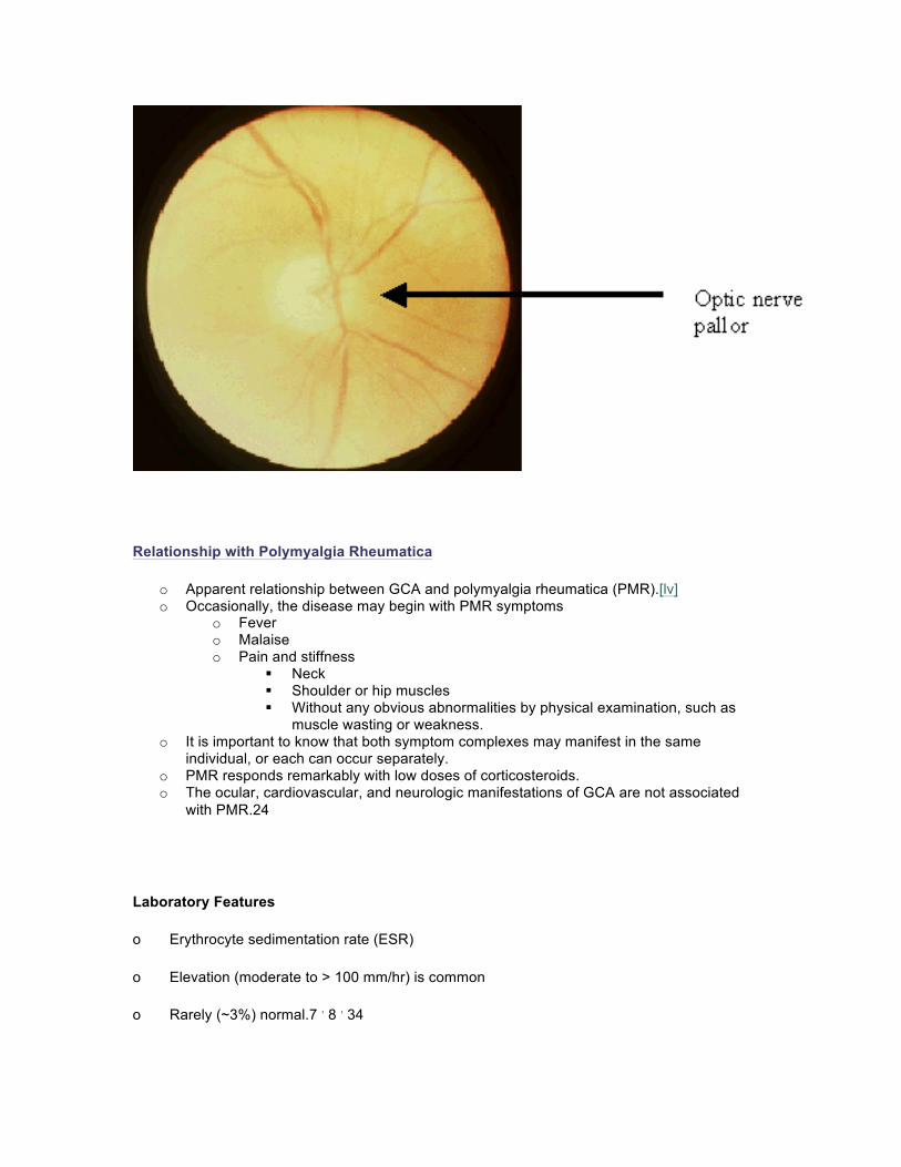
o Anterior ischemic optic neuropathy (AION) o The most common cause of vision loss. o Ischemia of the optic nerve head
Supplied mainly by the posterior ciliary arteries. o Majority of AION is non-arteritic (87% to 91%). [xlvii], [xlviii], [xlix] o GCA is an arteritic AION o History of sudden painless loss of vision. o Fundus examination
May reveal optic disc edema, with or without splinter hemorrhages along the disc margin.
Typically presents with a chalky white edematous optic disc. [l] o Automated visual field
Typically reveals an inferior altitudinal defect, inferior nasal sectoral defect or central scotoma.48
o Other important vascular ophthalmic presentations8 , 26 , 49 , [li], [lii], [liii] o Posterior ischemic (retrobulbar) optic neuropathy o Central retinal artery occlusion o Branch retinal artery occlusion o Choroidal ischemia.
o Neuroophthalmic manifestations7 , 24 , 29 , 25 , 27 , [liv] o Diplopia o Ptosis o Nystagmus o Internuclear ophthalmoplegia (INO) o Pupillary abnormalities.
Relationship with Polymyalgia Rheumatica
o Apparent relationship between GCA and polymyalgia rheumatica (PMR).[lv] o Occasionally, the disease may begin with PMR symptoms
o Fever o Malaise o Pain and stiffness
Neck Shoulder or hip muscles Without any obvious abnormalities by physical examination, such as
muscle wasting or weakness. o It is important to know that both symptom complexes may manifest in the same
individual, or each can occur separately. o PMR responds remarkably with low doses of corticosteroids. o The ocular, cardiovascular, and neurologic manifestations of GCA are not associated
with PMR.24
Laboratory Features
o Erythrocyte sedimentation rate (ESR)
o Elevation (moderate to > 100 mm/hr) is common
o Rarely (~3%) normal.7 , 8 , 34
o Highly elevated ESR results are characteristic of a GCA process rather than other vasculitic or rheumatologic entities.
o Other acute phase reactants
o C-reactive protein (CRP), are elevated and reflect the underlying inflammatory process.
o These acute phase reactants may be followed serially and may assist in monitoring for treatment dosing and response.[lvi], [lvii]
o CBC
o Most are mildly anemic (normochromic, normocytic).
o Lymphocytes hypothesized to proliferate in response to elastin, other arterial wall antigens, or muscle.
o Immunoglobulin levels
o Standard
o Immune complexes are absent.
o Hepatic enzymes, alkaline phosphatase and serum aspartate aminotransferase (SGOT)
o Elevated in 20% to 30% and 15% of cases respectively. [lviii], [lix]
o Imaging studies
o Aortic arch and cerebral angiography may show occlusion or alternating stenotic areas.
o Computed tomography (CT) and magnetic resonance imaging (MRI) of the brain are not first line diagnostic procedures for GCA, however, they may be useful in patients with a multi-infarct state secondary to cervicocephalic arteritis.56
o Superficial temporal artery biopsy (TAB)
o Focal granulomatous arteritis, often with giant cells and "skip areas" of normal arterial wall.[lx], [lxi]
o The technique for TAB is reported elsewhere.[lxii]
o The most symptomatic side should be biopsied initially.
o In a patient with suggestive symptoms and a negative initial biopsy on the symptomatic side, performing a TAB on the other side may confirm the diagnosis.57 , 58
o The TAB should be performed ASAP, in the appropriate patient.
o Therapy should not be withheld pending the performance or results of the TAB in patients with acute visual loss and extremely high clinical suspicion for GCA.
Pathophysiology
o In GCA, large elastic and medium-sized extracranial muscular arteries are most severely involved.[lxiii]
o The posterior ciliary and ophthalmic arteries are commonly affected. o The common, external and internal carotid, vertebral, subclavian axillary, proximal
brachial artery may be involved. o Less commonly, the descending aorta, mesenteric, renal, iliac, femoral, and pulmonary
arteries may be affected. 14 , 23 , 24 , 36
o GCA is a disease mediated primarily by an active cellular arm of immunity. o CD4+ T-helper cell response to macrophage-presented antigens lead to
inflammation typically beginning in the adventitia and progressing to involve the whole vessel wall.
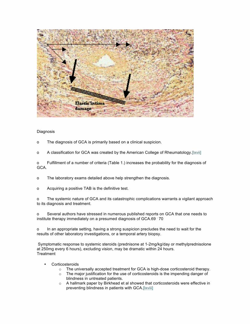
o The vascular occlusion occurs by focal narrowing, due to swelling of the wall without thrombosis or aneurysm formation.
o The internal elastic lamina is invaded and damaged by the primary inflammatory response.[lxiv]
o Giant cells while usually found histologically, is not required for the diagnosis. o After a period of active inflammation, scarring permanently narrows the vascular
lumen.[lxv] o The impetus for the destructive inflammation is still unknown today. Elastin is highly
suspected as being involved in the inciting events leading to this disease.[lxvi]

Diagnosis
o The diagnosis of GCA is primarily based on a clinical suspicion.
o A classification for GCA was created by the American College of Rheumatology.[lxvii]
o Fulfillment of a number of criteria (Table 1.) increases the probability for the diagnosis of GCA.
o The laboratory exams detailed above help strengthen the diagnosis.
o Acquiring a positive TAB is the definitive test.
o The systemic nature of GCA and its catastrophic complications warrants a vigilant approach to its diagnosis and treatment.
o Several authors have stressed in numerous published reports on GCA that one needs to institute therapy immediately on a presumed diagnosis of GCA.69 , 70
o In an appropriate setting, having a strong suspicion precludes the need to wait for the results of other laboratory investigations, or a temporal artery biopsy.
Symptomatic response to systemic steroids (prednisone at 1-2mg/kg/day or methylprednisolone at 250mg every 6 hours), excluding vision, may be dramatic within 24 hours. Treatment
• Corticosteroids o The universally accepted treatment for GCA is high-dose corticosteroid therapy. o The major justification for the use of corticosteroids is the impending danger of
blindness in untreated patients. o A hallmark paper by Birkhead et al showed that corticosteroids were effective in
preventing blindness in patients with GCA.[lxviii]

o Initially, high doses of corticosteroids may be given at 1 to 2 mg/kg/day until the disease activity is adequately suppressed.
• ESR determination o Sequential ESR determination may assist in determining the success of the high
dose corticosteroid therapy. o Once the signs of clinical inflammation is suppressed and the ESR is maintained
at a low level, corticosteroid levels may be tapered off slowly. • Length of treatment
o There is no agreement as to the length of treatment with corticosteroids for GCA. o It may be reasonable to maintain the patient on at least two years of treatment in
order to lessen the chances of relapses. o Even so, relapses have been reported.
• Immunosuppressives o Foster uses a cyclosporin-azathioprine or cyclosporin-methotrexate combination
as a steroid-sparing cocktail or therapy for steroid-resistant cases.[lxix] • Biopsy and treatment
o The disastrous nature of the disease may occasionally require the administration of treatment prior to a definitive superficial TAB.
o It is generally believed that the results of a TAB will not be altered if the procedure is performed within 3 to 5 days of initiating corticosteroid therapy. 59, 69 , [lxx]
Table 1.
Classification of giant cell arteritis. American College of Rheumatology Criteria for Classification of Giant cell arteritis.
Traditional (sensitivity, 93.5%; specificity, 91.2%; 3 0f 5 criteria must be met)
1. Age of onset of 50 years or older
2. Onset of new headache
3. Temporal artery tenderness or reduced pulsation
4. Elevated (>50 mmHg) Westergren erythrocyte sedimentation rate
5. Abnormal artery biopsy
Alternative (sensitivity, 95.3%; specificity, 90.7%; 3 of 6 criteria must be met)
1. Age at onset of 50 years or older
2. Onset of new headache
3. Temporal artery tenderness or reduced pulsation
4. Claudication of jaw
5. Scalp tenderness or nodules
6. Abnormal artery biopsy
References
[i] Hutchinson J. Diseases of the arteries. On a peculiar form of thrombotic arteritis of the aged which is sometimes productive of gangrene. Arch Surg (London) 1890;1:323-9.
[ii] Horton BT, Magath TB, Brown GE. An undescribed form of arteritis of the temporal vessels. Proc Staff Meet Mayo Clin 1932;7:700-1.
[iii] Bengtsson BA, Malmvall BE. The epidemiology of giant cell arteritis including temporal arteritis and Polymyalgia rheumatica. Incidences of different clinical presentations and eye complications. Arthritis Rheum 1981;24:899-904.
[iv] Nordberg E, Bengstonn BA. Epidemiology of biopsy-proven giant cell arteritis (GCA). J Intern Med 1990;227:233-6.
[v] Salvarani C, Gabriel SE, O®Fallon WM, et al. The incidence of giant cell arteritis in Olmsted Country, Minnesota: apparent fluctuations in a cyclic pattern. Ann Intern Med 1995;123:192-4.
[vi] Machado EB, Michet CJ, Ballard DJ, et al. Trends in incidence and clinical presentation of temporal arteritis in Olmstead County, Minnesota, 1950-1985. Arthritis Rheum 1988;31:745-9.
[vii] Huston KA, Hunder GG, Lie JT, et al. Temporal arteritis: a 25-year epidemiologic, clinical, and pathologic study. Ann Intern Med 1978;88:162-7.
[viii] Jonasson F, Cullen JF, Elton RA. Temporal arteritis. A 14-year epidemiological, clinical and prognostic study. Scott Med J 1979;24:111-7.
[ix] Franzen P, Sutinen S, von Knorring J. Giant cell arteritis and Polymyalgia rheumatica in a region of Finland: an epidemiologic, clinical and pathologic study, 1984-1988. J Rheumatol 1992;19:273-6.
[x] Berlit P. Clinical and laboratory findings with giant cell arteritis. J Neurol Sci 1992;111:1-12.
[xi] Noltorp S, Svensson B. High incidence of Polymyalgia rheumatica and giant cell arteritis in a Swedish community. Clin Exp Rheumatol 1991;9:351-5.
[xii] Salvarani C, Macchioni P, Zizzi F, et al. Epidemiologic and immunogenetic aspects of Polymyalgia rheumatica and giant cell arteritis in northern Italy. Arthritis Rheum 1991;34:351-6.
[xiii] Gonzalez EB, Varner WT, Lisse JR, et al. Giant cell arteritis in the southern United States. An 11-year retrospective study from the Texas Gulf Coast. Arch Intern Med 1989;149:1561-5.
[xiv] Boesen DR, Sorensen SF. Giant cell arteritis, temporal arteritis, and Polymyalgia rheumatica in a Danish county. A preospective investigation, 1982-1985. Arthritis Rheum 1987;30:294-9.
[xv] Smith CA, Fidler WJ, Pinals RS. The Epidemiology of giant cell arteritis. Report of a 10-year study in Shelby County, Tennessee. Artheritis Rheum 1983;26:1214-9.
[xvi] Friedman G, Friedman B, Benbassat J. Epidemiology of temporal arteritis in Israel. Isr J Med Sci 1982;18:241-4.
[xvii] Baldursson O, Steinsson K, Bjornsson J, et al. Giant cell arteritis in Iceland. An epidemiologic and histopahtologic analysis. Arthritis Rheum 1994;37:1007-12.
[xviii] Sonnenblick M, Nesher G, Friedlander Y, et al. Giant cell arteritis in Jerusalem: a 12-year epidemiological study. Br J Rheumatol 1994;33:938-41.
[xix] Gonzalez-Gay MA, Blanco R, Sanchez-Andrade A, et al. Giant cell arteritis in Lugo, Spain: a more frequent disease with fewer classic features. J Rheumatol 1997;24:2166-70.
[xx] Gran JT, Myklebust G. The incidence of Polymyalgia rheumatica and temporal arteritis in the county of Aust Agder, south Norway: a prospective study 1987-94. J of Rheumatol 1997;24:1739-43.
[xxi] Hollenhorst RW, Brown JR, Wagener HP, et al. Neurologic aspects of temporal arteritis. Neurology 1960;490-8.
[xxii] Rizzo J. (Resident®s lecture series 2000). Giant cell arteritis. Department of Ophthalmology. Massacusetts Eye and Ear Infirmary, Harvard Medical School, Boston, MA.
[xxiii] Dudenhoefer EJ, Cornblath WT, Schatz MP. Scalp necrosis with giant cell arteritis. Ophthalmology 1998;105(10):1875-8.
[xxiv] Paulley JW, Hughes JP. Giant cell arteritis, or arteritis of the aged. Br Med J 1960;2:1562-7.
[xxv] Graham E, Holland A, Avery A, et al. Prognosis in giant cell arteritis. Br Med J 1981;282:269-71.
[xxvi] Whitfield AG, Bateman M, Cooke WT. Temporal arteritis. Br J Ophthalmol 1963;47:555-66.
[xxvii] Reich KA, Giansiruacusa DF, Strongwater SL. Neurologic manifestations of giant cell arteritis. Am J Med 1990;89:67-72.
[xxviii] Hamilton CR, Shelley WM, Tumulty PA. Giant cell arteritis: including temporal arteritis and Polymyalgia rheumatica. Medicine 1971;50:1-27.
[xxix] Simons RJ, Cogan DG. Occult temporal arteritis. Arch Ophthalmol 1962;68:38-48.
[xxx] Warrel DA, Godfrey S, Olsen EGJ. Giant cell arteritis with peripheral neuropathy. Lancet 1968;1:1010-13.
[xxxi] Pascuzzi RM, Roos KL, Davis TE. Mental status abnormailities in temporal arteritis: a treatable cause of dementia in the elderly. Arthritis Rheum 1989;32:1308-11.
[xxxii] Andrews JM. Giant cell arteritis: a disease with variable clinical manifestations. Neurology 1966;16:963-71.
[xxxiii] Fauchald P, Rygvold O, Oystese B, et al. Temporal arteritis and Polymyalgia rheumatica: clinical and biopsy findings. Ann Intern Med 1972;77:845-52.
[xxxiv] Klein RG, Hunder GG, Stanson AW, et al. Large artery involvement in giant cell (temporal) arteritis. Ann Intern Med 1975;83:806-12.
[xxxv] Sorensen PS, Lorenzen I. Giant cell arteritis, temporal arteritis and Polymyalgia rheumatica: a retrospective study of 63 patients. Acta Medica Scandinavica 1977;201:207-13.
[xxxvi] Lie JT. Aortic and extracranial large vessel giant cell arteritis: a review of 72 cases with histopathologic documentation. Sem Arthritis Rheum 1995;24:422-31.
[xxxvii] Liu G, Shupak R, Chiu BK. Aortic dissection in giant cell arteritis. Sem Arthritis Rheum 1995;25:160-71.
[xxxviii] Evans JM, O®Fallon WM, Hunder GG. Increased incidence of aortic aneurysm and dissection in giant cell (temporal) arteritis. A population-based study. Ann Intern Med 1995;122:502-7.
[xxxix]Larson TS, Hall S, Hepper NG, et al. Respiratory tract symptoms as a clue to giant cell arteritis. Ann Intern Med 1984;101:594-7.
[xl] Olopade CO, Sekosan M, Schraufnagel DE. Giant cell arteritis manifesting as chronic cough and fever of unknown origin. Mayo Clinic Proc 1997;72:1048-50.
[xli] Gur H, Ehrenfeld M, Izsak E. Pleural effusion as a presenting manifestation of giant cell arteritis. Clin Rheumatol 1996;15:200-3.
[xlii] Goldberg JW, Lee ML, Sajjad SM. Giant cell arteritis of the skin simulating erythema nodosum. Ann Rheum Disease 1987;46:706-8.
[xliii] Lie JT, Tokugawa DA. Bilateral lower limb gangrene and stroke as initial manifestations of systemic giant cell arteritis in an African-American. J Rheumatol 1995;22:363-6.
[xliv] Alestig K, Barr J. Giant cell arteritis. Lancet 1963;1:1228-30.
[xlv] Tsuyuoka R, Takahashi T, Shinoda E, et al. Intestinal perforation in temporal arteritis, associated with paroxysmal nocturnal hemoglobinuria. Intern Med 1996;35:159-61.
[xlvi] Hayreh SS, Podhajsky PA, Zimmerman. Ocular manifestations of giant cell arteritis. Am J Ophthalmol 1998;125(4):509-20.
[xlvii] Guer DR, Miller NR, Auer CL, et al. The risk of Cerebrovascular and cardiovascular disease in patients with anterior ischemic optic neuropathy. Arch Ophthalmol 1985;103:1136-42.
[xlviii] Beri M, Klugman MR, Kohler JA, et al. Anterior ischemic optic neuropathy. Incidence of bilaterality and various influencing factors. Ophthalmology 1987;94:1020-8.
[xlix] Hayreh SS, Podhajsky P. Visual field defects in anterior ischemic optic neuropathy. Doc Ophthalmol Proc Ser 1979;19:53-71.
[l] Beck RW, Servais GE, Hayreh SS, Anterior ischemic optic neuropathy. Cup-to-disc ratio and its role in pathogenesis. Ophthalmology 1989;94:1503-8.
[li] Hayreh SS. Anterior ischemic optic neuropathy, differentiation of arteritic from non-arteritic type and its management. Eye 1990;4:25-41.
[lii] Raymond LA, Sacks JG, Choromokos E, et al. Short posterior ciliary artery insufficiency with hyperthermia (Uhthoff®s symptom). Am J Ophthalmol 1980;90:619-23.
[liii] Hayreh SS. Ophthalmic features of giant cell arteritis. Baillieres Clin Rheumatol 1991;5:431-59.
[liv] Crompton JL, Burrow DJ, Iyer PV. Bilateral internuclear ophthalmoplegia. An unusual initial presenting sign of giant cell arteritis. Aust N Z J Ophthalmol 1989;17:71-4.
[lv] Cohen MD, Ginsburg WW. Polymyalgia rheumatica. Rheum Dis Clin North Am 1990;16:325.
[lvi] Kyle V, Cawston TE, Hazleman BL. Erythrocyte sedimentation rate and C reactive protein in the assessment of Polymyalgia rheumatica/giant cell arteritis on presentation and during follow up. Ann Rheum Dis 1989;105:965-7.
[lvii] Jacobson DM, Slamovits TL. Erythrocyte sedimentation rate and its relationship to hematocrit in giant cell arteritis. Arch Opthalmol 1987;105:965-7.
[lviii] Calamia KT, Hunder GG. Clinical manifestations of giant cell (temporal) arteritis. Clin Rheum Dis 1980;6:389-415.
[lix] Albertini JG, Marks VJ. Temporal (Giant cell) arteritis. Emedicine.com 2000 topic 417.
[lx] Boyev LR, Miller NR, Green R. Efficacy of unilateral versus bilateral temporal artery biopsies for the diagnosis of giant cell arteritis. Am J Ophthalmol 1999;128(2)211-215.
[lxi] Lee AG. Efficacy of unilateral versus bilateral temporal artery biopsies for the diagnosis of giant cell arteritis. [letter] Am J Ophthalmol 2000;129(1):118-9.
[lxii] Levin LA. Ophthalmic surgery. Part V Ophthalmic Plastic Surgery Chapter 98:1606-12.
[lxiii] Wilkinson IM, Russell RW. Arteries of the head and neck in giant cell arteritis. A pathological study to show the pattern of arterial involvement. Arch Neurol 1972;27:378-91.
[lxiv] Ashton-Key M, Gallagher PJ. Surgical pathology of cranial arteritis and Polymyalgia rheumatica. Baillieres Clin Rheum 1991;5:387-404.
[lxv] Albert DM, Ruchman MC, Keltner JL. Skip areas in temporal arteritis. Arch Ophthalmol 1976;94:2072-7.
[lxvi] Kimmelstiel P, Gilmour MT, Hodges HH. Degeneration of elastic fibers in granulomatous giant cell arteritis (temporal arteritis). Arch Pathol 1952;54:157-68.
[lxvii] Hunder GG, Bloch DA, Michel BA, et al. The American College of Rheumatology 1990 criteria for the classification of giant cell arteritis. Arthritis Rheum 1990;33:1122-8.

[lxviii] Birkhead NC, Waener HP, Shick RM. Treatment of temporal arteritis with adrenal corticosteroids: Results in 55 cases in which the lesion was proved at biopsy. JAMA 1975;163:821.
[lxix] Foster CS. Giant cell arteritis. In: Albert, Jakobiec F, eds. Principles and Practice of Ophthalmology 1st ed. CD-ROM Mosby Clinical Ophthalmology Chap 328.
[lxx] Caselli RJ. Temporal/Giant cell arteritis. Emedicine.com 2000 topic 592.