Embed Size (px)
Citation preview
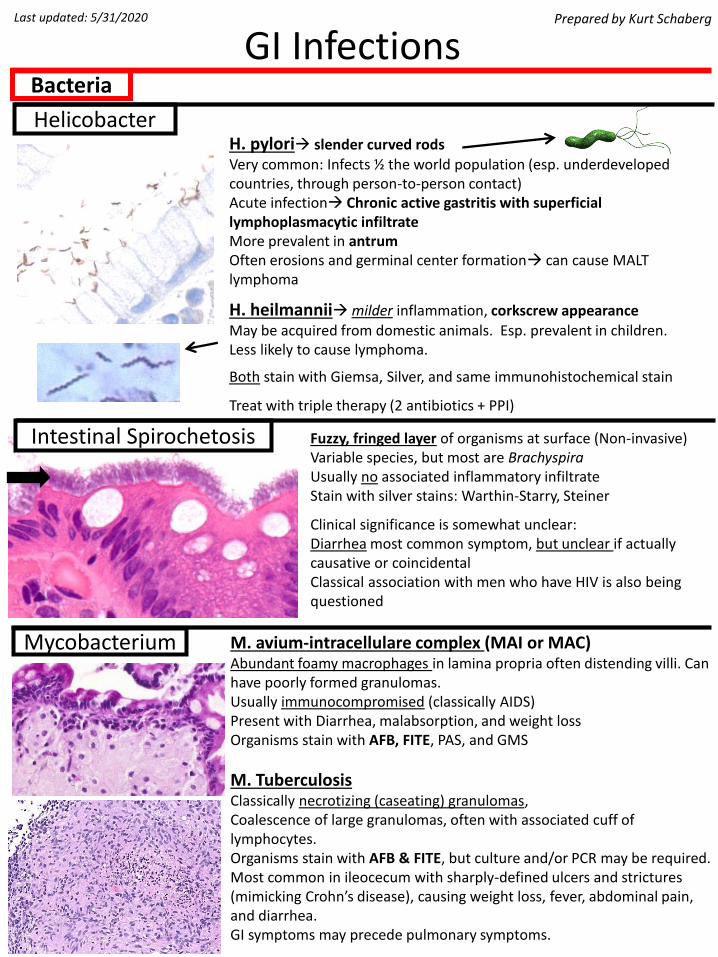
H. pylori→ slender curved rodsVery common: Infects ½ the world population (esp. underdeveloped countries, through person-to-person contact)Acute infection→ Chronic active gastritis with superficial lymphoplasmacytic infiltrateMore prevalent in antrumOften erosions and germinal center formation→ can cause MALT lymphoma
H. heilmannii→milder inflammation, corkscrew appearanceMay be acquired from domestic animals. Esp. prevalent in children.Less likely to cause lymphoma.
Both stain with Giemsa, Silver, and same immunohistochemical stain
Treat with triple therapy (2 antibiotics + PPI)
GI InfectionsPrepared by Kurt Schaberg
Intestinal Spirochetosis
Bacteria
Last updated: 5/31/2020
Helicobacter
Fuzzy, fringed layer of organisms at surface (Non-invasive) Variable species, but most are BrachyspiraUsually no associated inflammatory infiltrateStain with silver stains: Warthin-Starry, Steiner
Clinical significance is somewhat unclear:Diarrhea most common symptom, but unclear if actually causative or coincidentalClassical association with men who have HIV is also being questioned
Mycobacterium M. avium-intracellulare complex (MAI or MAC)Abundant foamy macrophages in lamina propria often distending villi. Can have poorly formed granulomas.Usually immunocompromised (classically AIDS)Present with Diarrhea, malabsorption, and weight lossOrganisms stain with AFB, FITE, PAS, and GMS
M. TuberculosisClassically necrotizing (caseating) granulomas,Coalescence of large granulomas, often with associated cuff of lymphocytes.Organisms stain with AFB & FITE, but culture and/or PCR may be required.Most common in ileocecum with sharply-defined ulcers and strictures (mimicking Crohn’s disease), causing weight loss, fever, abdominal pain, and diarrhea. GI symptoms may precede pulmonary symptoms.

Whipple disease
Acute infectious colitis
Gram-negative coccobacciform enteric bacteriaInfection caused by food contamination
Most commonly infects ilium, right colon, and appendix. Can cause ulcers and edema.
Abundant epithelioid granulomas with lymphoid cuffsTransmural lymphoid aggregates and giant cells commonUsually not necrotizing→Closely mimics Crohn’s diseaseStains not helpful→ consider culture, serologies, or PCRCommon cause of granulomatous appendicitis
Most commonly associated with bacterial enterocolitisUsually acute onset of diarrhea and abdominal pain. Often self-limited and resolves within several weeks.Often discriminated from one another by microbiology testing (classically culture, but now PCR NAATs)
Classically, Active colitis (cryptitis, crypt abscess formation, epithelial damage), without features of chronicity (preserved architecture, no metaplasia or basal lymphoplasmacytosis).Nevertheless, can mimic IBD, particularly in the resolving phase
Yersinia
Infection by Tropheryma whippleiPresent with weight loss, diarrhea, arthritis, lymphadenopathy, endocarditis, and neuropsychiatric issues.Most common in middle-aged white males with HLA-B27.
Most often infects small bowel, but can see changes throughout GI tract and also brain, heart, and lymph nodes.
Massive infiltration of lamina propria by foamy macrophagesVariable acute inflammation.
Organisms stain with PAS. Can also identify with PCR.(Negative for FITE and AFB, helping differentiate from MAI)
Most common bacteria include (Often food-borne illness):Campylobacter—most common stool isolate in US.Salmonella—can cause typhoid fever with hyperplastic Peyer’s patches, ulcers, and necrosis. Less PMNs.Enterohemorrhagic E. coli (O157:H7)—Shiga-like toxins cause epithelial and endothelial injury→ see fibrin thrombi and ischemic changes→ can cause hemolytic uremic syndrome (HUS) due to endothelial injury and platelet activation causing 1) Thrombocytopenia, 2) Hemolytic anemia, and 3) Kidney injuryClostridioides difficile—usually after recent antibiotic use. Watery diarrhea with pseudomembranesShigella, Yersinia
Also caused by some viruses (e.g., norovirus) and parasites
“Acute Self-limited Colitis”

Sarcina
Most “normal” bacteria in the oral cavity and intestines are gram-negative anaerobesOn GI biopsies, often see in esophagus and intestines
Bacteroides species are the most common, other common ones include Prevotella and Veillonella.Other organisms include gram-positive organisms like Streptococcus.
Usually, these are commensal and do not cause disease.
Can cause periodontal diseaseElsewhere, most disease is due to spread to other regions (e.g., endocarditis, abscesses, septic arthritis, pneumonia, etc…)
Often polymicrobial clusters/infectionsHighlighted by gram and silver stainsHistologic findings are nonspecific and further microbiology gests (e.g., culture, MALDI-TOF, or NAAT) are necessary for identification.
Normal Flora
Spherical cells 2-3 μm in diameter Occur in tetrads or packets of 8 or moreMost commonly found in the stomach
Unclear if pathogenic. Likely incidental finding.
Often seen in cases of delayed gastric emptying and gastric outlet obstruction→ Their presence can prompt further investigation as to
cause of dysfunction, such as occult malignancy
Long, filamentous bacteria that stain purpleLook like “dust bunnies”
Frequently seen as incidental bacteria on biopsies or part of mixed flora colonizinglesions, especially in oral cavity.Associated with poor hygiene.
Uncommon cause of appendicitis.
Positive on Gram stain and GMS. Negative on AFB.
Actinomyces

Cytomegalovirus (CMV)
Adenovirus
Normal hosts: Common cause of childhood diarrhea. Can cause intussusception due to lymphoid hyperplasia
Immunocompromised hosts: Diarrhea, potentially leading to disseminated disease (including hepatitis and pneumonitis) and death. Harder to control.
Characteristic smudgy inclusions that are basophilic to eosinophilic
Tubular GI tract: Inclusions in surface epithelium, often in goblet cells→ can be round or crescent shaped. Most often in colon with increased apoptosis and epithelial sloughing.
Liver: Inclusions in hepatocytes, often at edges of coagulative necrosis
Herpes Simplex Virus (HSV)
Viruses
Most common in immunocompromised hosts, esp. AIDS
Often causes ulcerations. Symptoms vary by site: Esophagus→ dysphagia, odynophagiaStomach/intestines→ Diarrhea, bloody or watery, pain
Ulceration, mixed inflammatory infiltrate with neutrophilsif severely immunocompromised, less inflammation
Viral inclusions, preferentially in mesenchymal cells:Most commonly endothelium or other stromal cellsNuclear→ “Owl’s eye” (Cowdry A), pink, nucleolus-likeCytoplasmic→ granular and pink to purple, hof-like
Be sure to evaluate for in refractory IBD and GVHD casesCan also look for with PCR
Most commonly causes ulceration with variable inflammation, predominantly acute. Can get vesicles in anorectum.
Viral inclusions at edges of ulcers in epithelial cells3M’s→Moulding, (chromatin) Margination, Multinucleation
#2 most common cause of infectious esophagitis→ dysphagiaSelf-limited in healthy patients; may cause esophageal perforation or disseminate in immunocompromised patientsFindings the same in HSV1&2

Candida
Cryptococcus Ubiquitous. Often from avian droppings (think “Pigeons”)Usually immunocompromised (e.g., AIDS, organ transplant, etc…)Can be localized or disseminated disease.Other common sites are lung and meninges
Variable inflammatory response (depending on immune state). Can have granulomas or suppurative necrosis.
4-7μm, very “pleomorphic” (lots of different sizes), round to oval,Narrow-based buds. Unstained, refringent capsules give “halo” or “soap bubble” appearance. Stain with GMS. Capsule stains with mucicarmine
Histoplasmosis
Fungus
Endemic to Ohio, Missouri, Mississippi river valleys.Can cause localized or disseminated disease (more common in immunocompromised). Lung most common site, but GI common too.
Most common GI site of involvement is ileum. May cause ulcers or mass.
Often lymphohistiocytic infiltrates without well-formed granulomasIntracellular 2-5 μm fungiPositive with GMS and PAS
Most common infection of the esophagusMore common in immunocompromisedPresents with dysphagia/odynophagia
Endoscopy: white plaques with underlying ulceration
Neutrophilic inflammation with ulceration, but less if immunocompromisedParakeratosis common→ highlighted by PAS-D and GMS stains→See mix of budding yeast and pseudohyphae
Coccidioides“Valley Fever.” Found in soil in southwestern United States and South and Central America. Higher risk if immunocompromised. Can have localized or disseminated disease.
In host, spores develop into large, thick-walled endospore-containing spherules, which enlarge and rupture. There is often associated granulomatous and chronic inflammation

Strongyloides
Schistosomiasis
Enterobius vermicularis
Parasites
Parasitic trematode (fluke)Any species of “schisto” can be found in the gutEndemic to Africa, Asia and parts of the Americas.Highest prevalence in Sub-Saharan Africa and Middle East
Infected by contaminated water through the skin→ snails are intermediate host
Most patients are asymptomatic, but can present with GI bleeding (or hematuria or portal hypertension)
Ova: Found in the wall of the GI or GU tract. Often calcifywith time. Variable acute, chronic, or granulomatous inflammation. Often prominent eosinophils.
Worms: often have no reaction to them, found in veins (of bowel or bladder) or in liver → lay eggs into urine/stool
Three main species in humans:Schistosoma mansoni-Usually GI tract. Lateral spineSchistosoma japonicum-Usually GI tract. Later knobSchistosoma haematobium-Usually GU tract. Terminal spine
“Pinworm”Spread by fecal-oral route. Humans are the only host.Most common in children. Often asymptomatic, but can cause anal pruritisMost commonly seen in appendix, often incidentallyThick cuticle on adult worm
characteristic lateral spikes (ala)Easily visible internal organsEven invasive worms cause minimal inflammation
Nematode with worldwide distribution. Very common in Tropics and southeastern US. Often get through skin when barefoot on contaminated soil. Skin→ Lung→ GI tract → Feces → next host (or autoinfect)Worse in immunocompromised patientsCan be asymptomatic and harbor for >30 yrsWhen symptomatic, diarrhea, pain, bleedingInflammation with neutrophils and eosinophils often, may resemble IBDAdult worms, larvae, and eggs all found IN crypts

Liver flukes
Echinococcus
Entamoeba Histolytica
Protozoan most common in subtropical and tropical regionsIn US, most common in immigrants and travelersInfected through fecal-oral route/contaminated food/water
Can be asymptomatic, or cause variably severe diarrheaCan cause amoebic liver abscesses
Cause deep “flask-shaped” ulcers, extending into submucosa, undermining nearby mucosa.Architectural distortion may mimic IBDOften abundant amorphous eosinophilic debris
Entamoeba: Round, red, eccentric nucleusDistinct cell membranes with foamy cytoplasmIngested RBCs.
Protozoans
Helminths occlude bile duct→ dilated ducts with wall thickening→ Signs of biliary obstruction (jaundice, fever, RUQ pain) → can cause cholangiocarcinoma long-term due to chronic inflammation
Clonorchis sinensis, Opisthorchis species, and Fasciola speciesEndemic primarily to Asia and acquired by eating raw or undercooked fish or crawfish
Worms visible to naked eye
Cestode (tapeworm) with wide geographic distributionDefinitive host = Dogs (or other carnivore)—humans infected through exposure to feces→ Eggs hatch → larvae travel to liver and form cysts→ cysts grow very slowly
Often asymptomatic, but can get symptoms from mass-effectTreated with surgical resection; Ruptured cysts are very antigenic→ can cause anaphylaxis
Inner most layer contains protoscolices (developing heads of adult tapeworms), which contain 2 circles of hooklets and suckerThis is surrounded by a layer of hyalinized, white laminated, acellular material

Giardia duodenalis
Cystoisospora Formerly just “isospora”Obligate intracellular world-wide parasite.Infected by contaminated food/waterCauses diarrhea, often chronic. Debilitating if immunocompromisedVillous blunting with mixed inflammation and prominent Eosinophils
Variable forms, all intraepithelial:Some crescent/banana shapedOthers are round with prominent nucleoli
Cryptosporidia
Most common protozoa infection in USUsually acquired from contaminated water. Can be STD.More common in kids, with travel, and immunocompromisedCauses diarrhea (unclear pathogenesis), often watery and foul-smelling. Can be chronic, esp. if immunocompromised
Usually see trophozoites with no associated inflammation(sometimes mild villous blunting and chronic inflammation)
Trophozoites are pair-shaped with 2 oval nucleiLook like “falling leaves” in bowel lumen
Obligate intracellular world-wide parasite.Can be from contaminated water or person-to-personDiarrhea→ self-limited in normal hosts, but often chronic/relapsing with weight loss and cramping in immunocompromised. No good therapy.
Parasites appear as 2-5μm basophilic “blue beads” on lumina apical surface.Can see villous blunting and variable inflammatory infiltrateEnveloped by microvilli→ less microvilli for absorption→ diarrhea
CyclosporaProtozoan with world-wide distribution that causes diarrhea. Infection often occurs through contaminated food/waterVariable villous blunting and inflammationRound (2-3 µm) forms and crescentic merozoites (5-6 µm) in parasitophorous vacuoles
MicrosporidiaFungus that causes intestinal infection, particularly in AIDS patients→DiarrheaSmall spores (2-3 μm) and larger plasmodiaLocated within supranuclear cytoplasm of epithelial cells