Embed Size (px)
Citation preview
Amburgey et al. Orphanet Journal of Rare Diseases 2013, 8:117http://www.ojrd.com/content/8/1/117
RESEARCH Open Access
Genotype-phenotype correlations in recessiveRYR1-related myopathiesKimberly Amburgey1, Angela Bailey1, Jean H Hwang2, Mark A Tarnopolsky3, Carsten G Bonnemann4, Livija Medne5,Katherine D Mathews6, James Collins7, Jasper R Daube8, Gregory P Wellman9, Brian Callaghan10, Nigel F Clarke2,11*
and James J Dowling1*
Abstract
Background: RYR1 mutations are typically associated with core myopathies and are the most common overallcause of congenital myopathy. Dominant mutations are most often associated with central core disease andmalignant hyperthermia, and genotype-phenotype patterns have emerged from the study of these mutations thathave contributed to the understanding of disease pathogenesis. The recent availability of genetic testing for theentire RYR1 coding sequence has led to a dramatic expansion in the identification of recessive mutations in coremyopathies and other congenital myopathies. To date, no clear patterns have been identified in these recessivemutations, though no systematic examination has yet been performed.
Methods: In this study, we investigated genotype-phenotype correlations in a large combined cohort ofunpublished (n = 14) and previously reported (n = 92) recessive RYR1 cases.
Results: Overall examination of this cohort revealed nearly 50% of cases to be non-core myopathy related. Ourmost significant finding was that hypomorphic mutations (mutations expected to diminish RyR1 expression) wereenriched in patients with severe clinical phenotypes. We also determined that hypomorphic mutations were morelikely to be encountered in non-central core myopathies. With analysis of the location of non-hypomorphicmutations, we found that missense mutations were generally enriched in the MH/CCD hotspots and specificallyenriched in the selectivity filter of the channel pore.
Conclusions: These results support a hypothesis that loss of protein function is a key predictive disease parameter.In addition, they suggest that decreased RyR1 expression may dictate non-core related pathology though, data onprotein expression was limited and should be confirmed in a larger cohort. Lastly, the results implicate abnormalion conductance through the channel pore in the pathogenesis in recessive core myopathies. Overall, our findingsrepresent a comprehensive analysis of genotype-phenotype associations in recessive RYR1-myopathies.
Keywords: Genotype-phenotype relationships, RYR1, Congenital myopathies
* Correspondence: [email protected]; [email protected] for Neuroscience and Muscle Research, Children’s Hospital atWestmead, University of Sydney, Level 3, Research Building Locked Bag 4001,Westmead, Sydney, NSW 2145, Australia1Department of Pediatrics, Taubman Medical Research Institute, University ofMichigan Medical Center, 5019 A. Alfred Taubman Biomedical ScienceResearch Building, 109 Zina Pitcher Place, Ann Arbor, MI 48109-2200, USAFull list of author information is available at the end of the article
© 2013 Amburgey et al.; licensee BioMed CenCreative Commons Attribution License (http:/distribution, and reproduction in any medium
tral Ltd. This is an Open Access article distributed under the terms of the/creativecommons.org/licenses/by/2.0), which permits unrestricted use,, provided the original work is properly cited.
Amburgey et al. Orphanet Journal of Rare Diseases 2013, 8:117 Page 2 of 12http://www.ojrd.com/content/8/1/117
BackgroundRyR1 is a skeletal muscle calcium release channel associ-ated with excitation-contraction coupling [1]. The RYR1gene is composed of 106 exons and encodes 5,038 aminoacids, making it one of the largest genes in the humangenome [2]. Mutations in RYR1 are the most commoncause of congenital myopathies [3]. Both dominant and re-cessive mutations have been reported in RYR1. Dominantmutations have traditionally been associated with centralcore disease (CCD) and/or a susceptibility to malignanthyperthermia (MHS) [2], while recessive mutations pre-dominate in patients with multiminicore disease (MmD),centronuclear myopathy (CNM), and congenital fiber typedisproportion (CFTD) [4-6]. At this time, no specific treat-ments are available for any RYR1-related myopathy,though modifying oxidative stress may be one therapeuticavenue [7].Until recently, the majority of research on RYR1-related
myopathies has focused on dominant mutations in RYR1that lead to CCD and MHS phenotypes. Dominant muta-tions are enriched in three hotspots, with mutations in theN-terminus and central regions most commonly associ-ated with MHS and mutations in the C-terminus associ-ated with CCD [8]. Previous literature may be biased dueto the fact that analysis was limited to the hotspot regions.Comprehensive studies of selected dominant mutationshave led to the hypothesis that MHS associated mutationscause RyR1 hyper-excitability, while CCD associatedmutations result in chronic channel dysfunction, eitherthrough excitation-contraction uncoupling or by persist-ent channel leakiness [9].Much less is known about recessive mutations and
their mechanism(s) of disease. Several case series havebeen published reporting patients with recessive muta-tions, though overall they have lacked sufficient patientnumber and power needed for more broad conclusions.The largest existing study was performed by Klein andcolleagues (2012), which included 36 families with reces-sive inheritance. They found, as compared to patients withdominant mutations, that patients with recessive RYR1mutations had (1) more severe presentations with earlieronset, (2) more significant widespread weakness, and(3) more involvement of the extraocular and bulbarmusculature. A smaller study from Zhou and colleagues(2007) observed that recessive RYR1 mutations are lo-cated throughout the gene and are associated with vari-able histological patterns and symptoms. An additionalfinding, from this and from other existing studies, isthat many recessive RYR1 mutations are hypomorphicsequence changes that lead to markedly reduced or ab-sent protein expression [1,10].Given the growing number of cases reported with re-
cessive RYR1 mutations, a larger study combining andcomparing these many reports is required in order to
understand how various recessive mutations influenceclinical phenotype, disease severity, and long term prog-nosis. The current study seeks to address this need byexamining genotype-phenotype correlations in a cohortof 106 patients with recessive RYR1 mutations. Thiscohort includes 14 previously unreported cases togetherwith published cases from the medical literature (n =92). We specifically analyzed whether associations existbetween mutation type and location, histopathologicdiagnosis, and severity of clinical features. In addition,we analyzed the distribution of recessive mutations inrelation to specific domains throughout the RyR1 pro-tein. Overall, several associations were identified, includ-ing an association between the presence of a hypomorphicallele and increased clinical severity, association of thediagnosis of CNM and/or hypomorphic alleles withophthalmoparesis, and an enrichment of missense muta-tions in the MH/CCD hotspots and the pore selectivityfilter. In all, this study provides a comprehensive analysisof genotype-phenotype relationships for recessive RYR1mutations.
MethodsApprovalsFor the previously unreported cases, all relevant infor-mation (clinical, diagnostic, etc.) and biologic sampleswere obtained using a protocol approved by the IRB atthe University of Michigan.
New patient ascertainmentClinical and diagnostic data from previously unreportedcases were gathered from clinical records and from anonline survey that was sent directly to colleagues orthat accompanied RYR1 genetic testing results fromPreventionGenetics. Information on recessive and non-classical dominant cases (i.e. cases with dominantinheritance, but variability of symptoms due to reducedpenetrance, monoallelic expression, etc.) was requested.
RYR1 gene sequencingRYR1 sequencing of all coding regions was performed byPreventionGenetics using standard Sanger sequencingmethods from patient genomic DNA. When possible,parental studies were performed to confirm inheritance.
Protein expressionLevels of RyR1 protein expression were assessed bywestern blot analysis of frozen muscle tissue (whenavailable) sent from participating physicians using pre-viously published methodology [11].
Literature reviewTo identify the known published cases of recessive RYR1-related myopathy, we searched the medical literature for
Amburgey et al. Orphanet Journal of Rare Diseases 2013, 8:117 Page 3 of 12http://www.ojrd.com/content/8/1/117
all previously reported recessive RYR1 cases and collatedthe clinical, histological and genetic information reported.A list of the references can be found at the end of themanuscript [1,2,4-6,9,10,12-33] and in Additional file 1:Table S1.
Basis for genotype-phenotype assessmentWe compiled the following information: RYR1 mutations,parental testing results, family history, features of musclebiopsy, RyR1 protein expression, clinical features, age ofonset, severity of weakness, motor function, respiratoryfunction, and clinical/pathologic diagnosis. Cases withinsufficient clinical or diagnostic information were ex-cluded from the subsequent group analyses. Cases wereclustered into core myopathies (MmCD, CCD, andother core myopathies), CNM and CNM-like myopathies,CFTD and other patterns. See Additional file 2: Table S2for full descriptions.
Disease severity ratingsA disease severity rating scale (Additional file 3: Table S3)was created for this study to investigate whether there arerelationships between mutation type/position, presence ofophthalmoparesis, histopathologic diagnosis and diseaseseverity. Ambulatory status and respiratory status wereused in this rating scale.
Stratification of recessive RYR1 mutationsRecessive mutations were divided into two groups basedon in-silico analysis. In the first group, we included allmutations that were predicted to abolish or markedly de-crease RyR1 protein production as hypomorphic alleles.These included nonsense mutations, frame-shift muta-tions, and splice mutations that lead to, or are predictedto cause, reduced levels of the mRNA transcript. The sec-ond group of recessive mutations included missense andsmall in-frame indels (insertions/deletions) which wouldlikely result in approximately full-length but functionallyabnormal RyR1 protein.
Analysis of recessive RYR1 mutationsWe initially compared mutation type (missense/indelsvs. hypomorphic alleles) with level of RyR1 proteinexpression (when available), histological diagnosis, clin-ical severity, and whether or not ophthalmoplegia waspresent using Chi-squared or Fisher’s Exact tests. Wethen investigated whether the position of missense andsmall in-frame indel mutations (i.e. mutations that arelikely to be incorporated into expressed RyR1 channels)correlates with clinical or histological features. Weexcluded hypomorphic mutations from this analysissince the position of these mutations in the RYR1 genesequence is predicted to have little relationship withRyR1 function domains. Information about amino acid
sequences that contribute to functional domains in RYR1was obtained from our recent review of the literature [34].For a list of functional domains and their amino acidsequences please see Additional file 4: Table S4. For theanalysis of functional domains, recurrent mutationsthat occur in multiple individuals or families were onlycounted once (the most frequent clinical or histologicalphenotype associated with each mutation was used) toavoid skewing the results. The observed percentages ofmutations residing in each domain and their 95% or99% Wilson confidence intervals were calculated usingSAS 9.2 software.
ResultsA newly identified cohort of 29 families with RYR1-relatedmyopathiesWe identified 29 new cases (14 with recessive inheritancefrom 12 families and 15 with dominant inheritance from14 families) with RYR1 mutations (Additional file 5: TableS5, Additional file 6: Table S6, Additional file 7: Table S7and Additional file 8: Table S8). Thirteen were diagnosedwith CCD, 7 with MmD, 8 with non-specific histologicalfeatures (classified as RYR1-related myopathy or RRM),and 1 with congenital muscular dystrophy. Onset rangedfrom birth to adulthood. The majority of patients withrecessive disease were non-ambulatory (10/14), whilemost patients with dominant inheritance were ambulatory(11/15). Age at clinical review was not ascertained in thedataset and, therefore, may be a confounder for this result.Four of the patients (3 recessive, 1 dominant) requiredventilatory support. Detailed clinical and pathologicinformation is included in Additional file 5: Table S5,Additional file 6: Table S6, Additional file 7: Table S7and Additional file 8: Table S8.There were 14 mutations found in the 14 dominant
families (Additional file 7: Table S7). 7 (all missense) hadbeen previously reported [8,35-39]. In terms of the novelvariants, 4 are missense changes that are predicted (basedon proximity to known mutations within the gene, onPolyPhen prediction, and on inheritance pattern) to bepathogenic/deleterious. One variant (patient N) is a splicesite change that is presumed to be pathogenic because itsegregates with disease in the family. The variant in pa-tient T is predicted to alter splicing and result in an in-frame deletion of 25 amino acids. This variant is found inmultiple affected family members.There were 25 total sequence variants found in the 12
recessive families (Additional file 5: Table S5). 4 of the var-iants (1 nonsense and 3 missense) have been previouslyreported [12,31], while 21 were novel. 6 of the 21 novelmutations are predicted to result in the introduction of apremature stop codon. 13/15 of the remaining variantsare missense changes, and there is one single amino aciddeletion and one duplication. The missense changes were
Amburgey et al. Orphanet Journal of Rare Diseases 2013, 8:117 Page 4 of 12http://www.ojrd.com/content/8/1/117
presumed to be pathogenic based on several factors,including clinical context, mutation inheritance, andPolyPhen prediction. Only the recessive mutationswere included in the subsequent genotype-phenotypeanalyses.
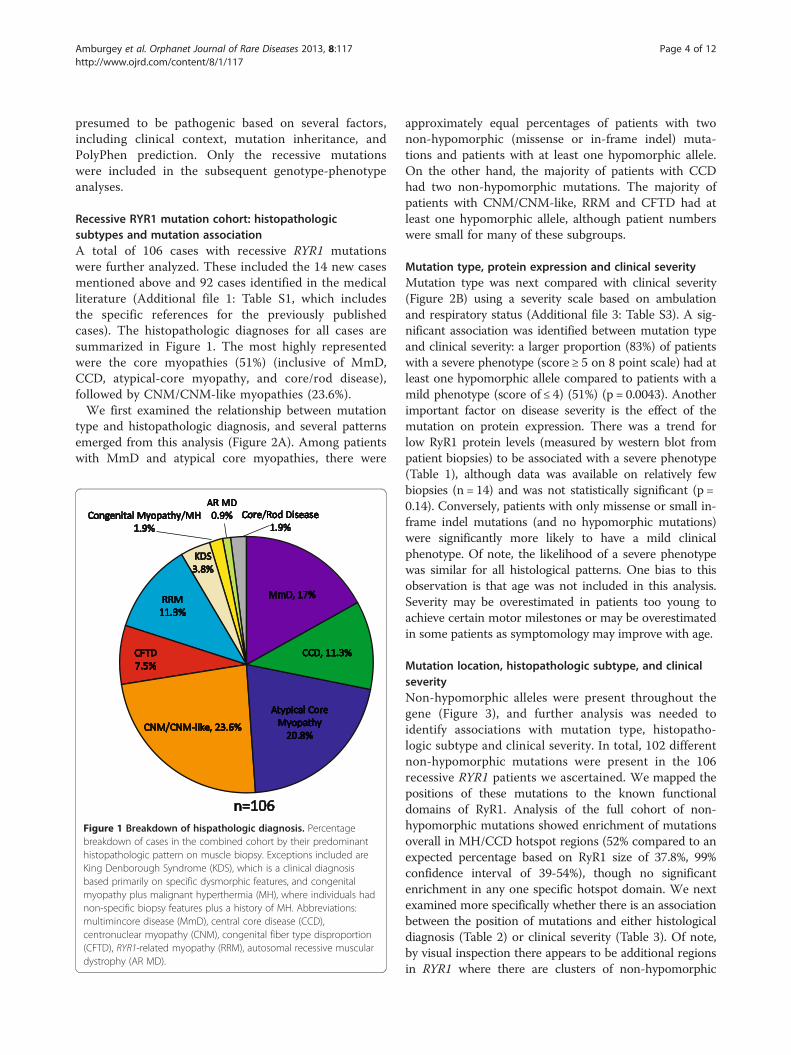
Recessive RYR1 mutation cohort: histopathologicsubtypes and mutation associationA total of 106 cases with recessive RYR1 mutationswere further analyzed. These included the 14 new casesmentioned above and 92 cases identified in the medicalliterature (Additional file 1: Table S1, which includesthe specific references for the previously publishedcases). The histopathologic diagnoses for all cases aresummarized in Figure 1. The most highly representedwere the core myopathies (51%) (inclusive of MmD,CCD, atypical-core myopathy, and core/rod disease),followed by CNM/CNM-like myopathies (23.6%).We first examined the relationship between mutation
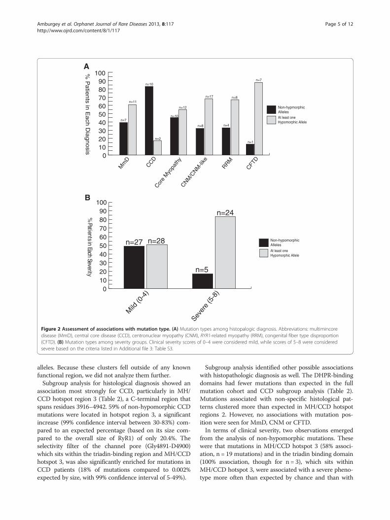
type and histopathologic diagnosis, and several patternsemerged from this analysis (Figure 2A). Among patientswith MmD and atypical core myopathies, there were
n=106
Congenital Myopathy/MH1.9%
AR MD0.9% Core/Rod Disease
1.9%
MmD, 17%
CNM/CNM-like, 23.6%
CCD, 11.3%
Atypical Core Myopathy
20.8%
RRM11.3%
CFTD7.5%
KDS3.8%
Figure 1 Breakdown of hispathologic diagnosis. Percentagebreakdown of cases in the combined cohort by their predominanthistopathologic pattern on muscle biopsy. Exceptions included areKing Denborough Syndrome (KDS), which is a clinical diagnosisbased primarily on specific dysmorphic features, and congenitalmyopathy plus malignant hyperthermia (MH), where individuals hadnon-specific biopsy features plus a history of MH. Abbreviations:multimincore disease (MmD), central core disease (CCD),centronuclear myopathy (CNM), congenital fiber type disproportion(CFTD), RYR1-related myopathy (RRM), autosomal recessive musculardystrophy (AR MD).
approximately equal percentages of patients with twonon-hypomorphic (missense or in-frame indel) muta-tions and patients with at least one hypomorphic allele.On the other hand, the majority of patients with CCDhad two non-hypomorphic mutations. The majority ofpatients with CNM/CNM-like, RRM and CFTD had atleast one hypomorphic allele, although patient numberswere small for many of these subgroups.
Mutation type, protein expression and clinical severityMutation type was next compared with clinical severity(Figure 2B) using a severity scale based on ambulationand respiratory status (Additional file 3: Table S3). A sig-nificant association was identified between mutation typeand clinical severity: a larger proportion (83%) of patientswith a severe phenotype (score ≥ 5 on 8 point scale) had atleast one hypomorphic allele compared to patients with amild phenotype (score of ≤ 4) (51%) (p = 0.0043). Anotherimportant factor on disease severity is the effect of themutation on protein expression. There was a trend forlow RyR1 protein levels (measured by western blot frompatient biopsies) to be associated with a severe phenotype(Table 1), although data was available on relatively fewbiopsies (n = 14) and was not statistically significant (p =0.14). Conversely, patients with only missense or small in-frame indel mutations (and no hypomorphic mutations)were significantly more likely to have a mild clinicalphenotype. Of note, the likelihood of a severe phenotypewas similar for all histological patterns. One bias to thisobservation is that age was not included in this analysis.Severity may be overestimated in patients too young toachieve certain motor milestones or may be overestimatedin some patients as symptomology may improve with age.
Mutation location, histopathologic subtype, and clinicalseverityNon-hypomorphic alleles were present throughout thegene (Figure 3), and further analysis was needed toidentify associations with mutation type, histopatho-logic subtype and clinical severity. In total, 102 differentnon-hypomorphic mutations were present in the 106recessive RYR1 patients we ascertained. We mapped thepositions of these mutations to the known functionaldomains of RyR1. Analysis of the full cohort of non-hypomorphic mutations showed enrichment of mutationsoverall in MH/CCD hotspot regions (52% compared to anexpected percentage based on RyR1 size of 37.8%, 99%confidence interval of 39-54%), though no significantenrichment in any one specific hotspot domain. We nextexamined more specifically whether there is an associationbetween the position of mutations and either histologicaldiagnosis (Table 2) or clinical severity (Table 3). Of note,by visual inspection there appears to be additional regionsin RYR1 where there are clusters of non-hypomorphic
ytir e
veS
hcaE
nist
nei t
aP%
n=28n=27
n=24
n=5
Mild
(0-4
)
Sever
e (5
-8)
B
n=7
n=11
n=10
% P
atie
nts in
Each
Dia
gnosis
At least one Hypomorphic Allele
Non-hypmorphic Alleles
CNM/C
NM-li
ke
RRM
CCDCor
e M
yopa
thy
A
n=2
n=10
n=8
n=8
n=1
0102030405060708090
100
Mm
D
CFTD
n=12
n=17
n=4
n=7
0102030405060708090
100
At least one Hypomorphic Allele
Non-hypomorphicAlleles
Figure 2 Assessment of associations with mutation type. (A) Mutation types among histopalogic diagnosis. Abbreviations: multimincoredisease (MmD), central core disease (CCD), centronuclear myopathy (CNM), RYR1-related myopathy (RRM), congenital fiber type disproportion(CFTD). (B) Mutation types among severity groups. Clinical severity scores of 0–4 were considered mild, while scores of 5–8 were consideredsevere based on the criteria listed in Additional file 3: Table S3.
Amburgey et al. Orphanet Journal of Rare Diseases 2013, 8:117 Page 5 of 12http://www.ojrd.com/content/8/1/117
alleles. Because these clusters fell outside of any knownfunctional region, we did not analyze them further.Subgroup analysis for histological diagnosis showed an
association most strongly for CCD, particularly in MH/CCD hotspot region 3 (Table 2), a C-terminal region thatspans residues 3916–4942. 59% of non-hypomorphic CCDmutations were located in hotspot region 3, a significantincrease (99% confidence interval between 30-83%) com-pared to an expected percentage (based on its size com-pared to the overall size of RyR1) of only 20.4%. Theselectivity filter of the channel pore (Gly4891-D4900)which sits within the triadin-binding region and MH/CCDhotspot 3, was also significantly enriched for mutations inCCD patients (18% of mutations compared to 0.002%expected by size, with 99% confidence interval of 5-49%).
Subgroup analysis identified other possible associationswith histopathologic diagnosis as well. The DHPR-bindingdomains had fewer mutations than expected in the fullmutation cohort and CCD subgroup analysis (Table 2).Mutations associated with non-specific histological pat-terns clustered more than expected in MH/CCD hotspotregions 2. However, no associations with mutation pos-ition were seen for MmD, CNM or CFTD.In terms of clinical severity, two observations emerged
from the analysis of non-hypomorphic mutations. Thesewere that mutations in MH/CCD hotspot 3 (58% associ-ation, n = 19 mutations) and in the triadin binding domain(100% association, though for n = 3), which sits withinMH/CCD hotspot 3, were associated with a severe pheno-type more often than expected by chance and than with
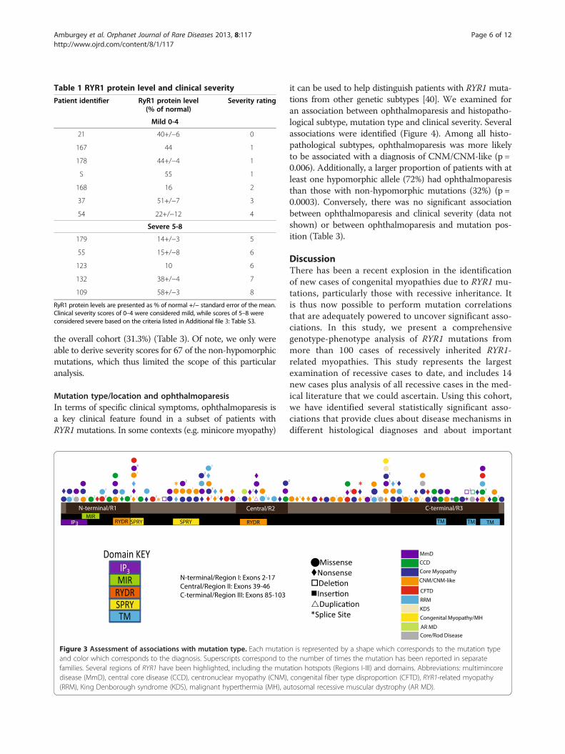
Table 1 RYR1 protein level and clinical severity
Patient identifier RyR1 protein level(% of normal)
Severity rating
Mild 0-4
21 40+/−6 0
167 44 1
178 44+/−4 1
S 55 1
168 16 2
37 51+/−7 3
54 22+/−12 4
Severe 5-8
179 14+/−3 5
55 15+/−8 6
123 10 6
132 38+/−4 7
109 58+/−3 8
RyR1 protein levels are presented as % of normal +/− standard error of the mean.Clinical severity scores of 0–4 were considered mild, while scores of 5–8 wereconsidered severe based on the criteria listed in Additional file 3: Table S3.
Amburgey et al. Orphanet Journal of Rare Diseases 2013, 8:117 Page 6 of 12http://www.ojrd.com/content/8/1/117
the overall cohort (31.3%) (Table 3). Of note, we only wereable to derive severity scores for 67 of the non-hypomorphicmutations, which thus limited the scope of this particularanalysis.
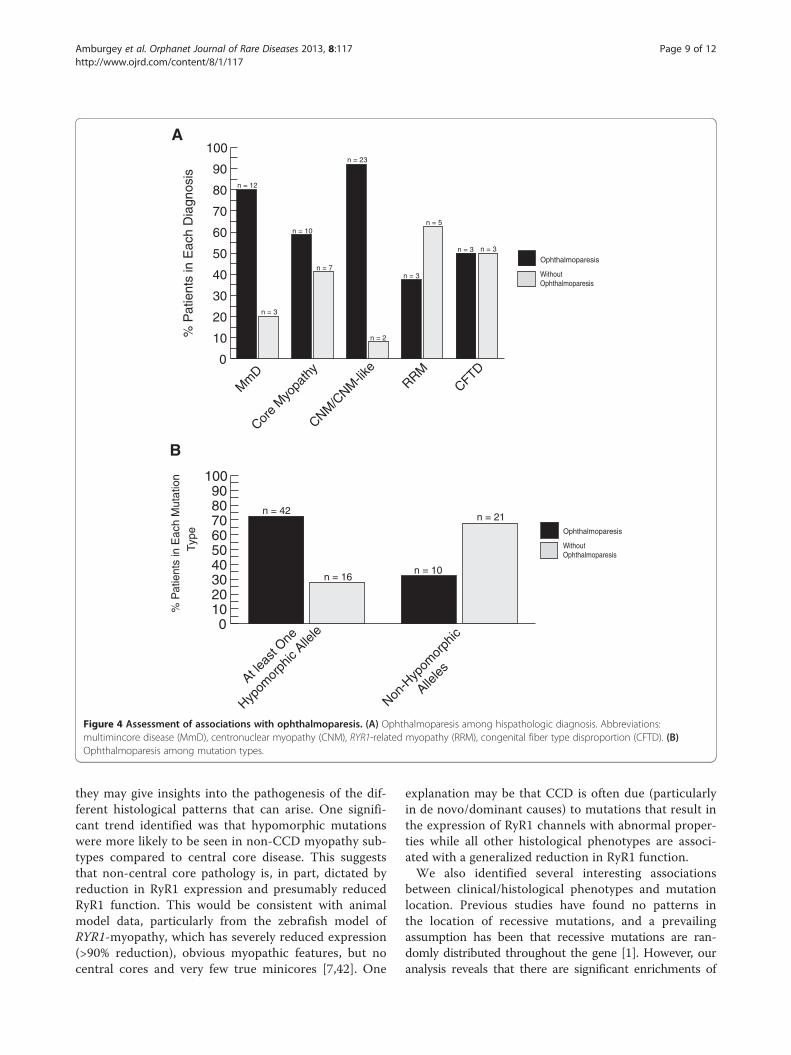
Mutation type/location and ophthalmoparesisIn terms of specific clinical symptoms, ophthalmoparesis isa key clinical feature found in a subset of patients withRYR1mutations. In some contexts (e.g. minicore myopathy)
Figure 3 Assessment of associations with mutation type. Each mutatioand color which corresponds to the diagnosis. Superscripts correspond tofamilies. Several regions of RYR1 have been highlighted, including the mutdisease (MmD), central core disease (CCD), centronuclear myopathy (CNM)(RRM), King Denborough syndrome (KDS), malignant hyperthermia (MH), au
it can be used to help distinguish patients with RYR1 muta-tions from other genetic subtypes [40]. We examined foran association between ophthalmoparesis and histopatho-logical subtype, mutation type and clinical severity. Severalassociations were identified (Figure 4). Among all histo-pathological subtypes, ophthalmoparesis was more likelyto be associated with a diagnosis of CNM/CNM-like (p =0.006). Additionally, a larger proportion of patients with atleast one hypomorphic allele (72%) had ophthalmoparesisthan those with non-hypomorphic mutations (32%) (p =0.0003). Conversely, there was no significant associationbetween ophthalmoparesis and clinical severity (data notshown) or between ophthalmoparesis and mutation pos-ition (Table 3).
DiscussionThere has been a recent explosion in the identificationof new cases of congenital myopathies due to RYR1 mu-tations, particularly those with recessive inheritance. Itis thus now possible to perform mutation correlationsthat are adequately powered to uncover significant asso-ciations. In this study, we present a comprehensivegenotype-phenotype analysis of RYR1 mutations frommore than 100 cases of recessively inherited RYR1-related myopathies. This study represents the largestexamination of recessive cases to date, and includes 14new cases plus analysis of all recessive cases in the med-ical literature that we could ascertain. Using this cohort,we have identified several statistically significant asso-ciations that provide clues about disease mechanisms indifferent histological diagnoses and about important
n is represented by a shape which corresponds to the mutation typethe number of times the mutation has been reported in separateation hotspots (Regions I-III) and domains. Abbreviations: multimincore, congenital fiber type disproportion (CFTD), RYR1-related myopathytosomal recessive muscular dystrophy (AR MD).
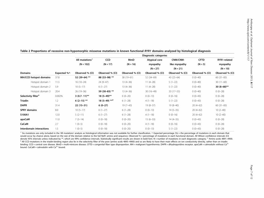
Table 2 Proportions of recessive non-hypomorphic missense mutations in known functional RYR1 domains analyzed by histological diagnosis
Diagnostic categories
All mutations+ CCD MmD Atypical core CNM/CNM- CFTD RYR1-related
(N = 102) (N = 17) (N = 16) myopathy like myopathy (N = 5) myopathy
(N = 27) (N = 21) (N = 10)
Domains Expected %^ Observed % (CI) Observed % (CI) Observed % (CI) Observed % (CI) Observed % (CI) Observed % (CI) Observed % (CI)
MH/CCD hotspot domains 37.8 52 (39–64) ** 88 (53–98) ** 38 (19–61) 52 (34–69) 43 (25–64) 0 (0–40) 60 (31–83)
Hotspot domain 1 11.5 16 (10–24) 24 (9–47) 13 (4–36) 11 (4–28) 5 (1–22) 0 (0–40) 30 (11–60)
Hotspot domain 2 5.9 10 (5–17) 6 (1–27) 13 (4–36) 11 (4–28) 5 (1–22) 0 (0–40) 30 (8–68)**
Hotspot domain 3 20.4 26 (19–36) 59 (30–83) ** 13 (4–36) 30 (16–49) 33 (17–55) 0 (0–40) 0 (0–28)
Selectivity filtero 0.002% 3 (0.7- 11)** 18 (5–49)** 0 (0–20) 0 (0–13) 0 (0–16) 0 (0–43) 0 (0–28)
Triadin 1.2 6 (2–15) ** 18 (5–49) **a 6 (1–28) 4 (1–18) 5 (1–22) 0 (0–43) 0 (0–28)
DHPR 31.4 22 (15–31) 6 (0–27) 19 (7–43) 19 (8–37) 19 (8–40) 20 (4–62) 60 (31–83)
SPRY domains 8.0 10 (5–17) 6 (1–27) 6 (1–28) 0 (0–13) 14 (5–35) 20 (4–62) 10 (2–40)
S100A1 12.0 5 (2–11) 6 (1–27) 6 (1–28) 4 (1–18) 0 (0–16) 20 (4–62) 10 (2–40)
apoCaM 11.0 7 (3–14) 0 (0–18) 0 (0–20) 15 (6–33) 14 (4–35) 0 (0–43) 0 (0–28)
CaCaM 2.7 1 (0–5) 0 (0–18) 0 (0–20) 4 (1–18) 0 (0–16) 0 (0–43) 0 (0–28)
Interdomain interactions 1.5 1 (0–5) 0 (0–18) 0 (0–20) 0 (0–13) 5 (1–22) 0 (0–43) 0 (0–28)+ Six mutations are only included in the ‘All mutations’ analysis as histological information was not available for further classification. ^ Expected percentage (%) = the percentage of mutations in each domain thatwould occur by chance alone, based on the size of the domain relative to the full RyR1 amino acid sequence. Observed % = percentage of mutations in each functional domain. All Wilson confidence intervals (CI)denote 95% intervals unless indicated by **, which are 99% confidence intervals. Statistically significant results are shown in bold font. N = number of mutations in each diagnostic category. o Amino acids 4891–4900.a All CCD mutations in the triadin-binding region also lie in the selectivity filter of the pore (amino acids 4895–4900) and so are likely to have their main effects on ion conductivity directly, rather than on triadinbinding. CCD = central core disease. MmD =multi-minicore disease. CFTD = congenital fiber type disproportion. MH =malignant hyperthermia. DHPR = dihydropyridine receptor. apoCaM = calmodulin without Ca2+
bound. CaCaM = calmodulin with Ca2+ bound.
Amburgey
etal.O
rphanetJournalof
RareDiseases
2013,8:117Page
7of
12http://w
ww.ojrd.com
/content/8/1/117
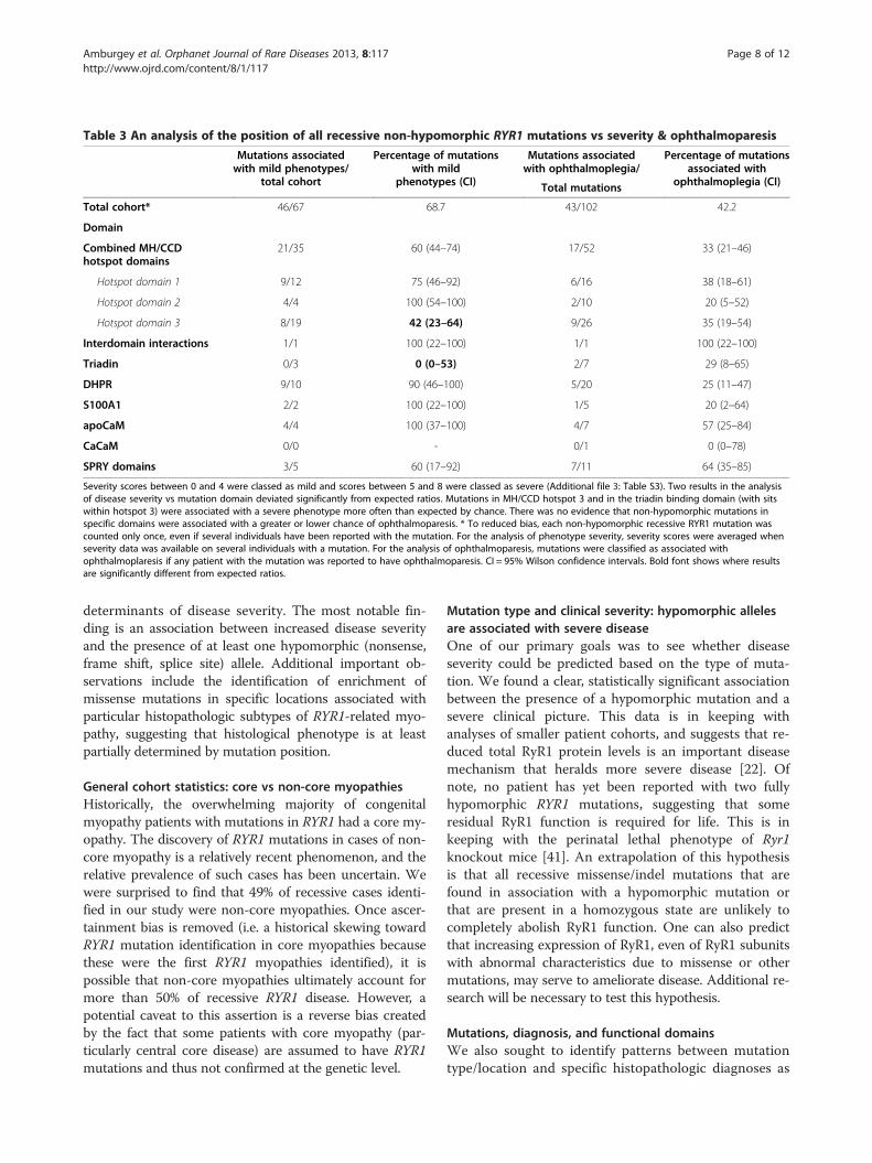
Table 3 An analysis of the position of all recessive non-hypomorphic RYR1 mutations vs severity & ophthalmoparesis
Mutations associatedwith mild phenotypes/
total cohort
Percentage of mutationswith mild
phenotypes (CI)
Mutations associatedwith ophthalmoplegia/
Percentage of mutationsassociated with
ophthalmoplegia (CI)Total mutations
Total cohort* 46/67 68.7 43/102 42.2
Domain
Combined MH/CCDhotspot domains
21/35 60 (44–74) 17/52 33 (21–46)
Hotspot domain 1 9/12 75 (46–92) 6/16 38 (18–61)
Hotspot domain 2 4/4 100 (54–100) 2/10 20 (5–52)
Hotspot domain 3 8/19 42 (23–64) 9/26 35 (19–54)
Interdomain interactions 1/1 100 (22–100) 1/1 100 (22–100)
Triadin 0/3 0 (0–53) 2/7 29 (8–65)
DHPR 9/10 90 (46–100) 5/20 25 (11–47)
S100A1 2/2 100 (22–100) 1/5 20 (2–64)
apoCaM 4/4 100 (37–100) 4/7 57 (25–84)
CaCaM 0/0 - 0/1 0 (0–78)
SPRY domains 3/5 60 (17–92) 7/11 64 (35–85)
Severity scores between 0 and 4 were classed as mild and scores between 5 and 8 were classed as severe (Additional file 3: Table S3). Two results in the analysisof disease severity vs mutation domain deviated significantly from expected ratios. Mutations in MH/CCD hotspot 3 and in the triadin binding domain (with sitswithin hotspot 3) were associated with a severe phenotype more often than expected by chance. There was no evidence that non-hypomorphic mutations inspecific domains were associated with a greater or lower chance of ophthalmoparesis. * To reduced bias, each non-hypomorphic recessive RYR1 mutation wascounted only once, even if several individuals have been reported with the mutation. For the analysis of phenotype severity, severity scores were averaged whenseverity data was available on several individuals with a mutation. For the analysis of ophthalmoparesis, mutations were classified as associated withophthalmoplaresis if any patient with the mutation was reported to have ophthalmoparesis. CI = 95% Wilson confidence intervals. Bold font shows where resultsare significantly different from expected ratios.
Amburgey et al. Orphanet Journal of Rare Diseases 2013, 8:117 Page 8 of 12http://www.ojrd.com/content/8/1/117
determinants of disease severity. The most notable fin-ding is an association between increased disease severityand the presence of at least one hypomorphic (nonsense,frame shift, splice site) allele. Additional important ob-servations include the identification of enrichment ofmissense mutations in specific locations associated withparticular histopathologic subtypes of RYR1-related myo-pathy, suggesting that histological phenotype is at leastpartially determined by mutation position.
General cohort statistics: core vs non-core myopathiesHistorically, the overwhelming majority of congenitalmyopathy patients with mutations in RYR1 had a core my-opathy. The discovery of RYR1 mutations in cases of non-core myopathy is a relatively recent phenomenon, and therelative prevalence of such cases has been uncertain. Wewere surprised to find that 49% of recessive cases identi-fied in our study were non-core myopathies. Once ascer-tainment bias is removed (i.e. a historical skewing towardRYR1 mutation identification in core myopathies becausethese were the first RYR1 myopathies identified), it ispossible that non-core myopathies ultimately account formore than 50% of recessive RYR1 disease. However, apotential caveat to this assertion is a reverse bias createdby the fact that some patients with core myopathy (par-ticularly central core disease) are assumed to have RYR1mutations and thus not confirmed at the genetic level.
Mutation type and clinical severity: hypomorphic allelesare associated with severe diseaseOne of our primary goals was to see whether diseaseseverity could be predicted based on the type of muta-tion. We found a clear, statistically significant associationbetween the presence of a hypomorphic mutation and asevere clinical picture. This data is in keeping withanalyses of smaller patient cohorts, and suggests that re-duced total RyR1 protein levels is an important diseasemechanism that heralds more severe disease [22]. Ofnote, no patient has yet been reported with two fullyhypomorphic RYR1 mutations, suggesting that someresidual RyR1 function is required for life. This is inkeeping with the perinatal lethal phenotype of Ryr1knockout mice [41]. An extrapolation of this hypothesisis that all recessive missense/indel mutations that arefound in association with a hypomorphic mutation orthat are present in a homozygous state are unlikely tocompletely abolish RyR1 function. One can also predictthat increasing expression of RyR1, even of RyR1 subunitswith abnormal characteristics due to missense or othermutations, may serve to ameliorate disease. Additional re-search will be necessary to test this hypothesis.
Mutations, diagnosis, and functional domainsWe also sought to identify patterns between mutationtype/location and specific histopathologic diagnoses as
0
10
20
30
40
50
60
70
80
90
100
Mm
D
Core
Myo
path
y
CNM/C
NM-lik
eRRM
CFTD
% P
atie
nts
in E
ach
Dia
gnos
is
Without Ophthalmoparesis
Ophthalmoparesis
0102030405060708090
100
% P
atie
nts
in E
ach
Mut
atio
n
Without Ophthalmoparesis
Ophthalmoparesis
At leas
t One
Hypom
orph
ic Alle
le
Non-H
ypom
orph
ic
Alleles
n = 42
n = 16n = 10
n = 21
n = 12
n = 3
n = 10
n = 7
n = 23
n = 2
n = 3
n = 5
n = 3 n = 3
A
B
Type
Figure 4 Assessment of associations with ophthalmoparesis. (A) Ophthalmoparesis among hispathologic diagnosis. Abbreviations:multimincore disease (MmD), centronuclear myopathy (CNM), RYR1-related myopathy (RRM), congenital fiber type disproportion (CFTD). (B)Ophthalmoparesis among mutation types.
Amburgey et al. Orphanet Journal of Rare Diseases 2013, 8:117 Page 9 of 12http://www.ojrd.com/content/8/1/117
they may give insights into the pathogenesis of the dif-ferent histological patterns that can arise. One signifi-cant trend identified was that hypomorphic mutationswere more likely to be seen in non-CCD myopathy sub-types compared to central core disease. This suggeststhat non-central core pathology is, in part, dictated byreduction in RyR1 expression and presumably reducedRyR1 function. This would be consistent with animalmodel data, particularly from the zebrafish model ofRYR1-myopathy, which has severely reduced expression(>90% reduction), obvious myopathic features, but nocentral cores and very few true minicores [7,42]. One
explanation may be that CCD is often due (particularlyin de novo/dominant causes) to mutations that result inthe expression of RyR1 channels with abnormal proper-ties while all other histological phenotypes are associ-ated with a generalized reduction in RyR1 function.We also identified several interesting associations
between clinical/histological phenotypes and mutationlocation. Previous studies have found no patterns inthe location of recessive mutations, and a prevailingassumption has been that recessive mutations are ran-domly distributed throughout the gene [1]. However, ouranalysis reveals that there are significant enrichments of
Amburgey et al. Orphanet Journal of Rare Diseases 2013, 8:117 Page 10 of 12http://www.ojrd.com/content/8/1/117
mutations in known functional domains of the proteinand in association with particular diagnoses. In particular,we detected an over-representation of recessive non-hypomorphic RYR1 mutations in the combined MH/CCDhotspots and in the subgroup analysis there was a strongassociation between CCD mutations and hotspot region 3that exceeded 99% confidence intervals. This is the samepattern that is seen for dominant CCD mutations, whichare more often in hotspot region 3 than regions 1 or 2 [8]and suggests that dominant and recessive forms of CCDmay share similar disease mechanisms.One association that provides a clue about disease
pathogenesis is the enrichment of recessive missensemutations in the selectivity filter of the channel pore, a 10amino acid motif that conserved in all RyRs and that iscritical for channel function [43]. This region only occu-pies 0.002% of the linear protein sequence, but accountsfor 3% of recessive non-hypomorphic mutations overalland almost 20% of recessive CCD mutations. This impli-cates abnormalities in ion conductance or ion selectivityin the pathogenesis of recessive CCD, a mechanism thathas been linked to dominant CCD mutations (reviewed in[9,27]). The enrichment of recessive missense mutationsin the triadin-binding domain is due to the fact that thisdomain contains the selectivity filter. The finding thatfewer mutations than expected were located in DHPRbinding regions may indicate that interactions with theseRyR1-binding proteins tend not to be important in thepathogenesis of the known RYR1 myopathies. Some asso-ciations are expected by chance when making multiplecomparisons and it would be ideal to replicate these find-ings in a second cohort of mutations as confirmation.
ConclusionsIn all, we present a comprehensive genotype-phenotypestudy of recessive RYR1 mutations. The insights we pro-vide reveal the utility of such a study, and show the firstassociations between mutation type and location andclinical severity and diagnosis. In particular, we highlightthe MH/CCD hotpspot regions, (and the selectivity filterthat lies within hotspot region 3), as likely to be importantin the pathogenesis of many recessive RYR1-related myop-athies. The study of additional patients in the future willconfirm and refine these associations.
Additional files
Additional file 1: Table S1. Literature review of recessive RYR1mutations. References for all cases in the literature are included at theend of the spreadsheet and in the manuscript. No diagnosis refers tothose cases where histologic information as not available to furtherdefine the diagnosis.
Additional file 2: Table S2. Grouping patients into broad diagnosticcategories for analysis.
Additional file 3: Table S3. Criteria for severity scores. Ambulatory andrespiratory ratings were added together to calculate the overall severity score.Abbreviations: pulmonary function testing (PFT), ventilator (Vent), continuouspositive airway pressure (CPAP), bilevel positive airway pressure (BiPAP).
Additional file 4: Table S4. Functional domains in the ryanodinereceptor type1. *Amino acids are numbered relative to the full RyR1amino acid sequence (NM_000540). DHPR = dihydropyridine receptor,apoCaM = calmodulin without bound Ca2+, CaCaM = calmodulin withbound Ca2+. Information summarized from Hwang et al., 2012.
Additional file 5: Table S5. Clinical characteristics of newly reportedrecessive RYR1 mutations. Severity scores based on criteria listed inAdditional file 3: Table S3. Also included are the PolyPhen2 predictions forall novel missense mutations (Probably Damaging (PRD), Possibly Damaging(POD)) and the severity (scale 0–1, 0 indicating least severe, 1 indicatingmost severe). Origin of the mutation is designated M for maternal and P forpaternal. For patients F&G, carrier testing was only performed on the father.Since one mutation was identified, the other mutation is presumed to bematernal, which is denoted by M*. Patients O and P (siblings) werepresumed recessive based on clinical presentation, lack of parentalsymptoms, and the presence of two mutations. Parental testing was notperformed, so it is formally possible (though unlikely) that the twomutations exist in cis. For patient Y, one mutation was identified asmaternal in origin. Carrier testing of the father did not identify the secondmutation, therefore, it is presumed to be de novo. Abbreviations: Patient ID(ID), siblings (F&G, O&P) (*), diagnosis (DX), multimincore disease (MmD),core myopathy (CM), RYR1-related myopathy (RRM), autosomal recessivemuscular dystrophy (AR MD), central core disease (CCD), first year of life(FYOL), Weakness: proximal (P), distal (D), facial (F), neck (N); rigid spine (RS),ophthalmoparesis (OPH), respiratory distress (RD), ventilator (Vent), feedingdifficulties (FD), malignant hyperthermia (MH), creatine kinase (CK).Previously reported mutations: aBevilacqua, et al., 2011, bZhou, et al., 2010.
Additional file 6: Table S6. Hispathologic findings of newly reportedrecessive RYR1 mutations. Diagnosis of cases without a muscle biopsywas based on family history of the disease. Mutations previously reportedin the medical literature (aBevilacqua, et al., 2011, bZhou, et al., 2010).Origin of the mutation is designated M for maternal and P for paternal.For patients F&G, carrier testing was only performed on the father. Sinceone mutation was identified, the other mutation is presumed to bematernal, which is denoted by M*. For patient Y, one mutation wasidentified as maternal in origin. Carrier testing of the father did notidentify the second mutation, therefore, it is de novo which is denotedby D. Abbreviations: Patient ID (ID), siblings (F&G, O&P) (*), diagnosis (DX),multimincore disease (MmD), core myopathy (CM), RYR1-relatedmyopathy (RRM), autosomal recessive muscular dystrophy (AR MD),central core disease (CCD), central cores (CC), minicores (MC), internalizednuclei (IN), central nuclei (CN).
Additional file 7: Table S7. Clinical characteristics of newly reporteddominant RYR1 mutations. Severity scores based on criteria listed inAdditional file 3: Table S3. Polyphen2 scores are also included for all novelmissense mutations (Probably Damaging (PRD)) and the severity (scale 0–1,0 indicating least severe, 1 indicating most severe). ^ indicates that thesilent mutation is predicted to create a new splice donor site resulting in a25 amino acid in-frame deletion. Previously reported mutations: aLynch,et al., 1999, bDavis, et al., 2003, cManning, et al., 1998, dChamley, et al., 2000,eMonnier, et al., 2001, fDavis, et al., 2002). Origin of the mutation isdesignated M for maternal, P for paternal, or D for de novo. Abbreviations:Patient ID (ID), siblings (B&C), diagnosis (DX), central core disease (CCD),RYR1-related myopathy (RRM), multimincore disease (MmD), first year of life(FYOL), Weakness: proximal (P), distal (D), facial (F), neck (N); rigid spine (RS),ophthalmoparesis (OPH), respiratory distress (RD), ventilator (Vent), feedingdifficulties (FD), malignant hyperthermia (MH), creatine kinase (CK).
Additional file 8: Table S8. Hispathologic findings of newly reporteddominant RYR1 mutations. Previously reported mutations: aLynch, et al.,1999, bDavis, et al., 2003, cManning, et al., 1998, dChamley, et al., 2000,eMonnier, et al., 2001, fDavis, et al., 2002) [8,36-39,44]. Origin of the mutationis designated M for maternal, P for paternal, or D for de novo. Abbreviations:Patient ID (ID), siblings (B&C), diagnosis (DX), central core disease (CCD),RYR1-related myopathy (RRM), multimincore disease (MmD), central cores(CC), minicores (MC), internalized nuclei (IN), central nuclei (CN).
Amburgey et al. Orphanet Journal of Rare Diseases 2013, 8:117 Page 11 of 12http://www.ojrd.com/content/8/1/117
Competing interestsThe authors declare that they have no competing interests. MT was paidconsulting fees by Biomarin in relation to an unrelated clinical trial, and waspaid by Genzyme/Sanofi for lecture and travel costs associated with theSteps Forward Meeting (2009, 2011, 2012).
Authors’ contributionsKA participated in the design and coordination of the study, assisted in datacollection and interpretation, and drafted the manuscript. AB wasresponsible for data collection and interpretation. JH assisted with datamanagement and helped to draft the manuscript. MT, CB, LM, DM, JC, JRD,and GW provided new cases of RYR1 myopathy and also provided criticalreading of the manuscript. BC performed the statistical analysis. JJD and NCconceived the study, participated in data analysis, and drafted and editedthe manuscript. All authors read and approved the final manuscript.
AcknowledgementsThis work has been supported by a grant from the NIH (K08AR054835 to JJD)and the National Health & Medical Research Council (NC; grants 1035828 &1022707). JH is also supported by the Albert Einstein College of MedicineSenior Research Fellowship. Additional support comes from the TaubmanResearch Institute Emerging Scholar fund. We thank Liz Barnes from theNHMRC Clinical Trials Centre at University of Sydney for the statistical analysis.
Author details1Department of Pediatrics, Taubman Medical Research Institute, University ofMichigan Medical Center, 5019 A. Alfred Taubman Biomedical ScienceResearch Building, 109 Zina Pitcher Place, Ann Arbor, MI 48109-2200, USA.2Institute for Neuroscience and Muscle Research, Children’s Hospital atWestmead, University of Sydney, Level 3, Research Building Locked Bag 4001,Westmead, Sydney, NSW 2145, Australia. 3Department of Neuromuscular andNeurometabolic Disease, McMaster University Medical Center, Hamilton,Ontario, Canada. 4Neuromuscular and Neurogenetic Disorders of ChildhoodSection, National Institutes of Health, Bethesda, MD, USA. 5Division ofNeurology, Children’s Hospital of Philadelphia, Philadelphia, PA, USA.6Departments of Pediatrics and Neurology, University of Iowa, Carver Collegeof Medicine, Iowa City, IA, USA. 7Department of Pediatric Neurology,Cincinnati Children’s Hospital Medical Center, Cincinnati, OH, USA.8Department of Neurology, Mayo Foundation for Medical Education andResearch, Rochester, MN, USA. 9The Delta Pathology Group, Shreveport, LA,USA. 10Department of Neurology, University of Michigan Medical Center, AnnArbor, MI, USA. 11Discipline of Paediatrics and Child Health, University ofSydney, Sydney, Australia.
Received: 6 March 2013 Accepted: 1 August 2013Published: 6 August 2013
References1. Zhou H, Jungbluth H, Sewry CA, Feng L, Bertini E, Bushby K, Straub V,
Roper H, Rose MR, Brockington M, et al: Molecular mechanisms andphenotypic variation in RYR1-related congenital myopathies.Brain 2007, 130:2024–2036.
2. Robinson R, Carpenter D, Shaw MA, Halsall J, Hopkins P: Mutations in RYR1in malignant hyperthermia and central core disease. Hum Mutat 2006,27:977–989.
3. Amburgey K, McNamara N, Bennett LR, McCormick ME, Acsadi G, DowlingJJ: Prevalence of congenital myopathies in a representative pediatricunited states population. Ann Neurol 2011, 70:662–665.
4. Jungbluth H: Multi-minicore Disease. Orphanet J Rare Dis 2007, 2:31.5. Wilmshurst JM, Lillis S, Zhou H, Pillay K, Henderson H, Kress W, Muller
CR, Ndondo A, Cloke V, Cullup T, et al: RYR1 mutations are a commoncause of congenital myopathies with central nuclei. Ann Neurol 2010,68:717–726.
6. Clarke NF, Waddell LB, Cooper ST, Perry M, Smith RL, Kornberg AJ, MuntoniF, Lillis S, Straub V, Bushby K, et al: Recessive mutations in RYR1 are acommon cause of congenital fiber type disproportion. Hum Mutat 2010,31:E1544–E1550.
7. Dowling JJ, Arbogast S, Hur J, Nelson DD, McEvoy A, Waugh T, Marty I,Lunardi J, Brooks SV, Kuwada JY, Ferreiro A: Oxidative stress and successfulantioxidant treatment in models of RYR1-related myopathy. Brain 2012,135(Pt 4):1115–1127.
8. Davis MR, Haan E, Jungbluth H, Sewry C, North K, Muntoni F, Kuntzer T,Lamont P, Bankier A, Tomlinson P, et al: Principal mutation hotspotfor central core disease and related myopathies in the C-terminaltransmembrane region of the RYR1 gene. Neuromuscul Disord 2003,13:151–157.
9. Treves S, Jungbluth H, Muntoni F, Zorzato F: Congenital muscle disorderswith cores: the ryanodine receptor calcium channel paradigm. Curr OpinPharmacol 2008, 8:319–326.
10. Monnier N, Marty I, Faure J, Castiglioni C, Desnuelle C, Sacconi S, EstournetB, Ferreiro A, Romero N, Laquerriere A, et al: Null mutations causingdepletion of the type 1 ryanodine receptor (RYR1) are commonlyassociated with recessive structural congenital myopathies with cores.Hum Mutat 2008, 29:670–678.
11. Anderson GL, Manson J, Wallace R, Lund B, Hall D, Davis S, Shumaker S,Wang CY, Stein E, Prentice RL: Implementation of the Women's HealthInitiative study design. Ann Epidemiol 2003, 13:S5–S17.
12. Bevilacqua JA, Monnier N, Bitoun M, Eymard B, Ferreiro A, Monges S,Lubieniecki F, Taratuto AL, Laquerriere A, Claeys KG, et al: Recessive RYR1mutations cause unusual congenital myopathy with prominent nuclearinternalization and large areas of myofibrillar disorganization.Neuropathol Appl Neurobiol 2011, 37:271–284.
13. Carpenter D, Ismail A, Robinson RL, Ringrose C, Booms P, Iles DE, Halsall PJ,Steele D, Shaw MA, Hopkins PM: A RYR1 mutation associated withrecessive congenital myopathy and dominant malignant hyperthermiain Asian families. Muscle Nerve 2009, 40:633–639.
14. Duarte ST, Oliveira J, Santos R, Pereira P, Barroso C, Conceicao I,Evangelista T: Dominant and recessive RYR1 mutations in adults withcore lesions and mild muscle symptoms. Muscle Nerve 2011,44:102–108.
15. Ducreux S, Zorzato F, Ferreiro A, Jungbluth H, Muntoni F, Monnier N,Muller CR, Treves S: Functional properties of ryanodine receptorscarrying three amino acid substitutions identified in patientsaffected by multi-minicore disease and central core disease,expressed in immortalized lymphocytes. Biochem J 2006,395:259–266.
16. Jungbluth H, Davis MR, Muller C, Counsell S, Allsop J, Chattopadhyay A,Messina S, Mercuri E, Laing NG, Sewry CA, et al: Magnetic resonanceimaging of muscle in congenital myopathies associated with RYR1mutations. Neuromuscul Disord 2004, 14:785–790.
17. Jungbluth H, Muller CR, Halliger-Keller B, Brockington M, Brown SC, Feng L,Chattopadhyay A, Mercuri E, Manzur AY, Ferreiro A, et al: Autosomalrecessive inheritance of RYR1 mutations in a congenital myopathy withcores. Neurol 2002, 59:284–287.
18. Jungbluth H, Zhou H, Hartley L, Halliger-Keller B, Messina S, Longman C,Brockington M, Robb SA, Straub V, Voit T, et al: Minicore myopathy withophthalmoplegia caused by mutations in the ryanodine receptor type 1gene. Neurol 2005, 65:1930–1935.
19. Klein A, Lillis S, Munteanu I, Scoto M, Zhou H, Quinlivan R, Straub V, ManzurAY, Roper H, Jeannet PY, et al: Clinical and genetic findings in a largecohort of patients with ryanodine receptor 1 gene-associatedmyopathies. Hum Mutat 2012, 33:981–988.
20. Klein A, Jungbluth H, Clement E, Lillis S, Abbs S, Munot P, Pane M, Wraige E,Schara U, Straub V, et al: Muscle magnetic resonance imaging incongenital myopathies due to ryanodine receptor type 1 genemutations. Arch Neurol 2011, 68:1171–1179.
21. Kossugue PM, Paim JF, Navarro MM, Silva HC, Pavanello RC, Gurgel-Giannetti J, Zatz M, Vainzof M: Central core disease due to recessivemutations in RYR1 gene: is it more common than described?Muscle Nerve 2007, 35:670–674.
22. Monnier N, Ferreiro A, Marty I, Labarre-Vila A, Mezin P, Lunardi J: Ahomozygous splicing mutation causing a depletion of skeletalmuscle RYR1 is associated with multi-minicore disease congenitalmyopathy with ophthalmoplegia. Hum Mol Genet 2003, 12:1171–1178.
23. Monnier N, Laquerriere A, Marret S, Goldenberg A, Marty I, Nivoche Y,Lunardi J: First genomic rearrangement of the RYR1 gene associatedwith an atypical presentation of lethal neonatal hypotonia. NeuromusculDisord 2009, 19:680–684.
24. Romero NB, Monnier N, Viollet L, Cortey A, Chevallay M, Leroy JP,Lunardi J, Fardeau M: Dominant and recessive central core diseaseassociated with RYR1 mutations and fetal akinesia. Brain 2003,126:2341–2349.
Amburgey et al. Orphanet Journal of Rare Diseases 2013, 8:117 Page 12 of 12http://www.ojrd.com/content/8/1/117
25. Sambuughin N, Holley H, Muldoon S, Brandom BW, de Bantel AM,Tobin JR, Nelson TE, Goldfarb LG: Screening of the entire ryanodinereceptor type 1 coding region for sequence variants associated withmalignant hyperthermia susceptibility in the north americanpopulation. Anesthesiol 2005, 102:515–521.
26. Schreuder LT, der Sanden MW N-v, de Hair A, Peters G, Wortmann S, BokLA, Morava E: Successful use of albuterol in a patient with central coredisease and mitochondrial dysfunction. J Inherit Metab Dis 2010.doi:10.1007/s10545-010-9085-7.
27. Treves S, Anderson AA, Ducreux S, Divet A, Bleunven C, Grasso C,Paesante S, Zorzato F: Ryanodine receptor 1 mutations, dysregulationof calcium homeostasis and neuromuscular disorders. NeuromusculDisord 2005, 15:577–587.
28. Wortmann SB, Rodenburg RJ, Jonckheere A, de Vries MC, Huizing M, HeldtK, van den Heuvel LP, Wendel U, Kluijtmans LA, Engelke UF, et al:Biochemical and genetic analysis of 3-methylglutaconic aciduria type IV:a diagnostic strategy. Brain 2009, 132:136–146.
29. Wu S, Ibarra MC, Malicdan MC, Murayama K, Ichihara Y, Kikuchi H,Nonaka I, Noguchi S, Hayashi YK, Nishino I: Central core disease is dueto RYR1 mutations in more than 90% of patients. Brain 2006,129:1470–1480.
30. Zhou H, Brockington M, Jungbluth H, Monk D, Stanier P, Sewry CA,Moore GE, Muntoni F: Epigenetic allele silencing unveils recessiveRYR1 mutations in core myopathies. Am J Hum Genet 2006,79:859–868.
31. Zhou H, Lillis S, Loy RE, Ghassemi F, Rose MR, Norwood F, Mills K, Al-Sarraj S, Lane RJ, Feng L, et al: Multi-minicore disease and atypicalperiodic paralysis associated with novel mutations in the skeletalmuscle ryanodine receptor (RYR1) gene. Neuromuscul Disord 2010,20:166–173.
32. Zhou H, Yamaguchi N, Xu L, Wang Y, Sewry C, Jungbluth H, Zorzato F,Bertini E, Muntoni F, Meissner G, Treves S: Characterization ofrecessive RYR1 mutations in core myopathies. Hum Mol Genet 2006,15:2791–2803.
33. Zorzato F, Jungbluth H, Zhou H, Muntoni F, Treves S: Functional effects ofmutations identified in patients with multiminicore disease. IUBMB Life2007, 59:14–20.
34. Bhat MB, Hayek SM, Zhao J, Zang W, Takeshima H, Wier WG, Ma J:Expression and functional characterization of the cardiac muscleryanodine receptor Ca(2+) release channel in Chinese hamster ovarycells. Biophys J 1999, 77:808–816.
35. Lynch PJ, Tong J, Lehane M, Mallet A, Giblin L, Heffron JJ, Vaughan P, ZafraG, MacLennan DH, McCarthy TV: A mutation in the transmembrane/luminal domain of the ryanodine receptor is associated with abnormalCa2+ release channel function and severe central core disease. Proc NatlAcad Sci U S A 1999, 96:4164–4169.
36. Manning BM, Quane KA, Ording H, Urwyler A, Tegazzin V, Lehane M,O'Halloran J, Hartung E, Giblin LM, Lynch PJ, et al: Identification ofnovel mutations in the ryanodine-receptor gene (RYR1) in malignanthyperthermia: genotype-phenotype correlation. Am J Hum Genet1998, 62:599–609.
37. Chamley D, Pollock NA, Stowell KM, Brown RL: Malignant hyperthermia ininfancy and identification of novel RYR1 mutation. Br J Anaesth 2000,84:500–504.
38. Monnier N, Romero NB, Lerale J, Landrieu P, Nivoche Y, Fardeau M, LunardiJ: Familial and sporadic forms of central core disease are associated withmutations in the C-terminal domain of the skeletal muscle ryanodinereceptor. Hum Mol Genet 2001, 10:2581–2592.
39. Davis M, Brown R, Dickson A, Horton H, James D, Laing N, Marston R,Norgate M, Perlman D, Pollock N, Stowell K: Malignant hyperthermiaassociated with exercise-induced rhabdomyolysis or congenitalabnormalities and a novel RYR1 mutation in New Zealand andAustralian pedigrees. Br J Anaesth 2002, 88:508–515.
40. Davis KG, Wertin TM, Schriver JP: The use of simvastatin for theprevention of gallstones in the lithogenic prairie dog model. Obes Surg2003, 13:865–868.
41. Marek KW, Davis GW: Controlling the active properties of excitable cells.Curr Opin Neurobiol 2003, 13:607–611.
42. Hirata H, Watanabe T, Hatakeyama J, Sprague SM, Saint-Amant L,Nagashima A, Cui WW, Zhou W, Kuwada JY: Zebrafish relatively relaxedmutants have a ryanodine receptor defect, show slow swimming andprovide a model of multi-minicore disease. Dev 2007, 134:2771–2781.
43. Zhao M, Li P, Li X, Zhang L, Winkfein RJ, Chen SR: Molecular identificationof the ryanodine receptor pore-forming segment. J Biol Chem 1999,274:25971–25974.
44. Nelson TJ, Zhao WQ, Yuan S, Favit A, Pozzo-Miller L, Alkon DL: Calexcitininteraction with neuronal ryanodine receptors. Biochem J 1999, 341(Pt 2):423–433.
doi:10.1186/1750-1172-8-117Cite this article as: Amburgey et al.: Genotype-phenotype correlations inrecessive RYR1-related myopathies. Orphanet Journal of Rare Diseases2013 8:117.
Submit your next manuscript to BioMed Centraland take full advantage of:
• Convenient online submission
• Thorough peer review
• No space constraints or color figure charges
• Immediate publication on acceptance
• Inclusion in PubMed, CAS, Scopus and Google Scholar
• Research which is freely available for redistribution
Submit your manuscript at www.biomedcentral.com/submit


![[PPT]PowerPoint Presentation · Web viewLearning Outcomes Explain the terms allele, locus, phenotype, genotype, dominant, codominant and recessive; Explain the terms linkage and crossing-over;](https://img.dokumen.tips/doc/110x75/5a9ef8f17f8b9a76178c2286/pptpowerpoint-presentation-viewlearning-outcomes-explain-the-terms-allele-locus.jpg)