Embed Size (px)
Citation preview

JOURNAL OF CLINICAL MICROBIOLOGY, Nov. 1993, p. 2873-2877 Vol. 31, No. 110095-1137/93/112873-05$02.00/0Copyright © 1993, American Society for Microbiology
Genomic Fingerprinting of Borrelia burgdorfeni Sensu Lato byPulsed-Field Gel Electrophoresis
J. BELFAIZA, D. POSTIC, E. BELLENGER, G. BARANTON, AND I. SAINT GIRONS*Unites de Bact6riologie Moleculaire et Me6dicale Institut Pasteur, 25 Rue du docteur Roux,
75724 Paris Cedex 15, France
Received 14 May 1993/Returned for modification 15 July 1993/Accepted 17 August 1993
A total of 46 Borrelia burgdorferi sensu lato isolates that were isolated from patients with Lyme borreliosisand infected animals or were extracted from ticks of the genus lxodes were analyzed. Large restrictionfragment patterns obtained after cleavage of genomic DNAs with Mlul were analyzed by pulsed-field gelelectrophoresis (PFGE). To eliminate the contribution ofplasmid DNA, only frmMents greater than 70 kb wereused for the analysis. The results indicated that each of the 14 B. burgdorferi sensu stricto isolates wererecognized by a band at 135 kbp, each of the 12 Borrelia garinii isolates by two bands (220 and 80 kbp), andeach of the 20 Borrelia afzelii isolates by three bands (460, 320, and 90 kbp). Whereas differences in the PFGEpatterns among B. burgdorferi sensu stricto isolates and B. garinii isolates were noted, B. afzelii isolates wereall similar. Identification of isolates by PFGE correlates with their belonging to a given species within B.burgdorferi sensu lato.
Borrelia burgdorferi was identified in 1982 as the agent ofLyme borreliosis (11) and was recognized as a new speciesof the genus Borrelia in 1984 (16). Three species wererecently delineated for B. burgdorferi sensu lato: B. burg-dorferi sensu stricto, Borrelia gainii, and Borrelia afzelii (4,20). This division into three species was based on DNA-DNA hybridizations, rRNA gene restriction patterns, andidentification by monoclonal antibodies, all tools recognizedcurrently by taxonomists (29). The results obtained bydifferent methods of typing isolates such as polymerasechain reaction (21, 23), arbitrarily primed polymerase chainreaction (30), multilocus enzyme electrophoresis (10), 16SrRNA sequencing (1, 2, 18, 19, 25), fla gene sequencing (21,28), and serotyping (31) are consistent with this new taxon-omy. Heterogeneity among isolates has also been observedby analysis of the profiles of linear and circular plasmids (6,27). Digestion of genomic DNAs with restriction endonu-cleases followed by conventional electrophoresis alsoshowed differences among isolates. However, the thousandsof bands obtained are difficult to interpret except whenSouthern analysis is used following hybridization with di-verse probes (17, 27, 28). The latter technique yieldedgroupings of isolates along the same lines as describedelsewhere (4, 20).
Pulsed-field gel electrophoresis (PFGE) was used forconstructing the physical map of B. burgdorferi (12). Thisgenome is unique, since it contains a 945-kbp linear chromo-some as well as linear and circular plasmids which vary insize and number among the isolates (7, 9, 13). In the presentarticle, we analyze several different isolates by PFGE. Theresults indicate that a given isolate can be readily identifiedas belonging to a specific species.
* Corresponding author. Electronic mail address: [email protected].
MATERIALS AND METHODSBacterial isolates and media. The B. burgdorferi sensu lato
isolates used in this study are listed in Table 1. The isolateswere grown in BSKII medium at 30°C (5).PFGE, digestion of DNA in agarose, and large restriction
faments patterns LRFP nomenclature. Previously describedprocedures were used for the preparation of high-molecular-weight genomic DNAs and PFGE (9, 12). Separation wasachieved with a pulse time ramped from 3 to 40 s for 20 h witha contour-clamped homogeneous electric field-DRII appara-tus (Bio-Rad laboratories, Richmond, Calif.).
Restriction endonucleases MluI (A/CGCGT) and SmaI(CCC/GGG) were purchased from Pharmacia. GenomicDNAs, in low-melting-temperature agarose, were digestedwith 20 to 40 U of restriction endonuclease for 20 h in 200 PI1of the buffer recommended by the supplier. The lambdaconcatemers used as size markers for Fig. 2 were those usedin reference 12 (monomer size, 44.3 kbp) and for Fig. 1 and3 were those commercialized by Tebu (monomer size, 48.5kbp).An LRFP is defined as a unique PFGE pattern. We
ignored bands lower than 70 kb, since these bands would bethe results of uncut or cut plasmids, which could complicatethe interpretation (24). The LRFPs obtained were designatedML or SM to identify the restriction endonuclease MluI orSmaI, respectively, with the suffix b, g, or a denoting B.burgdorferi sensu stricto, B. garinii, or B. afzelii, respec-tively. Different LRFPs of the same species are numbered 1or 2, etc.DNA-blot analysis. For blot analysis according to Southern
(26), DNA fragments separated by PFGE were submitted todepurination and then transferred to Hybond-N nylon mem-branes (Amersham International, Amersham, England). Toprepare the probe, the 135-kbp MluI band from isolate B31was excised from a low-melting-temperature agarose pulsed-field gel and washed with 1 ml of distilled water for 1 h atroom temperature. A reduced volume of 250 ,ul of the samplewas heated at 100°C for 10 min. The tube was placed on ice,and 25 pl was labelled by random priming with digoxigenin-11-dUTP. The probe thus generated was used in hybridiza-tion experiments as described elsewhere (8).
2873
on May 18, 2018 by guest
http://jcm.asm
.org/D
ownloaded from
2874 BELFAIZA ET AL.
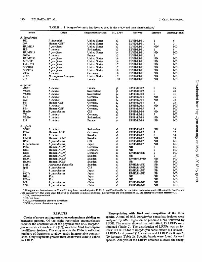
TABLE 1. B. burgdorferi sensu lato isolates used in this study and their characteristicsa
Isolate Origin Geographical location ML LRFP Ribotype Serotype Electrotype (ET)
B. burgdorferiB31297HUM115IRSHUM781420006HUM3336MEN115Lake 339SON188SON335Z13621305212
B. garnii20047VS185VS468N34G25PBiTNPBrIPer9OT25VS286153
B. afzeliiVS461PGauFlUMO1IPer3I. persulcatusPStoDK3DK8ACAlECM1ECM8SmSlIp2lJiP427aBFoxHFoxM72246
L damminiHuman CSFbL pacificusL ricinusL pacificusL. ncinusI. pacificusI. pacificusI. pacificusL pacificusI. pacificusL ncinusPeromyscus leucopus. ricinus
I. icinusL ncinusL ricinus. ricinusL ricinusHuman CSFL. icinusHuman CSFL persulcatusL. ncinusI. icinusL ricinus
I. icinusHuman ACAd. ricinusHuman ECMeI. persulcatusI. persulcatusHuman ACAdHuman ACAdHuman ACAdHuman ACAdHuman ECMdHuman ECMeApodemus flavicollisL persulcatusI. persulcatusI. persulcatusFoxFoxI. persulcatusI. persulcatus
United StatesUnited StatesUnited StatesSwitzerlandUnited StatesFranceUnited StatesUnited StatesUnited StatesUnited StatesUnited StatesGermanyUnited StatesFrance
FranceSwitzerlandSwitzerlandGermanySwedenGermanyGermanyGermanyCISGermanySwitzerlandFrance
SwitzerlandGermanySwedenSwedenCISJapanGermanyDenmarkDenmarkSwedenSwedenSwedenSwedenCISJapanJapanJapanJapanChinaChina
bl El/Hl/Rl/Plb2 El/H2/Rl/Plb3 El/H2/Rl/Plb3 E2/Hl/Rl/Plb4 El/H2/Rl/Plb5 El/Hl/Rl/Plb6 El/Hl/Rl/Plb6 El/Hl/Rl/Plb7 El/Hl/Rl/Plb7 El/Hl/Rl/Plb8 El/Hl/Rl/Plb8 El/Hl/Rl/Plb9 El/H2/Rl/PlblO El/Hl/Rl/Pl
gl E3/H3/R3/P2gl E3/H4/R3/P2g2 E4/H3/R2/P3g2 E3/H4/R2/P4g2 E3/H4/R2/P4g2 E3/H4/R2/P4g2 E4/H3/R2/P3g2 E3/H4/R3/P5g2 E5/H3/R3/P6g3 E3/H4/R3/P2g4 E3/H4/R3/P4g4 E3/H3/R3/P4
al E7/H5/R4/P7al E7/H5/R4/P7al E7/H5/R4/P7al E7/H5/R4/P7al E7/H5/R4/P7al E6/H5/R4/P7al NDal NDal E7/H5/R4/NDal NDal E7/ND/R4/NDal NDal E7/H5/R4/NDal E7/H6/R4/NDal E6/H5/R4/NDal E7/H5/R4/NDal NDal NDal E6/H5/R4/NDal E7/H5/R4/ND
a Ribotypes are from references 20 and 22; they have been designated E, H, R, and P to identify the restriction endonucleases EcoRI, HindIII, EcoRV, andPstI, respectively, that were used, followed by numbers to indicate subtypes. Electrotypes are from reference 10, and serotypes are from reference 31.
b CSF, cerebrospinal fluid.c ND, not done.d ACA, acrodermatitis chronica atrophicans.I ECM, erythema chronicum migrans.
RESULTSChoice of a rare cutting restriction endonuclease yielding an
evaluable pattern. Of the eight restriction endonucleasesused for the construction of the physical map of B. burgdor-feri sensu stricto isolate 212 (12), we chose MluI to comparethe different isolates. This enzyme cuts the DNA in sufficientnumbers of fragments to allow easily interpretable compar-isons. Only fragments greater than 70 kb were used to definean LRFP.
Fingerprinting with MluI and recognition of the threespecies. A total of 46 B. burgdorferi sensu lato isolates wereanalyzed by MluI digestion of genomic DNA followed byPFGE. The results showed that with MluI, 15 LRFPs were
obtained (Table 2). The distribution of LRFPs was as fol-lows: 10 LRFPs for B. burgdorferi sensu stricto (14 isolates),4 LRFPs for B. garinii (12 isolates), and 1 LRFP for B. afzelii(20 isolates) (Table 2). Specific bands were found for eachspecies. Analysis of the LRFPs obtained allowed the recog-
11NDc1ND1NDNDNDNDNDNDNDND
63ND8ND7NDNDNDNDNDNDNDND
066NDND4ND3ND7NDND
252431313333NDNDNDNDNDND
ND20NDNDND2222NDNDNDNDNDNDNDNDNDND
16171819NDNDNDNDND15NDNDNDNDNDNDNDNDNDND
J. CLIN. MICROBIOL.
on May 18, 2018 by guest
http://jcm.asm
.org/D
ownloaded from

GENOME FINGERPRINTING OF B. BURGDORFERI 2875
TABLE 2. Sizes of the bands obtained with MluI digestion of DNAs of the B. burgdorferi sensu lato isolates used in this studyPresence of band with the following sizes (kbp):
LRFP Isolate(s)460 410 400 380 320 280 220 170 160 145 135 110 100 90 80
MLb1 B31 + + +MLb2 297 + + + +MLb3 IRS and HUM115 + + + +MLb4 HUM7814 + + + +MLb5 20006 + + + + +MLb6 MEN115 and HUM3336 + + + +MLb7 Lake 339 and SON188 + + + +MLb8 Z136 and SON335 + + + + +MLb9 21305 + + + + +MLb1O 212 + + + +MLg1 20047 and VS185 + + + +MLg2 N34, PBi, G25, VS468, TN, PBr, and IPer9O + + + +MLg3 T25 + + + +MLg4 VS286 and 153 + + +MLa1 B. afzeliia + + +
a All 20 B. afzelii isolates in Table 1.
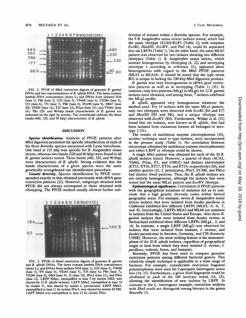
nition of a specific band at 135 kbp for B. burgdorferi sensustricto (Fig. 1A). Two bands at 220 and 80 kbp were specificto B. garinii (Fig. 2, lanes 1 to 12), while for B. afzelii, threebands (at 460, 320, and 90 kbp) were characteristic (Fig. 2,lanes 13 and 14; Table 2).The specific bands noted above for identification of a
species are identified by their size only. The question arisesas to whether the bands characteristic of a species are indeedgenetically similar. The 135-kbp MluI band of B. burgdorferisensu stricto isolate B31 hybridizes to the same 135-kbpband in each of the 14 isolates of that species (Fig. 1B). Asimilar experiment showed that the 220-kbp MluI bandcharacteristic of B. garinii is indeed the same for each of the12 isolates tested (data not shown). The experiments werenot performed for B. afzelii because of the unique andunambiguous MLal pattern consisting of most of the ge-nome (Table 2).
L 1 234 56789101121314L
291
145.597
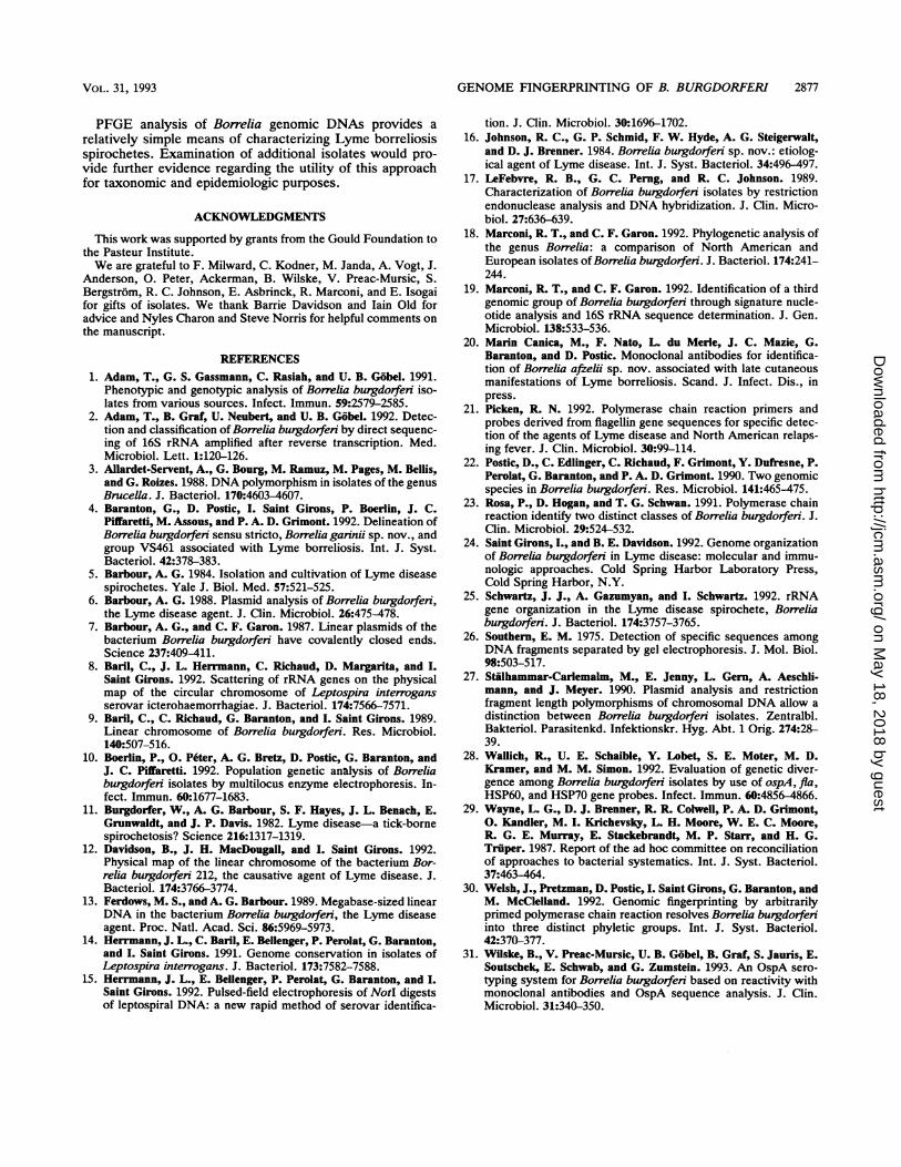
Fingerprinting with SmaI. Another rare cutting restrictionendonuclease was used to differentiate among isolates show-ing the same MluI profile. SmaI allowed the differentiation ofsome isolates from each other. For example, among the B.garinii isolates that were indistinguishable with MluI (Table2 [LRFP MLg2]), one isolate (PBr) had a unique LRFP withSmaI that was distinct from those of the other four isolatesN34, G25, VS468, and TN) (Fig. 3). On the other hand,isolates T25 and PBr, which had different MluI patterns,shared the same SmaI pattern.Among the 20 B. afzelii isolates which were indistinguish-
able with MluI, 15 isolates shared the SMal pattern, with acharacteristic quadruplet (Fig. 3, lane 9) while five isolateshad LRFPs which varied from that of SMal by one addi-tional or one missing band (LRFPs SMa2, SMa3, and SMa4[Fig. 3, lanes 10 to 12]).
12 3 4 5 6 78 91011121314
-ml 35
A BFIG. 1. (A) PFGE ofMluI restriction digests of genomic B. burgdorfen sensu stricto DNAs. The lanes contain lambda DNA concatemers
(lanes L) and DNAs from isolates B31 (lane 1), 297 (lane 2), IRS (lane 3), HUM115 (lane 4), HUM7814 (lane 5), 20006 (lane 6), MEN115 (lane7), HUM3336 (lane 8), Lake 339 (lane 9), SON188 (lane 10), Z136 (lane 11), SON335 (lane 12), 21305 (lane 13), and 212 (lane 14). The 135-kbpband characteristic of B. burgdorferi sensu stricto is indicated by an arrowhead on the right. (B) Blot of the PFGE gel hybridized with the135-kbp band from B. burgdorferi isolate B31 DNA.
VOL. 31, 1993
on May 18, 2018 by guest
http://jcm.asm
.org/D
ownloaded from
2876 BELFAIZA ET AL.
266 - _
177
FIG. 2. PFGE of MluI restriction digests of genoDNAs and two representatives of B. afzelii DNA. Thelambda DNA concatemers (lanes L) and DNAs fron(lane 1), PBi (lane 2), G25 (lane 3), VS468 (lane 4), V153 (lane 6), TN (lane 7), PBr (lane 8), IPer90 (lane c
10), VS185 (lane 11), T25 (lane 12), PGau (lane 13), an14). The 220- and 80-kbp bands characteristic of jindicated on the right by arrows. The arrowheads indbands (460, 320, and 90 kbp) characteristic of B. afze
DISCUSSION
Species identification. Analysis of PFGE pMluI digestion permitted the specific identificatiithe three Borrelia species associated with Lym4One band at 135 kbp was specific for B. burgcstricto, whereas two bands (220 and 80 kbp) werB. garinii isolates tested. Three bands (460, 320were characteristic of B. afzelii. Strong evidebands characteristic of a given species wer
genetically strengthened our identification procGenetic diversity. Species identification by I
sponded exactly to that obtained previously wit]restriction patterns (22). However, subgroupsPFGE did not always correspond to those olribotyping. The PFGE method usually allowed
L 1 23456 789101112L
1 94
97-
FIG. 3. PFGE of SmaI restriction digests of gencand B. afzelii DNAs. The lanes contain lambda DN)(lanes L), and DNAs from isolates N34 (lane 1), G25 ((lane 3), TN (lane 4), VS185 (lane 5), T25 (lane 6),VS286 (lane 8), DK8 (lane 9), Jl (lane 10), IPer3 (lane(lane 12). LRFP SMal, exemplified in lane 9 by isoshared by 15 B. afzelii isolates. LRFP SMa2, exemplby isolate Jl, was shared by isolate I. persulcatus.exemplified in lane 11 by isolate IPer3, was shared byLRFP SMa4 was exemplified in lane 12 by isolate P'
L division of isolates within a Borrelia species. For example,the 9 B. burgdorferi sensu stricto isolates tested, which hadthe same ribotype (El/Hl/Rl/Pl [Table 1]) with enzymes
-.460EcoRI, HindIII, EcoRV, and PstI (4), could be separated
460 into six LRFPs (Table 1). On the other hand, the same MLb3x 320 pattern was observed for two isolates showing two different
220 ribotypes (Table 1). B. burgdorfeii sensu stricto, whichseemed homogeneous by ribotyping (4, 22) and serotyping(serotype 1 according to reference 31), appeared more
80 heterogeneous with regard to the MluI PFGE patterns1
0(MLbl to MLblO). It should be noted that the type strainB31 is unique in lacking the 100-kbp MluI digestion product.
B. garinii was very heterogeneous in rRNA gene restric-
lanesB. gat~intion patterns as well as in serotyping (Table 1) (31). In
mlanes contain contrast, only four patterns (MLgl to MLg4) for 12 B. ganniin isolates N34 isolates were obtained, and among them, 7 isolates exhibitedtS286 (lane 5) the MLg2 profile.9), 20047 (lane B. afzelii appeared very homogeneous whatever theid VS461 (lane method used. For 14 isolates with the same MLal pattern,B. garinii are only two ribotypes were observed with EcoRI (E6 and E7)icate the three and HindIII (H5 and H6), and a unique ribotype was?lii. observed with EcoRV (R4). Furthermore, Wilske et al. (31)
found that six isolates, now known as B. afzelii, that hadbeen isolated from cutaneous lesions all belonged to sero-type 2 (31).The results of multilocus enzyme electrophoresis (10),
latterns after another technique used to type isolates, were incorporatedion of each of in the present study (Table 1). No correlation betweene borreliosis. electrotype (obtained by multilocus enzyme electrophoresis)lorferi sensu and either LRFP or ribotype could be shown.*e found in all A single MluI pattern was obtained for each of the 20 B., and 90 kbp) afzelii isolates tested. However, a quarter of them (ACA1,nce that the VS461, PGau, Fl, and UMO1) had distinct electrotypesre the same (ET15, ET16, ET17, ET18, and ET19, respectively) (10), andedure. another quarter (Jl, I. persulcatus, IPer3, ECM8, and PSto)PFGE corre- had distinct SmaI patterns. Thus, the B. afzelii isolates areh rRNA gene not entirely homogeneous, although the isolates that were)btained with tested had the same MLal pattern and EcoRV ribotype.btained with Epidemiological significance. Correlation of PFGE patternsfurther sub- with the geographical locations of isolation led us to con-
clude that a high genetic diversity exists within limitedgeographic areas. For example, seven B. burgdorferi sensustricto isolates that were isolated from Lxodes pacificus inCalifornia exhibited five different LRFPs (MLb3, -4, -6, -7,and -8). Interestingly, LRFPs MLb3 and MLb8 are commonto isolates from the United States and Europe. Also three B.garinii isolates that were isolated from Lxodes ricinus inSwitzerland exhibited three different LRFPs (MLgl, -2, and
.._145.5 -4). In contrast, a single LRFP (MLg2) was observed forisolates that were isolated from humans, I. icinus, andIxodes persulcatus in Sweden, Germany, and CIS (formerlyUSSR). However, the most striking feature is the monomor-phism of the 20 B. afzeldi isolates, regardless of geographicalorigin or host from which they were isolated (I. ncinus, Lpacificus, rodents, foxes, and humans).
Recently, PFGE has been used to compare genomicrestriction patterns among different bacterial genera. This
)mic B. garinii relatively simple technique is applicable to a wide range ofA concatemers bacteria. For example, considerable restriction fragment(lane 2), VS468 polymorphisms were seen for Leptospira interrogans sensua PBr (lane 7), lato (14, 15). Furthermore, a given NotI fingerprint could be11), and PSto attributed to each of the 160 serovars tested (14, 15),
ifiedKinlane 10 allowing the identification of new isolates by LRFP. IniLRFP SMa3n contrast to the L. interrogans example, restriction analysisisolate ECM8. with XbaI could not distinguish among biovars in the genusSto. Brucella (3).
J. CLIN. MICROBIOL.
on May 18, 2018 by guest
http://jcm.asm
.org/D
ownloaded from
GENOME FINGERPRINTING OF B. BURGDORFERI 2877
PFGE analysis of Borrelia genomic DNAs provides a
relatively simple means of characterizing Lyme borreliosisspirochetes. Examination of additional isolates would pro-vide further evidence regarding the utility of this approachfor taxonomic and epidemiologic purposes.
ACKNOWLEDGMENTS
This work was supported by grants from the Gould Foundation tothe Pasteur Institute.We are grateful to F. Milward, C. Kodner, M. Janda, A. Vogt, J.
Anderson, 0. Peter, Ackerman, B. Wilske, V. Preac-Mursic, S.Bergstrom, R. C. Johnson, E. Asbrinck, R. Marconi, and E. Isogaifor gifts of isolates. We thank Barrie Davidson and Iain Old foradvice and Nyles Charon and Steve Norris for helpful comments on
the manuscript.
REFERENCES1. Adam, T., G. S. Gassmann, C. Rasiah, and U. B. Gobel. 1991.
Phenotypic and genotypic analysis of Borrelia burgdorferi iso-lates from various sources. Infect. Immun. 59:2579-2585.
2. Adam, T., B. Graf, U. Neubert, and U. B. Gobel. 1992. Detec-tion and classification of Borrelia burgdorferi by direct sequenc-
ing of 16S rRNA amplified after reverse transcription. Med.Microbiol. Lett. 1:120-126.
3. Allardet-Servent, A., G. Bourg, M. Ramuz, M. Pages, M. Bellis,and G. Roizes. 1988. DNA polymorphism in isolates of the genus
Brucella. J. Bacteriol. 170:4603-4607.4. Baranton, G., D. Postic, I. Saint Girons, P. Boerlin, J. C.
Piffaretti, M. Assous, and P. A. D. Grimont. 1992. Delineation ofBorrelia burgdorfen sensu stricto, Borrelia gannii sp. nov., and
group VS461 associated with Lyme borreliosis. Int. J. Syst.Bacteriol. 42:378-383.
5. Barbour, A. G. 1984. Isolation and cultivation of Lyme diseasespirochetes. Yale J. Biol. Med. 57:521-525.
6. Barbour, A. G. 1988. Plasmid analysis of Borrelia burgdorfen,the Lyme disease agent. J. Clin. Microbiol. 26:475-478.
7. Barbour, A. G., and C. F. Garon. 1987. Linear plasmids of thebacterium Borrelia burgdorfeni have covalently closed ends.Science 237:409-411.
8. Baril, C., J. L. Herrmann, C. Richaud, D. Margarita, and I.
Saint Girons. 1992. Scattering of rRNA genes on the physicalmap of the circular chromosome of Leptospira interrogansserovar icterohaemorrhagiae. J. Bacteriol. 174:7566-7571.
9. Banl, C., C. Richaud, G. Baranton, and I. Saint Girons. 1989.Linear chromosome of Borrelia burgdorferi. Res. Microbiol.140:507-516.
10. Boerlin, P., 0. Peter, A. G. Bretz, D. Postic, G. Baranton, andJ. C. Piffaretti. 1992. Population genetic analysis of Borreliaburgdorferi isolates by multilocus enzyme electrophoresis. In-fect. Immun. 60:1677-1683.
11. Burgdorfer, W., A. G. Barbour, S. F. Hayes, J. L. Benach, E.Grunwaldt, and J. P. Davis. 1982. Lyme disease-a tick-bornespirochetosis? Science 216:1317-1319.
12. Davidson, B., J. H. MacDougall, and I. Saint Girons. 1992.Physical map of the linear chromosome of the bacterium Bor-relia burgdorferi 212, the causative agent of Lyme disease. J.Bacteriol. 174:3766-3774.
13. Ferdows, M. S., and A. G. Barbour. 1989. Megabase-sized linearDNA in the bacterium Borrelia burgdorfeni, the Lyme diseaseagent. Proc. Natl. Acad. Sci. 86:5969-5973.
14. Herimann, J. L., C. Baril, E. Bellenger, P. Perolat, G. Baranton,and I. Saint Girons. 1991. Genome conservation in isolates ofLeptospira interrogans. J. Bacteriol. 173:7582-7588.
15. Herrmann, J. L., E. Bellenger, P. Perolat, G. Baranton, and I.Saint Girons. 1992. Pulsed-field electrophoresis of NotI digestsof leptospiral DNA: a new rapid method of serovar identifica-
tion. J. Clin. Microbiol. 30:1696-1702.16. Johnson, R. C., G. P. Schmid, F. W. Hyde, A. G. Steigerwalt,
and D. J. Brenner. 1984. Borrelia burgdorferi sp. nov.: etiolog-ical agent of Lyme disease. Int. J. Syst. Bacteriol. 34:496-497.
17. LeFebvre, R. B., G. C. Perng, and R. C. Johnson. 1989.Characterization of Borrelia burgdorfen isolates by restrictionendonuclease analysis and DNA hybridization. J. Clin. Micro-biol. 27:636-639.
18. Marconi, R. T., and C. F. Garon. 1992. Phylogenetic analysis ofthe genus Borrelia: a comparison of North American andEuropean isolates of Borrelia burgdorfen. J. Bacteriol. 174:241-244.
19. Marconi, R. T., and C. F. Garon. 1992. Identification of a thirdgenomic group of Borrelia burgdorferi through signature nucle-otide analysis and 16S rRNA sequence determination. J. Gen.Microbiol. 138:533-536.
20. Marin Canica, M., F. Nato, L. du Merle, J. C. Mazie, G.Baranton, and D. Postic. Monoclonal antibodies for identifica-tion of Borrelia afrelii sp. nov. associated with late cutaneousmanifestations of Lyme borreliosis. Scand. J. Infect. Dis., inpress.
21. Picken, R. N. 1992. Polymerase chain reaction primers andprobes derived from flagellin gene sequences for specific detec-tion of the agents of Lyme disease and North American relaps-ing fever. J. Clin. Microbiol. 30:99-114.
22. Postic, D., C. Edlinger, C. Richaud, F. Grimont, Y. Dufresne, P.Perolat, G. Baranton, and P. A. D. Grimont. 1990. Two genomicspecies in Borrelia burgdorferi. Res. Microbiol. 141:465-475.
23. Rosa, P., D. Hogan, and T. G. Schwan. 1991. Polymerase chainreaction identify two distinct classes of Borrelia burgdorferi. J.Clin. Microbiol. 29:524-532.
24. Saint Girons, I., and B. E. Davidson. 1992. Genome organizationof Borrelia burgdorfen in Lyme disease: molecular and immu-nologic approaches. Cold Spring Harbor Laboratory Press,Cold Spring Harbor, N.Y.
25. Schwartz, J. J., A. Gazumyan, and I. Schwartz. 1992. rRNAgene organization in the Lyme disease spirochete, Borreliaburgdorfen. J. Bacteriol. 174:3757-3765.
26. Southern, E. M. 1975. Detection of specific sequences amongDNA fragments separated by gel electrophoresis. J. Mol. Biol.98:503-517.
27. Stiilhammar-Carlemalm, M., E. Jenny, L. Gern, A. Aeschli-mann, and J. Meyer. 1990. Plasmid analysis and restrictionfragment length polymorphisms of chromosomal DNA allow adistinction between Borrelia burgdorferi isolates. Zentralbl.Bakteriol. Parasitenkd. Infektionskr. Hyg. Abt. 1 Orig. 274:28-39.
28. Wallich, R., U. E. Schaible, Y. Lobet, S. E. Moter, M. D.Kramer, and M. M. Simon. 1992. Evaluation of genetic diver-gence among Borrelia burgdorferi isolates by use of ospA, fla,HSP60, and HSP70 gene probes. Infect. Immun. 60:4856-4866.
29. Wayne, L. G., D. J. Brenner, R R. Colwell, P. A. D. Grimont,0. Kandler, M. I. Krichevsky, L. H. Moore, W. E. C. Moore,R. G. E. Murray, E. Stackebrandt, M. P. Starr, and H. G.Truper. 1987. Report of the ad hoc committee on reconciliationof approaches to bacterial systematics. Int. J. Syst. Bacteriol.37:463-464.
30. Welsh, J., Pretzman, D. Postic, I. Saint Girons, G. Baranton, andM. McClelland. 1992. Genomic fingerprinting by arbitrarilyprimed polymerase chain reaction resolves Borrelia burgdorferiinto three distinct phyletic groups. Int. J. Syst. Bacteriol.42:370-377.
31. Wilske, B., V. Preac-Mursic, U. B. Gobel, B. Graf, S. Jauris, E.Soutschek, E. Schwab, and G. Zumstein. 1993. An OspA sero-typing system for Borrelia burgdorferi based on reactivity withmonoclonal antibodies and OspA sequence analysis. J. Clin.Microbiol. 31:340-350.
VOL. 31, 1993
on May 18, 2018 by guest
http://jcm.asm
.org/D
ownloaded from