Embed Size (px)
Citation preview

Gregory Alberts, Ph.D. Global Subject Matter ExpertLonza Pharma Bioscience Solutions
Genomic Editing, Nucleofection, and the Creation of Next-Generation Cell-Based Assays

slide 2
Content
■ Nucleofection and Next-Generation Cell-Based Assays
■ Introduction to Nucleofection
■ Introduction to Genome Editing
■ Nucleofection and Genome Editing- Best Practices
■ Nucleofection and iPSC Generation
■ Next Generation Cell-Based Assays and Models
■ Nucleofection Devices and Systems
■ The Nucleofector Platform- 4D, Shuttle, HTN
■ LV Nucleofector
■ Summary
slide 33
Jul-16
NotebookControl Unit
The Nucleofector™ Transfection Revolution
A Unique Combination: ■ Nucleofector™ Device■ Specific Nucleofector™ Kits and
cell type-specific solutions■ Detailed optimized protocols■ Enabling excellent transfection
performance combined with high functionality!
Higher quality standards for upstream GMP manufacturing■ GMP solution kits■ 21 CFR part 11 compliant 4D-
Nucleofector™ LogWare
■ Nucleofection is an advanced form of electroporation that excels at the transfection of primary cells and cell lines

slide 44
Jul-16
Physiologically Relevant Cells Efficiently Transfected by Nucleofection™
Research Area Disease Relevant CellsCancers Breast
ProstateMammary epithelial cellsProstate epithelial cells
Neurology Alzheimer’sParkinson’s
Cortical neuronsDorsal root ganglia
Immunology HIVInflammationArthritis
Human T cellsImmune system cellsChondrocytes
Metabolic Disorders DiabetesObesity
INS-1, MIN-6Adipocytes, 3T3-L1
Cardiology Heart DiseaseStroke
CardiomyocytesHUVEC, HMVEC, SMC
iPS Generation
hESC and iPS Transfection
Diseasepathogenesis
Human fibroblasts, CD34+ cells, monocytes hESC, hiPSC

slide 5
Using Nucleofection to Push Your Research to the Next Level: Custom Cell Line Generation
■ New trends in biomedical research are using primary cells in research.
■ Primary cells respond more appropriately to stimuli; express relevant pathways, and results are more likely to translate into in vivo models, creating better, more accurate model systems for cell-based assays
■ Nucleofection excels in the transfection of primary cells, making it an technology that can exploit these new trends.
■ Nucleofection is an excellent choice for iPSC generation, as well as for genome editing technologies (i.e. ZFNs, TALENs, CRISPRs)
■ Used in combination, Nucleofection and these technologies can:■ Generate normal or patient-derived iPSCs that can be genetically
modified for use as model systems or for use in cell-based assays■ Relevant cell line models can be genetically modified for cell-based
assays and to test and compare similar model systems■ Creation of isogenic cell populations differing in only in the
engineered modification for sophisticated analyses

slide 6
Why Nucleofection ®?■ High efficiency transfection of primary cells and physiologically-
relevant cell lines■ Use model systems and cells that previously were unavailable due to
limitations of technology- No Need to Compromise!■ Higher quality of data, higher quality of hits■ Ideal for ZFN, TALEN, CRISPR, Sleeping Beauty, PiggyBac delivery
■ Substrate Versatility: Use the same protocol for all substrates■ Efficient Transfection of plasmid DNAs (i.e. cDNAs), siRNAs, DNA
oligos, mRNAs, and even peptides and proteins
■ Flexibility of scale ■ Use the same technology and protocol across all Nucleofection
platforms, from single cuvette to 96-well to 384-well to Large Volume
■ Innovation in Transfection■ High Throughput 384-Well Transfection in Relevant Cell Types■ Adherent transfection in 24-well capacity for Neurons and other cells■ New LV Nucleofector: Large Scale Nucleofection for 108-109 cells
slide 7
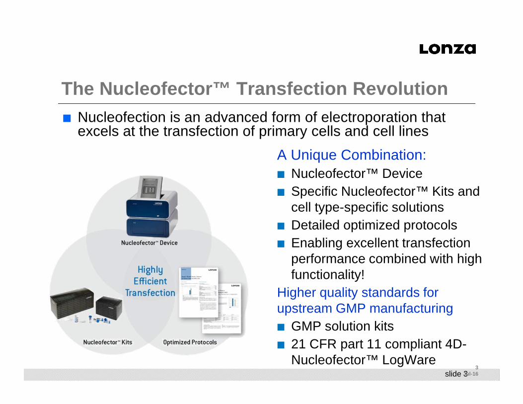
Amaxa ® Nucleofector ® Technology: Overview
Principle Efficient non-viral transport of DNA straightinto the nucleus of primary cells and cell lines
Major benefits ■ High gene transfer efficiency
■ Transfection independent of cell division
■ Allows transfection of non-dividing cells such as neurons
■ Faster gene expression
■ Versatility- can transfect plasmid DNA, DNA oligos, mRNA, miRNAs, or siRNAs, in either primary cells or cell lines

slide 8
Neurons and siRNA –Down -Regulation of eGFP Expression
eGFP (3 µg) eGFP (3 µg), siRNA (1.5 µg)
eGFP (3 µg), siRNA (3 µg)
DAPI stain
Data generated by Hyun-Ju Kim and Tim Vartanian, Beth Israel Deaconess Medical Center, Dept. of Neurology, Boston, MA, USA
■ Mouse cortical neurons (E17) were co-transfected with eGFP plasmid and siRNA oligonucleotide against eGFP
■ 4 days post Nucleofection® cells were fixed and analyzed by fluorescence microscopy

slide 9
Nucleofection ® of Neuroactive Peptides
Nucleofection ® No Nucleofection ®
Figure1: Fluorescence and transmitted light images of SH-SY5Y cells incubated with 50µm Alexa 488 dermorphin with (A, B) and without (C, D) Nucleofection®. In all cases the fluorescence and transmitted light images were of the same field of view.
Data Courtesy of Catherine Mollereau and Michel Roumy, Institute of Pharmacology and Structural Biology, CNRS, UMR, Toulouse, France
Investigation of G-coupled-protein receptor antagonists in SH-SY5Y cells: Nucleofection® of Neuropeptide FF Antagonizes NPFF2 Receptor Activity
Cys-dermorphin (YDAFGYPKC-NH2)
77% Transfectionefficiency of peptides
Relevant to Cas9Protein transfectionlater

slide 10
Lonza’s Break-Through Nucleofector ®
technology
Gene of interestCell of interest
+ –
›› ›
Gene transfer directly
into the nucleus
Stabilization of the pores generated in the cell and nuclear membranes during Nucleofection allows substrates to diffuse into the cell
››
Cell type-specific
Solutions
Nuanced Nucleofection
Pulses
slide 11
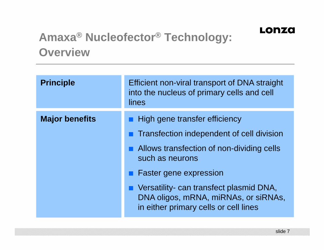
Gene transfer directly into the nucleus (and the cytoplasm)
Primary NHDF-neo cells were transfected with TMR-labeled plasmid DNA encoding GFP, fixed after 2h in 3.5% PFA and analyzed by confocal microscopy.
TMR-DNA GFP DAPI Merge
Amaxa ® Nucleofector ® Technology:Targeting the Nucleus
slide 1212
Jul-16
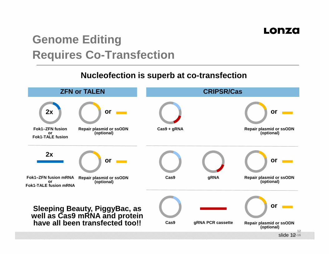
Genome Editing Requires Co -Transfection
ZFN or TALEN CRIPSR/Cas
Cas9 + gRNA
Cas9 gRNA
Cas9 gRNA PCR cassette
Fok1–ZFN fusionor
Fok1-TALE fusion
2x
Repair plasmid or ssODN (optional)
or
Fok1–ZFN fusion mRNAor
Fok1-TALE fusion mRNA
2x
Repair plasmid or ssODN (optional)
or
Repair plasmid or ssODN (optional)
or
Repair plasmid or ssODN (optional)
or
Repair plasmid or ssODN (optional)
or
Nucleofection is superb at co-transfection
Sleeping Beauty, PiggyBac, as well as Cas9 mRNA and protein have all been transfected too!!
slide 1313
Jul-16
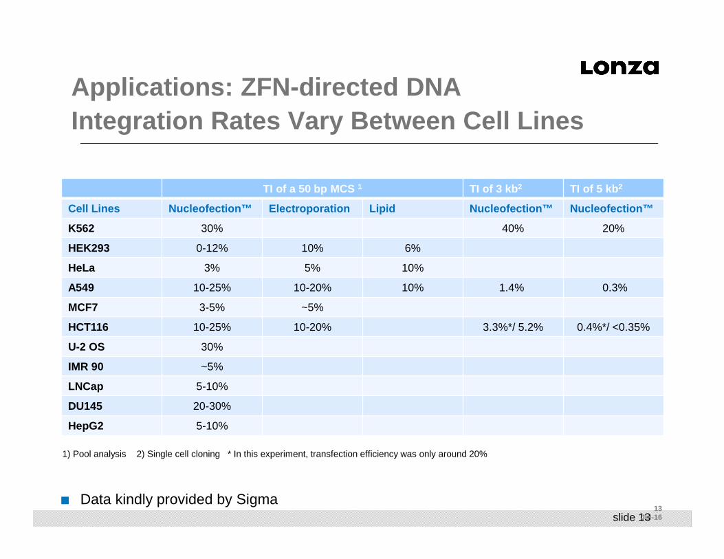
Applications: ZFN -directed DNAIntegration Rates Vary Between Cell Lines
TI of a 50 bp MCS 1 TI of 3 kb 2 TI of 5 kb 2
Cell Lines Nucleofection™ Electroporation Lipid Nucleof ection™ Nucleofection™
K562 30% 40% 20%
HEK293 0-12% 10% 6%
HeLa 3% 5% 10%
A549 10-25% 10-20% 10% 1.4% 0.3%
MCF7 3-5% ~5%
HCT116 10-25% 10-20% 3.3%*/ 5.2% 0.4%*/ <0.35%
U-2 OS 30%
IMR 90 ~5%
LNCap 5-10%
DU145 20-30%
HepG2 5-10%
■ Data kindly provided by Sigma
1) Pool analysis 2) Single cell cloning * In this experiment, transfection efficiency was only around 20%

slide 1414
Jul-16
Genome Editing Applications: Creation of Unique Model Systems
■ Custom generation of stable cell lines, e.g. for drug screening, pathway analysis etc.■ This should be even more efficient that the conventional generation of stable
clones in cell lines (Random integration/selection)
■ Stable modification of primary cells for in vitro disease modeling and for use in Cell-Based Assays■ Modification of normal iPSCs, patient-derived iPSCs, or cell lines, for
modeling disease pathogenesis and subsequent differentiation of modified iPSCs into cell types useful for comparison studies or cell-based assays
■ Stable modification of cells (e.g. iPSCs) for clinical applications ■ Autologous replacement/transplantation of diseased cells by healthy ones,
i.e. repair or replacement of defective genes in patient-derived iPSCs■ “Bar-coding of cells” by incorporation of unique DNA identifiers■ Generation of antigen-specific T cells (CAR T Cell Therapy)■ Cutting-edge technology that can improve “Speed to Patient”
slide 1515
Jul-16
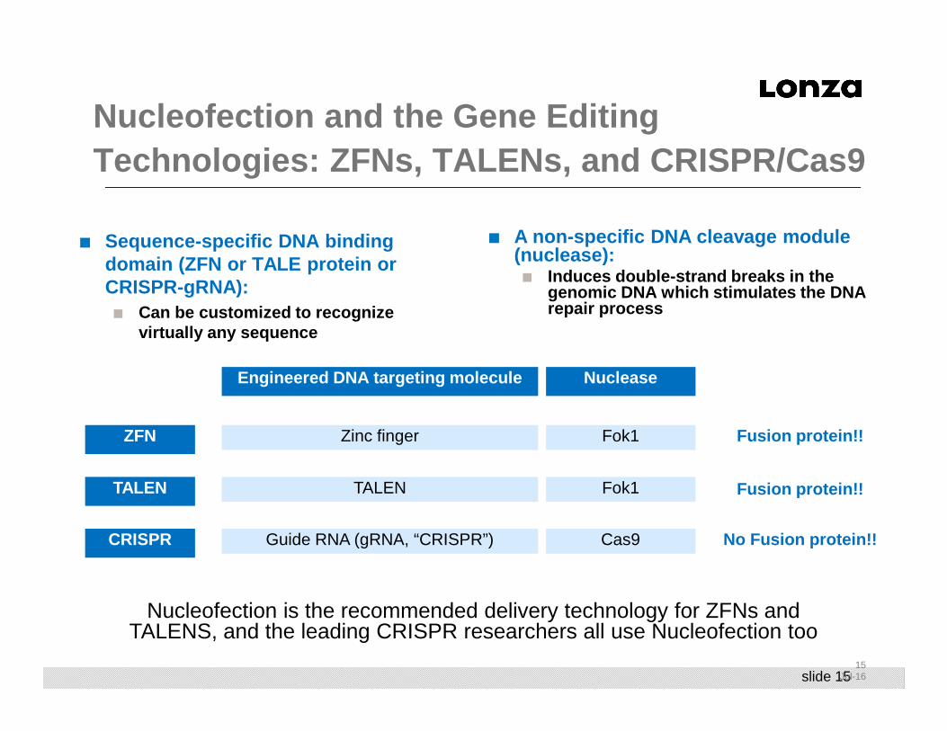
Nucleofection and the Gene Editing Technologies: ZFNs, TALENs, and CRISPR/Cas9
■ Sequence-specific DNA binding domain (ZFN or TALE protein or CRISPR-gRNA):
■ Can be customized to recognize virtually any sequence
NucleaseEngineered DNA targeting molecule
Fusion protein!!ZFN
TALEN
CRISPR
Fok1Zinc finger
Fok1TALEN
Cas9Guide RNA (gRNA, “CRISPR”)
Fusion protein!!
No Fusion protein!!
■ A non-specific DNA cleavage module (nuclease):
■ Induces double-strand breaks in the genomic DNA which stimulates the DNA repair process
Nucleofection is the recommended delivery technology for ZFNs and TALENS, and the leading CRISPR researchers all use Nucleofection too
slide 1616
Jul-16
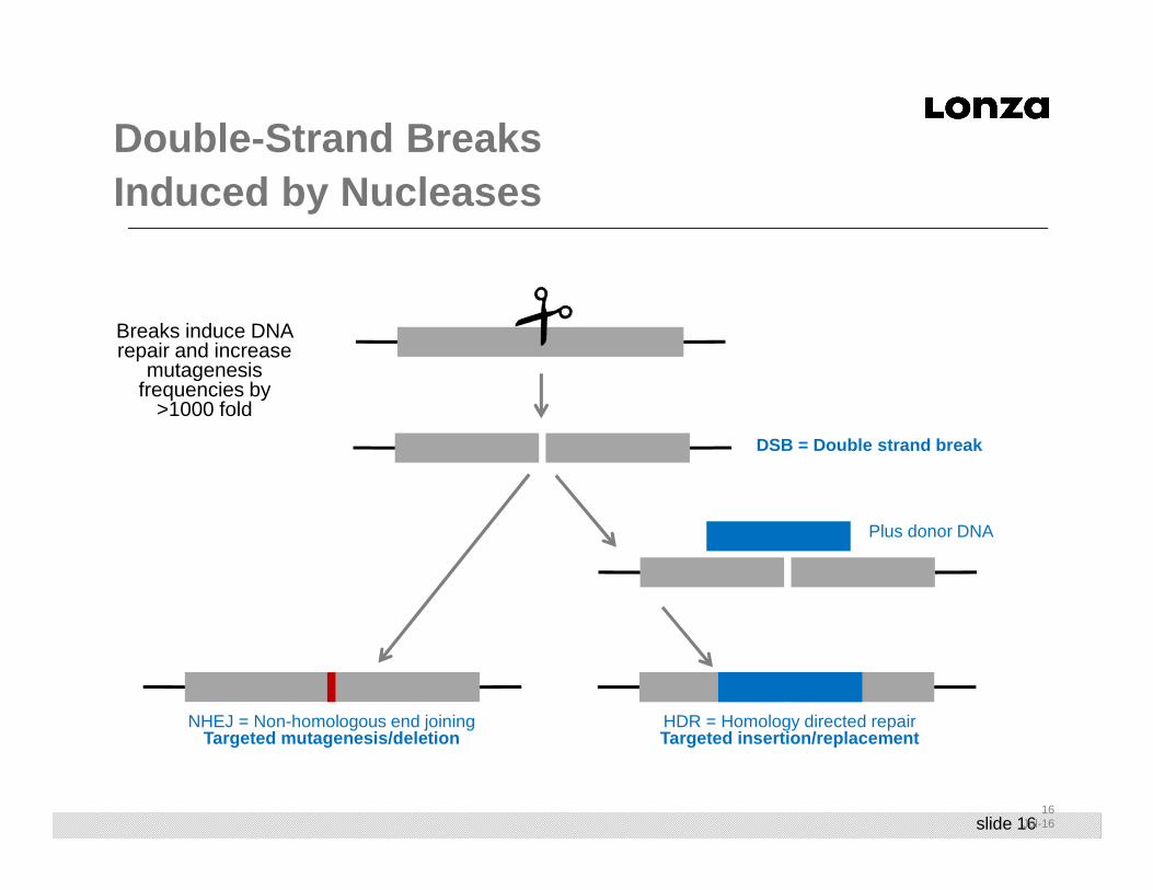
Double-Strand Breaks Induced by Nucleases
NHEJ = Non-homologous end joiningTargeted mutagenesis/deletion
DSB = Double strand break
Plus donor DNA
HDR = Homology directed repairTargeted insertion/replacement
Breaks induce DNA repair and increase
mutagenesis frequencies by
>1000 fold
slide 1717
Jul-16
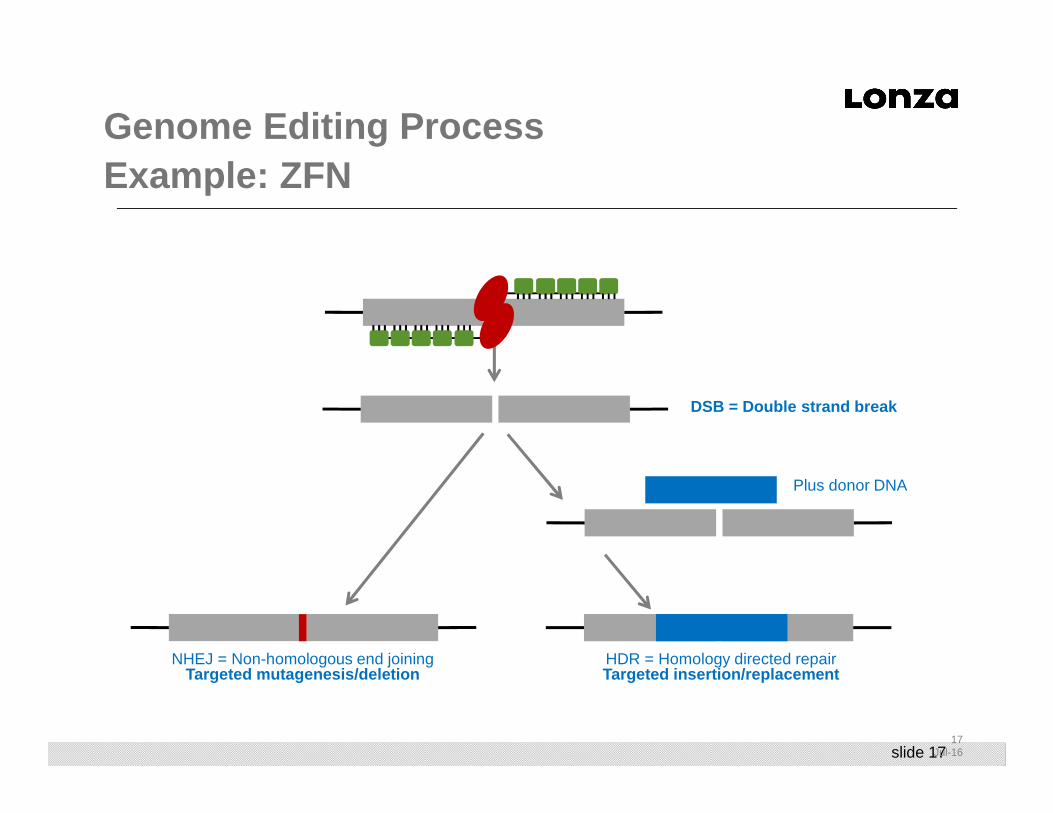
Genome Editing ProcessExample: ZFN
NHEJ = Non-homologous end joiningTargeted mutagenesis/deletion
DSB = Double strand break
Plus donor DNA
HDR = Homology directed repairTargeted insertion/replacement

slide 1818
Jul-16
Genome Editing ProcessExample: TALEN
NHEJ = Non-homologous end joiningTargeted mutagenesis/deletion
DSB = Double strand break
Plus donor DNA
HDR = Homology directed repairTargeted insertion/replacement
slide 1919
Jul-16
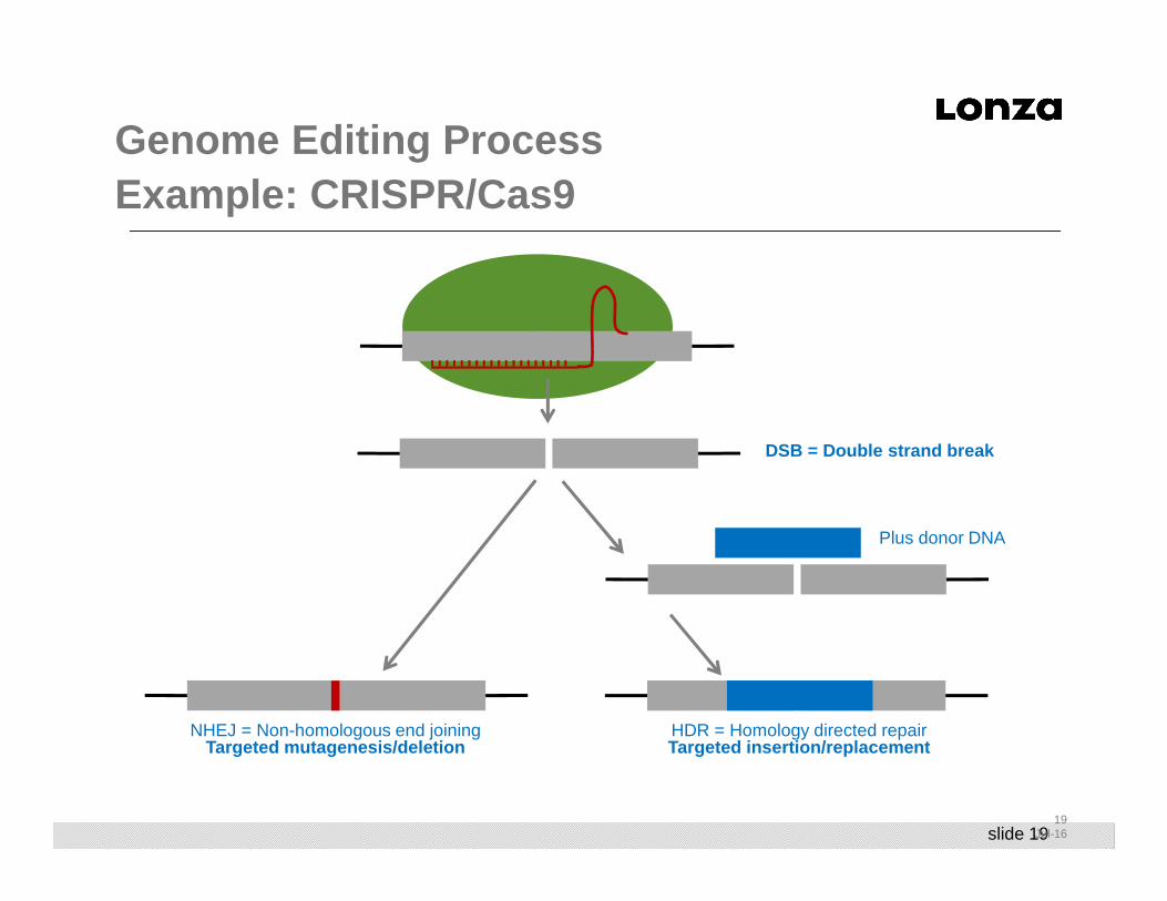
Genome Editing ProcessExample: CRISPR/Cas9
NHEJ = Non-homologous end joiningTargeted mutagenesis/deletion
DSB = Double strand break
Plus donor DNA
HDR = Homology directed repairTargeted insertion/replacement
slide 2020
Jul-16
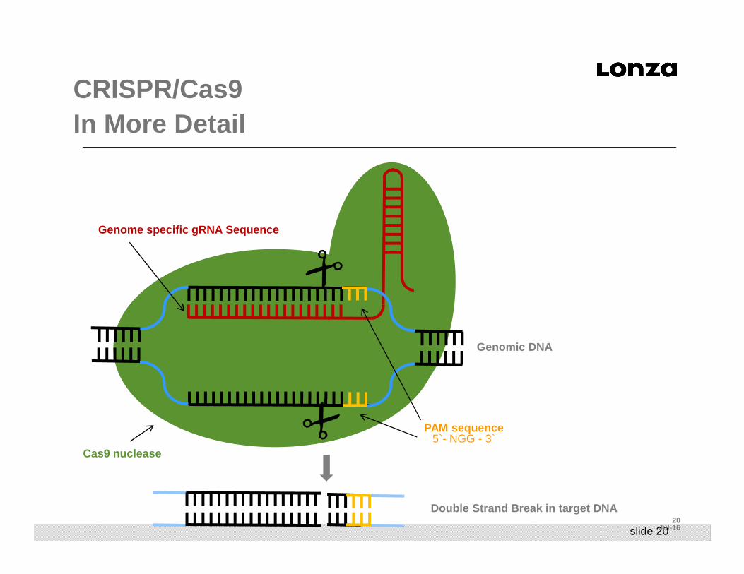
CRISPR/Cas9 In More Detail
Genome specific gRNA Sequence
PAM sequence5`- NGG - 3`
Cas9 nuclease
Genomic DNA
Double Strand Break in target DNA

slide 21
Content
■ Nucleofection and Next-Generation Cell-Based Assays
■ Introduction to Nucleofection
■ Introduction to Genome Editing
■ Nucleofection and Genome Editing- Best Practices
■ Nucleofection and iPSC Generation
■ Next Generation Cell-Based Assays and Models
■ Nucleofection Devices and Systems
■ The Nucleofector Platform- 4D, Shuttle, HTN
■ LV Nucleofector
■ Summary

slide 22
General Best Practices for Nucleofection■ Take care of your cells (i.e. passage number, confluence or cell density,
media, trypsin issues)■ Plan your experiment■ Prepare the culture plates with media and place at 37o C before you
start the experiment■ No ice needed! Everything is done at RT or at 37o C■ Count the cells each time■ Don’t spin the cells too fast: 90 g, 10 min!!
■ If transfecting iPSC or hESC, spin at 90 g for 3 min■ If transfecting neuronal cells, spin at 80 g for 10 min
■ Use endotoxin-free DNA with an OD ratio greater than 1.8 ■ if doing in vitro transcription of mRNAs, make sure the DNA
template is similarly endotoxin-free!!■ Handle the cells carefully and gently after nucleofection to minimize
shear stresses- Don’t be killing cells that would otherwise be surviving!!■ Plate cells at a density that makes scientific sense to your experiment

slide 2323
Jul-16
Nucleofection™ Tips and Tricks:Optimal Nucleofection™ Conditions
■ Use the same transfection conditions (Nucleofector Solution and program) that you would use for DNA transfections
■ If using an Optimized Protocol:■ first verify optimal conditions (given in the
ready-to-use protocol) using pmaxGFP™ Control
■ Otherwise, determine optimal conditions through an optimization strategy and use those conditions for transfection■ i.e. for iPSC clones or other cell types
For general Nucleofection™ Tips please refer to www.lonza.com/nucleofection
slide 24
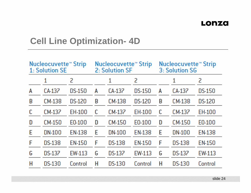
Cell Line Optimization - 4D
slide 25
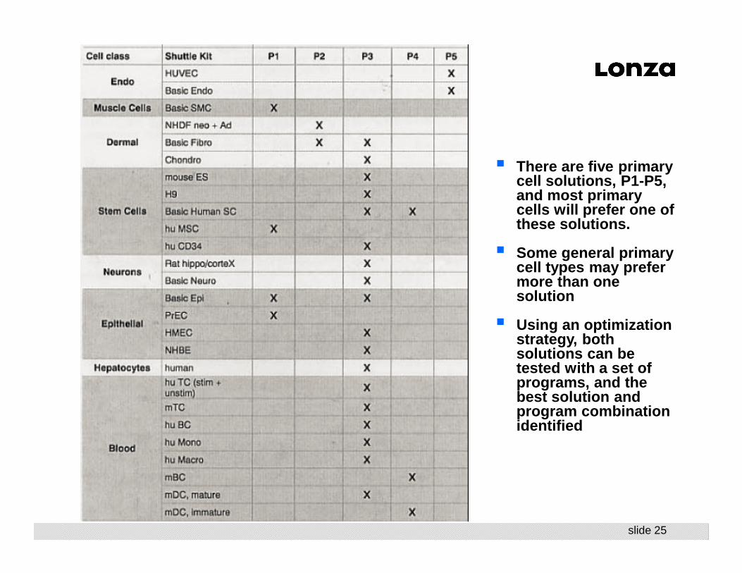
� There are five primary cell solutions, P1-P5, and most primary cells will prefer one of these solutions.
� Some general primary cell types may prefer more than one solution
� Using an optimization strategy, both solutions can be tested with a set of programs, and the best solution and program combination identified

slide 26
Basic Programs for Primary Cells
Basic FibroblastSolutions P2 and P3
CA-137CM-138DS-150 EH-100 EN-150 EO-114 FF-113
Basic EpithelialSolutions P1 and P3
CM-102DC-100EA-104EL-110ED-100CM-113DS-109
Basic EndothelialSolution P5
CA-167DY-138EH-100EP-114 FA-100 FF-138 FP-100
Basic SMCSolution P1
FF-130FG-113DS-137CM-137EH-106FP-113
Human ES BasicSolutions P3 and P4
CA-137CB-150CD-118CE-118 CM-113 DC-100 DN-100
Basic neuronSolution P3
CA-138CL-133CU-110DC-100DC-104DR-114EM-110

slide 2727
Jul-16
Nucleofection™ Tips and Tricks:Co-Transfection and How Much Substrate?
■ You may need to optimize substrate ratio and total substrate amount
■ Example ranges from publications:■ ZFN
■ TALEN
■ CRISPR
Plasmid mRNA Donor Plasmid:
0.01- 0.05 µg/µl each0.2 - 1 ug/20 ul
0.02 –0.2 µg/µl each0.4 – 4 ug / 20 ul
0.04-0.2 µg/µl0.8 – 4 ug / 20 ul
PlasmidDonorPlasmid:
DonordsDNA(lin)
DonorssODN
0.01 – 0.1 µg/µl each0.2 – 2 ug / 20 ul
0.02-0.2 µg/µl0.4 - 4 ug / 20 ul
0.1 µg/µl 2 ug / 20 ul
3-10 µM
PlasmidgRNA PCR Cassette
DonordsDNA (lin)
DonorssODN
Cas9: 0.025-0.5 µg/µlgRNA: 0.025-0.5 µg/µl
0.5 – 10 ug / 20 ul
Cas9/gRNA: 0.025-0.5 µg/µl 0.5 – 10 ug / 20 ul
0.5 ng/µl10 ng / 20 ul
0.02-0.2 µg/µl0.4 –4 ug /20 ul
10 µM
Ran, etal. recommends using 200K cells and 1 ug tot al DNA substrate for CRISPR

slide 2828
Jul-16
Nucleofection™ Tips and Tricks:Nucleofection of mRNA
■ Frequently, researchers choose to transfect mRNA encoding ZFNs, in order to limit nuclease activity in cells
■ Follow the same protocol and use the same program that is used for the transfection of DNA with your cells; it works fine with mRNA
■ The in vitro-transcribed mRNA must be capped and poly-adenylated
■ Optimal amount has to be titrated, might be higher than for plasmid (0.4 - 4 ug mRNA / 20 ul transfection)■ The volume of substrate added should not exceed 10% of the total sample
volume (i.e. 2 ul in 20 ul transfection)

slide 2929
Jul-16
Cas9 Protein vs. mRNA vs. DNA
■ The big advantages of using Cas9 mRNA or Cas9 protein is that they are labile, and do not persist in the cell, thereby limiting the activity of the Cas9 nuclease in a temporal fashion■ The longer the nuclease is active, as it is during DNA transfection, the more
opportunity there is for off-target effects
■ Nucleofection of mRNAs is typically is in the 90-95% TE range, so with mRNA, the vast majority of the cells will be transfected with Cas9 mRNA. ■ The gRNAs will transfect with 99% TE, so this combination will insure that
most of your cells will be able to be modified.
■ The Cas9 protein transfects at very high efficiencies (CPP-like activity?), and the kinetics for Cas9 protein activity are different, as the Cas9 protein is active immediately upon entry into the cell■ gRNAs are typically complexed with the Cas9 protein prior to transfection■ Most are using 100 pmol Cas9 protein and 50 pmol of ssODN per transfection
slide 30
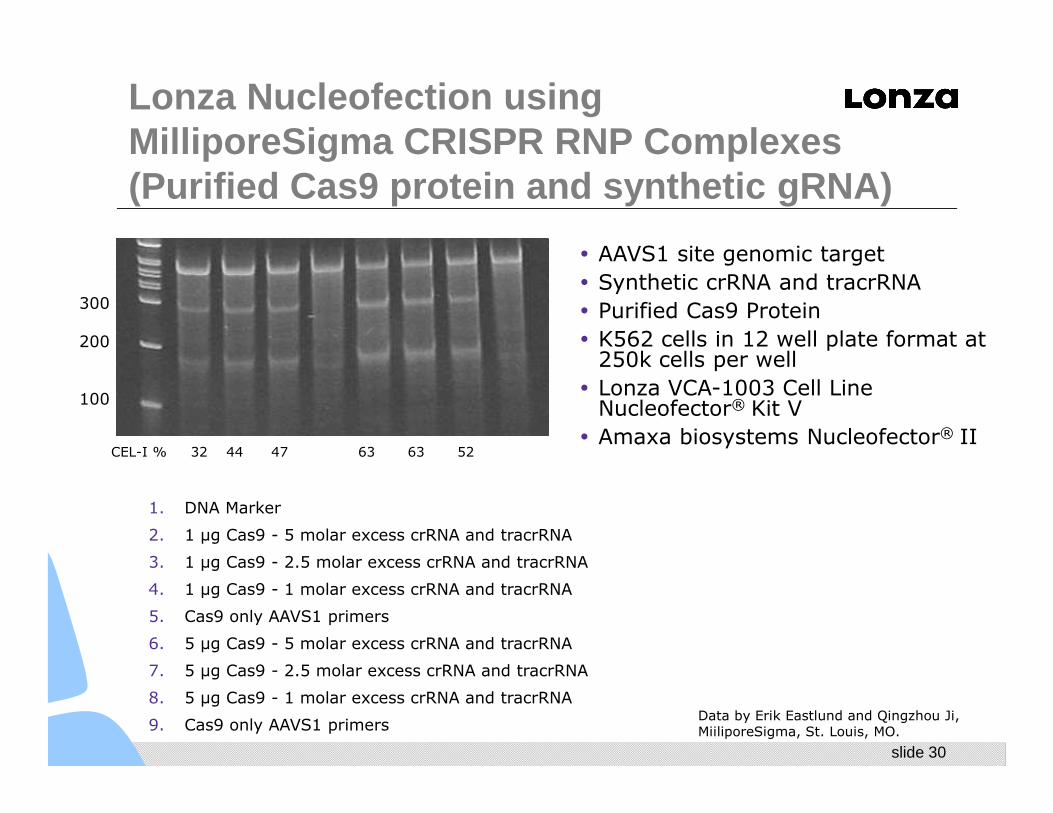
Lonza Nucleofection using MilliporeSigma CRISPR RNP Complexes(Purified Cas9 protein and synthetic gRNA)
1. DNA Marker
2. 1 µg Cas9 - 5 molar excess crRNA and tracrRNA
3. 1 µg Cas9 - 2.5 molar excess crRNA and tracrRNA
4. 1 µg Cas9 - 1 molar excess crRNA and tracrRNA
5. Cas9 only AAVS1 primers
6. 5 µg Cas9 - 5 molar excess crRNA and tracrRNA
7. 5 µg Cas9 - 2.5 molar excess crRNA and tracrRNA
8. 5 µg Cas9 - 1 molar excess crRNA and tracrRNA
9. Cas9 only AAVS1 primers
� AAVS1 site genomic target
� Synthetic crRNA and tracrRNA
� Purified Cas9 Protein
� K562 cells in 12 well plate format at 250k cells per well
� Lonza VCA-1003 Cell Line Nucleofector® Kit V
� Amaxa biosystems Nucleofector® II
Data by Erik Eastlund and Qingzhou Ji, MiiliporeSigma, St. Louis, MO.
32 44 47 63 63 52
300
200
100
CEL-I %

slide 31
Content
■ Nucleofection and Next-Generation Cell-Based Assays
■ Introduction to Nucleofection
■ Introduction to Genome Editing
■ Nucleofection and Genome Editing- Best Practices
■ Nucleofection and iPSC Generation
■ Next Generation Cell-Based Assays and Models
■ Nucleofection Devices and Systems
■ The Nucleofector Platform- 4D, Shuttle, HTN
■ LV Nucleofector
■ Summary

slide 32
Generation of Normal and Patient-Derived iPS Cells from Mononuclear Blood Cells
■ Used Nucleofection to reprogram human bone marrow or cord blood mononuclear cells using non-integrating oriP/EBNA1-based episomal vectors
■ The resulting iPSCs were free of transgene or vector sequences■ The cells lacked rearrangements of IGH and TCR, indicating that the cellular
origins were from non-B- or non-T-lymphoid cells■ Reprogramming was up to 100X more efficient, and occurred 1-3 weeks faster
compared to reprogramming of fibroblasts■ When cocultured on OP9, blood-derived iPSCs could be differentiated back to the
blood cells, however, with lower efficiency compared to fibroblast-derived iPSCs. ■ Also generated transgene-free iPSCs from the BM of a patient with chronic
myeloid leukemia (CML). CML iPSCs showed a unique c omplex chromosomal translocation (Bcr-Abl) identified in m arrow sample while displaying typical embryonic stem cell phenotype an d pluripotent differentiation potential
Efficient generation of transgene-free induced plur ipotent stem cells from normal and neoplastic bone marrow a nd cord blood mononuclear cellsYu, J and Hu, K; etal. Blood April 7, 2011, vol 117 no 14, e109-e119
slide 3333
Jul-16
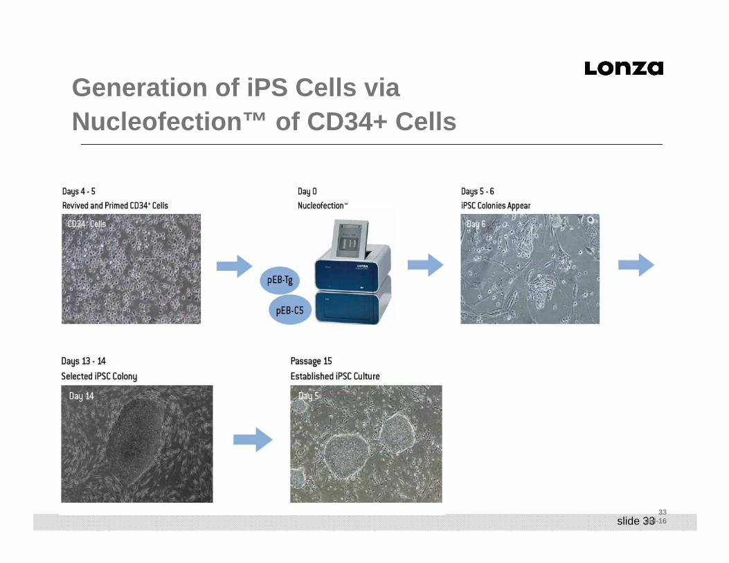
Generation of iPS Cells viaNucleofection™ of CD34+ Cells

slide 3434
Jul-16
iPCs can be differentiated into cells representative of all 3 germ layers
CD34+-derived iPSC CharacterizationEmbryoid Body (EB) Formation
slide 3535
Jul-16
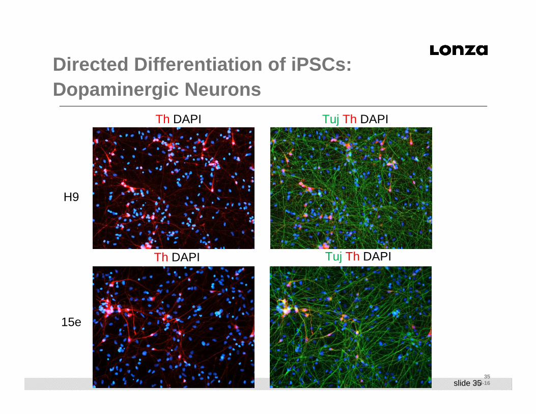
Directed Differentiation of iPSCs: Dopaminergic Neurons
H9
15e
Tuj Th DAPITh DAPI
Tuj Th DAPITh DAPI

slide 3636
Jul-16
Directed Differentiation of iPSCs: Definitive Endoderm Differentiation
Day40,40x Day40,20x
Day30,20xDay30,20x
FoxA2
FoxA2 DAPI SOX17 FoxA2

slide 37
Directed Differentiation of iPSCs:Hepatocytes

slide 3838
Jul-16
Directed Differentiation of iPSCs:Beating Cardiomyocytes
■ In vitro differentiation into spontaneously beating cardiomyocytes
Data courtesy of Dr. Chris Mayhew, Pluripotent Stem Cell Facility, Cincinnati Children's Hospital Medical Center

slide 3939
Jul-16
Generation of Next-Generation Cell-based Assays
■ Using Nucleofection as a central transfection technology allows the flexibility to work with primary human cells effectively
■ iPSC generation using Nucleofection is straightforward and easy
■ Nucleofection facilitates the transfection of CRISPR substrates into the iPSCs for specific modification, resulting in two isogenic populations of human iPSCs that are identical except for the engineered modification
■ The two iPSC populations can then be differentiated into terminally differentiated cell types for screening or cell-based assays
■ End Result- Two isogenic differentiated human cell t ypes, with a specific modification, in a dish for advance d analyses!!
■ Advanced analyses include plating into RAFT TM 3D Cell Culture for more physiological assays and results
slide 40
Content
■ Nucleofection and Next-Generation Cell-Based Assays
■ Introduction to Nucleofection
■ Introduction to Genome Editing
■ Nucleofection and Genome Editing- Best Practices
■ Nucleofection and iPSC Generation
■ Next Generation Cell-Based Assays and Models
■ Nucleofection Devices and Systems
■ The Nucleofector Platform- 4D, Shuttle, HTN
■ LV Nucleofector
■ Summary
slide 41
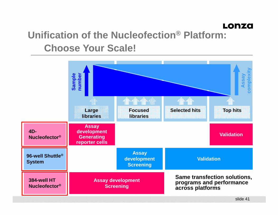
Sam
ple
num
ber
Large libraries
Focused libraries
Selected hits Top hits
Ass
ay
com
plex
ity
4D-Nucleofector ®
96-well Shuttle ®
System
384-well HT Nucleofector ®
Assay developmentGenerating
reporter cells
Assay development
Screening
Assay developmentScreening
Validation
Validation
Unification of the Nucleofection ® Platform: Choose Your Scale!
Same transfection solutions, programs and performance across platforms

slide 42
Minimal Development, Maximum Reproducibility: Same Solutions, Programs, and Results across All Three Platforms
Bench R&D
Basic Research
Assay Development
Screening
1o & 2o Screens
Library Screening
Hit Validation
Assay Analysis
Rescue Experiments
4D, Shuttle
Smaller Scale
Medium Throughput
Shuttle, HTN
Large Scale
High Throughput
Generation of Hits
4D, Shuttle
Small Scale
Medium Throughput
Analysis of HitsSame: Technology
Program Setting
Reagents and Liquid Volume
Cells and Cell Number
Experimental Process
Use the 4D and Shuttle for Assay Development and feed directly into the HT Nucleofector with Minimal Development Work
slide 43
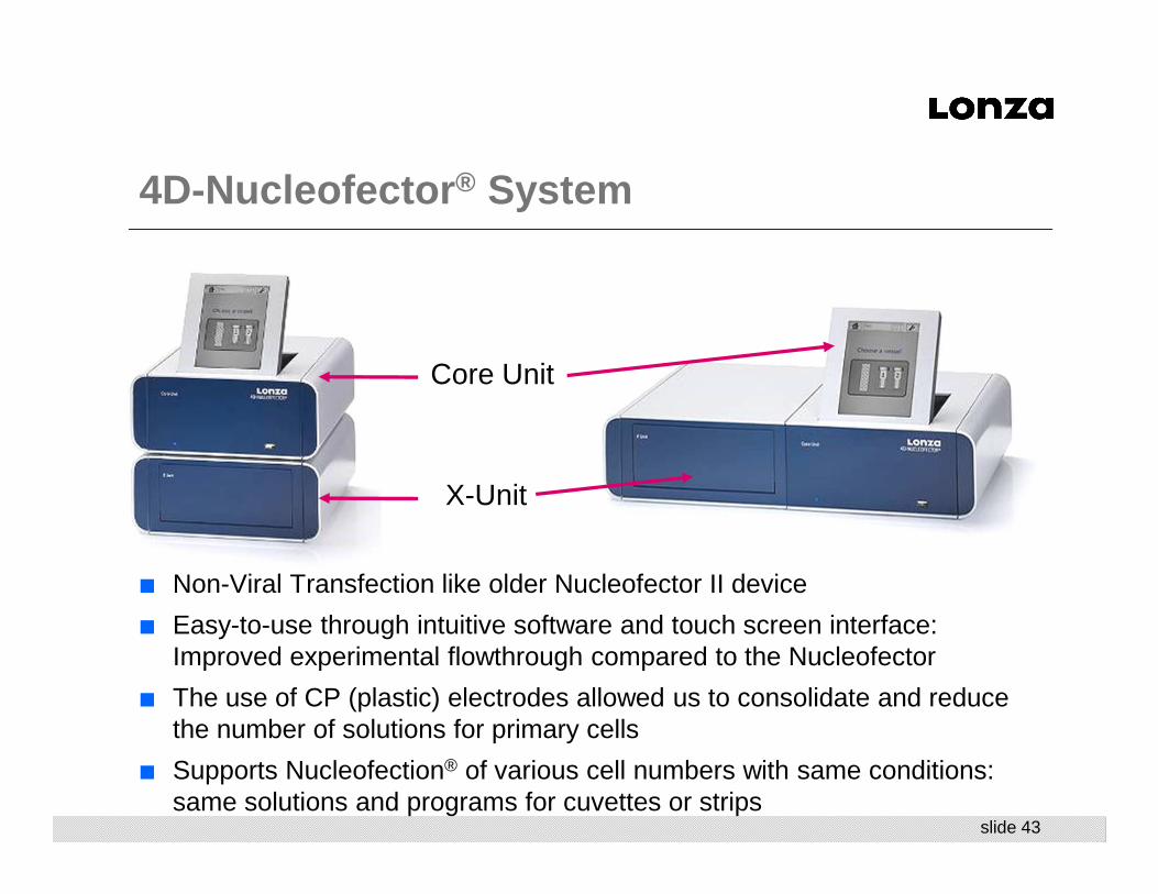
■ Non-Viral Transfection like older Nucleofector II device
■ Easy-to-use through intuitive software and touch screen interface: Improved experimental flowthrough compared to the Nucleofector
■ The use of CP (plastic) electrodes allowed us to consolidate and reduce the number of solutions for primary cells
■ Supports Nucleofection® of various cell numbers with same conditions: same solutions and programs for cuvettes or strips
4D-Nucleofector ® System
Core Unit
X-Unit
slide 44
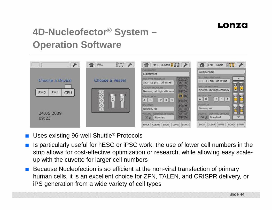
4D-Nucleofector ® System –Operation Software
■ Uses existing 96-well Shuttle® Protocols
■ Is particularly useful for hESC or iPSC work: the use of lower cell numbers in the strip allows for cost-effective optimization or research, while allowing easy scale-up with the cuvette for larger cell numbers
■ Because Nucleofection is so efficient at the non-viral transfection of primary human cells, it is an excellent choice for ZFN, TALEN, and CRISPR delivery, or iPS generation from a wide variety of cell types

slide 45
2Amaxa® Nucleofector®
96-well Shuttle® Device
Amaxa ® Nucleofector ® 96-well Shuttle ® System
NotebookControl Unit
3Laptop with software
(compatible with Windows XP and 2000)
A modular system
1Amaxa® 4D
Nucleofector® Device
- pulse delivery unit -

slide 46
■ 2x8 Amaxa® Nucleocuvette®
Modules, each strip consists of 16 wells
■ High flexibility: Combinations of strips for medium or high-throughput
■ Disposable – one time use■ Unique conductive polymer
electrodes: no generation of metal ions
■ Integrated electrodes – no contamination risk
■ Standard 96-well formatAmaxa® 96-well Nucleocuvette® Modules
Amaxa ® 96-well Nucleocuvette ® Plates

slide 47
The Amaxa ® 96-well Shuttle ®: Well-to -Well Standard Deviations
Transfection with pmaxGFP® Vector
Intra column SDs ~1%Column 1 1.26 %Column 2 0.95 %Column 3 0.88 %Column 4 0.94 %
Jurkat cells (ATCC®)
■ Program CL-120
■ 1 µg pmaxGFP® Vector
■ FACS analysis 24h post
Nucleofection®
0
10
20
30
40
50
60
70
80
90
100
Column 1 Column 2 Column 3 Column 4 (incl. 2controls)

slide 48
% E
xpre
ssio
n of
unt
reat
ed c
ontr
ol
0
10
20
30
40
50
60
70
80
90
100
1 2 3 4 5 6 7 8 9 10 11 12 13 14 15 16 17 18 19 20 21 22 23 24 25 26 27 28 29 31 C
Amaxa ® 96-well Shuttle ® System: siRNA -Mediated Depletion
External data kindly provided by Schering AG, Berlin, Germany
Jurkat E6-1 (ATCC® TIB 152™) SD 3 %
Jurkat E6-1 (ATCC® TIB 152™)
■ 2x10⁵ cells / sample
■ 20 µl transfection volume
■ Analysis 24 h post Nucleofection®
■ Relative expressions compared to untreated control sample (well C set to 100%)
■ Standard deviation is 2-3 % per module, and 3 % considering all samples
slide 49
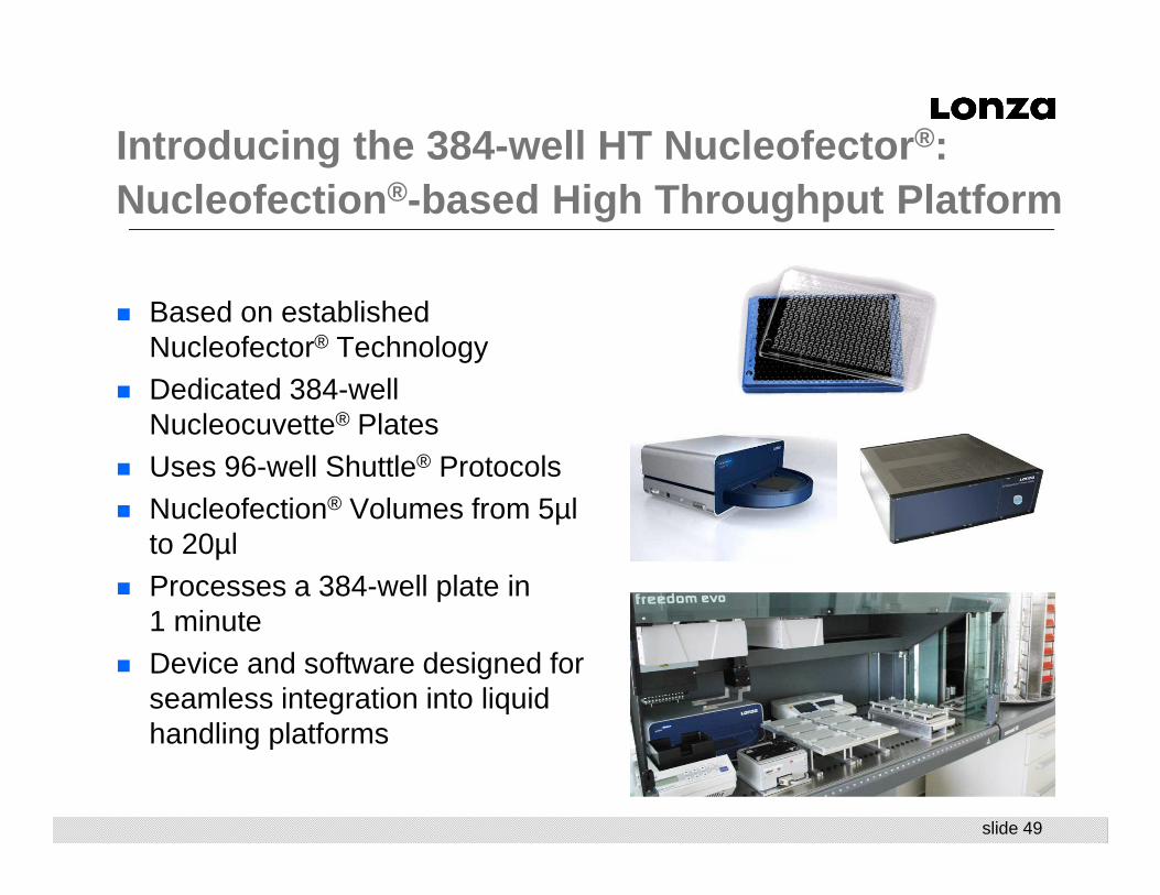
� Based on established Nucleofector® Technology
� Dedicated 384-well Nucleocuvette® Plates
� Uses 96-well Shuttle® Protocols� Nucleofection® Volumes from 5µl
to 20µl� Processes a 384-well plate in
1 minute� Device and software designed for
seamless integration into liquid handling platforms
Introducing the 384-well HT Nucleofector ®: Nucleofection ®-based High Throughput Platform

slide 5050
Jul-16
NEW! Large Scale Nucleofection4D-Nucleofector™ LV Unit
■ New functional unit for 4D-Nucleofector™ System
■ Allowing for closed, scalable Nucleofection of larger cell numbers in the range of 1x10⁷ to 1x10⁹
■ Conditions transferable from 4D-Nucleofector™ X Unit
■ Established protocols can be used■ Experimental setup can be optimized in
small volume
■ Can be operated via 21CFR part11 compatible software
■ Tested for various cell types, including human T cells, CHO-S, HEK293-S, K562
For research use only. Not for direct use in humans or for diagnostic purposes.

slide 5151
Jul-16
Scalability DataExample: Human T Cells
Data represent the mean of various independent experiments.
■ Transfection of human PBMCs with pmaxGFP™ Vector■ Cells stained and gated for CD3-positive T cells
0
20
40
60
80
100
100 µL 1 mL LV10 mL
100 µL 1 mL LV10 mL
EO-115 FI-115
Cell conc.: 3-5x10e7/mLSubstrate: 20 µg/mL
Effiency (%)
PI neg cells (%)
slide 5252
Jul-16
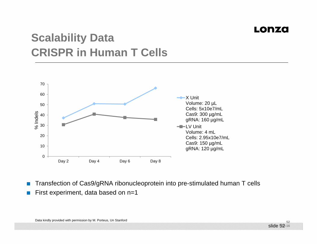
Scalability DataCRISPR in Human T Cells
Data kindly provided with permission by M. Porteus, Un Stanford
■ Transfection of Cas9/gRNA ribonucleoprotein into pre-stimulated human T cells■ First experiment, data based on n=1
0
10
20
30
40
50
60
70
Day 2 Day 4 Day 6 Day 8
% In
dels
X UnitVolume: 20 µLCells: 5x10e7/mLCas9: 300 µg/mLgRNA: 160 µg/mLLV UnitVolume: 4 mLCells: 2.95x10e7/mLCas9: 150 µg/mLgRNA: 120 µg/mL
slide 53
Content
■ Nucleofection and Next-Generation Cell-Based Assays
■ Introduction to Nucleofection
■ Introduction to Genome Editing
■ Nucleofection and Genome Editing- Best Practices
■ Nucleofection and iPSC Generation
■ Next Generation Cell-Based Assays and Models
■ Nucleofection Devices and Systems
■ The Nucleofector Platform- 4D, Shuttle, HTN
■ LV Nucleofector
■ Summary
slide 54
What does this mean for Your Research?
■ Nucleofection allows you to use the most relevant cells for your work,
■ Cells that respond more appropriately to stimuli, as they express all the relevant pathways with results that are more likely to translate into in vivo models
■ Nucleofection excels at the delivery of DNA and other substrates into primary cells and cell lines, and is an ideal companion technology for genomic-editing strategies like ZFN, TALEN, CRISPR
■ Nucleofection can be used in conjunction with genome editing technologies for the generation of unique and defined cell lines or iPSC-derived model systems for use in cell-based assays
■ Scalable transfection conditions across platforms
■ Development work can proceed with lower throughput devices, yet transition seamlessly into higher throughput platforms or the LV Nucleofector for large cell number applications

Thank you for your attentionVielen Dank für Ihre Aufmerksamkeit
www.lonza.com