Embed Size (px)
Citation preview
Genetics and Recombinant DNA
BIT 120
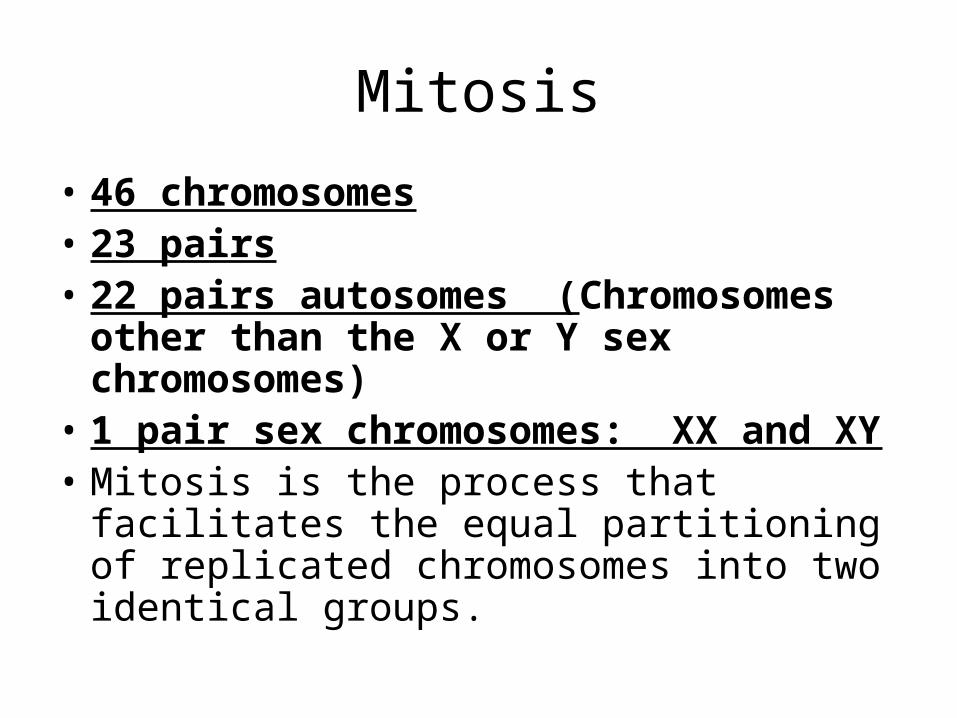
Mitosis
• 46 chromosomes• 23 pairs• 22 pairs autosomes (Chromosomes
other than the X or Y sex chromosomes)
• 1 pair sex chromosomes: XX and XY• Mitosis is the process that facilitates the
equal partitioning of replicated chromosomes into two identical groups.
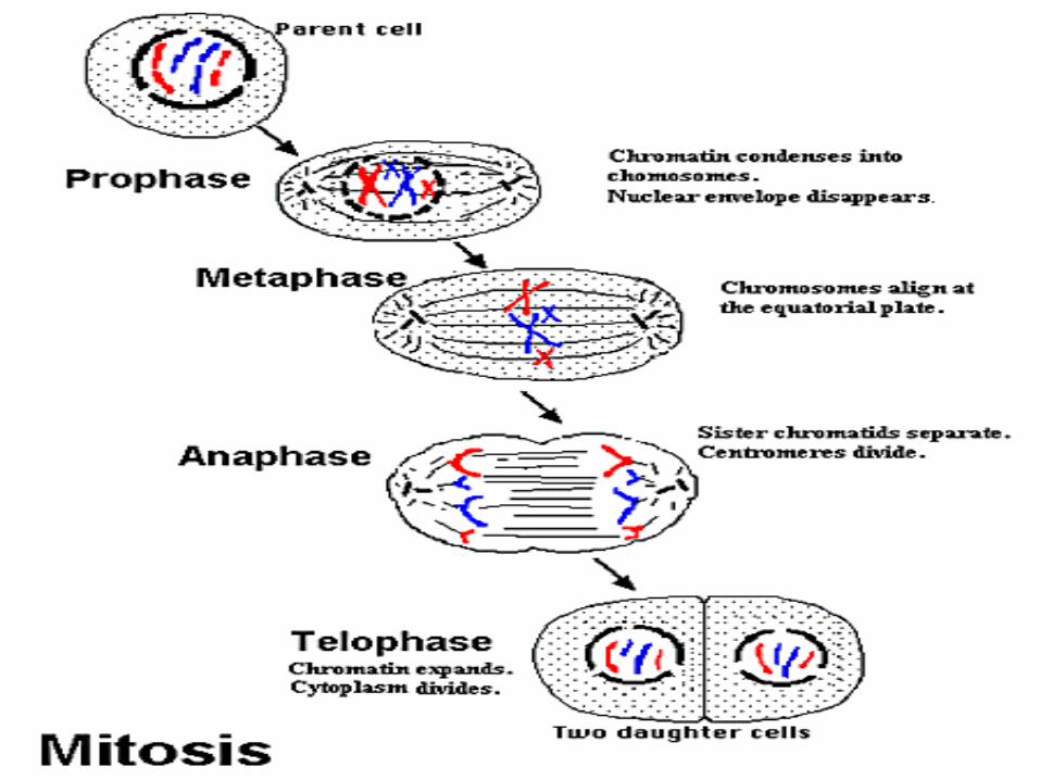
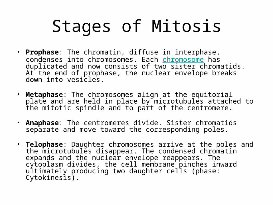
Stages of Mitosis• Prophase: The chromatin, diffuse in interphase, condenses into
chromosomes. Each chromosome has duplicated and now consists of two sister chromatids. At the end of prophase, the nuclear envelope breaks down into vesicles.
• Metaphase: The chromosomes align at the equitorial plate and are held in place by microtubules attached to the mitotic spindle and to part of the centromere.
• Anaphase: The centromeres divide. Sister chromatids separate and move toward the corresponding poles.
• Telophase: Daughter chromosomes arrive at the poles and the microtubules disappear. The condensed chromatin expands and the nuclear envelope reappears. The cytoplasm divides, the cell membrane pinches inward ultimately producing two daughter cells (phase: Cytokinesis).
Genes on Chromosomes
• Definition of Gene: The functional and physical unit of heredity passed from parent to offspring. Genes are pieces of DNA, and most genes contain the information for making a specific protein
• One gene one enzyme
• One gene one peptide
Meiosis - The Genetics of Reproduction
• Diploid – 2 chromosomes per pair• Haploid – set of one chromosome• The process of meiosis essentially
involves two cycles of division, essentially involving a gamete mother cell (diploid cell) dividing and then dividing again to form 4 haploid cells. These can be subdivided into four distinct phases which are a continuous process
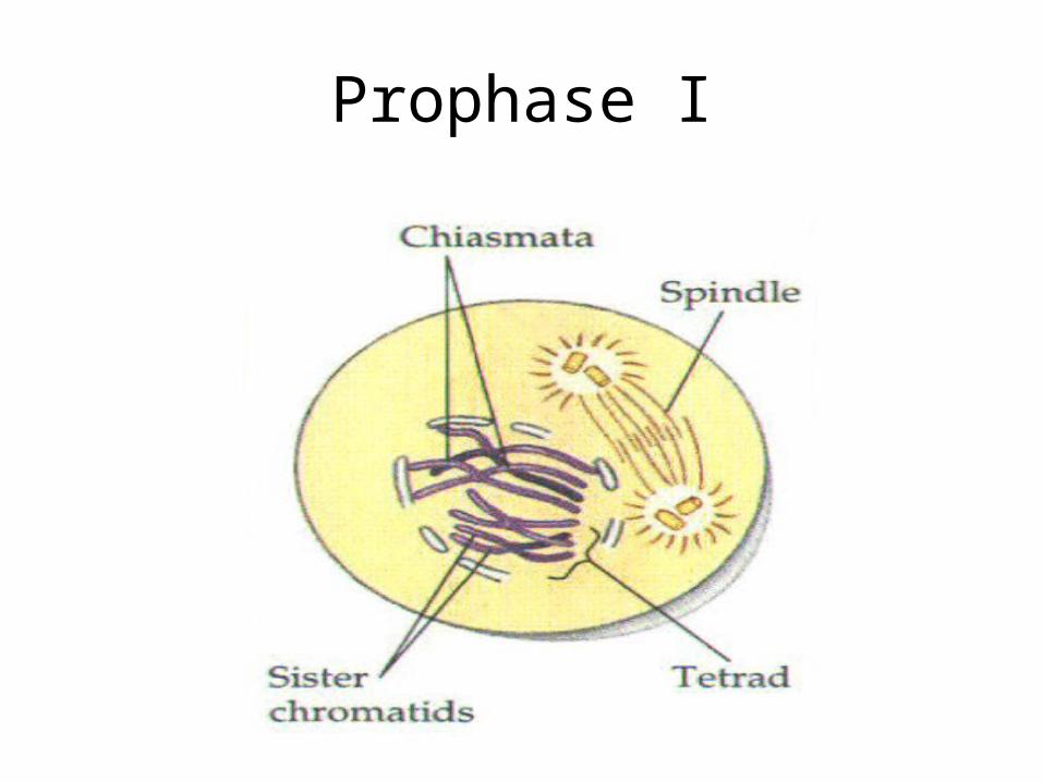
Prophase I
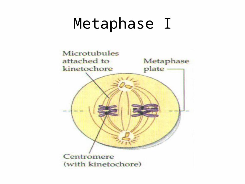
Metaphase I
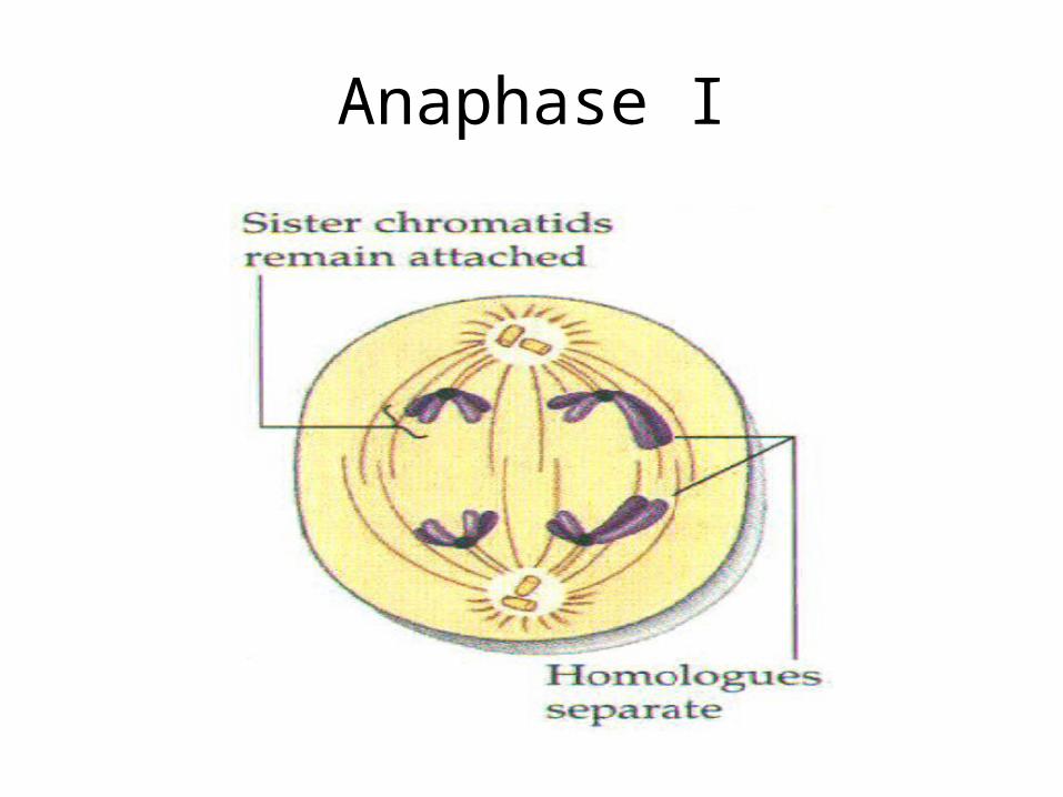
Anaphase I
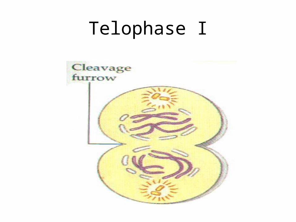
Telophase I
Meiosis II
• Meiosis II is simply a mitotic division of each of the haploid cells produced in Meiosis I. There is no interphase between Meiosis I and Meiosis II and the latter begins with:
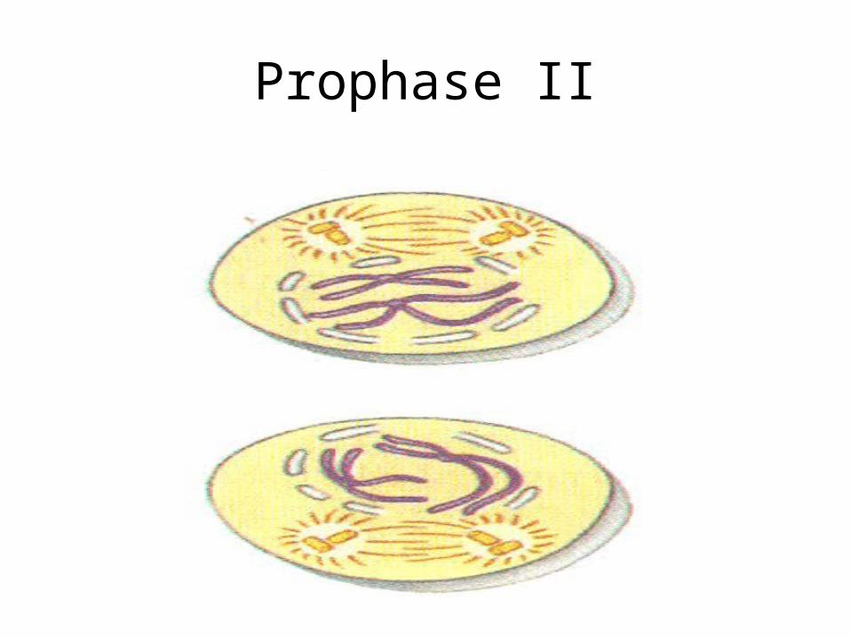
Prophase II
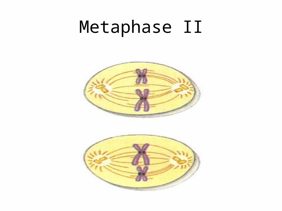
Metaphase II
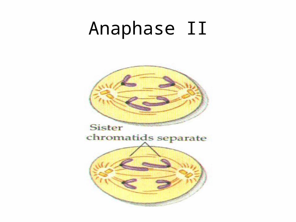
Anaphase II
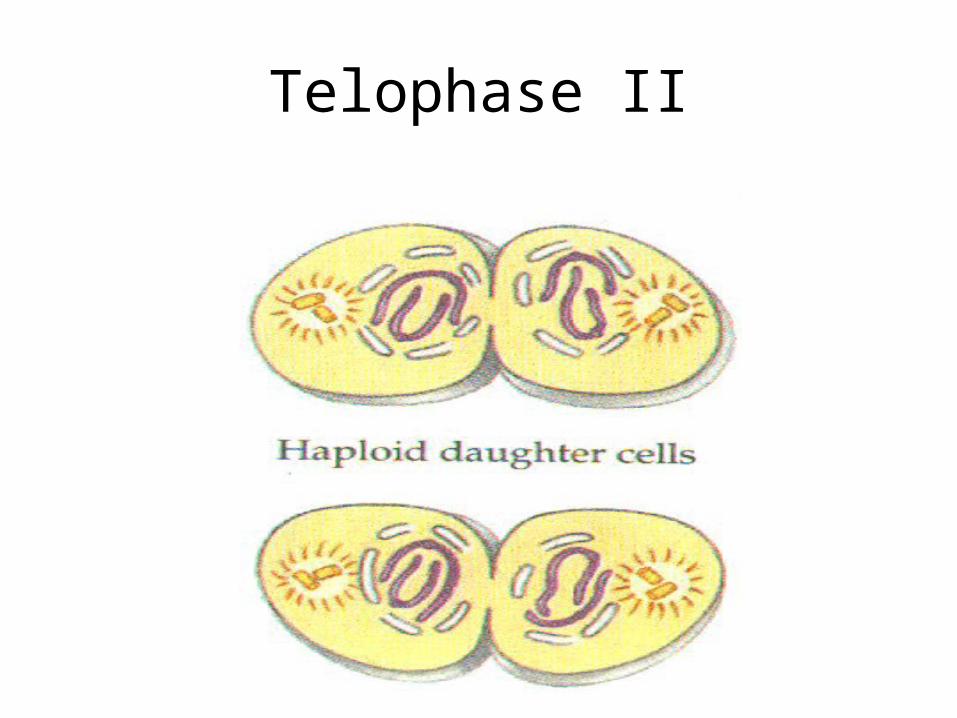
Telophase II
Summary: Meiosis Steps
• Prophase - Homologous chromosomes in the nucleus begin to pair up with one another and then split into chromatids (one half of a chromosome) where crossing over can occur. Crossing offer can increase genetic variation explained soon.
• Metaphase - Chromosomes line up at the equator of the cell, where the sequence of the chromosomes lined up is at random, increasing genetic variation via independent assortment explained soon.
Summary: Meiosis Steps
• Anaphase - The homologous chromosomes move to opposing poles from the equator
• Telophase - A new nuclei forms near each pole alongside its new chromosome compliment.
• At this stage two haploid cells have been created from the original diploid cell of the parent.
Summary: Meiosis Steps
• Prophase II - The nuclear membrane disappears and the second meiotic division is initiated.
• Metaphase II - Pairs of chromatids line up at the equator
Summary: Meiosis Steps
• Anaphase II - Each of these chromatid pairs move away from the equator to the poles via spindle fibres
• Telophase II - Four new haploid gametes are created that will fuse with the gametes of the opposite sex to create a zygote.
Summary: Meiosis Steps
• When Meiosis II is complete, there will be a total of four daughter cells, each with half the total number of chromosomes as the original cell.
• Increases Genetic diversity
Crossing Over
• During meiosis, when homologous chromosomes are paired together, there are points along the chromosomes that make contact with the other pair. This point of contact is deemed the chiasmata, and can allow the exchange of genetic information between chromosomes. This further increases genetic variation
Mendel’s Work
• Gregor Mendel, an Austrian monk is most famous in this field for his study of the phenotype of pea plants, including the shape of the peas on the pea plants.
• 2 laws:– Segregation– Independent Assortment
Mendel cont’d
• Mendel's goal was to have a firm scientific basis on the relationship of genetic information passed on from parents to offspring
• Did work with Pea plants - looked at traits for color and texture
• For texture - showed more round than wrinkled; for color - more green than yellow
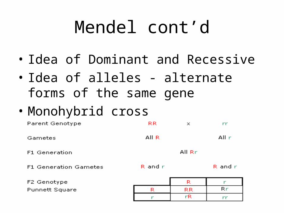
Mendel cont’d
• Idea of Dominant and Recessive
• Idea of alleles - alternate forms of the same gene
• Monohybrid cross
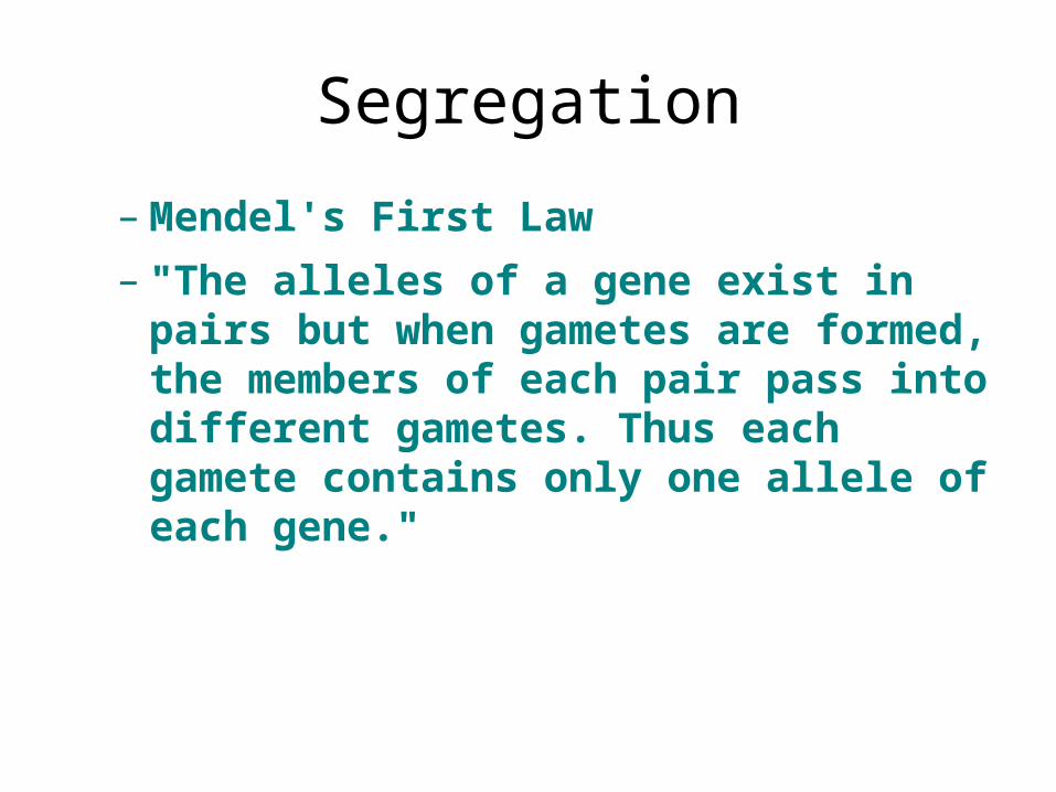
Segregation
– Mendel's First Law
– "The alleles of a gene exist in pairs but when gametes are formed, the members of each pair pass into different gametes. Thus each gamete contains only one allele of each gene."
Independent Assortment
• Genes affecting different traits will separate independently from one another during gamete formation.
• Dihybrid cross - see example next slide
• Can use to see how 2 or more genes will sort out
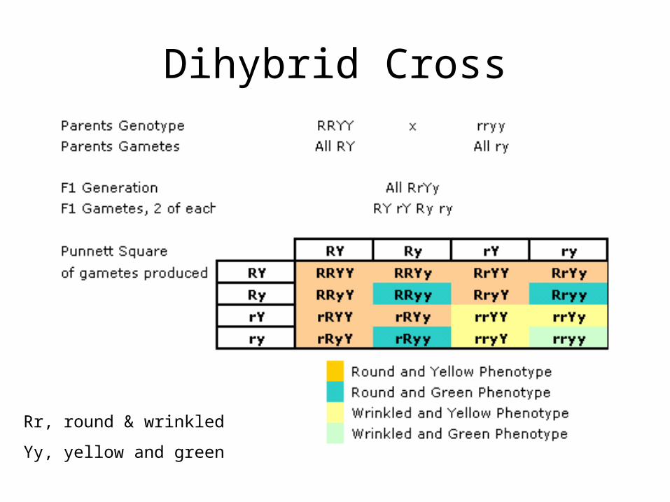
Dihybrid Cross
Rr, round & wrinkled
Yy, yellow and green
New convention for naming
• See overhead
• now would be Ww and Gg
• Exceptions:– incomplete dominance– multiple alleles– linked genes
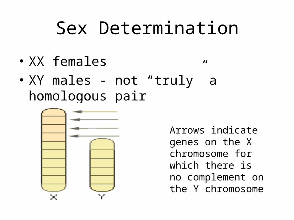
Sex Determination
• XX females
• XY males - not “truly” a homologous pair
Arrows indicate genes on the X chromosome for which there is no complement on the Y chromosome
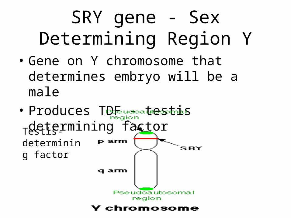
SRY gene - Sex Determining Region Y
• Gene on Y chromosome that determines embryo will be a male
• Produces TDF - testis determining factor
Testis-determining factor
Sex-linked Genes
• Some examples:Red-Green colour blindnessHemophilia - A condition which
prevents the clotting of the bloodDMD - muscular dystrophyHypertension
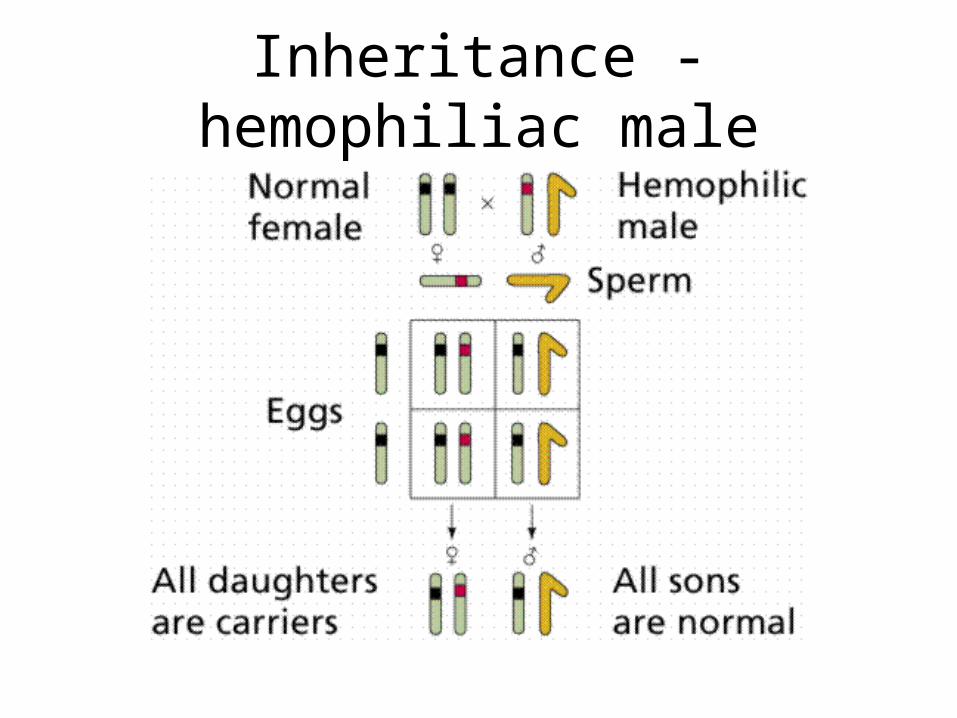
Inheritance - hemophiliac male
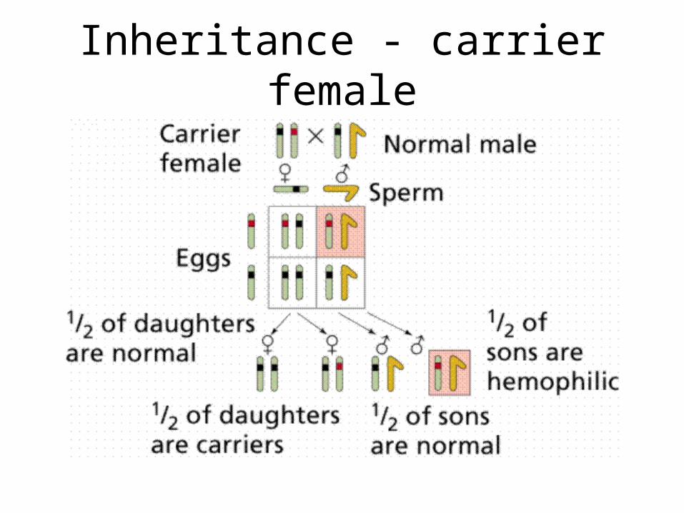
Inheritance - carrier female
Pedigree Analysis
• Charts to look at inheritance pattern of genes in humans
• http://www.blc.arizona.edu/courses/181gh/rick/human_genetics/pedigree.html
• http://www.ndsu.nodak.edu/instruct/mcclean/plsc431/mendel/mendel9.htm
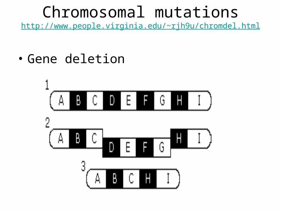
Chromosomal mutationshttp://www.people.virginia.edu/~rjh9u/chromdel.html
• Gene deletion
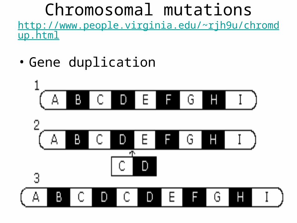
Chromosomal mutationshttp://www.people.virginia.edu/~rjh9u/chromdup.html
• Gene duplication
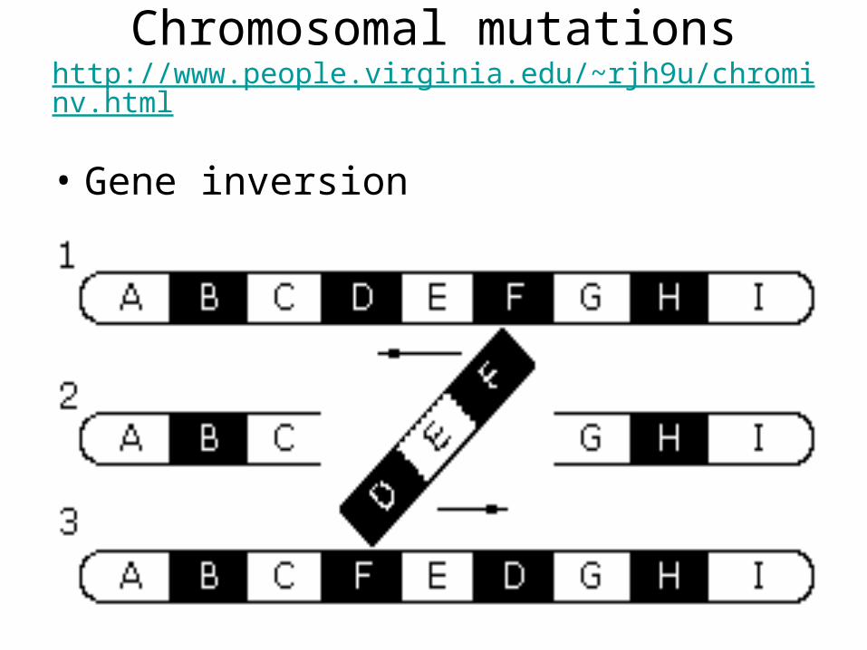
Chromosomal mutationshttp://www.people.virginia.edu/~rjh9u/chrominv.html
• Gene inversion
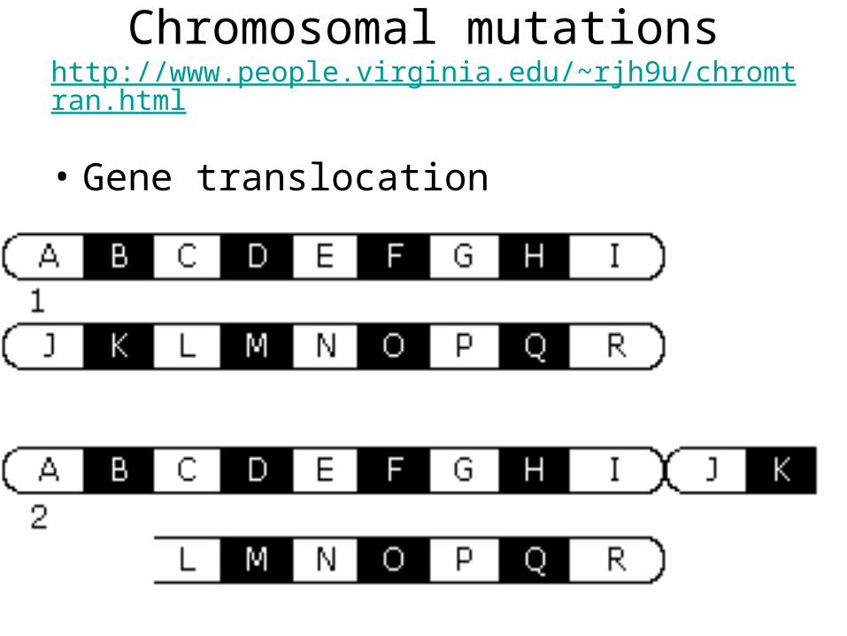
Chromosomal mutationshttp://www.people.virginia.edu/~rjh9u/chromtran.html
• Gene translocation
DNA-level mutation
• Previous examples chromosomal level changes
• changes can occur at DNA level
• also can have deletion, insertion, inversion and substitution
DNA mutation
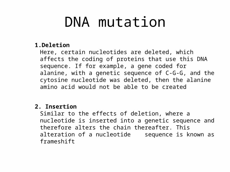
1.DeletionHere, certain nucleotides are deleted, which affects the coding of proteins that use this DNA sequence. If for example, a gene coded for alanine, with a genetic sequence of C-G-G, and the cytosine nucleotide was deleted, then the alanine amino acid would not be able to be created
2. InsertionSimilar to the effects of deletion, where a nucleotide is inserted into a genetic sequence and therefore alters the chain thereafter. This alteration of a nucleotide sequence is known as frameshift
DNA mutation
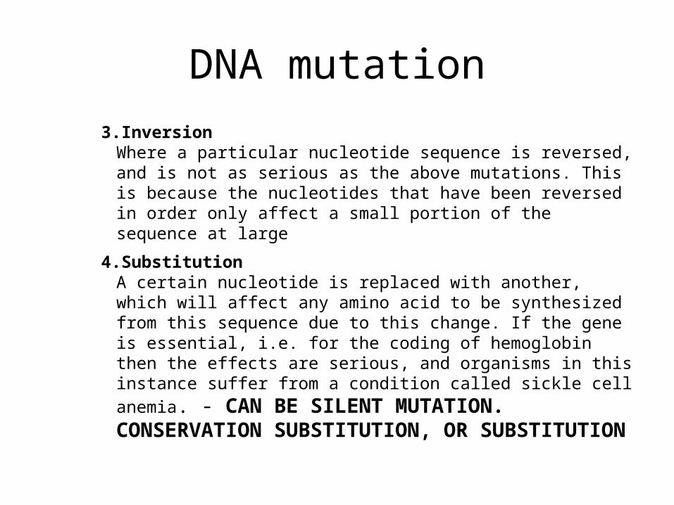
3.InversionWhere a particular nucleotide sequence is reversed, and is not as serious as the above mutations. This is because the nucleotides that have been reversed in order only affect a small portion of the sequence at large
4.SubstitutionA certain nucleotide is replaced with another, which will affect any amino acid to be synthesized from this sequence due to this change. If the gene is essential, i.e. for the coding of hemoglobin then the effects are serious, and organisms in this instance suffer from a condition called sickle cell anemia. - CAN BE SILENT MUTATION. CONSERVATION SUBSTITUTION, OR SUBSTITUTION
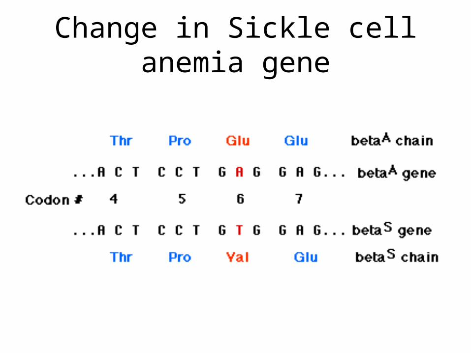
Change in Sickle cell anemia gene

Other related topics
• Polyploidy
• Mutation Frequency
Recessive human genetic disorders
• Brachydactly - first human genetic disorder characterized - short fingers and toes
• Albinism - absence of pigmentation
• Sickle cell anemia - inefficient oxygen transport due to abnormal shape red blood cells
• PKU
• Tay Sachs - neurodegenerative
Dominant Human genetic disorders
• Huntington’s disease - progressive destruction of brain cells
• Polydactly - extra fingers and toes
Intro to Recombinant DNA
• Some unmet medical needs:invasive fungi infections
drug resistant bacteria
hepatitis virus
new vaccines
HIVCancer
Recombinant DNA
– Definition : DNA molecule produced artificially and containing sequences from unrelated organisms.
• Genetic Engineering
• Use of techniques involving recombinant DNA technology to produce molecules and/or organisms with new properties.
• Biotechnology
• All inclusive term for several technologies including but not limited to recombinant DNA. Refers to the use of technology in applications for solving fundamental problems in biology.
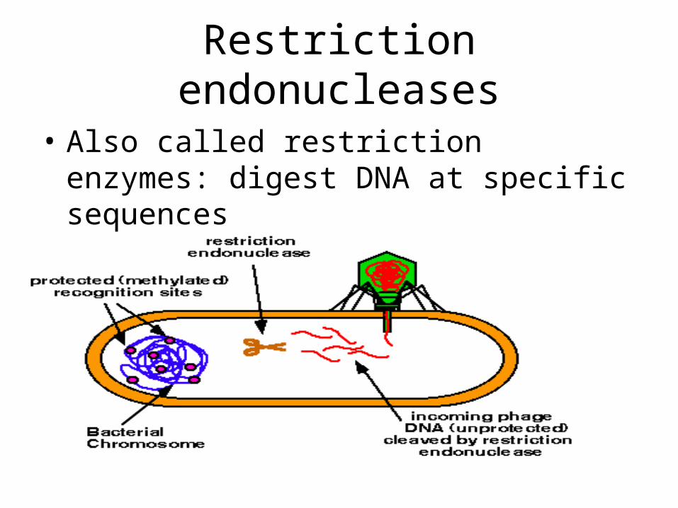
Restriction endonucleases
• Also called restriction enzymes: digest DNA at specific sequences
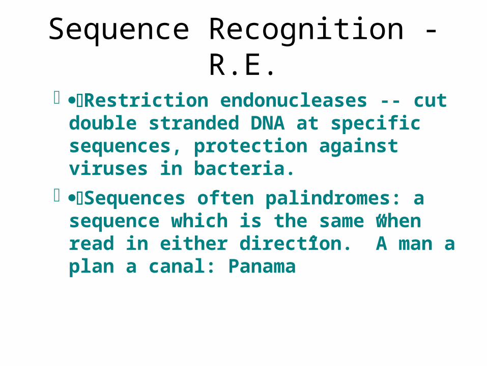
Sequence Recognition -R.E.
Restriction endonucleases -- cut double stranded DNA at specific sequences, protection against viruses in bacteria.
Sequences often palindromes: a sequence which is the same when read in either direction. ”A man a plan a canal: Panama”
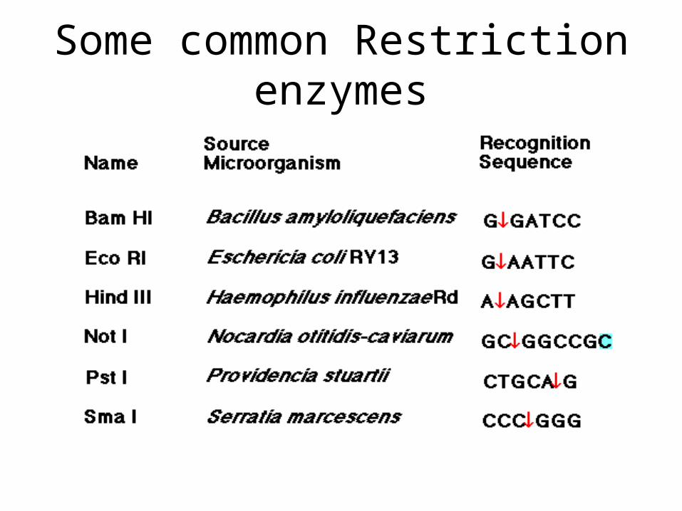
Some common Restriction enzymes
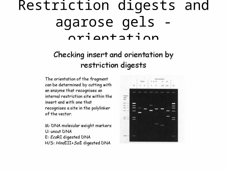
Restriction digests and agarose gels - orientation
DNA ligase
DNA ligase joins 5'-phosphate and 3'-hydroxyl ends of DNA
Two fragments formed by EcoRI can be rejoined by ligase.
• Similarly, Eco RI fragments from two different pieces of DNA can be joined
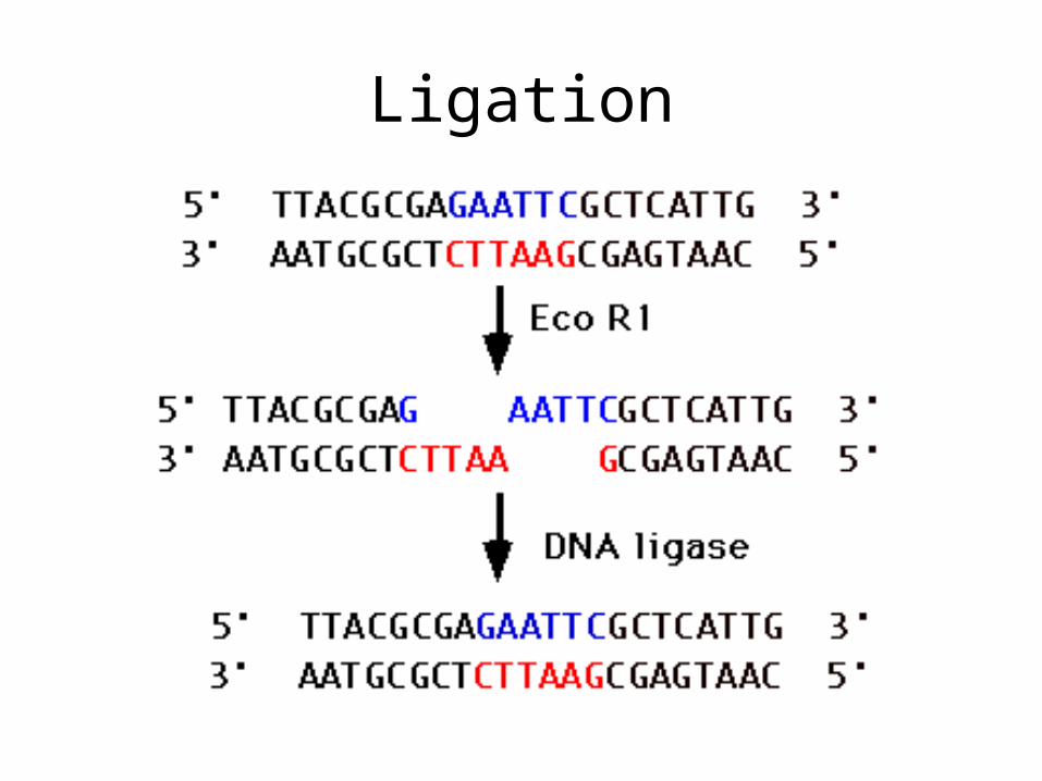
Ligation
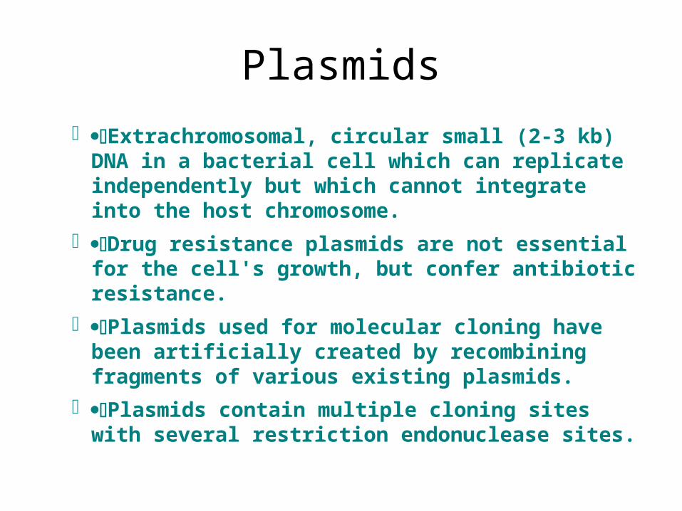
Plasmids
Extrachromosomal, circular small (2-3 kb) DNA in a bacterial cell which can replicate independently but which cannot integrate into the host chromosome.
Drug resistance plasmids are not essential for the cell's growth, but confer antibiotic resistance.
Plasmids used for molecular cloning have been artificially created by recombining fragments of various existing plasmids.
Plasmids contain multiple cloning sites with several restriction endonuclease sites.
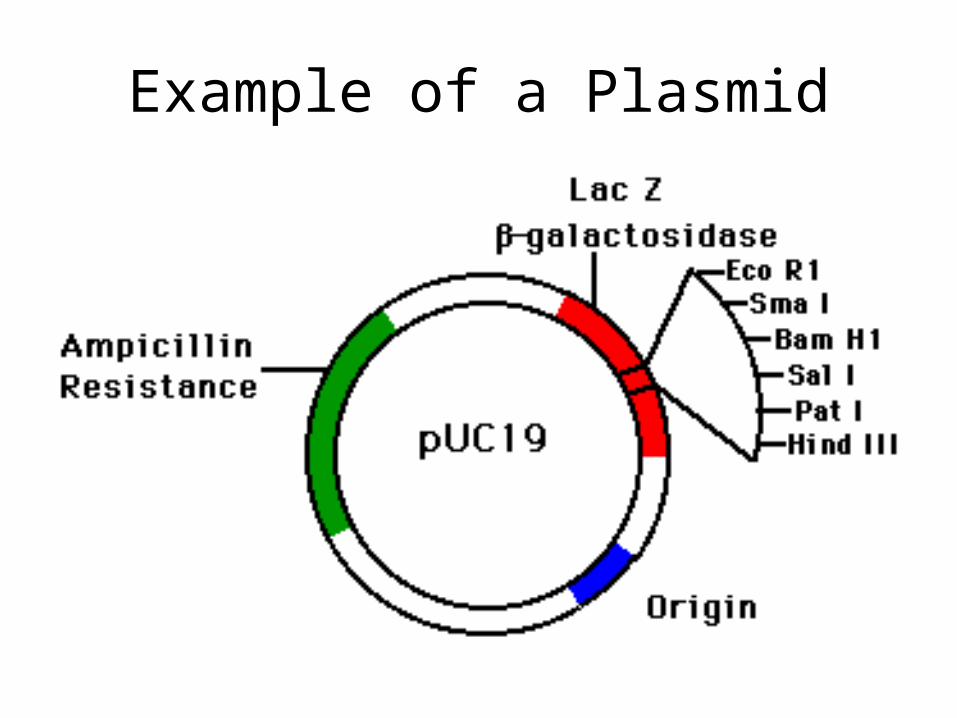
Example of a Plasmid
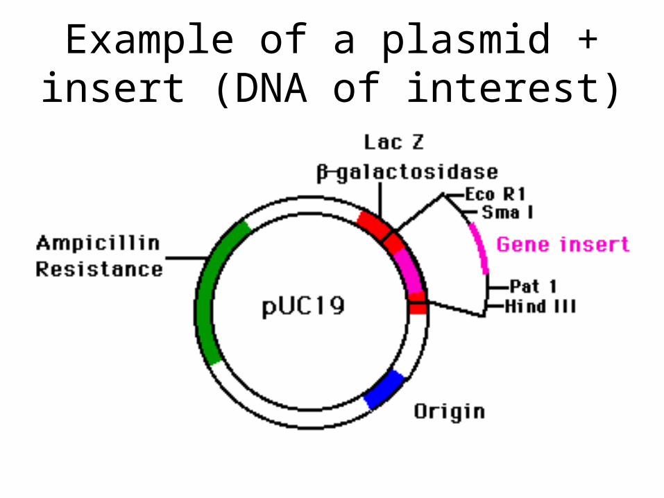
Example of a plasmid + insert (DNA of interest)
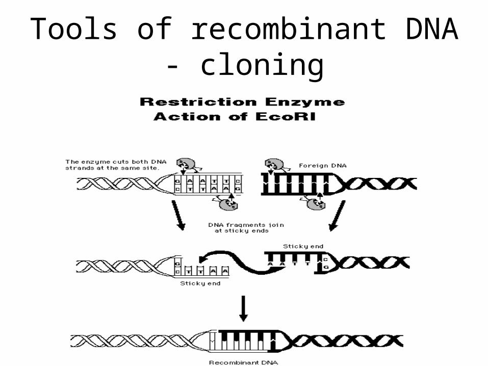
Tools of recombinant DNA - cloning
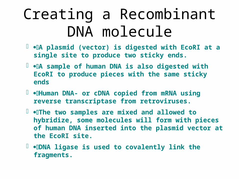
Creating a Recombinant DNA molecule
A plasmid (vector) is digested with EcoRI at a single site to produce two sticky ends.
A sample of human DNA is also digested with EcoRI to produce pieces with the same sticky ends
Human DNA- or cDNA copied from mRNA using reverse transcriptase from retroviruses.
The two samples are mixed and allowed to hybridize, some molecules will form with pieces of human DNA inserted into the plasmid vector at the EcoRI site.
DNA ligase is used to covalently link the fragments.
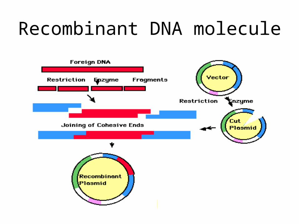
Recombinant DNA molecule
Inserting recombinant DNA into Host
· Transformation
– cell made competent to take up DNA
– competent cells: electroporation – poke holes in membrane and calcium chloride- make cells more permeable to DNA
· Transfection
– when the cloning vector used has aspects of a virus, the host cell can be infected (transfected) to insert the recombinant molecule
· Electroporation
– the cell is placed in an electric field such that small pores are temporarily opened in the membrane. Added DNA can enter through these pores.
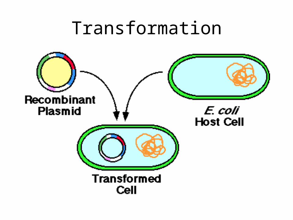
Transformation
Selection
– Antibotic resistance Plasmid vector contains an
ampicillin resistance gene making the cell resistant.
Growth of transformed cells (cells receiving the plasmid) can be identified on agar medium containing (e.g.) ampicillin.
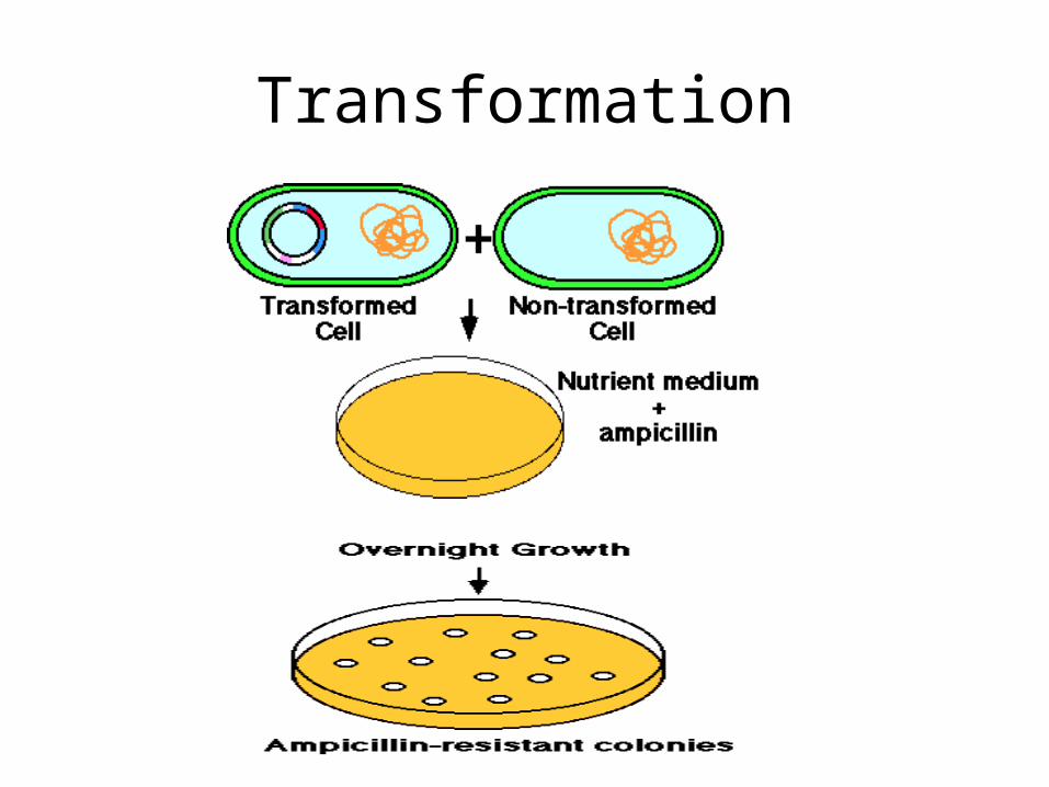
Transformation
Further selection
The plasmid vector contains another identifiable gene (e.g., a second drug resistance or an enzyme activity), with the coding sequence of this gene containing the restriction site for insertion.
Insertion of the foreign DNA at this site interrupts the reading frame of the gene and result in insertional mutagenesis.
In the following example, the -galactosidase gene is inactivated. The substrate "X-gal" turns blue if the gene is intact, ie. makes active enzyme. White colonies in X-gal imply the presence of recombinant DNA in the plasmid.

X-gal selection
Cells ready for DNA uptake
• Competent cells: Treat the cells with calcium chloride which makes the cell membranes more permeable to DNA. This technique succeeds with species that aren't naturally competent e.g. E. coli.
• Electroporation - alternate method
Finding the proper orientation of clone
• Insert can go in both directions
• How to determine correct orientation
• Perform restriction digests using enzymes outside the cloning fragment
• Add total fragments up
• Must add up to right size
Link to Orientation
• http://homepages.strath.ac.uk/%7Edfs99109/BB211/RDTSampleAnswers.html
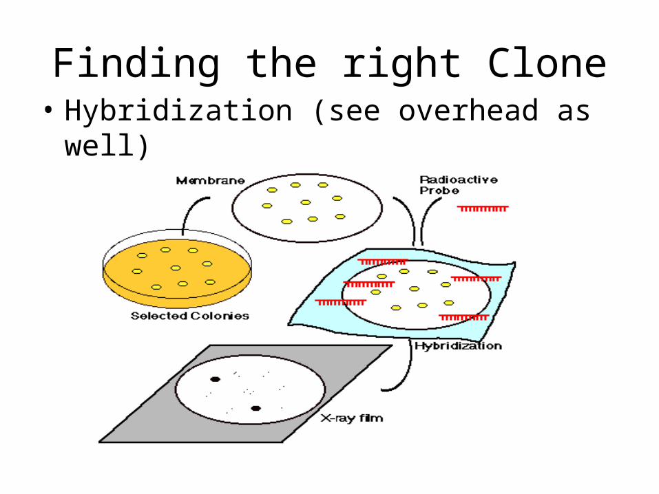
Finding the right Clone• Hybridization (see overhead as well)
Genomic library
• Source of DNA to clone
• all the cells in your body have identical DNA
• problem with this method is introns
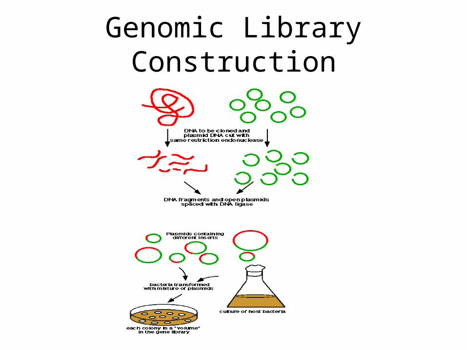
Genomic Library Construction
cDNA libraries: alternate source(complimentary DNA library)
• Made from RNA by reverse transcription (reverse transcriptase is enzyme)
• RNA made into double stranded DNA• comes from tissue that expresses gene(s) of
interest• no introns• source abundant in message• difficult to work with- RNA degrades more
rapidly than DNA
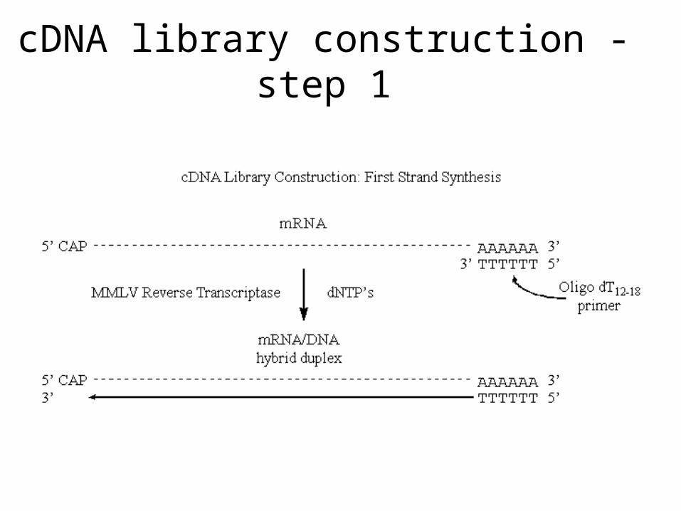
cDNA library construction - step 1
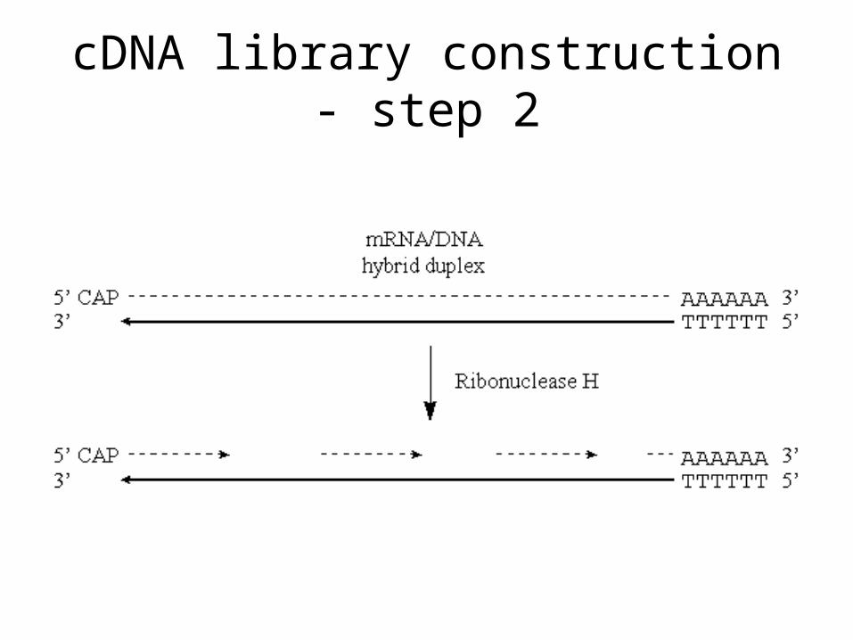
cDNA library construction - step 2
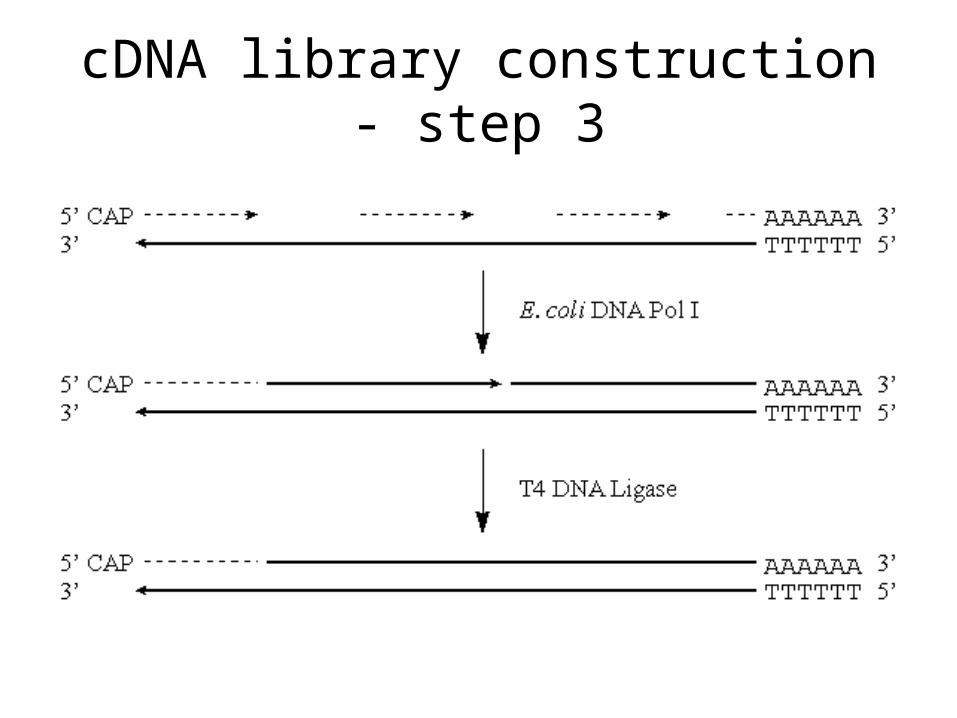
cDNA library construction - step 3
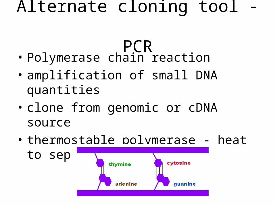
Alternate cloning tool - PCR
• Polymerase chain reaction
• amplification of small DNA quantities
• clone from genomic or cDNA source
• thermostable polymerase - heat to separate DNA strands
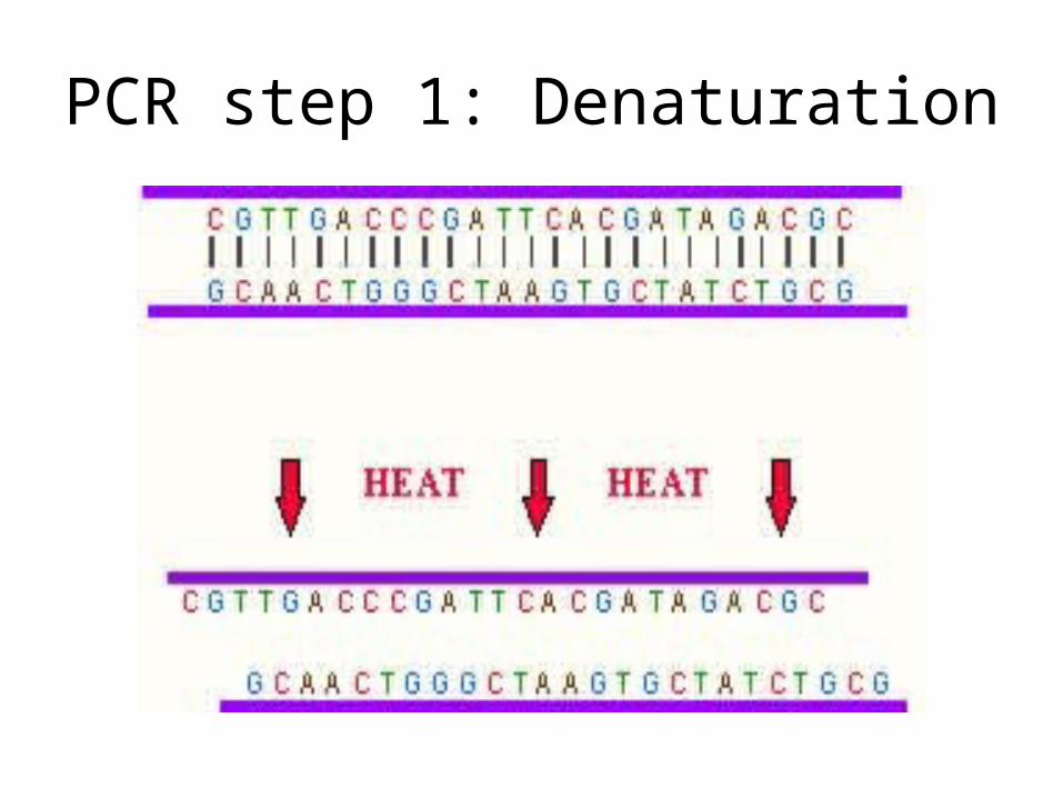
PCR step 1: Denaturation
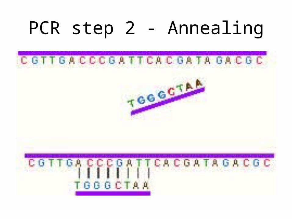
PCR step 2 - Annealing
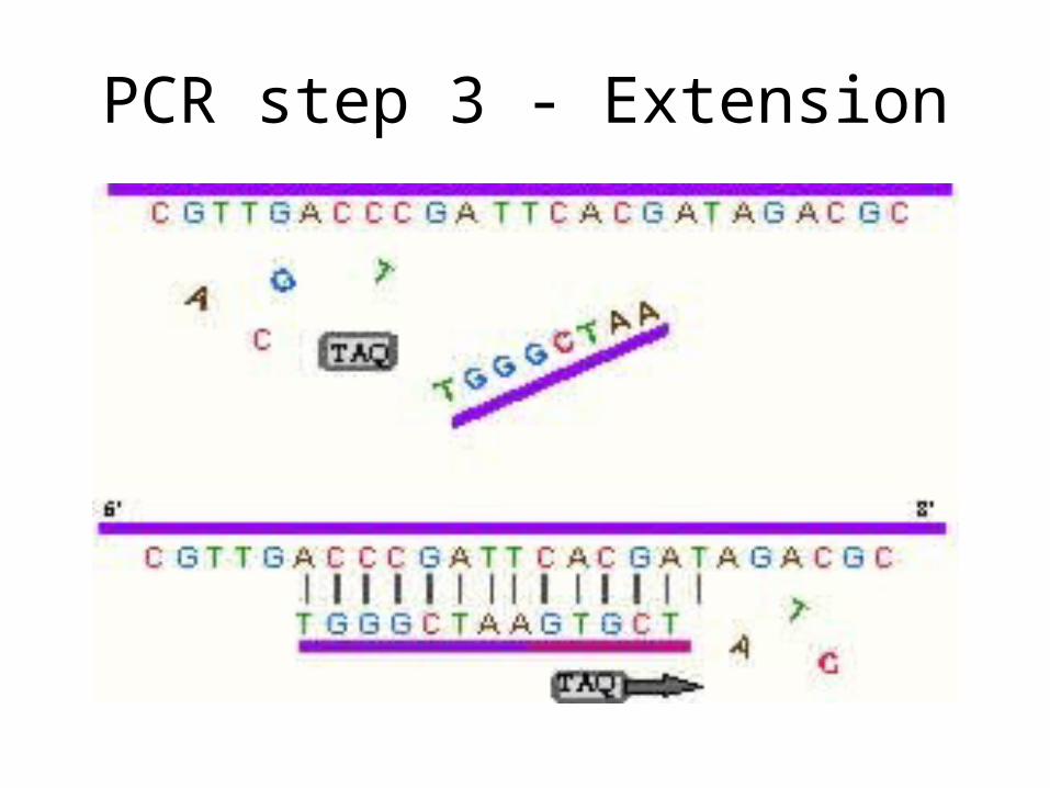
PCR step 3 - Extension
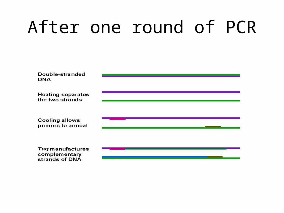
After one round of PCR
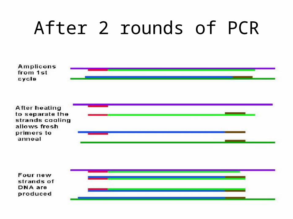
After 2 rounds of PCR
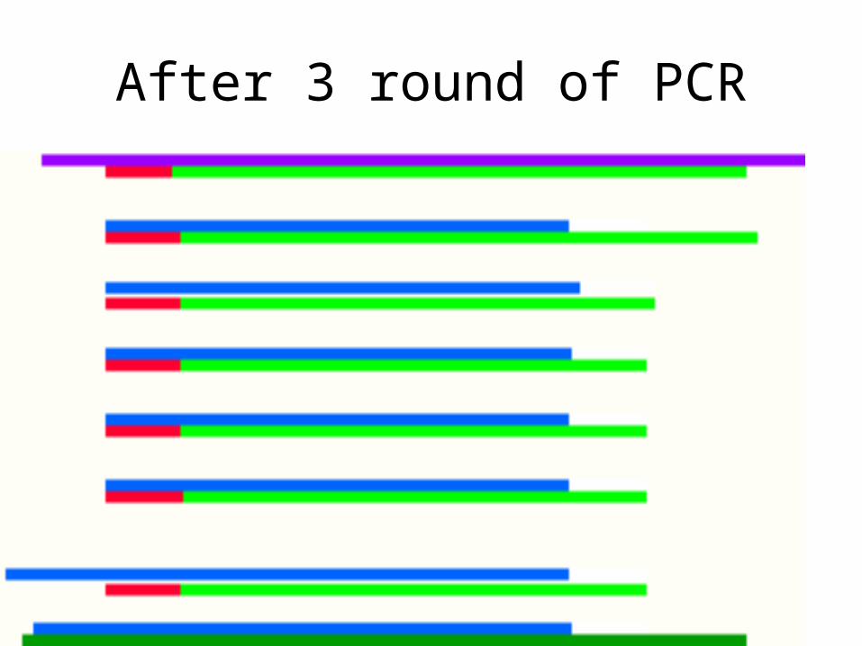
After 3 round of PCR
Required Components of PCR
• DNA template DNA
• thermocycler (or water baths)
• pool of free dNTPs
• Taq (or other heat-stable) DNA polymerase
• Primers - annealed at appropriate temperatures
Conditions for PCR
• Denature: 94C to 100C , 1 minute
• For anneal temperature, 2C for every A and T, 4 C for every C and G. 1minute - 2 minutes - GO 3-5 DEGREES BELOW THAT TEMPERATURE
• Extension: 72 C for 2 minutes
• Do this 30 cycles
• machine programmable
Problem
• What is the annealing temperature for the following primer (a 21 mer)?:
AAGCTTGTCCAGAATTTCGGC
Solution
• 11 A/T X 2 = 22
• 10 C/G X 4 = 40
• 22 + 40 + 62
• Go a few degrees below that number, so you would anneal at about 58C
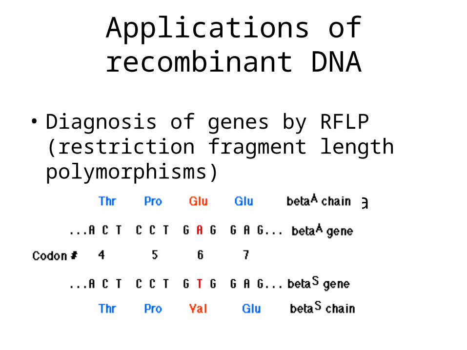
Applications of recombinant DNA
• Diagnosis of genes by RFLP (restriction fragment length polymorphisms)
• Example sickle cell anemia
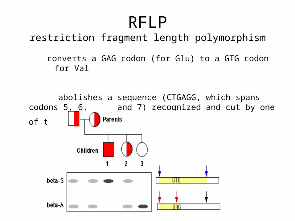
RFLPrestriction fragment length polymorphism
converts a GAG codon (for Glu) to a GTG codon for Val
abolishes a sequence (CTGAGG, which spans codons 5, 6, and 7) recognized and cut by one of the restriction
enzymes.
Other diseases identified by RFLP
• Cystic fibrosis
• Huntington’s disease
• Loss (or gain) of restriction enzyme sites when amino acid change in middle of codon, and thus, protein
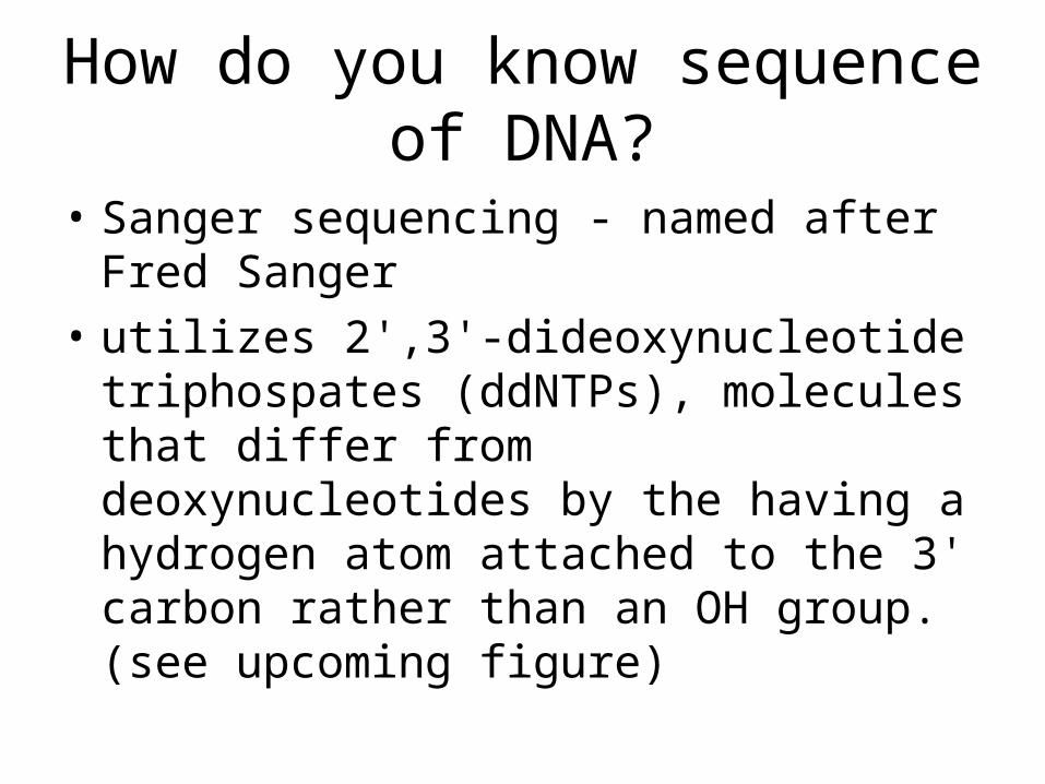
How do you know sequence of DNA?
• Sanger sequencing - named after Fred Sanger
• utilizes 2',3'-dideoxynucleotide triphospates (ddNTPs), molecules that differ from deoxynucleotides by the having a hydrogen atom attached to the 3' carbon rather than an OH group. (see upcoming figure)
Sanger (dideoxysequencing) sequencing
• Need polymerase
• dNTPs
• ddNTPs
• primer
• DNA template
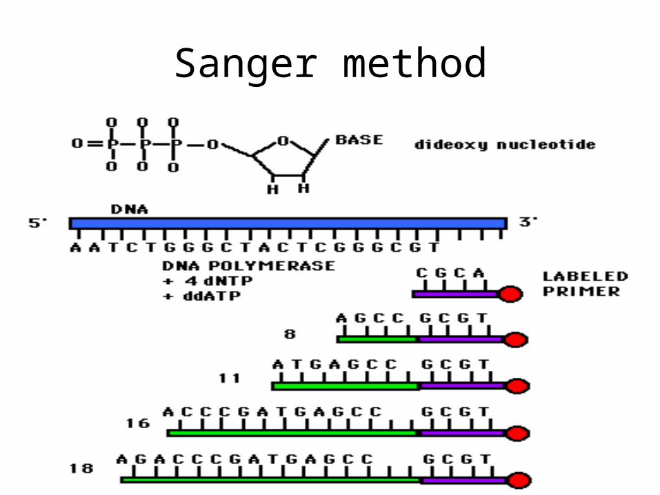
Sanger method
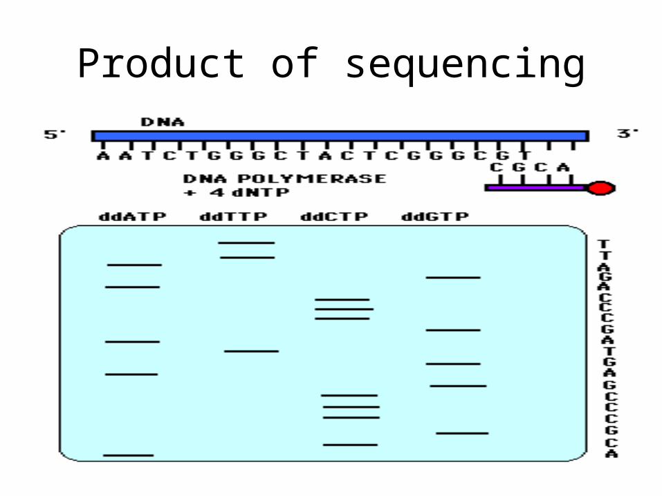
Product of sequencing
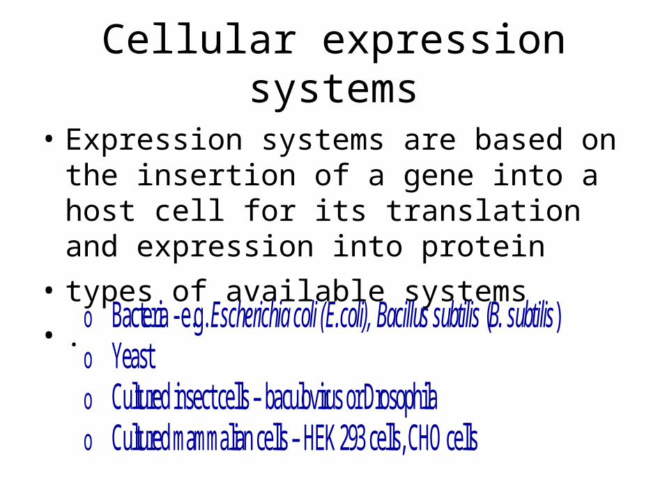
Cellular expression systems
• Expression systems are based on the insertion of a gene into a host cell for its translation and expression into protein
• types of available systems
• . o Bacteria - e.g. Escherichia coli (E.coli), Bacillus subtilis (B. subtilis) o Yeast o Cultured insect cells – baculovirus or Drosophila o Cultured mammalian cells – HEK 293 cells, CHO cells
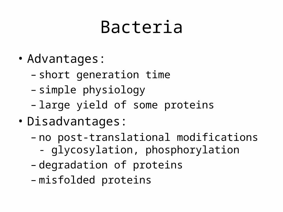
Bacteria
• Advantages:– short generation time– simple physiology– large yield of some proteins
• Disadvantages:– no post-translational modifications -
glycosylation, phosphorylation– degradation of proteins– misfolded proteins
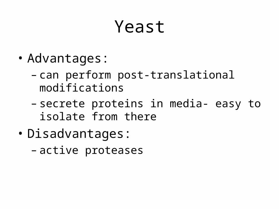
Yeast
• Advantages:– can perform post-translational modifications– secrete proteins in media- easy to isolate from
there
• Disadvantages:– active proteases
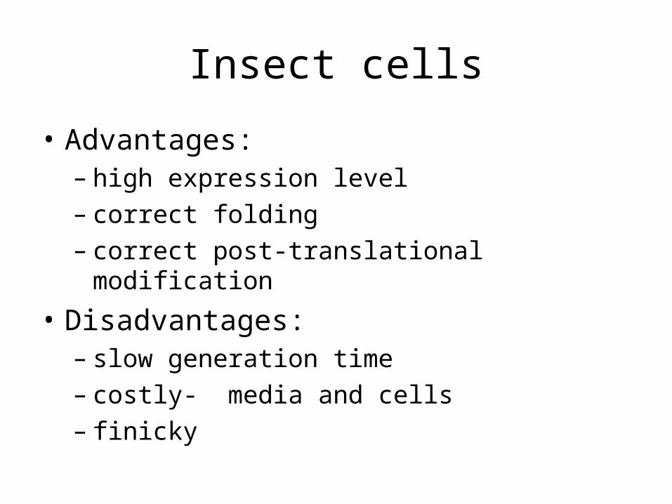
Insect cells
• Advantages:– high expression level– correct folding– correct post-translational modification
• Disadvantages:– slow generation time– costly- media and cells– finicky
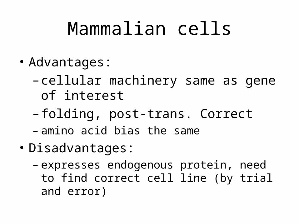
Mammalian cells
• Advantages:
– cellular machinery same as gene of interest
– folding, post-trans. Correct– amino acid bias the same
• Disadvantages:– expresses endogenous protein, need to find
correct cell line (by trial and error)
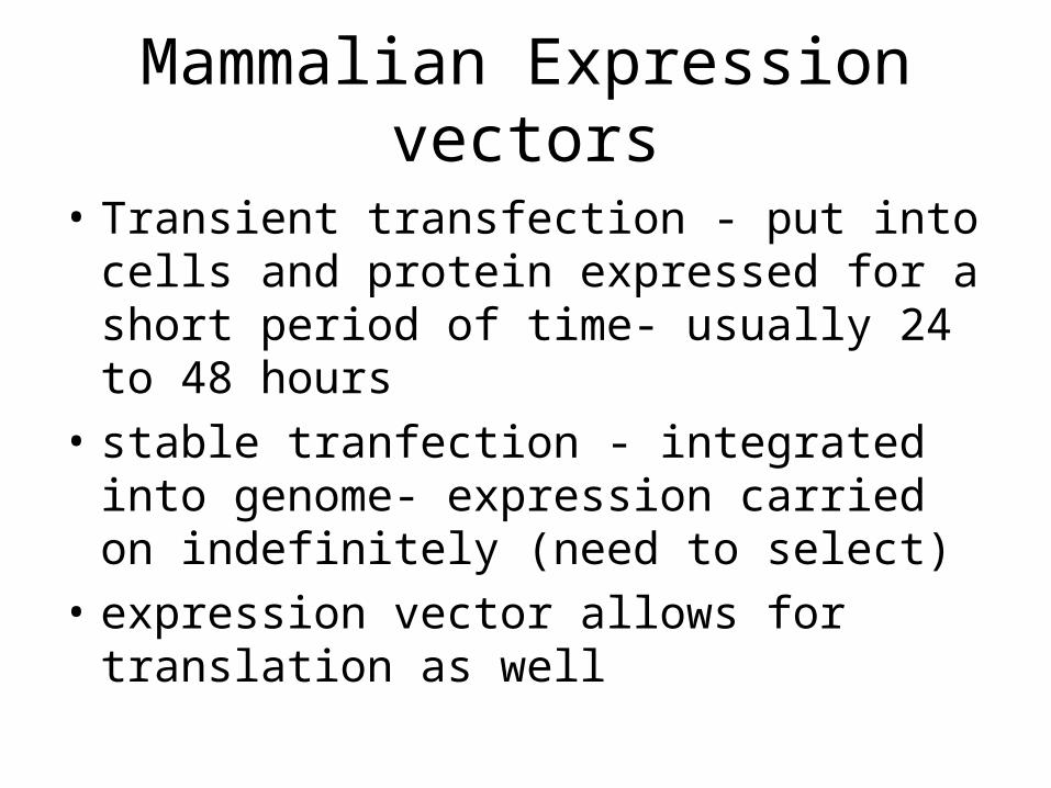
Mammalian Expression vectors
• Transient transfection - put into cells and protein expressed for a short period of time- usually 24 to 48 hours
• stable tranfection - integrated into genome- expression carried on indefinitely (need to select)
• expression vector allows for translation as well
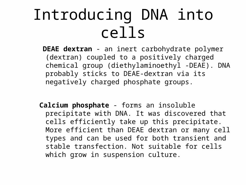
Introducing DNA into cells
DEAE dextran - an inert carbohydrate polymer (dextran) coupled to a positively charged chemical group (diethylaminoethyl -DEAE). DNA probably sticks to DEAE-dextran via its negatively charged phosphate groups.
Calcium phosphate - forms an insoluble precipitate with DNA. It was discovered that cells efficiently take up this precipitate. More efficient than DEAE dextran or many cell types and can be used for both transient and stable transfection. Not suitable for cells which grow in suspension culture.
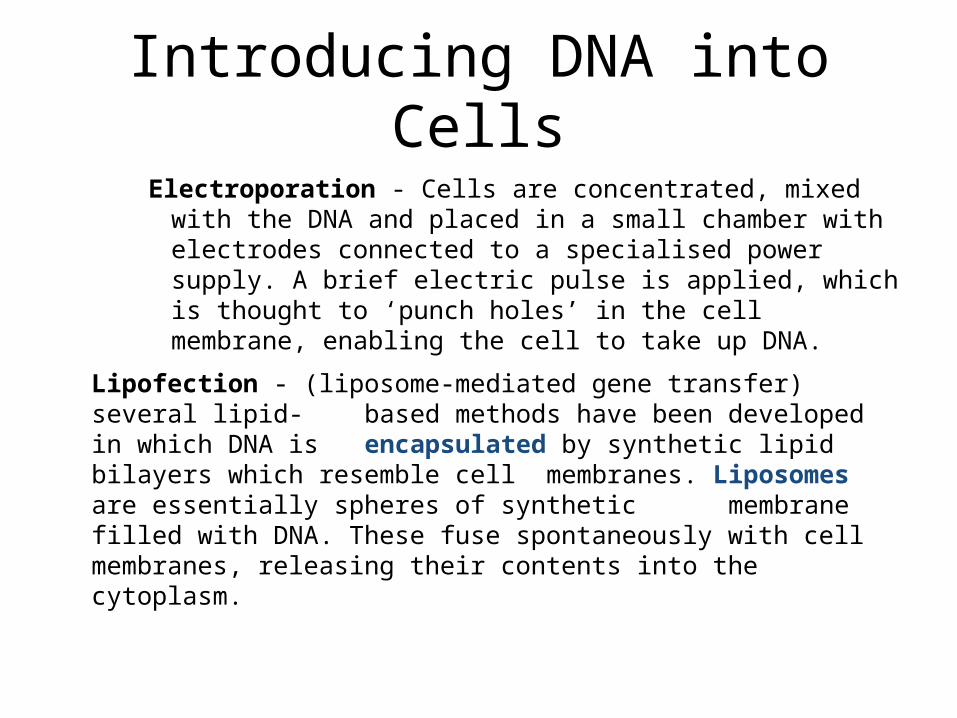
Introducing DNA into Cells
Electroporation - Cells are concentrated, mixed with the DNA and placed in a small chamber with electrodes connected to a specialised power supply. A brief electric pulse is applied, which is thought to ‘punch holes’ in the cell membrane, enabling the cell to take up DNA.
Lipofection - (liposome-mediated gene transfer) several lipid- based methods have been developed in which DNA is
encapsulated by synthetic lipid bilayers which resemble cell membranes. Liposomes are essentially spheres of synthetic membrane filled with DNA. These fuse spontaneously with cell membranes, releasing their contents into the cytoplasm.
Introducing DNA into cells
• Microinjection - The most efficient artificial means of getting DNA into cells. DNA is injected into the nucleus using a microelectrode needle. Very tedious method because each and every cell has to be injected individually. There are now computer-based systems which will assist in the process.
Creating a fusion protein
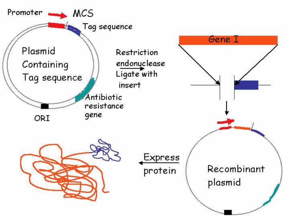
• Gene products are “fused” together, produced as a single polypeptide
• can then use a tag sequence to help isolate that protein
• can purify over a column and get rid of tag by cleavage (cutting)
Technique of Cell Culture
• Follow handout – 4 pages
• How did Tissue Culture develop
• What is Tissue Culture
• How is T.C. performed
• What can go wrong
• References