Embed Size (px)
Citation preview

Genetically Encoded Optochemical Probes for SimultaneousFluorescence Reporting and Light Activation of Protein Functionwith Two-Photon ExcitationJi Luo,†,‡ Rajendra Uprety,‡ Yuta Naro,† Chungjung Chou,‡ Duy P. Nguyen,§ Jason W. Chin,§
and Alexander Deiters*,†
†Department of Chemistry, University of Pittsburgh, Pittsburgh, Pennsylvania 15260, United States‡Department of Chemistry, North Carolina State University, Raleigh, North Carolina 27695, United States§Medical Research Council Laboratory of Molecular Biology, Francis Crick Avenue, Cambridge CB2 0QH, United Kingdom
*S Supporting Information
ABSTRACT: The site-specific incorporation of three new coumarin lysineanalogues into proteins was achieved in bacterial and mammalian cells usingan engineered pyrrolysyl-tRNA synthetase system. The genetically encodedcoumarin lysines were successfully applied as fluorescent cellular probes forprotein localization and for the optical activation of protein function. As aproof-of-principle, photoregulation of firefly luciferase was achieved in livecells by caging a key lysine residue, and excellent OFF to ON light-switchingratios were observed. Furthermore, two-photon and single-photonoptochemical control of EGFP maturation was demonstrated, enabling theuse of different, potentially orthogonal excitation wavelengths (365, 405, and760 nm) for the sequential activation of protein function in live cells. Theseresults demonstrate that coumarin lysines are a new and valuable class of optical probes that can be used for the investigation andregulation of protein structure, dynamics, function, and localization in live cells. The small size of coumarin, the site-specificincorporation, the application as both a light-activated caging group and as a fluorescent probe, and the broad range of excitationwavelengths are advantageous over other genetically encoded photocontrol systems and provide a precise and multifunctionaltool for cellular biology.
■ INTRODUCTION
Good photochemical properties, chemical stability, and ease ofsynthesis make coumarins an important class of fluorescentprobes for biological studies.1−3 In addition to being versatilefluorophores, coumarin chromophores can be used as light-removable protecting groups, so-called “caging groups”, that arephotolyzed through one- and two-photon irradiation.4 Cagedmolecules have been extensively applied in the optical controlof cellular processes.5−9 In particular, the 6-bromo-7-hydrox-ycoumarinmethyl caging group undergoes fast two-photonphotolysis at 740 nm and has been used to optically controlneurotransmitters, secondary messengers, and oligonucleoti-des.10−12 Two-photon irradiation enables optical activation ofbiological processes with enhanced tissue penetration of up to 1mm. Moreover, two-photon caging groups can be released withgreater precision in three-dimensional space than simple one-photon caging groups.4,13
Here we report the site-specific incorporation of threecoumarin amino acids into proteins via genetic code expansionwith unnatural amino acids (UAAs)14−16 to integrate theoptical properties of coumarin probes into cellular systems.Genetic code expansion requires the addition of orthogonaltranslational machinery to achieve site-specific UAA incorpo-ration into proteins. Recent advances in engineering pyrrolysyl-
tRNA synthetase/tRNA pairs for the incorporation of stericallydemanding amino acids17−20 prompted us to synthesizecoumarin lysines 1−3 (Figure 1A) and to test theirincorporation into proteins. The photochemical characteristicsof these UAAs complement and enhance the properties ofcaged and fluorescent amino acids that have been geneticallyencoded in bacterial and mammalian cells.19−25 Lysines 1−3were assembled in three steps from their correspondingcoumarin alcohols (Supporting Information, Scheme S1).Briefly, the coumarin alcohols were activated with nitrophenylchloroformate and coupled to commercially available Boc-lysine. A global deprotection under acidic conditions furnishedthe corresponding coumarin derivatives 1−3 in good yields.All three coumarin lysines 1−3 contain identical benzopyr-
one cores as fluorescent probes. However, subtle substitutionsresult in a set of coumarin derivatives with unique photo-chemical properties. Introduction of a bromine at the 6-position enables decaging not only with UV (single photon)light (in case of 1), but also near IR (two-photon) excitation(in case of 2).11 In contrast, extension of the coumarin−carbamate linker by a single carbon atom results in coumarin
Received: June 7, 2014Published: October 23, 2014
Article
pubs.acs.org/JACS
© 2014 American Chemical Society 15551 dx.doi.org/10.1021/ja5055862 | J. Am. Chem. Soc. 2014, 136, 15551−15558

lysine 3, which does not undergo photolysis and thusrepresents a stable coumarin amino acid probe. Thus, coumarinlysines 1 and 2 can be used as both fluorescent and light-activated probes for optochemical control of protein functionusing UV or near-IR light, while coumarin lysine 3 may serve asa stable fluorescent probe that does not decage under UVexcitation.
■ RESULTS AND DISCUSSIONThe Methanosarcina barkeri pyrrolysyl tRNA synthetase/tRNACUA (MbPylRS/tRNACUA) is functional and orthogonalin a wide range of organisms, such as Escherichia coli, yeast,mammalian cells, and animals such as Caenorhabditis elegansand Drosophila melanogaster.24,26−31 Furthermore, wild-typePylRS recognizes several unnatural amino acids withoutaccepting any of the 20 common amino acids as a substrate.32
The active site of the PylRS can be further engineered throughdirected evolution to enable the incorporation of additional
unnatural amino acids with new functions, including post-translational modifications, bioconjugation handles, photo-cross-linkers, photocaging groups, and others.14 Thus, wegenerated and screened a panel of MbPylRS mutants, guided bymutants that were previously reported,14 to direct theincorporation of 2 in response to a TAG amber codon inmammalian cells using a mCherry-TAG-EGFP reporter. Cellscontaining a MbPylRS mutant with only two amino acidmutations Y271A and L274M showed UAA-dependentexpression of full-length mCherry-EGFP-HA. The Y271Amutation has previously been reported to direct theincorporation of Nε-carbamate-linked lysines,33 while theL274M mutation34,35 was discovered to facilitate higheramber suppression activities with 2 in vivo, because it allowsgreater flexibility of the side chain and imposes less steric bulkat the back of the hydrophobic pocket. This synthetase, termedBhcKRS, enabled the site-specific incorporation of not only 2but also 1 and 3 in response to the amber codon TAG withinsfGFP-Y151TAG-His6 in E. coli (Figure 1). This is notsurprising, considering the very similar structures of 1−3 andprevious observations of the high promiscuity of PylRS.36,37 Tofurther rationalize the ability of BhcKRS to incorporate 1−3,molecular modeling was employed. The wild-type PylRSstructure (PDB: 2Q7H) was used as a starting template forwhich the Y271A and L274M mutations were introduced usingModeller.38 The mutant structure was energy minimized inAmber molecular dynamics39 before docking 1−3 into theactive site pocket using AutoDock4.40 As expected, 1−3 adoptvery similar poses, reflecting their similarity in structure (seeSupporting Information, Figure S1). The mutated synthetasemodel reveals that the Y271A and L274M mutations greatlyenlarge the binding pocket to accommodate the bulky bicycliccaging group, while also orienting it in a favorable π-stackinginteraction with W382. This orientation also benefits from afavorable H-bond interaction between the coumarin hydroxylgroup and D373. Similar to published crystal structures, theamino group’s positioning is maintained by interactions with astructural water and Y349.41 It has been previously shown thatinteractions with N311 and R295 play an important role inamino acid recognition by the PylRS system.31,41,42 The dockedstructure maintains these key interactions with the carbamatecarbonyl forming a H-bond with N311, while the carboxylicacid forms a H-bond with R295 (Figure 1B,C).SDS-PAGE analysis reveals coumarin fluorescence of the
expressed proteins containing the coumarin lysines 1−3. Nofluorescence is observed for wild-type sfGFP because itsexcitation wavelength (488 nm) does not match that of 1−3(365 nm) and because of the denaturing conditions of the gel.The dependence of protein expression on the presence of 1−3demonstrates that the engineered BhcKRS synthetase has ahigh specificity for coumarin lysines and does not significantlyincorporate any of the common 20 amino acids. Similar resultswere obtained for the incorporation of 1−3 into ubiquitin andmyoglobin in E. coli. Electrospray ionization mass spectrometry(ESI-MS, Supporting Information, Figures S2−S4) showed thatrecombinantly expressed sfGFP-1 and -3 have a mass of28446.22 and 28460.60 Da, in agreement with the expectedmasses of 28446.03 and 28460.04 Da, respectively. ESI-MSanalysis of sfGFP-2 showed a mass of 28445.97 Da, indicating apartial loss of bromine during E. coli expression, possibly due toreductive dehalogenation.43 Overall, these results demonstratethat 1−3 can be incorporated into proteins in E. coli in good
Figure 1. (A) Structures of the genetically encoded coumarin aminoacids for fluorescence reporting and light activation of proteinfunction. (B) Crystal structure of PylRS (2Q7H) with the pyrrolysinesubstrate (yellow) in the active site. (C) Structure of BhcKRS with 1(green) docked into the active site. Dashed blue lines represent H-bond interactions. (D) SDS-PAGE analysis of sfGFP-Y151TAGcontaining 1−3 through incorporation in E. coli. The gel was stainedwith Coomassie blue (top), and coumarin fluorescence was imaged viaexcitation at 365 nm (bottom). (E) Fluorescence micrographs of HEK293T cells expressing the BhcKRS/tRNACUA pair and mCherry-TAG-EGFP-HA in the presence or absence of 1−3. (F) Western blotanalysis of cell lysates using an anti-HA antibody and a GAPDHantibody as a loading control. Full-length protein expression is onlyobserved in the presence of 1−3, and incorporation efficiency with allthree amino acids is similar in mammalian cells.
Journal of the American Chemical Society Article
dx.doi.org/10.1021/ja5055862 | J. Am. Chem. Soc. 2014, 136, 15551−1555815552

yields (8.0 mg/L, 1.6 mg/L, and 2.5 mg/L, respectively, forsfGFP) and with high specificity.To demonstrate that the coumarin lysines 1−3 can also be
genetically incorporated into proteins in mammalian cells,pBhcKRS-mCherry-TAG-EGFP-HA and p4CMVE-U6-PylTwere cotransfected into human embryonic kidney (HEK)293T cells. Cells were incubated for 24 h in the absence of anyunnatural amino acid and in the presence of 1−3 (0.25 mM).Fluorescence imaging revealed EGFP expression only in thepresence of 1−3, indicating specific incorporation of thecoumarin lysines in response to the TAG codon, withoutmeasurable incorporation of endogenous amino acids (Figure1E). This was further confirmed by an anti-HA Western blot oncell lysates from the same experiment (Figure 1F).Furthermore, full-length mCherry-EGFP protein was immuno-precipitated from HEK 293T cells using an immobilizedantibody against the HA-tag and mass spectrometry sequencingconfirmed that 1−3 are site-specifically incorporated intoprotein in mammalian cells (Supporting Information, FigureS5). Importantly, the presence of bromine was verified forprotein containing 2, confirming the genetic encoding of theBhc-caged lysine.Because the coumarin groups on 1 and 2 are caging groups
that can be removed via light exposure, loss of their intrinsicfluorescence can be used as an indicator of protein decagingthrough UV irradiation, as shown in Figure 2A,B. This wasdemonstrated through a UV exposure time-course of purifiedsfGFP-1, followed by SDS-PAGE analysis. The coumarinfluorescence intensity of sfGFP-1 gradually decreases withextended UV exposure as more of the coumarin caging group is
removed from the protein, while the continued presence of theCoomassie-stained protein band indicates stability of theprotein. In a cellular context, this may enable experimentsthat allow for the determination of protein expression, proteinlocalization, and protein decaging using a single optochemicalprobe in a single experiment. In contrast, insertion of an extramethylene unit between the lysine and the fluorophore fullyabrogates photocleavage and thus establishes 3 as a stableamino acid for the site-specific fluorescent labeling of proteins.No change in coumarin fluorescence is observed after UVexposure of sfGFP-3 for 20 min (Figure 2C,D). Due to theidentical fluorophores in 1 and 3 and the stability of 3 to theUV irradiation conditions, the loss of protein fluorescence insfGFP-1 is due to decaging and not due to photobleaching.This is further supported by mass spectroscopic analysis of theproteins before and after UV exposure (see SupportingInformation, Figures S2 and S4).To demonstrate the ability of the genetically encoded
coumarin lysines to act as reporters for protein localization inlive cells, we investigated their utility as a protein nuclearlocalization marker. A plasmid was constructed to expressEGFP-HA with an N-terminal NLS (nuclear localization signal,pNLS-linker-EGFP-HA),44 which reliably localizes EGFP to thenucleus (Supporting Information, Figure S6). A TAG ambercodon was introduced in the linker between the NLS andEGFP, allowing for site-specific unnatural amino acidincorporation without affecting EGFP formation or nucleartranslocation. Cells cotransfected with the pNLS-KTAG-EGFPand BhcKRS/PyltRNACUA plasmid pair in the presence of 1(0.25 mM) were analyzed for coumarin fluorescence (405 nmexcitation, 450−480 nm emission) and EGFP fluorescence(488 nm excitation, 490−520 nm emission) by confocalmicroscopy. The observation of complete colocalization of bothfluorophores in the nucleus (merged micrographs) demon-strates the ability to use 1 as a reporter of protein localization(Figure 2E and Supporting Information, movie S1 and FigureS7).To apply the coumarin lysines 1−3 in the optical control of
protein function in live cells, firefly luciferase (Fluc) wasselected as an initial target because bioluminescence measure-ments afford low background, high sensitivity, and easyquantification. On the basis of the Fluc crystal structure, acritical lysine residue, K206, was identified, which is positionedat the edge of the substrate-binding pocket (Figure 3B). It hasbeen proposed that this residue stabilizes and orients ATP inthe active site.45,46 The ε-amino group on K206 provides ahydrogen-bond interaction with the γ-phosphate of ATP andpromotes the adenylation reaction with luciferin, thus beingessential for catalytic activity as shown by the dramatic decreasein enzymatic activity displayed by the K206R mutant.45
Therefore, we hypothesized that a sterically demandingcoumarin caging group placed on K206 would prevent theinteraction with ATP and limit the overall access of thesubstrates to the active site (Figure 3A). Photolysis of thecoumarin lysine would remove the caging group and produce anative lysine residue, restoring the catalytic activity of theenzyme (Figure 3B). A genetically encoded photocaged lysineat K206 would enable the enhanced regulation of the catalyticactivity of firefly luciferase via light activation.Site-directed mutagenesis of the corresponding K206 residue
to the amber codon (TAG) enabled incorporation of 1−3 intofirefly luciferase in mammalian cells. HEK 293T cells werecotransfected with the mutated firefly luciferase plasmid
Figure 2. SDS-PAGE fluorescence analysis shows photodecaging ofsfGFP-1 while sfGFP-3 is stable to UV exposure. (A) Loss of coumarinfluorescence after extended sfGFP-1 in-gel decaging for 0−50 min(365 nm, transilluminator). (B) Coomassie staining reveals identicalsfGFP-1 protein amounts in all lanes. (C) No loss of coumarinfluorescence is observed, since sfGFP-3 does not decage. (D)Coomassie staining reveals identical sfGFP-3 protein amounts in alllanes. (E) Nuclear colocalization of coumarin and EGFP fluorescencein CHO K1 cells cotransfected with pNLS-TAG-EGFP-HA and theBhcKRS/PylT pair (pBhcKRS-4PylT) in the presence of 1 (0.25mM). A DIC image and a merged image of all three channels areshown as well.
Journal of the American Chemical Society Article
dx.doi.org/10.1021/ja5055862 | J. Am. Chem. Soc. 2014, 136, 15551−1555815553
(pGL3-K206TAG) and the MbBhcKRS/PyltRNACUA pair(pBhcKRS-4PylT) in the presence of 1−3 (0.25 mM). After24 h incubation, the cells were either irradiated for 4 min (365nm, 25 W) or kept in the dark. The incorporation of 1−3 intoFluc caused complete inhibition of luciferase activity before UVirradiation, as determined by a Bright-Glo luciferase assay,comparable to the negative control (no unnatural amino acid).After UV irradiation, 1 and 2 were decaged to produce nativelysine, resulting in the activation of firefly luciferase by 34-foldand 31-fold, respectively (Figure 3C). As expected, 3 did notshow any activation of luciferase enzymatic activity uponillumination, as it does not undergo decaging. Therefore, theactivity of firefly luciferase can be tightly optochemicallyregulated by incorporation of a coumarin lysine residue into theactive site of the luciferase protein. Interestingly, attempts toapply 1 and 2 at position K529, another site that can be usedfor optical control of luciferase function,47 led to greatlydiminished luciferase activity, while introduction of ourpreviously reported o-nitrobenzyl-caged lysine24,48,49 workedat both positions K206 and K529. Western blots confirmed thatboth Fluc-K206 → 1 and Fluc-K529 → 1 were expressed atsimilar levels in mammalian cells (Supporting Information,Figures S8 and S9).To observe the optical triggering of protein function via
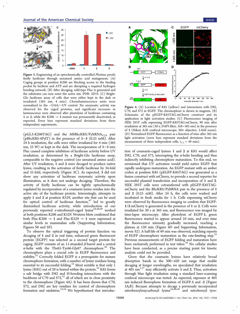
decaging of 1 and 2 in real time, enhanced green fluorescentprotein (EGFP) was selected as a second target protein forcaging. EGFP consists of an 11-stranded β-barrel and a centralα-helix with the Thr65-Tyr66-Gly67 chromophore.50 Thechromophore plays a crucial role in EGFP fluorescence andstability.51 Correctly folded EGFP is a prerequisite for maturechromophore formation, with a number of lysine residues beingessential to its successful folding.52 Most notable is that only 1lysine (K85) out of 20 is buried within the protein.52 K85 formsa salt bridge with D82 and H-bonding interactions with thebackbone of C70 and S72,52 all of which are in close proximityto the chromophore (Figure 4A). It has been shown that C70,S72, and D82 are key residues for control of chromophoreformation and oxidation.53,54 We hypothesized that introduc-
tion of coumarin-caged lysines 1 and 2 at K85 would affectD82, C70, and S72, interrupting the α-helix bending and thusindirectly inhibiting chromophore maturation. To this end, weenvisioned that UV activation would yield native EGFP thatrapidly undergoes maturation. An EGFP mutant with an ambercodon at position K85 (pEGFP-K85TAG) was generated as afusion construct with mCherry, to provide a second reporter forsuccessful plasmid transfection and incorporation of 1 and 2.HEK 293T cells were cotransfected with pEGFP-K85TAG-mCherry and the BhcKRS/PyltRNA pair in the presence of 1and 2 (0.25 mM). After 24 h, the cells were washed andincubated in fresh media for 1 h. Cells expressing mCherrywere observed by fluorescence imaging to confirm that EGFP-1/2-mCherry is generated in the presence of 1 or 2. Cells wereirradiated for 30 s at 365 nm, and fluorescence was imaged bytime-lapse microscopy. After photolysis of EGFP-1, greenfluorescence started to appear around 10 min, and over timethe fluorescence intensity gradually increased, reaching aplateau at 120 min (Figure 4D and Supporting Information,movie S2). A half-life of 49 min was observed, matching reportsof EGFP chromophore maturation as the rate-limiting step.55
Previous measurements of EGFP folding and maturation havebeen exclusively performed in test tubes.56 No cellular studieshave been conducted, as a precise starting point for kineticanalysis could not be provided.Given that the coumarin lysines have relatively broad
absorption bands in the 300−420 nm range that enabledecaging at longer wavelengths, we speculated that irradiationat 405 nm1,57 may efficiently activate 1 and 2. Thus, activationthrough blue light irradiation using a standard laser-scanningconfocal microscope was tested. As expected, exposure at 405nm induced fluorophore formation of EGFP-1 and -2 (Figure5A,B). Because attempts to decage a previously incorporatednitrobenzyloxycarbonyl lysine24,48,49 and nitrobenzyl tyro-
Figure 3. Engineering of an optochemically controlled Photinus pyralisfirefly luciferase through unnatural amino acid mutagenesis. (A)Caging groups at position K206 are blocking access to the bindingpocket by luciferin and ATP and are disrupting a required hydrogenbonding network. (B) After decaging, wild-type Fluc is generated andthe substrates can now enter the active site. PDB: 2D1S. (C) Bright-Glo luciferase assay of cells that were either kept in the dark orirradiated (365 nm, 4 min). Chemiluminescence units werenormalized to the −UAA/−UV control. No enzymatic activity wasobserved for the caged proteins, and significant increases inluminescence were observed after photolysis of luciferase containing1 or 2, while the K206 → 3 mutant was permanently deactivated, asexpected. Error bars represent standard deviations from threeindependent experiments.
Figure 4. (A) Location of K85 (yellow) and interactions with D82,C70, and S72 in EGFP. The chromophore is shown in magenta. (B)Schematic of the pEGFP-K85TAG-mCherry construct and itsapplication in light activation studies. (C) Fluorescence imaging ofHEK 293T cells expressing EGFP-K85TAG-mCherry, 90 min afterirradiation at 365 nm (30 s, DAPI filter, 358−365 nm) in the presenceof 1 (Nikon A1R confocal microscope, 20× objective, 2-fold zoom).(D) Normalized EGFP fluorescence as a function of time after 365 nmlight activation (error bars represent standard deviations from themeasurement of three independent cells, t1/2 = 49 min).
Journal of the American Chemical Society Article
dx.doi.org/10.1021/ja5055862 | J. Am. Chem. Soc. 2014, 136, 15551−1555815554

sine19,58,59 at 405 nm were not successful on comparable timescales and at comparable illumination power (data not shown),the caged lysines 1 and 2 may enable multiwavelengthactivation of proteins caged with the two different opticalprobes.Taking advantage of the two-photon decaging feature of 2,11
photocontrol of EGFP folding by two-photon activation ofEGFP-2 was performed. HEK 293T cells were cotransfectedwith pEGPF-K85TAG-mCherry and pBhcKRS-4PylT in theabsence or presence of 1 and 2 (0.25 mM). After a 24 hincubation, the cells were washed and incubated in fresh mediafor 1 h and irradiated with a multiphoton laser (760 nm, 130mW, 2 μm/s dwell time, 30 cycles, Olympus Fluoview FV1000MPE Multiphoton laser scanning microscope FV10-ASW,MaiTai DSBB-OL IR pulsed laser). Images were acquiredbefore and after two-photon irradiation using both EGFP (488nm) and mCherry (561 nm) excitation. Gratifyingly, an EGFPfluorescent signal was observed after photolysis of 2 at 760 nm(Figure 5C). The cells expressing EGFP containing 1, as acontrol, were also exposed to two-photon excitation (760 nm)and imaged in the same fashion (Figure 5D); no EGFPactivation was observed. In addition to the increased three-dimensional resolution that is provided through two-photonexcitation, effectively shifting the activation wavelength to thenear-IR will enable multiwavelength activation in conjunctionwith other optically triggered biological processes, while alsopreventing any overlap with established fluorescent reporterproteins.
■ SUMMARYThe site-specific genetic incorporation of three new coumarinlysine analogues 1−3 into proteins was achieved in bacterial
and mammalian cells using an engineered BhcKRS synthetasesystem. The genetically encoded coumarin lysines weresuccessfully applied as fluorescent cellular probes for proteinlocalization, and the small size of these coumarin lysines isexpected to minimally perturb protein structure and function,unless they are placed at critical sites. In addition to their smallsize, the spectral properties of 1−3 do not interfere withcommon fluorescent proteins (e.g., EGFP). While the aminoacid 3 showed stability under irradiation conditions, thecoumarins 1 and 2 were readily decaged, generating wild-typelysine residues. As a proof-of-principle, photoregulation offirefly luciferase was achieved in live cells by caging a key lysineresidue, and excellent OFF to ON light-switching ratios wereobserved for 1 and 2. As expected, the stable fluorescent aminoacid 3 did not undergo photolysis. Furthermore, two-photonand single-photon optochemical control of EGFP maturationwas demonstrated, enabling the use of different, potentiallyorthogonal, excitation wavelengths (365, 405, and 760 nm) forthe sequential activation of protein function in live cells. Whilethe caged lysine 2 could be activated using two-photonirradiation at 760 nm, the lysine 1 was stable under theseconditions. However, decaging of 1 was readily achieved withblue light of 405 nm, while a previously encoded o-nitrobenzyl-caged lysine requires UV activation.24,48,49 These resultsdemonstrate that coumarin lysines are a new and valuableclass of optical probes that can potentially be used for theinvestigation and regulation of protein structure, dynamics,function, and localization in live cells. The small size ofcoumarin, the application as both a light-activated caging groupand a fluorescent probe, and the broad range of excitationwavelengths are advantageous over other genetically encodedphotocontrol systems and provide a unique and multifunctionaltool for cellular biology. The ability to incorporate all threecoumarin lysines with the same PylRS/tRNACUA pair furtherfacilitates their application.
■ EXPERIMENTAL SECTIONCloning. (1) Construction of pNLS-TAG-EGFP-HA: The pTAG-
EGFP-HA fragment was amplified from pmCherry-TAG-EGFP-HAusing the PCR primers G1/G2, digested with HindIII and BglII, andligated into pEGFP-N1 (Clontech), generating the pTAG-EGFP-HAplamid. The pNLS PCR fragment was obtained by using primers N1/N2 and then ligated into the HindIII and XbaI sites of pTAG-EGFP-HA to generate the pNLS-TAG-EGFP-HA plasmid. (2) Constructionof pNLS-WT-EGFP-HA: Plasmids were obtained by converting theTAG codon of pNLS-TAG-EGFP-HA into an AAG (Lys) codon usingprimers QC1/QC2 and a QuikChange site-directed mutagenesis kit(Agilent). (3) Construction of pBhcKRS-4PylT: The plasmid wasobtained by ligating the p4CMVE-U6-PylT fragment from pMbPylTbetween the restriction sites NheI and MfeI sites of pMbBhcKRS.
Expression and Purification of Proteins in E. coli. The plasmid,pBAD-sfGFP-Y151TAG-pylT was cotransformed with pBK-BhcKRS24
into E. coli Top10 cells. A single colony was grown in LB mediaovernight, and 500 μL of the overnight culture was added to 25 mL ofLB media, supplemented with 1 mM of the designated unnaturalamino acid and 25 μg/mL of tetracycline and 50 μg/mL of kanamycin.Cells were grown at 37 °C, 250 rpm, and protein expression wasinduced with 0.1% arabinose when the OD600 reached ∼0.6. Afterovernight expression at 37 °C, cells were harvested and washed byPBS. The cell pellets were resuspended in 6 mL of phosphate lysisbuffer (50 mM, pH 8.0) and Triton X-100 (60 μL, 10%), gently mixed,and incubated for 1 h at 4 °C. The cell mixtures were sonicated, andthe cell lysates were centrifuged at 4 °C, 13 000 g, for 10 min. Thesupernatant was transferred to a 15 mL conical tube, and 100 μL ofNi-NTA resin (Qiagen) was added. The mixture was incubated at 4 °Cfor 2 h under mild shaking. The resin was then collected by
Figure 5. Fluorescence confocal imaging of COS-7 cells expressingEGFP-KTAG-mCherry, before and after irradiation at 405 nm (30mW diode laser, 20% laser power, 12.6 μs dwell time, 8 cycles) in thepresence of 2 (A) or 1 (B) (Zeiss confocal LSM710 microscope, 40×water objective). Similar light-activation experiments before and afterirradiation of HEK 293T cells at 760 nm (130 mW, 2 μm/s dwell time,30 cycles, Olympus Fluoview FV1000 MPE, MaiTai DSBB-OL IRpulsed laser), in the presence of 2 (C) or 1 (D), imaged with aOlympus Fluoview1000, 40× oil objective.
Journal of the American Chemical Society Article
dx.doi.org/10.1021/ja5055862 | J. Am. Chem. Soc. 2014, 136, 15551−1555815555

centrifugation (1000g, 10 min), washed twice with 400 μL of lysisbuffer, and followed by two washes with 400 μL of wash buffercontaining 20 mM imidazole. The protein was eluted with 400 μL ofelution buffer containing 250 mM imidazole. The purified proteinswere analyzed by 10% SDS-PAGE and stained with Coomassie Blue.Protein Analysis by ESI-MS. Two different instruments were
used: (A) Protein samples were analyzed using capillary LC ESI-TOFMS. The protein samples were loaded onto a PRLP-S column(Thermo Fisher 5 μm, 1000 A, 300 μm i.d. × 100 mm) on an LCsystem (Ultimate 3000, Dionex, Sunnyvale, CA). The LC system wasdirectly coupled to an electrospray ionization time-of-flight massspectrometer (microTOF, BrukerDaltonics, Billerica, MA). Chromato-graphic separation was performed at a constant flow rate of 3.5 μL/min using a binary solvent system (solvent A: 2.5% acetonitrile and0.1% formic acid; solvent B: 80% acetonitrile and 0.1% formic acid)and a linear gradient program (0−5 min, 5% B; 5−10 min, 5−30% B;10−30 min, 30−75% B; 30−35 min, 75−100% B; 35−45 min, 100−5% B; 45−60 min, 5% B). Mass spectra were acquired in positive ionmode over the mass range m/z 50 to 3000. ESI spectra weredeconvoluted with the MaxEnt algorithm (Data Analysis 3.3, BrukerDaltonics, Billerica, MA), obtaining molecular ion masses with a massaccuracy of 1−2 Da. (B) High-resolution exact mass measurementwere conducted on an Agilent Technologies (Santa Clara, CA) 6210LC-TOF mass spectrometer. Samples were analyzed via a 1 μL flowinjection at 300 μL/min in a water:methanol mixture (25:75 v/v) with0.1% formic acid. The mass spectrometer was operated in positive ionmode with a capillary voltage of 4 kV, nebulizer pressure of 35 psi, anda drying gas flow rate of 12 L/min at 350 °C. The fragmentor andskimmer voltages were 200 and 60 V, respectively. Reference ions ofpurine at m/z 121.0509 and HP-0921 at m/z 922.0098 weresimultaneously introduced via a second orthogonal sprayer and usedfor internal calibration.Coumarin Lysine Incorporation in Human Cells. Human
embryonic kidney (HEK) 293T cells were grown in DMEM(Dulbecco’s Modified Eagle Medium, Gibco) supplemented with10% FBS (Gibco), 1% Pen-Strep (Corning Cellgro), and 2 mM L-glutamine (Alfa Aesar) in 96-well plates (Costar) in a humidifiedatmosphere with 5% CO2 at 37 °C. HEK 293T cells were transientlytransfected with the pMbBhcKRS-mCherry-TAG-EGFP-HA andp4CMVE-U6-PylT24 at ∼75% confluency in the presence or absenceof 1, 2, and 3 (0.25 mM) in 96-well plates. Double transfections wereperformed with equal amounts of both plasmids. After an overnightincubation at 37 °C, the cells were washed by PBS and imaged with aZeiss Axio Observer.Z1Microscope (10× objective). To confirm theexpression of the fusion protein and also differentiate betweenexpression levels, a Western blot was performed. HEK 293T cells werecotransfected with pMbBhcKRS-mCherry-TAG-EGFP-HA andp4CMVE-U6-PylT in the presence or absence of 1, 2, and 3 (0.25mM) in six-well plates. After 24 h of incubation, the cells were washedby chilled PBS, lysed in mammalian protein extraction buffer (GEHealthcare) with complete protease inhibitor cocktail (Sigma) on ice,and the cell lysates were cleared at 13 200 rpm centrifugation (4 °C, 20min). The protein lysate was boiled with loading buffer and thenanalyzed by 10% SDS-PAGE. After gel electrophoresis and transfer toa PVDF membrane (GE Healthcare), the membrane was blocked inTBS with 0.1% Tween 20 (Fisher Scientific) and 5% milk for 1 h. Theblots were probed and incubated with the primary antibody, α-HA-probe (Y-11) rabbit polyclonal lgG (sc-805, Santa Cruz Biotech),overnight at 4 °C, followed by a fluorescent secondary antibody, goat-α-rabbit lgG Cy3 (GE Healthcare), for 1 h at room temperature. Thebinding and washing steps were performed in TBS with 0.1% Tween20.Protein Sequencing by LC-MS/MS. HEK 293T cells were
transfected with pBhcKRS-mCherry-TAG-EGFP-HA and p4CMVE-U6-PylT in a 10 cm Petri dish and incubated with DMEM containing1, 2, or 3 (0.25 mM) for 24 h. Cells were lysed with extraction buffer(GE Healthcare) and the mCherry-1/2/3-EGFP-HA protein wasimmunoprecipitated using the Pierce HA Tag IP/Co-IP kit (Pierce)according to manufacturer’s protocol. The proteins were separated onSDS-PAGE gels and stained with silver stain. Regions corresponding
to the expected molecular weight of mCherry-EGFP-HA were excised,washed with HPLC water, and destained with 50% acetonitrile/25mM ammonium bicarbonate until no visible staining. Gel pieces weredehydrated with 100% acetonitrile and reduced with 10 mMdithiothreitol at 56 °C for 1 h, followed by alkylation with 55 mMiodoacetamide at room temperature for 45 min in the dark. Gel pieceswere then again dehydrated with 100% acetonitrile to remove excessalkylating and reducing agents and rehydrated with 20 ng/μL trypsin/25 mM ammonium bicarbonate and digested overnight at 37 °C. Theresultant tryptic peptides were extracted with 70% acetonitrile/5%formic acid, speed-vac dried, and reconstituted in 18 μL of 0.1% formicacid. Tryptic digests were analyzed by reverse-phased LC-MS/MSusing a nanoflow LC (Waters nanoACQUITY UPLC system, WatersCorp., Milford, MA) coupled online to an LTQ/Orbitrap Velos hybridmass spectrometer (Thermo-Fisher, San Jose, CA). Separations wereperformed using a C18 column (PicoChip column packed with 10.5cm Reprosil C18 3 μm 120 Å chromatography media with a 75 μm IDcolumn and a 15 μm tip, New Objective, Inc., Woburn, MA). Mobilephase A was 0.1% formic acid in water, and mobile phase B was 0.1%formic acid in acetonitrile. Samples were injected onto a trap column(nanoACQUITY UPLC trap column, Waters Corp., Milford, MA) andwashed with 1% mobile phase B at a flow rate of 5 μL/min for 3 min.Peptides were eluted from the column using a 90 min gradient runningat 300 nL/min (5% B for 3 min, 5−36% B in 62 min, 36−95% B in 2min, 95% B for 8 min, 95%−5% B in 1 min, 5% B for 16 min). TheLTQ/Orbitrap instrument was operated in a data-dependent MS/MSmode in which each high resolution broad-band full MS spectra (R =60 000 at mass to charge (m/z) 400, precursor ion selection range ofm/z 300 to 2000) was followed by 13 MS/MS scans in the linear iontrap where the 13 most abundant peptide molecular ions dynamicallydetermined from the MS scan were selected for tandem MS using arelative collision-induced dissociation (CID) energy of 35%. Dynamicexclusion was enabled to minimize redundant selection of peptidespreviously selected for CID. MS/MS spectra were searched with theMASCOT search engine (version 2.4.0, Matrix Science Ltd.) against aUniProt jellyfish proteome database (June 2014 release) from theEuropean Bioinformatics Institute (http://www.ebi.ac.uk/integr8)combined with endogenous mCherry-EGFP fasta sequences. Thefollowing modifications were used: static modification of cysteine(carboxyamidomethylation, +57.0214 Da) and variable modification ofmethionine (oxidation, +15.9949 Da) for all searches, variablemodifications of lysine for mCherry-EGFP-HA (1, +218.17 Da; 2,+295.93 Da; 3, +231.03 Da). The mass tolerance was set at 20 ppm forthe precursor ions and 0.8 Da for the fragment ions. Peptideidentifications were filtered using PeptideProphet and ProteinProphetalgorithms with a protein threshold cutoff of 99% and peptidethreshold cutoff of 95% implemented in Scaffold (Proteome Software,Portland, OR).
Expression of Caged Firefly Luciferase and Light Activation.HEK 293T cells were cultured in DMEM (Dulbecco’s Modified EagleMedium, Gibco) supplemented with 10% FBS (Gibco), 1% Pen-Strep(Gibco), and 2 mM L-glutamine (Alfa Aesar) in 96-well plates (BDFalcon) in a humidified atmosphere with 5% CO2 at 37 °C. At 80−90% confluency, cells seeded on plates were transfected and themedium was changed to fresh DMEM supplemented without or with1, 2, or 3 (0.25 mM). The plasmid pMbBhcKRS-4PylT wasconstructed containing both CMV-MbBhcKRS and 4CMVE-U6-PylT. A TAG amber stop codon was introduced at the K206 siteusing primers GL1/GL2 and a QuikChange mutagenesis kit (AgilentTechnologies). A pGL3-control plasmid containing the gene encodingP. pyralis firefly luciferase with the TAG amber mutation at residueK206 (pGL3-K206TAG) was cotransfected into cells with the plasmidpBhcKRS-4PylT using linear PEI according to the manufacturer’sprotocol (Millipore). After double transfection and 24 h incubation,the medium was changed to DMEM without phenol red, and the cellswere irradiated with UV light (365 nm) for 4 min using a 365 nm UVlamp (high performance UV transilluminator, UVP, 25 W) or kept inthe dark. Cells were lysed by addition of 100 μL of substrate solution(Promega) in a 96-well plate (BD Falcon), and luminescence wasmeasured on a Synergy 4 multimode microplate reader with an
Journal of the American Chemical Society Article
dx.doi.org/10.1021/ja5055862 | J. Am. Chem. Soc. 2014, 136, 15551−1555815556

integration time of 2 s and a sensitivity of 150 or on a Tecan M1000microplate reader with an integration time of 1 s.Visualization of Nuclear Localization through Coumarin
Lysine Incorporation. CHO K1 cells were plated into a polylysine-coated four-well chamber slide (Lab-Tek) and, after incubation to 75%confluency, were transfected with 1 μg of pNLS-KTAG-EGFP andpBhcKRS-4PylT each. After 16 h incubation at 37 °C/5% CO2 inDMEM with 10% FBS in the presence of 1 (0.25 mM), cells werewashed with DMEM without phenol red and then incubated for 2 h.The cells were washed with PBS, fixed with 4% formaldehyde, andstained with rhodamine−phalloidin (Life Technologies). The chamberslide was dried in the dark overnight and cells were imaged on a Zeiss710 confocal microscope (40× water objective).One-Photon Light Activation of EGFP. HEK 293T cells were
plated into a poly-D-lysine-coated eight-well chamber slide (Lab-Tek).After incubation to 70% confluency, cells were transfected withpEGFP-K85TAG-mCherry and pBhcKRS-4PylT (200 ng each). Aftera 20 h incubation at 37 °C/5% CO2 in DMEM with 10% FBS in thepresence of 1 (0.25 mM), cells were washed with DMEM withoutphenol red and then incubated for 1 h. Before light activation,mCherry-expressing cells were identified using the TXRED channel,and imaged with a Nikon A1Rsi confocal microscope (20× objective,2-fold zoom, EGFP (ex. 488 nm) and mCherry (ex. 560 nm)channels). Subsequently, cells were illuminated for 15 s at 365 nmlight (DAPI filter, 358−365 nm), and EGFP and mCherryfluorescence was acquired by time-lapse imaging (every 1 min forthe first 15 min, every 5 min for the following 150 min, scan resolution512 × 512, scan zoom 2× , dwell time 1.9 ms). The mean EGFPfluorescence intensities were quantified using Nikon Elementssoftware.Two-Photon Light Activation of EGFP. HEK 293T cells were
plated into a polylysine-coated μ-dish (ibidi), and after incubation to50% confluency, the cells were transfected with 1 μg each of pEGFP-KTAG-mCherry and pBhcKRS-4PylT. After a 20 h incubation at 37°C/5% CO2 in DMEM with 10% FBS in the presence of 1 or 2 (0.25mM, 0.5% DMSO), cells were washed with DMEM without phenolred and then incubated for 1 h. Cells were imaged with an OlympusFluoview confocal microscope before two-photon irradiation (40× oilobjective, EGFP (ex. 488 nm) and mCherry (ex. 560 nm) channels),imaging positions for mCherry-expressing cells were recorded, and thecell μ-dish was transferred to an Olympus multiphoton microscope forirradiation (Olympus Fluoview FV1000 MPE). Cells were localized atthe previously recorded positions, focused using the mCherry channel,and then irradiated using a 760 nm laser (130 mW, 5% of laser power,30 cycles of scanning, 2 μm/s dwell time, MaiTai DSBB-OL IR pulsedlaser). After irradiation, the cell μ-dish was transferred back to theoriginal microscope for imaging.Mutant PylRS Structure Modeling and Energy Minimization.
The initial template structure of PDB 2Q7H was chosen as a startingpoint for all modeling. The missing loops were remodeled usingMODELER and the two point mutations (Y271A and L274M) wereconstructed using the mutate_model.py script provided by MODELER.Superposition of PDB 2Q7G on top of 2Q7H provided thecoordinates for the incorporation of ATP and magnesium ions intothe newly mutated structure. The ATP and magnesium ions wereparametrized in antechamber using previously developed parame-ters.60,61 The mutated structure was imported into AMBER12 softwareusing the AMBER FF99SBILDN force field.62 The protein was placedinto a cubic box with a 12.0-Å border, solvated with 17 316 watermolecules, and charge neutralized with the addition of six sodium ions.This system was energy minimized first with 5000 steps steepestdescent method, followed by 15 000 steps conjugate gradient methodwith 5 kcal/mol restraints on all atoms. This was followed by another5000 steps steepest descent method, followed by 15 000 stepsconjugate gradient method with 2 kcal/mol restraints on all atomsexcept Y271A and L274M. The resulting energy-minimized structurewas used as the starting structure for all our docking experiments. AllAMBER12 computational experiments were completed on the Centerfor Simulation and Modeling (SAM) Frank supercomputer at theUniversity of Pittsburgh.
Molecular Docking Experiments. The energy-minimized mutantstructure was prepared for docking with AutoDock4 by removing allsodium ions, and all water molecules except for a single water moleculewhich exists in the active site pocket of the protein. This structuralwater molecule is present in all available crystal structures and plays animportant role in amino acid recognition. The receptor input file wasprepared using AutoDock Tools software.40 The side chains for residueL274M were treated as flexible, while all other side chains were keptrigid. The unnatural amino acid ligands were constructed usingChemBioDraw3D, and the molecular geometry was optimized usingthe MMFF94 force field.63 The ligand input files were prepared fordocking using AutoDock Tools as well. Lamarkian genetic algorithmwas used for docking with the following parameters: number of runs:75, ga_pop_size 150, ga_num_evals 250 000 000, ga_num_genera-tions 27 000 were set, all other parameters were kept default. Dockingresults were clustered based on RMSD of each pose. Each coumarinlysine yielded a low energy cluster with binding scores of −9.63 kJ/mol, −6.18 kJ/mol, and −6.14 kJ/mol for 1, 2, and 3, respectively.
■ ASSOCIATED CONTENT*S Supporting InformationProtein mass spectrometry, additional micrographs, NMRspectra, oligonucleotide sequences, and synthesis protocols.This material is available free of charge via the Internet athttp://pubs.acs.org.
■ AUTHOR INFORMATIONCorresponding [email protected] authors declare no competing financial interest.
■ ACKNOWLEDGMENTSWe thank Dr. Dustin Lockney for preparation of the pEGFP-K85TAG plasmid. This work was supported in part by theNational Science Foundation (MCB-1330746, CHE-0848398)and the University of Pittsburgh. This research used the Centerfor Biologic Imaging, the Biomedical Mass SpectrometryCenter and UPCI Cancer Biomarker Facility that are supportedin part by the National Institutes of Health (P30CA047904).J.W.C. is supported by the Medical Research Council(U105181009, UD99999908). D.P.N. was supported by afellowship from Trinity College.
■ REFERENCES(1) Bort, G.; Gallavardin, T.; Ogden, D.; Dalko, P. I. Angew. Chem.,Int. Ed. 2013, 52, 4526−4537.(2) Krueger, A. T.; Imperiali, B. ChemBioChem 2013, 14, 788−799.(3) Goncalves, M. S. Chem. Rev. 2009, 109, 190−212.(4) Klan, P.; Solomek, T.; Bochet, C. G.; Blanc, A.; Givens, R.;Rubina, M.; Popik, V.; Kostikov, A.; Wirz, J. Chem. Rev. 2013, 113,119−191.(5) Brieke, C.; Rohrbach, F.; Gottschalk, A.; Mayer, G.; Heckel, A.Angew. Chem., Int. Ed. 2012, 51, 8446−8476.(6) Riggsbee, C. W.; Deiters, A. Trends Biotechnol. 2010, 28, 468−475.(7) Deiters, A. Curr. Opin. Chem. Biol. 2009, 13, 678−686.(8) Lee, H. M.; Larson, D. R.; Lawrence, D. S. ACS Chem. Biol. 2009,4, 409−427.(9) Baker, A. S.; Deiters, A. ACS Chem. Biol. 2014, 9, 1398−1407.(10) Furuta, T.; Takeuchi, H.; Isozaki, M.; Takahashi, Y.; Kanehara,M.; Sugimoto, M.; Watanabe, T.; Noguchi, K.; Dore, T. M.; Kurahashi,T.; Iwamura, M.; Tsien, R. Y. ChemBioChem. 2004, 5, 1119−1128.(11) Furuta, T.; Wang, S. S.; Dantzker, J. L.; Dore, T. M.; Bybee, W.J.; Callaway, E. M.; Denk, W.; Tsien, R. Y. Proc. Natl. Acad. Sci. U. S. A.1999, 96, 1193−1200.
Journal of the American Chemical Society Article
dx.doi.org/10.1021/ja5055862 | J. Am. Chem. Soc. 2014, 136, 15551−1555815557

(12) Ando, H.; Furuta, T.; Tsien, R. Y.; Okamoto, H. Nat. Genet.2001, 28, 317−325.(13) Helmchen, F.; Denk, W. Nat. Methods 2005, 2, 932−940.(14) Wan, W.; Tharp, J. M.; Liu, W. R. Biochim. Biophys. Acta 2014,1844, 1059−1070.(15) Liu, C. C.; Schultz, P. G. Annu. Rev. Biochem. 2010, 79, 413−444.(16) Chin, J. W. Annu. Rev. Biochem. 2014, 83, 379−408.(17) Xiao, H.; Peters, F. B.; Yang, P. Y.; Reed, S.; Chittuluru, J. R.;Schultz, P. G. ACS Chem. Biol. 2014, 9, 1092−1096.(18) Lang, K.; Davis, L.; Torres-Kolbus, J.; Chou, C.; Deiters, A.;Chin, J. W. Nat. Chem. 2012, 4, 298−304.(19) (a) Deiters, A.; Groff, D.; Ryu, Y.; Xie, J.; Schultz, P. G. Angew.Chem., Int. Ed. 2006, 45, 2728−2731. (b) Arbely, E.; Torres-Kolbus,J.; Deiters, A.; Chin, J. W. J. Am. Chem. Soc. 2012, 134, 11912−11915.(20) Chatterjee, A.; Guo, J.; Lee, H. S.; Schultz, P. G. J. Am. Chem.Soc. 2013, 135, 12540−12543.(21) Liu, W. R.; Wang, Y. S.; Wan, W. Mol. BioSyst. 2011, 7, 38−47.(22) Lemke, E. A.; Summerer, D.; Geierstanger, B. H.; Brittain, S. M.;Schultz, P. G. Nat. Chem. Biol. 2007, 3, 769−772.(23) Summerer, D.; Chen, S.; Wu, N.; Deiters, A.; Chin, J. W.;Schultz, P. G. Proc. Natl. Acad. Sci. U. S. A. 2006, 103, 9785−9789.(24) Gautier, A.; Nguyen, D. P.; Lusic, H.; An, W.; Deiters, A.; Chin,J. W. J. Am. Chem. Soc. 2010, 132, 4086−4088.(25) (a) Uprety, R.; Luo, J.; Liu, J.; Naro, Y.; Samanta, S.; Deiters, A.ChemBioChem 2014, 15, 1793−1799. (b) Nguyen, D. P.; Mahesh, M.;Elsaesser, S. J.; Hancock, S. M.; Uttamapinant, C.; Chin, J. W. J. Am.Chem. Soc. 2014, 136, 2240−2243. (c) Wu, N.; Deiters, A.; Cropp, T.A.; King, D.; Schultz, P. G. J. Am. Chem. Soc. 2004, 126, 14306−14307.(26) Greiss, S.; Chin, J. W. J. Am. Chem. Soc. 2011, 133, 14196−14199.(27) Bianco, A.; Townsley, F. M.; Greiss, S.; Lang, K.; Chin, J. W.Nat. Chem. Biol. 2012, 8, 748−750.(28) Parrish, A. R.; She, X. Y.; Xiang, Z.; Coin, I.; Shen, Z. X.; Briggs,S. P.; Dillin, A.; Wang, L. ACS Chem. Biol. 2012, 7, 1292−1302.(29) Kang, J. Y.; Kawaguchi, D.; Coin, I.; Xiang, Z.; O’Leary, D. D.M.; Slesinger, P. A.; Wang, L. Neuron 2013, 80, 358−370.(30) Hancock, S. M.; Uprety, R.; Deiters, A.; Chin, J. W. J. Am. Chem.Soc. 2010, 132, 14819−14824.(31) Yanagisawa, T.; Ishii, R.; Fukunaga, R.; Kobayashi, T.;Sakamoto, K.; Yokoyama, S. Chem. Biol. 2008, 15, 1187−1197.(32) Polycarpo, C. R.; Herring, S.; Berube, A.; Wood, J. L.; Soll, D.;Ambrogelly, A. FEBS Lett. 2006, 580, 6695−6700.(33) Yanagisawa, T.; Ishii, R.; Fukunaga, R.; Kobayashi, T.;Sakamoto, K.; Yokoyama, S. J. Mol. Biol. 2008, 378, 634−652.(34) Lang, K.; Davis, L.; Wallace, S.; Mahesh, M.; Cox, D. J.;Blackman, M. L.; Fox, J. M.; Chin, J. W. J. Am. Chem. Soc. 2012, 134,10317−10320.(35) Schmidt, M. J.; Borbas, J.; Drescher, M.; Summerer, D. J. Am.Chem. Soc. 2014, 136, 1238−1241.(36) Wang, Y. S.; Fang, X. Q.; Chen, H. Y.; Wu, B.; Wang, Z. Y. U.;Hilty, C.; Liu, W. S. R. ACS Chem. Biol. 2013, 8, 405−415.(37) Tharp, J. M.; Wang, Y. S.; Lee, Y. J.; Yang, Y.; Liu, W. R. ACSChem. Biol. 2014, 9, 884−890.(38) Eswar, N.; Webb, B.; Marti-Renom, M. A.; Madhusudhan, M. S.;Eramian, D.; Shen, M. Y.; Pieper, U.; Sali, A. Current Protocols inBioinformatics; Wiley: New York,2006; Chapter 5, Unit 5.6.(39) Case, D. A.; Cheatham, T. E.; Darden, T.; Gohlke, H.; Luo, R.;Merz, K. M.; Onufriev, A.; Simmerling, C.; Wang, B.; Woods, R. J. J.Comput. Chem. 2005, 26, 1668−1688.(40) Morris, G. M.; Huey, R.; Lindstrom, W.; Sanner, M. F.; Belew,R. K.; Goodsell, D. S.; Olson, A. J. J. Comput. Chem. 2009, 30, 2785−2791.(41) Kavran, J. M.; Gundllapalli, S.; O’Donoghue, P.; Englert, M.;Soll, D.; Steitz, T. A. Proc. Natl. Acad. Sci. U. S. A. 2007, 104, 11268−11273.(42) Schneider, S.; Gattner, M. J.; Vrabel, M.; Flugel, V.; Lopez-Carrillo, V.; Prill, S.; Carell, T. ChemBioChem. 2013, 14, 2114−2118.(43) Mohn, W. W.; Tiedje, J. M. Microbiol. Rev. 1992, 56, 482−507.
(44) Zou, Y.; Mi, J.; Cui, J.; Lu, D.; Zhang, X.; Guo, C.; Gao, G.; Liu,Q.; Chen, B.; Shao, C.; Gong, Y. J. Biol. Chem. 2009, 284, 33320−33332.(45) Conti, E.; Franks, N. P.; Brick, P. Structure 1996, 4, 287−298.(46) Fraga, H.; Fernandes, D.; Novotny, J.; Fontes, R.; Esteves daSilva, J. C. ChemBioChem. 2006, 7, 929−935.(47) Zhao, J.; Lin, S.; Huang, Y.; Zhao, J.; Chen, P. R. J. Am. Chem.Soc. 2013, 135, 7410−7413.(48) Gautier, A.; Deiters, A.; Chin, J. W. J. Am. Chem. Soc. 2011, 133,2124−2127.(49) Hemphill, J.; Chou, C.; Chin, J. W.; Deiters, A. J. Am. Chem. Soc.2013, 135, 13433−13439.(50) Ormo, M.; Cubitt, A. B.; Kallio, K.; Gross, L. A.; Tsien, R. Y.;Remington, S. J. Science 1996, 273, 1392−1395.(51) Stepanenko, O. V.; Stepanenko, O. V.; Kuznetsova, I. M.;Verkhusha, V. V.; Turoverov, K. K. Int. Rev. Cell Mol. Biol. 2013, 302,221−278.(52) Sokalingam, S.; Raghunathan, G.; Soundrarajan, N.; Lee, S. G.PLoS One 2012, 7, e40410.(53) Inouye, S.; Tsuji, F. I. FEBS Lett. 1994, 351, 211−214.(54) Pletnev, S.; Subach, F. V.; Dauter, Z.; Wlodawer, A.; Verkhusha,V. V. J. Am. Chem. Soc. 2010, 132, 2243−2253.(55) Iizuka, R.; Yamagishi-Shirasaki, M.; Funatsu, T. Anal. Biochem.2011, 414, 173−178.(56) Kutrowska, B. W.; Narczyk, M.; Buszko, A.; Bzowska, A.; Clark,P. L. J. Phys.: Condens. Matter 2007, 28, 285223.(57) Mastroberardino, P. G.; Orr, A. L.; Hu, X. P.; Na, H. M.;Greenamyre, J. T. Free Radic. Biol. Med. 2008, 45, 971−981.(58) Chou, C.; Deiters, A. Angew. Chem., Int. Ed. 2011, 50, 6839−6842.(59) Edwards, W. F.; Young, D. D.; Deiters, A. ACS Chem. Biol. 2009,4, 441−445.(60) Allner, O.; Nilsson, L.; Villa, A. J. Chem. Theory Comput. 2012,8, 1493−1502.(61) Meagher, K. L.; Redman, L. T.; Carlson, H. A. J. Comput. Chem.2003, 24, 1016−1025.(62) Lindorff-Larsen, K.; Piana, S.; Palmo, K.; Maragakis, P.; Klepeis,J. L.; Dror, R. O.; Shaw, D. E. Proteins 2010, 78, 1950−1958.(63) Halgren, T. A. J. Comput. Chem. 1996, 17, 616−641.
Journal of the American Chemical Society Article
dx.doi.org/10.1021/ja5055862 | J. Am. Chem. Soc. 2014, 136, 15551−1555815558


![3D Coumarin Systems Based on [2.2]Paracyclophane Synthesis](https://img.dokumen.tips/doc/110x75/617bd62db04f62341e536942/3d-coumarin-systems-based-on-22paracyclophane-synthesis-.jpg)


