Embed Size (px)
Citation preview

Genetically encoded molecules for inducibly inactivatingCaV channelsTingting Yang1, Yasir Suhail1, Stanislava Dalton1, Timothy Kernan1 & Henry M Colecraft1,2
Voltage-gated Ca2+ (CaV) channels are central to the biology of excitable cells, and therefore regulating their activity haswidespread applications. We describe genetically encoded molecules for inducibly inhibiting CaV channels (GEMIICCs).GEMIICCs are derivatives of Rem, a Ras-like GTPase that constitutively inhibits Ca2+ currents (ICa). C terminus–truncatedRem1–265 lost the ability to inhibit ICa owing to loss of membrane targeting. Fusing the C1 domain of protein kinase Cc toyellow fluorescent protein (YFP)-Rem1–265 generated a molecule that rapidly translocated from cytosol to plasma membranewith phorbol-12,13-dibutyrate in human embryonic kidney cells. Recombinant CaV2.2 and CaV1.2 channels were inhibitedconcomitantly with C1PKCc-YFP-Rem1–265 membrane translocation. The generality of the approach was confirmed by creatinga GEMIICC using rapamycin-dependent heterodimerization of YFP-FKBP-Rem1–265 and a constitutively membrane-targetedrapamycin-binding domain. GEMIICCs reduced ICa without diminishing gating charge, thereby ruling out decreased number ofsurface channels and voltage-sensor immobilization as mechanisms for inhibition. We introduce small-molecule–regulatedGEMIICCs as potent tools for rapidly manipulating Ca2+ signals in excitable cells.
Ca2+ influx through CaV1 and CaV2 channels (ICa) transduceselectrical signals into essential biological functions, including musclecontraction, synaptic transmission, hormone secretion and geneexpression regulation1. Given their critical role in signal transduction,it is not surprising that CaV channels are important targets forpharmacological molecules and physiological regulation1. Pharmaco-logical block of CaV1 and CaV2 channels is a therapeutic strategy for adiverse group of cardiovascular and neurological diseases, includingangina, hypertension, stroke, migraines and neuropathic pain2–5. Inthis regard, many classes of molecules target CaV channels: dihydro-pyridines, phenylalkylamines and benzothiazepines block CaV1 chan-nels, whereas CaV2 channels are specifically blocked by snail andspider venom toxins.
Despite the existence of many drugs that block CaV channels, thereis a salient need for genetically encoded molecules that induciblyinhibit ICa (GEMIICCs). GEMIICCs have distinct advantages thatwould fill several key gaps that cannot be resolved with the use oftraditional CaV channel blockers. First, their expression could beprecisely regulated in defined populations of excitable cells, therebyproviding customized specificity in inhibiting ICa in vivo. One poten-tial application of GEMIICCs in this regime would be the inducibleinhibition of synaptic transmission in specific neuron populations—adesirable functionality for studying the physiology of defined neuralcircuits6,7. Second, they could be targeted with subcellular precision,which could potentially permit selective inhibition of CaV channelssituated in spatially distinct enclaves within a single cell. Such a
capability would provide a powerful tool to engage an emergingparadigm of CaV channel signaling that has thus far been difficultto study—the idea that local decoding of Ca2+ signals within singleCaV-channel nano- and microdomains may permit individual cells togenerate distinct functional responses from a single messenger: Ca2+
(refs. 8–10). One example of this is the proposal that Ca2+ influxthrough CaV1.2 channels in heart cells may either evoke contraction orcouple to signal transduction cascades that lead to cardiac hypertro-phy, depending on their subcellular localization (apposed to ryano-dine receptors or localized in caveolae)8,11.
Prime candidates for generating GEMIICCs are the Rad/Rem/Gem/Kir (RGK) subfamily of Ras-like GTPases that were discovered topotently inhibit CaV channels by interacting with auxiliary CaVbsubunits12,13. Unfortunately, several characteristics of wild-type RGKproteins limit their utility as GEMIICCs. First, these proteins areconstitutively active, with no known signal capable of dynamicallyregulating their association with CaV channels. This prevents precisetemporal control of their inhibition of ICa. Second, their effect on ICa isdifficult to titrate because it can only be achieved at the level of proteinexpression14, a variable that is not easily regulated experimentally.Third, there is no mechanism to manage their specific subcellularlocalization, which precludes their potential value as discriminatinginhibitors of CaV channels located in distinct compartments withinsingle cells.
Here, we develop one member of the RGK GTPase family, Rem,into a GEMIICC. Starting with the observation that the truncated
Received 20 June; accepted 30 August; published online 21 October 2007; doi:10.1038/nchembio.2007.42
1Calcium Signals Laboratory, Department of Biomedical Engineering, Johns Hopkins University School of Medicine, 720 Rutland Avenue, 726 Traylor Building,Baltimore, Maryland 21205, USA. 2Present address: Department of Physiology and Cellular Biophysics and Department of Pharmacology, Columbia University,College of Physicians and Surgeons, 630 West 168th Street, P&S 7-422, New York, New York 10032, USA. Correspondence should be addressed toH.M.C. ([email protected]).
NATURE CHEMICAL BIOLOGY VOLUME 3 NUMBER 12 DECEMBER 2007 7 9 5
ART ICL ES

Rem protein Rem1–265 is unable to inhibit ICa solely owing to a loss oftargeting to the membrane, we generated fast-acting GEMIICCs byengineering Rem1–265 derivatives that could be inducibly targeted tothe plasma membrane with the use of small molecules. Surprisingly,the generated GEMIICC discriminated between distinct CaV1 andCaV2 channels, providing the unexpected bonus of built-in selectivityin ICa inhibition. Finally, GEMIICCs inhibited ICa without havingany impact on gating charge, thus providing an important newconstraint regarding the mechanism of RGK protein inhibition ofCaV channels.
RESULTSRem1–265 inhibition of CaV2.2 channelsWe determined the functional effects of Rem and various derivativeson ICa in tsA201 cells (T-antigen transformed human embryonickidney (HEK) 293 cells) stably expressing CaV2.2 channels (a1B/b3/a2d subunits)15. To permit assessment of the role played by subcellularlocalization in ICa inhibition, Rem constructs were tagged with YFP onthe N terminus. Control cells transiently transfected with YFP com-plementary DNA showed yellow fluorescence throughout the cell(Fig. 1a) and showed robust whole-cell currents (Fig. 1b) thatactivated at a threshold of �20 mV and peaked at +10 mV(Fig. 1c). In sharp contrast, cells transiently transfected with YFP-Rem showed fluorescence that was enriched in the plasma membrane(Fig. 1d) and, essentially, an ablated ICa across the entire range of testpulse voltages (Fig. 1e,f). What structural determinants are importantfor the potent effects of YFP-Rem on ICa? Compared with theprototypical Ras, RGK proteins are distinguished by characteristic Nand C termini extensions appending a conserved G-box domain16–18.
Although RGK proteins lack consensus sites for prenylation commonto many members of the Ras GTPase superfamily, they associate withthe plasma membrane by a combination of electrostatic and hydro-phobic interactions involving polybasic and aromatic residues in theirC termini19. Because membrane localization is known to be importantfor the function of certain members of the Ras GTPase superfamily20,we investigated the effect of deleting the C terminus of Rem, thusgenerating Rem1–265. Cells transfected with YFP-Rem1–265 cDNAshowed diffuse fluorescence signals predominantly throughout thecytosol (Fig. 1g), which confirms the necessary role of the C terminusextension in membrane targeting of Rem. Most importantly,in agreement with previous observations12, cells expressing YFP-Rem1–265 showed robust whole-cell currents that were essentiallyindistinguishable from those of control cells (Fig. 1h,i). The distincttargeting phenotypes of YFP-Rem and YFP-Rem1–265 were not due togross differences in expression, given that average YFP fluorescenceintensity was essentially the same for the two proteins (19,352 ± 682,n ¼ 24 for YFP-Rem; 18,722 ± 462, n ¼ 25 for YFP-Rem1–265).
The lack of efficacy of YFP-Rem1–265 in inhibiting ICa couldpotentially be explained by two competing mechanisms. First, theC terminus could have a direct role in transducing the inhibitoryeffect of Rem on ICa. Alternatively, the loss of function could ariseindirectly owing to the loss of membrane targeting of Rem1–265. Todistinguish between these two possibilities we introduced a 20-peptidemembrane-targeting amino acid sequence from neuromodulinonto the N terminus of YFP-Rem1–265, thus generating mem-YFP-Rem1–265. When expressed in cells, mem-YFP-Rem1–265 was almostexclusively localized to the plasma membrane (Fig. 1j) by virtue ofpost-translational palmitoylation of a dicysteine motif present inthe transplanted neuromodulin membrane-targeting sequence21.Moreover, mem-YFP-Rem1–265 abolished ICa with a potency similarto that of wild-type Rem (Fig. 1k,l), which explicitly demonstratesthat the loss of function observed with YFP-Rem1–265 was solelyattributable to its inability to target to the plasma membrane.A similar requirement for membrane targeting was previouslyreported for Rem2-mediated inhibition of ICa, albeit with a notabledifference compared with the results presented here. In that report,functional rescue of truncated Rem2 could only be obtained when amembrane-targeting sequence was appended to the C terminus butnot the N terminus22.
Overall, these results demonstrate two opposing features of theRGK GTPase–CaV channel crosstalk that motivates our study. On one
Voltage (mV)
J (p
A p
F–1
) 0
–20
–40
–60–40 –20 0 20 40 60
Voltage (mV)
0
–20
–40
–60–40 –20 0 20 40 60
Voltage (mV)
0
–20
–40
–60–40 –20 0 20 40 60
Voltage (mV)
0
–20
–40
–60–40 –20 0 20 40 60
5 ms 2 nA
5 ms 2 nA
5 ms 2 nA
a b c
d e f
g h i
j k l
YFP
YFP
YFP-Rem
YFP
YFP
YFP
YFP-Rem1–265
mem-YFP-Rem1–265
J (p
A p
F–1
)J
(pA
pF
–1)
J (p
A p
F–1
)
5 ms
–90
–50 mV–40 to +100 mV
2 nA
Figure 1 Role of membrane targeting in Rem-mediated inhibition of CaV2.2
channels. (a) Top, cartoon of YFP. Bottom, confocal image of a tsA201
cell expressing YFP. Scale bar represents 8 mm, here and throughout.
(b) Exemplar whole-cell currents from a tsA201 cell stably expressing
CaV2.2 channels and transiently transfected with YFP cDNA. Traces
shown were elicited with test pulse potentials of �30, �10, +10, +30
and +50 mV. (c) Population current density (J) versus voltage relationship
for control cells expressing YFP. Data are means ± s.e.m., n ¼ 9 for each
point. (d) Top, cartoon of YFP-Rem in which N- and C-terminal extensions
append a conserved G-box domain (hexagon). Bottom, confocal image
showing enrichment of YFP-Rem in the plasma membrane of a transfected
cell. (e) Exemplar currents from a tsA201 cell stably expressing CaV2.2
channels and transiently transfected with YFP-Rem cDNA. (f) Population
J�V relationship for cells expressing YFP-Rem (J). Data are means ±
s.e.m., n ¼ 8 for each point. Control trace from plot in c is reproduced tofacilitate visual comparison (cyan trace). (g–i) Data for YFP-Rem1–265. Same
format as for d�f. Data in i are means ± s.e.m., n ¼ 24 for each point.
(j–l) Data for a membrane-targeted YFP-Rem1–265. Same format as for
d�f. Data in l are means ± s.e.m., n ¼ 11 for each point.
ART ICL ES
79 6 VOLUME 3 NUMBER 12 DECEMBER 2007 NATURE CHEMICAL BIOLOGY

hand, the potent inhibition of ICa makes these proteins attractivecandidates as tools for regulating CaV channel activity in excitablecells. On the other hand, in the native state their potential utility islimited owing to the constitutive nature of CaV channel inhibition andthe inability to readily tune the degree of ICa block. The recognitionthat YFP-Rem1–265 was unable to inhibit ICa solely owing to loss ofmembrane targeting suggests that it might be possible to redress theseshortcomings by dynamically regulating association of YFP-Rem1–265
with the plasma membrane.
C1PKCc-YFP-Rem1–265 permits inducible ICa inhibitionTo generate a Rem1–265 derivative that could be induced to associatewith the plasma membrane by a small molecule, we fused theC1 domain from protein kinase Cg (PKCg) to the N terminus ofYFP-Rem1–265. In PKCg, the C1 domain permits the rapid phorbolester-induced translocation of the enzyme to the plasma membrane23.Before exploring the properties of C1PKCg-YFP-Rem1–265, we firstconducted control experiments to determine the effects of the phorbolester, phorbol-12,13-dibutyrate (PdBu, 1), on YFP-Rem1–265 sub-cellular localization and on basal ICa in tsA201 cells stably expressingCaV2.2 channels. As expected, treatment of cells with 1 mM PdBu hadno impact on the cytosolic distribution of YFP-Rem1–265 (Fig. 2a).Application of PdBu resulted in an increase in whole-cell ICa evoked by+10-mV test pulse depolarizations (Fig. 2b), in a slowly developingmanner (Fig. 2c). Population current density (J)-versus-voltage curvesindicated that the principal effect of PdBu was to cause a leftward shiftin the voltage dependence of channel activation (Fig. 2d). The effect ofPdBu on basal CaV2.2 currents is consistent with previous reports andis presumably mediated by PKC-mediated phosphorylation of CaV2.2channel subunits24,25.
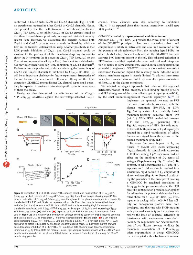
We next turned to our test case. In the basal state, cells transfectedwith C1PKCg-YFP-Rem1–265 showed fluorescence that was diffuselypresent throughout the whole cell (Fig. 3a). On exposure to 1 mMPdBu, C1PKCg-YFP-Rem1–265 rapidly relocated to the plasma (andnuclear) membrane (Fig. 3a), in agreement with previous observa-tions23. One important question was whether the induced targeting ofC1PKCg-YFP-Rem1–265 would translate into an inhibition of ICa.
Indeed, in cells expressing C1PKCg-YFP-Rem1–265, 1 mM PdBu resulted in a markeddecrease in ICa (Fig. 3b), thereby reversingthe polarity of the response observed in thecontrol condition. The PdBu-induceddecrease in ICa occurred rapidly (Fig. 3c),thus providing the first indication of thekinetics of CaV channel inhibition by anRGK protein. Notably, the inhibition of ICa
was faster than the PdBu-induced elevationof control CaV2.2 channels (Fig. 3c). Thisfinding is consistent with the expectation thatRGK proteins inhibit ICa by directly bindingthe CaV channel complex, without using anintermediary biochemical pathway. Popula-tion J�V plots indicated substantial inhibi-tion of ICa across the range of voltages, withno shifts in the voltage dependence of chan-nel activation (Fig. 3d). One desirable attri-bute of a successful GEMIICC is thecapability to readily tune the degree of chan-nel inhibition. This property was evident inthe C1PKCg-YFP-Rem1–265 molecule, as theamount of ICa inhibition was adjustable
with different concentrations of PdBu (Fig. 3e,f), with a half maximalinhibitory concentration (IC50) of 56 nM.
CaV2 channels are targets of two mechanistically distinct types ofphysiological modulation by heterotrimeric G proteins: voltage-dependent inhibition mediated by Gbg subunits26,27 and voltage-independent inhibition involving activated tyrosine kinases28. Todetermine whether CaV channel inhibition mediated by C1PKCg-YFP-Rem1–265 was more analogous to one form of heterotrimericG protein modulation than the other, we applied a discriminatingtest for the voltage dependence of ICa ablation (Fig. 3g). Voltage-dependent Gbg-mediated inhibition of ICa can be reversed by a strongdepolarizing prepulse, termed prepulse facilitation. For channelsinhibited by membrane-translocated C1PKCg-YFP-Rem1–265, a strongdepolarizing prepulse had no impact on the amplitude of ICa recordedat a test potential of +10 mV, which indicates a voltage-independentform of channel modulation (Fig. 3g).
Overall, these results demonstrate the successful generation ofa GEMIICC based on the principle of regulated association ofYFP-Rem1–265 to the plasma membrane.
Differential efficacy of C1PKCc-YFP-Rem1–265
We next determined whether the C1PKCg-YFP-Rem1–265 GEMIICCwas equally effective in inhibiting ICa carried through other CaV
channels. In these experiments, distinct CaV channel a1 subunitswere transiently transfected together with CaVb2a and C1PKCg-YFP-Rem1–265 into HEK 293 cells. Cells coexpressing CaV1.2 channels andC1PKCg-YFP-Rem1–265 responded to 1 mM PdBu with a substantialdecrease in current amplitude (Fig. 4a), which occurred rapidly(Fig. 4b) and was evident across the range of test potentials(Fig. 4c). By contrast, in control cells coexpressing CaV1.2 and YFP-Rem1–265, adding 1 mM PdBu had no appreciable effect on currentamplitude (Supplementary Fig. 1 online).
Surprisingly, similar experiments carried out on either CaV2.1(Fig. 4d–f) or CaV2.3 channels (Fig. 4g–i) showed currents thatwere refractory to inhibition despite visually observed clear-cutPdBu-induced translocation of C1PKCg-YFP-Rem1–265 to the plasmamembrane. So far, Rem protein inhibition of ICa has only been
a
YFP-Rem1–265
YFP
+ 1 µM PdBu
Step to +10 mV
+ 1 µM PdBu
10 ms
b
0 100 200 300 400
0
0.5 nA
c d
40 s 80 s 120 st = 0 s
J (p
A p
F–1
)
Voltage (mV)–20
–10
–20
–30
–400 20 40 60
I/I 0
0.00.20.40.60.81.01.21.41.6
Time (s)
Figure 2 Basal effects of PdBu on CaV2.2 channel currents. (a) Left, cartoon of YFP-Rem1–265. Right,
time series of confocal images showing no change in subcellular localization of YFP-Rem1–265 on
exposure to 1mM PdBu. Scale bar represents 8 mm. (b) Exemplar currents showing upregulation of ICa
by 1 mM PdBu in a tsA201 cell stably expressing CaV2.2 channels and transiently transfected with
YFP-Rem1–265. (c) Time course of the PdBu-induced upregulation of the exemplar current shown in b.
Red symbols represent currents recorded in the presence of PdBu. (d) Population J�V relationships
recorded before (m) and after (m) exposure to 1 mM PdBu. Data are means ± s.e.m., n ¼ 4 for
each point.
ART ICL ES
NATURE CHEMICAL BIOLOGY VOLUME 3 NUMBER 12 DECEMBER 2007 7 9 7
confirmed in CaV1.2 (refs. 12,29) and CaV2.2 channels (Fig. 1), withno experiments reported in either CaV2.1 or CaV2.3 channels. Hence,one possibility for the ineffectiveness of membrane-translocatedC1PKCg-YFP-Rem1–265 to inhibit CaV2.1 or CaV2.3 currents could bethat these channels have a previously unrecognized intrinsic immunityagainst Rem. However, we discounted this scenario because bothCaV2.1 and CaV2.3 currents were potently inhibited by wild-typeRem in the transient cotransfection assay. Another possibility is thatRGK protein inhibition of CaV2.1 and CaV2.3 channels could besensitive to the placement of the membrane-targeting domain toeither the N terminus (as it occurs in C1PKCg-YFP-Rem1–265) or theC terminus (as present in wild-type Rem). Precedent for such behaviorhas previously been noted for Rem2 inhibition of CaV2.2 channels22.Understanding the precise mechanisms underlying the insensitivity ofCaV2.1 and CaV2.3 channels to inhibition by C1PKCg-YFP-Rem1–265
will be an important challenge for future experiments. Irrespective ofthe mechanism, the unexpected differential efficacy of this first-generation GEMIICC among distinct CaV channel types could poten-tially be exploited to engineer customized specificity in future versionsof these molecules.
Finally, we also determined the effectiveness of the C1PKCg-YFP-Rem1–265 GEMIICC against the low-voltage-activated CaV3.1
channel. These channels were also refractory to inhibition(Fig. 4j–l), as expected given their known insensitivity to wild-typeRGK proteins12,13.
GEMIICC created by rapamycin-induced dimerizationAlthough C1PKCg-YFP-Rem1–265 provided the critical proof of conceptof the GEMIICC principle, it has two characteristics that couldcompromise its utility in native cells and also limit realization of thefull potential of this technology. First, the inducing ligand PdBu (orother phorbol ester) does not only activate the GEMIICC, but alsoactivates PKC isoforms present in the cell. This collateral activation ofPKC isoforms and their myriad substrates could confound interpreta-tion of results in some experiments. Second, in this configuration, thepotential to engineer a GEMIICC having a more refined control ofsubcellular localization beyond the relatively crude cytosolic versusplasma membrane regime is severely limited. To address these issueswe explored an alternative method to dynamically regulate associationof Rem1–265 to the plasma membrane.
We adapted an elegant approach that relies on the regulatedheterodimerization of two proteins, FK506-binding protein (FKBP)and FRB (a fragment of the mammalian target of rapamycin, mTOR),by the small immunosuppressant molecule rapamycin (2)30. To
implement the approach, we used an FRBthat was constitutively associated with theplasma membrane (Lyn-FRB, or LDR;Fig. 5a) by virtue of a covalently linkedmembrane-targeting sequence from Lyn(ref. 31). With FKBP sandwiched betweenYFP and Rem1–265 (generating YFR;Fig. 5a), exposure of HEK 293 cells trans-fected with both proteins to 1 mM rapamycinresulted in a rapid translocation of yellowfluorescence signal from the cytosol to theplasma membrane (Fig. 5a,b).
To assess functional impact on ICa weturned to tsA201 cells stably expressingCaV2.2 channels. In control cells expressingYFR alone, adding 1 mM rapamycin had noeffect on the amplitude of ICa across allvoltages (Supplementary Fig. 2 online). Bycontrast, in cells coexpressing LDR and YFR,exposure to 1 mM rapamycin resulted in asubstantial, rapid decline in ICa amplitude atall test voltages (Fig. 5c–e). Beyond confirm-ing the generality of the principle of creatinga GEMIICC by regulated association ofRem1–265 to the plasma membrane, the LDRplus YFR configuration provides clear optionsfor addressing the potential deficiencies iden-tified above for C1PKCg-YFP-Rem1–265. First,rapamycin analogs with 1,000-fold less affi-nity for endogenous proteins have beendeveloped, and their use with FKBP mutantswith preserved sensitivity to the analogs canresolve the issue of collateral activation orinterference with endogenous molecules32.Second, the requirement for heterodimeriza-tion of two distinct proteins to direct themembrane association of YFP-Rem1–265
offers opportunities to design GEMIICCsthat are targeted with sub-plasma membrane
C1PKCγ-YFP-Rem1–265
YFP
a
+ 1 µM PdBu
10 ms
b
0 100 200 300 400
0
–5
–10
–15
–20
–25–20 0 20 40 60
Time (s) Voltage (mV)
05 nM50 nM100 nM5 µM
e
1 10 100 1,000[PdBu] (nM)
[PdBu]
0.25 nA
0.5 nA
10 ms
+10 mV
+120 mV
–90 mV
20 ms
1 nA
* * * **
40 s 80 s 120 s
c d
f g
t = 0 s
+ 1 µM PdBu
Step to +10 mV
Step to +10 mV
I/I 0
0.00.20.40.60.81.01.21.41.6
J (p
A p
F–1
)
1.2
1.0
0.8
0.6
0.4
0.2
0.0
Fra
ctio
nal i
nhib
ition
C1PKCγ
Figure 3 Generation of a GEMIICC using PdBu-induced membrane translocation of C1PKCg-YFP-
Rem1–265. (a) Left, cartoon of C1PKCg-YFP-Rem1–265. Right, confocal images showing rapid PdBu-
induced relocation of C1PKCg-YFP-Rem1–265 from the cytosol to the plasma membrane in a transiently
transfected HEK 293 cell. Scale bar represents 8 mm. (b) Exemplar currents before (black trace)
and after (red trace) exposure to PdBu in a tsA201 cell stably expressing CaV2.2 channels and
transiently transfected with C1PKCg-YFP-Rem1–265. (c) Diary plot of ICa in a cell expressing C1PKCg-
YFP-Rem1–265 before (’) and after (’) exposure to 1 mM PdBu. Cyan trace is reproduced from
data in Figure 2c to facilitate visual comparison between the time courses of PdBu-induced decrease
and facilitation of ICa. (d) Population J�V curves recorded before (’) and after (’) 1 mM PdBu in
cells expressing C1PKCg-YFP-Rem1–265. Data are means ± s.e.m., n ¼ 6 for each point. *P o 0.05
compared to before PdBu data by two-tailed Student’s paired t-test. (e) Exemplar current showingdose-dependent inhibition of ICa by PdBu. (f) Population data showing dose-dependent fractional
inhibition of ICa by PdBu. Data are means ± s.e.m. (g) Exemplar currents evoked with a +10-mV step
depolarization recorded in the absence (black trace) or presence (cyan trace) of a strong (+120 mV)
depolarizing prepulse.
ART ICL ES
79 8 VOLUME 3 NUMBER 12 DECEMBER 2007 NATURE CHEMICAL BIOLOGY

precision. For example, this could potentially be accomplished bysimply altering the identity of the membrane-targeting element fusedto FRB.
GEMIICCs inhibit ICa without altering gating currentsThe precise mechanisms by which RGK proteins inhibit CaV channelsare not well understood. Two competing hypotheses have beenproposed. In the first, RGK proteins are suggested to bind andsequester CaVbs, thereby rendering them unavailable to interactwith a1 subunits and leading to loss of channel targeting to theplasma membrane13,33,34. In the second, RGK proteins bind to CaVbsassembled into mature channel complexes on the plasma membraneand silence electrical activity without diminishing channel expressionat the membrane22,29. The rapid kinetics with which GEMIICCsinhibit ICa is consistent with an acute effect on plasma membrane–localized channels. How does the acute inhibition of ICa arise? Basedon the consideration that the normal gating of CaV channels involvesvoltage-dependent closed-state transitions during which the voltagesensors move, followed by a conducting open state (Fig. 6a), there aretwo possibilities for the mechanism of ICa inhibition. RGK proteinscould act by immobilizing the voltage sensors, effectively restrainingthe channel from gating in response to membrane depolarization(depicted as shortened closed-state forward transition arrows inFig. 6a; Scheme I). Alternatively, RGK proteins could act at a stepbeyond the movement of voltage sensors, either preventing thechannel from opening or blocking the ion conduction pathway(Fig. 6a; Scheme II).
The rapid and inducible block of ICa by GEMIICCs offered a uniqueopportunity to discriminate between the candidate mechanisms forRGK protein inhibition of CaV channels. We simultaneously mon-itored gating charge (Qrev, which provides an index of the number ofchannels with movable voltage sensors on the plasma membrane) andtail current density (Itail, a measure of whole-cell current amplitude)by using test pulses to the reversal potential (typically +60 mV)(Fig. 6b). These experiments were conducted principally on CaV1.2channels because these express the most robust gating currents, andthey are also inhibited by GEMIICCs. In HEK 293 cells coexpressingCaV1.2 channels and C1PKCg-YFP-Rem1–265, 1 mM PdBu resulted in asubstantial and rapid decline in tail current amplitude with no effect
on gating charge (Fig. 6c–e). To ensure that this result held through-out the entire relevant voltage range, we isolated gating currents byblocking ionic currents with 2 mM CdCl2 and 0.1 mM LaCl3.Exemplar gating currents in cells coexpressing CaV1.2 channels andC1PKCg-YFP-Rem1–265 were similar under control conditions and inthe presence of PdBu (Supplementary Fig. 3 online). Moreover,population Q�V relationships for the two conditions were essentiallythe same (Supplementary Fig. 3). These results strongly support theposition that RGK proteins can inhibit ICa without diminishingsurface expression of CaV channels. Beyond providing this confirma-tion, the results also introduce a new constraint on the mechanism ofchannel inhibition: RGK proteins act to acutely block CaV channels ata step beyond the time at which all the voltage sensors have moved.
Our results are in disagreement with a previous study in whichadenoviral-mediated expression of Gem in heart cells resulted indiminished gating currents concomitant with inhibition of whole-cell L-type ICa (ref. 35). Possible reasons for the discrepancy includediffering time courses of RGK activation (constitutive versus acuteinhibition), the type of RGK protein used (Gem versus Rem) and theexperimental system (heart versus HEK 293 cells).
Role of Rem-CaVb interactions in GEMIICC actionA remaining question concerned the role played by the interaction ofthe different Rem configurations with CaVb in the mechanism ofaction of GEMIICCs. One possibility is that membrane targeting ofRem is necessary for a strong interaction with CaVb subunits. In thisscenario, Rem constructs that are cytosolic would have a low affinityfor CaVb (thus having minimal impact on ICa), and their inducedtargeting to the membrane (as embodied in the GEMIICC principle)would re-establish a high-affinity interaction between the two pro-teins, which would lead to ICa inhibition. To investigate the merit ofthis idea we developed a fluorescence resonance energy transfer(FRET)-based assay of Rem and CaVb binding that offered thefollowing advantages: (i) interactions are measured in live HEK 293cells, permitting direct correlation of observed binding characteristicsto functional electrophysiological experiments, (ii) the subcellularlocalization of interacting proteins can be directly observed and(iii) quantitative estimates of relative binding affinities as providedby effective Kd values (Kd,EFF) can be obtained.
0 100 200 300 400
0 0 0
a
b
c
d
e
f
g
h
i
j
k
l
1 nA 1 nA 1 nA 1 nA
* * * * * * *
CaV2.1 CaV2.3 CaV3.1Step to +10 mV Step to +10 mV Step to +10 mV Step to +10 mV
10 ms 10 ms 10 ms 10 ms
CaV1.2
I/I0
Time (s)
1.00.80.60.40.20.0
0 100 200 300 400
I/I0
Time (s)
1.00.80.60.40.20.0
0 100 200 300 400
I/I0
Time (s)
1.00.80.60.40.20.0
0 100 200 300 400
I/I0
Time (s)
1.00.80.60.40.20.0
Voltage (mV)–4
0
–30
–20
–10
–20 0 20 40 60
Voltage (mV)–4
0–2
0 0 20 40 60
Voltage (mV)–4
0–2
0 0 20 40 60
Voltage (mV)–4
0–6
0–2
0 0 20 40 60
J (p
A p
F–1
)
J (p
A p
F–1
)
J (p
A p
F–1
)
J (p
A p
F–1
)
–200–160–120–80–40
0
–100–80 –50
–40–30–20–10
–60–40–20
Figure 4 C1PKCg-YFP-Rem1–265 is a selective
GEMIICC. (a) Exemplar whole-cell currents before
(black trace) and after (red trace) addition of
1 mM PdBu in a HEK 293 cell transiently
transfected with CaV1.2 channel subunits (a1C/
b2a) and C1PKCg-YFP-Rem1–265. (b) Time course
of PdBu effect on exemplar CaV1.2 current.
(c) Population current density versus voltage
plot before (�) and after (�) 1 mM PdBu in cells
coexpressing CaV1.2 channels and C1PKCg-YFP-
Rem1–265. Data are means ± s.e.m., n ¼ 5 for
each point. *P o 0.05 compared with control
data by two-tailed paired Student’s t-test.
(d–f) Data for cells coexpressing CaV2.1 channel
subunits (a1A/b2a) and C1PKCg-YFP-Rem1–265.Same format as a�c. Data in f are means ±
s.e.m., n ¼ 4 for each point. (g–i) Data for cells
coexpressing CaV2.3 channel subunits (a1E/b2a)
and C1PKCg-YFP-Rem1–265. Same format as a�c.
Data in i are means ± s.e.m., n ¼ 4 for each
point. (j–l) Data for cells coexpressing CaV3.1
(a1G) and C1PKCg-YFP-Rem1–265. Same format
as a�c. Data in l are means ± s.e.m.
ART ICL ES
NATURE CHEMICAL BIOLOGY VOLUME 3 NUMBER 12 DECEMBER 2007 7 9 9

In control cells cotransfected with YFP and CFP-b3 (Fig. 7a, row 1),YFP fluorescence was uniformly present throughout the cell, whereasCFP-b3 was diffusely localized to the cytoplasm (Fig. 7b, row 1).FRET between donor and acceptor pairs was determined using thethree-cube FRET algorithm, which quantifies the YFP emission due toFRET in the metric FR, the FRET ratio36,37. FR¼ 1 indicates no FRET,whereas FR 4 1 signals detection of FRET, which is indicative of amolecular proximity that suggests an interaction between the CFP-and YFP-tagged molecules. For the control YFP and CFP-b3 pair, anFR value near unity (FR ¼ 1.24 ± 0.10, n ¼ 20; Fig. 7c, row 1)indicated minimal spurious FRET under our measurement conditions.YFP-Rem was targeted to the plasma membrane and promotedenrichment of coexpressed CFP-b3 at the membrane (Fig. 7a–c, row2). The relocation of CFP-b3 provided direct visual evidence of itsinteraction with YFP-Rem, a fact confirmed by a significantly elevatedFR relative to control (FR ¼ 2.15 ± 0.09, n ¼ 24; P o 0.001, two-tailed unpaired t-test). YFP-Rem1–265 and coexpressed CFP-b3 wereboth diffusely localized to the cytosol (Fig. 7a–c, row 3). Theimportant information came from the measurement of a significantlyelevated FR between YFP-Rem1–265 and CFP-b3 (FR ¼ 2.16 ± 0.10,n ¼ 25; P o 0.001 by two-tailed unpaired t-test relative to control)that was indistinguishable from the value obtained for the YFP-Rem/CFP-b3 pair. This result indicated that the inability of Rem1–265 toinhibit ICa could not be simply explained by an outright ablation of itsinteraction with b3. Similar FRET experiments with mem-YFP-Rem1–265 (Fig. 7a–c, row 4), and with C1PKCg-YFP-Rem1–265 in the
absence (Fig. 7a–c, row 5) or presence (Fig. 7a–c, row 6) of PdBu,yielded elevated FR values that were essentially the same as obtainedfor YFP-Rem/CFP-b3. The invariant FR values obtained for thedistinct Rem constructs hinted that there were no gross changes intheir interaction with CaVb3, irrespective of whether they weretargeted to the plasma membrane or localized in the cytosol. As apositive control, we evaluated the association between the intracellularloop connecting transmembrane domains I and II of CaV1.2 (YFP-a1C[I-II loop]) and CFP-b3 (Fig. 7a–c, row 7). The a1C[I-II loop]contains the a interaction domain, a high-affinity interaction site forCaVb subunits. As expected, this pair furnished a very robust FRETsignal (FR ¼ 4.66 ± 0.20).
It was still possible that the different Rem configurations haddivergent affinities for CaVb3 despite the similar FR values obtainedin FRET experiments. Detection of FRET as reported by mean FRvalues only provides information on whether an interaction occurs
YFP
a
bFKBP
YFP
Rem1–265
Lyn-FRB
Basal + rapamycin
40 s
PM
Basal + 1 µM rapamycin
(LDR) (YFR)
c
0 100 200 300 4000.00.20.40.60.81.01.2
−40 −20 0 20 40 60
−200−160−120
−80−40
0
e
Time (s)
I/I0
Voltage (mV)
J (p
A p
F–1
)LDR + YFR
Step to +10 mV
d
10 ms1 nA
* * * * *
60 s 80 s
a
b
C C C C O
C C C C O
C C C C O
Normal gating
arrested gatingScheme I:
arrested openingScheme II:
0.2 nA0.2 nA
2 ms2 ms
3 nA10 ms10 ms
to +60 mVControl
Step to +60 mV+ 1 µM PdBu
3 nAQrev
Qrev
I tail
I tail
0.0
0.2
0.4
0.6
0.8
1.0
dezila
mroN
Iliat
ro Q
ver
Time (s)
0
50
100
150
200
Q ver
)Cf (
0
2
4
6
Iliat
)An(
Contro
lPdB
u
Contro
l
PdBu
*
c d e
0 100 200 300 400
Step
Figure 5 Generation of a GEMIICC using a rapamycin-mediated
heterodimerization strategy. (a) Cartoon depicting the concept of the
rapamycin-induced GEMIICC. Left, a rapamycin-binding fragment of mTOR
(FRB) is constitutively anchored to the plasma membrane by virtue of a
fused membrane-targeting segment from Lyn (generating LDR). Another
rapamycin-binding protein, FKBP, is sandwiched between YFP and
Rem1–265, thus generating YFR. Right, rapamycin heterodimerizes LDR and
YFR, thereby translocating YFP-Rem1–265 to the plasma membrane.
(b) Confocal images showing rapamycin-induced rapid translocation of
YFP-Rem1–265 in HEK 293 cells coexpressing LDR and YFR. Scale bar
represents 8 mm. (c) Exemplar whole-cell currents from a stable CaV2.2
tsA201 cell coexpressing LDR and YFR before (black trace) and after (red
trace) application of 1 mM rapamycin. (d) Diary plot for the exemplar control
cell showing rapid rapamycin-induced inhibition of CaV2.2 channel current.
(e) Population current density versus voltage plots before (m) and after (m)rapamycin for stable CaV2.2 tsA201 cells transiently coexpressing LDR and
YFR. Data are means ± s.e.m., n ¼ 5 for each point. *P o 0.05 compared
with control (before rapamycin) data, by two-tailed Student’s paired t-test.
Figure 6 C1PKCg-YFP-Rem1–265 GEMIICC inhibits whole-cell ICa without
affecting gating current. (a) Alternative mechanistic schemes by which RGK
proteins could inhibit whole-cell CaV channel currents. (b) Left, exemplar
current showing simultaneous isolation of gating and tail currents (Itail) using
a test pulse depolarization to the reversal potential (+60 mV). Inset shows
gating current plotted on an amplified scale to facilitate visualization. Gating
charge, Qrev, is calculated as the time integral of the gating current. The cell
is coexpressing CaV1.2 channel subunits (a1C/b2a) and C1PKCg-YFP-Rem1–265.
Right, the same cell after addition of 1 mM PdBu shows marked reduction intail current amplitude without any appreciable change in gating current size.
(c) Diary plot showing relative effects of PdBu on Itail (’,’) and Qrev (m,m)
on the exemplar cell. (d) Bar charts showing the relative effects of PdBu on
Itail in the population data, n ¼ 7. *P ¼ 0.025 compared with control data
by two-tailed Student’s paired t-test. (e) Bar chart showing lack of effect of
PdBu on Qrev in the population data. Data are means ± s.e.m.
ART ICL ES
80 0 VOLUME 3 NUMBER 12 DECEMBER 2007 NATURE CHEMICAL BIOLOGY

between two proteins or not; it does not necessarily distinguishpossible differences in affinity. Fortunately, the three-cube FRETmethod permits estimation of the in situ dissociation constant byexploiting the unavoidable cell-to-cell variability in expression ratios ofinteracting CFP- and YFP-tagged proteins. Least-squares fitting of FRvalues measured from different cells to a 1:1 binding model providestwo important parameters: the relative dissociation constant (Kd,EFF)and the maximal FR achieved (FRmax) when the fraction of acceptorYFP-tagged molecules bound by a CFP-tagged molecule (Ab) isunity36,37. Applying this analysis to the YFP-Rem and CFP-b3 interac-tion (Fig. 7d) yielded a Kd,EFF value of 47,300. Revealingly, Kd,EFF valuesobtained for the different configurations of Rem (whether membranetargeted or cytosolic) were quite similar, which indicates comparableaffinities for b3. By contrast, binding analyses for the YFP-a1C[I-IIloop] and CFP-b3 interaction yielded a sharply lower Kd,EFF value of3,600 (Fig. 7e,f). This result suggested a relatively low affinity of b3 forthe various Rem proteins relative to the interaction with a1C[I-II loop].This idea was corroborated by immunoprecipitation experiments in
which a1C[I-II loop] and Rem differed sharplyin their ability to pull down coexpressed CaVb3
(Supplementary Fig. 4 online).Overall, these results indicate that the
mechanism of action of GEMIICCS does notinvolve changes in the affinity for CaVb sub-units. Instead, the overall lower affinity of theRem-CaVb interaction leads us to speculatethat targeting Rem to the plasma membraneleads to a higher effective concentration in thevicinity of mature channels that, by massaction effects, results in ICa inhibition.
DISCUSSIONWe have developed a new method for induci-bly inactivating CaV channels based on thepotent inhibition of ICa by RGK proteins.Because CaV channels are fundamental tothe physiology of excitable cells, and becausetheir dysregulation underlies disease,GEMIICCs have many potential biologicaland therapeutic applications. There are manyinstances when turning off the activity ofdefined excitable cells is a desirable goal. Forexample, the capability for tight temporalcontrol of neuronal activity in defined neuralcircuits is an important goal for neurophysiol-ogists. Several elegant approaches have beenrecently developed to attain this objective,including constitutive or inducible expressionof K+ or Cl� ion channels to suppress mem-brane excitability38–41, optical control of neu-ronal activity governed by light-sensitivechannels42–45, and use of molecules for inacti-vating synaptic transmission (MISTs) that relyon small-molecule-mediated cross-linking ofsynaptic fusion proteins7. We anticipate thatGEMIICCs will function as potent MISTsgiven the steep power law between synapticvesicle fusion and Ca2+ influx (transmitterrelease p ICa
n , where n ¼ 3�5)46–48. In theheart, GEMICCs could potentially be used tocreate inducible animal models of diseases
such as atrial fibrillation, in which a diminished ICa is a keycontributing element49. Moreover, focal gene delivery of constitutivelyactive Gem to the atrioventricular node of a porcine atrial fibrillationmodel was shown to effectively uncouple ventricular excitation fromatrial activity, which suggests a potential gene therapy for thisdisease35. The use of GEMIICCs in this regime would have theadded advantage of an ICa inhibitor that is inducible, thereby allowingexogenous control of activity. A necessary next step to realize thesepotential applications of GEMIICCs is to evaluate their efficacy inphysiological systems. We are currently in pursuit of this goal.
Beyond the macroscopic applications highlighted above, an excitingprospect is that GEMIICCs may be adapted to target spatially distinctCaV channels with nanometer-scale precision. This property is beyondthe capability of presently available ICa blockers but is necessary toengage an emerging central concept in CaV channel signaling: thenotion that in a single cell, Ca2+ influx through individual CaV
channels can signal disparate physiological responses based on thegeographical location of the channel and/or its organization into
0 5 10 15 20
1 2 3 4 5
YFP+
CFP
+ YFP
YFP-Rem
β3
+ YFP
YFP-Rem1–265
+
C1 YFP+
YFP+
YFP
mem-YFP-Rem1–265
C1PKCγ-YFP-Rem1–265
YFP-α1c[I-II loop]
YF
P-α
1c[I-
II lo
op]
CFP-β3 + YFP-Rem CFP-β3 + YFP-α1C[I-II loop]
C1PKCγ-YFP-Rem1–265
C1 P
KC
γ-Y
FP
-Rem
1–26
5
+ PdBu
YFP+
a b c
1
2
3
4
5
6
7
FR
EEFF (%)
d f
0
20
40
60
80
Kd,
EF
F (
× 10
3 )
mem
-YF
P-R
em1–
265
YF
P-R
em1–
265
YF
P-R
em
(20)
(23)
(26)
(23)
(25)
(24)
0.0 0.5 1.002468
1012
FR
Ab Ab
e
0.0 0.5 1.002468
1012
FR
Coexpressed proteins Subcellular localization
(20)
FRmax = 5.6
Kd,EFF = 3,600
FRmax = 4.4
Kd,EFF = 47,300
C1PKCγ
PKCγ
Figure 7 Cytosolic- and membrane-targeted Rem proteins have a similarly low affinity for CaVb3.
(a) Cartoons showing plasmid constructs transiently cotransfected with CFP-b3 into HEK 293 cells.
(b) Confocal images showing subcellular localization of CFP- and YFP-tagged constructs. Scale
bars represent 8 mm. (c) Identification of Rem-CaVb interactions as identified by population FR
measurements. Data are means ± s.e.m. (d,e) Fits of FR values to a 1:1 binding model in cells
coexpressing CFP-b3 + YFP-Rem (d) and YFP-a1C[I-II loop] + CFP-b3 (e). (f) Bar chart of Kd,EFF
values obtained from binding analyses for the interaction between the indicated proteins and CFP-b3.
ART ICL ES
NATURE CHEMICAL BIOLOGY VOLUME 3 NUMBER 12 DECEMBER 2007 8 0 1

distinct macromolecular complexes8–11. For example, in heart cells ithas been proposed that Ca2+ influx through CaV1.2 channels mayeither trigger contraction or signal to the nucleus to regulate geneexpression depending on whether the channels are apposed to ryano-dine receptors or present in caveolae8,11. Traditional CaV1.2 channelblockers cannot be used to directly evaluate this notion because they donot discriminate between spatially distinct pools of L-type calciumchannels on the cardiac cell membrane, and the possibility of engineer-ing this capability into them is remote. By contrast, GEMIICCs readilylend themselves to manipulations that would confer this capability.Our development of a GEMIICC induced by regulated heterodimer-ization should be particularly useful in this endeavor.
Our results also provide new insights and raise new questionsregarding the mechanisms of action of RGK proteins in inhibiting ICa.First, we show that these proteins have rapid kinetics of inhibition ofICa once they arrive at the plasma membrane. This is importantbecause in many cases expression of RGK proteins is inducible andtransient16,17,50, and it has been unclear whether such short-termchanges in RGK protein expression could acutely impact ICa tophysiological effect. The relatively fast effect of GEMIICCs on mem-brane translocation suggests that physiologically induced transientexpression of RGK proteins could have a significant effect on Ca2+
signaling in affected tissues. Second, we find that GEMIICCs inhibitICa without affecting gating charge amplitude. This observation rulesout a reduction in the number of channels at the plasma membrane asa viable candidate mechanism for the acute channel inhibition.Instead, we provide the new constraint that inhibition of ICa occursat a step that is after all the voltage sensors have moved. Defining theprecise mechanism of GEMIICC and RGK protein inhibition of ICa
remains a challenge for the future. Third, the fact that regulatedassociation of Rem1–265 to the plasma membrane successfully gives riseto a GEMIICC is a compelling validation of the idea that membranetargeting is important for the action of RGK proteins on ICa. Whydoes recruitment to the membrane enable Rem1–265 to inhibit ICa?Our results indicate that there are no intrinsic changes in affinity forCaVb irrespective of whether Rem is present in the cytosol or targetedto the plasma membrane. One possibility is that enrichment at themembrane increases the effective local concentration of Rem1–265 inthe vicinity of CaV channels. Alternatively, membrane targeting couldbe inherently important for transducing the functional effect. Fourth,we made the surprising observation that the Rem1–265 GEMIICCselectively inhibits CaV2.2 and CaV1.2 channels but is inert againstCaV2.1 and CaV2.3 channels. Nevertheless, all the channel types werepotently inhibited by wild-type Rem. This discrepancy suggests thatthe action of a particular RGK protein on distinct CaV channels maybe more nuanced than previously realized. A similar conclusion wasreached in a study evaluating concentration-dependent effects ofdifferent RGK proteins on CaV1.2 channels14. Mechanistic under-standing of the reasons underlying the CaV channel subtype selectivityof the GEMIICCs could potentially permit future rational design ofversions with customized specificity.
In conclusion, this work introduces GEMIICCs as a new method toinducibly inhibit CaV channels. The GEMIICC platform is flexible andcan incorporate future design modifications to generate new versionstailored for specific applications. GEMIICCs should provide a power-ful new tool for the manipulation and study of Ca2+ signals inexcitable cells.
METHODScDNA cloning. Hemagglutinin (HA)-tagged Rem and HA-Rem1–265 were gifts
(see Acknowledgments). To generate YFP-tagged Rem constructs, we PCR
amplified and cloned YFP into pcDNA4.1 (Invitrogen) using KpnI and BamHI
sites. Subsequently, Rem constructs were amplified by PCR and cloned down-
stream of the XFP molecule using BamHI and EcoRI sites. To generate C1PKCg-
YFP-Rem1–265 we used overlap extension PCR to produce a fusion product
comprised of residues 26–89 of mouse PKCg (ref. 23) appended to the
N terminus of YFP. The fusion product was subsequently cloned upstream of
Rem1–265 using KpnI and BamHI sites. To create YFP-FKBP-Rem1–265 we PCR
amplified YFP-FKBP using as template a YFP-FKBP-Rac1 fusion construct31.
The resulting PCR product was cloned upstream of Rem1–265 using KpnI and
BamHI sites. All PCR constructs were verified by sequencing.
Cell culture and transfection tsA201 cells stably expressing recombinant
N-type (CaV2.2) channels (a1B/b3/a2d) were a gift (see Acknowledgments).
To sustain selection pressure, cells were maintained in DMEM supplemented
with 10% fetal bovine serum (FBS) and 5 mg ml–1 blasticidin (CaV2.2
selection), 250 mg ml–1 zeocin (b3), and hygromycin (a2d)15. Typically, cells
were used for only a limited number of passages (o20) because the fraction of
cells expressing currents started to diminish at higher passage numbers. For
electrophysiology experiments, tsA201 cells cultured on cover slips in 6-cm
tissue culture dishes were transiently transfected with 8 mg of the appropriate
cDNA constructs using the calcium phosphate precipitation method. Cells were
washed 5–8 h after transfection and maintained in supplemented DMEM until
being assayed. Low-passage-number HEK 293 cells were maintained in DMEM
supplemented with 10% FBS and 100 mg ml–1 penicillin-streptomycin. For
electrophysiology experiments HEK 293 cells cultured in 6-cm tissue culture
dishes were transiently transfected with 8 mg of the appropriate CaV
channel subunits and 3 mg of T antigen by calcium phosphate precipitation.
For confocal microscopy applications, transfected HEK 293 cells were
replated onto fibronectin-coated culture dishes with No. 0 glass coverslip
bottoms (MaTek).
Electrophysiology. Whole-cell recordings were conducted 18–72 h after trans-
fection using an EPC-8 or EPC-10 patch clamp amplifier (HEKA Electronics)
controlled by PULSE software (HEKA). Micropipettes fashioned from 1.5-mm
thin-walled glass with filament (WPI Instruments) were filled with internal
solution containing (in mM): 135 cesium methanesulphonate (MeSO3),
5 CsCl, 5 EGTA, 1 MgCl2, 4 MgATP (added fresh) and 10 HEPES (pH 7.3).
Series resistance was typically 1.5–4 MO and was compensated 70�85%.
External solution contained (in mM): 140 tetraethylammonium-MeSO3,
10 BaCl2 and 10 HEPES (pH 7.3). Whole-cell I�V curves were generated
from a family of step depolarizations (�40 to +100 mV from a holding
potential of �90 mV). Currents were sampled at 25 kHz and filtered at 10 kHz.
Traces were acquired at a repetition interval of 6 s (or 20 s when assessing the
time course of GEMIICC inhibition of ICa). Leak and capacitive currents were
subtracted using a P/8 protocol. To measure gating currents, ionic currents
were blocked by adding 2 mM CdCl2 and 0.1 mM LaCl3 to the bath solution.
Confocal microscopy. Static or dynamic changes in the subcellular localization
of YFP-fused Rem constructs were observed using an Olympus Fluoview laser
scanning confocal microscope. HEK 293 cells expressing YFP fusion proteins
were imaged using a 515-nm Argon laser line for excitation and a 535-nm
bandpass filter.
FRET. Determination of Rem-CaVb protein interactions in live cells was
accomplished using the three-cube FRET algorithm as previously described36,37.
Cells transfected with XFP-tagged proteins were washed with Tyrode’s solution
(in mM: 138 NaCl, 4 KCl, 2 CaCl2, 1 MgCl2, 0.33 NaH2PO4, 10 HEPES,
10 glucose (pH 7.4)) and placed on an inverted microscope equipped for
epifluorescence. Individual cells were excited using a 150-W Xenon arc lamp
light source, and epifluorescence emission signals measured with a photo-
multiplier tube were integrated by a fluorometer and digitized. For each cell,
three successive measurements were taken with filter cube sets optimum for
measuring CFP, YFP and FRET signals, respectively. Background and auto-
fluorescence levels were determined by averages from single untransfected cells
and subtracted from experimental values from each cube. The FRET ratio (FR)
was calculated from background-corrected experimental measurements as
previously described36,37. Estimation of Kd,EFF and FRmax values by binding
analyses was performed as previously described37.
ART ICL ES
80 2 VOLUME 3 NUMBER 12 DECEMBER 2007 NATURE CHEMICAL BIOLOGY

Immunoprecipitation and immunoblotting. Confluent cultures of HEK 293
cells plated in 10-cm tissue culture dishes were harvested 48 h after transfection.
Cells were washed in phosphate-buffered saline and resuspended in 0.5 ml cold
lysis buffer (50 mM Tris-HCl, 150 mM NaCl, 1% NP-40) containing 1�protease inhibitor cocktail for 30 min. Cell lysates were centrifuged at 10,000g
for 15 min at 4 1C, and the supernatant was precleared by incubation with 50 ml
protein G beads slurry for 1 h. The mixture was centrifuged and the resulting
supernatant incubated with 4 mg primary antibody (anti-Xpress, Invitrogen;
anti-HA, Covance; anti-GFP, Covance) and 50 ml protein G slurry for 1 h
on a rotator. The mixture was again centrifuged, and the pellet was washed
four times with lysis buffer. 50 ml Laemmli sample buffer (BioRad Laboratories)
was added to the bead pellet, and the mixture was vortexed and heated
(90–100 1C for 10 min). The sample was centrifuged, and the supernatant
was loaded onto a gel for subsequent SDS-PAGE and western blot analyses.
For immunoblots, primary antibodies to GFP, Xpress-tag or HA-tag were
detected by horseradish peroxidase–conjugated secondary antibodies and
enhanced chemiluminescence.
Data and statistical analyses. Data were analyzed off-line using PulseFit
(HEKA) and Microsoft Excel. Statistical analyses were performed in Microsoft
Excel using built-in functions. Statistical significance (defined as P o 0.05) was
assessed using Student’s paired or unpaired t-test by comparison with control
(no drug) data. Data are presented as means ± s.e.m.
Note: Supplementary information and chemical compound information is available onthe Nature Chemical Biology website.
ACKNOWLEDGMENTSThe authors thank W. Wang for technical assistance; D. Andres (University ofKentucky) for HA-tagged Rem constructs; M. Tadross (Johns Hopkins University)for the mem-YFP construct; T. Meyer and T. Inoue (Stanford University) for FRBand FKBP constructs; D. Lipscombe (Brown University) for tsA201 cells stablyexpressing CaV2.2 channels; and I. Dick for comments on the manuscript.This work was supported by grants from the US National Institutes of Health(to H.M.C.).
AUTHOR CONTRIBUTIONST.Y. designed and performed experiments and analyzed results; Y.S. designed andperformed experiments and analyzed results; S.D. performed experiments andanalyzed results; T.K. performed experiments; H.M.C. designed and performedexperiments, analyzed results and wrote the paper.
Published online at http://www.nature.com/naturechemicalbiology
Reprints and permissions information is available online at http://npg.nature.com/
reprintsandpermissions
1. Catterall, W.A. Structure and regulation of voltage-gated Ca2+ channels. Annu. Rev.Cell Dev. Biol. 16, 521–555 (2000).
2. Leone, M. et al. Verapamil in the prophylaxis of episodic cluster headache: a double-blind study versus placebo. Neurology 54, 1382–1385 (2000).
3. Kochegarov, A.A. Pharmacological modulators of voltage-gated calcium channels andtheir therapeutical application. Cell Calcium 33, 145–162 (2003).
4. Triggle, D.J. Drug targets in the voltage-gated calcium channel family: why some areand some are not. Assay Drug Dev. Technol. 1, 719–733 (2003).
5. Valentino, K. et al. A selective N-type calcium channel antagonist protects againstneuronal loss after global cerebral ischemia. Proc. Natl. Acad. Sci. USA 90,7894–7897 (1993).
6. Marek, K.W. & Davis, G.W. Controlling the active properties of excitable cells. Curr.Opin. Neurobiol. 13, 607–611 (2003).
7. Karpova, A.Y., Tervo, D.G., Gray, N.W. & Svoboda, K. Rapid and reversible chemicalinactivation of synaptic transmission in genetically targeted neurons. Neuron 48,727–735 (2005).
8. George, M.S. & Pitt, G.S. The real estate of cardiac signaling: location, location,location. Proc. Natl. Acad. Sci. USA 103, 7535–7536 (2006).
9. Berkefeld, H. et al. BKCa-Cav channel complexes mediate rapid and localizedCa2+-activated K+ signaling. Science 314, 615–620 (2006).
10. Muller, A., Kukley, M., Uebachs, M., Beck, H. & Dietrich, D. Nanodomains of singleCa2+ channels contribute to action potential repolarization in cortical neurons.J. Neurosci. 27, 483–495 (2007).
11. Balijepalli, R.C., Foell, J.D., Hall, D.D., Hell, J.W. & Kamp, T.J. Localization of cardiacL-type Ca2+ channels to a caveolar macromolecular signaling complex is required forb2-adrenergic regulation. Proc. Natl. Acad. Sci. USA 103, 7500–7505 (2006).
12. Finlin, B.S., Crump, S.M., Satin, J. & Andres, D.A. Regulation of voltage-gated calciumchannel activity by the Rem and Rad GTPases. Proc. Natl. Acad. Sci. USA 100,14469–14474 (2003).
13. Beguin, P. et al. Regulation of Ca2+ channel expression at the cell surface by the smallG-protein kir/Gem. Nature 411, 701–706 (2001).
14. Seu, L. & Pitt, G.S. Dose-dependent and isoform-specific modulation of Ca2+ channelsby RGK GTPases. J. Gen. Physiol. 128, 605–613 (2006).
15. Lin, Y., McDonough, S.I. & Lipscombe, D. Alternative splicing in the voltage-sensingregion of N-Type CaV2.2 channels modulates channel kinetics. J. Neurophysiol. 92,2820–2830 (2004).
16. Maguire, J. et al. Gem: an induced, immediate early protein belonging to the Rasfamily. Science 265, 241–244 (1994).
17. Finlin, B.S. & Andres, D.A. Rem is a new member of the Rad- and Gem/Kir Ras-relatedGTP-binding protein family repressed by lipopolysaccharide stimulation. J. Biol. Chem.272, 21982–21988 (1997).
18. Reynet, C. & Kahn, C.R. Rad: a member of the Ras family overexpressed in muscle oftype II diabetic humans. Science 262, 1441–1444 (1993).
19. Heo, W.D. et al. PI(3,4,5)P3 and PI(4,5)P2 lipids target proteins with polybasicclusters to the plasma membrane. Science 314, 1458–1461 (2006).
20. Colicelli, J. Human RAS superfamily proteins and related GTPases. Sci. STKE 2004,RE13 (2004).
21. Arni, S., Keilbaugh, S.A., Ostermeyer, A.G. & Brown, D.A. Association of GAP-43 withdetergent-resistant membranes requires two palmitoylated cysteine residues. J. Biol.Chem. 273, 28478–28485 (1998).
22. Chen, H., Puhl, H.L., III, Niu, S.L., Mitchell, D.C. & Ikeda, S.R. Expression of Rem2,an RGK family small GTPase, reduces N-type calcium current without affectingchannel surface density. J. Neurosci. 25, 9762–9772 (2005).
23. Oancea, E., Teruel, M.N., Quest, A.F. & Meyer, T. Green fluorescent protein(GFP)-tagged cysteine-rich domains from protein kinase C as fluorescent indi-cators for diacylglycerol signaling in living cells. J. Cell Biol. 140, 485–498(1998).
24. Yang, J. & Tsien, R.W. Enhancement of N- and L-type calcium channelcurrents by protein kinase C in frog sympathetic neurons. Neuron 10, 127–136(1993).
25. Maeno-Hikichi, Y. et al. A PKC epsilon-ENH-channel complex specifically modulatesN-type Ca2+ channels. Nat. Neurosci. 6, 468–475 (2003).
26. Herlitze, S. et al. Modulation of Ca2+ channels by G-protein bg subunits. Nature 380,258–262 (1996).
27. Ikeda, S.R. Voltage-dependent modulation of N-type calcium channels by G-protein bgsubunits. Nature 380, 255–258 (1996).
28. Raingo, J., Castiglioni, A.J. & Lipscombe, D. Alternative splicing controls G protein-dependent inhibition of N-type calcium channels in nociceptors. Nat. Neurosci. 10,285–292 (2007).
29. Finlin, B.S. et al. Regulation of L-type Ca2+ channel activity and insulin secretion bythe Rem2 GTPase. J. Biol. Chem. 280, 41864–41871 (2005).
30. Crabtree, G.R. & Schreiber, S.L. Three-part inventions: intracellular signaling andinduced proximity. Trends Biochem. Sci. 21, 418–422 (1996).
31. Inoue, T., Heo, W.D., Grimley, J.S., Wandless, T.J. & Meyer, T. An inducible transloca-tion strategy to rapidly activate and inhibit small GTPase signaling pathways. Nat.Methods 2, 415–418 (2005).
32. Clackson, T. et al. Redesigning an FKBP-ligand interface to generate chemicaldimerizers with novel specificity. Proc. Natl. Acad. Sci. USA 95, 10437–10442(1998).
33. Beguin, P. et al. Nuclear sequestration of beta-subunits by Rad and Rem is controlledby 14–3-3 and calmodulin and reveals a novel mechanism for Ca2+ channel regulation.J. Mol. Biol. 355, 34–46 (2006).
34. Sasaki, T. et al. Direct inhibition of the interaction between alpha-interaction domainand beta-interaction domain of voltage-dependent Ca2+ channels by Gem. J. Biol.Chem. 280, 9308–9312 (2005).
35. Murata, M., Cingolani, E., McDonald, A.D., Donahue, J.K. & Marban, E. Creation of agenetic calcium channel blocker by targeted gem gene transfer in the heart. Circ. Res.95, 398–405 (2004).
36. Erickson, M.G., Alseikhan, B.A., Peterson, B.Z. & Yue, D.T. Preassociation of calmo-dulin with voltage-gated Ca2+ channels revealed by FRET in single living cells. Neuron31, 973–985 (2001).
37. Erickson, M.G., Liang, H., Mori, M.X. & Yue, D.T. FRET two-hybrid mapping revealsfunction and location of L-type Ca2+ channel CaM preassociation. Neuron 39, 97–107(2003).
38. Slimko, E.M., McKinney, S., Anderson, D.J., Davidson, N. & Lester, H.A. Selectiveelectrical silencing of mammalian neurons in vitro by the use of invertebrate ligand-gated chloride channels. J. Neurosci. 22, 7373–7379 (2002).
39. White, B., Osterwalder, T. & Keshishian, H. Molecular genetic approachesto the targeted suppression of neuronal activity. Curr. Biol. 11, R1041–R1053(2001).
40. White, B.H. et al. Targeted attenuation of electrical activity in Drosophila using agenetically modified K+ channel. Neuron 31, 699–711 (2001).
41. Johns, D.C., Marx, R., Mains, R.E., O’Rourke, B. & Marban, E. Induciblegenetic suppression of neuronal excitability. J. Neurosci. 19, 1691–1697(1999).
42. Herlitze, S. & Landmesser, L.T. New optical tools for controlling neuronal activity. Curr.Opin. Neurobiol. 17, 87–94 (2007).
43. Boyden, E.S., Zhang, F., Bamberg, E., Nagel, G. & Deisseroth, K. Millisecond-time-scale, genetically targeted optical control of neural activity. Nat. Neurosci. 8,1263–1268 (2005).
44. Zemelman, B.V., Lee, G.A., Ng, M. & Miesenbock, G. Selective photostimulation ofgenetically chARGed neurons. Neuron 33, 15–22 (2002).
ART ICL ES
NATURE CHEMICAL BIOLOGY VOLUME 3 NUMBER 12 DECEMBER 2007 8 0 3

45. Zemelman, B.V., Nesnas, N., Lee, G.A. & Miesenbock, G. Photochemical gating ofheterologous ion channels: remote control over genetically designated populations ofneurons. Proc. Natl. Acad. Sci. USA 100, 1352–1357 (2003).
46. Borst, J.G. & Sakmann, B. Calcium influx and transmitter release in a fast CNSsynapse. Nature 383, 431–434 (1996).
47. Dodge, F.A., Jr. & Rahamimoff, R. Co-operative action of calcium ions in trans-mitter release at the neuromuscular junction. J. Physiol. (Lond.) 193, 419–432(1967).
48. Wu, L.G. & Saggau, P. Presynaptic calcium is increased during normal synaptictransmission and paired-pulse facilitation, but not in long-term potentiation in areaCA1 of hippocampus. J. Neurosci. 14, 645–654 (1994).
49. Nattel, S. New ideas about atrial fibrillation 50 years on. Nature 415, 219–226(2002).
50. Hawke, T.J., Kanatous, S.B., Martin, C.M., Goetsch, S.C. & Garry, D.J. Rad istemporally regulated within myogenic progenitor cells during skeletal muscle regene-ration. Am. J. Physiol. Cell Physiol. 290, C379–C387 (2006).
ART ICL ES
80 4 VOLUME 3 NUMBER 12 DECEMBER 2007 NATURE CHEMICAL BIOLOGY