Embed Size (px)
Citation preview

GENES AND PROTEINS THAT CONTROL THE SECRETORY PATHWAY
Nobel Lecture, 7 December 2013
by
RANDY SCHEKMAN
Department of Molecular and Cell Biology, Howard Hughes Medical Institute, University of California,
Berkeley, USA.
Introduction
George Palade shared the 1974 Nobel Prize with Albert Claude and Christian de Duve for their
pioneering work in the characterization of organelles interrelated by the process of secretion in
mammalian cells and tissues. These three scholars established the modern field of cell biology and the
tools of cell fractionation and thin section transmission electron microscopy. It was Palade’s genius in
particular that revealed the organization of the secretory pathway. He discovered the ribosome and
showed that it was poised on the surface of the endoplasmic reticulum (ER) where it engaged in the
vectorial translocation of newly synthesized secretory polypeptides (1). And in a most elegant and
technically challenging investigation, his group employed radioactive amino acids in a pulse-chase
regimen to show by autoradiograpic exposure of thin sections on a photographic emulsion that secretory
proteins progress in sequence from the ER through the Golgi apparatus into secretory granules, which
then discharge their cargo by membrane fusion at the cell surface (1). He documented the role of vesicles
as carriers of cargo between compartments and he formulated the hypothesis that membranes template
their own production rather than form by a process of de novo biogenesis (1).
As a university student I was ignorant of the important developments in cell biology; however, I learned
of Palade’s work during my first year of graduate school in the Stanford biochemistry department. Palade
was a close friend of my graduate advisor Arthur Kornberg, who won the Nobel Prize in 1959 for his
discovery of DNA polymerase, the first enzyme found to take its instructions from a DNA template (2).
At first glance Kornberg and Palade had little in common. Palade was a classical anatomist and
physiologist who used the electron microscope as his primary tool of analysis. Kornberg was a classical
biochemist who cared deeply about the chemistry of life, which he probed exclusively through the study
of pure enzymes. However, in the late 1960s as the study of DNA synthesis began to focus on the
possible role of a membrane surface in organizing the segregation of replicating chromosomes, Kornberg
took a keen interest in membrane biochemistry and in 1969, the year before I started graduate school,
Kornberg traveled to several laboratories of membrane biologists including Palade’s, who was then at
The Rockefeller University. On return to Stanford, Kornberg turned his attention to membrane enzymes
in the hope that a membrane surface may provide a crucial link to the problem of DNA replication. Just
then, in the summer of 1969, the field of DNA replication was shaken with the discovery by John Cairns,
then Director of the Cold Spring Harbor laboratory, that Kornberg’s DNA polymerase was not required
for chromosome replication. I visited Cold Spring Harbor that summer and was swept up in the
excitement of the Cairns isolation of an E. coli pol1 mutant, lacking polymerase activity, but which grew

normally and yet was sensitive to UV irradiation, a clear sign that the classic polymerase could not be the
enzyme responsible for replication but instead played a role in DNA repair (3).
The power of genetics and biochemistry combined
Kornberg was a dominant figure with a powerful personality and intellect. His focus on enzyme
chemistry shaped a generation of students of DNA enzymology, including several former postdoctoral
fellows and associates who joined him to form the core of what was to become the preeminent
biochemistry department in the country at Stanford Medical School, where he moved from Washington
University, St. Louis in 1959, the year in which he was awarded his Nobel Prize. With the pure DNA
polymerase, Kornberg proved that it took its instructions from a template strand and copied DNA in an
antiparallel direction, as predicted from the Watson-Crick model of the DNA duplex (4). The most
persuasive evidence that it could be the replication enzyme came in1967 with the demonstration that
polymerase alone copied the circular single stand template of the bacteriophage φX174 to make a
complementary strand, which then also served as a template to make infectious viral strand DNA (5,6).
Thus the enzyme could faithfully take instructions from a template of around 5500 nucleotides and form,
essentially error-free, a complement to reproduce the viral infectious cycle in a living cell.
However, several features of the polymerase left some investigators skeptical that it was the authentic
replication enzyme. DNA chain elongation by the polymerase was quite slow in comparison to the
progression of a chromosome replication fork. The enzyme had properties that suggested an ability to
repair DNA damage, for example in the excision of thymine dimers on DNA isolated from cells exposed
to UV light (7). Another puzzling feature was the requirement for a complementary oligonucleotide that
forms a short duplex, which serves to launch the polymerase from a 3’OH provided by the primer (5).
Nonetheless, an enzyme much like the E. coli polymerase is encoded by the T4 bacteriophage and in that
case phage mutations in the polymerase gene show that it is clearly required for viral chromosome
replication (8).
Quite independently, bacterial geneticists found genes essential for chromosome replication by the
isolation and characterization of temperature sensitive (ts) mutations that arrest DNA synthesis in cells
warmed at 42C (9,10). Cells carrying the dna mutations can grow at 30 C but cease growing at 42 C. The
“dna” genes thus represented candidates for the authentic replication machinery quite distinct from the
pol1 gene identified as non-essential in the Cairns mutant. A grand union of the genetics and
biochemistry first developed through a twist of fate with the discovery by Tom Kornberg, Arthur’s
middle son, then a graduate student in the laboratory of Malcolm Gefter at Columbia University, of
another replication activity detected in lysates of the Cairns mutant (11). Gefter and Kornberg went on to
discover that the authentic polymerase is encoded by the dnaE gene, one of the approximately half-dozen
genes then known to be required for chromosome replication (12).
In 1970 I joined Arthur’s lab powerfully influenced by the two strands of investigation, enzymology as
practiced by the Kornberg school, and molecular biology and genetics, as best described in James
Watson’s textbook Molecular Biology of the Gene (13). I had read and reveled in the details in the first

edition of this book when I was a freshman at UCLA, and although I was drawn to the Kornberg
approach for graduate training, I was mindful that genetics and cellular physiology must inform the
biochemistry.
A stunning precedent for the value of a combined genetic and biochemical approach came from the
pioneering work of Robert Edgar, a bacterial geneticist who dissected the process of T4 phage assembly
with the isolation of mutations in the genes that encode subunits of the phage coat (14), and William
Wood, a new faculty colleague of Edgar’s at Cal Tech. Wood had trained with Paul Berg, a former post-
doctoral fellow of Kornberg’s and then a colleague in the new Biochemistry Department at Stanford. At
Cal Tech in the fall of 1965, Wood and Edgar joined forces to perform one of the classic experiments in
molecular biology. Edgar had found that some of the viral coat mutants accumulated incomplete viral
heads and tails within infected cells. Edgar used the standard cis-trans genetic complementation test, first
developed by Seymour Benzer for the characterization of phage rII genes (15), to characterize the genes
involved in T4 phage morphogenesis. Wood imagined that biochemical complementation might be
achieved by mixing extracts of different phage assembly mutant-infected cells. Indeed, starting with
separate extracts that had essentially no detectable infectious virions as assayed by the phage plaque test,
Edgar and Wood found that mixing lysates of genetically complementing mutants (i.e. biochemical
complementation) produced a thousand-fold increase in infectious particles (16). The team went on to
identify functional assembly intermediates and to map the pathway of virus assembly. Clearly, this
approach had the potential to dissect complex pathways and to reveal molecular details that might not
otherwise be elucidated by a strictly genetic or biochemical analysis.
In 1971, Doug Brutlag, a talented graduate student in Arthur’s lab, discovered that the conversion of the
M13 phage single stand circle to the double strand replicative form was blocked in infected cells by an
inhibitor of the transcription enzyme RNA polymerase, this in spite of the fact that no viral or host gene
expression is required at the first stage of chromosome replication. Brutlag and Kornberg suggested that
RNA polymerase might provide the missing primer to initiate the growth of a DNA chain (17). Brutlag
then established a replication reaction in a concentrated lysate of uninfected E. coli cells and found that
this faithfully reproduced the requirement for RNA polymerase in the conversion of M13 single strand
template to the duplex replicative form (18). A similar concentrated extract of E. coli had been developed
in the laboratory of Friedrich Bonhoeffer in Tübingen Germany, and found by one of Bonhoeffer’s
postdoctoral fellows, Baldomero Olivera, to be capable of replicating φX174 single strand circular
template (20). Both concentrated lysates contained membranes and cytosolic proteins and it seemed
possible that the reaction would require a membrane contribution. However, at the same time, Bruce
Alberts, then at Princeton University, found that soluble cytosolic lysates of T4 phage infected cells
replicated T4 DNA and applying the logic of Wood and Edgar, Jack Barry and Alberts showed
biochemical complementation of soluble protein fractions obtained from different T4 replication mutant
cells (21). At Stanford, a new postdoctoral fellow in the Kornberg group, William (Bill) Wickner, found
that the lysate capable of replicating M13 DNA could be centrifuged to produce a soluble fraction with no
loss of replication activity (18). All interest in membranes and DNA replication seemed to evaporate with
that result.

I joined the effort initiated by Brutlag and Kornberg, first on the replication of M13 DNA and then using
the cell-free reaction Doug Brutlag had developed, I found that φX174 double strand formation was
insensitive to the drug that blocks the standard RNA polymerase, suggesting perhaps an alternative RNA
polymerase for primer synthesis (18, 19). David Denhardt at Harvard University, with whom I had
worked for a summer, had reported that φX174 double strand formation was dependent on the E coli
dnaB gene; thus it seemed possible that the cell-free reaction might provide a functional assay for the
purification of the dnaB protein and for the remaining dna proteins. Indeed, it did, and this reaction
permitted the detection and fractionation of the full set of E. coli chromosome replication proteins (22).
One of the Dna proteins, DnaG, was found to catalyze a novel RNA synthesis reaction that provides the
primer for φX174 as well as for E. coli chromosome fork replication. Going forward I was confident that
the combined genetic and biochemical approach could prove crucial in the elucidation of other complex
cellular processes.
The cell division cycle represented one such complex pathway that was just beginning to be probed by
molecular genetic approaches. I was particularly taken by the efforts of Leland Hartwell who had
exploited the classical genetic tools available for baker’s yeast, Saccharomyces cerevisiae, to probe the
essential series of events that lead to yeast cell division. The key was a set of genes, identified by the
isolation of ts lethal mutations that focused attention on crucial control elements in the progression of the
cell cycle (23). Subsequent molecular genetic discoveries by Paul Nurse, Tim Hunt and others illuminated
the molecular basis of cell cycle control. Here again, the molecular insights that started with a classical
genetic approach proved crucial to the discovery of a protein kinase that controls the decision to initiate
the cell division cycle and then acts repeatedly in transitions throughout the division cycle. A billion years
of evolution conserved a similar pathway in mammals. From this it seemed most likely that studies on
yeast could pave the way for a mechanistic understanding of many, if not all, other essential eukaryotic
intracellular processes.
Investigating biological membranes as a macromolecular assembly
Although the replication reactions he investigated were not directly connected to membranes, Kornberg
remained interested in the problem of how to purify membrane enzymes and thus he was eager to
welcome an experienced membrane enzymologist, Bill Wickner, who joined the lab as a postdoctoral
fellow in 1971. Bill had trained as a medical student with Eugene Kennedy at Harvard Medical School
where he lost interest in clinical medicine but gained an abiding passion for biological membranes. He
and I shared endless hours in conversation about our work but importantly, I learned a great deal from
him about what was or was not known about how membranes are put together. In this context, I read the
work of Palade and his associates David Sabatini and Phillip Siekevitz who were then exploring the
mechanism of vectorial membrane translocation of secretory proteins as they are made on ribosomes
associated with the ER (24, 25). The Stanford biochemistry department was a focal point for visits by all
the leading figures in modern biology. I met Hartwell and two other memorable men who represented
different approaches to the study of membrane function: Efraim Racker who shared Arthur’s passion for
enzymes in his dissection of the mechanism of mitochondrial oxidative phosphorylation and Daniel
Koshland, who had exploited a genetic approach pioneered by Julius Adler to probe the mechanism of

bacterial chemotaxis, a process intimately linked to the detection of chemical gradients at the bacterial
cell surface.
As I considered my future research career directions, I was motivated by a desire to break away from the
field of DNA replication but to appropriate the tools and logic that had propelled the Kornberg group to a
successful resolution and reconstitution of the enzymes of the replication process. As I concluded
graduate work in 1974, the beginnings of a revolution in genetic engineering and recombinant DNA were
just emerging, largely from the work of Stanford biochemists Dale Kaiser and Paul Berg, Stanford
microbiologist Stanley Cohen and the UCSF biochemist Herbert Boyer. The tools of molecular cloning
were in prospect, thus it was appealing to consider how they may be applied to uncover essential genes in
any number of cellular processes.
And yet I was uncomfortable with the frenzy of activity that focused on all things DNA. I did not enjoy
the pressure of competing with other laboratories doing the same experiments. I nervously unwrapped
each new issue of the Proceedings of the National Academy of Sciences (PNAS) to see if our competitors
had beaten us to key discoveries. Basking in the glow of Kornberg’s influence had its advantages, but in
facing my own independent career I resolved to strike off in a new direction where I might have the
chance to establish my own identity and not be dependent on or overshadowed by Kornberg’s reputation.
In making a choice for future research, I was impressed with the work of S. Jonathan Singer at UC San
Diego, particularly with his greatly influential paper on the Fluid Mosaic Model of Membrane Structure
(26). Here was a grand synthesis that provided a conceptual framework to think about how a membrane
might be constructed. Singer’s lab had assembled tools to explore the topology of membrane proteins
using electron microscopy. His associate Kiyoteru Tokuyasu had developed an impressive cryoelectron
microscopic approach to the detection of antigens on membranes (27). They had demonstrated that
glycans on glycoproteins and glycolipids are asymmetrically displayed on the extracellular surface of red
cells and on the luminal surface of the ER membrane, thus fulfilling the prediction that transbilayer
movement of hydrophilic proteins and glycans was thermodynamically unlikely, but at the same time
explaining how the asymmetry of the plasma membrane may be achieved at the outset of the secretory
pathway (28). Singer enjoyed the warm support of my mentors at Stanford so I set off with my bride
Nancy, whom I had met through my friendship with Bill Wickner, to join Singer’s lab as a postdoctoral
fellow in the fall of 1974.
Singer was so different from Kornberg that I experienced a bit of culture shock while trying to identify a
research project of mutual interest. Although he made his career as a physical chemist, he had evolved
into a cell biologist focusing on questions of cellular organization. I was keen to use the reconstitution
approach of Kornberg to probe some aspect of membrane assembly or endocytosis but Singer pressed me
to pursue a morphological study using electron microscopy. Of course it was important to learn a new
discipline as well as a different approach so I took up a project to investigate the unusual behavior of
neonatal human erythrocytes, which unlike mature red cells are able to internalize antibody or lectin
molecules clustered on the cell surface (29). I found the work frustrating, and in spite of Tokuyasu’s
patience, my technical skills in thin section electron microscopy left much to be desired. It took two years

to obtain one precious Rh control sample of newborn cord blood. The prospect of a satisfying molecular
understanding of this process seemed remote and my dependence on cord blood from the local labor and
delivery ward slowed progress to a snail’s pace. I was spoiled by my previous experience with
microorganisms and the slow and cumbersome approaches then available for work with human samples
or even with cultured mammalian cells simply could not compare. So I had time to read and think, which
was perhaps the greatest benefit of my postdoctoral years.
Shortly after I started in Singer’s lab, the annual meeting of the American Society for Cell Biology
(ASCB) convened in San Diego. At the time the ASCB was a small and quite personal organization,
much more of a cottage industry than was the larger and more influential American Society for Biological
Chemistry (ASBC), subsequently renamed the ASBMB. Palade had just returned from Stockholm to
deliver a special lecture to an adoring crowd who rose to a standing ovation at the end of his presentation.
Although I knew then, and learned even more so later, how brilliant and broad Palade was in his
scholarship, I came away from the meeting feeling that cell biology had yet to enter the molecular world
of biochemical mechanism. Here was an enormously complex pathway of membrane transformation in
the secretory pathway and yet not a single protein had been ascribed a specific role in this essential
process.
The first crucial breakthrough that delivered Palade’s pathway into the molecular era came with the report
in 1975 by the Palade protégé Günter Blobel, of a cell-free system that reproduced the initiation and
translocation of a secretory precursor protein into the interior of isolated ER membranes. Two papers in
the Journal of Cell Biology by Blobel and Dobberstein paved the way to a mechanistic understanding of
the link between protein synthesis and the vectorial discharge of secretory proteins through what must
surely be a hydrophilic channel protein in the ER (30, 31). Although earlier work by Sabatini and Palade
had demonstrated the completion of the translocation event in vitro using rough microsomes isolated from
pancreatic tissue, Blobel’s breakthrough allowed the entire process to be replicated with the discovery of
an essential role for the N-terminal signal peptide in guiding the nascent chain to a special site on the ER
membrane. The signal hypothesis and the beautiful work that followed garnered a Nobel Prize for Blobel
in 1999.
Singer was quite excited by the Blobel discovery because it supported his view that the establishment of
protein asymmetry in the membrane must depend on a special channel in the ER that would convey
hydrophilic protein sequences through the hydrophobic bilayer. And yet, Singer remained skeptical that a
biochemical reconstitution approach would yield an essential understanding of the process. But to me,
this was precisely the way forward, though my own efforts in that direction would await an opportunity to
take the initiative.
Increasingly, I believed a unique opportunity lay in the evaluation of plasma membrane assembly in S.
cerevisiae and my reading of the literature focused on what was known before 1975, which outside of the
work of Gottfried Schatz and Walter Neupert on mitochondrial biogenesis was essentially nil. I read
about the organization of the yeast cell surface, particularly at the nascent division site, which had an
intriguing intermediate filament ring abutting the cytoplasmic surface of the bud neck membrane and a
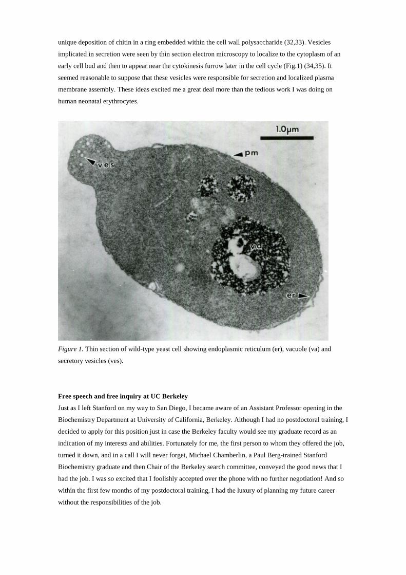
unique deposition of chitin in a ring embedded within the cell wall polysaccharide (32,33). Vesicles
implicated in secretion were seen by thin section electron microscopy to localize to the cytoplasm of an
early cell bud and then to appear near the cytokinesis furrow later in the cell cycle (Fig.1) (34,35). It
seemed reasonable to suppose that these vesicles were responsible for secretion and localized plasma
membrane assembly. These ideas excited me a great deal more than the tedious work I was doing on
human neonatal erythrocytes.
Figure 1. Thin section of wild-type yeast cell showing endoplasmic reticulum (er), vacuole (va) and
secretory vesicles (ves).
Free speech and free inquiry at UC Berkeley
Just as I left Stanford on my way to San Diego, I became aware of an Assistant Professor opening in the
Biochemistry Department at University of California, Berkeley. Although I had no postdoctoral training, I
decided to apply for this position just in case the Berkeley faculty would see my graduate record as an
indication of my interests and abilities. Fortunately for me, the first person to whom they offered the job,
turned it down, and in a call I will never forget, Michael Chamberlin, a Paul Berg-trained Stanford
Biochemistry graduate and then Chair of the Berkeley search committee, conveyed the good news that I
had the job. I was so excited that I foolishly accepted over the phone with no further negotiation! And so
within the first few months of my postdoctoral training, I had the luxury of planning my future career
without the responsibilities of the job.

The Berkeley Biochemistry Department was a perfect place for my interests. Daniel Koshland served as
Chair and the faculty included a distinguished group of classical biochemists such as Esmond Snell, Jesse
Rabinowitz, Clinton Ballou, Jack Kirsch and Howard Schachman, as well as a group with broader
interests in genetics and molecular biology such as Allan Wilson, Stuart Linn, Ed Penhoet, Chamberlin,
and Bruce and Giovanna Ames. Jeremy Thorner, a close friend from my Stanford years, had taken up a
study of yeast pheromone biology as a beginning faculty member in the bacteriology and immunology
department at Berkeley. Ballou was an expert in carbohydrate chemistry with a particular interest in the
yeast cell wall. Koshland, whom I had met at Stanford, and Ames were most appealing because they
blended genetics and biochemistry in a way that I found compatible with my temperament. I believed that
my future colleagues would allow me the freedom to explore a new direction quite different from my
graduate or postdoctoral work.
In the remaining time of my postdoctoral work, I completed a project and published a paper but all my
thoughts were directed to my future at Berkeley. Of course, I had no experience with yeast and knew
essentially no genetics so I planned to spend three weeks at the yeast genetics course offered at Cold
Spring Harbor and taught by Fred Sherman and Gerald Fink. Sherman and Fink were master geneticists
and were able to draw on all the major figures in the yeast community who dropped by to teach and
remain for a day or two. It was a thrill to meet Lee Hartwell and to share my thoughts about how yeast
cells may grow by vesicle traffic. On the other hand, like with thin section electron microscopy, my skills
in yeast tetrad dissection were inadequate. I believe I held the record for fewest tetrads dissected until
several years later when James Rothman took the course.
How to study secretion in yeast
As the time approached for the move to Berkeley, I worked feverishly to craft an NIH grant proposal that
included a range of ideas on how to study secretion and membrane growth in yeast. Published evidence
suggested that secretion was localized to the bud portion of the dividing cell but there were no tools
available to study the localization of a newly synthesized plasma membrane protein. My ideas were
fanciful but in the cold light of day, the NIH reviewing panel found my experience inadequate (I had no
preliminary data) and my ideas unproven. The rejection was crushing and my colleagues must have
wondered if their gamble on me was about to crash. Adding insult to injury, I was denied a Basil
O’Connor starter grant from the March of Dimes where the interviewer found me intelligent but regretted
that I had not proposed to work on cell division in Lesch Nyhan syndrome! Fortunately, the NSF, and
friendly reviews from Lee Hartwell and Susan Henry, a young yeast geneticist who studied phospholipid
regulation, rescued me with a grant in the princely amount of $35,000 for two years. With this and a small
internal University grant, a modest effort took shape.
What to do first? In the fall of 1976, two graduate students joined my lab: Janet Scott and Chris Greer.
Janet had transferred from another lab so she had to find something that would work quickly. I felt that in
order to study the yeast plasma membrane it would be necessary to have a clean way to remove the cell
wall avoiding the use of crude snail gut enzymes, Glusulase, that were used to convert cells to
spheroplasts. Another lytic enzyme secreted by a soil bacterium, Oerskovia xanthineolytica, seemed a

good source to begin a purification effort. Janet perfected the conditions of induction and purification of
an enzyme we called lyticase (36). Subsequently the bacterial gene was cloned and lyticase is still used as
a recombinant enzyme for experiments that require undamaged membranes. Chris also wanted to pursue a
biochemical project, so I set him off on an effort to purify yeast actin, which at the time seemed a logical
choice for a protein that may be involved in vesicle traffic. Chris completed the project but it was not
until years later that Peter Novick, then a postdoctoral fellow in David Botstein’s lab, showed that an
actin ts mutation delayed and mislocalized secretion at a restrictive growth temperature (37).
With a small lab, a little money and time free from other responsibilities, I started a couple of my own
projects to look at the localization of secretion with a focus on chitin, a polysaccharide in the division
septum, and invertase an enzyme secreted into the cell wall. My first undergraduate research student,
Vicki Brawley (now Chandler), helped me to study an unexpected surge in chitin synthesis that
accompanied the arrest of the yeast cell division cycle in response to the mating pheromone α-factor. That
work resulted in my first independent publication, a PNAS paper that was critically edited and
communicated by my colleague Clint Ballou (38). The notion of localized deposition and activation of the
plasma membrane enzyme chitin synthase seemed tractable but the subject excited little interest outside
of a small and contentious community of yeast investigators. Fortunately, a breakthrough in the study of
invertase secretion reinforced in my mind the importance of investigating a topic of general interest.
Within a few months, Peter Novick joined the group for his thesis work. Peter was quiet, focused and
technically superior. His background was impressive, having trained as an undergraduate at MIT and
during summers as a research student in the lab of Arthur Karlin at Columbia University where Peter’s
father was a Professor of Physics. Peter focused his studies on invertase, an enzyme that hydrolyzes
sucrose to glucose and fructose and which yeast cells use to mobilize hexose for uptake by active
transport at the cell surface. Invertase synthesis is repressed in cells growing on a medium containing
high (2%) glucose and is derepressed when cells are shifted to low glucose (0.1%). Peter found that
secretion of invertase is rapid: The pool of intracellular intermediates in the secretion of invertase is
depleted within five minutes after the addition of cycloheximide to block new protein synthesis. He then
looked at chemical agents that were reported to block secretion in animal cells to see if they could be used
in yeast. My first thought was to find a way to block the fusion of secretory vesicles at the cell surface to
see if both secretion and plasma membrane growth were arrested. Those experiments failed and we were
faced with a question of how to find secretion mutants.
During that first year, I followed up on an intriguing observation made by Susan Henry, then at Albert
Einstein Medical School, who showed that starvation of a yeast inositol auxotroph led to cell death and a
rapid arrest in cell growth. She demonstrated that starved cells increase in buoyant density, suggesting an
imbalance in macromolecule biosynthesis and net cell surface growth (39). I tested the possibility that
inositol may be required for secretion and cell surface growth by assaying invertase activity in intact
cells, a measure of enzyme in the cell wall (yeast cells are impermeable to and can not transport sucrose),
and in detergent lysed spheroplasts from which the cell wall material had been removed, a measure of
intracellular intermediates in secretion. Another dead end; I found that inositol starvation did not block
secretion.

Secretion mutants
During my postdoctoral years, I kept a box of cards with ideas about what to pursue in my lab at
Berkeley. One of many ideas was a search for secretion mutants. In retrospect, we could have initiated
that search right away, but I was not a geneticist and just did not think that way. And when Novick’s
work inevitably turned to that approach, we assumed that a block to secretion would be lethal and that
one would require a selection procedure to find what might be a rare ts lethal mutation. But what
advantage could a dying secretion defective cell have over a viable one? One thought was to select
against cells that could take up a toxic substance through a newly synthesized cell surface permease, one
whose export would be blocked in a secretion mutant such that the mutant cell would survive exposure to
the toxin. We settled on the yeast sulfate permease, which fails to discriminate sulfate and chromate.
Under the right conditions, chromate kills cells that express the sulfate permease. Indeed in a screen of
mutants that survived exposure to chromate at 37C, a standard non-permissive temperature for yeast,
Peter found a ts lethal mutation that also blocks invertase secretion. However, on reconstructing the
conditions of the selection, he found that this mutant died at 37C even more rapidly than the wild type
strain in the presence or absence of chromate. So this was no selection at all! From this we concluded that
the mutations may not be so rare after all and that a Hartwell style search among a set of random ts lethal
mutations might turn up more secretion specific lesions.
The mutant, sec1, that came from the aborted attempt at a selection, turned out to conform to all the
predictions we had made. At a permissive temperature, 24C, mutant cells behaved like wild type cells in
growth and rapid secretion of invertase and another conveniently assayed secreted enzyme, acid
phosphatase. The induction and appearance of sulfate permease was also normal. However, on shift to
37C, sec1 mutant cells arrested secretion of invertase and acid phosphatase (Fig. 2), which accumulated
to a high level within dying cells, and the sulfate permease failed to appear in intact cells. These blocks
were reversible and on return to 25C, the accumulated invertase and acid phosphatase were secreted even
in the absence of new protein synthesis. Thus, we concluded that the mutant Sec1 protein must be
thermally, but reversibly, unstable.

Figure 2. Secretion and accumulation of acid phosphatase in wild-type cells (open circles) and sec1
(closed circles) mutant cells (A) and spheroplasts (B). Reproduced from reference #40.
In May of 1978, George Palade visited Berkeley for two lectures in a series sponsored by the
pharmaceutical company Smith, Kline and French. This was the first opportunity I had had to meet
Palade personally and it was a thrill to be able to share with him what we were doing to study secretion in
yeast. He was not aware that yeast cells secrete glycoproteins. The graduate students hosted Palade for
dinner and in the course of the conversation, Peter Novick spoke of his new results on the sec1 mutant.
Palade encouraged Peter to examine the mutant by thin section microscopy. Shortly thereafter, Peter
called my office from the EM lab in the basement of the Biochemistry building, urging that I come
inspect the images of sec1 mutant cells. The picture was stunning; cells chock full of vesicles filling the
entire cytoplasmic compartment (Fig. 3). An enzyme-specific cytochemical stain for acid phosphatase
showed all the vesicles carried this enzyme and likely other proteins secreted by yeast cells. Mutant cells
grown at 24C behaved just like wild type cells did, with a small cluster of vesicles in the bud portion of
the cell. Short of the moments I witnessed the birth of my children, nothing in my life compares to the
excitement of that image in the EM room in the summer of 1978.

.
Figure 3. Wild-type (A) and sec 1 mutant cells at 24C (B) or 37C after 1h (C) or 3h (D, E). Reproduced
from reference #40.
Peter and I assembled a paper for publication in the PNAS, which was communicated by Dan Koshland
(40), and we continued a quest for more mutants of this sort because surely with a procedure that was
unfavorable for the selection of sec1, many more genes might be found with no selection whatsoever.
Peter collected 100 random ts mutant colonies by a standard mutagenesis protocol and found one more
mutant, sec2, which phenotypically resembled sec1 in accumulating a uniform population of vesicles.
Thus at least two proteins were implicated in some step in the delivery of vesicles to a target membrane,
possibly the plasma membrane. But surely there must be more such genes and the prospect of generating
thousands of ts colonies in a time before the robotic approaches we now enjoy, was a bit daunting.
For the next of what would be a brilliant string of observations, Peter noticed that sec1 mutant cells fail to
enlarge, fail to divide and become phase refractile during an hour or more of incubation at 37C. This

contrasts with the behavior of Hartwell’s cell cycle ts mutants that arrest with a unique cell morphology
characteristic of the cell cycle stage that is blocked, but that continue to enlarge into misshapen structures.
Peter reasoned that secretion defective cells may continue to produce macromolecules but by failing to
enlarge their buoyant density may increase, just as I had expected of the inositol auxotroph of Susan
Henry. Peter then performed a beautiful experiment to test his theory. Mutant cells were constructed with
a constitutively expressed form of acid phosphatase and an aliquot was incubated at 37C. A
corresponding wild type cell sample with a normally repressed phosphatase gene was mixed in a ratio of
100:1 with the mutant cells and the mixed cells were centrifuged on a self-forming gradient of Ludox, a
colloidal silica suspension that was then marketed as a commercial floor polish. Susan Henry had
exploited the same preparation of Ludox to separate inositol-starved and normal cells. Fractions of the
gradient plated on rich medium formed colonies that were then stained with a phosphatase-specific
histochemical reagent to reveal the distribution of phosphatase-constitutive and -repressed colonies. The
result was an absolute separation of sec1 mutant cells at the bottom of the gradient and wild type cells at
the top (Fig. 4). This density gradient then provided the opportunity Peter needed to enrich and screen
many more ts colonies for additional sec mutants.
Figure 4. Ludox gradient of wild-type (light stipples) and sec1 mutant cells (heavy stipples) separated
from bottom (high density) to top (low density) of centrifuge tube. Reproduced from ref. #41.
Over the next 18 months, Peter with Charles Field, a technician who was an expert in yeast genetics,
repeated the mutagenesis on a large scale with different mutagens and assembled a large collection of
density enriched ts colonies, 220 of which proved to be defective in secretion. Genetic complementation
tests uncovered 23 genes among these mutants and the distribution of alleles suggested yet more genes
were likely to be discovered. Electron microscopic inspection revealed three different phenotypic
categories of organelle disruption: Mutations in 10 genes, like sec1 and 2, accumulated secretory vesicles,
mutations in another 9 genes caused accumulation and distortion of the ER membrane, and another two
caused a toroid-shaped organelle, which Novick called the “Berkeley body”, to proliferate. One concern
we had was that the sec mutations might not represent components of the secretory machinery, but merely
defective biosynthetic cargo proteins that interfere with secretion. However, the simple complementation
tests used to establish the genes showed all the alleles to be genetically recessive, and thus unlikely to

represent dominant inhibitors of the process. Novick and Field completed a morphological and
physiological characterization of selected alleles of each of the 23 genes, and we put together a
comprehensive paper for the relatively new journal, Cell, which through the force of the personality of the
Editor, Benjamin Lewin, was changing the way life science research was evaluated and promoted (41).
In the following year, Novick and Susan Ferro, who later became Susan Ferro-Novick (the first of many
marriages within my laboratory), teamed up to apply a classic genetic epistasis test to establish the order
in which the SEC genes exert their function. In the course of this work Peter found that one of the mutants
sec7 that accumulates the odd “Berkeley bodies” appeared to define a stage equivalent to that of the Golgi
apparatus in mammalian cells. Quite by chance he found that this structure irreversibly blocked secretion
unless cells were incubated in medium containing low glucose in which case mutant cells accumulate a
classic, multi-cisternae Golgi structure (42). Some years later, Chris Kaiser, a talented postdoctoral fellow
with considerable experience in yeast genetics, revisited the SEC genes that govern traffic early in the
pathway and uncovered a distinct smaller vesicle species that mediates traffic between the ER and the
Golgi complex (43). He classified a set of SEC genes that governs vesicle formation and another set
required for vesicle consumption, presumably by a process of membrane fusion at the Golgi complex.
Importantly, he showed that the two sets of genes show extensive genetic interactions, with mutations in
each group exacerbating the mutant phenotype of other members of that group but not between the two
groups. This behavior, referred to as synthetic lethal interaction, suggested that the members of each
group function together, possibly by physical interaction with one another. These results led to a picture
of the secretory pathway in yeast that was essentially the same as Palade had shown for mammalian cells,
but with the crucial bonus that each step in the elaborate chain of events was now defined by genes and
thus proteins that would surely illuminate the molecular mechanisms of this pathway (Fig. 5, 6).
Figure 5. Yeast secretory pathway circa 1981. Reproduced from ref. #42.

Figure 6. ER – Golgi vesicular traffic pathway circa 1990. Reproduced from ref. #43.
Two other studies added molecular detail to the emerging view of the secretory pathway in yeast. Brent
Esmon, a graduate student in the lab, applied a histochemical stain for invertase on cell lysate samples
that were electrophoretically resolved on a native polyacryamide gel. He discovered that the mutants
defective in protein transport from the ER accumulate discrete forms of glycosylated invertase, distinct
from invertase that progressed to the Golgi compartment and into secretory vesicles. Using antibodies
that diagnose the “outer chain” carbohydrate epitopes of yeast glycoproteins, Brent learned that the
division of labor between the ER and the Golgi complex in yeast with respect to N-glycan maturation is
much the same as in mammalian cells (44). Tom Stevens, a postdoctoral fellow, studied the traffic of a
protein to the yeast vacuole and found that it is diverted from the Golgi complex, similar to the traffic of
lysosomal proteins in mammalian cells (45). Stevens and another postdoctoral fellow, Scott Emr, took
this part of the pathway to their own labs at the University of Oregon and Cal Tech, respectively, to
develop powerful genetic selections to uncover the genes that govern this sorting limb of the secretory
pathway. The VPS genes continue to illuminate the process of sorting from the Golgi complex to the
endosome and on to the vacuole or lysosome in all nucleated organisms.
Given our finding that the yeast and mammalian secretory pathways are fundamentally conserved, the
biotech industry was quick to exploit the fermentation possibilities of yeast culture to engineer the
expression of commercial quantities of important human secreted proteins. Chiron, near Berkeley in the
San Francisco Bay Area was the first to succeed. Recombinant expression of the hepatitis B surface
antigen in yeast resulted in the production of virus-like membrane particles that proved to be highly
immunogenic and which were commercialized as a potent hepatitis vaccine, the sole source of that
product in use today (46). As hepatitis B is the major cause of primary liver cancer, the successful
introduction of this product of the yeast secretory pathway could, if fully implemented, dramatically
reduce the incidence of liver cancer. Indeed this commercial product is considered the first anti-cancer
vaccine. Chiron next engineered the expression and secretion of human insulin in yeast and that product,
now marketed by Novo Nordisk, accounts for one-third of the world supply of human recombinant
insulin.
I never patented any of our discoveries or thought to do work directed to commercial application in my
laboratory because I was completely absorbed by the pursuit of fundamental knowledge. Nonetheless, as
a consultant to Chiron I did benefit financially and was enormously gratified to see our work applied to
such important practical goals. My view is that the work of drug discovery and practical application is
best left to the private sector and that University scientists should focus on basic discovery.
Important genes uncovered by other means
Although the initial set of SEC genes revealed the broad outline of the secretory pathway, it became clear
that key elements in the process were not reflected in the Novicks’ mutants. We had hoped to find
mutations that block the insertion of secretory polypeptides into the lumen of the ER and thus to define
genes that constitute the translocation channel predicted by the classic work of Palade, Sabatini and
Blobel. The key prediction was that mutations in a putative channel would accumulate unglycosylated
secretory precursor polypeptides in the cytoplasm. No such defects were found in the initial set of sec
mutations. Susan Ferro conducted a wider search for mutants using the density gradient technique and
turned up two that accumulated unglycosylated forms of invertase (47). However, on closer inspection
these mutations identified genes involved in the biosynthesis of glycans on secretory proteins rather than
bona fide catalysts of translocation (48, 49). Clearly, a different, more directed approach was needed.
Studies in E. coli and in yeast showed that the N-terminal signal peptide is necessary and sufficient for
the translocation of a secretory protein across the cytoplasmic membrane or ER membrane, respectively
(50, 51). The recombinant expression of a chimeric protein constructed by the fusion of a signal peptide
coding sequence and the E. coli β-galactosidase gene, encoding a soluble cytoplasmic enzyme, result in
the membrane translocation of the hybrid protein. Beckwith and colleagues found the expression of such
a hybrid protein in E. coli provided a selectable growth phenotype, which they used to isolate
translocation defective sec mutations, defining the novel cytoplasmic proteins SecA and SecB (52). Using
other genetic approaches, Silhavy, Ito and colleagues identified a gene encoding a membrane protein,
PrlA/SecY, a candidate for the bacterial translocation channel (53, 54).
Ray Deshaies, an unusually creative and confident graduate student joined the lab in the mid 1980s and
after an initial effort with the existing sec mutants, he decided to revisit the translocation problem. In
three brilliant but entirely independent efforts, he succeeded in defining a number of genes required in the
translocation process. Ray reasoned that if a signal peptide were appended to a cytoplasmic enzyme
required for the production of an essential nutrient, the enzyme would be sequestered in the ER, removed
from contact with its substrate. In this situation, cells would grow on the nutrient but not on its substrate
unless a mutation was introduced that blocked the translocation of the hybrid protein into the ER. Of
course, a mutation in an essential channel protein would likely kill the cell, so the quest was for mutations
that crippled but did not destroy proteins required for the assembly process. Temperature-sensitive lethal
mutations often exert a partial effect at a permissive temperature, thus the search was for mutations that

grow at 30C on the substrate, in this case histidinol, the substrate of the enzyme histidinol dehydrogenase,
the last step in the biosynthesis of histidine, but which fail to form colonies at 37C on rich growth
medium. Ray’s first mutant was called sec61 and further searches using the same selection identified five
other genes that encode additional functions essential for translocation, including other subunits of the
channel complex and a subunit of the signal recognition particle (SRP) (55, 56). Subsequent cloning of
these genes revealed that SEC61 is homologous to the PrlA/SecyY gene of E. coli (57). Comparable
genes are found in mammals, and biochemical analysis demonstrated that the Sec61 protein constitutes
the core of the channel protein through which secretory and membrane proteins pass during assembly in
the ER (58, 59).
Deshaies also tackled the question of how certain secretory proteins may be translocated post-
translationally in yeast. In contrast to the classical rule of co-translational translocation discovered by
Blobel, Peter Walter, a protégé of Blobel’s, discovered that at least one substrate, the precursor of the
yeast mating pheromone α-factor, could pass across the ER membrane after the completion of translation
(60). The assumption was that something extrinsic or intrinsic to α-factor precursor held it in a form that
could readily unfold during the translocation event.
In reading an influential review article by Hugh Pelham on the possible role of the heat shock protein
family hsp70 in dispersing protein aggregates (61), Ray imagined that hsp70 might also serve to retain
partially unfolded forms of post-translational substrates such as α-factor precursor. Fortunately, we were
in a position to test this in vivo because Margaret Werner-Washburn and Elizabeth Craig had just
constructed a yeast strain missing three members of the major hsp70 class of proteins and with a ts
mutation in the remaining fourth gene such that the quadruple mutant was ts lethal. Ray established in
short order that this mutant accumulated untranslocated α-factor precursor and as a bonus, he found that
the β subunit of the mitochondrial F1- ATPase, also post-translationally translocated into that organelle,
accumulated in the cytoplasm. Ray and independently Chirico and Blobel showed that the requirement
for Hsp70 could be reproduced in the cell-free reaction that reconstitutes the translocation of α-factor
precursor into isolated yeast ER membranes (62, 63).
In a third example of Deshaies’ creative instinct, he solved a problem that had bedeviled a postdoc, Peter
Bohni, who had struggled for two years to devise a selection for a mutation in the yeast signal peptidase,
the enzyme that Blobel demonstrated cleaves the signal on a secretory polypeptide as it emerges on the
luminal side of the ER. Neither the enzyme nor the gene for the peptidase had been obtained, thus it was
of interest to test the function of the protein, which at that time remained a candidate for a subunit of the
translocation channel. We knew that a mutation at the yeast invertase signal peptide cleavage site delayed
the secretion of active enzyme, which accumulates in a precursor form in the ER (64). Attempts to devise
a selection for mutations in the peptidase based on that secretion delay proved futile. Ray suggested that
some uncleaved cargo proteins might be delayed more seriously than others and that a peptidase mutant
could be in our original collection of sec mutants and would have the unusual characteristic of blocking
only a subset of cargo proteins. In Peter Novick’s last effort as a graduate student, he had devised a cell
surface chemical labeling procedure to assess the full range of major cargo proteins and how their cell
surface appearance is affected in sec mutant cells incubated at 37C (65). Curiously, one mutant in the

original collection, sec11, showed an anomalous effect with certain cargo proteins blocked and others less
so. With this insight, Bohni immediately investigated the sec11 mutant and found that it accumulated
uncleaved invertase at a restrictive temperature (66). The SEC11 gene was cloned and found to be the
prototype of all eukaryotic signal peptidases (67).
Cloning genes as an adjunct to functional analysis of Sec proteins
With the advent of cloning yeast genes by complementation, pioneered by Hinnen and Fink in 1978 (68),
we had the immediate prospect of a molecular description of the SEC genes and a possible alignment of
these genes with comparable functions in simple metazoans and perhaps even mammals. I resisted the
temptation to launch in this direction because it seemed unlikely that the SEC genes would look like
anything else then known. After all, DNA sequencing was still in its infancy and genome databases were
nonexistent. Almost from the outset of our characterization of the sec mutants, my focus was on
attempting to develop a cell-free reaction that reproduced the function of Sec proteins. Most students and
fellows who joined the lab resisted my entreaties or took up only half-hearted attempts. One initial effort
in this direction yielded a feeble signal that seemed unlikely to prove useful (69). And yet, just miles
away in his new lab at Stanford, Jim Rothman had succeeded in developing a reaction that appeared to
measure a significant limb of the Golgi traffic pathway reconstituted in a lysate of mammalian cells (70).
My own efforts remained on hold until I found a courageous student to take up the challenge.
SEC53 was the first SEC gene cloned and identified with a biochemical function. Although sec53 was
isolated and initially characterized as a mutant defective in translocation, the gene sequence predicted a
soluble protein (71), which on closer inspection proved to be to the enzyme phosphomannomutase
involved in the production of GDP-mannose, the precursor of N- and O-glycans in yeast (48). Other SEC
genes were cloned but other than predicting that SEC12 encoded an ER membrane protein and SEC18
encoded a soluble cytoplasmic protein, no functional biochemical role could be seen in the sequences (72,
73).
The first real breakthrough with respect to vesicular traffic came in 1987 when Novick, now in his own
lab at Yale, cloned and sequenced SEC4, which he showed encoded a small GTP- binding protein of the
RAS family (74). Novick’s focus on SEC4 was no accident. We had agreed that he could take charge of
the group of sec mutants that block late in the pathway and accumulate mature secretory vesicles.
Salminen and Novick found that SEC4 overexpression suppressed the growth defect of several members
of the group of late acting sec mutants, and that double mutants constructed among the members of this
class displayed a synthetic lethal form of genetic interaction. As the genomes of other organisms were
sequenced, it became clear that SEC4 was a prototype of what are now called Rab proteins, each of which
defines a unique destination for the fusion of vesicles to a target membrane. Continuing on the brilliant
path he established right from the start of his graduate work, Novick has built a substantial body of highly
original work that reveals detailed mechanisms associated with the production, migration and fusion of
transport vesicles at the yeast cell surface. And given the fundamental conservation of the SEC gene
sequences, it is no surprise that Novick’s insights extend to all comparable vesicle targeting/fusion events
in metazoans and mammals. Indeed, SEC1 was found to be related to the unc-18 gene isolated in the

original collection of uncoordinated mutants of C. elegans isolated by Sydney Brenner (75, 76). And the
Sec1 protein is known to play a universal role in the control of SNARE protein action in vesicle fusion.
Jim Rothman’s pioneering initial effort to purify proteins required for vesicle fusion yielded the soluble
ATPase, NSF (NEM-sensitive factor), which on cloning revealed a striking similarity to the yeast Sec18
protein, a gene that had been cloned by Scott Emr in his own lab at Cal Tech (73, 77). At around the same
time, Chris Kaiser in my lab had detected a vesicle intermediate between the ER and Golgi, whose
consumption by fusion required the genetically interacting genes SEC18, SEC17 and SEC22 (43). In a
joint paper, our labs showed that SEC17 encodes the yeast equivalent of α-SNAP, a protein Rothman’s
lab discovered as the factor required for NSF to bind a membrane site, later defined as the SNARE
protein (78). Later work showed that SEC22 encodes one such yeast SNARE protein. These results made
it clear that the two labs were working on fundamentally the same problem and forged a persuasive link
between the mechanism of vesicle targeting/fusion in yeast and mammalian cells.
The mechanism of secretory vesicle budding was now accessible to molecular analysis. Palade had seen
coated vesicles at the ER exit site in sections of pancreatic exocrine cells and the view was that the
mechanism of budding would involve a coat similar to the classic clathrin coat first visualized as a coated
pit engaged in yolk protein internalization in insect oocytes and characterized molecularly by Barbara
Pearse with isolated bovine brain clathrin coated vesicles (79, 80). Rothman had evidence to suggest a
role for clathrin in the transport of vesicular stomatitis virus G protein from the ER in cultured
mammalian cells (81). Thus, clathrin or a similar coat protein was a candidate for one or more of the SEC
genes required for traffic from the ER.
Greg Payne decided to assess the role of clathrin directly by cloning the gene for the heavy chain and
characterizing the phenotype of a clathrin gene knockout in yeast. Given the expected role of clathrin in
vesicular traffic, we assumed the gene would – like the SEC genes – be essential for cell viability. Yet,
after disruption of the heavy chain gene in a diploid strain, Greg was shocked to see 2 disrupted spores in
each tetrad growing after a several day lag phase. Clathrin deficient cells were sickly but continued to
secrete even when the gene was knocked out in a number of different genetic backgrounds (82). Lemmon
and Jones reported a strain in which the heavy chain gene was essential but it now seems likely this strain
carried an additional mutation that exerted a synthetic lethal effect in the absence of clathrin (83). Further
analysis showed that clathrin was required for the proper sorting/retention of a Golgi-localized dibasic
peptidase essential for the proteolytic maturation of α-factor precursor (84). These results conformed
nicely to the suggestion by Lelio Orci that clathrin coats mediate the retrieval of the proinsuln processing
protease from condensing granules in pancreatic β cells (85). The search continued for a coat mechanism
in the formation of secretory transport vesicles.
A yeast cell-free vesicular transport reaction
I knew that the full potential of the sec mutant collection awaited the development of a cell-free reaction
to recapitulate at least a portion of the pathway in vitro. Finally, in 1985, I recruited a brilliant and
creative graduate student, David Baker, who shared my vision and had the talent to make it happen. Up to
that point we had relied on the accumulation of precursor glycoproteins in sec mutant cells arrested at
37C to serve as substrates in in vitro reactions. Immature glycoproteins become modified by specific
outer chain glycan decorations en route through the Golgi complex when cells are returned to the
permissive temperature, and we assumed the same would be true in vitro. This assumption proved wrong.
The first hint of a problem came in the evaluation of sec53 mutant phosphomannomutase, which proved
to be inactive even in lysates of cells that were grown at a permissive temperature (48). But without such
a block to accumulate substrates in the ER, the assay for traffic would have to rely on a low level of
immature glycoproteins radiolabelled for a brief time during biosynthesis. Rothman had succeeded with
just such an approach (71), but the transit time of glycoproteins in yeast is much quicker than in
mammalian cells.
David had a fresh idea. Peter Walter’s lab (as well as the labs of David Meyer and Blobel) had
reconstituted the translocation of radiolabled α-factor precursor into ER membranes prepared by
mechanical disruption of yeast spheroplasts (61). The product of this incubation was a core N-
glycosylated species that migrated at a discrete position on SDS-PAGE separation. David guessed that
membranes prepared by a more gentle lysis procedure, basically a quick freeze-thaw of yeast
spheroplasts, might preserve membrane organization well enough to permit vesicular traffic of the core
glycan modified synthetic α-factor precursor. Within a few weeks of starting, David observed the
production of a heterogeneous spread of low electrophoretic mobility forms of the radioactive precursor,
which importantly was precipitated by antibodies directed against mannose epitopes added to N-glycans
in the yeast Golgi complex. The reaction required cytosol, ATP and incubation at a physiological
temperature. The results were most promising and the assay was amenable to quantification and easy
repetition with many samples.
The crucial test of Baker’s reaction was to examine the effect of an ER-blocked sec mutant in the cell-free
reaction. Linda Hicke, an ambitious and technically gifted graduate student had cloned SEC23, one of the
four genes Kaiser found to interact in the formation of ER-derived transport vesicles. She collaborated
with Baker to reproduce the α-factor precursor transport reaction in separate incubations containing
membranes from wild type (wt) cells mixed with cytosol fractions from wild type (wt), mutant and
mutant cells complemented with the wt gene. The results were stunning, with a clear ts defect in transport
complemented by a wt copy of Sec23p supplied in the mutant cytosol fraction (86) (Fig. 7). Amazingly,
Susan Ferro-Novick and her graduate student Hannele Ruohola, developed virtually the same
methodology yielding similar results in their laboratory at Yale (87).
Baker and Hicke’s results were precisely what I had dreamed of and the experimental design was
modeled on my own graduate research, in which I used complementation of mutant lysates as an assay to
purify functional DNA replication enzymes (19). With her assay, Linda was able to purify overexpressed
recombinant Sec23p and then to show that it copurified with anther protein that was not represented in
our original mutant collection but which proved to be encoded by another essential gene, which we then
called SEC24 (88).
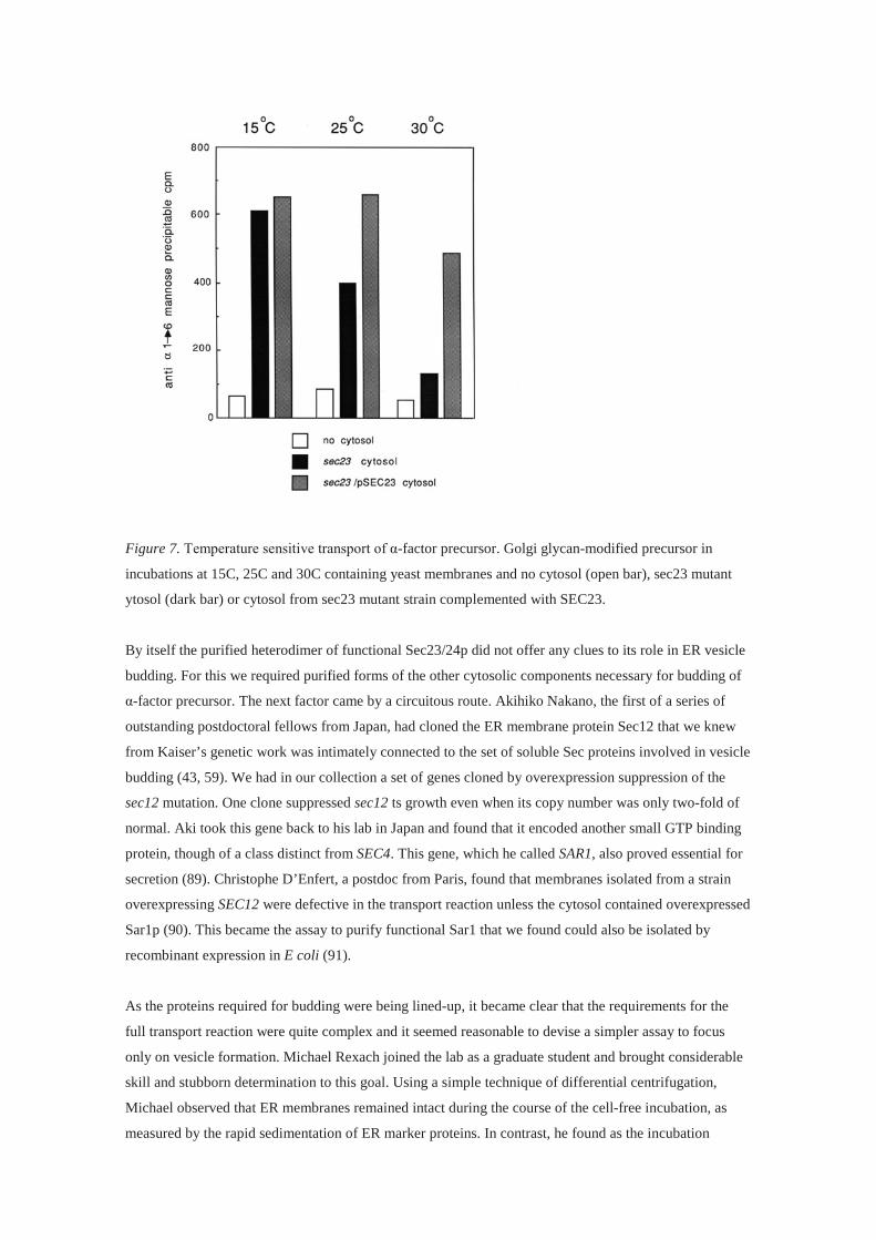
Figure 7. Temperature sensitive transport of α-factor precursor. Golgi glycan-modified precursor in
incubations at 15C, 25C and 30C containing yeast membranes and no cytosol (open bar), sec23 mutant
ytosol (dark bar) or cytosol from sec23 mutant strain complemented with SEC23.
By itself the purified heterodimer of functional Sec23/24p did not offer any clues to its role in ER vesicle
budding. For this we required purified forms of the other cytosolic components necessary for budding of
α-factor precursor. The next factor came by a circuitous route. Akihiko Nakano, the first of a series of
outstanding postdoctoral fellows from Japan, had cloned the ER membrane protein Sec12 that we knew
from Kaiser’s genetic work was intimately connected to the set of soluble Sec proteins involved in vesicle
budding (43, 59). We had in our collection a set of genes cloned by overexpression suppression of the
sec12 mutation. One clone suppressed sec12 ts growth even when its copy number was only two-fold of
normal. Aki took this gene back to his lab in Japan and found that it encoded another small GTP binding
protein, though of a class distinct from SEC4. This gene, which he called SAR1, also proved essential for
secretion (89). Christophe D’Enfert, a postdoc from Paris, found that membranes isolated from a strain
overexpressing SEC12 were defective in the transport reaction unless the cytosol contained overexpressed
Sar1p (90). This became the assay to purify functional Sar1 that we found could also be isolated by
recombinant expression in E coli (91).
As the proteins required for budding were being lined-up, it became clear that the requirements for the
full transport reaction were quite complex and it seemed reasonable to devise a simpler assay to focus
only on vesicle formation. Michael Rexach joined the lab as a graduate student and brought considerable
skill and stubborn determination to this goal. Using a simple technique of differential centrifugation,
Michael observed that ER membranes remained intact during the course of the cell-free incubation, as
measured by the rapid sedimentation of ER marker proteins. In contrast, he found as the incubation

proceeded that a substantial fraction of the core glycosylated α-factor precursor, which was initially
contained within large ER envelopes, was transferred into a slowly-sedimenting vesicle species, which
lacked translocation activity and other marker proteins of the ER membrane and lumen. Importantly, the
formation of this vesicle species was blocked in the mutants that Kaiser showed to be defective in the
production of the vesicle intermediate in vivo (sec12 and sec23), but not in mutants blocked later (sec18)
(92). Again, similar results were obtained in Ferro-Novick’s lab (93). Rexach’s work provided us with the
essential tool we needed to complete the purification and functional analysis of the proteins required for
vesicle budding from the ER.
Two other genes required for ER vesicle formation remained to be functionally identified: SEC13 and
SEC16. In his own lab at MIT, Kaiser cloned and characterized SEC16 and learned that it encodes a
240kD peripheral membrane protein, not readily released into the cytosol (94). Nancy Pryer, a postdoc in
our lab, cloned SEC13 and found that it encodes a small cytosolic protein that contains a series of WD-40
repeats, very similar to the G protein β subunit (95). Members of this family have a 7-member β propeller
structure common to proteins that engage in reversible multi-subunit protein interactions. Nina Salama,
an effervescent graduate student in the lab, used Rexach’s budding reaction to purify a functional form of
Sec13p and found that it co-purifies with an additional subunit, which we cloned and characterized as a
novel SEC gene, SEC31 (96). With this last piece of the puzzle, we found that the budding reaction was
sustained with isolated membranes and pure, recombinant Sar1p, Sec23/24p and Sec13/31p, with Sec16p
presumably being supplied by the membrane fraction. A complete functional analysis of the mechanism
of vesicle budding was now at hand.
COPII mediates vesicle budding from the ER
We had few clues as to the mechanism of vesicle budding mediated by the pure Sec proteins in our
collection. Rothman’s lab had identified and characterized a novel coat protein complex, coatomer,
required for vesicle budding in transport within the Golgi complex (97). He suggested that this coat may
also be required for vesicular traffic from the ER, but we found no evidence for subunits of the coatomer
in our purified set of Sec proteins. In addition, we had cloned and characterized a different SEC gene,
SEC21, that encodes a subunit of coatomer and although the sec21 mutant is blocked in traffic from the
ER, it did not fit neatly into one of Kaiser’s mutant classes, and we attributed its effect on traffic from the
ER to a backlog of cargo that accumulates when Golgi function is disrupted (98).
Several key insights developed in the1990s that consolidated our efforts. Two wonderful new postdocs in
the lab, Charles Barlowe and Tohru Yoshihisa, discovered a cycle of GTP hydrolysis and exchange on
Sar1p. Tohru found that the Sec23 subunit is a GTP hydrolysis catalyst (GAP) specific for Sar1p and
Charlie found that the cytoplasmic domain of Sec12p catalyzes nucleotide exchange on Sar1p (99, 100).
Several years later Bruno Antonny, a tremendously skilled and perceptive biophysicist, discovered that
the Sec31 subunit of the 13/31 heterotetramer complex accelerates the GAP activity of Sec23 10-fold
(101). Clearly, a coordinated assembly event controlled by GTP binding and hydrolysis, served to frame
the budding process. Given Rothman’s discovery of a role for GTP binding in the control of coatomer
assembly and vesicle budding on Golgi membranes, we were primed for the prospect of a novel coat
complex (102).
Fate intervened again in the form of a phone call from the maestro of membrane morphology, Lelio Orci
at the University of Geneva Medical School. Orci was instrumental in the effort to discover the
morphologic stages in vesicle formation and fusion in the Golgi complex uncovered in the Rothman lab
cell-free transport reaction. His skills were so extraordinary that I had attempted, unsuccessfully, to
engage his interest when our work uncovered a role for clathrin in the retrieval of a Golgi enzyme similar
to his discovery of the organization of clathrin and proinsulin processing in β-cells of the pancreas (85).
His call in 1990 was prompted by our recent publication of Kaiser’s analysis of the vesicle species that
mediates traffic from the ER. Lelio took pity on us for the primitive standards of our thin section EM
analysis and graciously offered his help in a collaboration to examine the organization of the Sec proteins
involved in ER vesicle formation. His first success was in using our antibody against the yeast Sec23p to
localize the mammalian homolog precisely at the ER exit site in sections of pancreatic tissue (103). But
the greatest excitement came when he discovered a novel coat that surrounded the vesicles formed in a
reaction with yeast membranes and our purified Sec proteins. Barlowe isolated these vesicles and we saw
a hint of a coat in thin sections prepared by my skilled EM technician, Susan Hamamoto, but the images
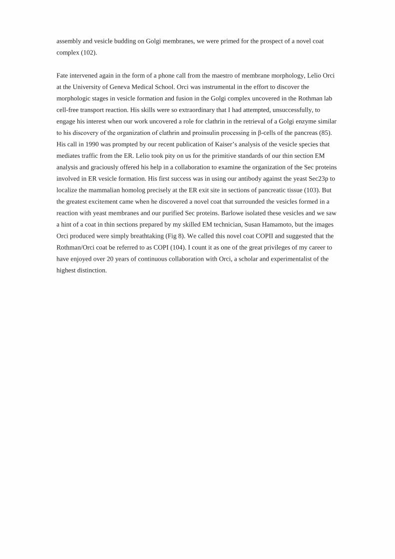
Orci produced were simply breathtaking (Fig 8). We called this novel coat COPII and suggested that the
Rothman/Orci coat be referred to as COPI (104). I count it as one of the great privileges of my career to
have enjoyed over 20 years of continuous collaboration with Orci, a scholar and experimentalist of the
highest distinction.

Figure 8. Thin section transmission and scanning EM images of COPII vesicles. Bar, 100nm. Courtesy of
Lelio Orci, Univ. of Geneva.
In a crucial initial collaboration, Orci and a new postdoc in the lab, Sebastian Bednarek, defined the ER
as the morphological site of COPII budding. Sebastian purified yeast nuclei as a source of pure ER
membrane and with Orci showed that COPII proteins, and curiously also COPI, form buds and
incorporate cargo molecules from the outer nuclear membrane. Using a sequential binding assay,
Sebastian demonstrated that the COPII assembles in pieces with Sar1p and Sec23/24p binding first and
constituting an inner layer of the coat with Sec13/31p forming the outer layer of the coat (105).
With a purified ensemble of cytosolic proteins in hand, we turned our attention to the contribution of
membrane proteins and lipids in an effort to define the minimum requirements for vesicle budding.
Sec16p represented the most obvious part of the machinery not accounted for in our reconstituted
reaction. Two successive postdoctoral fellows, Joe Campbell and Frantisek Supek, found conditions in
which a role for Sec16p in the budding reaction could be observed (106, 107). Eugene Futai succeeded in
purifying recombinant Sec16p and found conditions in which it controlled the GTPase cycle mediated by
the interaction of the full set of COPII proteins and Sar1 (108). Yet even now, it is not clear if Sec16
participates actively in the cycle of vesicle budding or rather plays a regulatory role in organizing COPII
proteins at the ER exit site.
We considered the possibility that coat assembly may be regulated by the availability of membrane cargo
proteins. Tom Yeung, a graduate student in the lab, found that membranes isolated from cells treated with

cycloheximide, and thus purged of newly-synthesized cargo, were perfectly active in budding COPII
vesicles as assayed by the incorporation of a SNARE protein (109). Although biosynthetic cargo may not
be essential for vesicle budding, we suggested that proteins cycling between the ER and Golgi might
constitute an essential element of the membrane contribution to the formation of a COPII bud (110).
To test directly the role of membrane proteins and lipids in the budding event, Yeung initiated an effort to
solubilize the membrane with detergents to see if membrane proteins and lipids could be reconstituted
into liposomes capable of budding synthetic COPII vesicles. To our surprise, Yeung and a meticulous
new postdoctoral fellow, Ken Matsuoka, systematically documented that synthetic COPII vesicles bud
and could be isolated by density gradient sedimentation from reactions conducted with pure phospholipid
liposomes of defined composition provided the reaction was conducted in the presence of a non-
hydrolyzable analog of GTP (111). Bruno Antonny developed an elegant real time light scattering assay
to monitor the stepwise assembly and disassembly of the coat in incubations containing GTP or a
nonhydrolyzable analog (101). Eugene Futai then showed that GTP could replace a nonhydrolyzable
analog to produce a stable COPII coated membrane provided the reaction was supplemented with the
cytoplasmic domain of the Sar1p nucleotide exchange catalyst, Sec12p, presumably stabilizing the coat
by repeated rounds of GTP nucleotide exchange. Curiously, these reactions arrested with buds on
liposomes, but few if any completed COPII vesicles (112). More recently, Kirsten Bacia, another
postdoctoral fellow, reconstituted the budding reaction on giant unilamellar vesicles where the process
may be visualized in real time by light and fluorescence microscopy without potentially damaging
manipulation, e.g. centrifugation. In these conditions, incubations containing the COPII proteins and
nonhydrolyzable GTP produce long, multi-lobed, coated tubules with regular points of constriction but
with little evidence of vesicle fission (113). The nature of the COPII fission reaction remains unresolved
but appears to hinge on spatial regulation of GTP binding and hydrolysis at the vesicle bud neck.
The initial event that leads to a bud may begin when Sar1p acquires GTP through interaction with Sec12.
Structural analysis showed that the soluble GDP-bound form of Sar1 shields an N-terminal amphipathic
helix in a cleft of the folded protein (114). Nucleotide exchange displaces the N-terminus and renders
activated Sar1p highly insoluble and prone to membrane insertion. Marcus Lee, an insightful postdoctoral
fellow, reasoned that the embedment of the N-terminus in the bilayer may laterally displace
phospholipids and create a local asymmetry in the surface area of the two leaflets, much as Sheetz and
Singer had proposed decades earlier in the bilayer couple hypothesis (115). In a series of elegant
experiments conducted in collaboration with Orci, Lee showed that Sar1p promotes the formation of
membrane tubules from synthetic liposomes dependent on the insertion of the amphipathic N-terminal
helix and that this insertion is required for COPII vesicle formation in vitro and protein transport in vivo
(116).
Tremendous progress has been made on the structural analysis of the COPII coat, principally by the
laboratories of Jonathan Goldberg and William Balch (117,118,119). We now have a detailed
understanding of the mechanism of polymerization of the two layers of the coat and a key insight
concerning the scaffold complex that forms the outer layer, a regular polyhedral lattice that Balch
discovered in a self-assembly reaction with purified mammalian Sec13/31 heterotetramer. A former

postdoctoral co-worker, Giulia Zanetti, using cryoelectron microscopy has now visualized the lattice
network of COPII formed on the surface of a synthetic liposome (120). Although little evidence suggests
any significant structural or functional differences between the yeast and mammalian COPII proteins,
mammals have the capacity to regulate the size of the coat to accommodate large or irregularly-shaped
cargo complexes such as lipoproteins and pro-collagen. A posttranslational modification, ubiquitylation
of Sec31, may serve to regulate some aspect of coat assembly to create a more flexible carrier (121).
An unexpected connection developed between the structure and function of the two layers of the COPII
coat in the discovery of a mutation in the human Sec23A subunit. Simeon Boyadjiev and Waffa Eyaid, a
Saudi colleague, examined a Bedouin family in which children have a rare craniofacial disorder. The
recessive mutation maps to an invariant phenylalanine residue corresponding to a position on the structure
of yeast Sec23p facing away from the surface predicted to abut the cytoplasmic face of the ER, a residue
not at that time known to have any particular role in coat function or assembly. Orci examined primary
skin fibroblasts from one of the afflicted children and observed a profound distortion of the ER and an
accumulation of procollagen consistent with a severe defect in secretion (122). Fortunately for us, Jinoh
Kim, a courageous postdoc in the lab, had systematically perfected a COPII vesicle budding reaction
using membranes isolated from cultured mammalian cells (123). Chris Fromme, another ambitious and
skilled postdoctoral fellow, took up the effort to recapitulate the defect seen in the human F382L mutant
Sec23A. Chris found conditions that reproduce a budding defect and showed that the defect could be
suppressed by increasing the level of recombinant human Sec13/31 in a budding reaction. Further he
showed, with all pure mammalian COPII proteins, that the F382L mutant Sec23A has trouble making
contact with the Sec13/31 complex as reflected in reduced stimulation of Sar1p GTP hydrolysis (124). At
the same time, Goldberg’s lab had solved the structure of the yeast Sec23/24 heterodimer in complex with
a fragment of yeast Sec31 that stimulates the GAP activity of Sec23p (125). The point of closest contact
between Sec31and Sec23 was located within angstroms of the position corresponding to the human F382
residue. Thus, the structure of the yeast protein and the functional deficit resulting from mutation in
humans could be perfectly reconciled.
The COPII coat guides cargo selection in yeast and in mammalian cells and tissues
In the early 90s the prevailing view was that sorting of secretory and ER resident proteins occurs after
cargo exits the ER, mediated by retrieval receptors that return escaped resident proteins back to the ER.
Powerful support for this model came with the discovery and characterization of a retrieval signal and a
receptor for soluble resident ER proteins such as the luminal hsp70 chaperone, BiP (126). Measurements
of the rate of traffic of certain artificial proteins introduced into the secretory pathway argue against the
need for active sorting of secretory proteins en route through the pathway (127). Furthermore, two major
proteins secreted in the liver appear not to be concentrated in buds that form at the ER exit site, but
instead later at the point of COPI-mediated resident protein retrieval (128). Although this issue continues
to be the subject of considerable disagreement (129), the results of our vesicle budding reaction where
resident proteins are largely excluded from COPII vesicles formed in vitro support an alternative view
that active protein sorting accompanies the budding reaction and that resident protein retrieval mediated
by sorting receptors in the Golgi membrane may represent a back up mechanism to reinforce the primary

event in the ER (92, 104). Substantial evidence developed over the past 15 years documents a role for
ER-localized secretory cargo receptors and one particular subunit of the COPII coat, Sec24p, in the
concentrative sorting of membrane and soluble luminal cargo proteins into COPII transport vesicles
(130).
If secretory proteins are actively sorted into COPII vesicles, it should be possible to define a sorting
signal by the isolation of point mutant forms that produce properly folded precursors that persist in the
ER lumen. In practice this has proved difficult because of the uncertainly that a mutant protein may be
subject to the quality control retention of misfolded proteins in the ER. Irene Schauer, one of the early
graduate students in my lab, isolated just such a mutant of invertase that accumulates in the ER in what
appears to be a perfectly active, properly assembled and fully soluble enzyme, but which is secreted from
the ER 4–5 fold more slowly than normal (65).
With respect to membrane cargo proteins, early evidence supported a direct interaction with the inner
subunits of the COPII coat, Sar1p and Sec23/24p, prior to the complete formation of the coated vesicle.
Meta Kuehn, a postdoc in the lab, detected an interaction of plasma membrane permease and SNARE
proteins but not luminal ER resident proteins with the inner COPII subunits dependent on incubation of
membranes in the presence of a non-hydrolyzable analog of GTP (131). Bill Balch’s lab observed a
similar interaction of mammalian Sec23/24 with a transit intermediate of the VSV G protein, and
discovered the interaction depends on a C-terminal sorting sequence, ..DxE., in the G-protein (132).
Sebastian Springer reinforced this idea with the observation of a stable and selective complex of Sar1p,
Sec23/24p and pure recombinant forms of the cytosolic domain of two ER SNARE proteins, Bet1p and
Bos1p (133).
For secretory proteins, the best evidence for selective sorting comes from the discovery of ER sorting
receptors. David Ginsburg’s laboratory identified the genes involved in a rare, combined hemophilia in
which two blood-clotting factors, V and VIII, are delayed in the ER. One gene encodes a lectin-binding
membrane protein, ERGIC53 (or LMAN1) (134), that cycles between the ER and Golgi and which is
actively packaged into COPII vesicles in a cell-free budding reaction prepared from permeabilized
cultured mammalian cells (previous ref). The heavily glycosylated protein domains of factors V and VIII
are suggested to interact with the luminal lectin-binding domain of ERGIC 53 to promote their exit from
the ER (135). An appreciation of the exact role of ERGIC53 as a sorting receptor awaits the development
of an approach to measure the incorporation of a blood-clotting factor into transport vesicles.
A breakthrough in yeast came with the discovery by Charles Barlowe, now in his own laboratory at
Dartmouth, of the sorting receptor necessary for the transport of α-factor precursor. In a survey of
membrane proteins in isolated COPII vesicles, Barlowe characterized Erv29p, a protein that had not
turned up in any genetic screen (136). Deletion of ERV29 produced a viable strain with a pronounced
defect in the secretion of α-factor, the mature species produced by proteolytic processing of the precursor
in the trans Golgi (137). Unfortunately, ERV29 had evaded detection in classic selections for pheromone
deficient yeast mutants because even the 30-fold delay in secretion of α-factor seen in the erv29 deletion
strain is inadequate to reduce the steady state level of secreted pheromone below that necessary to

produce an infertile strain of yeast. Other work showed that ERV29 speeds the transport of a vacuolar
protease from the ER and likely several other secreted proteins, though notably not invertase (138).
Barlowe went on to demonstrate that Erv29p is required to package α-factor precursor into COPII
vesicles in vitro and to map the residues responsible for Erv29p interaction with the precursor (137).
One could argue that ERGIC53 and Erv29p serve primarily as species-specific folding chaperones that
accompany cargo molecules into the cis Golgi and then are recycled for reuse in the ER and that in their
absence the cognate cargo molecules remain subtly unfolded and subject to quality control retention.
Such appears to be the case for a large number of species-specific ER membrane chaperones, .eg. Shr3p
required for the transport of amino acid permeases in yeast, which remain in the ER and do not
accompany cargo into COPII vesicles (139). However, Per Malkus, a graduate student in my lab, showed
that α-factor precursor is chemically concentrated 3-fold with respect to a soluble bulk flow marker, a
glycotripeptide, within COPII vesicles produced in a budding reaction. This result favors a model of
active sorting as opposed to bulk flow in the capture of cargo proteins into COPII vesicles (140).
A complementary line of evidence demonstrates that the COPII coat, specifically the Sec24p subunit,
directs the selection of cargo molecules during the budding event. Yeast has three paralogs of Sec24,
mammals have four, and genetic and biochemical evidence shows that several are responsible for the
capture of particular subsets of cargo membrane proteins. Chris Kaiser’s lab at MIT was the first to
recognize the important role of the SEC24 paralog he called LSTI in the transport of the major plasma
membrane ATPase, Pma1p (141). Although deletion of LST1 is not a lethal event, cells are sickly and
deficient in the surface presentation of the ATPase, which instead accumulates in the ER. Kaiser
suggested that Lst1p might form an alternate complex with Sec23p to favor the packaging of Pma1p and
that in its absence, the normal Sec24p may not properly sort Pma1p into COPII vesicles. Yuval Shimoni,
a postdoc in my lab, proved that directly using a Pma1p budding reaction programmed with either Sec23/
Sec24p or Sec23/Lst1p. Lst1p dramatically promoted the packaging of Pma1p into COPII vesicles in
vitro (142).
Liz Miller, a wonderfully enthusiastic and talented postdoc, joined the lab to explore the details of cargo
sorting mediated by COPII. Following Shimoni’s isolation of functional Sec23/Lst1p, Liz found a
remarkably different spectrum of membrane proteins packaged into vesicles produced by the alternative
heterodimer, including a defect in the incorporation of α-factor precursor, presumably because Lst1p is
not required to recognize most membrane cargo or sorting receptor proteins, including Erv29p (143). Liz
then undertook a detailed mutagenesis study designed to identify the residues of Sec24p devoted to the
sorting of particular cargo proteins (144). She found mutant alleles that were synthetically lethal when
one or both of the other SEC24 paralogs was deleted. One mutation mapped to a binding pocket Jonathan
Goldberg had defined structurally on a lateral surface of Sec24p that interacts with the ..DXE.. sorting
signal Bill Balch had discovered as important for the traffic of mammalian VSV G protein from the ER
(132, 145). Using the budding reaction, Liz showed that Sec24p mutations in this binding site were fully
capable of budding certain cargo but not those dependent on the ..DXE.. sorting motif. Per Malkus had
identified a ..DXD.. motif in the C-terminal cytoplasmic domain of the yeast amino acid permeases which
Liz showed was recognized perfectly adequately by a Sec24p mutant that failed to recognize the ..DXE..

motif (140). These and many results since then have built up a picture of a Sec24p coat subunit with
multiple independent cargo binding sites which combined with the two Sec24p paralogs, helps to explain
how the diverse repertoire of cargo molecules may be deciphered by a combinatorial code.
An even greater range of cargo proteins is encountered in the mammalian ER. Two striking examples of
cargo specificity in sorting mediated by mammalian SEC24 paralogs have been reported. Chain
terminating mutations in the mouse SEC24B gene cause an extreme form of neural tube closure defect
referred to as craniorachiscisis (146, 147). The same arrest is seen in deletions of the neural forms of such
signaling receptors as Frizzled and Vangl, two neural epithelium surface proteins that are assembled on
the distal and proximal plasma membranes of neural epithelial cells, respectively (148). Using
permeabilized cultured mammalian cells, Devon Jensen, a graduate student in my lab collaborating with
the laboratory of David Ginty, found that the Sec24B protein stimulated the packaging specifically of
Vangl2 protein into COPII vesicles, again consistent with the sequence or structure-selective sorting of
membrane proteins at the ER (146). Mutant alleles of human SEC24B may appear in children afflicted
with a genetic form of spina bifida. Xiaowei Chen in David Ginsburg’s lab found a striking cargo
preference mediated by another paralog, SEC24A. Deletion of SEC24A in the mouse leads to a striking
decrease in cholesterol levels in the blood that Chen was able to attribute to a defect in ER transport and
secretion of a soluble serum protein, PCSK9, which controls the itinerary of the LDL receptor (149).
Lower levels of PCSK9 allow the LDL receptor to cycle efficiently and control cholesterol biosynthesis,
thus explaining the low cholesterol in animals deficient in PCSK9 secretion. Chen’s results argue that the
export of PCSK9 from the ER is mediated by a sorting receptor that is recognized and packaged into
COPII vesicles by SEC24A. The nature of this receptor and its role in sorting of other cargo molecules
remain to be discovered. It seems likely that many other such sorting receptors in the ER will be found,
adding to the picture of an active process of cargo selection by the COPII coat, and by extension, by other
coats involved in the intracellular traffic of membrane and soluble proteins.
Lessons learned and credit given
Summarizing almost 40 years of work is a daunting experience, but if I may, three key conclusions follow
from the work I have described:
1. Secretion and plasma membrane assembly are physically and functionally linked through a
series of obligate organelle intermediates.
2. The polypeptide translocation and vesicular traffic machinery has been conserved over a billion
years of evolution.
3. The COPII coat sorts cargo molecules by the recognition of transport signals and physically
deforms the ER membrane to create budded vesicles.
Limitations of space and time have made it impossible to acknowledge all the many contributions of the
nearly 200 students, fellows and colleagues with whom it has been my privilege to collaborate over the
years. Although I end this story here, the work continues in my lab in spite of the many distractions that
the call from Stockholm has brought to my life. I am grateful to the present members of my lab for their

patience with me this year but even more importantly for the enthusiasm and dedication they bring to the
work at hand. None of this would have been possible without the steadfast love and support of my family
and friends, and the wise investment that the U.S. and California made in building educational and
research opportunities second to none.
The subject of membrane assembly and vesicular traffic is rich with opportunity and remains an area with
great potential for molecular and even atomic resolution in the years ahead. The connections between
basic discovery and practical, medical application are certainly more tangible now than when I began my
independent work in 1976. However, I trust the pursuit of basic discovery unconnected to any practical
application will continue to motivate young scholars and that the agencies, government and private, that
made discovery an adventure for me will continue to do so for as long as we thirst for knowledge of the
natural world.

REFERENCES
1. Palade, G. (1975). “Intracellular aspects of protein secretion.” Science 189: 347–358.
2. Kornberg, A., Lehman, I. R., Bessman, M. J. & Simms, E. S. “Enzymatic synthesis of
desoxyribonucleic acid.” Biochim. Biophys. Acta 21, 197–198 (1956).
3. De Lucia, P. and Cairns, J. (1969). “Isolation of an E. coli Strain with a Mutation affecting
DNA Polymerase.” Nature 224, 1164–1166.
4. Josse, J., Kaiser, A.D. and Arthur Kornberg (1961). “Enzymatic Synthesis of
Deoxyribonucleic Acid: VIII. Frequencies of the nearest neighbor base sequences in
deoxyribonucleic acid.” J. Biol. Chem. 236: 864–875.
5. Goulian, M. and Kornberg, A. (1967). “Enzymatic synthesis of DNA, XXIII, Synthesis of
circular replicative form phage φX174 DNA.” Proc. Natl. Acad. Sci. USA 58: 1723–1730
6. Goulian, M., Kornberg, A. and Sinsheimer, R.L. (1967). “Enzymatic synthesis of DNA,
XXIV. Synthesis of infectious phage φX174 DNA.” Proc. Natl. Acad. Sci.,USA 58 (6) 2321–
2328.
7. Kelly, R., Atkinson, M., Huberman, J., and Kornberg, A. (1969). “Excision of Thymine
Dimers and Other Mismatched Sequences by DNA Polymerase of Escherichia coli.” Nature
224, 495–501.
8. De Waard, A., Paul, A., and Lehman, I.R. (1965). “The structural gene for deoxyribonucleic
acid polymerase in bacteriophages T4 and T5.” Proc. Natl. Acad. Sci. USA 54: 1241–1248.
9. Hirota, Y., Ryter, A., and Jacob F. “Thermosensitive mutants of E. coli affected in the
processes of DNA synthesis and cellular division.” Cold Spring Harb Symp Quant Biol.
(1968) 33: 677–693.
10. Fangman, W. and Novick, A. (1968). “Characterization of two bacterial mutants with
temperature-sensitive synthesis of DNA.” Genetics 60: 1–17.
11. Kornberg, T. & Gefter, M. L. “DNA synthesis in cell-free extracts of a DNA polymerase-
defective mutant.” Biochem. Biophys. Res. Commun. 40, 1348–1355 (1970).
12. Gefter, M., Hirota, Y., Kornber, T., Wechsler, J. and Barnoux, C. (1971). “Analysis of DNA
polymerases II and III in mutants of Escherichia coli thermosensitive for DNA Synthesis.”
Proc. Natl. Acad. Sci., USA. 68: 3150–3153.
13. Watson, J.D. (1966). The Molecular Biology of the Gene (W.A. Benjamin, New York).
14. Epstein, R., Bolle, A., Steinberg, C., Kellenberger, E., Boy de la Tour, E., Chevalley, R.,
Edgard, R., Susman, M., Denhardt, G., and Leilausis, A. (1963). “Physiological Studies of
Conditional Lethal Mutants of Bacteriophage T4D.” Cold Spring Harbor Symposia on
Quantitative Biology, 28: 375–394.
15. Benzer, S. (1957). The elementary units of heredity. In a symposium on the chemical basis of
heredity, edited by W.D. McElroy and B. Glass. Baltimore: The Johns Hopkins Press, p. 70–
133.
16. Edgar, R. and Wood, W., (1966). “Morphogenesis of bacteriophage T4 in extracts of mutant–
infected cells.” Proc. Natl. Acad. Sci. USA 55: 498–505.
17. Brutlag, D., Schekman, R. W., and Kornberg, A. (1971). “A possible role for RNA
polymerase in the initiation of M13 DNA synthesis.” Proc. Natl. Acad. Sci. USA 68, 2826.
18. Wickner, W., Brutlag, D., Schekman, R., and Kornberg, A. (1972). “RNA synthesis initiates
in vitro conversion of M13 DNA to its replicative form.” Proc. Natl. Acad. Sci. USA 69, 965.
19. Schekman, R. W., Wickner, W., Westergaard, O., Brutlag, D., Geider, K., Bertsch, L. L., and
Kornberg, A. (1972). “Initiation of DNA synthesis: synthesis of φX174 replicative form
requires RNA synthesis resistant to rifampicin.” Proc. Natl. Acad. Sci. USA 69, 2691.
20. Olivera, B. and Bonhoeffer, F. (1972). “Replication of φX174 DNA by polA- in vitro.” Proc.
Natl. Acad. Sci USA 69: 25–29.
21. Barry, J. and Alberts, B. (1972). “In vitro complementation as an assay for new proteins
required for bacteriophage T4 DNA replication: Purification of the complex specified by T4
genes 44 and 62.” Proc. Natl. Acad. Sci. USA 69: 2717–2721.
22. Schekman, R., Weiner, A., and Kornberg, A. (1974). “Multienzyme systems of DNA
replication.” Science 186, 987.
23. Hartwell, L., Mortimer, R., Culotti, J., and Culotti, M. (1973). “Genetic control of cell
division cycle in yeast: Genetic analysis of cdc mutants.” Genetics 74: 267–286.
24. Redman, C., Siekevitz, P., and Palade, G. (1966). “Synthesis and Transfer of Amylase in
Pigeon Pancreatic Microsomes.” J. Biol. Chem. 241: 1150–1158.
25. Redman, C. and Sabatini, D. (1966). “Vectorial discharge of peptides released by puromycin
from attached ribosomes.” Proc. Natl. Acad. Sci. USA 56: 608–615.
26. Singer, S.J. and Nicholson, G. (1972). “The fluid mosaic model of membrane structure.”
Science 175 (4023): 720–31.
27. Tokuyasu, K. (1973). “A technique for ultracryotomy of cell suspensions and tissues.” J. Cell
Biol. 57: 551–565.
28. Hirano, H., Parkhouse, B, Nicholson, G., Lennox, E., and Singer, S.J. (1972). “Distribution
of saccharide residues on membrane fragments from a melanoma cell homogenate: Its
implications for membrane biogenesis.” Proc. Natl. Acad. Sci. USA 69: 2945–2949.
29. Schekman, R. and Singer, S. J. (1976). “Clustering and endocytosis of membrane receptors
can be induced in mature erythrocytes of neonatal humans but not adults.” Proc. Natl. Acad.
Sci. USA 73, 4075.
30. Blobel, G. and Dobberstein B. (1975a). “Transfer of proteins across membranes. I. Presence
of proteolytically processed and unprocessed nascent immunoglobulin light chains on
membrane-bound ribosomes of murine myeloma.” J. Cell Biol. 67: 835–51.
31. Blobel G. and Dobberstein B. (1975b). “Transfer of proteins across membranes. II.
Reconstitution of functional rough microsomes from heterologous components.” Journal of
Cell Biology 67: 852–862.
32. Byers, B. and Goetsch, L. (1976). “A highly ordered ring of membrane-associated filaments
in budding yeast.” J. Cell Biol. 69: 7171–721.
33. Cabib, E. and Bowers, B. (1971). “Chitin and yeast budding.” J. Biol Chem. 246: 152–159.
34. Byers, B. and Goetsch, L. (1974). “Duplication of spindle plaques and integration of the
yeast cell cycle.” Cold Spring Harb Symp Quant Biol.:38: 123–131
35. Boer, P., Van Rijn, H.J., Reinking, A., Seryn-Parvé, E.P. (1975). “Biosynthesis of acid
phosphatase of baker's yeast. Characterization of a protoplast-bound fraction containing
precursors of the exo-enzyme.” Biochim Biophys Acta. Feb 19;377(2): 331–342.

36. Scott, J. H. and Schekman, R. (1980). “Lyticase: Endoglucanase and protease activities that
act together in yeast cell lysis.” J. Bact. 142, 414.
37. Novick, P. and Botstein, D. (1985). “Phenotypic analysis of temperature-sensitive yeast actin
mutants.” Cell 40(2): 405–16.
38. Schekman, R. and Brawley, V. (1979). “Localized deposition of chitin on the yeast cell
surface in response to mating pheromone.” Proc. Natl. Acad. Sci. USA 76, 645.
39. Henry, S., Atkinson, K., Kolat, A., and Culbertson, M. (1977). “Growth and metabolism of
inositol-starved Saccharomyces cerevisia .” J. Bact. 130: 472–484.
40. Novick, P. and Schekman, R. (1979). “Secretion and cell surface growth are blocked in a
temperature sensitive mutant of Saccharomyces cerevisiae.” Proc. Natl. Acad. Sci. USA 76,
1858–1862.
41. Novick, P., Field, C., and Schekman, R. (1980). “The identification of 23 complementation
groups required for post-translational events in the yeast secretory pathway.” Cell 21, 205–
215.
42. Novick, P., Ferro, S., and Schekman, R. (1981). “Order of events in the yeast secretory
pathway.” Cell 25, 461–469.
43. Kaiser, C. A. and Schekman, R. (1990). “Distinct sets of SEC genes govern transport vesicle
formation and fusion early in the secretory pathway.” Cell 61, 723–733.
44. Esmon, B., Novick, P., and Schekman, R. (1981). “Compartmentalized assembly of
oligosaccharides on exported glycoproteins.” Cell 25, 451–460.
45. Stevens, T., Esmon, B., and Schekman, R. (1982). “Early stages in the yeast secretory
pathway are required for transport of carboxypeptidase Y to the vacuole.” Cell 30, 439–448.
46. Valenzuela, P., Medina, M., Rutter, W., Ammerer, G., and Hall, B. (1982). “Synthesis and
assembly of hepatitis B surface antigen particles in yeast.” Nature 298: 347–350.
47. Ferro-Novick, S., Novick, P., Field, C., and Schekman, R. (1984). “Yeast secretory mutants
that block the formation of active cell surface enzymes.” J. Cell Biol. 98, 35–43.
48. Kepes, F. and Schekman, R. (1988). “The yeast SEC53 gene encodes
phosphomannomutase.” J. Biol. Chem. 263, 9155–9161.
49. Bernstein, M., Kepes, F., and Schekman, R. (1989). “SEC59 encodes a membrane protein
required for core glycosylation in yeast.” Mol. Cell. Biol. 9, 1191–1199.
50. Silhavy, T.J., Casadaban, M.J., Shuman, H.A., and Beckwith, J.R. “Conversion of beta-
galactosidase to a membrane-bound state by gene fusion.” Proc Natl Acad Sci U S A. 1976
Oct;73(10): 3423–3427.
51. Emr, S. D., Schauer, I., Hansen, W., Esmon, P., and Schekman, R. (1984). “Invertase β-
galactosidase hybrid proteins fail to be transported from the endoplasmic reticulum in yeast.”
Mol. Cell Biol. 4, 2347–2356.
52. Oliver, D.B. and Beckwith, J. (1981). “E. coli mutant pleiotropically defective in the export
of secreted proteins.” Cell. Sep;25(3):765–72.
53. Emr, S.D. and Silhavy, T.J. (1980). “Mutations affecting localization of an Escherichia coli
outer membrane protein, the bacteriophage lambda receptor.” J. Mol. Biol.; 141: 63–90.

54. Shiba, K., Ito, K., Yura, T., and Cerretti, D.P. “A defined mutation in the protein export gene
within the spc ribosomal protein operon of Escherichia coli: isolation and characterization of
a new temperature-sensitive secY mutant.” EMBO J. 1984 Mar;3(3):631–635.
55. Deshaies, R. and Schekman, R. (1987). “A yeast mutant defective at an early stage in import
of secretory protein precursors into the endoplasmic reticulum.” J. Cell Biol. 105, 633–645.
56. Rothblatt, J. A., Deshaies, R. J., Sanders, S., Daum, G., and Schekman, R. (1989). “Multiple
genes are required for proper insertion of secretory proteins into the endoplasmic reticulum in
yeast.” J. Cell Biol. 109, 2641–2652.
57. Stirling, C. A., Rothblatt, J., Hosobuchi, M., Deshaies, R., and Schekman, R. (1992). “Protein
translocation mutants defective in the insertion of integral membrane proteins into the
endoplasmic reticulum.” Mol. Biol. Cell 3, 129–142.
58. Gorlich, D., Prehn, S., Hartmann, E., Kalies, K-U., and Rapoport, T. (1992). “A mammalian
homolog of SEC61p and SECYp is associated with ribosomes and nascent polypeptides
during translocation.” Cell 71: 489–503
59. Nakano, A., Brada, D., and Schekman, R. (1988). “A membrane glycoprotein, Sec12p,
required for protein transport from the endoplasmic reticulum to the Golgi apparatus in
yeast.” J. Cell Biol. 107, 851–863.
60. Gorlich, D. and Rapoport, T. (1993). “Protein translocation into proteoliposomes
reconstituted from purified components of the endoplasmic reticulum membrane.” Cell 75:
615–630.
61. Hansen, W., Garcia, P., and Walter, P. (1986). “In vitro protein translocation across the yeast
endoplasmic reticulum: ATP-dependent posttranslational translocation of the prepro-alpha-
factor.” Cell 45: 397–406.
62. Pelham, H. (1986). “Speculations on the functions of the major heat shock and glucose-
regulated proteins.” Cell 46: 959–961.
63. Deshaies, R. J., Koch, B. D., Werner-Washburne, M., Craig, E. A., and Schekman, R. (1988).
“A subfamily of stress proteins facilitates translocation of secretory and mitochondrial
precursor polypeptides.” Nature 332, 800–805.
64. Chirico, W. J., Waters, M. G., and Blobel, G. (1988). “70K heat shock related proteins
stimulate protein translocation into microsomes.” Nature 332, 805−810.
65. Schauer, I., Emr, S., Gross, C., and Schekman, R. (1985). “Invertase signal and mature
sequence substitutions that delay intercompartmental transport of active enzyme.” J. Cell
Biol., 100, 1664–1675.
66. Novick, P. and Schekman, R. (1983). “Export of major cell surface proteins is blocked in
yeast secretory mutants.” J. Cell Biol. 96, 541–547.
67. Böhni, P. C., Deshaies, R. J., and Schekman, R. W. (1988). “SEC11 is required for signal
peptide processing and yeast cell growth.” J. Cell Biol. 106, 1035–1042.
68. Greenberg, G., Shelness, G and Blobel, G. (1989). “A subunit of the mammalian signal
peptidase is homologous to the yeast SEC11 protein.” J. Biol. Chem. 264: 15762–5.
69. Hinnen, A., Hicks, J., and Fink, G. (1978). “Transformation of yeast.” Proc. Natl. Acad. Sci.
USA 75: 1929–1933.

70. Haselbeck, A. and Schekman, R. (1986). “Interorganelle transfer and glycosylation of yeast
invertase in vitro.” Proc. Natl. Acad. Sci. USA 83, 2017–2021.
71. Fries, E. and Rothman, J.E. (1980). “Transport of vesicular stomatitis virus glycoprotein in a
cell-free extract.” Proc. Natl. Acad. Sci. USA 77: 3870–3874
72. Bernstein, M., Hoffmann, W., Ammerer, G., and Schekman, R. (1985). “Characterization of
a gene product (Sec53p) required for protein assembly in the yeast endoplasmic reticulum.”
J. Cell Biol. 101, 2374–2382.
73. Eakle, K., Bernstein, M., and Emr, S. (1988). “Characterization of a component of the yeast
secretion machinery: identification of the SEC18 gene product.” Mol. Cell Biol. 8: 4098–
4109.
74. Salminen, A. and Novick, P.J. (1987). “A ras-like protein is required for a post-Golgi event
in yeast secretion.” Cell 49, 527–538.
75. Gengyo-Ando, K., Kamiya, Y., Yamanaka, A., Kodaira, I. K., Nishiwaki, K., Miwa, J., Hori,
I., and Hosono R. “The C. elegans unc-18 gene encodes a protein expressed in motor
neurons.” Neuron. 1993;11: 703–711.
76. Brenner, S. “The genetics of Caenorhabditis elegans.” Genetics. 1974;77: 71–94.
77. Wilson, D.W., Wilcox, C.A., Flynn, G.C., Chen, E., Kuang, W.J., Henzel, W.J., Block, M.R.,
Ullrich, A., and Rothman, J.E. (1989). “A fusion protein required for vesicle-mediated
transport in both mammalian cells and yeast.” Nature 339: 355–359.
78. Griff, I. C., Schekman, R., Rothman, J. E., and Kaiser, C. A. (1992). “The yeast SEC17 gene
product is functionally equivalent to mammalian α-SNAP protein.” J. Biol. Chem. 267,
12106–12115.
79. Roth, T. and Porter, K., (1964). “Yolk protein uptake in the oocyte of the mosquito Aedes
aegypti.” J. Cell Biol. 20: 313–331.
80. Pearse, B. (1976). “Clathrin: a unique protein associated with intracellular transfer of
membrane by coated vesicles.” Proc. Natl. Acad. Sci USA 73: 1255–1259.
81. Rothman, J. and Fine, R. (1980). “Coated vesicles transport newly-synthesized membrane
glycoproteins to plasma membrane in two successive stages.” Proc Natl. Acad. Sci USA 77:
280–284.
82. Payne, G. S. and Schekman, R. (1985). “A test of clathrin function in protein secretion and
cell growth.” Science 230, 1009–1014.
83. Lemmon, S. and Jones, E. (1987). “Clathrin requirement for normal growth of yeast.”
Science 238: 504–509.
84. Payne, G. S., and Schekman, R. (1989). “Clathrin: A role in the intracellular retention of a
Golgi membrane protein.” Science 245, 1358–1365.
85. Orci, L., Ravazzola, M., Storch, M-J., Anderson, R., Vassalli, J-D., and Perrelet, A. (1987).
“Proteolytic maturation of insulin is a post-Golgi event which occurs in acidifying clathrin-
coated secretory vesicles.” Cell 49: 865–868.
86. Baker, D., Hicke, L., Rexach, M., Schleyer, M., and Schekman, R. (1988). “Reconstitution of
Sec gene product-dependent intercompartmental protein transport.” Cell 54, 335–344.
87. Ruohola, H., Kabcenell, A., and Ferro-Novick, S. (1988). “Reconstitution of protein transport
from the endoplasmic reticulum to the Golgi complex in yeast: The acceptor Golgi
compartment is defective in the sec23 mutant.” J. Cell Bio. 107: 1465–76.
88. Hicke, L., Yoshihisa, T., and Schekman, R. (1992). “Sec23p and a novel 105 kD protein
function as a multimeric complex to promote vesicle budding and protein transport from the
ER.” Mol. Biol. Cell 3, 667–676.
89. Nakano, A. and Muramatsu, M. (1989). “A novel GTP-binding protein, Sar1p, is involved in
transport from the endoplasmic reticulum to the Golgi apparatus.” J Cell Biol, 109: 2677–
2691.
90. d'Enfert, C., Wuestehube, L. J., Lila, T., and Schekman, R. (1991). “Sec12p-dependent
membrane binding of the small GTP-binding protein Sar1p promotes formation of transport
vesicles from the ER.” J. Cell Biol. 114, 663–670.
91. Barlowe, C., d'Enfert, C., and Schekman, R. (1993). “Purification and characterization of
SAR1p, a small GTP-binding protein required for transport vesicle formation from the
endoplasmic reticulum.” J. Biol. Chem. 268, 873–879.
92. Rexach, M. and Schekman, R. (1991). “Distinct biochemical requirements for the budding,
targeting, and fusion of ER-derived transport vesicles.” J. Cell Biology 114, 219–229.
93. Groesch, M., Ruohola, H., Bacon, R., Rossi, G., and Ferro-Novick, S. (1990). “Isolation of a
functional vesicle intermediate that mediates ER-Golgi transport in yeast.” J Cell Biol. 111:
45–53.
94. Espenshade P., Gimeno R. E., Holzmacher E., Teung P., and Kaiser C. A., (1995). “Yeast
SEC16 gene encodes a multidomain vesicle coat protein that interacts with Sec23p.” J. Cell
Biol. 131: 311–324
95. Pryer, N. K., Salama, N. R., Schekman, R., and Kaiser, C. A. (1993). “Cytosolic Sec13p
complex is required for vesicle formation from the endoplasmic reticulum in vitro.” J. Cell
Biol. 120, 865–875.
96. Salama, N. R., Yeung, T., and Schekman, R. (1993). “The Sec13p complex and
reconstitution of vesicle budding from the ER with purified cytosolic proteins.” EMBO J. 12,
4073–4082.
97. Malhotra, V., Serafini, T., Orci, L., Shepherd, J.C., and Rothman, J.E. (1989). “Purification
of a novel class of coated vesicles mediating biosynthetic protein transport through the Golgi
stack.” Cell. 58: 329–336
98. Hosobuchi, M., Kreis, T., and Schekman, R. (1992). “SEC21 is a gene required for ER to
Golgi protein transport that encodes a subunit of a yeast coatomer.” Nature 360, 603–605.
99. Yoshihisa, T., Barlowe, C., and Schekman, R. (1993). “Requirement for a GTPase-activating
protein in vesicle budding from the endoplasmic reticulum.” Science 259, 1466–1468.
100. Barlowe, C. and Schekman, R. (1993). “SEC12 encodes a guanine nucleotide exchange
factor essential for transport vesicle formation from the ER.” Nature 365, 347–349.
101. Antonny, B., Madden, D., Hamamoto, S., Orci, L., and Schekman, R. (2001). “Dynamics of
the COPII coat with GTP and stable analogues.” Nature Cell Biol. 3, 531–537.

102. Melancon, P., Glick, B.S., Malhotra, V., Weidman, P.J., Serafini, T., Gleason, M.L., Orci,
L., and Rothman, J.E. (1987). “Involvement of GTP-binding “G” proteins in transport
through the Golgi stack.” Cell. 51: 1053–1062.
103. Orci, L., Ravazzola, M., Meda, P., Holcomb, C., Moore, H-P., Hicke, L., and Schekman, R.
(1991). “Mammalian Sec23p homologue is restricted to the endoplasmic reticulum
transitional cytoplasm.” Proc. Natl. Acad. Sci. USA 88, 8611–8615.
104. Barlowe, C., Orci, L.,Yeung, T., Hosobuchi, M., Hamamoto, S., Salama, N., Rexach, M. F.,
Ravazzola, M., Amherdt, M., and Schekman, R. (1994). “COPII: A membrane coat formed
by Sec proteins that drive vesicle budding from the endoplasmic reticulum.” Cell 77, 895–
907.
105. Bednarek, S. Y., Ravazzola, M., Hosobuchi, M., Amherdt, M., Perrelet, A., Schekman, R.,
and Orci, L. (1995). “COPI- and COPII-coated vesicles bud directly from the endoplasmic
reticulum in yeast.” Cell 83, 1183–1196.
106. Campbell, J. L. and Schekman, R. (1997). “Selective packaging of cargo molecules into
endoplasmic reticulum-derived COPII vesicles.” Proc. Natl. Acad. Sci. USA 94, 837–842.
107. Supek, F., Madden, D. T., Hamamoto, S., Orci, L., and Schekman, R. (2002). “Sec16p
potentiates the action of COPII proteins to bud transport vesicles.” J. Cell Biol. 158, 1029–
1038.
108. Kung, L. F., Pagant, S., Futai, E., D’Arcangelo, J. G., Buchanan, R., Dittmar, J. C., Reid, R.
J. D., Rothstein, R., Hamamoto, S., Snapp, S., Schekman, R., and Miller, E. A. (2011).
“Sec24p and Sec16p cooperate to regulate the GTP cycle of the COPII coat.” EMBO J. 31,
1014–1027.
109. Yeung, T., Barlowe, C., and Schekman, R. (1995). “Uncoupled packaging of targeting and
cargo molecules during transport vesicle budding from the endoplasmic reticulum.” J. Biol.
Chem. 270, 30567–30570.
110. Springer, S., Spang, A., and Schekman, R. (1999). “A primer on vesicle budding.” Cell 97,
145–148.
111. Matsuoka, K., Orci, L., Amherdt, M., Bednarek, S.Y., Hamamoto, S., Schekman, R., and
Yeung, T. (1998). “COPII-Coated vesicle formation reconstituted with purified coat proteins
and chemically defined liposomes.” Cell 93, 263–275.
112. Futai, E., Hamamoto, S., Orci, L., and Schekman, R. (2004). “GTP/GDP exchange by
Sec12p enables COPII vesicle bud formation on synthetic liposomes.” EMBO J., 4146–4155
113. Bacia, K., Kutai, E., Prinz, D., Meister, A., Daum, S., Glatte, D., Briggs, J.A.G., and
Schekman, R. (2011). “Multibudded tubules formed by COPII on artificial liposomes.”
Scientific Reports, 1, 17.
114. Huang, M., Weissman, J., Beraud, S., Luan, P., Wang, C., Chen, W., Aridor, M., Wilson, I.,
and Balch, W. (2001). “Crystal structure of Sar1-GDP at 1.7 Å.” J Cell Biol. 155: 937–948.
115. Sheetz, M. and Singer, S.J. (1974). “Biological membranes as bilayer couples. A mechanism
of drug-erythrocyte interactions.” Proc. Natl. Acad. Sci USA 71: 4457–4461.
116. Lee, M. C, Orci, L., Hamamoto, S., Futai, E., Ravazzola, M., and Schekman, R. (2005).
“Sar1p N-terminal helix initiates membrane curvature and completes the fission of a COPII
vesicle.” Cell. Aug 26;122(4): 605–17.

117. Bi, X., Corpina, R., and Goldberg, J. (2002). “Structure of the Sec23/24-Sar1 pre-budding
complex of the COPII coat.” Nature 419: 271–277.
118. Stagg, S. M., Gürkan, C., Fowler, D. M., LaPointe, P., Foss, T. R., Potter, C.S., Carragher,
B., and Balch, W.E. (2006). “Structure of the Sec13/31 COPII coat cage.” Nature 439: 234–
238.
119. Fath, S., Mancias, J. D., Bi, X., and Goldberg, J. (2007). “Structure and organization of coat
proteins in the COPII cage.” Cell 129: 1325–1336.
120. Zanetti, G., Prinz, S., Daum, S., Meister, A., Schekman, R., Bacia, K., and Briggs, J.A.G.
(2013). “The structure of the COPII coat assembled on membranes.” eLife (2013) 2, e00951.
121. Jin, L., Pahuja, K. B., Wickliffe, K. E., Gorur, A., Baumgartel, C., Schekman, R., and Rape,
M. (2012). “biquitin-dependent regulation of COPII coat size and function.” Nature 482,
495–500.
122. Boyadjiev, S. A., Fromme, J. C., Ben, J., Chong, S. S., Nauta, C., Hur, D. J., Zhang, G.,
Hamamoto, S., Schekman, R., Ravazzola, M., Orci, L., and Eyaid, W. (2006). “Cranio-
lenticulo-sutural dysplasia is caused by a SEC23A mutation leading to abnormal
endoplasmic-reticulum-to-Golgi trafficking.” Nature Gen. 38, 1192–1197.
123. Kim, J., Hamamoto, S., Ravazzola, M., Orci, L.,, and Schekman, R. (2004). “Uncoupled
packaging of amyloid precursor protein and presenilin 1 into COPII vesicles.” J. Biol. Chem.,
published online, December 2004, 10.1074.
124. Fromme, J. C., Ravazzola, M., Hamamoto, S., Al-Balwi, M., Eyaid, W., Boyadjiev, S. A.,
Cosson, R., Schekman, R., and Orci, L. (2007). “he genetic basis of a craniofacial disease
provides insight into COPII coat assembly.” Developmental Cell, 13, 623–634.
125. Bi, X. et al. (2007). “Insights into COPII coat nucleation from the structure of Sec23–Sar1
complexed with the active fragment of Sec31.” Dev. Cell 13, 635–645.
126. Semenza, J., Hardwick, K., Dean, N. and Pelham, H. (1990). “ERD2, a yeast gene required
for the receptor-mediated retrieval of ER luminal proteins from the secretory pathway.” Cell
61: 1349–1357.
127. Wieland, F., Gleason, M., Serafini, T., and Rothman, J. (1987). “The rate of bulk flow from
the endoplasmic reticulum to the cell surface.” Cell 50: 289–300.
128. Martinez-Menarguez, J., Geuze, H., Slot, J., and Klumpermann, J., (1999). “Vesicular
tubular clusters between the ER and Golgi mediate concentration of soluble secretory
proteins by exclusion from COPI-coated vesicles.” J. Cell Biol. 98: 81–90.
129. Thor, F., Gautschi, M., Geiger, R. and Helenius, A. (2009). “Bulk flow revisited: Transport
of a soluble protein in the secretory pathway. Traffic 10: 1819–1830.
130. Zanetti, G., Pahuja, K. B, Studer, S., Shim, S., and Schekman, R. (2011). “COPII and the
regulation of protein sorting in mammals.” Nature Cell Biol. 14, 20–28.
131. Kuehn, M. T., Herrmann, J. M., and Schekman, R. (1998). “COPII-cargo interactions direct
protein sorting into ER-derived transport vesicles.” Nature 391, 187–190.
132. Nishimura, N. and Balch, W. (1997). “A di-acidic signal required for selective export from
the endoplasmic reticulum.” Science 277: 556–558.
133. Springer, S. and Schekman, R. (1998). “Nucleation of COPII vesicular coat complex by ER
to Golgi v-SNAREs.” Science 281, 698–700.

134. Nichols, W.C., Seligsohn, U., Zivelin, A., Terry, V.H., Hertel, C.E., Wheatley, M.A.,
Moussalli, M.J., Hauri, H.P., Ciavarella, N., Kaufman, R.J., and Ginsburg, D. (1998).
“Mutations in the ER-Golgi intermediate compartment protein ERGIC-53 cause combined
deficiency of coagulation factors V and VIII.” Cell: 93: 61–70.
135. Moussalli, M., Pipe, S.W., Hauri, H.P., Nichols, W.C., Ginsburg, D., and Kaufman, R.J.
(1999). “Mannose-dependent endoplasmic reticulum (ER)-Golgi intermediate compartment-
53-mediated ER to Golgi trafficking of coagulation factors V and VIII.” J Biol Chem. 12:
32539–42.
136. Belden, W.J. and Barlowe, C. (1996). “Erv25p, a component of COPII-coated vesicles,
forms a complex with Emp24 that is required for efficient endoplasmic reticulum to Golgi
transport.” J Biol Chem.271: 26939–26946.
137. Belden, W. J. and Barlowe, C. (2001). “Role of Erv29p in collecting soluble secretory
proteins into ER-derived transport vesicles.” Science 294, 1528–1531.
138. Caldwell, S., Hill, K., and Cooper, A. (2001). “Degradation of ER quality control substrates
requires transport between the ER and Golgi.” J. Biol. Chem. 276: 23296–23303.
139. Kuehn, M., Schekman, R., and Ljungdahl, P. (1996). “Amino acid permeases require COPII
components and the ER resident membrane protein in Shr3p for packaging into transport
vesicles in vitro.” J. Cell Biol. 135, 585–595
140. Malkus, P., Jiang, F., and Schekman, R. (2002). “Concentrative sorting of secretory cargo
proteins into COPII-coated vesicles.” J. Cell Biol. 159, 915–921.
141. Roberg, K., Crotwell, P., Espenshade, R., Gimeno, C., and Kaiser, C. (1999). “LST1 is a
SEC24 homologue used for selective export of the plasma membrane ATPase from the
endoplasmic reticulum.” J. Cell Biol.145: 659–672.
142. Shimoni, Y., Kurihara, T., Ravazzola, M., Amherdt, M., Orci, L., and Schekman, R. (2000).
“Lst1p and Sec24p cooperate in sorting of the plasma membrane ATPase into COPII vesicles
in Saccharomyces cerevisiae.” J. Cell Biol. 151, 973–984.
143. Miller, E., Antonny, B., Hamamoto, S., and Schekman, R. (2002). “Cargo selection into
COPII vesicles is driven by the Sec24p subunit.” EMBO J. 21, 6105–6113.
144. Miller, E., Beilharz, T. H., Malkus, P. N., Lee, M.C.S., Hamamoto, S., Orci, L., and
Schekman, R. (2003). “Multiple cargo binding sites on the COPII subunit Sec24p ensure
capture of diverse membrane proteins into transport vesicles.” Cell 114, 1–20.
145. Mosseseva, E., Bickford, L.C., and Goldberg, J., (2003). “SNARE selectivity of the COPII
coat.” Cell 114: 483–495
146. Merte, J., Jensen, D., Wright, K., Sarsfield, S., Wang, Y., Schekman, R., and Ginty, D. D.
(2010). “Sec24b selectively sorts Vang12 to regulate planar cell polarity during neural tube
closure.” Nature Cell Biol. 12: 4–46.
147. Wansleeben, C., Feitsma, H., Montcouquiol, M., Kroon, C., Cuppen, E., and Meljlink, F.
(2010). “Planar cell polarity defects and defective Vangl2 trfficking in mutants for the COPII
gene Sec24b.” Development 137: 1067–1073.
148. Wu, J. and Mlodzik, M. (2009). “A quest for the mechanism regulating global planar cell
polarity.” Trends in Cell Biology 19: 295–305.
149. Chen, X.-Y,E., Baines, E., Yu, G., Sartor, M.A., Zhang, B., Yi, Z., Lin, J., Young, S. G.,

Schekman, R., and Ginsburg, D. (2013). “SEC24A Deficiency Lowers Plasma Cholesterol
through Reduced PCSK9 Secretion.” eLife, April 9; 2:e00444.doi:10.7554/eLife.00444.