Embed Size (px)
Citation preview
Universitat Autònoma de Barcelona
Facultat de Ciències
Departament de Biologia Cel·lular, Fisiologia i Immunologia
Generation of Tolerogenic Dendritic Cells
for Cell Therapy in Multiple Sclerosis
Dàlia Raïch Regué
Laboratory of Immunobiology for Research and Diagnosis (LIRAD)
Blood and Tissue Bank (LIRAD BST)
Germans Trias i Pujol Health Sciences Research Institute (IGTP)
Germans Trias i Pujol University Hospital (HUGTiP)
Badalona (Barcelona), SPAIN
2012
Thesis to obtain the PhD degree in Immunology of the
Universitat Autònoma de Barcelona
Directors: Francesc E. Borràs Serres & Eva M. Martínez Cáceres

Design and layout: Dàlia Raïch Regué
This thesis has been supported by a grant from ‘Fundació La Marató de TV3’
(07/2410).
The printing of this thesis was made possible by the financial aid of the UAB.

Institut d’Investigació Germans Trias i Pujol
Dr. Francesc E. Borràs Serres and Dr. Eva M. Martínez Cáceres, biomedical
researchers in the Immunology Unit LIRAD BST of Germans Trias i Pujol
Health Sciences Research Institute (IGTP) and associate professors of the
Universitat Autònoma de Barcelona (UAB), certify:
That the thesis entitled “Generation of Tolerogenic Dendritic Cells for Cell
Therapy in Multiple Sclerosis” has been performed by Dàlia Raïch Regué under
their direction and guidance, and is able to be presented and defended in order
to apply for the PhD degree in Immunology of the Universitat Autònoma de
Barcelona.
Badalona, 30th April 2012
Dr. Francesc E. Borràs Serres Dr. Eva M. Martínez Cáceres
~
Departament de Biologia Cel·luar,Fisiologia i Immunologia
Institut d’Investigació Germans Trias i Pujol
Universitat Autònoma de Barcelona
Laboratori d’Immunologia per ala Recerca i Aplicacions
Diagnòstiques (LIRAD)
Banc de Sang i Teixits (BST)
Ctra. del Canyet s/n08916 Badalona
Telf. 34.497.86.66Fax. 34.497.86.68
e-mail: [email protected]
Laboratoris d’Immunologia
i Diagnòstic Molecular – LIRAD
BANC DE SANG i TEIXITS
Institut d’Investigació
Germans Trias i Pujol
Laboratoris d’Immunologia
i Diagnòstic Molecular – LIRAD
BANC DE SANG i TEIXITS
Laboratoris d’Immunologia
i Diagnòstic Molecular – LIRAD
BANC DE SANG i TEIXITS
Institut d’Investigació
Germans Trias i Pujol
Francesc E. Borràs Serres, Investigador Biomédico en la Unidad de Inmunología
LIRAD-BST de l’Institut d’Investigació Germans Trias i Pujol
Certifica:
Que la tesis titulada: “Respuesta alogénica inducida por células dendríticas
plasmacitoides. Factores implicados en su activación” ha sido realizada por M. Mar
Naranjo Gómez bajo su dirección, en la Unitat d’Immunologia LIRAD-Banc de Sang i
Teixits (BST) de l’Institut d’Investigació Germans Trias i Pujol. Y que es apta para ser
presentada y optar al grado de Doctor en Inmunología por la Universitat Autònoma de
Barcelona.
Y para que quede constancia firma la presente certificación a Badalona, 8 de Junio
del 2009.
Director,
Dr. Francesc E. Borràs Serres

“
” Friedensreich Hundertwasser
(1928 – 2000, Austrian architect and artist)
A tots els que m’heu acompanyat al llarg del camí
i heu compartit amor i il·lusió
When a single man dreams,
it is just a dream.
But when many people dream together,
this is the beginning of a new reality.

Abbreviations
Ads: Autoimmune diseases
APCs: Antigen-presenting cells
APLs: Altered peptide ligands
BBB: Blood-brain barrier
BM: Bone marrow
CCR7: chemokine receptor 7
CNS: Central nervous system
CSF: Cerebrospinal fluid
CTL: Cytotoxic T lymphocytes
CTLA-4: Cytotoxic T-lymphocyte-associated protein 4
DCs: Dendritic cells
DNA: Deoxyribonucleic acid
DNA: Deoxyribonucleic acid
EAE: Experimental autoimmune encephalomyelitis
ELISA: Enzyme-linked immunosorbent assay
FACS: Fluorescence-activated cell sorting
FITC: Fluorescein isothiocyanate
G-CSF: Granulocyte colony–stimulating factor
GA: Glatiramer acetat
GM-CSF: Granulocyte-macrophage colony-stimulating factor
GMP: Good manufacturing practices
HGF: Hepatocyte growth factor
HSCs: Hematopoietic stem cells
ICAM: Intercellular adhesion molecule
ICOSL: Inducible T cell co-stimulator ligand
iDCs: Immature monocyte-derived dendritic cells
IDO: Indoleamine 2,3-dioxygenase
IFN: Interferon
Ig: Immunoglobulin
IL: Interleukin
ILT: Immunoglobulin like transcript
IR: Immune response
IRF: Interferon responsive factor
IS: Immune system
LCs: Langerhans cells
LPS: Lipopolysaccharides
mAb: Monoclonal antibody
MBP: Myelin basic protein
mDCs: Mature monocyte-derived dendritic cells
MDDCs: Monocyte-derived dendritic cells
MHC-I: Class I major histocompatibility complex
MHC-II: Class II major histocompatibility complex
MHC: Major histocompatibility complex
MRI: Magnetic resonance imaging
MS: Multiple sclerosis
mTECs: Medullar thymic epithelial cells

NF!-B: Nuclear factor kappa B
NK: natural killer
PAMPS: Pathogen associated molecular patterns
PBMCs: Peripheral-blood mononuclear cells
PBS: Phosphate buffer saline
PD-L1: Programmed Death Ligand 1
PHA: Phytohaemagglutinin
PLP: Proteolipid protein
PRRs: Pattern-recognition receptors
RNA: Ribonucleic acid
RR-MS: Relapsing-remitting multiple sclerosis
TCR: T cell receptor
TCR: T cell receptor
TGF: Transforming growth factor
Th: T helper
TLRs: Toll-like receptors
TNF: Tumor necrosis factor
TRA: Tissue-restricted antigens
TSLP: Thymic stromal lymphopoietin
VDR: Vitamin D receptor

Table of Contents

1
SUMMARY 3
INTRODUCTION 5
1. Immune system and immune response 5 1.1. Dendritic cells (DCs) 6
1.1.1. DCs subsets 7 1.1.2. DC functions 9
1.2. T cell response 12 1.3. Immunological tolerance 17
1.3.1. Central tolerance 17 1.3.2. Peripheral tolerance 18 1.3.3. Tolerogenic DCs (tolDCs) 19 1.3.4. Lost of tolerance 22
2. Multiple sclerosis 22 2.1. Immunopathology 24 2.2. Autoreactive T cells 26 2.3. Myelin antigens 28 2.4. Treatments for RR-MS 30
2.4.1. Disease-modifying drugs in MS 30 2.4.2. Antigen-specific therapies in MS 34
3. DC-based immunotherapy (from bench to bedside) 39 3.1. Cell sources used for DC generation 39 3.2. Role of cytokines in DCs generation 40 3.3. Maturation stimuli for DC generation 41 3.4. TolDCs generation 43
3.4.1. Generation of tolDCs using biologics 44 3.4.2. Pharmacologically induced tolDCs 45 3.4.3. Genetically induced tolDCs 47
3.5. Clinical-grade tolDCs 48
HYPOTHESIS AND OBJECTIVES 50

2
RESULTS 52
Result s I . Comparative study of clinical grade human tolerogenic dendritic cells.
Resul t s II . Differential effects of monophosphoryl lipid A and cytokine cocktail as maturation stimuli of immunogenic and tolerogenic
dendritic cells for immunotherapy.
Resul t s III . Stable antigen-specific T cell hyporesponsiveness induced by tolerogenic dendritic cells from multiple sclerosis patients.
DISCUSSION 56
1. Standardization of tolDC generation protocol 56 1.1. Source of DCs and growth factors 56 1.2. GMP compliance 56 1.3. Tolerance-inducing agent 56 1.4. Quality control of tolerogenicity 58 1.5. Functional stability and maturation stimulus 59 1.6. Choice and loading of auto-antigens 60 1.7. Specificity and mechanisms of tolDCs 62
2. Considerations relating to tolDCs clinical application 65 2.1. Route of administration and migration of tolDCs 65 2.2. Biomarker of tolerance induction 67 2.3. Combined therapy? Error ! Bookmark not def ined.
CONCLUSIONS 69
BIBLIOGRAPHY 71
PUBLICATIONS 97

Summary

3
Summary
Current treatments for relapsing-remitting multiple sclerosis (RR-MS)
patients decrease the frequency of relapses and reduce inflammatory
activity in a nonspecific manner, but their effect on disease progression is
still unclear. Therefore, in order to modify the course of MS, new and
more specific therapeutic approaches are necessary. Specific inhibition or
deletion of autoreactive T cells represents an interesting goal for restoring
peripheral tolerance in autoimmune diseases such as MS.
The main goal of this work has been to generate and characterize
tolerogenic dendritic cells (tolDCs) from RR-MS patients, loaded with
myelin peptides as specific antigen, as a therapeutic tool to re-establish
tolerance to myelin-antigens in these patients.
Our results show that using different immunosuppressive drugs and
different maturation stimulus permitted the generation of clinical-grade
tolDCs products with differences that are relevant to therapeutic
applicability. We evaluated the viability, phenotype, cytokine profile,
stability and functionality of these tolDCs.
The comparison of different pharmacological grade tolerogenic
agents (dexamethasone, rapamycin and vitamin-D3) led to the
observation that dexamethasone-treated DCs showed a semi-mature
phenotype and high IL-10 secretion; that rapamycin-treated DCs impaired
IFN-! in co-cultured T cells and expanded T regulatory cells; and finally
that vitamin-D3-treated DCs presented a semi-mature phenotype,
produced IL-10, and reduced IFN-! in co-cultured T cells. These features,
along with their reproducibility among different samples, made vitamin-
D3 considered as the most convenient of the three compared agents to
generate tolDCs for MS therapy. Regarding the maturation stimulus, the
cytokine cocktail (composed by TNF-", IL-1# and PGE-2) was determined
as the optimal maturation stimulus to generate tolDCs (induced by

4
vitamin-D3 treatment) in our setting, since these tolDCs were the unique
exhibiting functional stability and capability to suppress an immune
response in vitro.
The generation and characterization of tolDCs from RR-MS patients
(generated with vitD3 and maturated with the pro-inflammatory cytokine
cocktail), showed that there are no significant differences between tolDCs
generated from healthy controls and from MS patients’ cells, both
presenting a tolerogenic profile. Importantly, myelin peptide-loaded
tolDCs from MS patients induced antigen-specific and stable
hyporesponsiveness in autologous myelin-reactive T cells in vitro.
Altogether this work has conducted to the development of a protocol
to generate clinical-grade tolDCs and set up the bases for their use as a
therapeutic tool to re-establish tolerance in RR-MS patients.

Introduction
5
Introduction
1. Immune system and immune response
The immune system is composed by a complex group of tissues, cells
and molecules that protect the organism from infectious agents like fungi,
bacteria and viruses. To do so, the immune system has the ability to
recognize danger signals from the infectious agents and to activate
multiple mechanisms to eliminate them while preserving homeostasis.
The immune system has developed different mechanisms to deal with
invading pathogens from the milieu. These mechanisms are collectively
named immune response (IR), and can be divided in two types: the innate
or antigen-unspecific response and the adaptive or antigen-specific
response (1,2). Each type of response has its own characteristics and
includes cellular and soluble components.
The innate response takes place at the primary stage of an infection
and it is constituted by quick and unspecific defenses beyond the physical
barriers (epidermis, mucosa, etc.). The innate immune cells (such as mast
cells, natural killer cells, granulocytes, monocytes, macrophages, dendritic
cells and epithelial cells), together with the complement system, respond
rapidly to invading microorganisms in the exposed tissues, releasing
inflammatory cytokines and initiating antimicrobial activity. These innate
immune cells detect pathogens through the pattern-recognition receptors
(PRRs) that recognize pathogen-associated molecular patterns (PAMPs), a
set of evolutionary conserved hallmarks on the pathogens, or damage
associated molecular patterns (DAMPs), released within tissues as a
consequence of cellular distress. Some examples of PRRs are: toll-like
receptors (TLRs), which bind to RNA or DNA from several pathogens
and bacterial cell wall components such as lipopolysaccharides (LPS) (3,4),
NOD-like receptors and RIG-I-like receptors, among others (5).
Surveillance receptors also include scavenger receptors and C-type lectin
receptors that induce endocytosis. Engagement of surveillance receptors

6
to pathogen domains leads to the activation of some of the immune cells,
triggering the secretion of chemokines and inflammatory cytokines (i.e.
tumor necrosis factor (TNF)-" and interleukin (IL)-1#), as well as the
maturation and migration of antigen-presenting cells (APCs). This creates
an inflammatory environment that allows the development of specific
adaptive immune response.
The adaptive immune response is specifically designed for each
pathogen, and provides long-lasting immunological memory that allows
for a faster and more efficient response in later encounters. To start an
adaptive immune response, APCs have to present a foreign antigen to T
lymphocytes. Depending on the nature of the antigen, antigen
presentation may occur through the classic pathway, involving the major
histocompatibility complex (MHC), or the non-classic pathway through
the CD1 family. Activation of APCs renders the clonal expansion of
antigen-specific T and B lymphocytes, which are the main effector
immune cells (together with APCs) in adaptive immunity. Thus, APCs
orchestrate and act as a link between innate and adaptive immunity to
achieve an effective immune response.
APCs could be classified into two categories: professional or non-
professional. Dendritic cells, macrophages, monocytes, and B lymphocytes
are classified as professional APCs, since they express MHC class II
molecules constitutively and co-stimulatory molecules required for
priming naive T cells. Oppositely, fibroblasts, glial cells, and endothelial
cells are classified as non-professional APCs since they do not present
such features. Remarkably, dendritic cells are the most potent activators
of T cells (6).
1.1 Dendritic cells (DCs)
DCs were first described by P. Langerhans in 1868, and re-discovered
by R. Steinman and Z. Cohn on 1973 (7). Nowadays, we know that DCs
are a heterogeneous group of APCs widely distributed through different

7
tissues (8), and they are specialized in the regulation of immune
responses.
DCs are generated from bone marrow (BM) precursors and are
present in all mucosal tissues, typically sites of pathogen entry, like the gut
and the lungs. DCs are also present in the skin, internal organs, blood,
lymph, and all lymphoid tissues, including bone marrow (BM). DCs
display a high degree of plasticity within organs and lymphoid tissues, and
effector functions of DCs are often regulated by the microenvironment of
the tissue (9).
1.1.1 DCs subsets
The population of DCs in humans is composed by distinct subsets
with a great complexity of anatomical distribution, immunological
function and cell-surface marker expression (10). Although DCs are
widely distributed, they are present at very low frequencies in blood
representing a 0.5–2% of the total peripheral-blood mononuclear cells
(PBMCs) (11).
The different routes of DC differentiation from hematopoietic stem
cells (HSCs) are yet another layer of complexity to the heterogeneity of
DC populations and, as a matter of fact, classification of DCs
subpopulations is still a matter of discussion (12,13). DCs were initially
classified into ‘conventional DCs’ (found in steady-state conditions) and
‘non-conventional DCs’ (that arose in response to inflammatory stimuli),
and are differentiated respectively by the expression or not of CD11c
(14,15). Monocyte-derived DCs (MDDCs) and plasmacytoid DCs (pDCs)
however, are considered non-conventional DCs despite also being
encountered under steady-state condition (10,12,15).
According to work by Liu et al. (12) and Kushwah et al. (13), HSCs
differentiate into ‘common lymphoid progenitors’ and ‘common myeloid
progenitors’. The latter, further differentiate into monocytes and pre-DCs
in the BM and, subsequently, enter the blood and migrate to lymphoid

8
organs and peripheral tissues, giving rise to lymphoid DCs and tissue-
resident DCs. Nonetheless, some studies have shown that lymphoid
progenitors could also give rise to pDCs and conventional DCs (16). In
addition, distinct cytokine signals produced during steady state or
inflammation can have a different outcome on DC lineage commitment
and differentiation (17,18).
As human DCs could have multiple routes of development, those
that arise from pre-DCs can be regarded as conventional DCs, whereas
non-conventional DCs can include pDCs and MDDCs (13).
Conventional DCs (cDCs), also initially termed ‘myeloid DCs’, are
characterized by a monocytic morphology, due to secretion of high levels
of interleukin-12 (IL-12), expression of CD11c, CD13, low levels of
CD123, and also due to preferential presentation of TLR-1, 2, 3 and 8
(11). cDCs can be further subdivided into lymphoid DCs (found in
thymus, spleen and lymph nodes) and migratory DCs (that can be found
in the skin, lung, intestinal tract, liver and kidneys). Skin DCs include
Langerhans cells and dermal DCs, which are widely distributed
throughout the mucosal surfaces. In the circulatory system, cDCs
subtypes can be distinguished on the basis of their expression of blood
DC antigens (BDCA, types: BDCA-2, -3, and -4) (19).
Plasmacytoid DCs (pDCs), mainly circulate in blood and lymphoid
tissue, but can be recruited to sites of inflammation, acquiring typical DC
morphology after activation (11). These cells are phenotypically
characterized by the expression of CD4, CD62L, CD123 and the lack of
expression of CD11c. In humans, pDCs selectively express the activating
fragment crystallizable receptor (FcR), as well as TLR-7 and TLR-9 but no
other TLRs (20). When pDCs are bound to immune complexes
containing DNA or RNA, they signal high levels of type I interferons
(IFNs), promoting antiviral responses (20).
Monocyte-derived DCs (MDDCs); Monocytes can give rise to DCs
under inflammatory as well as steady state conditions (21). MDDCs are
9
found in peripheral tissues such as the intestine, lung, skin and kidneys.
During inflammation and infection it has been shown that monocytes
can be mobilized to the dermis and differentiate into dermal DCs, which
subsequently migrate into the lymph nodes (22). In fact, some studies
support that Langerhans cells are originated from monocytes (23,24).
Moreover, a recent study demonstrated that a subset of CD14+
monocytes can migrate across the inflamed human blood-brain barrier
(BBB) and differentiate into CD83+ CD209+ DCs, under the influence
of BBB-secreted transforming growth factor (TGF)-# and GM-CSF (25).
Owing to the low abundance of DCs in vivo, modeling of their
immunological function is often approached using in vitro generated
MDDCs, which exhibit similar characteristics to cDCs (26).
1.1.2 DC functions
From BM, DCs precursors home to sites of potential antigen entry,
where they can differentiate locally into immature DCs (henceforth called
iDCs) (27). The different subtypes of DCs are distributed through the
body acting as sentinels in peripheral tissues or in lymphoid organs,
where they scatter possible antigens. When pathogen invasion takes place,
iDCs can capture microorganisms via endocytic surveillance receptors,
through two distinct mechanisms: phagocytosis (internalization of large
size particles) or pinocytosis (ingestion of fluid or solutes) (28).
After pathogen uptake, DCs initiate a process of pathogen
degradation rendering antigenic peptides that are finally expressed in the
context of the MHC) molecules (28). “Danger” signaling through PRRs in
the presence of pro-inflammatory cytokines (released by cells of the innate
IS) prompts a complex maturation process of iDCs. This maturation
process (Figure 1) consists of profound phenotypic and functional
modifications driven by changes in gene expression, including the
activation of the nuclear factor kappa B (NF$-B) and interferon
responsive factor (IRF) members (29). During the maturation process,
antigen–MHC complexes are redistributed from intracellular

10
compartments to the cell surface along with a down-regulation of the
endocytosis, thus permitting a maximal antigen presentation (28,30). In
summary, upon maturation, DCs redistribute the complex MHC-peptide
to the cell-surface and increase the expression of T cell co-stimulatory
molecules such as CD40, CD80, CD86, OX40L, and inducible T cell co-
stimulator ligand (ICOSL or CD275), together with the secretion of
cytokines including IL-1ß, IL-2, IL-6, IL-8, IL-12, and IL-18 (8,28). Such
changes endow DCs with stimulatory capacity on effector immune cells.
In vivo, this maturation process is paralleled by a change in the
repertoire of traffic molecules, such as the up-regulation of CCR7, a
© 2004 Nature Publishing Group2 6 | JANUARY 2004 | VOLUME 4 www.nature.com/reviews/immunol
R E V I EW S
cellular activation and proliferation (for example, p70 S6kinase and cyclin-dependent kinases)22. Therefore,analysis of the consequences of MTOR inhibition onDC function might provide new insights into the actionof rapamycin.
Rapamycin suppresses the generation of granulo-cyte–macrophage colony stimulating factor (GM-CSF)-expanded human monocyte-derived DC populations in vitro23,24 and the generation of fms-like tyrosine kinase3 ligand (FLT3L)-expanded DC populations in mice in vivo25. Importantly, and in contrast to differentiationinhibitors such as corticosteroids, rapamycin quantita-tively reduces growth-factor-induced expansion of DCpopulations, but does not impair DC differentiationqualitatively. This conclusion is supported by data indi-cating that DCs differentiated in the presence ofrapamycin have typical DC surface markers, such asCD1a (in humans) and CD11c (in mice), and down-regulate the expression of monocyte markers, such asCD14 (REFS 24,25). With respect to in vitro GM-CSF-generated human DCs, rapamycin also decreases DCrecovery by the induction of apoptosis23,26.
Besides these two classical immunosuppressiveagents, the short-chain fatty-acid (SCFA) butyratealso interferes with DC differentiation. Butyrate is aphysiological SCFA that arises from bacterial fermen-tation of dietary fibre in the intestine. Owing to itsanti-inflammatory properties, it is thought to beinvolved in the regulation of gut immune responses.At physiological intestinal concentrations, butyratesuppresses the differentiation of CD14+ monocytesinto DCs as indicated by impaired upregulation ofexpression of the DC marker CD1a20,21 and persistentexpression of the monocyte marker CD14 (REF. 20).
Rapamycin (sirolimus) is an immunosuppressiveimmunophilin ligand that inhibits downstream signalling from the mammalian targets of rapamycin(MTOR) proteins by forming complexes with its intra-cellular receptor FK506-binding protein 12 (FKBP12)22.Traditionally, rapamycin has been believed to mediateits immunosuppressive action mainly through inhibitionof cytokine-induced lymphocyte responses. However,MTOR is a common effector protein shared by manysignal transduction pathways that are important for
CCR2
Antigen Antigen processingin endosome
CCR1
Phenotypicallymature DC
Fully matureDC
B cell
HSC
Bone marrow
Peripheraltissues
MigrationDifferentiation Antigen uptake
PhenotypematurationMigration
Functionalmaturation CD40
Peptide–MHC class II
CCR7
IL-12TNFIL-10
CD80
CD86
Draining lymph nodeNK cell
iDCiDC
T cell
Figure 1 | Checkpoints of dendritic-cell immunology. Haematopoietic stem cells (HSCs) differentiate into immature dendriticcells (iDCs) that are recruited to peripheral tissues, where they continuously internalize antigens that can be processed by anendosomal, MHC class-II-restricted pathway. After antigen capture and depending on the nature of the antigen, DCs migrate to thedraining lymphoid tissue and mature phenotypically, upregulating the expression of CD40, CD80, CD86, MHC class II moleculesand CC-chemokine receptor 7 (CCR7). In the draining lymphoid tissue, they present peptide–MHC class II complexes on the cellsurface, interact with antigen-specific lymphocytes and mature functionally, activating T cells, B cells and natural killer (NK) cells andproducing pro-inflammatory cytokines, such as interleukin-12 (IL-12) and tumour-necrosis factor (TNF).
Figure 1. Maturation process of DCs (adapted from Nature Reviews Immunology (241)). iDCs in peripheral tissues continuously internalize antigens that can be processed by an endosomal MHC-II-restricted pathway. After antigen capture and depending on the nature of the antigen, DCs migrate to the draining lymphoid tissue and mature phenotypically, upregulating the expression of CD40, CD80, CD86, MHC class II molecules and CCR7. In the draining lymphoid tissue, they present peptide–MHC-II complexes on the cell surface, interacting with antigen-specific lymphocytes and mature functionally, producing pro-inflammatory cytokines, such as IL-12 and TNF.

11
chemokine receptor that enables migration of DCs to the lymph nodes
(31). There, mature DCs (mDCs) present antigen-derived peptides in
association with MHC-II molecules to naive T helper (Th) lymphocytes,
which recognize the MHC-II/peptide complex via the T cell receptor
(TCR) (8,32). After recognition, and with the appropriate additional
interactions mediated by co-stimulatory molecules, naive T lymphocytes
become effector CD4+ T cells. The antigen specific crosstalk established
between DCs and CD4+ T cells generates the “immunological synapse”,
which is supported by further cell contacts through adhesion molecules
(33) (further explained in the section below).
Beyond pathogen capture and processing, DCs are also able to
present antigens derived from endogenous proteins (degraded in the
cytosol by the proteasome) to CD8+ T cells in MHC class I (MHC-I)
molecules. This endogenous pathway allows DCs to trigger cytotoxic
responses. Interestingly, mature DCs can also present exogenous antigens
to CD8+ T cells through the MHC-I endogenous pathway (34). This
process, known as cross-presentation, allows DCs to induce CD8+ T cell
responses to immune complexes and dying cells, and also to non-
replicating forms of microbes (35). DCs provide the link between the
pathogen entry locations and the lymph nodes, presenting antigens to T
cells in secondary lymphoid tissues that otherwise would not be able to
respond to the distant invasion. Furthermore, a recent study showed that
DCs also control the entry of naive lymphocytes to lymph nodes by
modulating the phenotype of high endothelial venules, which are blood
vessels specialized in lymphocyte recruitment (36). These results
emphasize the role of DCs in the regulation of lymphocyte recirculation
for immune surveillance.
In addition to the antigen-specific activation of T cells, DCs can
influence the functions of other immune cells (37). DCs have been shown
to play an important role in B-cell activation and differentiation into
antibody forming cells (38), and also in priming and proliferation of
natural killer (NK) cells and NKT cells (39).
12
1.2 T cell response
T cell response during antigen presentation depends on three
different and integrative signals, involved in the identification and
reciprocal activation of DCs and T cells:
• Signal 1: Antigen specific activation provided by MHC-II
molecules (on APC surface) displaying a peptide to the cognate T cell
receptor on the CD4+ T lymphocyte. This signal is of low affinity and
needs the presence of adhesion molecules, such as LFA-1, CD11b, CD2,
CD54/ICAM-1, CD58/LFA-3 or DC-SIGN, which stabilize the first step
of the immunological synapsis (40).
• Signal 2: Co-stimulatory signal by recognition and binding of co-
stimulatory molecules on the surface of DCs such as CD40L and
CD80/CD86 to their cognate receptors on the surface of T lymphocytes,
CD40 and CD28 respectively. Interaction of CD40 with CD40L induces
the total maturation of DCs, triggering an increase of CD80 and CD86
expression on DC surface and the secretion of cytokines IL-1, TNF and
IL-12. Other molecules of the TNF family, such as 4-1BB-Ligand and
OX40-Ligand, can also co-stimulate CD8+ and CD4+ T-cells respectively.
The B7 family members ICOSL (Inducible Co-stimulator Ligand), PD-L1
(Programmed Death Ligand 1, B7-H1), PD-L2 (B7-DC), B7-H3 and B7-
H4 can also play an important role during the T-cell priming (41).
• Signal 3: secretion of cytokines, such as IL-12/IFN-! or IL-4/IL-
10, by DCs or other micro-environmental sources that will bias the
resultant immune response (42).
The ‘3 signals’ theory is in line with the finding that the signal
strength of the DC-T cell interaction is an important determinant of the
fate of responding T cells (43). Low signal strength induces proliferation
of naive T cells, a higher signal strength induces effector function and the
capacity to migrate to inflamed tissues and an even higher signal strength
induces T cell death (43).

13
Effector T lymphocytes could be differentiated in two types, CD8+ or
T cytotoxic cells, and CD4+ or T helper cells, both of which are key
mediators in multiple phases of the protective immune response. While
CD8+ T cells recognize antigens presented in MHC class I molecules,
CD4+ T cells recognize antigens in MHC class II molecules of an APC,
mainly in secondary lymphoid organs. After antigen-recognition, the
selected T cells undergo clonal expansion and differentiation to an array
of cytotoxic and helper activities (Figure 2).
After priming, CD8+ T cells become effector cells that could be
phenotypically and functionally heterogeneous, depending on the signals
received from cytokines such as IL-2, IL-21, IL-12 and IL-27 (44,45). IL-2
signaling promotes the differentiation of short-lived effector cells (SLEC),
representing the major population of effector cells that will mostly die off
when infection is cleared. However, in the absence or decreased amounts
of IL-2, CD8+ T cells preferentially become memory precursor effector cells
(MPEC), contributing to the T cell memory pool (46). To achieve
maximal expansion, CD8+ T cells need to integrate multiple signals
including the TCR, co-stimulatory signals, and inflammatory cytokines,
such as IL-12 and type I IFN (46). Effector CD8+ T cells (often termed
cytotoxic T lymphocytes, CTL) can kill infected cells using granzymes and
perforin. They can also release anti-viral cytokines such as IFN! and TNF-
" upon TCR ligation (47), and recent reports have shown that they may
also play a regulatory role in preventing excessive tissue injury (IL-
10+CD8+ T cells) (48). Based on expression of the lymph-node homing
receptors CD62L and CCR7, memory CD8+ T cells are often subdivided
into non-lymphoid tissue resident effector memory cells (TEM,
CD62LloCCR7lo) and lymphoid tissue resident central memory cells
(TCM, CD62LhiCCR7hi) (46). However, it is still unclear how different
cell subsets are maintained, and which is the extent of their plasticity.
Like CD8+ T cells, CD4+ T cells need co-delivered antigenic and
cytokine signals for optimal differentiation (49). Thus, CD4+ T cells can
acquire the capacity to generate several responses depending on the

14
received signals, and they are defined according to their pattern of
cytokines and functions, as detailed below:
T helper 1 (Th1) cells induction occurs via the ligation of TLRs (on
APCs) and bacterial or viral products, which trigger the production of IL-
12 (the essential Th1 cell differentiation factor) by DCs (50). Th1 cells
produce IFN-!, IL-2 and TNF-" among other cytokines, promoting a
cellular immune response (type-1 immunity), such as the activation of
macrophages to resist intracellular bacteria infections (51). Th1 cells
express on their surface the CD40-ligand that activate the target cells, and
the Fas-ligand that induce the cell death of Fas-expressing cells.
T helper 2 (Th2) cells secrete IL-4, IL-5, IL-9 and IL-13, and are
required to fight parasitic infection but also promote allergic asthma
responses in the lung (52). Numerous studies have shown that the type-2
effector cytokines are important for many aspects of type-2 immunity,
including eosinophilic inflammation, mast cell proliferation and excessive
mucus production at mucosal surfaces, IgE class-switching by B cells, and
smooth muscle contraction (53). However, until recently, it was unclear
how innate activation elicited this response. IL-25 and IL-33 were
identified as type-2-inducing cytokines (54), and novel innate cell
populations, such as ‘nuocytes’ or ‘NHCs’, have been recently discovered
to be essential for type-2 responses against helminthic parasites (55).
These novel populations respond to IL-25 and/or IL-33 (secreted by tissue
resident cells), producing high amounts of IL-13 and IL-5, which in turn
can trigger the differentiation of Th2 cells (56).
T helper 17 (Th17) cells develop through stimulation via IL-6 and
TGF-ß. The former activates STAT3, enhancing expression of the
transcription factors ROR!t and ROR", which promote the expression of
the cytokine products: IL-17A, IL-17F, IL-21, and IL-22. Th17 cells also
secrete IL-23, which promote their own expansion and sustaining (57).
Th17 cells are involved in promoting inflammation and host defense
against certain infectious agents, and have been related with the immuno-
pathogenesis of some autoimmune diseases, such as multiple sclerosis

15
(58). Furthermore, non-CD4+ T cell populations such as !%T cells and NK
cells are also capable of producing Th17 cytokines.
T regulatory cells (Tregs) can be discriminated between two major
types based on their origin (59): ‘natural’ and ‘adaptive’ Tregs. Natural
Tregs (nTregs) originate during thymic development (60) and, upon
recognition of self-antigens in the thymus, they up-regulate the
transcription factor Foxp3, which controls the phenotype and suppressive
program of CD4+ nTregs (61). Innocuous self- and non-self-antigens that
appear post-natally (like hormones, food, and commensal flora) may be
transported into the thymus by migratory iDCs (62), which may induce
new nTregs. In addition, effector T cells can be converted to adaptive Tregs
(aTregs) in extra-thymic sites such as secondary lymphoid organs (SLOs)
or peripheral tissues. aTregs are phenotypically heterogeneous, could be
CD4+ or CD8+ and include Tr1 cells (that secrete IL-10), Th3 cells (that
secrete TGF-# and IL-10), and Foxp3+ Treg cells (63). Tregs may have
different regulatory modes of action that include: secretion of anti-
inflammatory cytokines, granzyme-perforin-induced apoptosis of effector
lymphocytes, and inhibitory receptors such as cytotoxic T-lymphocyte
antigen 4 (CTLA4), lymphocyte-activation gene-3 (LAG-3), glucocorticoid-
induced tumor necrosis factor receptor (GITR), CD39, or CD73, among
others, that can inhibit DCs function (64). Other lymphocyte subsets
with regulatory function include: inducible CD8+ Tregs, CD3+CD4-CD8-
Tregs (double-negative), CD4+V"14+ (NKTreg), and !%T-cells (63).
T helper 9 (Th9) cells are induced by TGF-# plus IL-4 and produce
exceptionally large quantities of IL-9, high levels of IL-10 but only trace
amounts of IL-17 or IFN-!. Indeed Th9 cells act as key players in onset
and progression of asthma, serving as strong inducers of mast cell
responses (65).
16
A remaining question is whether these T cell lineages are
phenotypically and functionally stable. Recent studies have described
certain plasticity of these T cell subsets. For example, it has been reported
that the axis of PD-1 with its ligand PDL-1 converts human Th1 cells into
regulatory T cells (66). Within the Treg subset there is also functional
plasticity, since the loss of IL-10 and IL-35 by Tregs (double-deficient) did
not deprive them to be fully functional in vitro and in vivo, but it was
compensated for an increase in cathepsin E and TRAIL expression (67).
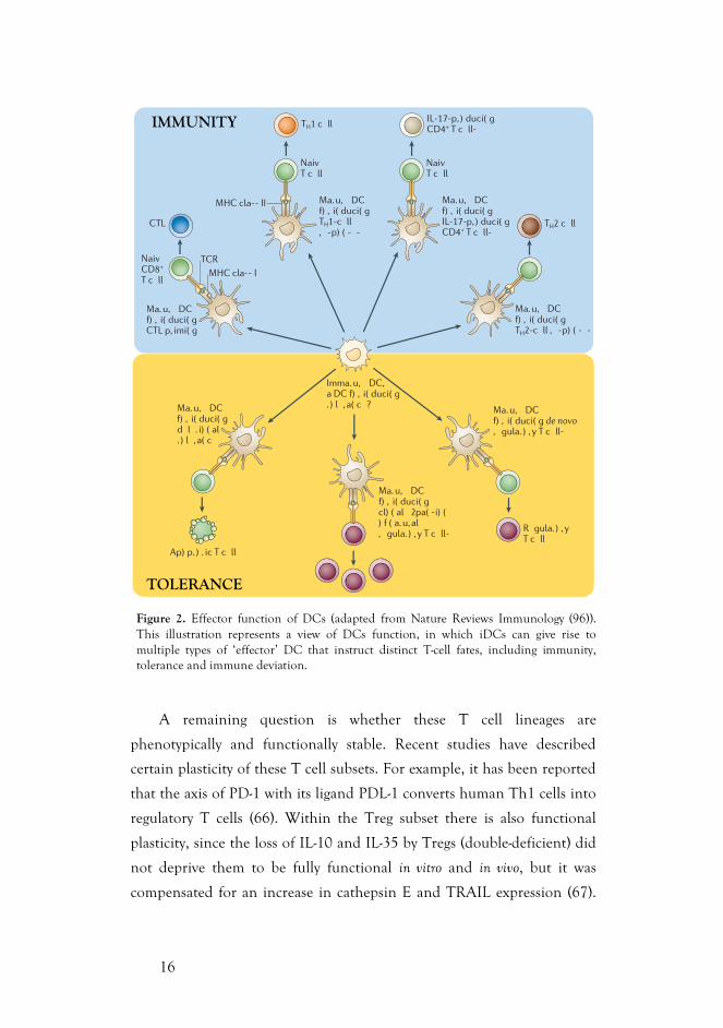
Figure 2. Effector function of DCs (adapted from Nature Reviews Immunology (96)). This illustration represents a view of DCs function, in which iDCs can give rise to multiple types of ‘effector’ DC that instruct distinct T-cell fates, including immunity, tolerance and immune deviation.
� � � � & &
� ' ' � . / , � � � �� � � � � ! ) , � $ ( � / � $ ( ". ) & , � ( � �
� � . / , � � �! ) , � $ ( � / � $ ( "� & ) ( � & � 2* � ( - $ ) () ! � ( � . / , � &, " / & � . ) , 3 � � � � & & -
� � . / , � � �! ) , � $ ( � / � $ ( " � � � � � � � �, " / & � . ) , 3 � � � � & & -
� � . / , � � �! ) , � $ ( � / � $ ( "� & . $ ) ( � &. ) & , � ( �
� � . / , � � �! ) , � $ ( � / � $ ( "� � � � * , $ ' $ ( "
� � � � � & � -- � �� � �
� � . / , � � �! ) , � $ ( � / � $ ( "� � � � & &, - * ) ( - -
� � . / , � � �! ) , � $ ( � / � $ ( "� � � � & & � , - * ) ( - -
� � $ 0 � � �
� � � & &
� � �
� � � � & &
� � $ 0 � � � & &
� * ) * . ) . $ � � � � � & &
� " / & � . ) , 3� � � & &
� � � � � & � -- � � � � � . / , � � �! ) , � $ ( � / � $ ( "� � � � � * , ) � / � $ ( "� � � � � � � � & & -
� � � � � * , ) � / � $ ( "� � � � � � � � & & -
� � $ 0 � � � & &
is unimportant and it would be unwise to be overly prescriptive in a field that is domi-nated by personal preferences. The point is that, ultimately, all these terminologies define DCs empirically by what they do to T cells. In other words, there is a clear trend in the field to complement the use of the ‘maturation’ nomenclature with one that defines DCs by their ‘effector function’.
In the simplest view, DCs are ‘effector cells’ when they have the potential to inter-act with lymphocytes and regulate their function (although this does not exclude other possible effector roles for DCs, for
example, in innate immune responses81). However, ‘effector DCs’ can direct dis-tinct lymphocyte responses and should be further defined by reference to these responses. In this respect, it is illuminating to consider T cells, because so much of the effector activity of DCs seems to be focused on that leukocyte compartment. So, just as there are TH1 cells, TH2 cells and CTLs, there could be distinct DCs for TH1-cell, TH2-cell and CTL priming. Similarly, perhaps even DCs for peripheral tolerance or for the induction of regulatory T cells (FIG. 4). This open-ended view of DC
function could easily integrate future types of effector DC as they are discovered.
Is this just semantics? After all, what is the difference between a ‘mature DC for inducing TH1-cell priming’ and a ‘mature DC’? Depending on the context, the two descriptions could denote precisely the same cell. But terminology shapes thinking and the first nomenclature, although awkward, forces one to focus on DC function and thereby complements the second, which is all too often used merely as a phenotypic descrip-tion. A focus on effector activity also adds to the concept of ontogenetically distinct DC lineages. Therefore, one can allow for the fact that plasmacytoid DCs (pDCs) constitute a unique DC subset, but refer to them in different contexts: for example, as pDCs for CTL priming82,83 or as pDCs for regulating T-cell responses to inhaled antigens84. The advantage of complementing definitions of DC maturity (in terms of phenotype) with statements about effector function becomes clearest when the role of DCs in tolerance is considered. By accepting that phenotypically mature DCs can be tolerogenic, tolerogenic-ity does not need to be cited as a property of immature DCs. Therefore, the possibility that certain environmental signals can ‘mature’ DCs into a tolerogenic mode can be accepted. This would offer a solution to the issues raised earlier, by allowing for the exis-tence of tolerogenic maturation signals that allow DCs to selectively upregulate the ability to deliver signal 1 and thereby promote tolerization of the low-affinity repertoire of self-reactive T cells that is most likely to pose a problem during infection.
Is there any evidence that tolerance induc-tion by DCs in vivo requires a stimulus? It is notable that most protocols used to show tolerance induction by steady-state DCs involve delivery of potential signals, be they in the form of antibodies that crosslink cell-surface signalling molecules or Fc receptors, or as cell-associated antigens that can trigger receptors involved in the uptake of cellular material85–89. One apparent exception is the elegant model of Probst et al.69,90, in which antigen expression by steady-state DCs is acutely induced by tamoxifen administration and promotes peripheral tolerance of CD8+ T cells. Given that tamoxifen is unlikely to be acting as a signal for the generation of tolerogenic DCs, these data are generally seen as evidence for peripheral tolerance induction by DCs being a default pathway. Therefore, some populations of immature DCs might possess an intrinsic tolerogenic effector function, especially the blood-derived DCs that are present in secondary
� $ " / , � � � 4� � � � � � � � � � � � � � � � � � � � � � � � � � � � � � � � # $ - � 0 $ 1 � ) ! � � ( � , $ . $ � � � & & � � � � � � ! / ( � . $ ) ( � - / * * ) - -� . # � . �$ ' ' � . / , � � � - � � � ( � " $ 0 � , $ - � . ) � ' / & . $ * & � . 3 * - � ) ! � � ! ! � . ) , � � � � � . # � . � $ ( -. , / � . � � $ -. $ ( � . � � � � & & � ! � . - � �$ ( � & / � $ ( " � $ ' ' / ( $ . 3 � � . ) & , � ( � � � ( � � $ ' ' / ( � � 0 $ � . $ ) ( � � � � . / , � . $ ) ( � , ! , - � . ) � . # � � # � ( " - � . # � . �� � � ) ' * � ( 3 � $ ' ' � . / , � � � � . , � ( - $ . $ ) ( � . ) � � � " $ 0 ( � ! ! � . ) , � -. � . � $ ( � , - * ) ( - � . ) � ( 0 $ , ) ( ' ( . � & � - $ " ( � & - �) ! � 2) " ( ) / - � � ! ) , � 2� ' * & � � ' $ � , ) � $ � & � � ) , � ( � ) " ( ) / - � � ! ) , � 2� ' * & � � � 3 . ) % $ ( - � � # ) , ' ) ( -� � ( � � � 3 $ ( " �� & & - � � ) , $ " $ ( � � � # � +/ � & $ . 3 � ) ! � . # - � - $ " ( � & - � & � , " & 3 � � . , ' $ ( - � . # � � # ) $ � � ) ! � ! ! � . ) , � � � � � � & . # ) / " # �) ( . ) " ( 3 � � � ( � � & -) � # � 0 � � � , ) & � � ( � � � , . � $ ( � � � � - / � . 3 * - � ' $ " # . � � � * , � $ - * ) - � � . ) 1 � , � - � * � , . $ � / & � , � ! ! � . ) , � -. � . - � ) , � # � 0 � ) ( & 3 � � � , -. , $ � . � � , * , . ) $ , � ) ! � ! ! � . ) , � ! / ( � . $ ) ( - � � � � � � � . $ 0 � . $ ) ( � ' $ " # . � � & -) �) � � / , � - * ) ( . � ( ) / - & 3 � � . # � . � $ - � � $ ( � . # � � � - ( � � ) ! � ( 0 $ , ) ( ' ( . � & � - $ " ( � & - � � � / . ) ' � . $ � � & & 3 � * , ) ' ) . $ ( " � � � �. , � ( - $ . $ ) ( � . ) � � � " $ 0 ( � ! ! � . ) , � -. � . � � ! . , � � � ! $ 2 � � * , $ ) � � ) ! � . $ ' � � � ) . � . # � . � . # $ - � ' ) � & � � $ ! ! , - � ! , ) ' �. # � � ) ( 0 ( . $ ) ( � & � ) ( � � 3 � - / * * ) - $ ( " � . # � . � . ) & , ) " ( $ � � � � - � � � ( � � � � � . 3 * � ) ! � ' � . / , � � � � � � ( � ) . # , �1 ) , � - � � -) ' � ' � . / , � . $ ) ( � - $ " ( � & - � � � ( � * , ) ' ) . � . # � " ( , � . $ ) ( � ) ! � . ) & , ) " ( $ � � � � - � � � # $ - � � ) - � ( ) . �( " � . � . # � * ) -- $ � $ & $ . 3 � . # � . � -) ' � $ ' ' � . / , � � � - � # � 0 � � ( � $ ( . , $ ( - $ � � . ) & , ) " ( $ � � ! / ( � . $ ) ( � � � � � � � � 3 . ) �. ) 2$ � � � � & 3 ' * # ) � 3 . � � � � � � � � $ ( . , & / % $ ( � � � � � � � � � � � � & & � , � * . ) , � � � � � � � � # & * , �
� � � � � � � � � � � �
NATURE REVIEWS | � � � � � � � � VOLUME 6 | JUNE 2006 | � � �©� 2006 � Nature Publishing Group�
�
IMMUNITY
TOLERANCE

17
Therefore, cross-regulatory compensating pathways may exist to control
the suppressive mechanisms of Tregs. In addition, the balance between
Tregs and Th17 cells in culture is controlled by various factors (i.e. IL-6,
IL-1, IL-23, and retinoic acid (68)), and Th17 cells are capable of
converting into Tregs and vice versa. Therefore, effector Th17 cells are
highly adaptable to their cytokine microenvironment, which may partially
explain their association with pro- and anti-inflammatory functions (69).
Interestingly, a recent study reported that plasticity of human Th17 cells
and iTregs is orchestrated by different subsets of CD14+ myeloid cells
(70), shaping the outcome of immune reaction from inflammation to
tolerance.
1.3 Immunological tolerance
The immune system is not only responsible to protect the organism
from invading pathogens, but also to avoid self-responses that could
destroy self-tissues. Thus, ‘immunological tolerance’ is also an active form
of immune response aimed at avoiding autoimmune diseases. Based on
their organic distribution, the immunological tolerance is divided in two
scenarios, known as ‘central’ and ‘peripheral’ tolerance.
1.3.1 Central tolerance
Central tolerance is based on the positive and negative selection of T
cell-precursors in the thymus. Only T-cells bearing receptors (TCRs) that
recognize the own MHC molecules (on thymic epithelium) receive a
survival signal leading to their positive selection. Those T lymphocytes
that bear TCRs strongly reactive to self-peptides (autoreactive cells) are
deleted and therefore eliminated from the repertoire by negative selection
(71). Different cells contribute to this thymocite negative selection, such
as medullar thymic epithelial cells (mTECs) that ectopically express tissue-
restricted antigens (TRA) (i.e. insulin) under the control of a nuclear factor
called autoimmune regulator (Aire). Medullary DCs also contribute to this
process cross-presenting TRA (72), and peripheral DCs presenting

18
antigens from peripheral tissues when passing through the thymus (62),
therefore expanding the antigen repertoire. Furthermore, the cytokine
milieu in the thymus, like the influence of thymic stromal lymphopoietin
(TSLP) produced by epithelial cells in Hassall’s corpuscles, can promote
the conversion of low-autoreactive CD4+ CD25- thymocytes to CD4+
CD25+ Foxp3+ Tregs (73). Thus, within the thymus, T cells go through
positive and negative selection processes to shape the entire peripheral T-
cell repertoire.
1.3.2 Peripheral tolerance
Central tolerance is essential but not enough, since some
autoreactive T cells escape from thymic deletion and, once in the
periphery, may contribute to the development of autoimmune responses.
Hence, peripheral tolerance mechanisms are designed to safe guard
against these autoreactive T cells in peripheral tissues (basically lymph
nodes and the spleen). Mechanisms of peripheral tolerance (74) can be
divided into ‘T cell-extrinsic’ and ‘T cell-intrinsic’ mechanisms.
T cell-intrinsic mechanisms include:
a) Immunological ignorance to an antigen that could not be
presented efficiently by APCs in secondary lymphoid organs, due to its
low concentration. This phenomenon has been described to occur in the
initial phases of some peripheral solid tumors (75).
b) Anergy is a state of long-term hyporesponsiveness in T cells that
is characterized by an active repression of TCR signaling and IL-2
expression (76). Induction of anergy in T cells was initially described as
the result of TCR (signal 1) without concomitant co-stimulatory signaling
(signal 2). However, recent studies have demonstrated that the T cell
actively sense its microenvironment, through mTOR dependent and
independent mechanisms, for available nutrients and negative cues such
as adenosine. This regulates the T cell commitment to switch its
metabolic machinery and enter the S phase of the cell cycle, inducing

19
anergy and long-term tolerance in the T cell (77). Several forms of anergy
have been described, and Schwartz et al proposed to classify them into
two broad categories: clonal anergy (growth arrest state that can be reversed
by adding IL-2) and adaptive tolerance or in vivo anergy (inhibition of
proliferative and effector functions not reversible by IL-2) (76).
c) Deletion of T cell clones through induction of apoptosis is
induced by the engagement of counter-regulatory receptors on T cells
surface, such as CTLA4 with CD80/CD86, or PD1 with PD1L/ PDL2,
during antigen presentation. Another mechanism to induce apoptosis is
the Fas receptor engagement by FasL, and triggering of a Bcl-2 and Bcl-
xL–regulated mitochondrial death pathway (78).
d) Phenotypic skewing or immune deviation is the shift of T cells
towards a different effector subset expressing different cytokine patterns,
i.e. from pro-inflammatory to anti-inflammatory phenotype, after
activation. While Th1 and Th17 effector T cells are considered relevant
for pathogenic immune response, it has been suggested that development
of a Th2 effector T cell would counteract autoimmunity by promoting
anti-inflammatory cytokines (79).
T cell-extrinsic mechanisms include the induction of Treg cells
(explained earlier) and the induction of tolerogenic DCs. In fact, some
authors suggested that tolerogenic DCs and Tregs regulate each other’s
homeostasis (80).
1.3.3 Tolerogenic DCs (tolDCs)
DCs are important not only in the generation of T-cell immune
responses, but also in immune tolerance. The ability of DCs to induce
tolerance was initially demonstrated by experiments on iDCs residing in
peripheral lymphoid tissues (81). Therefore, under steady-state
conditions, iDCs may capture apoptotic bodies derived from natural cell
turnover and, after migration to the draining lymph nodes, DCs present
self antigens and silenciate autoreactive T cells (81). However iDCs are

20
not in a final differentiation state and can give rise to both
immunogenic/pro-inflammatory mDCs as well as semi-mature DCs,
which have the ability to establish and maintain tolerance. These natural
tolDCs maintain tolerance in peripheral tissues against commensal
microorganisms, antigens from food and airways, etc. within a steady-state
environment. In addition, many pathogens and tumors can mimic or
produce tolerogenic factors and instruct tolDCs as an immune escape
mechanism (reviewed by Maldonado and von Adrian (82)). pDCs have
also been described to play an important role in inducing immune
tolerance (83).
TolDCs are characterized by reduced expression of co-stimulatory
molecules (mainly CD40, CD80, CD86) and, usually, by reduced
production of IL-12 and increased IL-10 secretion (84) together with a
reduced ability to induce T cell proliferation. While these properties can
explain their ability to induce Tregs rather than T effector cells, several
other mechanisms may play a role in tolerance induction and regulation
(85).
The molecular mechanisms involved in the tolerogenic function of
DCs in the periphery include:
a) Antigen presentation with inappropriate co-stimulation
(induction of anergy).
b) Presentation of very low levels of antigen in the absence of other
stimuli, which promotes Treg differentiation (86)).
c) Production of cytokines such as IL-10, TGF-", TNF-#, or
granulocyte colony–stimulating factor (G-CSF) can induce functional
properties to DCs (87). For instance, presence of IL-10 during
differentiation of CD4+ T cells results in the development of Tr1 cells
(82). In addition, some tissue-DCs can synthesize retinoic acid (RA), a
metabolite of vitamin A that, besides imprinting T cells to express gut
homing receptors, promote the differentiation to Foxp3+ Tregs (88).

21
d) Expression of some molecules that induce T cell death (deletion),
such as the indoleamine 2,3-dioxygenase (IDO), the rate-limiting enzyme of
tryptophan catabolism that increases a cytotoxic metabolite for activated
T cells (89). In this sense, some studies support that IDO-expressing DCs
contribute to peripheral tolerance by depleting autoreactive T cells (90).
Furthermore, DCs also express membrane receptors that may instruct T-
cell deletion, such as the interaction through the Fas-L, or the PDL-1 and
PDL-2 (mentioned earlier).
e) Expression of inhibitory receptors such as ILT3 (immunoglobulin
like transcript 3), that contain a cytoplasmic tyrosine-based inhibitory
motif (ITIM), have shown to negatively regulate activation of DCs (91).
Furthermore, CD8+CD28- suppressor T cells have demonstrated to up-
regulate ILT3 and ILT4 expression on DCs rendering them tolerogenic
(92).
Importantly, the role of DCs in maintaining tolerance is
independent of their maturation state, since immature DCs, semi-mature
DCs and also mature DCs have shown to expand antigen-specific Treg
cells both in vitro and in vivo (93–95). For this reason, some authors
recommend to define DCs based on their effector function on T cells
rather than on their phenotype (87,96). Hence we can differentiate
‘immunogenic DCs’ from ‘tolerogenic DCs’ according to their functional
properties.
DCs play key roles not only as an initiator of the immune response
but also as a regulator of adaptive responses, ensuring the balance
between immunity and maintenance of peripheral tolerance. This dual
functionality of DCs is achieved by the integration of different signals:
antigen dose, DC lineage and maturation status, DC stimulation by
pathogen derived products, and cytokine milieu at sites of inflammation.

22
1.3.4 Lost of tolerance
Under homeostatic conditions, central and peripheral tolerance
ensure the selective generation and regulation of functional, non-self-
reactive T cells. However, despite these multiple mechanisms to maintain
tolerance, some situations can elicit an eventual activation of autoreactive
T cells, which can engage an IR against antigens produced from self-
tissues. This breakdown of immune tolerance could trigger an adaptive
autoimmune response, leading to the development of an autoimmune
disease (97).
Autoimmune diseases (ADs) could be classified in ‘organ-specific’
when the IR is against a tissue-specific antigen (such as in multiple
sclerosis or type-I diabetes), or ‘systemic’ when the IR is against an
ubiquitous antigen (i.e. systemic lupus erythematosus). In addition, ADs
could be also classified based on their mechanism of action: mediated by
antibodies, by immunocomplexes or by autoreactive T cells (98).
An important issue in the field of autoimmunity is to identify the
auto-antigens recognized by T and B lymphocytes, and use this
information to design new strategies to induce antigen-specific
therapeutic tolerance. One of the most studied ADs and their auto-
antigens has been multiple sclerosis.
2. Multiple sclerosis
Multiple sclerosis (MS) is a chronic autoimmune disease of the
central nervous system (CNS). Epidemiologic studies show that MS is the
most common cause of neurological disability among young adults (20-40
years), with a prevalence of 1/1000 in Caucasian populations (99).
The etiology of MS remains unclear, but according to current data,
environmental and genetic factors are involved in the development of MS
(100). Research of susceptibility genes has involved the MHC molecules,
specifically the HLA-DR15 haplotype in Caucasians, which is thought to

23
account for 10%–60% of the genetic risk of MS (101,102). In addition,
non-genetic factors also contribute to MS etiology, such as infectious
agents and behavioral influences (103).
The autoimmune response is thought to be responsible for the
pathological features of the disease, which include demyelination,
oligodendrocyte loss and axonal injury (104).
The disease is characterized by episodes of inflammatory activity in
the CNS associated with neurological impairment such as progressive
paralysis in most patients (105). These episodes are caused by localized
CNS demyelination plaques, and the symptoms of MS vary depending on
the location of plaques within the CNS. Common symptoms include
sensory disturbances in the limbs, optic nerve dysfunction, pyramidal
tract dysfunction, bladder or bowel dysfunction, sexual dysfunction,
ataxia, and diplopia (106).
The diagnosis is made primarily on the basis of the medical history
and physical exam (formalized as the McDonald criteria and later
reviewed) (107,108), and may take into account laboratory data, such as
the characteristic oligoclonal bands in the cerebrospinal fluid (CSF). Over
the past two decades, the diagnosis has included the identification of
white matter lesions via evaluation of T2-hyperintense lesions and
gadolinium-enhancing T1 lesions on magnetic resonance imaging (MRI)
(109), the later serving as a marker of focal inflammation due to the local
permeability and breakdown of the BBB.
According to Lublin et al (110) there are four different courses of MS
(schematized in Figure 3):
a) Relapsing-remitting (RR-MS) is characterized by acute attacks of
neurologic dysfunction (relapses). Over the following weeks to months
after the attacks, most patients experience a partial or complete recovery
of function. Between the attacks the patient is neurologically and

24
symptomatically stable. This is the initial clinical form of 85% of MS
patients.
b) Secondary-progressive (SP-MS), begins as RR, but at some point the
attack rate is reduced and the course becomes characterized by a steady
deterioration in function without frequent relapses. 85% of RR-MS
patients develop this form after 25 years of disease evolution (111).
c) Primary-progressive (PP-MS) is characterized by a progressive
neurological worsening from the beginning without clear relapses. It is
the initial clinical form of 15% patients.
d) Progressive-relapsing (PR-MS) also begins with a progressive course
although these patients also experience occasional attacks.
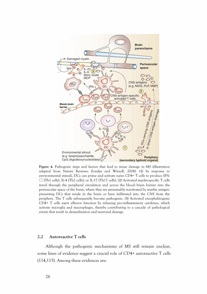
2.1 Immunopathology
The disease process in MS (reviewed in (112,113), and schematized
in Figure 4) may start with the activation of autoreactive CD4+ T cells in
the periphery, for example by epitope mimicry, in an inflammatory
context. Then, activated autoreactive T cells adhere to the BBB
Figure 2. Frequent courses of MS (adapted from Lancet (106)). This scheme illustrates how the pathological processes of inflammation, demyelination, and axon degeneration explain the clinical course of multiple sclerosis. The ‘clinical threshold’ illustrates the fact that inflammatory lesions in CNS not always are manifested as a clinical episode; below this threshold the effects of individual inflammatory lesions can be compensated and above it they cause symptoms.
� � � ! � $
� � � � 6 6 6 � 3) & , " . $& 3� $/ - � � � � " � � � � � � � � � & " � � $ � � � � � � � � � � � �
�
��
�
�
�
�
� 1" * . � 5/ , 4- &
� & $/ . % " 18� 01/ ( 1& 22* 5&� & , " 02* . ( � 1& - * 33* . (
� , * . * $" , � 3) 1& 2) / , %
� . = " - - " 3* / .
� � & � " � " � )
� 7/ . " , � , / 22
�
��
�
�
�
�
Progression of pathology
Relapsing remitting Secondary progressive
Inflammation
Clinical threshold
Axonal loss

25
endothelium via adhesion molecules such as LFA-1 and VLA-4, and
transmigrate into the brain parenchyma. There, these autoreactive T cells
are presumably reactivated by myelin antigen-presenting DCs that reside
in the brain or have infiltrated into the CNS from the periphery.
Activated encephalitogenic CD4+ T cells exert effector function by
releasing pro-inflammatory cytokines (i.e. IFN-!, IL-23, TNF-") and
chemokines (RANTES, IP-10, IL-8, and others), which activate microglia
and macrophages in the CNS, and recruit other immune cells, such as
monocytes, CD8+ T cells, B cells, and mast cells from the peripheral
blood. These pathological events result in formation of the inflammatory
lesion, which is characterized by the release of proteases, pro-
inflammatory molecules, and oxygen and nitrogen radicals from mast
cells, monocytes, and T cells. Altogether lead to demyelination, neuronal
damage, and axonal loss that is closely related to neurological disability
(112).
26
2.2 Autoreactive T cells
Although the pathogenic mechanisms of MS still remain unclear,
some lines of evidence suggest a crucial role of CD4+ autoreactive T cells
(114,115). Among these evidences are:
Figure 4. Pathogenic steps and factors that lead to tissue damage in MS (illustration adapted from Nature Reviews; Zozulya and Wiendl, 2008). (1) In response to environmental stimuli, DCs can prime and activate naive CD4+ T cells to produce IFN-" (Th1 cells), IL-4 (Th2 cells), or IL-17 (Th17 cells). (2) Activated myelin-specific T cells travel through the peripheral circulation and across the blood–brain barrier into the perivascular space of the brain, where they are presumably reactivated by myelin antigen-presenting DCs that reside in the brain or have infiltrated into the CNS from the periphery. The T cells subsequently become pathogenic. (3) Activated encephalitogenic CD4+ T cells exert effector function by releasing pro-inflammatory cytokines, which activate microglia and macrophages, thereby contributing to a cascade of pathological events that result in demyelination and neuronal damage.
394 NATURE CLINICAL PRACTICE NEUROLOGY ZOZULYA AND WIENDL JULY 2008 VOL 4 NO 7
www.nature.com/clinicalpractice/neuro
lie in the isolation and use of human antigen-specific TREG cells,—the durability and function of these cells has not yet been tested. In addition,
it might be difficult in practice to determine the optimum antigen dose to induce ex vivo expan-sion of antigen-specific TREG cells. The best route
6
5
4 1
2
3CNS-derivedor peripheral ¡TREG
CNS milieu
Blood!brainbarrier
DC
s ca
rryi
ng C
NS
ant
igen
sor
dire
ct C
NS
-ant
igen
dra
inag
e
IL-10TGF-
IL-23IL-6Nitric oxideMCP
IFN-IL-10Soluble HLA-G
M
SolubleHLA-G
Repaired myelin Damaged myelin
Tolerance Immunity
Brainparenchyma
Perivascularspace
CNS antigens(e.g. MOG, PLP, MBP)nTREG
¡TREG
TREG TREG
TREG
???
?
IL-10
ICOSLILT3ILT4
TREGTREGTREG
TREG
¡TREG
HLA-G+
nTREG
CD4+CD25+
nTREG
Circulating in blood nTREG
CNS-antigen-specificactivated T cells
Environmental stimuli(e.g. lipopolysaccharide,CpG oligodeoxynucleotides)
Periphery(secondary lyphoid organs)
DCtol
CD8+
CD28–
TR1
TH3
?
DCtol
IL-23
IL-6
TGF-
OX40L
TEFFTEFF
TEFF
TH2TH17
TH1
IL-12
DC
TEFF
Mc
TEFF
DC
TEFF TEFF
Figure 3 Balance between immunogenic and tolerogenic mechanisms in multiple sclerosis: a hypothesis. Autoimmune neuroinflammation is considered to result from a disrupted balance between immune cells that cause damage (TEFF cells) and cells with suppressive capabilities (TREG cells and tolerogenic DCs). (1) In response to environmental stimuli, DCs (red) can prime and activate naive CD4+ T cells to produce IFN-! (TH1 cells), IL-4 (TH2 cells), or IL-17 (TH17 cells). (2) Activated myelin-specific T cells travel through the peripheral circulation and across the blood–brain barrier into the perivascular space of the brain, where they are presumably reactivated by myelin-antigen-presenting DCs that reside in the brain or have infiltrated into the CNS from the periphery. The T cells subsequently become pathogenic. (3) Activated encephalitogenic CD4+ T cells exert effector function by releasing proinflammatory cytokines (e.g. IFN-!, IL-6, IL-23), which activate microglia and macrophages, thereby contributing to a cascade of pathological events that result in demyelination and neuronal damage. The presentation of myelin antigen epitopes by resident CNS DCs causes further myelin breakdown, leading to epitope spreading.65 (4) Tolerogenic DCs (blue) cause naive T cells to become regulatory (TH3, TR1 or CD8+CD28–). The resulting iTREG cell populations can enter the CNS. In addition, nTREG cells (HLA-G+ or CD4+CD25+) that are circulating in the blood can enter the perivascular space of the CNS. The mechanisms and triggers that cause nTREG and iTREG cells to traffic across the blood–brain barrier into the brain are poorly understood. (5) TREG cells in the CNS can either directly suppress encephalitogenic T cells through cytokine secretion (IL-10, TGF-", soluble HLA-G) and T cell–T cell interaction, or act on local DCs, thereby rendering them tolerogenic. The tolerogenic DCs in turn could generate or propagate TREG-cell expansion or suppressive function (iTREG) that contributes to the suppression of ongoing autoinflammation. (6) TREG-cell populations that become expanded in response to specific CNS antigens suppress ongoing autoimmune inflammation in the CNS. It is not known whether or how TREG cells act directly within the CNS and whether TREG-cell populations can be generated within the brain parenchyma. Abbreviations: DC, dendritic cell; DCtol, tolerogenic dendritic cell; HLA-G, human leukocyte antigen G; M#, macrophage; Mc, microglial cell; ICOSL, inducible costimulatory molecule ligand; IFN-!, interferon !; IL-, interleukin; ILT, immunoglobulin-like transcript; iTREG, inducible regulatory TREG cell; MBP, myelin basic protein; MCP, monocyte chemotactic protein; MOG, myelin oligodendrocyte glycoprotein; nTREG, natural regulatory TREG cell; PLP, proteolipid protein; TEFF, T effector cell; TGF-", transforming growth factor "; TH, T-helper cell; TR1, T-regulatory 1 cell; TREG, regulatory T cell.
REVIEW
Blood−brainbarrier
27
• The HLA-DR15 haplotype in Caucasians (DRB1-1501, DRB5-
0101, DQA1-0102, DQB1-0602) carries the strongest genetic risk for MS,
presumably via their role as antigen-presenting molecules to pathogenic
CD4+ T cells (101).
• In experimental autoimmune encephalomyelitis (EAE) in rodents
it is known that myelin reactive T cells are encephalitogenic, since the
transfer of these cells from EAE mice to a healthy receptor developed the
disease (116).
• A clinical trial demonstrated that MS patients treated with an
altered peptide ligand based on the MBP(83-99) epitope developed
clinical worsening, increased brain inflammation and increased anti-MBP
responses (117).
• T cell responses to myelin antigens have been extensively studied in
MS and control subjects. Interestingly, a higher level of activation of T
cells reactive to myelin basic protein (MBP), an important constituent of
CNS myelin, was found in MS patients as compared to healthy controls
(118). In fact, many groups have confirmed the CD4+ T cell reactivity to
myelin antigens in MS patients (119–121).
Some studies have reported evidences that these autoreactive CD4+ T
cells in MS patients have been previously activated in vivo. (114,122). The
in vivo activation of this pathogenic autoreactive T cells could be
explained by several microbial infection-mediated mechanisms, including
molecular mimicry (i.e. the activation of autoreactive cells by cross-
reactivity between self-antigens and foreign agents), bacterial
superantigens and/or bystander activation (autoreactive cells are activated
due to nonspecific inflammatory events during infections) (123,124).
Recent studies have reported Th1 and Th17 as effector T cell subsets that
play a key role in the regulation of IR during CNS infection and disease
(125,126).

28
2.3 Myelin antigens
A lot of effort has been put in identifying myelin antigens that could
be recognized by autoreactive T cells in MS patients (reviewed in (120)).
Myelin basic protein (MBP) and proteolipid protein (PLP) rapidly gained
interest due to their abundance and, most probably, MBP is the best-
studied myelin protein in MS. Some reports have demonstrated that the
frequencies and Th1-bias of MBP and PLP-specific T cells are increased in
MS (115,127,128). Myelin oligodendrocyte glycoprotein (MOG), a less
abundant myelin compound, has been reported to induce strong
responses in MS patients as well (129). Similarly, elevated myelin-associated
glycoprotein (MAG) specific T and B cell responses have been observed in
the cerebrospinal fluid of MS patients by ELISPOT assays (130).
Bielekova et al. demonstrated the presence of a higher proportion of high
avidity myelin-specific CD4+ T cells in MS patients compared to controls,
which were mostly memory cells with a Th1 phenotype (115). Reactivity
of these myelin-specific T cells was predominantly directed towards a
group of immunodominant epitopes of myelin (MBP13–32, MBP11–129,
MBP146–170, PLP139–154, MOG1–20 and MOG35–55), and also less
dominant epitopes such as the C-terminal area of 2’,3’-Cyclic Nucleotide 3’-
phosphodiesterase (CNP-ase) (115).
Our group has recently confirmed a distinct reactivity to a selection
of these immunodominant peptides (Table 1) in peripheral blood T-cells
from a cohort of RR-MS patients compared to healthy controls (HC)
(131) (Figure 5). A significant correlation was found between positive T-
cell proliferation and the clinical score (a higher disability or EDSS score,
a shorter relapse-free time and a higher frequency of relapses), and this
reactivity was sustained over time in the majority of patients (131). These
results supported the pathogenic significance of these set of myelin
peptides in MS. Furthermore, these myelin peptides are expressed in the
thymus (132) but are predicted to bind with low affinity to main MS-
associated HLA-DR alleles (115,133), supporting a defective negative

29
selection in the thymus and consequently permitting a higher frequency
in the periphery of these autoreactive CD4+ T cells.
Other myelin peptides involved in the T cell response and/or
recognized by autoantibodies in cerebrospinal fluid and in serum of MS
Description Sequence aa
MBP(13-32) KYLATASTMDHARHGFLPRH 20
MBP(83-99) ENPVVHFFKNIVTPRTP 17
MBP(111-129) LSRFSWGAEGQRPGFGYGG 19
MBP(146-170) AQGTLSKIFKLGGRDSRSGSPMARR 25
PLP(139-154) HCLGKWLGHPDKFVGI 16
MOG(1-20) GQFRVIGPRHPIRALVGDEV 20
MOG(35-55) MEVGWYRPPFSRVVHLYRNGK 21
Table 1. Selected myelin peptides
Proliferative response to myelin peptides
&0 -6 <: 16/ ;0- 5 1? 7. 8-8;1, - : ) ; l� + 76+ -6;9) �;176� ) 87: 1;1=- 9-) + ;176 > ) : , - ;- + ;- , 16 � 7. �8) ;1-6; : > 1;0 � $ � � � � � � 7. + 76;974: � � � � ) 6, �7. � 8) ;1-6; : > 1;0 ! � � � � � P � � � � � ; � l� �;0- 9- : 876, -9: > -9- � 7. � 8) ;1-6; : � � � � � � 7. + 76;974: � � � ) 6, � 7. � 8) ;1-6; : > 1;0 ! � � � � �� P � � � � � � 1/ � � �
T-cell proliferation, clinical and MRI disease
characteristics in the MS cohort
� : 1/ 61C+ ) 6; ) : : 7 + 1) ;176 > ) : .7<6, *- ; > - -6 87: 1;1=-%� + -44 89741.-9) ;176 ) 6, 01/ 0-9 , 1: ) *141; @ � � � $ $ : + 79- �� � ' E� � � ( =: � � ' E� � � (� P � � � � 01/ 0-9 6<5 *-97.9-4) 8: - : � � B � =: � � B � � � P � � � � ) 6, : 079;-9;15 - .975 ;0- 4) : ; 9-4) 8: - � � B � � � =: � � � B � � � 5 76;0: � P � � � � %) *4- � � � � $ $ : + 79- > ) :16, -8-6, -6;4@ ) : : 7 + 1) ;- , > 1;0 %� + -4489741.-9) ;176 � 7, , :9) ;17 � � � ' � � � � � � � � � E� � � (� ) .;-9 ) , 2<: ;5 -6; .796<5 *-9 7. 9-4) 8: - : � , 1: - ) : - , <9) ;176 ) 6, ;15 - .975 ;0-4) : ; 9-4) 8: - �
Longitudinal analysis of T-cell proliferation during
follow-up
� 6) 4@ : 1: 7. ;0- -=74<;176 7. %� + -44 9-) + ;1=1;@ ) / ) 16: ;5 @ -416 8-8;1, - : : 07> - , ;0) ; + -44<4) 9 9-) + ;1=1;@ � 87: 1�;1=- � 6- / ) ;1=- � > ) : 5 ) 16;) 16- , 16 � � 8) ;1-6; : > 1;0� $ � � � � � 7=-9 ;15 - � � 1=- 89-=17<: 4@ 87: 1;1=- 8) ;1-6; :*- + ) 5 - 676� 9- : 876, -9: , <916/ .7447> � <8� ;09-- 7. ;0-5) .;-9 : ;) 9;16/ ;9-) ;5 -6; > 1;0 16;-9.-976� *- ;) � � - : 81;-;0- + 76;16<7<: <: - 7. 16;-9.-976� *- ;) � ;0- @ 9-5 ) 16- ,676� 9- : 876, -9: .79 764@ ) : 079; 8-917, 7. ;15 - � .975 �;7 � 5 76;0: � � � 76=-9: -4@ � .7<9 89-=17<: 4@ 6- / ) ;1=-8) ;1-6; : : 07> - , ) 87: 1;1=- 89741.-9) ;176 ) ; 4) ;;-9 ;15 -8716; : � ; > 7 7. ;0-5 ) .;-9 : <!-916/ ) 9-4) 8: - � � 6 *7;08) ;1-6; : � 9-4) 8: - : > -9- 67; ;9-) ;- , > 1;0 : ;-971, : ) : ;0- @)!- + ;- , 5 ) 164@ : -6: 79@ .<6+ ;176: � %01: 87: 1;1=1;@ > ) :5 ) 16;) 16- , , <916/ .7447> � <8�
Discussion
%0- 9- : <4; : 7. ;01: : ;<, @ : 07> ;0) ; 87: 1;1=- %� + -449-) + ;1=1;@ ) / ) 16: ; ) : -4- + ;- , 5 1? 7. 5 @ -416 8-8;1, - : 1:5 79- + 75 5 76 16 8) ;1-6; : > 1;0 # # � $ ;0) 6 16 � � � ;01:9- ) + ;1=1;@ 1: : <: ;) 16- , 7=-9 ;15 - ) 6, 1: + 47: -4@ 9-4) ;- , ;7/ 9-) ;-9 , 1: ) *141; @ �%7 ) : : - : : ;0- : 8- + 1C+ 1; @ 7. ;01: %� + -44 9-) + ;1=1;@ .79
� $ � > - ) 6) 4@ : - , 8) ;1-6; : > 1;0 ! � � > 01+ 0 5 ) @ 4- ) , ;7, -5 @ -416) ;176 ) 6, � ;0-79- ;1+ ) 44@ � %� + -449- : 876: - : : 15 �14) 9 ;7 � $ � & - .7<6, ;0) ; %� + -44 9-) + ;1=1;@ > ) : 5 7, �-9) ;-4@ : 8- + 1C+ .79 � $ � / 1=-6 ;0) ; ;0- 8-9+ -6;) / - 7.9- : 876, -9: 16 ;0- ! � ) 6, � � / 97<8 > ) : : 15 14) 9�# - / ) 9, 16/ + 4161+ ) 4+ 0) 9) + ;-91: ;1+ : 7. ;0- 8) ;1-6; : � ) 6,
;) 316/ 16;7 + 76: 1, -9) ;176 ;0) ; ;0- : -4- + ;- , / 97<8 � 76*) : 1: 7. ;0- @ > -9- 67; 9- + -1=16/ 15 5 <675 7, <4) ;79@;9- ) ;5 -6; � > ) : 5 7: ;4@ 5 1, , 4- � ) / - , .-5 ) 4- : > 1;0 9-4) �;1=- 476/ , 1: - ) : - , <9) ;176� > - .7<6, ;0) ; 87: 1;1=- %� + -4489741.-9) ;176 ;7 5 @ -416 8-8;1, - : : 07> - , ) : ;976/ + 79�9-4) ;176 > 1;0 / 9-) ;-9 , 1: ) *141; @ � : 079;-9 9-4) 8: - � .9- - ;15 -) 6, 01/ 0-9 6<5 *-97. 9-4) 8: - : � � 79-7=-9� 8) ;1-6; : > 1;087: 1;1=- %� + -449-) + ;1=1;@ 0) , 5 79- %� *9) 16 4- : 176: > -9-74, -9 ) 6, 0) , ) 476/ -9 , 1: - ) : - , <9) ;176 � ) 4;07</ 0 ;0- : -, 1!-9-6+ - : , 1, 67; ) + 01-=- : ;) ;1: ;1+ ) 4 : 1/ 61C+ ) 6+ - � �: </ / - : ;16/ ;0) ; 87: 1;1=- %� + -44 89741.-9) ;176 5 1/ 0; *-) 4: 7 9-4) ;- , ;7 476/ � 4) : ;16/ 16D) 5 5 ) ;176 ) 6, , 1: - ) : -897/ 9- : : 176�� 6 ;01: + 76;- ? ; � 1; 1: 9-4-=) 6; ;7 8716; 7<; ;0) ; 764@
� � 7. ;0- 8) ;1-6; : ) 6) 4@ : - , > -9- � � � � # � � � �: <8879;16/ ;0) ; ;0- 15 5 <67, 75 16) 6; 8-8;1, - : <: - , ) 9-67; � � � � � # � 9- : ;91+ ;- , ' � (�%� + -44 9-) + ;1=1;@ ;7 5 @ -416 ) 6;1/ -6: 0) : *- -6 - ? ;-6�
: 1=-4@ : ;<, 1- , 16 � $ � 9-=1- > - , 16 � � � � 7 : ; 7. ;0- : ;<, 1- :.7<6, 67 , 1!-9-6+ - : *- ; > - -6 8) ;1-6; : ) 6, + 76;974:' E� ( ) 6, , 1, 67; C6, ;01: 9-4) ;176: 018 *- ; > - -6 87: 1;1=-89741.-9) ;176 ) 6, + 4161+ ) 4 , ) ;) ' � (� � 6 ;0- 5 ) 2791; @ 7.
� � �� � � � � � �
� � �
�
� � �
� � � � � � �
� � � � �
� � �
� �
� �
� � �
� � �
� �
��������������� ������������� ��������������������
� � � � � � � � � � � � � � � � � � � � � � � � �� � � � �
Figure 1 $ <5 5 ) 9@ 7. ;0- 89741.-9) ;176 ) : : ) @ : � %0- C/<9- : <5 �
5 ) 91A- : ;0- 897879;176 7. 8) ;1-6; : > 1;0 9-4) 8: 16/ � 9-5 1;;16/ 5 <4�
;184- : + 4-97: 1: � *4) + 3 *) 9: � n � � � � 7;0-9 6-<9747/ 1+ ) 4 , 1: - ) : - :
� ! � � 8) ;1-6; : � / 9- @ *) 9: � n � � � ) 6, 0-) 4;0@ , 7679: � � � �
� > 01;- *) 9: � n � � : 07> 16/ 87: 1;1=- 89741.-9) ;176 ;7 ;0- 5 1? 7.
8-8;1, - : <: - , ) ; � l� � � � � � $ � � � ! � ) 6, � � � �
P � � � � ) 6, ) ; l� � � � � $ � � ! � ) 6, � � � � �
P � � � � � : ) 87: 1;1=- + 76;974� > - <: - , " � � � � 7 � � 7.
, 7679: 87: 1;1=- � ) 6, <6: ;15 <4) ;- , + -44: ) : ) 6- / ) ;1=- + 76;974�
# - ) + ;1=1;@ ;7 5 @ -416 8-8;1, - : 16 5 <4;184- : + 4-97: 1: �
! 2010 The Author(s)European Journal of Neurology ! 2010 EFNS European Journal of Neurology 18, 1101–1104
Figure 5. Proliferative response to myelin peptides. The figure summarizes the proportion of patients with RR-MS (black bars, n=42), other neurological diseases (OND) patients (grey bars, n=15) and healthy donors (HD) (white bars,
n=40) showing positive proliferation to the mix of peptides used at 2µM (38%
MS, 20% OND and 10% HD, P = 0.008) and at 10µM (74% MS, 40% OND and 30% HD, P=0.01). (Figure from Grau-López et al, J Neurol 2011).
30
patients include: alpha-B cristallin, oligodendrocyte-specific glycoprotein
(OSP), myelin-associated oligodendrocytic basic protein (MOBP), non-
mielinic proteins as S-100 beta and transaldolase-H (Tal-H), heat shock
proteins (HSP), or lipid components such as gangliosides (reviewed by
(112)).
Furthermore, there is solid evidence that autoimmune response in
MS changes with time and involves different antigens arising via “epitope
spreading”, both in humans (134) and in EAE (135). Therefore the ideal
tolerization protocol in MS should be applied early in the disease course,
and the strategy should allow tolerization of autoreactive T cells specific
for multiple antigens.
2.4 Treatments for RR-MS
Current approved treatments for RR-MS are based in
immunomodulatory and immunosuppressive drugs, which inhibit
unspecifically the autoimmune response and show a modest effect on the
natural progression of the disease. These MS treatments are summarized
below.
Treatment of RR-MS typically consists of direct symptom
management, brief corticosteroid administration for acute exacerbations,
and regular use of disease-modifying drugs (136). Although corticosteroids
are useful in reducing the severity and duration of relapses, they are not
used as long treatment due to their fault of efficacy in disease progression
and to have secondary effects (137).
2.4.1 Disease-modifying drugs in MS
The currently approved treatments that modify MS natural history
can be classified as ‘first-line’ and ‘second-line’ treatments, depending on
the severity of the disease (reviewed by (138).
31
First-line treatments are:
a) Interferon beta (IFN-ß): IFN-ß has been approved by the Food
and Drug Administration (FDA) as treatment for RR-MS and has been
used as a first-line therapy for many years. Although its mechanism of
action has not been fully elucidated, this drug has been demonstrated
efficacious by different clinical assays in reducing clinical relapses and
number of active lesions (with MRI) in different clinical assays of 2-3 years
of follow-up (139–141). However, besides the adverse effects that IFN-#
can produce (being the most common flu-like symptoms, injection-site
reactions, and lymphopenia (142)), interferon is not able to completely
abolish disease activity neither administrated alone nor combined with
azathioprine or prednisone (143). Although clearly a step forward in MS
therapy, frequency of subcutaneous injections, adverse effects and
treatment failures still motivate the search for alternative agents.
b) Glatiramer acetate (GA): GA is a synthetic copolymer made up
of a random mixture of four aminoacids (glutamic acid, lysine, alanine,
and tyrosine) in a specific molar ratio. Although the mechanism is still
not well known, it is hypothesized that GA competes with the MBP for
the binding to HLA molecules and it is able to induce immune deviation
from a Th1/Th17 to a Th2 cell-type response (144). Some clinical trials
demonstrated a reduction in relapses and in neural lesions (gadolinium
capture) (145), RR-MS patients taking GA continuously for up to 22 years
experience minimal disability progression. Although GA appears to be
well tolerated, this treatment requires daily subcutaneous injections and is
beneficial to only a minority of MS patients (146).
IFN-# and glatiramer acetate are generally selected based on clinician
impression and patient preference as first-line therapies in RR-MS. When
these agents are unable to stabilize disease progression with ongoing
relapses, alternatives have to be considered (136).

32
Second line treatments are:
a) Immunosupressive drugs:
i. Azathioprine: It is a purine analog derived from the mercapto-
purine, and it mainly targets activation, proliferation, and differentiation
of both T and B lymphocytes (147). There has been off-label use of
azathioprine to treat MS for more than 30 years, and a systematic review
of their efficacy indicated that it reduced relapses and disease progression
(147). A major safety concern with chronic azathioprine treatment is the
increased risk of non-Hodgkin lymphoma or other malignancies during
long-term use (148).
ii. Cyclophosphamide: Reduce the lymphocytes traffic through the
blood and the synthesis of IgG in the CNS. Although it presents
moderate adverse effects (149), it is an approved treatment for primary-
progressive MS.
iii. Mitoxantrone: Synthetic derived from the Anthracycline, with
anti-tumoral activity. Suppresses the T lymphocytes proliferation and
migration, antigen presentation and reduces the production of pro-
inflammatory cytokines. It has been shown to increase the time between
relapses, and has yielded promising results in induction regimens of
mitoxantrone followed by IFN or GA preparations to stabilize disease and
delay progression (150). It has however, serious adverse effects as
cardiotoxicity and risk of leukaemia (142).
b) Natalizumab: It is a recombinant and humanized monoclonal
antibody that binds integrin VLA-4 (on lymphocytes) blocking its
complexing with receptor VCAM-1 (on endothelium). This blocking
inhibits crossing of autoreactive T cells through the BBB, avoiding the
CNS damage. Natalizumab was approved by the European Commission
as RR-MS treatment after showing reduction in relapses and in neural
lesions (151). Its use, however, has been associated with some cases of
progressive multifocal leukoencephalopathy (PML), a CNS disease caused by
Natalizumab-reactivated JC virus (152), which could be diagnosed on the

33
patient sera (JC virus antibodies) prior to the Natalizumab administration
(153). Other adverse reactions documented include allergic and
hypersensitivity reactions at the time of infusion and possibly more severe
respiratory tract illnesses.
c) Fingolimod: It is a sphingosine 1-phosphate (S1P) receptor
agonist, which binds with high affinity to S1P receptors, thereby
sequestering lymphocytes in the lymph nodes and preventing their egress
into the peripheral circulation (154). As a consequence, there is a
reduction in the infiltration of autoreactive lymphocytes into the CNS. In
large multinational trials in patients with RR-MS, oral fingolimod was
more effective than oral placebo and intramuscular IFN-# in reducing the
annualized relapse rate. Fingolimod was generally well tolerated in these
trials of up to 2 years' duration. However, further clinical experience is
required to fully determine its long-term safety profile (155). Common
adverse events described in trials of fingolimod are bradycardia and
atrioventricular conduction blockade during initial introduction of the
drug, macular edema, liver function test abnormalities, lymphocytopenia,
hypertension and also herpes virus infections have been documented
(156).
d) Alemtuzumab: anti-CD52 monoclonal antibody present in
lymphocytes and monocytes. Pulsed administration 12 months apart has
been shown to significantly deplete T cells and modulate the lymphocyte
repertoire (157). A phase II trial versus IFN-# 1a in patients with early
RR-MS showed significant reductions in annualized relapse rate and
sustained increase in disability. However it was to be ceased early due to
reports of patients with immune thrombocytopenic purpura (ITP) (157).
There was also a significant increase in infections, probably due to the
profound lymphocyte depletion that occurs in the first 6 months after
infusion.
Other monoclonal antibodies, as Rituximab (against CD20+ cells)
and Daclizumab (against the IL-2 receptor alfa chain) have demonstrated
efficacy in the treatment of RR-MS (158) but present some adverse effects

34
that may restrict their use (159). Currently, several new treatment
strategies for MS are being evaluated in clinical assays. Some of them are
oral drugs as Cladribine (approved in Australia but not in Europe or
USA), Laquinimod, Teriflunomide or Dimethyl Fumarate (136). These
treatments are effective in modifying MS natural course but reduction of
long-term disability of MS patients has not been demonstrated yet.
Moreover, they require long-term regular injection or parenteral
infusions. All these inconveniences indicate that new therapeutic
approaches need to be investigated for the treatment of MS.
2.4.2 Antigen-specific therapies in MS
The induction of antigen-specific tolerance has been considered the
“holy grail” of immune therapy, because aims to specifically delete/inhibit
pathogenic autoreactive cells while avoiding generalized long-term
immunosuppression and, therefore, trying to correct the causes of
autoimmune diseases at their roots. Thus, the ideal treatment would be
an early intervention using an antigen-specific tolerance protocol that
selectively targets both activated and naive autoreactive T cells, ideally
blocking epitope spreading at an early stage and preventing progression of
the disease.
Since CD4+ T cells are key contributors to the underlying pathogenic
mechanisms responsible for the onset and progression of MS, they are
also logical target for therapeutic intervention. Most strategies of antigen-
specific tolerance interfere at the level of antigen presentation and
activation of effector T cells by antigen presenting cells (APC). In this
context they can modulate T cell activation either through direct
interaction with the trimolecular complex (TCR/Ag/HLA) or via
regulatory mechanisms through the induction of cytokines and regulatory
cells. The basic tolerizing mechanisms include anergy, clonal deletion,
immune deviation and induction of regulatory cells.
Despite many successes of antigen-specific therapies in animals, so far
the attempts in humans resulted in several difficulties including lack of
35
efficacy, disease exacerbation and hypersensitivity reactions (160). The
applied strategies in MS are summarized below:
a) Tolerance induced by the mucosal (oral/nasal) route: T cells
found within the gastrointestinal surfaces are constantly exposed to
exogenous foreign Ags and allow for protective tolerance against some
(primarily food) Ags while at the same time serving as an immunological
defense against other harmful (pathogenic) Ags. For this reason, the
induction of tolerance using the mucosal route for the administration of
soluble Ags is appealing, as it is antigen-specific, relatively easy to
administrate, and it carries low risk of toxicity. In fact, orally administered
antigens showed to induce deletion or Treg conversion depending on the
dose treatment (161). Although the encouraging results obtained in EAE
models (162) and in clinical trials phase I/II, a large phase III (placebo
controlled) trial with oral MBP and GA failed to demonstrate significant
effects on the clinical and immunological outcomes (163).
b) Administration of myelin peptides: Intravenous injection of
high doses of soluble MBP leaded to the deletion of autoreactive T cells
upon re-stimulation with the cognate peptide in EAE mice (164).
Therefore, tolerance induced by soluble peptides may be useful for Ag-
specific immunotherapy for human autoimmune diseases. The induction
of tolerance to soluble MBP peptides was examined in two phase I clinical
trials in primary-progressive MS patients, being well tolerated and
showing favorable effect on disease progression (165,166). Based on these
results, a phase III trial to assess the effect of the synthetic peptide
MBP8298 i.v. injected was started, but results showed no efficacy in SP-
MS patients (167).
c) Intravenous injection of a solubilized MHC-peptide complex:
This strategy has the aim to engage the TCR of autoreactive T cells
without delivering co-stimulatory signals, thereby inducing clonal anergy.
Administration of the solubilized MHC-MBP84-102 complex ameliorated
EAE in mice (168), but did not show significant effect in patients with SP-
MS (169).

36
d) Altered peptide ligands (APLs): are analogues of immunogenic
peptides that have been modified by introducing one or more amino-acid
substitutions in positions essential for the contact with TCR, but retain
the MHC binding motifs. APLs typically bind with lower affinity to the
TCR than the native peptide, and function as either antagonists or partial
agonists. APLs can block T cell responses or induce immune deviation
from Th1/Th17 to Th2-/Th3-cell dependent responses, or bystander
suppression through the induction of Tregs (170). In vivo administration
of these myelin APLs were reported to prevent or reverse clinical disease
progression in EAE (171). Based on this results, a phase II trial with an
APL derived from the immunodominant MBP peptide 83-99 was started
in MS, but the trial had to be halted because some patients developed
relapses (117). Immunologic studies demonstrated that these relapses
were driven by encephalitogenic T cells reactive to MBP83-99 that were
stimulated by the APL. A second trial using another APL of MBP83-99
was suspended because of hypersensitivity reactions in 9% of the patients
(172). In contrast to the aforementioned trials, Glatiramer acetate, which
is thought to act as an APL, is the only approved semi-Ag-specific drug for
the treatment of MS.
e) T cell or TCR vaccination: This treatment is based on the
injection of inactivated antigen-specific T cells or TCR peptides with the
aim of inducing an IR against autoreactive T cells of MS patients. The
potential use of T cell vaccination has been tested in several clinical trials
to treat MS patients (173–175), being well tolerated and accompanied by
a reduction in the frequency of autoreactive T cells. Similarly, a pilot trial
of a TCR peptide vaccine also boosted peptide-reactive T cells in patients
with progressive MS, achieving a reduction of MBP response and
remaining clinically stable without side effects during one year of therapy
(176). These results hold promise for treatment of MS.
f) DNA vaccination: is based on the intramuscular injection of a
plasmid encoding an auto-antigen, leading to a low-level expression of it
in muscle cells. A DNA vaccine of whole MBP was well tolerated and
37
provided favorable trends on MRI in MS patients with active disease. This
effect was paralleled by a marked decrease in peripheral blood Th1 CD4+
T cells reacting against myelin and reduced titers of myelin-specific auto-
antibodies in CSF (177). A larger phase II trial is currently under way to
further assess its clinical efficacy. As an advantage compared to other
tolerization strategies, DNA vaccine offers the opportunity to combine
expression of the auto-antigen with expression of anti-inflammatory
cytokines in a single plasmid, or to act on different myelin epitopes at the
same time, which might enhance the efficacy. In fact, a DNA vaccine
encoding full-length human MBP has already been translated into the
clinic, with the name of BHT-3009 (178). A phase II clinical trial showed
that this DNA vaccine was safe, induced immune tolerance to MBP and
to other myelin antigens, and reduced the number of active lesions,
which was accompanied by a decrease in clinical relapse rates (178). BHT-
3009 appears to be a promising new approach for the treatment of MS,
although further clinical trials are warranted to confirm the early findings.
g) Administration of ECDI-peptide-coupled cells: Administration
of peripheral blood lymphocytes (PBL) coupled with seven
immunodominant peptides (MBP13-32, MBP111-129, MBP146-170,
MBP83-99, MOG1-20, MOG35-55, PLP139-154) with the coupling agent
1-ethyl-3-(3-dimethylaminopropyl)-carbodiimide (ECDI) has been tested in the
EAE model. The results shown that a single i.v. injection of murine
myelin peptides-coupled splenocytes is highly efficient in inducing long-
lived peptide-specific tolerance in vivo (179). This protocol effectively
reduced the onset and severity of all subsequent relapses when given after
EAE induction, indicating that specific tolerance can down-regulate an
ongoing autoimmune response (180). Moreover, these studies in EAE
have shown that tolerance can be simultaneously induced to multiple
epitopes using a cocktail of encephalitogenic myelin peptides, thus
providing the capacity to target autoreactive T cells with multiple
specifities. In humans, tolerization of T cells by autologous antigen-
coupled APCs treated with ECDI has shown to be effective in vitro (181).
38
Based on the current success in treating EAE, this therapy is near to be
tested in a Phase I/IIa clinical trial for treating new-onset RR-MS patients
(182).
There are other approaches under development that also aim to
achieve antigen-specific immunoregulation in MS, for example:
• Transfer of regulatory T cells, which is typically achieved by
initially extraction from patients, in vitro modulation and/or selection of
autologous T cells, and subsequent administration of T cells back to the
patients, has demonstrated some promising data both in animals and in
humans (183,184). However, the main hurdles of this approach are the
selective targeting of relevant autoreactive T cells, and that Treg-mediated
suppressive activity can also contribute to the immune escape of
pathogens or tumors.
• Transfer of tolDCs, with the aim of inducing tolerance to the
specific antigens that elicit pathologic immune responses, without
compromising the immune defense against pathogens or tumors. This
approach has been demonstrated to be safe and effective in different
animal models of autoimmunity (reviewed in (185)) and will be tested
soon in clinical trials, as further explained in the next section.
• Targeting of DCs in vivo with monoclonal antibodies, such as
anti-DEC205-mediated delivery of the PLP139-151, which attempts to
induce differentiation of tolDCs in vivo (186). However, accurate targeting
of DCs in vivo and ensuring the function of modulated DCs remain
difficult to accomplish.

39
3. DC-based immunotherapy (from bench to bedside)
As DCs are involved in the regulation of both immunity and
tolerance, they could have many clinical applications for treatment of
immune-based diseases. In fact, following the establishment of protocols
for the generation of DCs from murine bone marrow (187) or human
peripheral blood (188), the potential of DCs for clinical applications has
been under extensive investigation (189–191). Up to now, DCs have been
tested for therapy of infectious diseases such as HIV-1 (192,193), various
cancers such as lymphoma, melanoma and renal carcinoma (190,194),
post-transplant graft versus host disease (GVHD) (195,196) and various
autoimmune diseases (reviewed in (185,197)). Many clinical trials of DC-
based immunotherapies show that this method is reliable, safe and
therapeutically efficient (198,199). However, there are still some
challenges to the therapeutic development of DCs, such as the
standardization of protocols for DCs generation. This standardization
includes the definition of tissue sources, of growth factors and cytokines,
the maturation stimulus or not, and the identification of antigens to be
loaded, which are further developed below.
3.1 Cell sources used for DC generation
The first approach to obtain DCs for a DC-based therapy was the use
of peripheral blood DCs, but they are present at very low numbers in
human blood (around 1% of the PBMCs) (11), which add a difficulty
working with them. Interestingly, it is well established that DCs can be
differentiated in vitro from various cellular sources, including bone
marrow (BM), cord blood (CB) or PBMCs, from which it can be obtained
both CD34+ (stem cells) and CD14+ (monocytes) cells. The most widely
used cell source for DC generation are monocytes (MDDCs) since the
first protocol reported by Sallusto and colleagues (200), probably due to
they can be easily obtained from peripheral blood without previous
mobilization.
40
Monocytes are usually isolated from PBMCs, which are usually
obtained performing a leukapheresis (method based on differential
centrifugation that permits to separate millions of PBMCs) (201). The
techniques used for enrichment and purification of monocytes from
PBMCs (and subsequent DCs generation) are plastic adherence (202)],
density gradient centrifugation (203), and the most used for DC-based
therapy protocols: positive and negative immunomagnetic selection (204)
and elutriation (technique based on a counter-flow centrifugation to
physically separate cells depending on their size and density) (201), both
showing high monocyte recovery from the leukapheresis product (around
80% and 109 of monocytes) (201,205).
A different way to obtain DCs in vitro has been recently proposed by
Silk et al., using human induced pluripotent stem cells (iPSCs) or human
embryonic stem cells (hESCs) as progenitors (206), but it will require
further work to be fully characterized.
3.2 Role of cytokines in DCs generation
Although a wide variety of conditions have been reported to be able
to support DC generation, the majority of research and clinical protocols
to date differentiate DCs from precursors using granulocyte-macrophage
colony stimulating factor (GM-CSF). GM-CSF is involved in monocyte
survival and differentiation both in humans and in mice (207). Lower
concentrations of GM-CSF promote only cell survival, while higher
concentrations of GM-CSF lead to cell survival and cell proliferation,
differentiation or functional activation (208). This factor has been usually
used in combination with either TNF-" or IL-4 (200,209).
Several cytokines have been shown to be able to induce DC
differentiation under a variety of conditions. According to recent reports,
cytokines such as IL-2, IL-6, IL-7, IL-13, IL-15 and hepatocyte growth
factor (HGF), can contribute to the generation of DCs from monocytes
(210). For instance, IL-3 is an important cytokine for generating DCs
from monocytes (211), by promoting their survival and exhibiting

41
phenotypic and functional properties highly similar to those DCs
generated with GM-CSF and IL-4 (212). As well, Flt3 ligand (Flt3-L)
combined with stem cell factor has been reported to enhance in vivo DC
recovery (213), according with recent findings (17,18). However, Flt3-L
seems to act on a distinct population of DCs compared to GM-CSF
(214,215).
Monocytes cultured with GM-CSF and IL-4 or TNF-" render
immature DCs (200) that are inefficient in antigen presentation and
migration. Upon treatment with maturation factors, DCs acquire the
characteristics of mature DC including morphology, loss of monocyte
markers (such as CD14+), up-regulation of co-stimulatory molecules and
MHC-II to the cell surface, and finally the capacity to efficiently prime
naive T cells. Also, maturation induces de novo expression of CD83 that
is therefore used as a surrogate marker for mature DCs (216). Maturation
increases the antigen presentation and migratory capacity of DCs.
Certainly the ability of DCs to generate an effective immune response has
been correlated with their maturation state (217,218). For this reason, the
generation of DCs for immunotherapy requires the addition of a potent
maturating stimulus.
3.3 Maturation stimuli for DC generation
A wide variety of stimuli are able to induce DC maturation and these
include signals from T cells such as CD40L (219), inflammatory cytokines
such as TNF-", IFN-! and IL-1, bacterial stimuli such as LPS (200), live
Gram positive bacteria (220), bacterial DNA and double-stranded RNA
(221). In addition, infection by pathogens such as the influenza virus can
also directly induce the maturation of DCs (217).
Some molecules such as CD40 ligand (CD40L) can play a dual
function in DC activity: to generate DCs from monocytes, and to be a
potent maturation factor for DCs, enhancing their stimulatory ability
(222). However, some studies suggest that CD40 ligation might not be
sufficient for optimal maturation (223). Hence, a combination of

42
signaling through CD40 and IFN-! was required to optimally mature DCs
that produced high levels of IL-12p70, which the authors suggest as a
more accurate marker for mature DCs than CD83 expression (223). In
fact, TNF-"-matured DCs have also shown a lack of IL-12 secretion and
consequently, a reduced ability to activate T cell responses in vitro (224).
LPS, due to its bacterial origin and its predominant role as a PAMP,
represents a prototypical model for DC maturation. Hence it has been
described that iDCs generated with TNF-" and GM-CSF can be
converted into mature DCs in the presence of LPS, which are then able
to stimulate both Th1 and Th17 immune responses (225). Additionally,
new TLR agonists have been brought to the clinical setting as a new
generation of vaccine adjuvants and immunomodulators (226,227). These
clinical-grade TLR agonists may also be used under the restrictive good
manufacturing practice (GMP) conditions to generate MDDC for clinical
treatments (228). In this sense, the low toxicity LPS-derivative monophos-
phoryl lipid A (LA) in combination with IFN-! showed to induce potently
immunogenic MDDCs (229).
The first maturation medium in which iDCs were cultured was
monocyte-conditioned medium (MCM) (230). In fact, MCM is the
supernatant of the cultured monocytes and contains a variety of cytokines
that one cannot have control over its composition. Various cytokine
cocktails have therefore been developed to replace MCM, with the goal of
defining a standard procedure for clinical and experimental purposes.
The first and most frequently used cocktail for DC maturation contains
TNF-", IL-1ß, IL-6, and prostaglandin E2 (PGE2). The rationale for the
use of this cocktail is to enhance the pro-inflammatory effects of TNF-" in
an attempt to mimic the physiologically inflammatory environment. IL-6
is a potent, pleiotropic, inflammatory cytokine that mediates many
physiological functions (231). PGE2 is believed to play an important role
in DC migration and lymph node homing (31), but it is also considered
to have some inhibitory properties on DCs, such as the induction of IDO
and the increased secretion of IL-10 (232). These properties may permit

43
the expansion of Tregs or the induction of a Th2 response by such
matured DCs (233), which could be a reason for the limited efficacy of
MDDC-based tumor vaccines (194).
An alternative cytokine cocktail contains IL-1#, TNF-", Poly (I:C),
IFN-" and IFN-! (234). This maturation regime seems to generate DCs
with the capacity to induce a Th1 response, but its in vitro T-cell
stimulation failed to show a clear improvement in relation to DCs
matured with the PGE2 cocktail (235). Another cytokine cocktail without
PGE2 is the composed by LPS and IFN-! (236) that has shown to
generate DCs much more stimulators of antigen-specific T cells in vitro
than PGE2-matured DCs (237).
Other cytokines such as IL-16 or IL-17 have been involved in
promoting DC maturation in vitro. IL-16 is a natural ligand for the CD4
receptor and has the ability to up-regulate CD25 and CD83 expression in
MDDC (238). IL-17 is a T cell-derived cytokine that stimulates stromal
cells and macrophages to secret pro-inflammatory cytokines (239). In fact,
it has been hypothesized that IL-17 may contribute to allogeneic immune
responses during organ and stem cell transplantation (240).
Remarkably, molecular gene expression analyses (using DNA array
and SAGE: Serial Analysis of Gene Expression) suggested that exist
fundamental differences between DC populations differently matured,
which may have functional implications (241). In this sense, the
comparison and study of the effect of different maturation protocols will
permit to improve the effectiveness of DC-based immunotherapy (242).
3.4 TolDCs generation
In the last decades, researchers attempted to emulate the conditions
leading to tolDCs differentiation and function, in order to understand
the underlying biology and to use tolDCs for immunotherapy
(84,243,244). But, what constitutes a tolDC? Generally, the low
constitutive expression of surface MHC and co-stimulatory molecules,

44
resistance to maturation in response to ‘danger signals’, the ability to
acquire and present antigen to T cells, and finally the ability to expand
Tregs and/or to delete/anergize T cells. All these features can be induced
in vitro using anti-inflammatory biologicals, pharmacologic agents, or
genetic modification of DCs (82,244) (Figure 6).
3.4.1 Generation of tolDCs using biologics
Diverse biomolecules that are physiologically encountered in
tolerogenic situations in vivo, can also induce tolDC differentiation in
vitro, and are commented below.
Incubation of DCs with IL-10, alone or in combination with other
cytokines, confers a certain capacity to induce Tregs, including
CD4+CD25+, CD8+, and invariant NK cells (245). The suppressive
capacity of these Tregs has been extensively tested in models of allograft
rejection, allergies, and graft-versus-host disease (recently reviewed in
(82)). Signaling through the IL-10 receptor maintains iDCs in their
immature state even in the presence of maturation signals (246).
Cytokines, growth factors• ! GM-CSF• " IL-10• " TGF#1• " VEGF
! Antigen internalization and processing
! NF-$B! DC maturation
! Co-stimulatory molecules(CD80/CD86)
! IL-12p70 secretion
" Inhibitory molecules (PDL1)
" IL-10 secretion " T cell death-inducing
molecules (CD95L)
" CCR7
" Migration of tolerogenic DCs to secondary lymphoid organs
" TGF#1 secretion
Genetic engineering• Recombinant viral vectors or naked DNA: CD95L, CTLA4–Ig, IL-10, TGF#1, IDO, soluble TNFR, CCR7, dominant-negative I$B kinase• ODNs: NF-$B-specific decoy ODNs• RNA interference: RELB, IL-12
Tolerogenic DC
Pharmacological mediators• Immunosuppressive or anti-inflammatory drugs: cyclosporine, rapamycin, tacrolimus, deoxyspergualin, mycophenolate mofetil, sanglifehrin A, corticosteroids, aspirin• 1%,25-dihydroxyvitamin D3• N-acetyl-L-cysteine• Cyclic AMP inducers: PGE2, histamine, #2 agonists, neuropeptides• Glucosamine, cobalt protoporphyrin• Ligands for ILT receptors (HLA-G)
BMDC precursors(rodents)
Monocytes(humans, NHP)
DC
MHCclass I/II
Expansion orde novo generationof regulatory T cells
" IDO " HO1
FMS-like tyrosine kinase 3 ligand(FLT3L). An endogenous cytokine that stimulates the proliferation of stem and progenitor cells through binding to the FLT3 receptor (a type III receptor tyrosine kinase member of the PDGF family). FLT3L administration substantially increases the number of DCs in lymphoid and non-lymphoid tissues.
ChimerismThe presence of donor-derived cells (normally of haematopoietic origin) in the tissues of allograft recipients.
Chronic rejectionLate graft rejection that is associated with tissue injury, mediated by chronic inflammation, alloantibodies and vascular pathology, which is believed to be caused by T- and B-cell-mediated immunity.
‘Two-signal’ hypothesisThe concept that both the MHC–peptide complex (signal 1) and co-stimulatory signals delivered by B7 family molecules expressed by APCs (signal 2) are required for T-cell activation. The absence of signal 2 results in the induction of T-cell anergy or deletion.
� � � � � � � � � � � � � � � � � � � � � � � � � � � � � � � � After transplantation, DCs present alloantigen to T cells through the direct, indirect or semi-direct pathways of allorecognition37,38. Donor DCs that migrate from the graft present intact donor MHC molecules to allospe-cific T cells through the direct pathway. Recipient DCs that have processed donor alloantigen present allo-peptides on self (recipient) MHC molecules to donor-reactive T cells through the indirect pathway. Through the semi-direct pathway, recipient T cells recognize donor MHC molecules that are transferred, intact, from donor cells to the surface of recipient DCs38. DCs can acquire MHC molecules or allopeptides from other cells by the transfer of vesicles39 or fragments of the plasma membrane, a process known as cell ‘nibbling’40. The level of participation of each pathway in the devel-opment of T-cell immunity or tolerance depends on the time after transplantation, the type of graft and the experimental model.
The direct pathway has classically been considered to be the most powerful mechanism that instigates acute graft rejection but that decreases in influence with time after transplantation. The indirect pathway predominates at later times after transplantation and is the main mechanism of allorecognition that underlies late graft rejection (known as chronic rejection). Donor and recipient DCs that are rendered tolerogenic in vitro
have been used to interfere with the direct, indirect and semi-direct pathways of allorecognition with the aim of prolonging allograft survival, while reducing depend-ency on immunosuppressive drugs. Those studies in which indefinite murine allograft survival has been achieved using either donor- or recipient-derived DCs are listed in Supplementary information S1 and S2 (tables), respectively. The development of techniques to propagate large numbers of DCs in vitro paved the way for the generation of tolerogenic DCs to downregu-late host-versus-graft and graft-versus-host immune responses that are mediated by T cells.
Donor DCs. Based on the ‘two-signal’ hypothesis of T-cell activation41, the generation of donor (myeloid) DCs that express MHC antigens but are deficient in surface expression of T-cell co-stimulatory molecules was the first approach to generating tolerogenic DCs for transplantation (FIG. 4). In mice, bone-marrow-derived (myeloid) immature DCs induce alloantigen-specific T-cell anergy in vitro42–44 and drive de novo differen-tiation of naturally occurring TReg cells or T regulatory type 1 (TR1) cells45,46. Bone-marrow-derived mature DCs are more efficient than their immature counterparts at inducing the proliferation of TReg cells47, and have been used in vitro to expand antigen-specific TReg cells that suppress GVHD48 and autoimmunity49.
� (& 30 $ � � 8 � � � � � � � � � � � � � � � � � � � � � � � � � � � � � � � � � � � � � � � � � $ , # 0 (2(" � " $ * * 1� � � � 1� � 2' 2� 0 $ � & $ , $ 0 2$ # � � � � � � � � � � % 0 - + � ! - , $ �+ 0 0 - 5 � . 0 $ " 30 1- 0 1� � � � � � � . 0 $ " 30 1- 0 1� � (, � 0 - # $ , 21� - 0 � ! * - - # � + - , - " 72$ 1� (, � ' 3+ , 1� , # � , - , � ' 3+ , � . 0 (+ 2$ 1� � � � � � �' 4$ � ! $ $ , � 0 $ , # $ 0 $ # � 2- * $ 0 - & $ , (" � ! 7� " - , 20 - * * (, & � 2' $ (0 � " 3* 230 $ � " - , # (2(- , 1� 2' 0 - 3& ' � $ 6 . - 130 $ � 2- � " 72- ) (, $ 1� � & 0 - 5 2' �% " 2- 0 1� - 0 � . ' 0 + " - * - & (" * � + $ # ( 2- 0 1� � - 0 � ! 7� & $ , $ 2(" � $ , & (, $ $ 0 (, & � � � � 1� & $ , $ 0 2$ # � 3, # $ 0 � 13" ' � " - , # (2(- , 1� " , �# - 5 , 0 $ & 3* 2$ � 2' $ � - 32" - + $ � - % � 2' $ � � � " $ * * � 0 $ 1. - , 1$ � ! 7� � 1(, & * $ � . 0 $ # - + (, , 2� % 3, " 2(- , � - 0 � � + - 0 $ � % 0 $ / 3$ , 2* 7� � ! 7� �" - + ! (, 2(- , � - % � " - + . * $ + $ , 2 0 7� + $ " ' , (1+ 1� � � � � � � � � � � " ' $ + - ) (, $ � 0 $ " $ . 2- 0 � � � � � � � � 9 � & � � " 72- 2- 6 (" � � � * 7+ . ' - " 72$ � , 2(& $ , � � 9 (+ + 3, - & * - ! 3* (, � % 31(- , � . 0 - 2$ (, � � � � � � � � � & 0 , 3* - " 72$ � + " 0 - . ' & $ � " - * - , 7� 12(+ 3* 2(, & � % " 2- 0 �� � � � � ' $ + � - 6 7& $ , 1$ � � � � � � � � (, # - * $ + (, $ � � � # (- 6 7& $ , 1$ � � $ � � � (, ' (! (2- 0 � - % � � � � $ � � � � � � (, 2$ 0 * $ 3) (, � � � � � �(+ + 3, - & * - ! 3* (, � * () $ � 20 , 1" 0 (. 2 � � � � $ � � � , 3" * $ 0 � % " 2- 0 � $ � � � � � � � - * (& - # $ - 6 7, 3" * $ - 2(# $ � � � � � � � . 0 - & 0 + + $ # �" $ * * � # $ 2' � * (& , # � � � � � � � � . 0 - 12 & * , # (, � � � � � � # � � � 20 , 1% - 0 + (, & � & 0 - 5 2' � % " 2- 0 � # � � � � � � � � 23+ - 30 � , $ " 0 - 1(1�% " 2- 0 � 0 $ " $ . 2- 0 � � � � � � � 4 1" 3* 0 � $ , # - 2' $ * ( * � & 0 - 5 2' � % " 2- 0 �
� � � � � � �
NATURE REVIEWS | � � � � � � � � ADVANCE ONLINE PUBLICATION | �
� � � � � � � � � � � � � � � � � � �
BiologicsPharmacological mediatorsGenetic engineering
Figure 6. Generation of tolDCs in vitro (illustration adapted from Nature Reviews Immunology; 244). DCs that are generated in vitro from bone marrow (BMDC) precursors in rodents or blood monocytes in humans and non-human primates (NHP) have been rendered tolerogenic by controlling their culture conditions through exposure to biologics, pharmacological mediators, or by genetic engineering.
45
TGF-#, a cytokine produced by Tregs and other cells, has also
profound effects on DCs in vitro. The use of animal models has revelead
that TGF-# allows DCs to attenuate the neuropathology associated with
EAE (247).
Other bioderivatives instructing tolDCs are HGF and the active
metabolite of vitamin D: 1",25-dihydroxyvitamin D3 (vitD3). When
treated in vitro with these compounds, DCs initiate the expression of
gene products that have been implicated in immune tolerance, including
IDO, CCL2, IL-10, TGF-#, TRAIL, and the inhibitory receptors
CD300LF and CYP24A1 (87,248).
Several other factors, such as estrogen, vasoactive intestinal peptide
(VIP), binding immunoglobulin protein (BiP), thymic stromal
lymphopoietin (TSLP), GM-CSF, PGE2, and TNF", may also promote
Treg-inducing ability on tolDCs. For instance, DCs generated in the
presence of GM-CSF and TSLP may induce proliferation of naive CD4+
T cells and also CCL22 and CCL17 chemokine production, both
attracting CCR4-expressing Th2 cells (249). Culture of murine BM
precursors in low doses of GM-CSF has also shown to produce DCs that
induce alloantigen-specific T cell hyporesponsiveness in vitro (250).
TolDCs could be also generated using antibodies and synthetic
soluble ligands of surface receptors. As an example, HLA-G (a non-
classical histocompatibility molecule and the natural ligand for ILT4)
impaired the maturation of human MDDCs in vitro and induced
suppressive autologous CD25+CTLA4+ T cells (251). Co-stimulatory
blockers such as anti-CD40L have also shown to induce tolDCs (252).
3.4.2 Pharmacologically induced tolDCs
The use of immunosuppressive drugs and anti-inflammatory agents
has been crucial for the treatment of many diseases. Not surprisingly,
both frequently affect DC immunogenicity often by intervening with
their maturation. Possibly, this effect is via inhibition of the NF-kB

46
transcription factor family, which down-regulates many cytokines, such as
IL-1, IL-6, and TNF-", but increase IL-10 production (243,253,254).
Thus, pharmacological agents have been successfully employed to
manipulate DCs function both in vitro and in vivo in many disease
models (243,255), and include: anti-inflammatory agents (such as
acetilsalicylic acid), histamine, adenosin receptor agonists, and
immunosuppressive drugs including corticosteroids, cyclosporin A,
rapamycin, deoxyspergualin, tacrolimus (FK506), mycophenolate mofetil
(MMF), and BAY-117085 (256). Apart from these classical
immunosuppressive drugs, there are other agents less addressed, such as
resveratrol, imiquimod, curcumin, or triptolide, that have also shown to
induce tolDCs (recently reviewed by Svajger et al (257)).
Glucocorticoids (GCs) were the first immunosuppressants to be used
in a clinical setting (258). Treatment of human MDDCs or mouse
BMDCs with prednisolone or dexamethasone leads to tolDCs
differentiation with the ability to instruct aTregs (259,260). The binding
of GCs to their receptor regulates DC activation, negatively modulating
the canonical NF$B pathway, inflammatory cytokines, chemokines, and
antigen presentation molecules (258). In addition to repress DC
maturation, dexamethasone also induces a discrete set of anti-
inflammatory gene products and chemo-attractants, including IL-10,
GITRL, IDO, CCL2 (MCP-1), CCL8 (MCP-2), CCR2, CCL9 (MIP-1c),
and CCLl2 (MIP-2) (261). This impairs the DCs’ ability to migrate and
provokes them to assume a tolerogenic phenotype capable of instructing
T cells to express CD25, Foxp3, and IL-10.
Recent observations suggest that cellular metabolism also plays a role
in DC immunogenicity, for example the pathway of serine/threonine
kinase mammalian target of rapamycin (mTOR). Inhibition of mTOR by
rapamycin, a macrolide from Streptomyces hygroscopicus, exerts
immunosuppressive effects in humans and animals (262) and has shown
efficacy in both clinical and preclinical settings of autoimmunity and
47
inflammatory disease (263,264). Treatment of DCs with rapamycin has
shown to stimulate Treg expansion in vivo and in vitro (263,265,266).
One of the most efficient pharmacological agents is the vitD3 which,
besides its function as an important secosteroid hormone for the
regulation of body calcium homeostasis, exerts a large number of
biological effects as an essential organic compound with important
immunoregulatory properties (267). The biological effects of vitD3 are
mediated by the vitamin D receptor (VDR), which function as a
transcription factor binding to specific genes (268). DCs are key targets of
VDR agonists both in vivo and in vitro, and modulate their phenotype and
function towards semi-mature DCs (269–271). The phenotype of vitD3-
conditioned MDDCs showed a down-regulated expression of MHC class
II molecules and the co-stimulatory molecules CD40, CD80 and CD86;
the up-regulation of inhibitory molecules ILT3; impaired production of
IL-12 and enhanced IL-10 production (recently reviewed in (271)). This
tolDC phenotype resist further differentiation into a mature one, even
when exposed to strong maturation stimuli including pro-inflammatory
cytokines, LPS or CD40L (272,273). In addition, Széles et al. (248)
suggested that vitD3 regulates a large set of its targets autonomously and
not via the inhibition of differentiation and maturation of MDDCs,
leading to the tolDC state. Different mechanisms have been attributed to
vitD3-treated DCs, as the induction of T cell anergy (due to the reduction
in antigen presentation and T cell co-stimulation (274)), the dampening
of Th1 responses, and recruitment and differentiation of regulatory T
cells (270).
3.4.3 Genetically induced tolDCs
Several genetic manipulations have been used to modulate the
maturation of DCs and inducing tolDCs (244). With this end, the
selected genes can be transferred to DCs through viral or nonviral
delivery systems (including liposomes and electroporation) (275), or could
be knock-down by selective gene silencing such as anti-sense
48
oligodeoxynucleotides (ODNs) and small interfering RNAs (siRNA)
(276). Using these techniques, tolDCs have been generated by either
inducing the expression of different immunomodulatory molecules (such
as IL-4, IL-10, TGF-#, CTLA-4, or PDL-1 among others) or, in contrast, by
inhibiting specific molecules involved in DC activation (i.e. IL-12p35,
CD40, or CD86) (reviewed in (185,244)). These genetically induced
tolDCs have shown in some cases to induce hyporesponsiveness and
prolong allograft survival in mice (277), to induce Treg differentiation
(278), and to suppress diabetes or delayed-type hypersensitivity (DTH) in
mice (279). However such technology will require further developments
and studies before establishing clinical-grade tolDCs.
3.5 Clinical-grade tolDCs
Clinical-grade DCs were initially designed and tested in clinical trials
for cancer treatment. Since then, the knowledge and expertise around
DC-based vaccines have increased considerably, and tolDCs have been
explored for their potential use in transplantation and in treating
autoimmunity. Indeed, the prophylactic and therapeutic potential of
tolDCs has been proven predominantly in experimental animal models of
human autoimmune diseases, such as type-I diabetes, MS, and
rheumatoid arthritis (RA) (summarized in (185)) and also in
transplantation (195,196,244). Importantly, the results obtained by these
studies aimed to consider the application of tolDC therapy to human
autoimmune pathologies.
As clinical studies progress, the regulatory agencies require to
accomplish the restrictive good manufacturing practice (GMP) guidelines
when preparing DCs (as well as other cell types) for immunotherapy
(212). When manufacturing clinical-grade DCs it is important to monitor
the quality of DCs before their release to the clinical setting. The
predefined criteria for release of clinical-grade DCs are sterility testing and
the evaluation of viability, purity, stability, and potency to ensure
consistency and quality of the cell product (280).
49
Currently, two clinical trials are ongoing to test tolDC-based
immunotherapy in patients with autoimmune diseases. Specifically,
tolDCs designed to express low levels of CD40, CD80, and CD86 (using
ODN) are in a Phase I clinical trial to evaluate safety in an adult cohort
with insulin-requiring type-1 diabetes (M.Trucco, University of Pittsburgh
(PE, USA), ClinicalTrials.gov identifier: NCT00445913) (281). Soon will
start a clinical trial to treat patients with rheumatoid arthritis using
autologous tolDCs treated with dexamethasone and vitD3 (C.M.U.
Hilkens and J.D. Isaacs, University of Newcastle (U.K.), ClinicalTrials.gov
identifier: NCT01352858 (282)). Another clinical trial is being prepared
in rheumatoid arthritis with BAY117082 treated-DCs (256) (Thomas et
al., University of Queensland (Australia)).
Considering this scenario, we believe that these imminent clinical
trials will reveal the security and efficacy of treating autoimmune diseases
with tolDC-based vaccines, and will permit to do a step torwards their
cure.

Hypothesis and Objectives

50
Hypothesis and Objectives
Current treatments for RR-MS patients decrease the frequency of
relapses and reduce inflammatory activity in a nonspecific manner, but
their effect on disease progression is still unclear. Therefore, in order to
modify the course of MS, new and more specific therapeutic approaches
are necessary. Specific inhibition or deletion of autoreactive T cells is an
interesting goal for restoring peripheral tolerance in autoimmune diseases
such as MS.
We hypothesize that infusion of antigen-specific autologous tolDCs
(previously generated in vitro) to MS patients may inhibit autoreactive T
cells, hence re-establishing tolerance to self-antigens. The main goal of this
work is to generate and characterize tolDCs from RR-MS patients, loaded
with myelin peptides as specific antigen, as a prof-of-concept study for
evaluating the feasibility of re-establishing tolerance using such cells in a
future clinical trial.
The specific objectives pursued in this thesis are outlined below:
1) Standardization of a protocol to generate tolDCs in GMP
conditions.
a) Determination of the most convenient tolerogenic agent to
generate tolDCs for MS therapy.
b) Determination of the optimal maturation stimulus to generate
tolDCs for MS therapy.
2) Generation and characterization of tolDCs from MS patients
following the pre-established protocol, specifically:
a) Comparison of phenotype and function of HC and MS
patient-derived tolDCs;
b) Determination of the best conditions for loading tolDCs with
myelin peptides;

51
c) Investigation antigen-specificity and tolerogenic function of
tolDCs.

Results
52
Results
Results are presented as a compendium of three published
manuscripts on the generation of tolDCs for cell therapy in MS.
Altogether these manuscripts have contributed to the development of a
protocol to generate clinical-grade tolDCs and have set up the bases for
the use of tolDCs as a therapeutic tool to re-establish tolerance in RR-MS
patients. A summary of these publications is detailed below.
Results I
Comparative study of clinical grade human tolerogenic dendritic cells.
Naranjo-Gómez M, Raïch-Regué D, Oñate C, Grau-López L, Ramo-Tello C, Pujol-Borrell
R, Martínez-Cáceres E, Borràs FE. Journal of Translational Medicine. 9, 89 (2011)
In this study we compared the capabilities of three different GMP-
grade immunosuppressive drugs (dexamethasone, rapamycin and vitamin
D3) in obtaining tolDCs. Our results show relevant differences exhibited
by the three types of clinical-grade tolDCs. Regarding the maturation
phenotype, only Dexa-and VitD3-DCs showed a reduced expression of
surface maturation markers, oppositely to Rapa-DCs that express co-
stimulatory molecules at similar level than fully mature DCs. Cytokine
profile experiments revealed that Dexa-DCs and moderately VitD3-DCs
produced IL-10, in contrast to Rapa-DCs, whereas the secretion of IL-
12p70 was not detected in any case. Functionally, all three tolDCs
sustained a poor alloantigen T cell proliferation compared to mature
DCs, and reduced the secretion of IFN-!. In addition, only Rapa-DCs
promoted CD4+CD127lo/negCD25hi Foxp3+ T cells.
These contrasting influences of the three clinical-grade
pharmacological agents on tolDCs generation should be considered when
applying these tolDCs for a particular disease. In our case, we chose vitD3
as a tolerance-inducing agent for generating tolDCs for MS therapy.

53
Results II
Differential effects of monophosphoryl lipid A and cytokine cocktail as
maturation stimuli of immunogenic and tolerogenic dendritic cells for
immunotherapy.
Raïch-Regué D, Naranjo-Gómez M, Grau-López L, Ramo-Tello C, Pujol-Borrell R,
Martínez-Cáceres E, Borràs FE. Vaccine. 30(2), 378-87 (2012)
In this work we performed a comparative study to find the best
maturation stimulus to generate tolDCs. We compared three clinical-
grade maturation stimuli: the classic cytokine cocktail (CC)—composed of
IL-1b, TNF-a and PGE-2—, the monophosphoril lipid A (LA) and their
combination (CC+LA). We evaluated the in vitro effects of these
maturation stimuli on both immunogenic and tolerogenic (1",25-
dihydroxyvitamin D3-treated) MDDCs.
The results point to the combination of CC+LA as the best stimuli
for immunogenic DCs, since they induced the production of IFN-g and
IL-17 in allogeneic co-cultures, besides a fully mature phenotype. In
contrast, we identified the CC as the best stimuli for tolerogenic DCs,
since CC permitted to obtain functionally stable tolDCs along with a
strikingly capability of suppressing an immune response.
Altogether these results demonstrate the importance of choosing an
appropriate stimulus to modulate the therapeutic potential of DCs
suitable for immunotherapy.
Results III
Stable antigen-specific T cell hyporesponsiveness induced by tolerogenic
dendritic cells from multiple sclerosis patients.
Raïch-Regué D, Grau-López L, Naranjo-Gómez M, Ramo-Tello C, Pujol-Borrell R,
Martínez-Cáceres E, Borràs FE. European Journal of Immunology, 42(3):771-82 (2012)

54
In this study we investigate the generation and characterization
(phenotypic and functionally) of tolDCs from relapsing-remitting (RR)-
MS patients with the aim of developing a cellular immunotherapy for MS.
Based on our previous studies, we generated tolDCs from monocytes in
the presence of vitD3 and maturated with the pro-inflammatory cytokine
cocktail.
We demonstrate that there are no significant differences between
tolDCs generated from HC and from MS patients’ cells, both presenting
a tolerogenic profile. We also investigate the optimal conditions for
myelin-peptide loading to tolDCs. Importantly, myelin peptide-loaded
tolDCs from MS patients induced stable antigen-specific tolerance, or
hyporesponsiveness state, in autologous myelin-reactive T cells in vitro.
These results suggest that myelin peptide-loaded tolDCs may be a
powerful tool for inducing myelin-specific tolerance in RR-MS patients.
As a global summary of the results, we have standardized a protocol
to generate clinical-grade tolDCs. First we defined the best tolerogenic
inducing agent (vitD3). Secondly, we have set the optimal maturation
stimulus (proinflammatory cytokine cocktail) in our setting. We have used
this protocol to generate tolDCs from RR-MS patients. These cells will be
ultimately loaded with the pool of immunodominant myelin peptides for
tolerogenic antigen presentation. Importantly, these tolDCs
demonstrated to induce stable antigen-specific hyporesponsiveness in
myelin-reactive T cells of MS patients. Altogether, these findings provide
the basis for the use of autologous tolDCs as a therapeutic tool to re-
establish myelin-tolerance in RR-MS patients.

Results I

RESEARCH Open Access
Comparative study of clinical grade humantolerogenic dendritic cellsM Naranjo-Gómez1, D Raïch-Regué1, C Oñate1, L Grau-López2, C Ramo-Tello2, R Pujol-Borrell1, E Martínez-Cáceres1†
and Francesc E Borràs1*†
Abstract
Background: The use of tolerogenic DCs is a promising therapeutic strategy for transplantation and autoimmunedisorders. Immunomodulatory DCs are primarily generated from monocytes (MDDCs) for in vitro experimentsfollowing protocols that fail to fulfil the strict regulatory rules of clinically applicable products. Here, we comparedthe efficacy of three different tolerance-inducing agents, dexamethasone, rapamycin and vitamin D3, on DCbiology using GMP (Good Manufacturing Practice) or clinical grade reagents with the aim of defining their use forhuman cell therapy.
Methods: Tolerogenic MDDCs were generated by adding tolerogenic agents prior to the induction of maturationusing TNF-a, IL-b and PGE2. We evaluated the effects of each agent on viability, efficiency of differentiation,phenotype, cytokine secretion and stability, the stimulatory capacity of tol-DCs and the T-cell profiles induced.
Results: Differences relevant to therapeutic applicability were observed with the cellular products that wereobtained. VitD3-induced tol-DCs exhibited a slightly reduced viability and yield compared to Dexa-and Rapa-tol-DCs. Phenotypically, while Dexa-and VitD3-tol-DCs were similar to immature DCs, Rapa-tol-DCs were notdistinguishable from mature DCs. In addition, only Dexa-and moderately VitD3-tol-DCs exhibited IL-10 production.Interestingly, in all cases, the cytokine secretion profiles of tol-DCs were not modified by a subsequent TLRstimulation with LPS, indicating that all products had stable phenotypes. Functionally, clearly reduced alloantigen Tcell proliferation was induced by tol-DCs obtained using any of these agent. Also, total interferon-gamma (IFN-g)secretion by T cells stimulated with allogeneic tol-DCs was reduced in all three cases, but only T cells co-culturedwith Rapa-tol-DCs showed impaired intracellular IFN-g production. In addition, Rapa-DCs promoted CD4+ CD127low/negative CD25high and Foxp3+ T cells.
Conclusions: Our results demonstrate contrasting influences of different clinical-grade pharmacological agents onhuman tol-DC generation. This should be taken into account for decisions on the use of a specific agent for theappropriate cellular therapy in the context of a particular disease.
BackgroundAutoimmune diseases are characterized by the loss oftolerance toward self-antigens and the induction ofdestructive immune responses leading to tissue damage.Most patients with autoimmune diseases are treatedwith immunosuppressive drugs that induce a generalized
immune suppression, which increases the risk of infec-tious diseases and cancer [1]. Thus, induction of toler-ance is an important goal for treating autoimmunedisorders or to prevent undesirable immune responsesagainst allogeneic transplants [2-8].Research in recent years has primarily focused on
developing more selective immunosuppressive or immu-nomodulatory therapies with fewer side effects and withthe potential for long-term disease remission. In thiscontext, the use of antigen-specific tolerogenic dendriticcells (tol-DCs) that target autoreactive T cells is anattractive strategy, with the aim of reprogramming the
* Correspondence: [email protected]† Contributed equally1Laboratory of Immunobiology for Research and Diagnosis (LIRAD). Bloodand Tissue Bank (BTB); Dept. of Cell Biology, Physiology and Immunology,Universitat Autònoma de Barcelona, Institut Investigació Germans Trias iPujol, SpainFull list of author information is available at the end of the article
Naranjo-Gómez et al. Journal of Translational Medicine 2011, 9:89http://www.translational-medicine.com/content/9/1/89
© 2011 Naranjo-Gómez et al; licensee BioMed Central Ltd. This is an Open Access article distributed under the terms of the CreativeCommons Attribution License (http://creativecommons.org/licenses/by/2.0), which permits unrestricted use, distribution, andreproduction in any medium, provided the original work is properly cited.

immune system for the treatment of autoimmune disor-ders [9-11].Dendritic cells (DCs) are professional antigen-present-
ing cells that have the potential to either stimulate orinhibit immune responses [12-15]. Their broad range ofpowerful immune stimulatory and regulatory functionshas placed DCs at centre stage of active immunotherapy[16-23]. Dendritic cells maintain immune tolerance toself-antigens by deleting or controlling the pathogenicityof autoreactive T-cells. Modifications of DCs in thelaboratory can enhance and stabilise their tolerogenicproperties, and several pharmacological agents, such asdexamethasone (Dexa), rapamycin (Rapa) and vitaminD3 (VitD3), may promote the tolerogenic activities ofDCs [24,25]. It has been widely reported that suchmaturation-resistant DCs can regulate autoreactive oralloreactive T-cell responses and promote or restoreantigen-specific tolerance in experimental animal models[26-36].Yet, the current challenge is to move tol-DCs from the
bench to the bedside [37-41], and one of the major tasksis to translate laboratory protocols into clinically-applic-able procedures. Currently, information on different tol-erogenic cellular products can be found at the researchlevel. Therefore, a systematic comparison of the requiredfunctional characteristics of the various clinical tolero-genic DCs is necessary.In this study, we compared the effects of three immu-
nomodulatory agents: Dexa, Rapa and VitD3, on tol-DCs generation using clinical grade reagents. Wedescribe both the convenient and inconvenient aspectsof each different “tolerogenic cellular products” toinduce tolerance and discuss the eligibility of each cellu-lar product for particular therapeutic scenarios.
MethodsCulture Media and reagentsCulture medium used was X-VIVO 15 (BioWhittaker®,Lonza, Belgium) supplemented with 2% (vol/vol) heat-inactivated AB human serum (BioWhittaker®, Lonza,Belgium), 2 mM L-glutamine (Sigma-Aldrich CompanyLTD, Saint Louis, MO, USA), 100 U/mL penicillin(Cepa S.L, Madrid, Spain), and 100 !g/mL streptomycin(Laboratorios Normon S.A, Madrid, Spain).
Monoclonal AntibodiesThe following murine mAbs were used. FITC-labelledmAbs: CD86 and Foxp3 (BD Biosciences, CA, USA);PE-labelled mAbs: CD14 (ImmunoTools GmbH, Ger-many), CD40 and CD127 (BD Biosciences); PerCP-labelled mAb: CD3 (BD Biosciences); PE-Cyanine dye 5-labelled mAb: CD25 (BD Biosciences); PE-Cyanine dye7-labelled mAb: CD14 (BD Biosciences); Allophycocya-nin (APC)-labelled mAbs: CD83, CD4 and anti-IFN-g
(BD Biosciences); APC-H7-labelled mAb: HLA-DR (BDBiosciences).
Immunostaining and flow cytometryCells were washed, resuspended in 50 !l of PBS andincubated with mAbs for 15-18 minutes at room tem-perature (RT). After washing, acquisition used a Facs-Canto II flow cytometer with Standard FacsDivasoftware (BD Biosciences). Subsequent analyses usedFlowJo software (Tree Star, Inc, OR, USA). Sampleswere gated using forward (FSC) and side (SSC) scatterto exclude dead cells and debris.
Cell IsolationBuffy coats, provided by our Blood Bank department,were obtained from healthy blood donors following theinstitutional Standard Operating Procedures for blooddonation and processing. Peripheral Blood MononuclearCells (PBMCs) were isolated by Ficoll-Paque (Lympho-prep, Axis Shield, Oslo, Norway) density gradient centri-fugation at 400 ! g for 25 min. Recovered cells werewashed twice in PBS and counted using Perfect Countmicrospheres (Cytognos SL, Salamanca, Spain) followingthe manufacturer’s instructions. The Ethical Committeeof Germans Trias i Pujol Hospital approved the study,and all subjects gave their informed consent accordingto the Declaration of Helsinki (BMJ 1991; 302: 1994).
Establishing Monocyte-derived DCsPBMCs were depleted of CD3+ T cells using a Rosette-Sep™ Human CD3 Depletion Cocktail (StemCell Tech-nologies, Seattle, WA, USA). Monocytes were obtainedby positive selection using an EasySep® Human CD14Positive Selection Kit (StemCell Technologies, Seattle,WA, USA). For all samples, the purity and viability ofthe monocyte populations were greater than 95% and90% respectively, as assessed by the expression of speci-fic markers and Annexin V + and 7-Amino-actinomycinD (7AAD) labelling (BD Biosciences).Monocytes were cultured at 1-1.1 !106/ml for 6 days
in cGMP-grade XVIVO15 containing penicillin (100 U/ml) and streptomycin (100 !g/ml) in the presence ofclinical-grade granulocyte-macrophage colony-stimulat-ing factor (GM-CSF: 1000 U/ml; CellGenix, Freiburg,Germany) and interleukin 4 (IL-4: 1000 U/ml; Cell-Genix, Freiburg, Germany). Cells were replenished onday 2 with a half volume of fresh medium and cyto-kines, and complete fresh medium and cytokines on day4. To induce mature DCs (Mat-DCs), DCs were treatedwith a cGMP-grade cytokines cocktail: TNF-a (1000 U/mL) and IL-b (10 ng/mL) (both from CellGenix); andPGE2 (1 !M) (Pfizer, New York, USA) on day 4. Tol-DCs were established by treatment with either Dexa (1!M, Fortecortín, Merck Farma y Química, S.L, Spain),
Naranjo-Gómez et al. Journal of Translational Medicine 2011, 9:89http://www.translational-medicine.com/content/9/1/89
Page 2 of 14

Rapa (10 nM, Rapamune, Wyeth Farma S.A, Spain) ondays 2 and 4, or VitD3 (1 nM, Calcijex, Abbott) on days0 and 4. Tol-DCs were stimulated as mature DCs at day4 with the cytokine cocktail. On day 6, DCs were har-vested and washed extensively twice before functionalassays were performed.
Allostimulatory assaysPBMCs were labelled with CFSE and plated (105 cells/well) in 96-well round-bottom plates. Mononuclear cellswere co-cultured for 6 days with MDDCs at a 1:20 ratio(DC: PBMC). Cell proliferation was determined by thesequential loss of CFSE fluorescence of CD3 positivecells, as detected by flow cytometry.
Intracellular cytokine stainingMononuclear cells isolated from healthy donors wereseeded in 96-well round bottom plates (Nunc) at a den-sity of 1 ! 105 cells/well and stimulated for 6 days withallogeneic DCs (5 ! 103 DC/well). Then, total cells werestimulated with 50 ng/mL phorbol 12-myristate 13-acet-ate (PMA, Sigma) plus 500 ng/mL ionomycin (Sigma)for 5 h in the presence of 10 !g/ml brefeldin A (Sigma).After stimulation, cells were washed with PBS andstained for 18 min at RT with PerCP-conjugated anti-human CD3 mAb (BD Biosciences). Cells were thenwashed, fixed and permeabilised using an IntraStain kit(Dako) and incubated for 28 min at RT with anti-human IFNg APC mAb (eBioscience). Cells were washedand analysed with a BD-FACScanto II flow cytometerequipped with FACSDiva software (Becton-Dickinson).
Measurements of cytokine productionInterleukin 10 (IL-10), IL-12p70 and IL-23 were deter-mined in supernatants of activated DCs using MILLI-PLEX Multi-Analyte Profiling (MAP; MilliporeCorporate Headquarters, MA, USA) following the man-ufacturer’s instructions. These supernatants were col-lected after 48 h upon maturation and also after strongTLR (LPS: 100 ng/mL from E. Coli 0111:B4, Sigma.Reference: L4391) re-stimulation for 24 h and analysedfor the presence of the indicated cytokines.Supernatants from allogeneic co-cultures were col-
lected after 6 days, stored at -20°C, and analyzed byMILLIPLEX Multi-Analyte Profiling (IL-10) and ELISA(TGFb, eBioscience).
Determination of CD4+ CD127 low/negative CD25highand Foxp3+ T cellsCD3+ T lymphocytes were purified from mononuclearcells by negative selection using an EasySep® Human TCell Enrichment Kit (StemCell Technologies) followingthe manufacturer’s instructions. Purity was > 95% in allexperiments. Enriched T cells were plated (105 cells/
well) in 96-well round-bottom plates. After 6 days ofco-culture (1DC:20T), we used flow cytometry to deter-mine the percentages of Tregs defined as CD4+,CD127low/negative, CD25high and intracellular Foxp3+, aspreviously reported [42] (Human Regulatory T CellStaining Kit; eBioscience, San Diego, CA, USA).
Statistical analysesResults are given as means ± standard deviations (SD)for n samples per group. Results are the means of atleast 5 replicates for each experiment. Comparisonsused either parametric paired t-tests or non-parametricWilcoxon tests, as appropriate. A p-value ! 0.05 wasconsidered statistically significant. Prism software(GraphPad v4.00 software. CA, USA) was used for sta-tistical analysis.
ResultsDexa, Rapa and VitD3 generate tol-DCs under GMPconditionsMost clinical studies use MDDCs to obtain adequatenumbers of cells to warrant clinical doses for patients.We first evaluated the viabilities and yields of the differ-entiation processes using parallel conditions for thesame individual for each of 5 different donors. In orderto establish a common, objective baseline for compara-tive purposes, dose-dependent experiments were set upto obtain the optimal concentration of each immunomo-dulatory agent that induced an arbitrary 50% reductionof allostimulatory capacity compared to mature DCs(similar to immature DCs) with high viability (" 85%viable cells) (additional file 1:, Figure S1). Rapa-andVitD3-tol-DCs exhibited 50-70% reductions of T prolif-eration at 10 nM and 1 nM, respectively, while Dexarequired a concentration 100-1000 times higher (1 !M)to achieve similar results. These criteria allowed us toevaluate equivalent tolerogenic products using the fol-lowing final concentrations: 1 !M Dexa, 10 nM Rapaand 1 nM VitD3.Simultaneous staining of cells with PE-annexin V and
with the non-vital dye 7AAD was used to discriminateviable cells (Figure 1A). These results showed that, com-pared to mature DCs, only VitD3 treatment slightlyreduced the cell viability (80 ± 13% vs. 87 ± 11% ofmature DCs, p = 0.01, paired t-test; Figure 1B) and yieldof DCs (45 ± 17% vs. 70 ± 19%, p = 0.0071, paired t-test;Figure 1C) (n = 5). Treatment with Dexa and Rapa didnot affect these outcomes (viability: 89 ± 6% and 90 ± 8%and yield: 60 ± 23% and 83 ± 16%; respectively, n = 5).
Dexa-and Vit D3-tol-DC phenotypes change and produceIL-10The tolerogenic functions of DCs may depend on theirmaturation stage and their anti-inflammatory profile.
Naranjo-Gómez et al. Journal of Translational Medicine 2011, 9:89http://www.translational-medicine.com/content/9/1/89
Page 3 of 14

Thus, in our initial studies, we investigated the surfacephenotypes and cytokine milieus of tol-DCs obtainedusing the 3 different immunomodulatory agents.After 6 days of differentiation, immature DCs (Im-
DCs) expressed low surface levels of MHC II and co-sti-mulatory molecules (CD86 and CD83; n = 15) as com-pared with mature DCs (Mat-DCs) (Table 1 and Figures2A and 2B). Tol-DC generation in the presence of Dexaand VitD3 was associated with an immature phenotypeas compared to Mat-DCs. This phenotypic impairmentmay affect the whole population or may be observedas a partial maturation induced in a relatively low
!"!#
"!$"!%"!&
'()*+,
!
"!#
"!$
"!%
"!&
'(-./(*/0&*&*+,
!1&2 21#%
#1!%231"
!"!#
"!$"!%"!&
'()*+,
!
"!#
"!$
"!%
"!&
'(-./(*/0&*&*+,
!1$ 31%
&1#"2&1"
!"!#
"!$"!%"!&
'()*+,
!
"!#
"!$
"!%
"!&
'(-./(*/0&*&*+,
"1% "!14
#1%2&1&
!"!#
"!$"!%"!&
'()*+,
!
"!#
"!$
"!%
"!&
'(-./(*/0&*&*+,
!1$2 %1"#
#1223#15
!"!#
"!$"!%"!&
'()*+,
!
"!#
"!$
"!%
"!&
'(-./(*/0&*&*+,
"1"$ "&15
&1#&42
! "!!! #!!! $!!! %!!!
67/*+
!
"!!!
#!!!
$!!!
%!!!
77/*+
&!1&
$!
8-9:;
/-<<;
! "!!! #!!! $!!! %!!!
67/*+
!
"!!!
#!!!
$!!!
%!!!
77/*+
&$1#
#41%
8-9:;
/-<<;
! "!!! #!!! $!!! %!!!
67/*+
!
"!!!
#!!!
$!!!
%!!!
77/*+
&&1$
#%1%
8-9:;
/-<<;
! "!!! #!!! $!!! %!!!
67/*+
!
"!!!
#!!!
$!!!
%!!!
77/*+
%21"
$#
8-9:;
/-<<;
! "!!! #!!! $!!! %!!!
67/*+
!
"!!!
#!!!
$!!!
%!!!
77/*+
&&1#
##1$
8-9:;
/-<<;
=>*?@@/ ?9A*?@@/ @-B9*?@@/ C9D9*?@@/ EFA @$*?@@/
+GG-BFG E
4H++@
!"!#
"!$"!%"!&
'()*+,
!
"!#
"!$
"!%
"!&
'(-./(*/0&*&*+,
!1&2 21#%
#1!%231"
!"!#
"!$"!%"!&
'()*+,
!
"!#
"!$
"!%
"!&
'(-./(*/0&*&*+,
!1$ 31%
&1#"2&1"
!"!#
"!$"!%"!&
'()*+,
!
"!#
"!$
"!%
"!&
'(-./(*/0&*&*+,
"1% "!14
#1%2&1&
!"!#
"!$"!%"!&
'()*+,
!
"!#
"!$
"!%
"!&
'(-./(*/0&*&*+,
!1$2 %1"#
#1223#15
!"!#
"!$"!%"!&
'()*+,
!
"!#
"!$
"!%
"!&
'(-./(*/0&*&*+,
"1"$ "&15
&1#&42
! "!!! #!!! $!!! %!!!
67/*+
!
"!!!
#!!!
$!!!
%!!!
77/*+
&!1&
$!
8-9:;
/-<<;
! "!!! #!!! $!!! %!!!
67/*+
!
"!!!
#!!!
$!!!
%!!!
77/*+
&!1&
$!
8-9:;
! "!!! #!!! $!!! %!!!
67/*+
!
"!!!
#!!!
$!!!
%!!!
77/*+
&!1&
$!
! "!!! #!!! $!!! %!!!
67/*+
!
"!!!
#!!!
$!!!
%!!!
77/*+
&!1&
$!
8-9:;
/-<<;
! "!!! #!!! $!!! %!!!
67/*+
!
"!!!
#!!!
$!!!
%!!!
77/*+
&$1#
#41%
8-9:;
/-<<;
! "!!! #!!! $!!! %!!!
67/*+
!
"!!!
#!!!
$!!!
%!!!
77/*+
&$1#
#41%
8-9:;
! "!!! #!!! $!!! %!!!
67/*+
!
"!!!
#!!!
$!!!
%!!!
77/*+
&$1#
#41%
! "!!! #!!! $!!! %!!!
67/*+
!
"!!!
#!!!
$!!!
%!!!
77/*+
&$1#
#41%
8-9:;
/-<<;
! "!!! #!!! $!!! %!!!
67/*+
!
"!!!
#!!!
$!!!
%!!!
77/*+
&&1$
#%1%
8-9:;
/-<<;
! "!!! #!!! $!!! %!!!
67/*+
!
"!!!
#!!!
$!!!
%!!!
77/*+
&&1$
#%1%
8-9:;
! "!!! #!!! $!!! %!!!
67/*+
!
"!!!
#!!!
$!!!
%!!!
77/*+
&&1$
#%1%
! "!!! #!!! $!!! %!!!
67/*+
!
"!!!
#!!!
$!!!
%!!!
77/*+
&&1$
#%1%
8-9:;
/-<<;
! "!!! #!!! $!!! %!!!
67/*+
!
"!!!
#!!!
$!!!
%!!!
77/*+
%21"
$#
8-9:;
/-<<;
! "!!! #!!! $!!! %!!!
67/*+
!
"!!!
#!!!
$!!!
%!!!
77/*+
%21"
$#
8-9:;
! "!!! #!!! $!!! %!!!
67/*+
!
"!!!
#!!!
$!!!
%!!!
77/*+
%21"
$#
! "!!! #!!! $!!! %!!!
67/*+
!
"!!!
#!!!
$!!!
%!!!
77/*+
%21"
$#
8-9:;
/-<<;
! "!!! #!!! $!!! %!!!
67/*+
!
"!!!
#!!!
$!!!
%!!!
77/*+
&&1#
##1$
8-9:;
/-<<;
! "!!! #!!! $!!! %!!!
67/*+
!
"!!!
#!!!
$!!!
%!!!
77/*+
&&1#
##1$
8-9:;
! "!!! #!!! $!!! %!!!
67/*+
!
"!!!
#!!!
$!!!
%!!!
77/*+
&&1#
##1$
! "!!! #!!! $!!! %!!!
67/*+
!
"!!!
#!!!
$!!!
%!!!
77/*+
&&1#
##1$
8-9:;
/-<<;
=>*?@@/ ?9A*?@@/ @-B9*?@@/ C9D9*?@@/ EFA @$*?@@/
+GG-BFG E
4H++@
!
" #
Figure 1 Survival of tol-DCs after clinical protocol differentiation. (A) Viability of MDDCs with or without immunomodulatory treatmentafter 6 days of differentiation. Plots are representative of 5 independent experiments. (B) Surviving cells are annexin V and 7AAD negative cells.(C) Yield obtained calculated as the number of MDDCs obtained from the initial number of monocytes that were cultured (n = 5). (paired t-test.* p ! 0.05; ** p ! 0.001; ***! 0.0001).
Table 1 Surface markers on tolerogenic DCsCD86 CD83 HLA-DR n
Im-DC 15737 ± 7681 *** 1316 ± 673 *** 39405 ± 33712 ** 15
Mat-DC 22704 ± 13632 4371 ± 3189 70692 ± 66038 15
Dexa-DC 12291 ± 11364 *** 2811 ± 2343 * 50928 ± 62830 11
Rapa-DC 23782 ± 10961 4785 ± 2786 75297 ± 56014 15
VitD3-DC 6398 ± 6243 ** 1941 ± 3096 ** 20851 ± 38803 ** 11
Surface markers expression was measured by flow cytometry on MDDC.Results are the averages ± SDs of Mean Fluorescence Intensity (MFI) fromdifferent donors; n (number of samples). Mature DCs were used as a referencegroup for all comparisons. * p ! 0,05; ** p ! 0,001; *** p ! 0,0001 (paired t-test) indicating significant differences compared to MDDCs.
Naranjo-Gómez et al. Journal of Translational Medicine 2011, 9:89http://www.translational-medicine.com/content/9/1/89
Page 4 of 14
!"#$%%& $'(#$%%& %)*'#$%%& +','#$%%& -.( %/#$%%&
0102 10/ 103 10456!7Y
0
20
30
:0
;0
100
<=>?=$'*
24;@@
0102 10/ 103 10456!7Y
0
20
30
:0
;0
100
<=>?=$'*
/30:4
0102 10/ 103 10456!7Y
0
20
30
:0
;0
100
<=>?=$'*
:A0:
0102 10/ 103 10456!7Y
0
20
30
:0
;0
100
<=>?=$'*
1;@02
0102 10/ 103 10456!7Y
0
20
30
:0
;0
100
<=>?=$'*
3111
&%;:
&%;/
0102 10/ 103 10458BY
0
20
30
:0
;0
100
<=>?=$'*
10A3
0102 10/ 103 10458BY
0
20
30
:0
;0
100<=>?=$'*
:3@4
0102 10/ 103 10458BY
0
20
30
:0
;0
100
<=>?=$'*
14;:
0102 10/ 103 10458BY
0
20
30
:0
;0
100
<=>?=$'*
:304
0102 10/ 103 10458BY
0
20
30
:0
;0
100
<=>?=$'*
;:A
0102 10/ 103 10458B&#&C@#89
0
20
30
:0
;0
100
<=>?=$'*
/40@A
0102 10/ 103 10458B&#&C@#89
0
20
30
:0
;0
100
<=>?=$'*
A330:
0102 10/ 103 10458B&#&C@#89
0
20
30
:0
;0
100<=>?=$'*
//@3@
0102 10/ 103 10458B&#&C@#89
0
20
30
:0
;0
100
<=>?=$'*
A1@4;
0102 10/ 103 10458B&#&C@#89
0
20
30
:0
;0
100
<=>?=$'*
10;A/
DE8#%+
!"#$%%& $'(#$%%& %)*'#$%%& +','#$%%& -.( %/#$%%&
0102 10/ 103 10456!7Y
0
20
30
:0
;0
100
<=>?=$'*
24;@@
0102 10/ 103 10456!7Y
0
20
30
:0
;0
100
<=>?=$'*
24;@@
0102 10/ 103 10456!7Y
0
20
30
:0
;0
100
<=>?=$'*
/30:4
0102 10/ 103 10456!7Y
0
20
30
:0
;0
100
<=>?=$'*
/30:4
0102 10/ 103 10456!7Y
0
20
30
:0
;0
100
<=>?=$'*
:A0:
0102 10/ 103 10456!7Y
0
20
30
:0
;0
100
<=>?=$'*
:A0:
0102 10/ 103 10456!7Y
0
20
30
:0
;0
100
<=>?=$'*
1;@02
0102 10/ 103 10456!7Y
0
20
30
:0
;0
100
<=>?=$'*
1;@02
0102 10/ 103 10456!7Y
0
20
30
:0
;0
100
<=>?=$'*
3111
0102 10/ 103 10456!7Y
0
20
30
:0
;0
100
<=>?=$'*
3111
&%;:&%;:
&%;/
0102 10/ 103 10458BY
0
20
30
:0
;0
100
<=>?=$'*
10A3
0102 10/ 103 10458BY
0
20
30
:0
;0
100
<=>?=$'*
10A3
0102 10/ 103 10458BY
0
20
30
:0
;0
100<=>?=$'*
:3@4
0102 10/ 103 10458BY
0
20
30
:0
;0
100<=>?=$'*
:3@4
0102 10/ 103 10458BY
0
20
30
:0
;0
100
<=>?=$'*
14;:
0102 10/ 103 10458BY
0
20
30
:0
;0
100
<=>?=$'*
14;:
0102 10/ 103 10458BY
0
20
30
:0
;0
100
<=>?=$'*
:304
0102 10/ 103 10458BY
0
20
30
:0
;0
100
<=>?=$'*
:304
0102 10/ 103 10458BY
0
20
30
:0
;0
100
<=>?=$'*
;:A
0102 10/ 103 10458BY
0
20
30
:0
;0
100
<=>?=$'*
;:A
0102 10/ 103 10458B&#&C@#89
0
20
30
:0
;0
100
<=>?=$'*
/40@A
0102 10/ 103 10458B&#&C@#89
0
20
30
:0
;0
100
<=>?=$'*
/40@A
0102 10/ 103 10458B&#&C@#89
0
20
30
:0
;0
100
<=>?=$'*
A330:
0102 10/ 103 10458B&#&C@#89
0
20
30
:0
;0
100
<=>?=$'*
A330:
0102 10/ 103 10458B&#&C@#89
0
20
30
:0
;0
100<=>?=$'*
//@3@
0102 10/ 103 10458B&#&C@#89
0
20
30
:0
;0
100<=>?=$'*
//@3@
0102 10/ 103 10458B&#&C@#89
0
20
30
:0
;0
100
<=>?=$'*
A1@4;
0102 10/ 103 10458B&#&C@#89
0
20
30
:0
;0
100
<=>?=$'*
A1@4;
0102 10/ 103 10458B&#&C@#89
0
20
30
:0
;0
100
<=>?=$'*
10;A/
0102 10/ 103 10458B&#&C@#89
0
20
30
:0
;0
100
<=>?=$'*
10;A/
DE8#%+DE8#%+
%)*'#$%%& +','#$%%& -.(=%/#$%%&
!
"
&%;:
&%;/
DE8#%+
Figure 2 Dexa-and VitD3-DCs exhibit a semi-mature phenotype as compared with Mat-DCs. (A) DC expression of maturation-associatedmarkers of immature DCs (Im-DCs), mature DCs (Mat-DCs) and tol-DCs. Surface expression of CD86-FITC, CD83-APC and HLA-DR-APCH7 stainingon MDDCs. Each histogram is representative of 15 independent experiments. Isotype controls are shown in grey. (B) Results are meanfluorescence intensities from n = 11 cultures in the presence of Dexa, n = 15 cultures with Rapa-DCs and n = 11 cultures with VitD3-DCs. (pairedt-test. * p ! 0.05; ** p ! 0.001; ***! 0.0001).
Naranjo-Gómez et al. Journal of Translational Medicine 2011, 9:89http://www.translational-medicine.com/content/9/1/89
Page 5 of 14

proportion of cells compared to the mature situation.The latter was often observed in most cases of our results.Indeed, in several experiments the percentage of cells withlow CD83 and HLA DR levels ("semi-mature”) was over75%. As our study aimed for the comparison of the popu-lations obtained under different tolerogenic regimes, weconsidered that the analyses of the whole populationwould better reflect these comparisons. VitD3-DCsshowed a significantly reduced expression of CD86, CD83and HLA-DR (n = 11). Dexa-tol-DCs exhibited a similarpattern, although only CD86 and CD83 showed signifi-cantly reduced expression levels (n = 11). In contrast,Rapa-tol-DCs were not phenotypically different from Mat-DCs (n = 15) (Table 1 and Figures 2A and 2B).In addition, we measured the secretion of IL-10 and
IL-12p70 after 48 h upon maturation. We found IL-10production in cultures with either Dexa or VitD3, butnot with Rapa (Figure 3A). Of note, the production ofIL-10 in the presence of dexamethasone was 6 timeshigher compared to mature DCs (1305 ± 846 pg/mL vs.204.5 ± 160.5 pg/mL; p = 0.0135, n = 6, paired t-test).Also, VitD3 tol-DCs produced slightly more IL-10 thanmature cells (243 ± 272.9 pg/mL vs. 204.5 ± 160.5 pg/mL, n = 11). In contrast, IL-12 was notably undetectablein all culture conditions (data not shown).
Stability of Tol-DCs after restimulation with LPSTo evaluate whether DCs were resistant to an exogen-ous maturation stimulus, tol-DC stability was investi-gated by culturing tol-DCs for 24 h in XVIVO mediumcontaining LPS (without immunomodulatory agent). Asshown in Figure 3B, tol-DCs were phenotypically refrac-tory to secondary stimulation, and retained their typicalcytokine profile of IL-10 production. Dexa tol-DCs resti-mulated with LPS produced 19 times more IL-10 thanDexa-DCs (165.1 ± 203.7 pg/mL vs. 3244 ± 828.6 pg/mL, p = 0.0046, n = 4, paired t-test). Regarding VitD3-DCs, LPS-restimulation did not greatly modified the IL-10 production. Again, Rapa tol-DCs did not exhibit anyIL-10 production.Importantly, while primary stimulation of the DCs
with this strong TLR4 ligand induced greater IL-23 pro-duction by immature DCs (10.86 ± 6.5 fold increase), noincreased IL-23 production was detected by tol-DCs inany culture condition (Dexa-DC: 1.11 ± 0.46; Rapa: 1.22± 0.84; VitD3: 1.08 ± 0.51 fold changes), which sup-ported a stable non-proinflamatory profile for tol-DCs.Mat-DC also showed some refractoriness to the ulteriorstimulation with LPS, meaning there was a faint produc-tion of cytokines “de novo” as opposite to Im-DCs.
DC-tols do not promote a Th1 profileTo analyze the effect of the different tol-DCs, allostimu-lated T cells were further studied. An example of the
proliferation of T cells allostimulated by tol-DCs isshown in Figure 4A. We have also summarized the rela-tive results achieved using mature-DCs for differentdonors in Figure 4B. Of mention, we found that Dexa-DCs inhibited T cell proliferation only partially in somedonors (4/12 subjects, data not shown).To further investigate the effect of tol-DCs on T cells,
we also determined whether inhibition of T cell prolifera-tion was due to increased T cell apoptosis. We found thatthe reduced stimulation of T cell proliferation was not dueto a reduction in cell viability induced by a particular typeof tol-DC (% of both Annexin V and 7AAD negative cells)of allostimulated T cells (Im: 61.76 ± 9.28%; Mat: 65.92 ±10.13%; Dexa: 62.08 ± 9.21%; Rapa: 61.02 ± 11.12% andVitD3: 60.43 ± 11.72%; n = 4) (Figure 4C).To gain some insight into the cytokines secreted by
these responding T cells, CFSElow alloproliferative Tlymphocytes were re-stimulated with PMA + ionomycinand IFN-g production was measured by intracellularstaining. These results confirmed a reduction of about50-60% in IFN-g production relative to mature DCs forall conditions tested (Figures 5A and 5B: 50.18 ± 16.65%IFN-g producing cells among T cells allostimulated byDexa-DC, p = 0,0093, n = 4, paired t-test; 39.83 ±16.76% Rapa-DC, p < 0,0001, n = 7, paired t-test; and37.97 ± 44.08 VitD3-DC, p = 0,0098, n = 7, paired t-test). When only CFSElow proliferating T cells were ana-lysed, Rapa-DCs stimulated T cells showed a significantdecrease in IFN-g production relative to Mat-DCs (Fig-ure 5C: 40.99 ± 9.2% vs. 52.47 ± 10.85% IFN-g amongCFSElow CD3+ cells, n = 7, p = 0,0057, paired t-test).VitD3-DCs also suppressed IFN-g production in co-cul-tures with allogeneic mononuclear cells, but only insome donors and Dexa-DCs did not reduce the capabil-ity of responding T cells to produce IFN-g in any of theexperiments.In addition, we determined the production of IL-10
and TGFb in the supernatants from T cells co-culturedwith tol-DC. We could measure IL-10 production inallostimulated T cells by Dexa-DC in 3 of 4 donors.Interleukin 10 values obtained were 57.47 ± 29.47 pg/mL (T cells + Dexa-DCs) compared to 33.37 ± 2.66 pg/mL (T cells allostimulated with Mat-DCs). Conversely,we did not find major differences in T cells stimulatedwith Rapa-DC (15.7 ± 13.61 pg/mL) or VitD3-DC (38.7± 7.28 pg/mL) compared to mature DCs (n = 3).Regarding TGFb, all the measures were below the limitof detection of the assay (60 pg/mL) in the different sti-mulatory conditions analyzed.Finally, the presence of Tregs cells defined as CD4+
CD127 low/negative CD25high and Foxp3+ as reportedbefore (72) was estimated in these culture conditions.After one round of stimulation for 6 days, we analysed theinduction of CD4+ Foxp3+ and CD25high, CD127low/negative
Naranjo-Gómez et al. Journal of Translational Medicine 2011, 9:89http://www.translational-medicine.com/content/9/1/89
Page 6 of 14

cells as shown in Figure 6A. Then, as depicted, onlythose T cells stimulated by Rapa-DCs showed a signifi-cantly increase of the percentages of CD4+ Foxp3+ andCD25high, CD127low/negative cells (5.4 ± 1.9% vs. 3.5 ±1.7% with Mat-DCs, p = 0.0211, n = 6, paired t test)(Figure 6B).
DiscussionInduction of therapeutic tolerance is of increasing inter-est in autoimmunity, allograft rejection, allergy, asthma,and various forms of hypersensitivity. Because of theircapacity to orchestrate immune responses, DCs can beused as therapeutic agents. The classical concept that
!
"
Figure 3 Tolerogenic dendritic cells (tol-DCs) exhibit an anti-inflammatory cytokine profile and stable phenotype. (A) IL-10 release byDCs in the presence or absence of immunomodulatory agents (Dexa, Rapa or VitD3) was measured after 48 h stimulation with a maturationcocktail. Supernatants were harvested and analysed for IL-10 production by MILLIPLEX (Dexa: n = 6; Rapa: n = 7 and VitD3: n = 11). (B) Stabilityof tol-DCs was evaluated after culture for 24 h in XVIVO medium containing LPS (without immunomodulatory agent). IL-10 and IL-23 productionwas determined for all DC conditions (with or without LPS). (n = 4. Statistical significance derived from a paired t-test. * p ! 0.05).
Naranjo-Gómez et al. Journal of Translational Medicine 2011, 9:89http://www.translational-medicine.com/content/9/1/89
Page 7 of 14
immature DCs induce tolerance and that mature DCsinduce immune responses has changed completely, andseveral lines of evidence demonstrate that the maturationstate of DCs does not always correlate with their toleris-ing or activating functions [43]. In this sense, the
definition of tol-DCs must include a maturation-resistantcell that acts as “an immature DC” with a stable pheno-type that is preserved, even in the presence of pro-inflam-matory signals. This tolerogenic state of DCs can beinduced using several pharmacological agents [44-46].
! "!!! #!!! $!!! %!!!&'()*
!
"!!!
#!!!
$!!!
%!!!
''()*
++,-
!"!# "!$ "!% "!+.&/0()*1
!"!#
"!$
"!%
"!+
.2345654789:;)*1
! !
-%"<
! "!!! #!!! $!!! %!!!&'()*
!
"!!!
#!!!
$!!!
%!!!
''()*
<!,<
!"!# "!$ "!% "!+.&/0()*1
!"!#
"!$
"!%
"!+
.2345654789:;)*1
! !,!"-
<",-$-,#
! "!!! #!!! $!!! %!!!&'()*
!
"!!!
#!!!
$!!!
%!!!
''()*
++,=
!"!# "!$ "!% "!+.&/0()*1
!"!#
"!$
"!%
"!+
.2345654789:;)*1
! !
=-,>#","
! "!!! #!!! $!!! %!!!&'()*
!
"!!!
#!!!
$!!!
%!!!
''()*
+<,=
!"!# "!$ "!% "!+.&/0()*1
!"!#
"!$
"!%
"!+
.2345654789:;)*1
! !
>",<-,$+
! "!!! #!!! $!!! %!!!&'()*
!
"!!!
#!!!
$!!!
%!!!
''()*
+=,<
!"!# "!$ "!% "!+.&/0()*1
!"!#
"!$
"!%
"!+
.2345654789:;)*1
! !
>=,##,=<
/?)@AA( @3B)@AA( A;C3)@AA( D3E3)@AA( F5B A$)@AA(
! "!!! #!!! $!!! %!!!&'()*
!"!#
"!$
"!%
"!+
.*2()*1
-#,<
(A$
! "!!! #!!! $!!! %!!!&'()*
!"!#
"!$
"!%
"!+
.*2()*1
-=,=
(A$
! "!!! #!!! $!!! %!!!&'()*
!"!#
"!$
"!%
"!+
.*2()*1
-<,%
(A$
! "!!! #!!! $!!! %!!!&'()*
!"!#
"!$
"!%
"!+
.*2()*1
-+,=
(A$
! "!!! #!!! $!!! %!!!&'()*
!"!#
"!$
"!%
"!+
.*2()*1
-=
(A$
(&'G
! "!!! #!!! $!!! %!!!&'()*
!
"!!!
#!!!
$!!!
%!!!
''()*
++,-
!"!# "!$ "!% "!+.&/0()*1
!"!#
"!$
"!%
"!+
.2345654789:;)*1
! !
-%"<
! "!!! #!!! $!!! %!!!&'()*
!
"!!!
#!!!
$!!!
%!!!
''()*
<!,<
!"!# "!$ "!% "!+.&/0()*1
!"!#
"!$
"!%
"!+
.2345654789:;)*1
! !,!"-
<",-$-,#
! "!!! #!!! $!!! %!!!&'()*
!
"!!!
#!!!
$!!!
%!!!
''()*
++,=
!"!# "!$ "!% "!+.&/0()*1
!"!#
"!$
"!%
"!+
.2345654789:;)*1
! !
=-,>#","
! "!!! #!!! $!!! %!!!&'()*
!
"!!!
#!!!
$!!!
%!!!
''()*
+<,=
!"!# "!$ "!% "!+.&/0()*1
!"!#
"!$
"!%
"!+
.2345654789:;)*1
! !
>",<-,$+
! "!!! #!!! $!!! %!!!&'()*
!
"!!!
#!!!
$!!!
%!!!
''()*
+=,<
!"!# "!$ "!% "!+.&/0()*1
!"!#
"!$
"!%
"!+
.2345654789:;)*1
! !
>=,##,=<
/?)@AA( @3B)@AA( A;C3)@AA( D3E3)@AA( F5B A$)@AA(
! "!!! #!!! $!!! %!!!&'()*
!"!#
"!$
"!%
"!+
.*2()*1
-#,<
(A$
! "!!! #!!! $!!! %!!!&'()*
!"!#
"!$
"!%
"!+
.*2()*1
-#,<
(A$
! "!!! #!!! $!!! %!!!&'()*
!"!#
"!$
"!%
"!+
.*2()*1
-=,=
(A$
! "!!! #!!! $!!! %!!!&'()*
!"!#
"!$
"!%
"!+
.*2()*1
-=,=
(A$
! "!!! #!!! $!!! %!!!&'()*
!"!#
"!$
"!%
"!+
.*2()*1
-<,%
(A$
! "!!! #!!! $!!! %!!!&'()*
!"!#
"!$
"!%
"!+
.*2()*1
-<,%
(A$
! "!!! #!!! $!!! %!!!&'()*
!"!#
"!$
"!%
"!+
.*2()*1
-+,=
(A$
! "!!! #!!! $!!! %!!!&'()*
!"!#
"!$
"!%
"!+
.*2()*1
-+,=
(A$
! "!!! #!!! $!!! %!!!&'()*
!"!#
"!$
"!%
"!+
.*2()*1
-=
(A$
! "!!! #!!! $!!! %!!!&'()*
!"!#
"!$
"!%
"!+
.*2()*1
-=
(A$
(&'G
!
" #
Figure 4 Tolerogenic dendritic cells (tol-DCs) suppress T cell proliferation without apoptosis induction. (A and B) Allogeneic T cells werestimulated with tol-DCs and compared for proliferation with stimulation by Mat-DCs and Im-DCs in mixed-lymphocyte reactions. Compared toMat-DCs, tol-DCs potently inhibited allogeneic T cell proliferation at a level similar to Im-DCs (Dexa: n = 7; Rapa: n = 10; and Vit D3: n = 10). (C)Viability results (%Annexin V and 7AAD negative) for T cells co-cultured with different cellular products (n = 4).
Naranjo-Gómez et al. Journal of Translational Medicine 2011, 9:89http://www.translational-medicine.com/content/9/1/89
Page 8 of 14

!
"
#
! "!!! #!!! $!!! %!!!&'()*
!"!#
"!$
"!%
"!+
,-.(/)+)*0
12
!"!# "!$ "!% "!+,&34()*0
!"!#
"!$
"!%
"!+
,*-()*0
$567
"5#6 #6
1!5$#5%%
!"!# "!$ "!% "!+,&34()*0
!"!#
"!$
"!%
"!+
,*-()*0
$$57 !
!665"
! "!!! #!!! $!!! %!!!&'()*
!"!#
"!$
"!%
"!+
,-.(/)+)*0
1!57
!"!# "!$ "!% "!+,&34()*0
!"!#
"!$
"!%
"!+,*-()*0
"$
+52+ #$
6%15"7
!"!# "!$ "!% "!+,&34()*0
!"!#
"!$
"!%
"!+
,*-()*0
%+5" !
!+%57
! "!!! #!!! $!!! %!!!&'()*
!"!#
"!$
"!%
"!+
,-.(/)+)*0
1$56
!"!# "!$ "!% "!+,&34()*0
!"!#
"!$
"!%
"!+
,*-()*0
651+
#567 #656
6651%5!6
!"!# "!$ "!% "!+,&34()*0
!"!#
"!$
"!%
"!+,*-()*0
$757 !
!6!5"
! "!!! #!!! $!!! %!!!&'()*
!"!#
"!$
"!%
"!+
,-.(/)+)*0
1+57
!"!# "!$ "!% "!+,&34()*0
!"!#
"!$
"!%
"!+
,*-()*0
65"6
"57$ #!5"
1$51%5#$
!"!# "!$ "!% "!+,&34()*0
!"!#
"!$
"!%
"!+
,*-()*0
$"5% !
!6256
! "!!! #!!! $!!! %!!!&'()*
!"!#
"!$
"!%
"!+
,-.(/)+)*0
1%5#
!"!# "!$ "!% "!+,&34()*0
!"!#
"!$
"!%
"!+
,*-()*0
%52+
"5$6 #+52
675%$5+
!"!# "!$ "!% "!+,&34()*0
!"!#
"!$
"!%
"!+
,*-()*0
#2 !
!1#
(8$ (8$ (8$ (8$ (8$
39):88( :;<):88( 8=>;):88( ?;@;):88( AB< 8$):88(
! "!!! #!!! $!!! %!!!&'()*
!"!#
"!$
"!%
"!+
,-.(/)+)*0
12
!"!# "!$ "!% "!+,&34()*0
!"!#
"!$
"!%
"!+
,*-()*0
$567
"5#6 #6
1!5$#5%%
!"!# "!$ "!% "!+,&34()*0
!"!#
"!$
"!%
"!+
,*-()*0
$$57 !
!665"
! "!!! #!!! $!!! %!!!&'()*
!"!#
"!$
"!%
"!+
,-.(/)+)*0
1!57
!"!# "!$ "!% "!+,&34()*0
!"!#
"!$
"!%
"!+,*-()*0
"$
+52+ #$
6%15"7
!"!# "!$ "!% "!+,&34()*0
!"!#
"!$
"!%
"!+
,*-()*0
%+5" !
!+%57
! "!!! #!!! $!!! %!!!&'()*
!"!#
"!$
"!%
"!+
,-.(/)+)*0
1$56
!"!# "!$ "!% "!+,&34()*0
!"!#
"!$
"!%
"!+
,*-()*0
651+
#567 #656
6651%5!6
!"!# "!$ "!% "!+,&34()*0
!"!#
"!$
"!%
"!+,*-()*0
$757 !
!6!5"
! "!!! #!!! $!!! %!!!&'()*
!"!#
"!$
"!%
"!+
,-.(/)+)*0
1+57
!"!# "!$ "!% "!+,&34()*0
!"!#
"!$
"!%
"!+
,*-()*0
65"6
"57$ #!5"
1$51%5#$
!"!# "!$ "!% "!+,&34()*0
!"!#
"!$
"!%
"!+
,*-()*0
$"5% !
!6256
! "!!! #!!! $!!! %!!!&'()*
!"!#
"!$
"!%
"!+
,-.(/)+)*0
1%5#
!"!# "!$ "!% "!+,&34()*0
!"!#
"!$
"!%
"!+
,*-()*0
%52+
"5$6 #+52
675%$5+
!"!# "!$ "!% "!+,&34()*0
!"!#
"!$
"!%
"!+
,*-()*0
#2 !
!1#
(8$ (8$ (8$ (8$ (8$
39):88( :;<):88( 8=>;):88( ?;@;):88( AB< 8$):88(
3&CD
(&'.
$
$$
$$$
Figure 5 Decreased production and secretion of IFN-g by T lymphocytes stimulated with tol-DCs. Proliferating T lymphocytes wereobtained from allostimulatory cultures. The production of interferon (IFN)-g was measured by intracellular staining after restimulating the cellswith PMA+Io in the presence of brefeldin for 5 h. (A) First row (i) shows gating CD3+ cells. The second row plots (ii) indicate the proportion oftotal IFN-g producing cells. Third row (iii) shows the percentages of cells that responded to allostimulation (CFSElow) and produced IFN-g. Thenumbers inside the plots indicate the percentage of cells in each quadrant or boxes (a representative experiment). (B) Summary of the results ofthe total intracellular IFN-g (Upper Left, UL) production with Dexa-(n = 4), Rapa-(n = 7) and Vit D3 (n = 7) activated cultures relative to Mat-DCs(taken as 100% production). (C) Percentage of IFN-g producing T cells that responded to allostimulation (CFSElow CD3+ cells). Each symbolrepresents an individual sample. Significant differences are indicated (** p < 0,001; paired t-test).
Naranjo-Gómez et al. Journal of Translational Medicine 2011, 9:89http://www.translational-medicine.com/content/9/1/89
Page 9 of 14

!
"
!"#$!%%&
%'("$!%%&
)"*"$!%%&
+,#%-$!%%&
./(*-
&%0
./(*-
&%0
&%12&%314
&%12&%314
56"7#89'667
:/:$56"7#89'667
-;0-<
=>;?<
0;1><
=@;4<
-;10<
=?;><
-;==<
42;><
0;@><
>@;2<
-;?1<
=1;><
=2;><
1;1-<1;?3<
40;2<
Figure 6 Rapa-DCs promote CD4+CD25hiCD127lowFoxP3+ induction from blast T cells. After 6 days of culture without re-stimulation andany supplemental cytokines, cell sizes were evaluated by FACS by plotting forward scatter (FSC) versus side scatter (SSC) parameters. Small (solidline) non-blast cells and large (dotted line) blast cells are circled. (A) Phenotype of T cells as CD4+, Foxp3+ and CD25+ with low or null CD127expression. One of 6 representative experiments is shown. (B) Summary of percentages of T cells in non-blast (left) and blast (right) cells. (* p !0.05, n = 6, paired t-test).
Naranjo-Gómez et al. Journal of Translational Medicine 2011, 9:89http://www.translational-medicine.com/content/9/1/89
Page 10 of 14
At present, scattered knowledge from different tolero-genic cellular products can be found. A better under-standing of clinical grade cellular therapies may offernew opportunities for treating different disorders. How-ever, several gaps in our knowledge remain to be filled-in before a perfect tolerogenic DC (one best suited fortargeting a particular process) may be envisaged. Thus,our work aimed to determine the capabilities of thoseGMP-grade immunosuppressive drugs (dexamethasone,rapamycin and vitamin D3) that are used to obtain tol-DCs in comparative scenarios and identify the “array” oftheir individual characteristics, such as phenotypes, cyto-kine profiles, resistance to maturation, and T-cell pro-files, in order to define the best DCs for a particularsituation.Hence, we report for the first time a comparative
study of clinical-grade tolerogenic cellular products fortherapeutic applications that fulfil the regulatory medicalrules for human therapy. Our results show that all clini-cal-grade tol-DCs that were analysed function as “nega-tive cellular vaccines,” which are comparable topreviously characterised research-grade tol-DCs [47]. Interms of viability, we observed that VitD3 had a slighttendency to promote DC apoptosis, in accordance withprevious reports [48]. However, this minor reduction incell viability does not compromise either DC functional-ity or the eventual use of these cells in therapy.Although apoptosis induction in DCs by pharmacologi-cal agents has been controversial, several reportsdemonstrated that Dexa did not induce cell death inMDDCs at any of the tested concentrations [49,50].Also, use of Rapa for DC maturation did not increaseapoptosis [51], in agreement with our results.When analysing the phenotypes of the generated tol-
DCs, we observed that only Dexa-and VitD3-DCs hadreduced classical markers of mature cells on their sur-faces. However, Rapa-DCs did not show an immaturephenotype, thus being characterized as “mature DCs”with respect to their exhibited phenotype. In this con-text, it is obvious that the definition of DC maturationusing phenotype markers is not a distinguishing featureof immunogenicity nor tolerogenicity [40]. Thus, a setof “biomarkers” for tolerance induction in our cellularproducts have to be defined to better monitor the puta-tive tolerogenic cells [17,37], as phenotypic identificationof tol-DCs may not be as accurate as expected. Ideally,quality controls for tol-DCs should be based on markersthat are quickly and readily detectable and that arereliable.From the cytokine profile results, Dexa-and moder-
ately VitD3-derived DCs showed increased IL-10 pro-duction, whereas the secretion of IL-12p70 was notdetected in all cases. It is well known that IL-10 blocksIL-12 synthesis by DCs, downregulates the expression of
co-stimulatory molecules and potentiates their tolero-genicity [43,52]. This tolerogenic feature was notobserved with Rapa-DCs, as was previously reported[53]. Most likely, DCs modified by Rapa use some othermechanism to induce tolerance, as discussed below.Resistance to maturation is considered a prerequisite
of tolerogenic potential for ‘’negative cellular vaccines’’.Under the influence of inflammation, the administeredimmature DCs should potentially undergo maturationand lose their tolerogenic function. Thus, for good clini-cal applications, tol-DCs should show a stable immuno-suppressive phenotype that will not be transformed toimmunostimulatory DCs after injection into patients. Inthis context, several methods have been described fordesigning maturation-resistant DCs [54-57]. Our resultsshow that Dexa-DCs, and to a lesser extent VitD3-DCs,exhibit a durable “immaturity,” as high IL-10 productionand no IL-12/IL-23 production was maintained uponsubsequent TLR stimulation. In agreement with this,Xia et al. previously demonstrated that this tolerogenicproduct preserves this feature up to 5 days after remov-ing Dexa [58]. As described in the literature, immatureDCs undergo maturation and lose their tolerogenicfunctions. Interestingly, the cytokine profiles of the gen-erated tol-DCs were not modified by a strong TLR sti-mulation, indicating that they maintained a stableprofile.Another functional property of tol-DCs is their
decreased T cell-stimulatory capability. We furtherinvestigated the immunoregulatory capability of clinical-grade tol-DCs using direct T cell activation in mixed-lymphocyte reactions. Our results showed differentialpotentials for reducing proliferation: Rapa and VitD3worked in the nM range, while Dexa required higherconcentrations in the !M range. In fact, tolerogenicMDDCs conditioned with Dexa from 1/3 of the indivi-duals (4/12) did not acquire regulatory properties at theconcentration used, and even showed a “semi-mature”phenotype. In this regard, the possibility of combiningDexa with VitD3 to prevent de-sensitization of the DCsto the actions of Dexa has been reported [11]. Further-more, both immunomodulatory agents used in combina-tion inhibit DC maturation and function in an additivemanner [7,59,60].In addition, total IFN-g production was significantly
reduced when these T cells were stimulated by tol-DCs.To extend our analyses, we evaluated IFN-g in T cellsthat had responded to allostimulation and observed thatIFN-g production was only reduced when Rapa-DCswere used as stimulators. This property in the deviationof Th differentiation was also observed previously byMonti P. et al [61].It has been described that tolerogenic DCs induce
immune tolerance through several pathways, including
Naranjo-Gómez et al. Journal of Translational Medicine 2011, 9:89http://www.translational-medicine.com/content/9/1/89
Page 11 of 14

clonal T cell depletion or exhaustion, anergy, deviationof Th differentiation or generation of Tregs [15,62-68].To deduce which mechanisms that tol-DCs might haveexerted, the possibility of apoptosis induction was evalu-ated. However, we did not find any differences in celldeath by allostimulated T cells, indicating that thismechanism was not acting in our cellular products. Incontrast, it has been reported that Dexa-and VitD3-DCsinduced a hyporesponsiveness as a strategy to dampenautoreactive responses [50], and our own observations(Raïch-Regué D. et al) support these results.Finally, we tested for the induction of CD4+CD25hiC-
D127lowFoxP3+ T cells. Regulatory T cells suppress theresponses of alloreactive or self-reactive CD4+ T cellsand are supposed to maintain immunologic self-toler-ance or control autoimmunity [69-71]. Rapa-DC-primedT cells exhibited reduced alloproliferation along with aconcomitant expansion of CD4+CD25hiCD127lowFoxP3+cells [72-74]. This effect may have been in response tothe expression of high levels of CD86 and is consistentwith previous reports that described that co-stimulationis required for induction and expansion of FoxP3+Tregs [53,75,76]. In contrast, Dexa and VitD3 did notinduce this phenotype on T cells. This discrepancy withthe literature could be due to the particular experimen-tal approaches. It is important to note that we analyzedthese T cells in co-cultures of MDDCs with allogenic Tcells for one round of stimulation. However, it has beendemonstrated that VitD3-DCs convert naive T cells intoTregs after several rounds of priming and boosting [77].Another possibility to explore was the presence of otherCD4+ Treg subsets, including CD4+CD25-FoxP3-IL-10producing Tr1 cells [78,79] and transforming growthfactor-b (TGF-b+) Th3 cells [80]. In this sense, ourresults show IL-10 production on T cells stimulated byDexa-DCs but not TGF-b in any of cultured conditions.
ConclusionsIn summary, in these comparative analyses of clinicalgrade tol-DCs, Dexa-and VitD3-DCs exhibited a “semi-immature” phenotype and IL-10 secretion. In contrast,Rapa-DCs induced CD4+CD25hiCD127lowFoxP3+ andinhibited IFN-g secretion by allostimulated T cells. Thiscomparative study emphasises the fact that a simplephenotypic determination of maturation markers doesnot guarantee a tolerogenic function and that a com-plete set of functional determinations is mandatory inorder to clearly define a tolerogenic “functional” pheno-type. This also stresses the necessity to define reliablebiomarkers for applications in GMP labs. Finally, thismay also help with decisions on which tolerogenic pro-duct will be the best for a particular situation. Phase I-IIstudies with quality control measures and appropriate
clinical and immunological outcomes must be per-formed to evaluate potential tol-DC functions.
Additional material
Additional file 1: Figure S1-Dose-dependent experiments toestablish equivalent tol-DCs. Summary of the dose-dependentexperiments set up to obtain the optimal concentration of eachimmunomodulatory agent. The results reflected the relative values of thealloproliferation of T cells co-cultured with different tol-DCs (A: Dexa-DCs,n ! 2; B: Rapa-DCs, n = 3; C: VitD3-DCs, n = 4).
List of abbreviationsDC: dendritic cell; Dexa: dexamethasone; GMP: Good Manufacturing Practice;IFN-!: Interferon-gamma; Io: ionomycin; MDDC: Monocyte Derived DC;PBMCs: Peripheral Blood Mononuclear Cells; PMA: phorbol 12-myristate 13-acetate; Rapa: rapamycin; tol-DC: tolerogenic DCs; Tregs: regulatory T cells;VitD3: vitamin D3.
Acknowledgements and FundingThe authors thank Marco Fernández for his helpful advice with flowcytometry experiments (Cytometry Unit of the IGTP). We also thank theresearchers of the Advanced Therapies Division (Banc Sang i Teixits) for theircontinuous support. Grant Support: This work was supported, in part, by agrant from Fundació La Marató de TV3 (07/2410) and Fundació GAEM (toEMC). MNG is supported by a grant from the Spanish Ministry of Scienceand Innovation and Blood and Tissue Bank (PTQ-09-02-017050). DRR is apredoctoral fellow supported by project 07/2410 Fundació La Marató deTV3. LGL is supported by a Rio Hortega grant from Instituto de Salud CarlosIII (ISCIII) Spanish Ministry of Health (CM07/00196). FEB is co-funded by thestabilization program of Biomedical researches (CES07/015) of the ISCIII andDirecció d’ Estratègia i Coordinació, Health Dept. of Catalonia.
Author details1Laboratory of Immunobiology for Research and Diagnosis (LIRAD). Bloodand Tissue Bank (BTB); Dept. of Cell Biology, Physiology and Immunology,Universitat Autònoma de Barcelona, Institut Investigació Germans Trias iPujol, Spain. 2Multiple Sclerosis Unit. Department of Neurosciences, HospitalUniversitari Germans Trias i Pujol Badalona Barcelona. Spain.
Authors’ contributionsMNG conceived and designed the study, performed most of theexperiments and drafted the manuscript. DRR carried out theimmunophenotyping and the determination of Tregs, participated in thedesign of the study and helped in writing the manuscript. CO contributed incell culture techniques and analysed data. LGL participated in the statisticalanalysis and interpretation of data. CR participated in the analysis andrevised the manuscript. RPB, head of the lab, critically revised themanuscript. EMC participated in the coordination of the study and helpedto draft manuscript. FEB, author for correspondence, participated in thedesign of the study, supervised the research, and revised the manuscript. Allauthors read and approved the final manuscript.
Competing interestsThe authors declare that they have no competing interests.
Received: 18 January 2011 Accepted: 9 June 2011Published: 9 June 2011
References1. Miller SD, Turley DM, Podojil JR: Antigen-specific tolerance strategies for
the prevention and treatment of autoimmune disease. Nat Rev Immuno2007, 7(9):665-77.
2. van Duivenvoorde LM, van Mierlo GJ, Boonman ZF, Toes RE: Dendriticcells: vehicles for tolerance induction and prevention of autoimmunediseases. Immunobiology 2006, 211(6-8):627-32.
Naranjo-Gómez et al. Journal of Translational Medicine 2011, 9:89http://www.translational-medicine.com/content/9/1/89
Page 12 of 14

3. Morelli AE, Thomson AW: Tolerogenic dendritic cells and the quest fortransplant tolerance. Nat Rev Immunol 2007, 7(8):610-21.
4. Chuang JJ, Kleist C, Sandra-Petrescu F, Iancu M, Wang D, Opelz G,Terness P: Suppressive dendritic cells as a tool for controlling allograftrejection in organ transplantation: promises and difficulties. HumImmunol 2008, 69(3):p. 165-73.
5. van Kooten C, Lombardi G, Gelderman KA, Sagoo P, Buckland M, Lechler R,Cuturi MC: Dendritic Cells as a Tool to Induce Transplantation Tolerance:Obstacles and Opportunities. Transplantation 2011, 15(91(1)):2-7.
6. Thomson AW, Robbins PD: Tolerogenic dendritic cells for autoimmunedisease and transplantation. Ann Rheum Dis 2008, 67(Suppl 3):90-6, iii.
7. Hilkens CM, Isaacs JD, Thomson AW: Development of dendritic cell-basedimmunotherapy for autoimmunity. Int Rev Immunol 29(2):p. 156-83.
8. Torres-Aguilar H, Blank M, Jara LJ, Shoenfeld Y: Tolerogenic dendritic cellsin autoimmune diseases: crucial players in induction and prevention ofautoimmunity. Autoimmun Rev 2010, 10(1):8-17.
9. Torres-Aguilar H, Aguilar-Ruiz SR, González-Pérez G, Munguía R, Bajaña S,Meraz-Ríos MA, Sánchez-Torres C: Tolerogenic dendritic cells generatedwith different immunosuppressive cytokines induce antigen-specificanergy and regulatory properties in memory CD4+ T cells. J Immunol2010, 184(4):1765-75.
10. Luther C, Adamopoulou E, Stoeckle C, Brucklacher-Waldert V, Rosenkranz D,Stoltze L, Lauer S, Poeschel S, Melms A, Tolosa E: Prednisolone treatmentinduces tolerogenic dendritic cells and a regulatory milieu inmyasthenia gravis patients. J Immunol 2009, 183(2):841-8.
11. Harry RA, Anderson AE, Isaacs JD, Hilkens CM: Generation andcharacterisation of therapeutic tolerogenic dendritic cells for rheumatoidarthritis. Ann Rheum Dis 2010, 69(11):2042-50.
12. Banchereau J, Steinman RM: Dendritic cells and the control of immunity.Nature 1998, 392(6673):245-52.
13. Morelli AE, Thomson AW: Dendritic cells: regulators of alloimmunity andopportunities for tolerance induction. Immunol Rev 2003, 196:125-46.
14. Steinman RM: The dendritic cell system and its role in immunogenicity.Annu Rev Immunol 1991, 9:271-96.
15. Steinman RM, Hawiger D, Nussenzweig MC: Tolerogenic dendritic cells.Annu Rev Immunol 2003, 21:685-711.
16. Tatsumi T, Storkus WJ: Dendritic cell-based vaccines and therapies forcancer. Expert Opin Biol Ther 2002, 2(8):919-28.
17. Figdor CG, de Vries IJ, Lesterhuis WJ, Melief CJ: Dendritic cellimmunotherapy: mapping the way. Nat Med 2004, 10(5):475-80.
18. Palucka K, Banchereau J, Mellman I: Designing vaccines based on biologyof human dendritic cell subsets. Immunity 2010, 33(4):464-78.
19. Palucka K, Ueno H, Fay J, Banchereau J: Harnessing dendritic cells togenerate cancer vaccines. Ann N Y Acad Sci 2009, 1174:88-98.
20. Peng JC, Thomas R, Nielsen LK: Generation and maturation of dendriticcells for clinical application under serum-free conditions. J Immunother2005, 28(6):599-609.
21. Napoletano C, Pinto D, Bellati F, Taurino F, Rahimi H, Tomao F, Panici PB,Rughetti A, Frati L, Nuti M: A comparative analysis of serum and serum-free media for generation of clinical grade DCs. J Immunother 2007,30(5):567-76.
22. D’Argenio DA, Wilson CB: A decade of vaccines: Integrating immunologyand vaccinology for rational vaccine design. Immunity 33(4):p. 437-40.
23. Germain RN: Vaccines and the future of human immunology. Immunity2010, 33(4):441-50.
24. Hackstein H, Thomson AW: Dendritic cells: emerging pharmacologicaltargets of immunosuppressive drugs. Nat Rev Immunol 2004, 4(1):24-34.
25. Turnquist HR, Fischer RT, Thomson AW: Pharmacological modification ofdendritic cells to promote their tolerogenicity in transplantation.Methods Mol Biol 2010, 595:135-48.
26. Verginis P, Li HS, Carayanniotis G: Tolerogenic semimature dendritic cellssuppress experimental autoimmune thyroiditis by activation ofthyroglobulin-specific CD4+CD25+ T cells. J Immunol 2005,174(11):7433-9.
27. Min WP, Zhou D, Ichim TE, Strejan GH, Xia X, Yang J, Huang X, Garcia B,White D, Dutartre P, Jevnikar AM, Zhong R: Inhibitory feedback loopbetween tolerogenic dendritic cells and regulatory T cells in transplanttolerance. J Immunol 2003, 170(3):1304-12.
28. Huang H, Dawicki W, Zhang X, Town J, Gordon JR: Tolerogenic dendriticcells induce CD4+CD25hiFoxp3+ regulatory T cell differentiation fromCD4+CD25-/loFoxp3-effector T cells. J Immunol 2010, 185(9):5003-10.
29. Hill M, Cuturi MC: Negative vaccination by tolerogenic dendritic cells inorgan transplantation. Curr Opin Organ Transplant 2010.
30. Stoop JN, Harry RA, von Delwig A, Isaacs JD, Robinson JH, Hilkens CM:Therapeutic effect of tolerogenic dendritic cells in established collagen-induced arthritis is associated with a reduction in Th17 responses.Arthritis Rheum 2010, 62(12):3656-65.
31. Quintana FJ, Murugaiyan G, Farez MF, Mitsdoerffer M, Tukpah AM, Burns EJ,Weiner HL: An endogenous aryl hydrocarbon receptor ligand acts ondendritic cells and T cells to suppress experimental autoimmuneencephalomyelitis. Proc Natl Acad Sci USA 2010, 107(48):20768-73.
32. van Duivenvoorde LM, Louis-Plence P, Apparailly F, van der Voort EI,Huizinga TW, Jorgensen C, Toes RE: Antigen-specific immunomodulationof collagen-induced arthritis with tumor necrosis factor-stimulateddendritic cells. Arthritis Rheum 2004, 50(10):3354-64.
33. van Duivenvoorde LM, Han WG, Bakker AM, Louis-Plence P,Charbonnier LM, Apparailly F, van der Voort EI, Jorgensen C, Huizinga TW,Toes RE: Immunomodulatory dendritic cells inhibit Th1 responses andarthritis via different mechanisms. J Immunol 2007, 179(3):1506-15.
34. Martin E, Capini C, Duggan E, Lutzky VP, Stumbles P, Pettit AR, O’Sullivan B,Thomas R: Antigen-specific suppression of established arthritis in miceby dendritic cells deficient in NF-kappaB. Arthritis Rheum 2007,56(7):2255-66.
35. Adorini L: Tolerogenic dendritic cells induced by vitamin D receptorligands enhance regulatory T cells inhibiting autoimmune diabetes. AnnN Y Acad Sci 2003, 987:258-61.
36. Marin-Gallen S, Clemente-Casares X, Planas R, Pujol-Autonell I, Carrascal J,Carrillo J, Ampudia R, Verdaguer J, Pujol-Borrell R, Borràs FE, Vives-Pi M:Dendritic cells pulsed with antigen-specific apoptotic bodies preventexperimental type 1 diabetes. Clin Exp Immunol 2010, 160(2):207-14.
37. Nestle FO, Banchereau J, Hart D: Dendritic cells: On the move from benchto bedside. Nat Med 2001, 7(7):761-5.
38. Cerundolo V, Hermans IF, Salio M: Dendritic cells: a journey fromlaboratory to clinic. Nat Immunol 2004, 5(1):7-10.
39. Bluestone JA, Thomson AW, Shevach EM, Weiner HL: What does the futurehold for cell-based tolerogenic therapy? Nat Rev Immunol 2007,7(8):650-4.
40. Rescigno M: Dendritic cells in tolerance induction for the treatment ofautoimmune diseases. Eur J Immunol 2010, 40(8):2119-23.
41. Steinman RM: Some active areas of DC research and their medicalpotential. Eur J Immunol 2010, 40(8):2085-8.
42. Soldevila B, Alonso N, Martínez-Arconada MJ, Morillas RM, Planas R,Sanmartí AM, Martínez-Cáceres EM: A prospective study of T-and B-lymphocyte subpopulations, CD81 expression levels on B cells andregulatory CD4(+) CD25(+) CD127(low/-) FoxP3(+) T cells in patientswith chronic HCV infection during pegylated interferon-alpha2a plusribavirin treatment. J Viral Hepat 2010.
43. Rutella S, Danese S, Leone G: Tolerogenic dendritic cells: cytokinemodulation comes of age. Blood 2006, 108(5):1435-40.
44. Adorini L, Penna G: Induction of tolerogenic dendritic cells by vitamin Dreceptor agonists. Handb Exp Pharmacol 2009, , 188: 251-73.
45. van Kooten C, Stax AS, Woltman AM, Gelderman KA: Handbook ofexperimental pharmacology “dendritic cells": the use of dexamethasonein the induction of tolerogenic DCs. Handb Exp Pharmacol 2009, , 188:233-49.
46. Fischer R, Turnquist HR, Taner T, Thomson AW: Use of rapamycin in theinduction of tolerogenic dendritic cells. Handb Exp Pharmacol 2009, , 188:215-32.
47. Anderson AE, Swan DJ, Sayers BL, Harry RA, Patterson AM, von Delwig A,Robinson JH, Isaacs JD, Hilkens CM: LPS activation is required formigratory activity and antigen presentation by tolerogenic dendriticcells. J Leukoc Biol 2009, 85(2):243-50.
48. Penna G, Adorini L: 1 Alpha,25-dihydroxyvitamin D3 inhibitsdifferentiation, maturation, activation, and survival of dendritic cellsleading to impaired alloreactive T cell activation. J Immunol 2000,164(5):2405-11.
49. Fazekasova H, Golshayan D, Read J, Tsallios A, Tsang JY, Dorling A,George AJ, Lechler RI, Lombardi G, Mirenda V: Regulation of rat andhuman T-cell immune response by pharmacologically modified dendriticcells. Transplantation 2009, 87(11):1617-28.
50. Woltman AM, van der Kooij SW, de Fijter JW, van Kooten C: Maturation-resistant dendritic cells induce hyporesponsiveness in alloreactive
Naranjo-Gómez et al. Journal of Translational Medicine 2011, 9:89http://www.translational-medicine.com/content/9/1/89
Page 13 of 14

CD45RA+ and CD45RO+ T-cell populations. Am J Transplant 2006,6(11):2580-91.
51. Hackstein H, Taner T, Zahorchak AF, Morelli AE, Logar AJ, Gessner A,Thomson AW: Rapamycin inhibits IL-4–induced dendritic cell maturationin vitro and dendritic cell mobilization and function in vivo. Blood 2003,101(11):4457-63.
52. Steinbrink K, Wölfl M, Jonuleit H, Knop J, Enk AH: Induction of tolerance byIL-10-treated dendritic cells. J Immunol 1997, 159(10):4772-80.
53. Haidinger M, Poglitsch M, Geyeregger R, Kasturi S, Zeyda M, Zlabinger GJ,Pulendran B, Hörl WH, Säemann MD, Weichhart T: A versatile role ofmammalian target of rapamycin in human dendritic cell function anddifferentiation. J Immunol 2010, 185(7):3919-31.
54. Karimi MH, Ebadi P, Pourfathollah AA, Moazzeni M, Soheili ZS, Samiee S:Comparison of Three Techniques for Generation of TolerogenicDendritic Cells: siRNA, Oligonucleotide Antisense, and AntibodyBlocking. Hybridoma (Larchmt) 2010, 29(6):473-80.
55. Svajger U, Obermajer N, Jeras M: Novel findings in drug-induced dendriticcell tolerogenicity. Int Rev Immunol 2010, 29(6):574-607.
56. Xiao BG, Huang YM, Link H: Tolerogenic dendritic cells: the ins and outsof outcome. J Immunother 2006, 29(5):465-71.
57. Pulendran B, Tang H, Manicassamy S: Programming dendritic cells toinduce T(H)2 and tolerogenic responses. Nat Immunol 2010, 11(8):647-55.
58. Xia CQ, Peng R, Beato F, Clare-Salzler MJ: Dexamethasone induces IL-10-producing monocyte-derived dendritic cells with durable immaturity.Scand J Immunol 2005, 62(1):45-54.
59. Xing N, L Maldonado ML, Bachman LA, McKean DJ, Kumar R, Griffin MD:Distinctive dendritic cell modulation by vitamin D(3) and glucocorticoidpathways. Biochem Biophys Res Commun 2002, 297(3):645-52.
60. Griffin MD, Xing N, Kumar R: Gene expression profiles in dendritic cellsconditioned by 1alpha,25-dihydroxyvitamin D3 analog. J Steroid BiochemMol Biol 2004, 89-90(1-5):443-8.
61. Monti P, Mercalli A, Leone BE, Valerio DC, Allavena P, Piemonti L:Rapamycin impairs antigen uptake of human dendritic cells.Transplantation 2003, 75(1):137-45.
62. Jonuleit H, Schmitt E, Steinbrink K, Enk AH: Dendritic cells as a tool toinduce anergic and regulatory T cells. Trends Immunol 2001, 22(7):394-400.
63. Kubsch S, Graulich E, Knop J, Steinbrink K: Suppressor activity of anergic Tcells induced by IL-10-treated human dendritic cells: association with IL-2-and CTLA-4-dependent G1 arrest of the cell cycle regulated byp27Kip1. Eur J Immunol 2003, 33(7):1988-97.
64. Battaglia M, Stabilini A, Roncarolo MG: Rapamycin selectively expands CD4+CD25+FoxP3+ regulatory T cells. Blood 2005, 105(12):4743-8.
65. Maldonado RA, von Andrian UH: How tolerogenic dendritic cells induceregulatory T cells. Adv Immunol 2010, 108:111-65.
66. Steinbrink K, Graulich E, Kubsch S, Knop J, Enk AH: CD4(+) and CD8(+)anergic T cells induced by interleukin-10-treated human dendritic cellsdisplay antigen-specific suppressor activity. Blood 2002, 99(7):2468-76.
67. Cobbold SP, Adams E, Nolan KF, Regateiro FS, Waldmann H: Connectingthe mechanisms of T-cell regulation: dendritic cells as the missing link.Immunol Rev 2010, 236:203-18.
68. Francisco LM, Sage PT, Sharpe AH: The PD-1 pathway in tolerance andautoimmunity. Immunol Rev 2010, 236:219-42.
69. Sakaguchi S: Regulatory T cells: key controllers of immunologic self-tolerance. Cell 2000, 101(5):455-8.
70. Shevach EM, McHugh RS, Thornton AM, Piccirillo C, Natarajan K,Margulies DH: Control of autoimmunity by regulatory T cells. Adv ExpMed Biol 2001, 490:21-32.
71. Maloy KJ, Powrie F: Regulatory T cells in the control of immunepathology. Nat Immunol 2001, 2(9):816-22.
72. Liu W, Putnam AL, Xu-Yu Z, Szot GL, Lee MR, Zhu S, Gottlieb PA,Kapranov P, Gingeras TR, Fazekas de St Groth B, Clayberger C, Soper DM,Ziegler SF, Bluestone JA: CD127 expression inversely correlates withFoxP3 and suppressive function of human CD4+ T reg cells. J Exp Med2006, 203(7):1701-11.
73. Sakaguchi S, Miyara M, Costantino CM, Hafler DA: FOXP3+ regulatory Tcells in the human immune system. Nat Rev Immunol 2010, 10(7):490-500.
74. Simonetta F, Chiali A, Cordier C, Urrutia A, Girault I, Bloquet S, Tanchot C,Bourgeois C: Increased CD127 expression on activated FOXP3+CD4+regulatory T cells. Eur J Immuno 2010, 40(9):2528-38.
75. Kang J, Huddleston SJ, Fraser JM, Khoruts A: De novo induction ofantigen-specific CD4+CD25+Foxp3+ regulatory T cells in vivo following
systemic antigen administration accompanied by blockade of mTOR. JLeukoc Biol 2008, 83(5):1230-9.
76. Munn DH, Sharma MD, Mellor AL: Ligation of B7-1/B7-2 by human CD4+T cells triggers indoleamine 2,3-dioxygenase activity in dendritic cells. JImmunol 2004, 172(7):4100-10.
77. Penna G, Roncari A, Amuchastegui S, Daniel KC, Berti E, Colonna M,Adorini L: Expression of the inhibitory receptor ILT3 on dendritic cells isdispensable for induction of CD4+Foxp3+ regulatory T cells by 1,25-dihydroxyvitamin D3. Blood 2005, 106(10):3490-7.
78. Roncarolo MG, Bacchetta R, Bordignon C, Narula S, Levings MK: Type 1 Tregulatory cells. Immunol Rev 2001, 182:68-79.
79. Roncarolo MG, Gregori S, Battaglia M, Bacchetta R, Fleischhauer K,Levings MK: Interleukin-10-secreting type 1 regulatory T cells in rodentsand humans. Immunol Rev 2006, 212:28-50.
80. Weiner HL: Induction and mechanism of action of transforming growthfactor-beta-secreting Th3 regulatory cells. Immunol Rev 2001, 182:207-14.
doi:10.1186/1479-5876-9-89Cite this article as: Naranjo-Gómez et al.: Comparative study of clinicalgrade human tolerogenic dendritic cells. Journal of Translational Medicine2011 9:89.
Submit your next manuscript to BioMed Centraland take full advantage of:
• Convenient online submission
• Thorough peer review
• No space constraints or color figure charges
• Immediate publication on acceptance
• Inclusion in PubMed, CAS, Scopus and Google Scholar
• Research which is freely available for redistribution
Submit your manuscript at www.biomedcentral.com/submit
Naranjo-Gómez et al. Journal of Translational Medicine 2011, 9:89http://www.translational-medicine.com/content/9/1/89
Page 14 of 14

Results II

Vaccine 30 (2012) 378– 387
Contents lists available at SciVerse ScienceDirect
Vaccine
j ourna l ho me pag e: www.elsev ier .com/ locate /vacc ine
Differential effects of monophosphoryl lipid A and cytokine cocktail asmaturation stimuli of immunogenic and tolerogenic dendritic cells forimmunotherapy
Dàlia Raïch-Reguéa, Mar Naranjo-Gómeza, Laia Grau-Lópezb, Cristina Ramob, Ricardo Pujol-Borrell a,Eva Martínez-Cáceresa,!,1, Francesc E. Borràsa,!,1
a Laboratory of Immunobiology for Research and Diagnosis (LIRAD), Blood and Tissue Bank (BST), Dept. of Cell Biology, Physiology and Immunology, Universitat Autònoma deBarcelona, Institut Investigació Germans Trias i Pujol (IGTP), Badalona, Barcelona, Spainb Multiple Sclerosis Unit, Dept. of Neurosciences, Hospital Universitari Germans Trias i Pujol, Dept. of Medicine, Universitat Autònoma de Barcelona, Barcelona, Spain
a r t i c l e i n f o
Article history:Received 18 April 2011Received in revised form 10 August 2011Accepted 28 October 2011Available online 12 November 2011
Keywords:Human dendritic cellsMaturation stimuliClinical-grade TLR-agonistDC-based vaccineCell therapyInterleukin-17
a b s t r a c t
Immunotherapy using monocyte-derived dendritic cells (MDDC) is increasingly being considered as alter-native therapeutic approach in cancer, infectious diseases and also in autoimmunity when patients arenot responsive to conventional treatments. In general, generation of MDDC from monocytes is inducedin the presence of GM-CSF and IL-4, and a maturation stimulus is added to the culture to obtain matureDCs suitable for therapy. For DC maturation, different combinations of pro-inflammatory mediators andToll-like receptor ligands have been tested, obtaining DCs that differ in their properties and the type ofimmune response they promote. Therefore, it is necessary to find an optimal cytokine environment forDC maturation to obtain a cellular product suitable for DC-based immunotherapeutic protocols.
In this study, we have evaluated in vitro the effects of different maturation stimuli on the viability,phenotype, cytokine profile, stability and functionality of immunogenic and tolerogenic (1!,25-dihydroxyvitamin D3-treated) MDDC. Maturation was induced using the clinical grade TLR4-agonist:monophosphoryl lipid A (LA), compared to the traditional cytokine cocktail (CC; clinical grade TNF-!,IL-1", PGE2) and a combination of both.
Our results showed the combination of CC + LA rendered a potent immunogenic DC population thatinduced the production of IFN-# and IL-17 in allogeneic co-cultures, suggesting a Th17 polarization.Moreover, these immunogenic DCs showed a high surface expression of CD83, CD86, HLA-DR and secre-tion of IL-12p70. When aiming to induce tolerance, using LA to generate mature TolDC did not represent aclear advantage, and the stability and the suppressive capability exhibited by CC-matured TolDC may rep-resent the best option. Altogether, these findings demonstrate the relevance of an appropriate maturationstimulus to rationally modulate the therapeutic potential of DCs in immunotherapy.
© 2011 Elsevier Ltd. All rights reserved.
1. Introduction
Since dendritic cells (DCs) were identified as master cells in themodulation of the immune response, a lot of effort has been madeto bring their potential to the clinical setting [1–3]. The definitionof culture methods to derive DCs from monocytes (MDDC) [4,5]
Abbreviations: MDDC, monocyte-derived dendritic cells; DCs, dendritic cells;TolDC, tolerogenic DCs; MatDC, immunogenic DCs; TLR, toll-like receptor; vitD3,1!,25-dihydroxyvitamin D3; LA, monophosporyl lipid A; LPS, lipopolysaccharide;CC, cytokine cocktail; GMP, good manufacturing practices; IFN-#, interferon gamma.
! Corresponding authors at: LIRAD-BST, Institut d’Investigació Germans Trias iPujol, Ctra. del Canyet s/n, Edifici “Escoles”, 08916 Badalona (Barcelona), Spain. Tel.:+34 93 497 86 66 8671; fax: +34 93 497 86 68.
E-mail address: [email protected] (F.E. Borràs).1 Shared senior co-authorship.
established the starting point to the development of new poten-tial therapeutic approaches for several diseases that are refractoryto conventional treatments. Among those, cancer and infectiousdiseases were first treated with MDDC, reporting partially satis-factory results [1,6–8]. The extensive knowledge generated in thepast has even increased the interest in considering immunother-apy using MDDC as an alternative therapeutic approach in patientsnon-responding to current treatments in cancer, infectious diseasesand lately, in autoimmune disorders.
Initially, MDDC are obtained using similar procedures, but eachtarget disease must be particularly envisaged, as the final desiredeffect would be different. To date, most clinical studies have used acombination of pro-inflammatory cytokines to obtain the so-called“mature dendritic cells” in vitro from monocyte precursors [9]. Thestandard cytokine cocktail (CC) containing TNF-!, IL-1", IL-6 andPGE-2, has largely proved to effectively mature DCs in vitro [10].
0264-410X/$ – see front matter © 2011 Elsevier Ltd. All rights reserved.doi:10.1016/j.vaccine.2011.10.081
D. Raïch-Regué et al. / Vaccine 30 (2012) 378– 387 379
However, it has been argued that the effectiveness of these DCsregarding their capacity to produce the pro-inflammatory cytokineIL-12p70, which is involved in the generation of Th1 immuneresponses (as requested in the cancer setting), may require addi-tional activation via toll-like receptor (TLR) signalling [11].
In the last 10 years, TLR agonists have been brought to theclinical setting as a new generation of vaccine adjuvants andimmunomodulators, due to their ability to stimulate innate andadaptive immune responses [12,13]. In fact, TLR3- (poly I:C) andTLR4- (LPS) ligand clinical-derivatives have been recently intro-duced in clinical trials. Apart from their direct use as adjuvants invaccination [14,15], clinical-grade TLR agonists may also be usedunder the restrictive good manufacturing practices (GMP) condi-tions to generate MDDC for clinical treatments [16]. In this sense,it has been shown that the low toxicity LPS-derivative monophos-phoryl lipid A (LA) in combination with IFN-! generates MDDC thatproduce IL-12p70 and induce Th1 polarization, which may favouranti-tumour responses [17,18].
In this work, we report the use of LA in the generation of bothtolerogenic and immunogenic MDDC, compared to the traditionalCC and the combination of both (CC + LA). Throughout this arti-cle, we named ‘MatDC’ the immunogenic DCs that promote theimmune response, and ‘TolDC’ the tolerogenic DCs with the capac-ity to modulate the immune response promoting the inductionof tolerance [19]. Comparison among the different DCs has beenconducted taking in account their state of maturation and theirfunctional role. Hence, MatDC have been compared to immatureDC along the study, while TolDC have been compared to MatDCand/or immature DC to better define their characteristics and func-tion. Our results point to some fine-tuning features of DCs that maybe important for the specific therapeutic activity to which the cellsare being designed for.
2. Material and methods
2.1. Isolation of cells from peripheral blood
Buffy coats, provided by our Blood Bank department, wereobtained from healthy blood donors following the InstitutionalStandard Operating Procedures for blood donation and process-ing. Peripheral blood mononuclear cells (PBMC) were isolatedby density gradient centrifugation on Ficoll-Paque PLUSTM (GE-Healthcare®, Sweden). Monocytes were isolated by positivemagnetic selection using anti-CD14 (Easysep®, Stemcell Technolo-gies, France). T cells were isolated by CD3+ negative magneticselection (Easysep®, Stemcell Technologies). The Ethical Commit-tee of HUGTIP approved the study and all subjects gave theirconsent according to the declaration of Helsinki (BMJ 1991;302:1994).
2.2. Generation of monocyte-derived DCs
Monocytes were cultured at 1–1.5 ! 106 cells/mL in presenceof GM-CSF and IL-4 (1000 U/mL for both, CellGenixTM GmbH,Germany) for 6 days to obtain monocyte-derived DCs (MDDC).The culture medium was X-VIVOTM 15 medium (BioWhittaker®,Lonza, Belgium) supplemented with pooled AB human serum (2%,supplied by Blood and Tissue Bank), l-glutamine (2 mM, SigmaAldrich, USA), penicillin (100 U/mL, Cepa, Spain) and streptomycin(100 "g/mL, Normon Lab., Spain). Medium supplemented withcytokines was refreshed at day 2 (half volume) and 4 (total vol-ume). Maturation was induced at day 4 using LA (1 "g/mL, AvantiPolar Lipids Inc., AL, USA), or a proinflammatory cytokine cocktail(CC) consisting of TNF-# (100 ng/mL), IL-1$ (10 ng/mL, both fromCellGenix), and prostaglandin-E2 (1 "M, Pfizer, NY, USA), or the
combination of both (CC + LA) for 48 h. TolDC were generated byadding 1#,25-dihydroxyvitamin D3 (1 nM, Abbot) to the culture atdays 0 and 4.
2.3. Flow cytometry analysis
The following monoclonal antibodies were used for cell surfacemarker analysis: CD83-APC, CD86-FITC, CD40-PE, HLA-DR-APCH7,CD4-APC, CD25-PECy5, CD127-PE (all from BD Pharmingen, SanDiego, CA, USA). Staining followed standard protocols (incubationfor 30 min at 4 "C, and extensively wash in PBS). Regulatory T cells(CD4+ CD25high Foxp3+ CD127low/#) were stained as previouslyreported [20] (Foxp3 Ab from eBioscience, San Diego, CA, USA).Events were acquired on a FacsCanto II cytometer (BD) using thestandard FacsDiva software (BD) and analyzed using FlowJo soft-ware (Tree Star, Ashland, OR, USA). MDDC were gated according totheir Forward scatter (FSC) and Side scatter (SSC) properties. Cellviability was assessed by Annexin V-PE apoptosis detection kit (BDPharmingen).
2.4. Assessment of DCs stability
All generated DCs were extensively washed with PBS, and thenre-stimulated for 24 h in complete medium with or without LPS(100 ng/mL, Sigma–Aldrich), with no other reagent added. Subse-quently, DCs were recollected, washed and further evaluated fortheir viability, phenotype, cytokine secretion profile and allostim-ulatory inducing ability.
2.5. Allostimulatory assays
DC were washed twice with an excess of PBS to exclude deadcells and debris and to avoid a possible direct effect of residual vitD3on PBMCs. Allogeneic PBMC cells (105) were stimulated in vitrowith the indicated DCs at a 20:1 ratio in 96 round-well plate.After 4 days, the cells were pulsed with 1 "Ci of (3H)-thymidine(Amersham, Germany) for additional 16 h. Cells were harvested(Harvester 96®, Tomtec, USA) and analyzed using a scintillationcounter (1450 Microbeta reader, Trilux Wallac, Finland).
2.6. TolDC-suppression assay
PBMCs from blood donors were tested for CMV-specific positiveproliferation (CMV, dilution 1:300, Peptivator-CMV pp65, MiltenyiBiotec). Some of the donors were used for the suppression assay.Peripheral blood T cells (105) from CMV-responders were cocul-tured with autologous CMV-loaded MatDC (CC-matured, 5 ! 103)in presence or not of CMV-loaded TolDC (CC, LA or CCLA-matured)at different ratios (2:1, 1:1, 1:5 relative to MatDC) for 5 days.Proliferation was then determined as above by (3H)-thymidineincorporation for 16 h.
2.7. Cytokine production
Supernatants of DCs were collected 48 h after activation with thematuration stimuli and stored at #20 "C. Cytokine production wasdetermined by multiplex assay (MilliplexTM MAP, Millipore Cor-poration, MA, USA) and analyzed by Luminex 100TM IS (MilliporeCorp.).
Supernatants from allogeneic co-cultures were collected after96 h (time prior to addition of thymidine to the culture), stored at#20 "C, and analyzed by cytometric bead array (CBA, BD).
To determine the intracellular production of cytokines, day 6alloproliferative T cells were washed and re-stimulated for 5 h withphorbol-12-myristate 13-acetate (PMA, 50 ng/ml, Sigma) and iono-mycin (500 ng/ml, Sigma) in the presence of brefeldin A (10 "g/mL,

380 D. Raïch-Regué et al. / Vaccine 30 (2012) 378– 387
Sigma). Cells were washed with PBS and stained with PerCP-conjugated anti-human CD3 mAb (BD Biosciences). Then, cellswere washed, fixed and permeabilized using an IntraStain kit fol-lowing the manufacturer instructions (Dako Cytomation, Glostrup,Denmark). Finally, cells were labelled with anti-human IFN-! APCand anti-IL17 FITC mAb (eBioscience) or the corresponding fluores-cence minus one (FMO) control. Cells were washed and analyzedwith a FACScanto II flow cytometer equipped with FACSDiva soft-ware (BD).
2.8. Statistical analysis
Data are given as mean ± standard deviation (SD) of n sam-ples. Statistics were performed using the Prism 4.0 software(GraphPad software Inc., San Diego, CA). Analysis of variance(ANOVA) was used for comparisons between multiple groups, andMann–Whitney U or Student’s t test for comparisons between twogroups. A value of p < 0.05 was considered statistically significant.
3. Results
3.1. MDDC morphology, viability and yield
Comparison of the three different maturation strategies (CC,LA and CC + LA) was firstly evaluated by the cellular morphology,viability and yield. MDDC matured using the CC predominantlyshowed round-shape core morphology, exhibiting prominent den-drites (Fig. 1A). Conversely, in both LA and CC + LA conditions, ahigher number of cells remained attached to the culture plateshowing elongated-shape morphology (Fig. 1A). Nevertheless,these cells were otherwise easily detached by moderate pipetting. Asimilar pattern was obtained when cells were cultured in the pres-ence of 1",25-dihydroxyvitamin D3 (vitD3) to generate tolerogenicDCs (TolDC) in each condition (Fig. 1A).
Best differentiation yield (72% ±10) for immunogenic DCs wasobserved when using CC + LA, while lower cell recovery (49% ±15)occurred when using only LA as maturation stimulus (Fig. 1B).Under tolerogenic conditions, yield was not massively modified byany of the three maturation combinations studied (50–60%, Fig. 1B).In addition, no major differences were found either when cell via-bility was examined (Fig. 1C). In all conditions, either immunogenicand tolerogenic viable cells were about 80–90% of total counts(Fig. 1C), as determined by Annexin-V and 7AAD negative staining(Fig. 1D).
3.2. MDDC phenotype and cytokine profile
Phenotypic changes, such as up-regulation of CD83, costim-ulatory molecules and HLA-DR among others, are consideredgold-standard markers of DCs activation. In this study (CC + LA)-immunogenic DCs expressed higher levels of CD83, CD86 andHLA-DR compared to CC or LA alone, and this increase was sta-tistically significant when compared to immature DC expression(Fig. 2A). Regarding TolDC, no significant differences were observedamong the three maturation stimuli used (Fig. 2A). In all TolDC, theexpression of CD83, CD86 and HLA-DR was reduced when com-pared to their immunogenic counterparts (MatDC), as expected forthe tolerogenic phenotype (Fig. 2A and B).
As for cytokine secretion, using CC + LA on immunogenic DCsresulted in a higher production of IL-12p70 compared to CC or LAstimuli (Fig. 2C). Moreover, IL-10 was detected in some sampleswhen maturated in the presence of LA and CC + LA, but less withCC alone (Fig. 2C). For tolerogenic regime, all TolDC produced verylow levels of IL-12p70 under the different maturation conditions(less than 1.5 pg/mL per 106 cells). Interestingly, IL-10 was prefer-entially produced by (LA)- or (CC + LA)-TolDC, when compared with
(CC)-TolDC secretion (Fig. 2C). However, when comparing the IL-10 secretion by TolDC with their immunogenic DCs counterparts(CC)-TolDC showed 1.4 times more of IL-10 production (144 ± 120vs. 101 ± 79 pg/mL per 106 cells, respectively), in contrast with (LA)-TolDC and (CCLA)-TolDC that produced about 50% less IL-10 thantheir MatDC counterparts.
3.3. MDDC allostimulatory capability and T cell cytokinepolarization
T lymphocyte induced alloresponse are often considered a sur-rogate marker of the DC’s immunomodulatory potential. Thus, thealloresponse induced in allogeneic T cells was evaluated usingMDDC matured with each combination. The maturation regimesCC + LA and CC, but not LA alone, endowed a significant higherallostimulatory capacity to immunogenic DCs, compared to imma-ture DCs (Fig. 3A, left panel). As expected, TolDC were modestinducers of T cell alloproliferation, similar to immature DCs, but nomajor differences were found among the three groups (Fig. 3A, rightpanel). However, only (CC)-TolDC induced a statistically significantlower alloproliferation than their immunogenic counterparts (CC)-MatDC (19,290 ± 10,330 vs. 40,570 ± 9925 cpm, respectively; n ! 7;p < 0.05, Tukey’s multiple comparison test).
Analyses of the supernatants from allostimulatory culturesrevealed that IL-6 production was slightly (but not significantly)increased when using LA alone or CC + LA, compared to CC aloneand to immature DCs values, by both MatDC (60 ± 60, 70 ± 52,61 ± 68 pg/mL respectively, n = 6) and TolDC (47 ± 40, 56 ± 42,40 ± 36 pg/mL respectively, n = 6) (Fig. 3B). However, IL-6 secre-tion was lower in presence of TolDC than MatDC (p < 0.05, pairedT test, for CC + LA condition). The secretion capacity of IL-10 wasincreased in T cells primed by (CC + LA)-MatDC (27 ± 18 pg/mL,n = 5) in comparison with all TolDC (p < 0.05 paired T test) (Fig. 3B).Strikingly, the secretion of IFN-! and IL-17 showed a particularbehaviour. IFN-! was mostly detected in T cells stimulated withimmunogenic DCs, being CC or CC + LA matured DCs the mostefficient (1204 ± 1306 and 1359 ± 1169 pg/mL respectively, n = 5)(Fig. 3B). Interestingly, IL-17 was mainly secreted by T cells stimu-lated by (LA)-immunogenic DCs (9 ± 10 pg/mL) and more efficientlyby (CC + LA)-immunogenic DCs (11 ± 4 pg/mL), in contrast to TolDC(CC: 2 ± 2 pg/mL, LA: 4 ± 6 pg/mL, CC + LA: 3 ± 5 pg/mL) and imma-ture DC (2 ± 3 pg/mL) (Fig. 3B). The results obtained for IFN-! andIL-17 secreted in supernatants were further confirmed by intracel-lular staining upon re-stimulation (Fig. 3C).
3.4. TolDC suppressive ability on MatDC-induced T cellproliferation
To test whether the differently matured TolDC were also able tosuppress in vitro proliferation, T cell proliferation was induced onCMV-responders using autologous CMV-loaded MatDC. Differentlymatured TolDC (CC, LA or CC + LA) were added to these culturesat different ratios. In two independent experiments, CC-maturedTolDCs were the unique cells able to suppress the CMV-loadedMatDC induced proliferation at all ratios tested (Fig. 4). In con-trast, (LA)-matured TolDC only suppressed proliferation at ratio2:1 (TolDC:MatDC), whereas (CC + LA)-matured TolDC could notsuppress the proliferation induced by CMV-loaded MatDC (Fig. 4).Therefore, the different maturation regimes confer different sup-pressive ability to matured TolDC.
3.5. Phenotypic and functional stability of MDDC
Due to their potential use in the clinical setting, we aimed tostudy whether an ulterior activation would modify the phenotype

D. Raïch-Regué et al. / Vaccine 30 (2012) 378– 387 381
Fig. 1. Morphology, yield and viability of different MDDC. (A) MDDC morphology was observed the last day of the differentiation protocol (1000!). (B) MDDC differentiationyield, calculated as the percentage of MDDC obtained from the initial monocytes (n = 6; *p < 0.05, Tukey’s multiple comparison test). (C) MDDC viability, determined as thepercentage of both Annexin-V and 7AAD negative cells were also measured. (D) One representative plot of (CC + LA)-maturated DCs is shown. Graphs represent the resultsas mean ± SD of six independent experiments.
and function of MDDC. To do so, MatDC and TolDC werematured as mentioned above, and then re-stimulated using LPS for24 h.
Re-stimulation using LPS did not induce significant changes inMDDC viability in any of the situations studied (Fig. 5A). Pheno-typically, cells did not show any remarkable change, although bothimmunogenic and tolerogenic (LA)-matured DCs showed a mod-est increase of CD83 and CD86 markers upon LPS re-stimulation(Fig. 5B).
With regard to the cytokine secretion, immunogenic DCsshowed refractoriness to the ulterior stimulation with LPS, mean-ing there was no detectable IL-10, IL-12p70 and IL-23 production“de novo”. In contrast, an interesting fact was that in some exper-iments, LPS-restimulated TolDC still produced some IL-10 (around500 pg/mL ! 106 cells, Fig. 5C), but no IL-12p70 nor IL-23 (data not
shown), thus confirming their stable non-proinflammatory profile.As a control of the LPS functionality on DCs, we evaluated the secre-tion of IL-10 (2149 pg/mL ! 106 cells), IL-12 (28 pg/mL ! 106 cells)and IL-23 (621 pg/mL ! 106 cells) on immature DCs upon first LPSstimulation (Fig. 5C).
Alloproliferation experiments with the re-stimulated MDDCproduced an interesting observation: in most experiments,immature DCs alloproliferative induction increased after LPStreatment (1.6 times higher, p < 0.05, paired T test), however LPS-restimulation of immunogenic DCs did not result in an increase ofT cell alloproliferation (1.1 times for CC; 1.3 for LA; 0.8 for CCLA-matured DCs) (Fig. 6A). Similarly to the immunogenic DC behaviour,TolDC did not increase their alloproliferative induction abilityafter LPS challenge, but (LA) and (CCLA)-TolDC failed to maintaintheir capacity to induce lower levels of alloproliferation after 24 h,
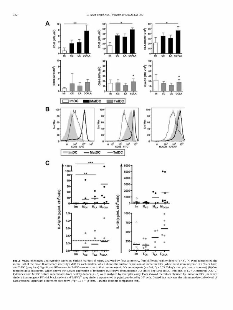
382 D. Raïch-Regué et al. / Vaccine 30 (2012) 378– 387
Fig. 2. MDDC phenotype and cytokine secretion. Surface markers of MDDC analyzed by flow cytometry, from different healthy donors (n ! 5). (A) Plots represented themean ± SD of the mean fluorescence intensity (MFI) for each marker, which shows the surface expression of immature DCs (white bars), immunogenic DCs (black bars)and TolDC (grey bars). Significant differences for TolDC were relative to their immunogenic DCs counterparts (n = 5–9; *p < 0.05, Tukey’s multiple comparison test). (B) Onerepresentative histogram, which shows the surface expression of immature DCs (grey), immunogenic DCs (thick line) and TolDC (thin line) of CC + LA matured DCs. (C)Cytokines from MDDC culture supernatants from healthy donors (n ! 5) were analyzed by multiplex assay. Plots showed the values obtained by immature DCs (Im, whitecircles), immunogenic DCs (M, black circles) and TolDC (T, grey circles), represented as pg/mL produced by 106 cells. Dotted line indicates the minimum detectable level ofeach cytokine. Significant differences are shown (**p < 0.01, ***p < 0.001, Dunn’s multiple comparison test).

D. Raïch-Regué et al. / Vaccine 30 (2012) 378– 387 383
Fig. 3. Allostimulatory capacity of MDDC. (A) Allogeneic T cell proliferation induced by differentially matured DC, at 1:20 ratio. Proliferation was measured by (3H)-thymidineincorporation after 4 days of co-culture. Plots show the mean (line) of n ! 7 independent experiments (circles, which in torn are the mean of 5 replicates each). Results aredepicted as the proliferation value relative to immature DCs’ one, to minimize alloresponse mismatch variability. Significant differences for TolDC (grey circles) wererelative to their immunogenic DCs (black circles) counterparts (*p < 0.05, Tukey’s multiple comparison test). (B) The cytokine profile was determined in supernatants fromalloproliferative assays induced by DC, collected after 96 h of culture and quantified by multiplex assay (Millipore). To minimize donor variability, the results were representedas fold of immature DCs (Im) cytokine production for each cytokine (n ! 5; *p < 0.05 paired T test). The detection limit for IL-6, IL-10, IFN-! and IL-17 is 3.2 pg/mL. (C) IFN-!and IL17 production by the different DC-primed T cells was detected intracellularly, at day 6 of coculture, using a cytokine secretion assay upon re-stimulation (as describedin Section 2). Box plots represent the mean, percentiles and SD of four experiments with different donors’ cells. We considered the IFN-! and IL-17 secretion by immatureDC-stimulated T cells as the basal levels.
independently of the LPS-restimulation (Fig. 6B). Interestingly, only(CC)-TolDC exhibited similar levels of induced alloproliferation(comparable with the initial levels), hence showing stability of theirtolerogenic functionality (Fig. 6B).
4. Discussion
Despite the fact that DCs loaded with tumour-associated anti-gens induce anti-tumoral cytotoxic T cells in vitro and in vivo [21],

384 D. Raïch-Regué et al. / Vaccine 30 (2012) 378– 387
Fig. 4. TolDC suppressive ability of T cell proliferation. T cells (105) were cocultured with autologous CMV-loaded MatDC (CC-matured, 5 ! 103) in presence of CMV-loaded(CC)-TolDC (A), or (LA)-TolDC (B) or (CCLA)-TolDC (C) at different ratios (1:2, 1:1, 5:1 relative to MatDC) for 5 days. Proliferation was determined by (3H)-thymidineincorporation for 16 h. The results of a representative experiment out of two are shown (*p < 0.05, **p < 0.01, ***p < 0.0001 Mann–Whitney test).
and DCs-immunotherapy has provided a proof of principle fortherapeutic immunity in cancer [1], clinical trials using ex vivo-generated DCs have failed to demonstrate wide clinical efficacy.Several reasons may account for this disappointing result. Amongthose, authors consider that the full potential of DCs has not beeneffectively exploited [22]. Thus, although some cancer treatmentsare envisaged to target DCs with tumour antigens in vivo, improvedex vivo generated immunogenic DCs may have an important role toplay in cellular therapy. An important contribution to the immuno-genicity of ex vivo DCs was the use of TLR-activating agents. In fact,different TLR-agonists have been shown to instruct DCs to inducedifferent types of T-cell responses [23–25]. Thus, the definition ofGMP-compliant TLR-agonists has permitted new combinations ofmaturating regimens allowing the definition of more immunogenicDCs [17,26]. One of these GMP-compliant TLR agonists is the lowtoxicity LPS-derivative monophosphoryl lipid A (LA), which acti-vates TLR4 on both DCs and T cells [27]. It has been well establishedthat MDDC matured with LA plus IFN-! are endowed with a highercapacity of Th1 polarization compared to the traditional CC and theIFN-" cocktail [17].
In our experiments, the combination of CC + LA rendered apotent MDDC population with higher surface expression of CD83,CD86, HLA-DR and secretion of IL-12p70. A relevant novel find-ing was the identification of IL-17 secretion, additional to IFN-!,mainly in supernatants from allostimulated T cells responding to(CC + LA)-immunogenic DCs. Intracellular staining confirmed theresults, thus suggesting the generation of Th17 cells (as a majorsource of IL-17) induced by these immunogenic DCs. In fact, theinduction of human IL-17 producing cells mediated by DCs hasbeen reported in the context of myeloma patients [28] and alsoin the inflamed joints of rheumatoid arthritis patients [29]. Hence,the maturation stimulus CC + LA it is likely to “simulate” an envi-ronment of pathogen-induced inflammation, activating the MDDCand entitling them to promote a Th17 response. Though the roleplayed by Th17 cells in cancer is still controversial [30], it ispostulated that IL-17-producing cells may have a decisive rolein anticancer immune responses [31]. Furthermore, some stud-ies showed the important role that Th17 cell play in immunityand host responses to extracellular [32,33] and also to intracellularpathogens [34,35]. Thus, the induction of IL-17 producing T cellscould contribute to immunotherapy. Taking these results together,(CC + LA)-immunogenic DCs may yield superior anti-tumour oranti-pathogen immune responses than the immunity induced byDCs matured with CC or LA alone.
Beyond the induction of potent immunoresponses, DCs mayalso induce tolerance. In this sense, the use of ex vivo tolero-genic DCs as cellular treatment in autoimmune disorders and intransplantation is just at the initial steps, and will undoubtedly
benefit from the vast knowledge generated in DC-based tumourvaccination. Some studies reported the use of immature DCs fortolerance immunotherapy [36], but under inflammatory conditionsthese cells could undergo maturation and acquire immunogenicDCs features. To overcome this undesired effect, it is necessaryto generate mature DCs with a stable tolerogenic profile. Hence,one important feature to consider is the maturation stimuli used,which would condition the TolDC behaviour in terms of the sta-bility of tolerogenicity. In this sense, not all maturation stimulimay confer such stability. For example, it has been reported thatTNF-" matured-DCs could still be further activated in vitro andin vivo [37]. Therefore, we evaluated on TolDC (generated in thepresence of vitD3) the effect of the different maturation regimenstested in immunogenic DCs (in matched donors). As expected,all TolDC were phenotypically “semi-mature”, failed to producethe proinflammatory cytokine IL-12p70, and induced a moderatealloresponse in T cells compared to donor-matched immunogenicDCs. However, fine-tuning of TolDC features using differential mat-uration regimens may be observed. It is remarkable that (LA) or(CC + LA)-matured TolDC produced higher levels of IL-10 (Fig. 2C)than (CC)-matured TolDC, which could be an important feature inDC tolerance immunotherapy. In sharp contrast, (CC)-TolDC exhib-ited the most suppressive potential of T cell proliferation in vitro,when compared to (CC + LA)-matured TolDC, whereas (LA)-TolDCalso showed certain suppressive capability, which is partially inline with a previous report [38]. Furthermore, only (CC)-TolDCshowed functional stability since did not induce increased T cellproliferation after LPS challenge or after additional 24 h of cul-ture. Such stability was not observed in (LA) and in (CC + LA)-TolDC(Fig. 6B). These results suggest that CC-matured TolDC may be bestsuited as negative cellular vaccine for immunotherapy. A previ-ous report has also studied the use of LA as maturation signal forTolDC generation [38]. Harry et al. used Dexamethasone and a lowdose of VitD3 to generate LA-matured TolDC that exhibited sta-ble phenotype and cytokine secretion when exposed to differentproinflammatory stimuli [38]. In a recent study, our group com-pared different tolerogenic pharmacological agents and found thatdexamethasone and vitD3 TolDCs were similar in terms of phe-notype and alloproliferative responses [39] and therefore similarfunctional properties could be expected. However, dexamethasoneinduced-TolDC secreted high levels of IL10 compared to VitD3-TolDCs [39]. Therefore, the different reagents used to induce TolDCmay undoubtedly account for minor differences found betweenHarry et al. and our study. As different tolerogenic agents maybe best suited for specific treatments, our results extent theknowledge on maturation signals to generate functionally stableTolDC to use them as negative cellular vaccine for immunother-apy.
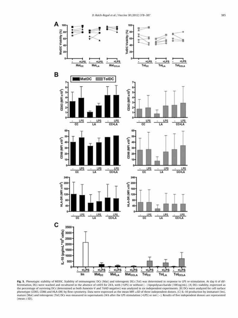
D. Raïch-Regué et al. / Vaccine 30 (2012) 378– 387 385
Fig. 5. Phenotypic stability of MDDC. Stability of immunogenic DCs (Mat) and tolerogenic DCs (Tol) was determined in response to LPS re-stimulation. At day 6 of dif-ferentiation, DCs were washed and recultured in the absence of vitD3 for 24 h, with (+LPS) or without (!) lipopolysaccharide (100 ng/mL). (A) DCs viability, expressed asthe percentage of surviving DCs (determined as both Annexin-V and 7AAD negative) was analyzed in six independent experiments. (B) DCs were analyzed for cell surfacephenotype (CD83, CD86 and HLA-DR) by flow cytometry. Data were expressed as the mean MFI ±SD of three independent donors. (C) IL-10 production by immature (Im),mature (Mat) and tolerogenic (Tol) DCs was measured in supernatants 24 h after the LPS stimulation (+LPS) or not (!). Results of five independent donors are represented(mean ± SD).

386 D. Raïch-Regué et al. / Vaccine 30 (2012) 378– 387
Fig. 6. Functional stability of MDDC. Following 24 h in culture with or without LPS, DCs were washed extensively with PBS, and then cocultured with allogeneic peripheralblood mononuclear cells (PBMC) for 5 days. (3H)-thymidine was added to the culture for the last 16 h. Data were expressed as counts per minute (cpm) (A), showing themean of five replicates for 6 different donors. Each donor is represented by a different symbol (*p < 0.05, paired T test). (B) The results for TolDC proliferation are expressedas the percentage of the donor-matched MatDC proliferation, considered as 100%.
In summary, this study compares the features of clinical-gradegenerated immunogenic and tolerogenic DCs differently maturedusing the traditional CC in combination or not with the TLR-activating LA. Fine-tuning features observed may be relevant in thetranslation of these DCs to human immunotherapy. Specifically,in our experimental conditions, the maturation stimulus CC + LArendered fully immunogenic DCs that induced IFN-! and IL-17production by allostimulated T cells, suggesting a Th17 polariza-tion. Clinical immunotherapy strategies that are aimed to induceimmune activation against tumours and infectious agents may takeadvantage of this feature. Conversely, in the tolerance field it isimportant to notice that DCs maturation using LA promoted thesecretion of high levels of IL-10, while CC-matured TolDC producedIL10 at lower levels but were unique to show a potent suppressiveability and stable tolerogenic functionality. Altogether, these find-ings point to the selection of appropriate maturation stimuli foroptimizing the therapeutic potential of DCs, when aiming to designa DC-based vaccine.
Contributions
DRR performed the experiments, analyzed data, made the fig-ures and wrote the manuscript; MNG performed some experimentsand revised the manuscript; LGL performed flow cytometry; CRTdirected statistical analyses; RPB and EMC revised the manuscript;FEB designed and conducted the study, organized the data andwrote the paper.
Conflict of interest
The authors declare no competing financial interests.
Acknowledgements
This work was supported in part by a grant from Fundació LaMarató de TV3 (07/2410) and Fundació Grup d’Afectats d’EsclerosiMúltiple (to EMC). DRR is supported by project 07/2410 Fun-dació La Marató de TV3. MNG is supported by a grant from theSpanish Ministry of Science and Innovation and Blood and Tis-sue Bank (PTQ-09-02-017050). LGL is supported by a Rio Hortegagrant from Instituto de Salud Carlos III, Spanish Ministry of Health(CM07/00196). FEB is co-founded by the stabilization program ofBiomedical researches (CES07/015) of the Instituto de Salud CarlosIII and Direcció d’ Estratègia i Coordinació, Health Dept. of Catalonia.
The authors thank Marco Fernández for his helpful advise withflow cytometry experiments (Cytometry Unit of IGTP), and alsothank to Dr. Maria Rosa Sarrias for helpful discussions.
References
[1] Palucka K, Ueno H, Zurawski G, Fay J, Banchereau J. Building on dendritic cellsubsets to improve cancer vaccines. Curr Opin Immunol 2010;22:258–63.
[2] Hilkens CM, Isaacs JD, Thomson AW. Development of dendritic cell-basedimmunotherapy for autoimmunity. Int Rev Immunol 2010;29:156–83.
[3] Huang F-PP, Chen Y-XX, To CKK. Guiding the misguided – functional condition-ing of dendritic cells for the DC-based immunotherapy against tumours. Eur JImmunol 2011;41:18–25.
[4] Peters JH, Ruppert J, Gieseler RK, Najar HM, Xu H. Differentiation of humanmonocytes into CD14 negative accessory cells: do dendritic cells derive fromthe monocytic lineage? Pathobiology 1991;59:122–6.
[5] Zhou LJ, Tedder TF. CD14+ blood monocytes can differentiate into functionallymature CD83+ dendritic cells. Proc Natl Acad Sci U S A 1996;93:2588–92.
[6] Kalinski P, Okada H. Polarized dendritic cells as cancer vaccines: directingeffector-type T cells to tumors. Semin Immunol 2010;22:173–82.
[7] Farag MM, Hoyler B, Encke J, Stremmel W, Weigand K. Dendritic cells can effec-tively be pulsed by HBVsvp and induce specific immune reactions in mice.Vaccine 2010;29:200–6.
[8] Garcia F, Climent N, Assoumou L, Gil C, González N, Alcamí J, et al. A thera-peutic dendritic cell-based vaccine for HIV-1 infection. J Infect Dis 2011;203:473–8.
[9] Skalova K, Mollova K, Michalek J. Human myeloid dendritic cells for cancertherapy: does maturation matter? Vaccine 2010;28:5153–60.
[10] Landi A, Babiuk LA, van Drunen Littel-van den Hurk S. Dendritic cells maturedby a prostaglandin E2-containing cocktail can produce high levels of IL-12p70and are more mature and Th1-biased than dendritic cells treated with TNF-"or LPS. Immunobiology 2011;216:649–62.
[11] Dauer M, Lam V, Arnold H, Junkmann J, Kiefl R, Bauer C, et al. Combined use oftoll-like receptor agonists and prostaglandin E(2) in the FastDC model: rapidgeneration of human monocyte-derived dendritic cells capable of migrationand IL-12p70 production. J Immunol Methods 2008;337:97–105.
[12] De Becker G, Moulin V, Pajak B, Bruck C, Francotte M, Thiriart C, et al. Theadjuvant monophosphoryl lipid A increases the function of antigen-presentingcells. Int Immunol 2000;12:807–15.
[13] Boccaccio C, Jacod S, Kaiser A, Boyer A, Abastado JP, Nardin A. Identifica-tion of a clinical-grade maturation factor for dendritic cells. J Immunother2002;25:88–96.
[14] Garay RP, Viens P, Bauer J, Normier G, Bardou M, Jeannin JF, et al. Cancer relapseunder chemotherapy: why TLR2/4 receptor agonists can help. Eur J Pharmacol2007;563:1–17.
[15] Cluff CW. Monophosphoryl lipid A (MPL) as an adjuvant for anti-cancer vac-cines: clinical results. Adv Exp Med Biol 2010;667:111–23.
[16] Schreibelt G, Benitez-Ribas D, Schuurhuis D, Lambeck AJ, van Hout-Kuijer M,Schaft N, et al. Commonly used prophylactic vaccines as an alternative for syn-thetically produced TLR ligands to mature monocyte-derived dendritic cells.Blood 2010;116:564–74.
[17] Ten Brinke A, Karsten ML, Dieker MC, Zwaginga JJ, van Ham SM. The clinicalgrade maturation cocktail monophosphoryl lipid A plus IFNgamma generatesmonocyte-derived dendritic cells with the capacity to migrate and induce Th1polarization. Vaccine 2007;25:7145–52.
[18] Ten Brinke A, van Schijndel G, Visser R, de Gruijl TD, Zwanginga JJ, van HamSM, et al. Monophosphoryl lipid A plus IFNgamma maturation of dendritic cellsinduces antigen-specific CD8+ cytotoxic T cells with high cytolytic potential.Cancer Immunol Immunother 2010;59:1185–95.
[19] Reis e Sousa C. Dendritic cells in a mature age. Nat Rev Immunol 2006;6:476–83.[20] Soldevila B, Alonso N, Martínez-Arconada MJ, Morillas RM, Planas R, Sanmartí
AM, et al. A prospective study of T- and B-lymphocyte subpopulations, CD81expression levels on B cells and regulatory CD4(+) CD25(+) CD127(low/!)FoxP3(+) T cells in patients with chronic HCV infection during pegylatedinterferon-alpha2a plus ribavirin treatment. J Viral Hepat 2011;18:384–92.

D. Raïch-Regué et al. / Vaccine 30 (2012) 378– 387 387
[21] Dauer M, Schnurr M, Eigler A. Dendritic cell-based cancer vaccination: quovadis? Expert Rev Vaccines 2008;7:1041–53.
[22] Koski GK, Cohen PA, Roses RE, Xu S, Czerniecki BJ. Reengineering dendriticcell-based anti-cancer vaccines. Immunol Rev 2008;222:256–76.
[23] Agrawal S, Agrawal A, Doughty B, Gerwitz A, Blenis J, Van Dyke T, et al.Cutting edge: different toll-like receptor agonists instruct dendritic cellsto induce distinct Th responses via differential modulation of extracellularsignal-regulated kinase-mitogen-activated protein kinase and c-Fos. J Immunol2003;171:4984–9.
[24] Coccia EM, Severa M, Giacomini E, Monneron D, Remoli ME, Julkunen I, et al.Viral infection and toll-like receptor agonists induce a differential expression oftype I and lambda interferons in human plasmacytoid and monocyte-deriveddendritic cells. Eur J Immunol 2004;34:796–805.
[25] Re F, Strominger JL. Heterogeneity of TLR-induced responses in dendritic cells:from innate to adaptive immunity. Immunobiology 2004;209:191–8.
[26] Vanderlocht J, Van Elssen CH, Senden-Gijsbers BL, Meek B, Cloosen S, LibonC, et al. Increased tumor-specific CD8+ T cell induction by dendritic cellsmatured with a clinical grade TLR-agonist in combination with IFN-gamma.Int J Immunopathol Pharmacol 2010;23:35–50.
[27] Ismaili J, Rennesson J, Aksoy E, Vekemans J, Vincart B, Amraoui Z, et al.Monophosphoryl lipid A activates both human dendritic cells and T cells. JImmunol 2002;168:926–32.
[28] Dhodapkar KM, Barbuto S, Matthews P, Kukreja A, Mazumder A, Vesole D, et al.Dendritic cells mediate the induction of polyfunctional human IL17-producingcells (Th17-1 cells) enriched in the bone marrow of patients with myeloma.Blood 2008;112:2878–85.
[29] Evans HG, Gullick NJ, Kelly S, Pitzalis C, Lord GM, Kirkham BW, et al. In vivo acti-vated monocytes from the site of inflammation in humans specifically promoteTh17 responses. Proc Natl Acad Sci U S A 2009;106:6232–7.
[30] Wilke CMM, Kryczek I, Wei S, Zhao E, Wu K, Wang G, et al. Th17 cells in cancer:help or hindrance? Carcinogenesis 2011;32:643–9.
[31] Ma Y, Aymeric L, Locher C, Mattarollo SR, Delahaye NF, Pereira P, et al. Con-tribution of IL-17-producing gammadelta T cells to the efficacy of anticancerchemotherapy. J Exp Med 2011;208:491–503.
[32] Cheng P, Zhou J, Gabrilovich D. Regulation of dendritic cell differentiation andfunction by Notch and Wnt pathways. Immunol Rev 2010;234:105–19.
[33] Sellge G, Magalhaes JG, Konradt C, Fritz JH, Salgado-Pabon W, Eberl G, et al.Th17 cells are the dominant T cell subtype primed by Shigella flexneri mediatingprotective immunity. J Immunol 2010;184:2076–85.
[34] Lin Y, Ritchea S, Logar A, Slight S, Messmer M, Rangel-Moreno J, et al.Interleukin-17 is required for T helper 1 cell immunity and host resis-tance to the intracellular pathogen Francisella tularensis. Immunity 2009;31:799–810.
[35] Curtis MM, Way SSS. Interleukin-17 in host defence against bacterial, mycobac-terial and fungal pathogens. Immunology 2009;126:177–85.
[36] Dhodapkar MV, Steinman RM, Krasovsky J, Munz C, Bhardwaj N. Antigen-specific inhibition of effector T cell function in humans after injection ofimmature dendritic cells. J Exp Med 2001;193:233–8.
[37] Voigtlander C, Rossner S, Cierpka E, Theiner G, Wiethe C, Menges M, et al.Dendritic cells matured with TNF can be further activated in vitro andafter subcutaneous injection in vivo which converts their tolerogenicity intoimmunogenicity. J Immunother 2006;29:407–15.
[38] Harry RA, Anderson AE, Isaacs JD, Hilkens CM. Generation and characterisationof therapeutic tolerogenic dendritic cells for rheumatoid arthritis. Ann RheumDis 2010;69(11):2042–50.
[39] Naranjo-Gómez M, Raïch-Regué D, Onate C, Grau-López L, Ramo-Tello C, Pujol-Borrell R, et al. Comparative study of clinical grade human tolerogenic dendriticcells. J Transl Med 2011;9:89.

Results III
772 Dalia Raıch-Regue et al. Eur. J. Immunol. 2012. 42: 772–783DOI: 10.1002/eji.201141835
Stable antigen-specific T-cell hyporesponsivenessinduced by tolerogenic dendritic cells from multiplesclerosis patients
Dalia Raıch-Regue1, Laia Grau-Lopez2, Mar Naranjo-Gomez1,Cristina Ramo-Tello2, Ricardo Pujol-Borrell1, Eva Martınez-Caceres!1
and Francesc E Borras!1
1 Laboratory of Immunobiology for Research and Diagnosis (LIRAD), Blood and Tissue Bank
(BST). Department of Cell Biology, Physiology and Immunology, Universitat Autonoma de
Barcelona, Institut d’Investigacio Germans Trias i Pujol (IGTP), Badalona, Spain2 Multiple Sclerosis Unit, Department of Neurosciences, Hospital Universitari Germans Trias i
Pujol, Badalona, Spain
Multiple sclerosis (MS) is a chronic demyelinating autoimmune disease of the centralnervous system. Current therapies decrease the frequency of relapses and limit, to someextent, but do not prevent disease progression. Hence, new therapeutic approachesthat modify the natural course of MS need to be identified. Tolerance induction to self-antigens using monocyte-derived dendritic cells (MDDCs) is a promising therapeutic strat-egy in autoimmunity. In this work, we sought to generate and characterize tolerogenicMDDCs (tolDCs) from relapsing-remitting (RR) MS patients, loaded with myelin peptidesas specific antigen, with the aim of developing immunotherapeutics for MS. MDDCs weregenerated from both healthy-blood donors and RR-MS patients, and MDDC maturationwas induced with a proinflammatory cytokine cocktail in the absence or presence of1!,25-dihydroxyvitamin-D3, a tolerogenicity-inducing agent. tolDCs were generated frommonocytes of RR-MS patients as efficiently as from monocytes of healthy subjects. TheRR-MS tolDCs expressed a stable semimature phenotype and an antiinflammatory profileas compared with untreated MDDCs. Importantly, myelin peptide-loaded tolDCs inducedstable antigen-specific hyporesponsiveness in myelin-reactive T cells from RR-MSpatients. These results suggest that myelin peptide-loaded tolDCs may be a powerfultool for inducing myelin-specific tolerance in RR-MS patients.
Keywords: 1!,25-dihydroxyvitamin D3 ! Autoimmune disease ! Cell immunotherapy ! DCs !
Tolerance induction
See accompanying Commentary by Wiendl et al.
Supporting Information available online
Correspondence: Dr. Francesc E Borrase-mail: [email protected]
!Francesc E Borras and Eva Martınez-Caceres share senior co-authorship on this
paper.
C" 2012 WILEY-VCH Verlag GmbH & Co. KGaA, Weinheim www.eji-journal.eu

Eur. J. Immunol. 2012. 42: 772–783 Clinical immunology 773
Introduction
Multiple sclerosis (MS) is a chronic autoimmune disease of the
central nervous system, characterized by leucocyte infiltration,
demyelination, and axonal loss. Although the pathogenic mech-
anisms of MS remain unclear, some lines of evidence suggest a
crucial role of autoreactive CD4+ T cells [1]. In fact, some groups
have confirmed CD4+ T-cell reactivity to myelin antigens [2,3].
In addition, a higher proportion of high-avidity myelin-specific
CD4+ T cells was demonstrated in MS patients in comparison
with controls, and their specificities were predominantly directed
toward a group of immunodominant epitopes of myelin basic
protein (MBP), proteolipid protein (PLP), or myelin oligodendro-
cyte glycoprotein (MOG) [4]. Furthermore, our group has recently
confirmed the reactivity to the aforementioned set of immun-
odominant peptides in peripheral blood T cells from MS patients
compared with healthy controls (HCs) [5]. Hence, specific inhibi-
tion or depletion of autoreactive T cells represents an interesting
goal for future therapies aiming to restore peripheral tolerance in
autoimmune diseases such as MS.
Current treatments for relapsing-remitting MS (RR-MS)
patients decrease the frequency of relapses and reduce inflamma-
tory activity in a nonspecific manner, but their effect on disease
progression is still unclear [6,7]. Therefore, in order to modify
the course of MS, new and more specific therapeutic approaches,
such as the induction of antigen-specific tolerance are necessary.
So far, current strategies for induction of antigen-specific toler-
ance in MS include T-cell vaccination [8], T-cell receptor blockade
using antibodies [9], and immunization with DNA-encoding MBP
[10]. These have been effective in the experimental autoimmune
encephalomyelitis (EAE) model, but not in MS patients [11]. The
administration of an altered peptide ligand also prevented EAE
induction in animal models [12], but clinical trials in MS patients
were discontinued due to worsening of the disease [13]. As has
recently been reviewed by Turley and Miller [14], other antigen-
specific strategies used to induce tolerance in the EAE model
include the mucosal administration of antigens, and the intra-
venous injection of soluble peptide or ECDI-peptide coupled cells.
A novel approach for targeting autoreactive T cells is the use
of antigen-loaded dendritic cells (DCs) [15,16]. DCs are a het-
erogeneous group of antigen presenting cells (APCs) involved in
immunity and tolerance. Tolerogenic DCs induce tolerance
through antigen presentation along with inadequate costimula-
tion and biased cytokine production, resulting in T-cell silenc-
ing, deletion, immune deviation, and/or induction of regulatory
T (Treg) cells [17]. Use of tolDCs to induce antigen-specific anergy
and/or Treg cells has been tested in several autoimmune animal
models [18], including the EAE model [19]. Hence, several groups
have designed immunotherapeutic approaches involving adminis-
tration of tolDCs to humans to induce T-cell tolerance in transplan-
tation and autoimmune diseases [20,21]. Different strategies have
been evaluated to generate tolDCs in vitro, as recently reviewed
[18,22]. We have focused on the use of 1!,25-dihydroxyvitamin
D3 (vitD3) to generate tolDCs. In vitro treatment of DCs with
vitD3 or other vitamin D-receptor agonists decreased expression
of costimulatory molecules such as CD40, CD80, CD86, reduced
interleukin (IL)-12 production, enhanced IL-10 secretion, and
down-modulated T-cell activation [23], indicating a tolerogenic
functional phenotype. Moreover, these effects have recently been
suggested to occur in vivo as well [24]. Also, high serum levels of
vitD3 in MS patients have been correlated with a slower progres-
sion of the disease [25]. Finally, our own results have confirmed
the possibility of obtaining clinical grade vitD3-tolDCs [26].
Here we sought to generate and characterize tolDCs from
RR-MS patients’ monocytes, as a pilot study to investigate the
feasibility of reestablishing tolerance using such cells in a future
clinical trial.
Our results show that tolDCs generated from RR-MS patients
behave similarly to those of HCs, and are able to induce a
stable state of hyporesponsiveness in myelin-specific autologous
T cells.
Results
Differentiation to tolDCs is equally efficient frommonocytes of both HCs and MS patients
Generation of tolDCs was evaluated in a group of eleven RR-MS
patients and eleven HCs to assess the possible differences in differ-
entiation efficiencies (percentage of monocyte-derived dendritic
cells (MDDCs) obtained from the initial cultured monocytes).
No statistical difference was observed in differentiation yield
((number tolDC / number initial monocytes) ! 100), or in the
percentage of surviving tolDCs at day 6 of culture, between MS
patients and HCs (Table 1). Therefore, differentiation of mono-
cytes to tolDCs was as efficient in MS patients as it was in HCs.
TolDCs from RR-MS patients inducehyporesponsiveness in allogeneic T cells
During the study, tolDCs (matured in presence of vitD3) were
compared with immunogenic dendritic cells (matDCs; matured
without vitD3) and/or immature dendritic cells (imDCs; with-
out vitD3 and not matured) to better define their characteris-
tics and function. In relation to cell surface phenotype, the ex-
pression of CD83, CD86, CD40, and human histocompatibility
leukocyte Ag (HLA)-DR was significantly upregulated in matDCs
compared with imDCs, except for CD40 in MS patients (Fig. 1A).
Table 1. TolDC differentiation yield ((number tolDC / number initialmonocytes) !100) and viability (annexin-V and 7AAD double negative)obtained from healthy controls (HCs) and RR-MS patients (MS) cells
n tolDC differentiation tolDCyield (%) viability (%)
HC 11 45 ± 15 83 ± 8MS 11 32 ± 13 79 ± 7
C" 2012 WILEY-VCH Verlag GmbH & Co. KGaA, Weinheim www.eji-journal.eu

774 Dalia Raıch-Regue et al. Eur. J. Immunol. 2012. 42: 772–783
Figure 1. Characterization of tolDCs from MS patients reveals a semimature phenotype and antiinflammatory cytokine profile. (A) Expression ofDCs surface markers (CD83, CD86, CD40, and HLA-DR) in imDCs, matDCs, and tolDCs of MS patients (n = 9) and controls (n = 10) were analyzed byflow cytometry. Dots represent the MFI of the analyzed marker for each individual (*p < 0.05, **p < 0.01, ***p < 0.001 Unpaired t-test). The gray barrepresents the mean. (B) IL-10 and IL-12p70 levels in culture supernatants of DCs isolated from MS patients (n = 5) and HCs (n = 7) as determinedby multiplex assay. The minimum detectable level of IL-12p70 is indicated by a dotted line.
Conversely, the expression of all these markers remained unal-
tered in tolDCs in contrast to imDCs (Fig. 1A). When comparing
HCs and RR-MS patients, an intriguing low expression level of
the maturation marker CD83 was observed in matDCs generated
from RR-MS patients, compared with HCs (2954 ± 989 vs. 7175
± 4654 mean fluorescence intensity (MFI), n = 9, p < 0.05, un-
paired t-test) (Fig. 1A) while no significant changes were observed
in the other markers analyzed.
When analyzing cytokine production, although differences did
not reach statistical significance, it was observed that tolDCs pro-
duced higher levels of IL-10 (3.1 times more for MS patients and
1.6 times more for HCs) and produced very low IL-12p70 levels
compared with matDCs (Fig. 1B). Additionally, IL-23 production
by matDCs and tolDCs was also evaluated, but most samples were
below the detection level of the technique (23 pg/mL) (data not
shown). These data suggest an antiinflammatory profile of tolDCs
from both HCs and MS patients (Fig. 1B).
TolDCs from RR-MS patients inducehyporesponsiveness in allogeneic T cells
The immunostimulatory capability of tolDCs was first determined
in allogeneic mixed leukocyte reaction experiments. Compared
with matDCs, tolDCs showed a reduced ability to stimulate T-cell
proliferation, observed equally in MS patients and HC subjects
(51.6 ± 20.1% and 53.5 ± 19.9%, respectively, n = 9) (Fig. 2A).
The supernatants of these allogeneic cocultures contained signif-
icantly reduced amounts of tumor necrosis factor (TNF)-!, IL-6,
and interferon (IFN)-" in the presence of tolDCs, compared with
those of matDCs (p < 0.05 paired t-test, n = 6) (Fig. 2B). The IL-4
and IL-10 levels were below the detection limit (data not shown).
To confirm that this reduction in proinflammatory cytokine secre-
tion was not uniquely due to the lower number of proliferating
T cells in the presence of tolDCs, intracellular cytokine experi-
ments were performed. tolDCs were less potent in inducing IFN-"
secretion by T cells than matDCs, for total CD3+ cells (p < 0.05
Wilcoxon test, n = 6; Fig. 2C) and also for proliferating CD3+
cells (carboxyfluorescein diacetate succinimidyl ester (CFSE)low)
(p < 0.05 paired test, n = 6; Fig. 2D).
tolDCs have a stable tolerogenic profile in vitro
Due to their potential use in the clinical setting, we evalu-
ated whether the vitD3 induced-DCs were phenotypically and
functionally stable after a potent proinflammatory stimulus.
MatDCs and tolDCs were restimulated for 24 h with or with-
out lipopolysaccharide (LPS). Under these conditions, matDC
and tolDC surface expression of CD83, CD86, and HLA-DR
remained stable following LPS restimulation in comparison to the
nonstimulated phenotype (Fig. 3A). Furthermore, tolDCs
C! 2012 WILEY-VCH Verlag GmbH & Co. KGaA, Weinheim www.eji-journal.eu

Eur. J. Immunol. 2012. 42: 772–783 Clinical immunology 775
Figure 2. tolDCs from RR-MS patients induced hyporesponsiveness of and impaired proinflammatory cytokine secretion by allogeneic T cells.(A) Allogeneic T-cell proliferation induced by matDCs and tolDCs, at a 1:20 ratio, from MS patients (n = 9) and HCs (n = 9). Proliferation wasmeasured by (3H)-thymidine incorporation and data were expressed as cpm. The discontinuous line indicates the reduction of alloproliferation inthe presence of tolDCs compared with that of matDCs derived from the same patient (**p < 0.01 Wilcoxon test). The background levels (unstimulatedT cells) were 297 ±180 cpm for HCs and 459 ±207 cpm for MS, and the positive control (pokeweed mitogen, 1 µg/mL): 18.89 ± 25.22 ! 103 cpmfor HCs and 37.56 ± 38.47 ! 103 cpm for MS. (B) IL-2, TNF-!, IL-6, and IFN-" profiles were analyzed in supernatants of the alloproliferative assaysinduced by either matDCs or tolDCs (from MS patients), collected after 96 h and quantified by cytometric bead array. The minimum detectionlevel of each cytokine is indicated by a dotted line (*p < 0.05 paired t-test, n = 6). (C and D) IFN-" production by matDC- and tolDC-primedT cells was measured on day 6 of coculture by intracellular staining and flow cytometry, after restimulating the cells (phorbol myristate acetate(PMA) + ionomycin) in the presence of brefeldin A for 5h. A representative flow cytometry plot (left) and percentage of IFN-" producing cells fromn = 6 independent experiments are shown for (C) the total T cell population and (D) those T cells that responded to allostimulation (CFSElow).The numbers inside the plots indicate the percentage of cells in each quadrant and dots represent the mean of five replicates per experiment(*p < 0.05 Wilcoxon and paired t-test for CD3+ and CD3+CFSElow cells, respectively).
maintained a lower expression of CD83, CD86, and HLA-DR com-
pared with matDCs (Fig. 3A).
Additionally, LPS did not induce the secretion of IL-23 from
tolDCs but small amounts of IL-10 were still detectable in these
supernatants (Fig. 3B). As positive control, LPS-stimulated imDCs
secreted significant amounts of IL-10 and IL-23 whereas non-
stimulated imDCs did not produce detectable levels of these
cyto-kines (p < 0.005 Mann–Whitney Test, n = 5) (Fig. 3B).
Secretion of IL-12p70 was only detected for LPS-stimulated imDCs
(28 pg/mL per 106 cells in two assays out of five, data not
shown).
Furthermore, alloproliferation experiments indicated that allo-
proliferative responses induced by imDCs increased after LPS
treatment (1.6-fold higher, p < 0.05 paired t-test, n = 5), whereas
no significant increase was observed in alloproliferative responses
induced by matDCs after LPS restimulation (Fig. 3C). Similarly,
the alloproliferative induction ability of tolDCs after LPS chal-
lenge was not increased, indicating stability of their tolerogenic
functionality (Fig. 3C).
Binding of myelin peptides to DCs
To assess tolDC hyporesponsiveness induction to autologous
myelin-reactive T cells, we first estimated the peptide-loading
capability of tolDCs using a biotinylated form of a single myelin
peptide (PLP(139–154)) in time-course experiments on both matDCs
and tolDCs. Results from RR-MS patients’ cells (n = 3) showed
C" 2012 WILEY-VCH Verlag GmbH & Co. KGaA, Weinheim www.eji-journal.eu

776 Dalia Raıch-Regue et al. Eur. J. Immunol. 2012. 42: 772–783
Figure 3. tolDCs have a highly stabletolerogenic profile. DCs were washedand recultured for 24 h with (+) orwithout (-) LPS (100 ng/mL) on day6 of differentiation. (A) Cell surfacephenotype (CD83, CD86, and HLA-DR) of imDCs, matDCs, and tolDCswere analyzed by flow cytometry.Data are expressed as the mean MFI+SD of three independent donors.(B) IL-10 and IL-23 production by DCsat 24h after LPS stimulation (+LPS)or unstimulated (–) was measuredby multiplex assay. Results are rep-resented as the mean + SD of fiveindependent donors (**p < 0.01 Mann–Whitney Test). (C) Alloproliferation-inducing ability of DCs, following LPSstimulation (+LPS) or not (–), on cocul-tured PBMCs for 108 h was measuredby (3H)-thymidine uptake during thefinal 16 h. Data are expressed as cpmshowing the mean of five replicatesfor five different donors analyzed(indicated with different symbols) (*p< 0.05 paired t-test).
that optimal binding was obtained between 12 and 18 h after
peptide loading at 37!C (Fig. 4A). No significant difference was
observed in the loading capacity exhibited by matDCs or tolDCs for
any patient (Fig. 4B and C). To ascertain the specificity of peptide
loading, a competition experiment was performed. When increas-
ing amounts of unlabeled PLP(139–154) were added to tolDCs in the
presence of a constant concentration of biotinylated PLP(139–154),
a gradual decline of the biotinylated signal was observed
Figure 4. Time course of myelin pep-tide binding to DCs. (A) DCs (indi-cated by FSC/SSC) were incubated withbio-tinylated PLP(139–154) (50 µM) for theindicated times. After extensive wash-ing, bound peptide was detected withStreptavidin-Alexa488 by flow cytom-etry (MFI). Time course of binding ofbiotinylated PLP(139–154) to (B) matDCsand (C) tolDCs from RR-MS patients(n = 3, indicated with different sym-bols) are shown.
C" 2012 WILEY-VCH Verlag GmbH & Co. KGaA, Weinheim www.eji-journal.eu
Eur. J. Immunol. 2012. 42: 772–783 Clinical immunology 777
(Supporting Information Fig. 1), showing specificity in peptide
binding.
Myelin peptide-loaded tolDCs induce stablehyporesponsiveness in autologous RR-MS patients
We investigated whether the immunomodulatory ability of tolDCs
observed in allogeneic cultures was also maintained in autolo-
gous conditions. For this purpose, enriched myelin-specific T cells
from RR-MS patients (n = 3) were stimulated for 6 days with
either autologous matDCs or tolDCs, which had been previously
loaded with the same mix of myelin peptides. It was observed that
T cells stimulated by myelin peptides-loaded tolDCs exhibited a
lower proliferation (reduction of 45 ± 17%, n = 3) compared
with those induced by myelin peptides-loaded matDCs (Fig. 5A).
Supernatants from coculture experiments were analyzed for se-
creted cytokines and a reduction of IL-17 (62%), IL-6 (76%),
and IFN-! (58%) production in the presence of tolDCs compared
with that of matDCs was observed (Table 2). Therefore, tolDCs
from RR-MS patients induced hyporesponsiveness in autologous
myelin-reactive T cells accompanied by a reduction of proinflam-
matory cytokines.
To further evaluate the T-cell response promoted by tolDCs,
T cells were recovered from the primary coculture with myelin-
loaded matDCs (Tmat+ptd) or tolDCs (Ttol+ptd), and following 3 days
of resting, were restimulated with autologous myelin-loaded pe-
ripheral blood mononuclear cells (PBMCs) as APCs. In the three
patients tested, Tmat+ptd readily responded after restimulation with
myelin-loaded PBMCs, in contrast to Ttol+ptd, as shown by the
reduced proliferation (35 ± 8% reduction compared with Tmat+ptd,
p < 0.01 paired t-test, n = 3) (Fig. 5B). Taken together, these
results support the induction of stable hyporesponsiveness of
myelin-reactive T cells by TolDCs from RR-MS patients.
tolDC-induced hyporesponsiveness is antigen specific
Finally, we evaluated whether the induction of hyporesponsive-
ness by tolDCs was antigen specific, meaning that tolDC-primed
T cells would still respond to an unrelated antigen. This possibil-
ity was tested using T cells from four donors reactive to tetanus
toxin (TT) and two MS patients reactive to myelin peptides (ptd).
In both cases, the unrelated antigen was cytomegalovirus (CMV).
T cells were cocultured with either TT- or ptd-loaded matDCs
(TT/ptd-matDCs) or TT- or ptd-loaded tolDCs (TT/ptd-tolDCs).
Following a resting period, the T cells were restimulated with
unloaded, TT/ptd-loaded, or CMV-loaded matDCs separately,
and the induced response was measured by IFN-! enzyme-
linked immunospot (ELISpot) assay. In both HCs and patients,
the results showed that TT/ptd-matDC-primed T cells (Tmat+TT
in Fig. 6A, and Tmat+ptd in Fig. 6B) secreted large amounts of
IFN-! when restimulated with TT/ptd-matDCs or CMV-matDCs
(Fig. 6A and B). In contrast, TT/ptd-tolDC-primed T cells (Ttol+TT
Figure 5. Myelin-peptide-loaded tolDCs induce hyporesponsiveness inautologous T cells from RR-MS patients. (A) The graph shows the mean+ SD (of 21 replicates) of myelin-specific proliferation induced by DCsfrom three different RR-MS patients. Specific T cells generated by se-quential myelin-peptide stimulation were cocultured with unloadedmatDCs (matDC), with myelin-peptide-loaded matDCs (matDC+ptd)or with myelin-peptide-loaded tolDCs (tolDC+ptd), at a 1:20 ratio for6 days. Proliferation was determined by (3H)-thymidine uptake (cpm)during the final 16 h. The proliferation of unstimulated T cells was705 ±599 cpm and 23645 ±4517 cpm for the positive control (poke-weed mitogen, 1 µg/mL). (B) T cells primed as described in (A), after 3days of resting, were restimulated (ratio 1:10) with irradiated autolo-gous PBMCs loaded with the mix of myelin peptides. Proliferation wasdetermined by (3H)-thymidine uptake after 72 h of culture. Bars repre-sent the mean + SD of three replicates for each of the RR-MS patients(**p < 0.005 Wilcoxon test).
Table 2. Cytokine secretion analyzed in supernatants fromautologous proliferation assays induced by either matDC or tolDCfrom RR-MS patients loaded with the mix of myelin peptides, collectedafter 96 h and quantified by multiplex assay (detection limit:3.2 pg/mL) (paired t-test)
(pg/mL) n matDC tolDC pmean (range) mean (range)
IL-17 5 4.2 (0.3–12) 1.6 (0–6.98) 0.057IL-6 3 124 (21–289) 29 (10–44) 0.236IFN-! 3 306 (224–407) 128 (34–256) 0.336IL-10 5 646 (14–1975) 576 (25–2529) 0.724
C! 2012 WILEY-VCH Verlag GmbH & Co. KGaA, Weinheim www.eji-journal.eu

778 Dalia Raıch-Regue et al. Eur. J. Immunol. 2012. 42: 772–783
Figure 6. tolDCs induce antigen-specific hyporesponsiveness. Unla-beled CD3+ lymphocytes, from (A) donors or (B) MS patients with posi-tive reaction to both CMV and TT or myelin peptides (ptd), respectively,were cocultured with (A) autologous TT-loaded or (B) ptd-loaded matDCor tolDC at a 20:1 T cell/DC ratio for 5 days. After this time, lymphocytes((A) Tmat+TT and Ttol+TT or (B) Tmat+ptd and Ttol+ptd) were harvested andleft to rest for three additional days, and then were rechallenged ina second proliferation assay with autologous unloaded (DC(-)), TT/ptd-loaded (DCTT/DCptd), or CMV-loaded (DCCMV) matDCs at a 20:1 T cell/DCratio for 24 h. The secretion of IFN-! in these cocultures was analyzedwith ELISpot. Data show the number of counted IFN-! spots (upperpanel) and the corresponding IFN-! ELISpot of one representative as-say of (A) a healthy donor (n = 4) and (B) a MS patient (n = 2).
in Fig. 6A, and Ttol+ptd in Fig. 6B) secreted large amounts of IFN-!
only when restimulated with CMV-matDCs but not when restim-
ulated with TT/ptd-matDCs (Fig. 6A and B). Hence, these results
indicate that TT/ptd-tolDCs selectively induced hyporesponsive-
ness to TT/ptd-specific T cells but did not alter the ability of other
T cells to respond to an unrelated antigen such as CMV. There-
fore, these results demonstrate that myelin peptides loaded-tolDCs
from MS patients are able to induce an antigen-specific state of
hyporesponsiveness to myelin-autoreactive T cells.
Discussion
Given the limited effectiveness of the approved treatments for MS,
the design of new and more specific therapeutic approaches is nec-
essary. To this end, induction of T-cell antigen-specific tolerance
administering tolDCs has been tested in the EAE model [19,27].
Different strategies have been evaluated to generate tolDCs in vitro
using different tolerogenic agents as recently reviewed [18,20].
Among those agents, some benefits of vitD3 in MS patients have
been previously reported [28]. As a potential tolerogenic agent,
we have focused on the use of vitD3 to generate tolDCs from
RR-MS patients. The role of vitD3 in the generation of tolDCs has
been previously reported in HCs [29,30].
Our in vitro data demonstrate that vitD3 has a comparable
effect on the differentiation and function of DCs derived from ei-
ther HCs or MS patients, indicating that it is able to overcome
any immune-activating factors associated with the autoimmune
response in RR-MS patients. Comparative analyses of MDDCs
between healthy donors and MS patients did not show any sig-
nificant difference, except for the reduced expression of CD83
observed in matDCs and tolDCs from MS patients. Intrigu-
ingly, this phenomenon has also been reported in myeloid DCs
from systemic lupus erythematosus patients [31]. Additionally, a
proinflammatory phenotype of blood-derived DCs from MS
patients has been described (higher levels of IFN-!, IL-6,
TNF-", and IL-23 than their counterparts from HCs) [32,33]. In
contrast, the results obtained in this study and by another group
[34] clearly indicate that monocyte-derived DCs from MS patients
became tolDCs and exhibited an antiinflammatory profile follow-
ing treatment with vitD3.
In addition to their tolerogenic potential, a key consideration
for tolDC therapy should be the ability to maintain a tolerogenic
function under inflammatory conditions in vivo, once tolDCs
are reinfused into the patient. In vitro tolDCs maintained their
tolerogenic phenotype and cytokine profile and secreted neither
IL-12p70 nor IL-23, even when rechallenged with LPS in the
absence of vitD3. Moreover, in agreement with other studies
[26,35], these tolDCs also retained their reduced T-cell allostim-
ulatory ability after challenge with LPS, hence showing stability
of their tolerogenic phenotype and functionality.
Our study reports for the first time that tolDCs from RR-MS
patients are capable of inducing stable hyporesponsiveness in
both allogeneic and myelin-autoreactive T cells. MS patients’
autoreactive T cells are generally considered strong secretors
of proinflammatory cytokines [36]. Interestingly, our results
showed that T cells primed by tolDCs were refractory to further
stimulation using myelin peptides-loaded APC, thus emphasizing
the tolerogenic potential of tolDCs in MS patients. We evaluated
some putative mechanisms involved in the hyporesponsive state
induced by tolDCs in T cells (data not shown). The results ruled
out specific deletion (apoptosis) of autoreactive T cells as shown
with dexamethasone induced-DCs [37]. The phenotype of these
primed T cells did not support the induction of Treg cells, in
agreement with a previous report [38], but in contrast to the
results of other authors [35]. Finally, the inability of exogenous
C! 2012 WILEY-VCH Verlag GmbH & Co. KGaA, Weinheim www.eji-journal.eu
Eur. J. Immunol. 2012. 42: 772–783 Clinical immunology 779
IL-2 to restore proliferation of tolDC-primed T cells suggests it is
not T-cell clonal anergy [39] in accordance with previous findings
[40]. However, our results clearly suggest that the reduction in
secretion of proinflammatory cytokines is a key mechanism of
tolerance induction by tolDCs in T cells. While MS patients’ autore-
active T cells are strong secretors of proinflammatory cytokines
including IFN-! [41], tolDCs obtained from RR-MS patients
are able to readily reduce the secretion of IFN-! in antigen-
experienced T cells. Thus, both hyporesponsiveness and reduction
in the secretion of proinflamatory cytokines are key mechanisms
of tolerance induction by vitD3-treated DCs in our setting.
In the design of an antigen-specific cellular therapy, antigen
loading of APC is of important relevance. Current cancer treat-
ments undergoing investigation include DCs loaded with tumor
lysates [42] and genetically engineered DC vaccines expressing
relevant tumour antigens [43]. Another approach for tolerance-
inducing therapies is the use of apoptotic cells as a “pool of
antigens,” which has the potential to induce a tolerogenic profile
in DCs [44,45]. Induction of therapeutic tolerance in EAE has also
been reported by targeting of DCs with monoclonal antibodies,
such as anti-DEC205-mediated delivery of the PLP139–151 [46], and
also administration of splenocytes coupled with a myelin-peptide
cocktail [47]. In our experiments, mature DCs loaded with a
selected pool of seven immunodominant myelin peptides [4]
induced specific proliferation of autoreactive T cells while tolDCs
loaded with the same pool of myelin peptides induced hypore-
sponsiveness of autoreactive T cells from RR-MS patients. Impor-
tantly, this hyporesponsiveness shown by autoreactive antigen-
experienced T cells obtained from MS patients was stable, since it
was not overcome by myelin-loaded autologous APC. Moreover,
using a combination of two different antigens, we could demon-
strate the antigen specificity of this hyporesponsiveness in HCs
(TT–CMV) and MS patients (myelin peptides–CMV). Therefore,
tolDCs had the ability to induce specific tolerance to the antigen
they present to T cells. These results confirm previous observations
on the induction of antigen-specific hyporesponsiveness [16].
In summary, our data show that tolDCs generated from
RR-MS patients using vitD3 exhibit a stable tolerogenic pheno-
type and antiinflammatory cytokine profile, with the capability to
induce stable and antigen-specific hyporesponsiveness in autore-
active (myelin-primed) T cells from RR-MS patients. The tolero-
genic potential of these autologous tolDCs may be an effective tool
to reestablish myelin tolerance in RR-MS patients and provide the
basis for future designs of clinical trials.
Material and methods
Patients
Eleven patients (ten females and one male) with RR-MS (age:
44 ± 9 years, median of the Expanded Disability Status Scale
(EDSS): 4.0 (2.5–4.5) points, and illness duration: 11.8 ± 5.8
years) were included. All patients were in remission and had not
received any immunosuppressive or immunomodulatory therapy
for 3 months prior to their inclusion in the study. Sixteen control
individuals (ten females and six males) aged 25–40 years were in-
cluded. The Ethical Committee of Germans Trias i Pujol Hospital
approved the study, and all subjects gave their consent accord-
ing to the Declaration of Helsinki (59th WMA General Assembly,
Korea, 2008).
Isolation of cells from peripheral blood
Samples were obtained from buffy coats from healthy blood
donors (HCs) supplied by Blood and Tissue Bank (Spain), or
by vein puncture of RR-MS patients (70 mL). PBMCs were iso-
lated by density gradient centrifugation on Ficoll-Paque PLUSTM
(GE-Healthcare R!, Life Sciences, Uppsala, Sweden). Monocytes
were isolated by positive selection using anti-CD14 (Miltenyi
Biotec GmbH, Cologne, Germany). T cells were isolated by CD3+
negative magnetic selection (Easysep R!, Stemcell Technologies,
Seattle, WA, USA). Purity of both CD14+ and CD3+ was always
>85%.
Generation of monocyte-derived DCs
Monocytes were cultured at 1–1.5 " 106 cells/mL in the pres-
ence of granulocyte-macrophage colony forming unit and IL-4
(1000 U/mL both, CellGenixTM GmbH, Freiburg, Germany) for
6 days to obtain MDDCs. Complete medium contained X-VIVOTM
15 medium (BioWhittaker R!, Lonza, Walkersville, MD, USA) sup-
plemented with pooled AB human serum (2% v/v, supplied by
Blood and Tissue Bank), L-glutamine (2 mM, Sigma-Aldrich, St.
Louis, MO, USA), penicillin (100 U/mL, Cepa S.L., Madrid, Spain),
and streptomycin (100 µg/mL, Normon Lab. S.A., Madrid, Spain).
Complete medium was refreshed at days 2 (half volume) and 4
(total volume). Maturation was induced at day 4 using a proin-
flammatory cytokine cocktail containing TNF-" (100ng/mL), IL-1#
(10 ng/mL, both from CellGenix), and prostaglandin E2 (1 µM,
Pfizer, New York, NY, USA) for 48 h to obtain matDCs. TolDCs
were generated by adding vitD3 (1 nM, Calcijex R!, Abbott Lab.,
North Chicago, IL, USA) to the culture on days 0 and 4, and
cytokine cocktail on day 4. imDCs were generated without either
vitD3 or the proinflammatory stimulus. At day 6, DCs were loaded
with the mix of myelin peptides for 18 h for the indicated assays.
Flow cytometry analysis
The following monoclonal antibodies were used for cell
surface marker analysis: CD83-allophycocyanin, CD86-FITC,
CD40-PE, HLA-DR-APC-H7, CD4-allophycocyanin, CD25-PECy5,
CD127-PE (BD Pharmingen, San Diego, CA, USA). Staining fol-
lowed standard protocols (incubation for 30 min at 4#C, and
wash in phosphate-buffered saline (PBS)). CD4+ CD25high Foxp3+
CD127low/$ Treg cells were stained as previously reported [48]
(Foxp3-Ab from eBioscience, San Diego, CA, USA). Events were
C! 2012 WILEY-VCH Verlag GmbH & Co. KGaA, Weinheim www.eji-journal.eu

780 Dalia Raıch-Regue et al. Eur. J. Immunol. 2012. 42: 772–783
acquired on a FacsCanto II cytometer using the standard FacsDiva
software and analyzed using FlowJo software (Tree Star, Ashland,
OR, USA). DCs were gated according to their forward scatter (FSC)
and side scatter (SSC) properties. Cell viability was assessed using
the Annexin V-PE apoptosis detection kit.
Assessment of DC stability
DCs were extensively washed with PBS, and then restimulated for
24 h in complete medium with or without LPS (100 ng/mL, from
Escherichiacoli 0111:B4, Sigma-Aldrich) in the absence of vitD3.
Subsequently, DCs were collected, washed, and further evalu-
ated for their viability, phenotype, cytokine secretion profile, and
allostimulatory inducing ability.
Allostimulatory assays
DCs were washed twice with an excess of PBS to exclude dead
cells and debris, and to avoid a possible effect of residual vitD3.
Allogeneic PBMCs were stimulated in vitro with the indicated DCs
at a 20:1 ratio in 96 round-well plates. After 4 days, the cells
were pulsed with 1 µCi of (3H)-thymidine (Amersham Pharmacia
Biotech, Munich, Germany) for an additional 16 h. Cells were
harvested (Harvester 96 R!, Tomtec Inc., Hamden, CT, USA) and
analyzed using a scintillation counter (1450 Microbeta R!, Trilux
Wallac, Turku, Finland).
Cytokine production
Supernatants were collected as indicated and stored at –20"C.
Cytokine production was determined by multiplex assay
(MilliplexTM MAP, Millipore Co., Bedford, MA, USA) and an-
alyzed by Luminex 100TM IS . Supernatants from allogeneic
cocultures were collected after 96 h (prior to addition of thymi-
dine), stored at –20"C, and analyzed by cytometric bead array
(CBA). To determine the intracellular production of cytokines,
day 6 alloproliferative T cells were washed and restimulated
for 5 h with phorbol-12-myristate 13-acetate (PMA, 50 ng/mL,
Sigma) and ionomycin (500 ng/mL, Sigma) in the presence
of brefeldin A (10 µg/mL). Cells were washed, fixed, and
permeabilized using the IntraStain kit following the manufacturer
instructions (Dako Cytomation, Glostrup, Denmark). Finally,
cells were labeled with anti-IFN-! allophycocyanin monoclonal
antibodies, or the corresponding isotype antibody (fluorescence
control), and analyzed by flow cytometry.
Peptide-binding assays
For peptide binding assays (based on previous literature [49]),
matDCs and tolDCs were incubated in complete medium with
the biotinylated myelin peptide PLP(139–154) (PolyPeptide group,
Strasbourg, France) at different time points (from 2 to 18 h) at
37"C.
Peptide competition assay (based on previous literature [50])
was performed adding 50 µM of biotinylated PLP(139–154) with
increasing concentrations of unlabeled PLP(139–154) (0, 50, 250,
500 µM) to either MatDCs or TolDCs (2 # 105 DCs in 100 µL
of complete medium), and incubated the mixture for 18 h at
37"C. After peptide excess was washed off with PBS, the bound
peptide was detected by staining with Alexa-488 labeled strep-
tavidin (1/800 dilution, Molecular Probes, Inc., Eugene, OR,
USA), and with Annexin V-PE to only analyze living cells by flow
cytometry.
Enrichment of myelin-specific T-cell lymphocytes
For enrichment of myelin-specific T cells from MS patients, we
followed the protocol described by Bielekova et al. [4] that
expanded the high-avidity myelin-specific T cells, which were
>90% CD45RA$/RO+. Briefly, PBMCs were isolated from pe-
ripheral blood of RR-MS patients by density gradient centrifu-
gation. A total of 2 # 105 PBMCs/well were seeded into fresh
96-well plates in X-vivo 15 medium (BioWhittaker, Walkersville,
MD, USA) enriched with IL-7 (10 ng/mL; PeproTech, Rocky Hill,
NJ, USA) and a mix of seven myelin peptides: MBP13–32, MBP83–99,
MBP111–129, MBP146–170, MOG1–20, MOG35–55, PLP139–154 (5 µM
each peptide, NeoMPS, San Diego, CA, USA). Sixty wells without
antigen served as negative control. Following 7 days of incuba-
tion at 37"C at 5% CO2, 50% of the individual cultures were
transferred into new 96-well plates (“daughter plates”), which
were pulsed for 16 h with 1 µCi/well (3H)-thymidine to mea-
sure incorporated radioactivity (counts per minute (cpm)). Indi-
vidual wells were considered positive if their cpm was at least
three standard deviation (SD) above the average cpm of the 60
control wells. Hundered microliter of X-VIVO15 medium contain-
ing IL-2 (20 IU/mL; ImmunoTools GmbH, Friesoythe, Germany),
2-mM L-glutamine, and 100-U/mL penicillin/streptomycin and
5% pooled human AB sera were added to original plates. Posi-
tive cultures were restimulated on day 12 with 105/well autol-
ogous irradiated Ag-pulsed (5 µM of each peptide) PBMCs with
IL-2 (20 IU/mL). Cell culture was refreshed every 3–4 days adding
IL-2 (20 IU/mL). On days 24 and 36, T cells were restimulated
again in 48 or 24 well plates with 1 # 106/well or 2 # 106/well
(respectively) autologous irradiated Ag-pulsed (5 µM of each pep-
tide) PBMCs with IL-2 (20 IU/mL). The phenotypic analysis of
these cells, at the end of the third in vitro restimulation cycle (days
45–80 ex vivo), showed that 80–90% were CD3+ and 75–80% of
them were CD4+.
Myelin-specific proliferation assay
Enriched myelin-specific T lymphocytes from three RR-MS patients
were cocultured with matDCs or tolDCs (DC:T ratios 1:20 and 1:40
for both) loaded or not with the same mix of peptides. Proliferation
was determined after 6 days by (3H)-thymidine incorporation for
the final 16 h.
C! 2012 WILEY-VCH Verlag GmbH & Co. KGaA, Weinheim www.eji-journal.eu
Eur. J. Immunol. 2012. 42: 772–783 Clinical immunology 781
T-cell restimulation
Following stimulatory cultures, myelin-loaded matDC- and/or
tolDC-primed T cells were counted and equal numbers were
plated. After 3 days of resting (without IL-2), T cells were res-
timulated with irradiated (60Gy) (IBL 437C, CIS Bio Interna-
tional S.A., Gif-sur-Yvette, France) autologous PBMCs as APC (ra-
tio 1:10) loaded with the mix of myelin peptides. Proliferation was
determined after 84 h of culture by (3H)-thymidine incorporation
during the final 16 h.
Analysis of antigen-specific responsiveness
imDCs from HCs with positive reaction (significant proliferation
of PBMCs) to both TT (from Clostridium tetani, Sigma-Aldich) and
CMV (PepTivator R! CMV pp65, Miltenyi Biotec) were loaded with
TT, CMV, or no antigen for 8 h prior to the addition of the matura-
tion cocktail. To detect antigen-specific responsiveness, unlabeled
CD3+ lymphocytes were cocultured with autologous TT-loaded
matDCs or tolDCs at a 20:1 T cell/DC ratio for 5 days. Following
this, lymphocytes were harvested and rested in fresh medium for
2 additional days. After that, T cells were rechallenged in a sec-
ond stimulation assay with autologous TT-loaded, CMV-loaded,
or unloaded matDCs at a 20:1 T cell/DC ratio for 24 h. Secre-
tion of IFN-! was analyzed with ELISpotPRO (Mabtech AB, Nacka
Strand, Sweden) after 20 h of additional incubation, following
the manufacturer’s instructions. Spots were counted and analyzed
with a CTL ImmunoSpot R! Analyzer using the CTL ImmunoSpot R!
Academic software (CTL-Europe GmbH, Bonn, Germany). The
same experiment was performed using the mix of myelin peptides
instead of TT to detect antigen-specific responsiveness with MS
patients’ cells with positive reaction to both myelin peptides and
CMV.
Statistical analysis
Data are given as mean ± SD of n samples. Comparisons
between two groups were performed using the paired and un-
paired t-test for parametric data, and the Wilcoxon test (paired
data) or Mann–Whitney test (unpaired data) for nonparametric
data. Comparisons among multiple groups were performed using
analysis of variance. Statistical analysis was performed using the
Prism 4.0 software (GraphPad software Inc., San Diego, CA, USA).
The preset limit of statistical significance was p < 0.05.
Acknowledgements: This work was supported by grants from
Fundacio La Marato de TV3 (07/2410) and Fundacio Grup
d’Afectats d’Esclerosi Multiple (to EMC). DRR is supported by
Fundacio La Marato de TV3 (07/2410). MNG is supported
by the Spanish Ministry of Science and Innovation, and the
Blood and Tissue Bank (PTQ-09–02-017050). LGL was sup-
ported by a Rio Hortega grant from Instituto de Salud Car-
los III, Spanish Ministry of Health (CM07/00196). FEB is co-
founded by the stabilization program of Biomedical researchers
(CES07/015) of the Instituto de Salud Carlos III and Direccio
d’Estrategia i Coordinacio, Health Deptartment of Catalonia. The
authors thank Marco Fernandez for advice with flow cytom-
etry (Cytometry Unit of IGTP), Dr. Albericio, and Dr. Acosta
for their help with myelin peptides, and also Drs. Roland Mar-
tin, Mireia Sospedra, Teresa Gallart, Dolores Jaraquemada, and
Raquel Planas for helpful discussions. We thank Steven Laurie for
proofreading.
Conflict of interest: The authors declare no financial or commer-
cial conflicts of interest.
References
1 Martin, R., Mcfarland, H. F. and Mcfarlin, D. E., Immunological aspects
of demyelinating diseases. Ann. Rev. Immunol. 1992. 10: 153–187.
2 Grau-Lopez, L., Raıch, D., Ramo-Tello, C., Naranjo-Gomez, M., Davalos,
A., Pujol-Borrell, R., Borras, F. E. et al., Myelin peptides in multiple scle-
rosis. Autoimm. Rev. 2009. 8: 650–653.
3 Bahbouhi, B., Pettre, S., Berthelot, L., Garcia, A., Elong Ngono, A.,
Degauque, N., Michel, L. et al., T cell recognition of self-antigen present-
ing cells by protein transfer assay reveals a high frequency of anti-myelin
T cells in multiple sclerosis. Brain 2010. 133: 1622–1636.
4 Bielekova, B., Sung, M. H., Kadom, N., Simon, R., Mcfarland, H. and
Martin, R., Expansion and functional relevance of high-avidity myelin-
specific CD4+ T cells in multiple sclerosis. J. Immunol. 2004. 172: 3893–
3904.
5 Grau-Lopez, L., Raıch, D., Ramo-Tello, C., Naranjo-Gomez, M., Davalos,
A., Pujol-Borrell, R., Borras, F. E. et al., Specific T-cell proliferation to
myelin peptides in relapsing-remitting multiple sclerosis. Eur. J. Neurol.
2011. 18: 1101–1104.
6 Katrych, O., Simone, T. M., Azad, S. and Mousa, S. A., Disease-modifying
agents in the treatment of multiple sclerosis: a review of long-term out-
comes. CNS Neurol. Dis. Drug Targets 2009. 8: 512–519.
7 Ulzheimer, J. C., Meuth, S. G., Bittner, S., Kleinschnitz, C., Kieseier,
B. C. and Wiendl, H., Therapeutic approaches to multiple sclerosis: an
update on failed, interrupted, or inconclusive trials of immunomodula-
tory treatment strategies. BioDrugs 2010. 24: 249–274.
8 Hong, J., Zang, Y. C., Nie, H. and Zhang, J. Z., CD4+ regulatory T cell re-
sponses induced by T cell vaccination in patients with multiple sclerosis.
Proc. Natl. Acad. Sci. USA 2006. 103: 5024–5029.
9 Sinha, S., Subramanian, S., Proctor, T. M., Kaler, L. J., Grafe, M., Dahan,
R., Huan, J., et al., Recombinant T-Cell receptor ligands modulate ex-
perimental autoimmune encephalomyelitis by reducing interleukin-17
production and inhibiting migration of encephalitogenic cells into the
CNS. J. Neurosci. 2007. 27: 12531–12539.
10 Garren, H., Robinson, W. H., Krasulova, E., Havrdova, E., Nadj, C.,
Selmaj, K., Losy, J. et al., Phase 2 trial of a DNA vaccine encod-
ing myelin basic protein for multiple sclerosis. Ann. Neurol. 2008. 63:
611–620.
C! 2012 WILEY-VCH Verlag GmbH & Co. KGaA, Weinheim www.eji-journal.eu
782 Dalia Raıch-Regue et al. Eur. J. Immunol. 2012. 42: 772–783
11 Lutterotti, A., Sospedra, M. and Martin, R., Antigen-specific therapies in
MS – current concepts and novel approaches. J. Neurol. Sci. 2008. 274:
18–22.
12 Fischer, F. R., Santambrogio, L., Luo, Y., Berman, M. A., Hancock,
W. W. and Dorf, M. E., Modulation of experimental autoimmune
encephalomyelitis: effect of altered peptide ligand on chemokine
and chemokine receptor expression. J. Neuroimmunol. 2000. 110: 195–
208.
13 Bielekova, B., Goodwin, B., Richert, N., Cortese, I., Kondo, T., Afshar,
G., Gran, B., et al., Encephalitogenic potential of the myelin basic pro-
tein peptide (amino acids 83–99) in multiple sclerosis: results of a phase
II clinical trial with an altered peptide ligand. Nat. Med. 2000. 6: 1167–
1175.
14 Turley and Miller. Prospects of antigen-specific tolerance based therapies
for the treatment of multiple sclerosis. Results Probl. Cell Differ. 2010. 51:
217–235.
15 Thomson, A. W. and Robbins, P. D., Tolerogenic dendritic cells
for autoimmune disease and transplantation. Ann. Rheum. Dis. 2008.
67(Suppl. 3): 90–96.
16 Torres-Aguilar, H., Sanchez-Torres, C., Jara, L. J., Blank, M. and Shoen-
feld, Y., IL-10/TGF-beta-treated dendritic cells, pulsed with insulin,
specifically reduce the response to insulin of CD4+ effector/memory
T cells from type 1 diabetic individuals. J. Clin. Immunol. 2010. 30: 659–
668.
17 Maldonado, R. A. and von Andrian, U. H., How tolerogenic dendritic cells
induce regulatory T cells. Ad. Immunol. 2010. 108: 111–165.
18 Hilkens, C. M., Isaacs, J. D. and Thomson, A. W., Development of den-
dritic cell-based immunotherapy for autoimmunity. Int. Rev. Immunol.
2010. 29: 156–183.
19 Menges, M., Rossner, S., Voigtlander, C., Schindler, H., Kukutsch, N. A.,
Bogdan, C., Erb, K., et al., Repetitive injections of dendritic cells matured
with tumor necrosis factor alpha induce antigen-specific protection of
mice from autoimmunity. J. Exp. Med. 2002. 195: 15–21.
20 Hill, M. and Cuturi, M. C., Negative vaccination by tolerogenic den-
dritic cells in organ transplantation. Curr. Opin. Organ. Transp. 2010, doi:
10.1097/MOT.0b013e32833f7114 [Epub ahead of print].
21 Harry, R. A., Anderson, A. E., Isaacs, J. D. and Hilkens, C. M., Genera-
tion and characterisation of therapeutic tolerogenic dendritic cells for
rheumatoid arthritis. Ann. Rheum. Dis. 2010. 69: 2042–2050.
22 Gill, D. and Tan, P. H., Induction of pathogenic cytotoxic T lymphocyte
tolerance by dendritic cells: a novel therapeutic target. Expert. Opin. Ther.
Targets 2010. 14: 797–824.
23 Adorini, L. and Penna, G., Dendritic cell tolerogenicity: a key mechanism
in immunomodulation by vitamin D receptor agonists. Hum. Immunol.
2009. 70: 345–352.
24 Bartels, L. E., Hvas, C. L., Agnholt, J., Dahlerup, J. F. and Agger, R., Hu-
man dendritic cell antigen presentation and chemotaxis are inhibited by
intrinsic 25-hydroxy vitamin D activation. Int. Immunopharmacol. 2010. 10:
922–928.
25 Burton, J. M., Kimball, S., Vieth, R., Bar-Or, A., Dosch, H. M., Cheung,
R., Gagne, D., et al., A phase I/II dose-escalation trial of vitamin D3 and
calcium in multiple sclerosis. Neurology 2010. 74: 1852–1859.
26 Naranjo-Gomez, M., Raıch-Regue, D., Onate, C., Grau-Lopez, L., Ramo-
Tello, C., Pujol-Borrell, R., Martınez-Caceres, E. et al., Comparative study
of clinical grade human tolerogenic dendritic cells. J. Transl. Med. 2011. 9:
89.
27 Yang, J. S., Xu, L. Y., Huang, Y. M., Van Der Meide, P. H., Link, H. and Xiao,
B. G., Adherent dendritic cells expressing high levels of interleukin-10
and low levels of interleukin-12 induce antigen-specific tolerance to ex-
perimental autoimmune encephalomyelitis. Immunology 2000. 101: 397–
403.
28 Correale, J., Ysrraelit, M. C. and Gaitan, M. I., Immunomodulatory effects
of vitamin D in multiple sclerosis. Brain 2009. 132: 1146–1160.
29 Adorini, L. and Penna, G., Induction of tolerogenic dendritic cells by vi-
tamin D receptor agonists. Handb. Exp. Pharmacol. 2009. 188: 251–273.
30 Pedersen, A. W., Holmstrøm, K., Jensen, S. S., Fuchs, D., Rasmussen, S.,
Kvistborg, P., Claesson, M. H. et al., Phenotypic and functional markers
for 1alpha,25-dihydroxyvitamin D(3)-modified regulatory dendritic cells.
Clin. Exp. Immunol. 2009. 157: 48–59.
31 Jin, O., Kavikondala, S., Sun, L., Fu, R., Mok, M. Y. Y., Chan, A., Yeung, J.
et al., Systemic lupus erythematosus patients have increased number of
circulating plasmacytoid dendritic cells, but decreased myeloid dendritic
cells with deficient CD83 expression. Lupus 2008. 17: 654–662.
32 Huang, Y. M., Stoyanova, N., Jin, Y. P., Teleshova, N., Hussien, Y., Xiao,
B. G., Fredrikson, S. et al., Altered phenotype and function of blood den-
dritic cells in multiple sclerosis are modulated by IFN-beta and IL-10. Clin.
Exp. Immunol. 2001. 124: 306–314.
33 Vaknin-Dembinsky, A., Murugaiyan, G., Hafler, D. A., Astier, A. L. and
Weiner, H. L., Increased IL-23 secretion and altered chemokine produc-
tion by dendritic cells upon CD46 activation in patients with multiple
sclerosis. J. Neuroimmunol. 2008. 195: 140–145.
34 Bartosik-Psujek, H., Tabarkiewicz, J., Pocinska, K., Stelmasiak, Z. and
Rolinski, J., Immunomodulatory effects of vitamin D on monocyte-
derived dendritic cells in multiple sclerosis. Mult. Scler. 2010. 16: 1513–
1516.
35 Unger, W. W., Laban, S., Kleijwegt, F. S., van der Slik, A. R. and Roep,
B. O., Induction of Treg by monocyte-derived DC modulated by vitamin
D3 or dexamethasone: differential role for PD-L1. Eur. J. Immunol. 2009.
39: 3147–3159.
36 Hermans, G., Stinissen, P., Hauben, L., Van den Berg-Loonen, E., Raus,
J. and Zhang, J., Cytokine profile of myelin basic protein-reactive T cells
in multiple sclerosis and healthy individuals. Ann. Neurol. 1997. 42: 18–
27.
37 Woltman, A. M., van der Kooij, S. W., de Fijter, J. W. and van Kooten,
C., Maturation-resistant dendritic cells induce hyporesponsiveness in
alloreactive CD45RA+ and CD45RO+ T-cell populations. Am. J. Transpl.
2006. 6: 2580–2591.
38 Sochorova, K., Budinsky, V., Rovzkova, D., Tobiasova, Z., Dusilova-
Sulkova, S., vSpivsek, R. and Bartruvnkova, J., Paricalcitol (19-nor-1,25-
dihydroxyvitamin D2) and calcitriol (1,25-dihydroxyvitamin D3) exert po-
tent immunomodulatory effects on dendritic cells and inhibit induction
of antigen-specific T cells. Clin. Immunol. 2009. 133: 69–77.
39 Beverly, B., Kang, S. M., Lenardo, M. J. and Schwartz, R. H., Reversal of
in vitro T cell clonal anergy by IL-2 stimulation. Int. Immunol. 1992. 4:
661–671.
40 Penna, G. and Adorini, L., 1 Alpha,25-dihydroxyvitamin D3 inhibits dif-
ferentiation, maturation, activation, and survival of dendritic cells lead-
ing to impaired alloreactive T cell activation. J. Immunol. 2000. 164: 2405–
2411.
41 Kebir, H., Ifergan, I., Alvarez, J. I. I., Bernard, M., Poirier, J., Arbour,
N., Duquette, P. et al., Preferential recruitment of interferon-gamma-
expressing TH17 cells in multiple sclerosis. Ann. Neurol. 2009. 66: 390–402.
42 Van Gool, S., Maes, W., Ardon, H., Verschuere, T., Van Cauter, S. and
De Vleeschouwer, S., Dendritic cell therapy of high-grade gliomas. Brain.
Pathol. 2009. 19: 694–712.
43 Smits, E. L., Anguille, S., Cools, N., Berneman, Z. N. and Van Tendeloo,
V. F., Dendritic cell-based cancer gene therapy. Hum. Gene Therapy 2009.
20: 1106–1118.
C! 2012 WILEY-VCH Verlag GmbH & Co. KGaA, Weinheim www.eji-journal.eu

Eur. J. Immunol. 2012. 42: 772–783 Clinical immunology 783
44 Morelli, A. E. and Larregina, A. T., Apoptotic cell-based therapies against
transplant rejection: role of recipient’s dendritic cells. Apoptosis. 2010. 15:
1083–1097.
45 Marin-Gallen, S., Clemente-Casares, X., Planas, R., Pujol-Autonell, I.,
Carrascal, J., Carrillo, J., Ampudia, R., et al., Dendritic cells pulsed with
antigen-specific apoptotic bodies prevent experimental type 1 diabetes.
Clin. Exp. Immunol. 2010. 160: 207–214.
46 Stern, J. N., Keskin, D. B., Kato, Z., Waldner, H., Schallenberg, S., An-
derson, A., von Boehmer, H., et al., Promoting tolerance to proteolipid
protein-induced experimental autoimmune encephalomyelitis through
targeting dendritic cells. Proc. Natl. Acad. Sci. USA 2010. 107: 17280–17285.
47 Smith, C. and Miller, S., Multi-peptide coupled-cell tolerance ameliorates
ongoing relapsing EAE associated with multiple pathogenic autoreactiv-
ities. J. Autoimm. 2006. 27: 218–231.
48 Soldevila, B., Alonso, N., Martınez-Arconada, M. J., Morillas, R. M.,
Planas, R., Sanmartı, A. M. and Martınez-Caceres, E. M., A prospective
study of T- and B-lymphocyte subpopulations, CD81 expression levels on
B cells and regulatory CD4(+) CD25(+) CD127(low/-) FoxP3(+) T cells in
patients with chronic HCV infection during pegylated interferon-alpha2a
plus ribavirin treatment. J. Viral Hepat. 2011. 18: 384–392.
49 Rothbard, J. B. and Busch, R., Binding of biotinylated peptides to MHC
class II proteins on cell surfaces. Curr. Protoc. Immunol. 2001. Chapter 18:
Unit 18.1.
50 Kessler, J. H., Benckhuijsen, W. E., Mutis, T., Melief, C. J. M., van der
Burg, S. H. and Drijfhout, J. W., Competition-based cellular peptide
binding assay for HLA class I. Curr. Protoc. Immunol. 2004. Chapter 18:
Unit 18.12.
Abbreviations: HC: healthy control · MS: multiple sclerosis · RR-MS:
relapsing-remitting multiple sclerosis · MDDC: monocyte-derived den-
dritic cell · imDC: immature dendritic cell · MatDC: matured without
vitD3 dendritic cell · MBP: myelin basic protein · MOG: myelin oligoden-
drocyte glycoprotein · PGE-2: prostaglandin E2 · PLP: proteolipid protein
· ptd: myelin peptides · TolDC: tolerogenic dendritic cell · TT: tetanus
toxin · vitD3: 1!,25-dihydroxyvitamin D3
Full correspondence: Dr. Francesc E. Borras, LIRAD-BST. Institut
d’Investigacio Germans Trias i Pujol, Ctra. del Canyet s/n, Edifici
“Escoles,” 08916 Badalona (Barcelona) Spain
Fax: +34-93-4978668
e-mail: [email protected]
Additional correspondence: Dr. Eva Martınez-Caceres, LIRAD-BST.
Institut d’Investigacio Germans Trias i Pujol, Ctra. del Canyet s/n,
Edifici “Escoles,” 08916 Badalona (Barcelona) Spain
Fax: +34-93-4978668
e-mail: [email protected]
See accompanying Commetary by Wiendl:
http://dx.doi.org/10.1002/eji.201142402
Received: 7/6/2011
Revised: 20/10/2011
Accepted: 22/12/2011
Accepted article online: 4/1/2012
C! 2012 WILEY-VCH Verlag GmbH & Co. KGaA, Weinheim www.eji-journal.eu

Discussion
55
Discussion
Given the limited efficacy of the approved treatments for MS, the
design of new and more specific therapeutic approaches is necessary. To
this end, several authors proposed tolDC administration for inducing T-
cell antigen-specific tolerance, as a potential treatment for autoimmune
pathologies (87,185,283,284). Altogether the findings of this thesis, set up
the bases for using autologous tolDCs as a therapeutic tool to re-establish
tolerance in RR-MS patients. During last decade the amount of
bibliography concerning the DC-based therapy in tolerance has increased,
and some key considerations relating to their clinical application have
emerged (185,254), summarized below.
Our studies have covered some of these issues focusing on MS
therapy, with a special interest on the cell-product manufacturing process,
discussed below.
• Standardizing protocols for their generation:
- Choice of type and source of DC
- Choice of growth factors, tolerance-inducing agent and maturation stimulus
- Choice and loading of auto-antigen/s
• Ensuring:
- Stability of tolerogenicity and specificity
- Safety: GMP compliance
• Identification of quality control/s of tolerogenicity
• Identifying the optimal route, timing, dose and frequency of administration
• Migration after administration
• Identification of biomarker of tolerance induction

56
1. Standardization of tolDC generation protocol
1.1. Source of DCs and growth factors
Our first decision to standardize the protocol for tolDC generation
was the type and source of cells to be used. As mentioned in the
introduction section, DCs can be differentiated in vitro from different
cellular sources, including bone marrow, cord blood and PBMCs. In
humans, blood monocytes are the most common source for tolDC
generation, although tolerance induction with pDCs is being investigated
too (285). MDDCs offer the advantages of being the most accessible,
since they do not require previous precursor mobilization, and are very
well characterized both from animal models and from humans. For these
reasons we decided to use autologous dendritic cells derived from
monocytes as a therapeutic tool in our setting. Regarding the growth
factors to differentiate monocytes to MDDCs, we used the classical
combination of IL-4 and GM-CSF, like most of the studies performed
with MDDCs (286,287).
1.2. GMP compliance
The standardized protocol for generating tolDCs that has been
developed fulfills the regulatory medical rules for human therapy. All
reagents used are (or are available) in GMP-grade, including growth
factors (IL-4 and GM-CSF), tolerance-inducing agents (vitD3,
dexamethasone and rapamycin) used in the clinical practice, maturation
agents (IL-1β, TNF-α, PGE-2 and MPLA), cell-culture medium and
human serum.
1.3. Tolerance-inducing agent
To decide which tolerance-inducing agent was the most convenient
to generate tolDCs for MS therapy, we performed a comparative study of
three different clinical-grade tolDCs products (Part I of Results). Several
immunosuppressive drugs were investigated for tolDC generation:

57
dexamethasone (Dexa), rapamycin (Rapa) and vitamin D3 (vitD3). Our
results show that all clinical-grade tolDCs were functionally tolerogenic in
comparison with previously characterized research-grade tolDCs (260).
Only Dexa-DCs and VitD3-DCs presented reduced expression of surface
maturation markers and increased IL-10 production, in contrast with
Rapa-DCs that showed a “mature phenotype” and no IL-10 secretion. In
agreement with previous reports (288) Rapa-DCs also exhibited reduced
ability to induce T-cell alloproliferation along with a concomitant
decrease of T-cell IFN-! secretion and CD4+ CD25hi CD127lo FoxP3+ Tregs
expansion.
Despite its interesting features, we ruled out the use of Rapa since
Rapa-tolDCs showed a phenotype and cytokine profile comparable to that
of immunogenic DCs, not allowing the distinction between tolDCs and
immunogenic DCs. Nevertheless, some groups reported reduced
expression of co-stimulatory molecules (especially CD86) on Rapa-treated
DCs (266,289), which could be explained for differences on the DC
source or in the DC differentiation protocol. Other works suggest that
Rapa selectively up-regulates CCR7 expression on MDDCs (290) and that
it could be used as a distinguishable biomarker. In fact, some groups
selected Rapa-conditioned DCs for tolerance promotion in animal
models of transplantation (291,292).
We also ruled out the use of Dexa because in approximately 1/3 of
the tested samples, Dexa-conditioned DCs did not acquire regulatory
properties at the concentration used. These findings could be explained
by a de-sensitization of DCs to the immunomodulatory action of Dexa in
some individuals (293). Precisely to overcome this phenomena Hilkens
and co-workers decided to use a combination of Dexa and vitD3 in
rheumatoid arthritis (294). The combined use of both
immunomodulatory agents has been shown to inhibit DC maturation
and function in an additive manner (185,295), rendering effective tolDCs
in a colitis murine model (273).
58
We selected vitD3 for tolDCs-generation in the MS setting, although
the different features presented by tolDCs obtained with Dexa or Rapa
may be suitable for other disease contexts. VitD3 as tolerogenic agent,
besides having immunomodulatory potential for MS treatment (296), has
been repeatedly shown to generate tolDCs with anti-inflammatory profile
and tolerogenic function. In a similar study, Pedersen and co-workers also
compared differently generated tolDCs, treated with IL-10,
dexamethasone or vasoactive intestinal peptides, concluding that,
compared to others, vitD3-treated DCs were superior in terms of
tolerogenic functional stability (271), finding that reinforced our choice
for vitD3.
1.4. Quality control of tolerogenicity
Our comparative study of differently generated tolDCs revealed that
features such as phenotype markers and cytokine profiles do not always
permit to distinguish immunogenic from tolerogenic DCs, as is the case
of Rapa-DCs. For this reason, a set of “biomarkers” or quality control
(QC) measures of tolerogenicity in cellular products have to be defined
for a better monitoring of putative tolerogenic cells (297,298). The
definition of QC measures will be especially important for clinical trials.
Unlike recent efforts in developing QC for immunogenic DCs (297), QC
measures on clinically applicable tolDCs are still in its infancy. Ideally,
QC for DCs should be based on reliable markers that can be assessed
rapidly prior to release of the generated tolDC product, validating their
suitability for clinical use. Current in vitro functional assays for assessing
functional tolerogenicity of DCs (such as generation of Tregs) are labor-
intensive and take several days, being not suitable as QC of a tolDC-based
vaccine (299). In this line, some studies proposed PD-L1 and ILT
molecules as markers for vitD3-treated DCs (92,259,300), but they were
not confirmed. Pedersen and co-workers (271) recently suggested the lack
of IL-23 secretion and the up-regulated expression of microRNA-155
(miR-155) as markers to distinguish vitD3-modified regulatory DCs (273).

59
Importantly, these markers can be analyzed with standard laboratory
techniques such as ELISA or RT-PCR, and may be combined with the
classical phenotype markers for tolDC characterization (i.e. IL-10
production).
1.5. Functional stability and maturation stimulus
Another issue of critical importance for the clinical use of tolDCs is
the functional stability of the tolDC product. We refer to functional
stability as the incapacity of tolDC to revert into immunogenic DC in
response to inflammatory signals (such as pro-inflammatory cytokines,
TLR ligands or CD40 ligation) when injected back to the patient. This
major concern comes up with the finding that some regulatory DCs
showed a versatile function, such as immature DCs or TNF-α matured
DCs, which could be reverted into immunogenic DCs under potent
inflammatory conditions (301,302). For this reason, ensuring the
functional stability of in vitro-generated tolDCs is extremely crucial prior
to patient administration.
Maturation stimulus used for obtaining tolDCs is an important
feature to be considered, since it conditions the stability of their
tolerogenicity. In this regard, DCs with contrasting properties have been
obtained using different combinations of pro-inflammatory mediators
and TLR ligands, but not all maturation stimulus confer such stability to
tolDCs (227,242,303). Therefore, to find an optimal cytokine
environment for DC maturation to obtain a cellular product suitable for
DC-based immunotherapeutic protocols, we performed a comparative
study of three different maturation stimuli on vitD3-induced DCs and on
matched immunogenic DCs (Part II of Results). Using differential
maturation regimens: the traditional CC in combination or not with the
TLR4-activating LA, we obtained tolDCs (and also MatDCs) with
distinctive features. Specifically, we found that CC-matured tolDCs were
unique in showing stable tolerogenic functionality, since they did not
induce T cell proliferation after LPS challenge or after additional 24
60
hours of culture. Such functional tolerogenic stability was not observed in
(LA) neither in (CC+LA)-TolDC, which could be explained by a study of
Griffin and co-workers (304). They suggest that DCs cultured with vitD3
(as other VDR ligands) selectively suppress the expression of the promoter
RelB (a NF-$B family member), inhibiting DCs maturation (305).
Interestingly, RelB is up-regulated by the presence of LPS (304). This
downstream regulation could explain our finding that vitD3-induced DCs
matured with the TLR4 agonist LA did not result in functionally stable
tolDCs. These results are supported by the fact that treatment of human
monocytes with vitD3 suppressed the expression of TLR2 and TLR4
mRNAs and proteins (306,307). This mechanism may permit to prevent
excessive TLR activation and inflammation at a later stage of an infection
(306).
Thus, we selected to stimulate our tolDCs with the CC, since CC-
matured tolDCs were unique to show stable tolerogenic functionality
besides a potent suppressive ability on T-cell induced proliferation. CC-
matured tolDCs were the best suited to be used as negative cellular
vaccine for immunotherapy in our setting.
1.6. Choice and loading of auto-antigens
Since the main aim of tolDC-based vaccines is to induce antigen-
specific tolerance, loading of tolDCs with the relevant autoantigen(s) will
have to be taken into consideration. In the cancer setting, immunogenic
DC-vaccines tested in clinical trials have been loaded with disease-specific
antigens in the form of DNA, RNA, peptides, proteins, cell lysates, or
exosomes (308–310). Best options for antigen loading of DCs need to be
exhaustively studied and analyzed for each application (311). On the
other hand, many of the animal studies conducted so far for tolDC
vaccination did not employ antigen-loaded DCs (94). Administration of
unloaded tolDCs can lead to either untargeted tolerance induction or to
tolerance to non-related disease antigens taken up in vivo. Therefore it is
desirable that tolDCs to be used in human clinical settings are loaded
61
with defined antigens. Although several methods of Ag-pulsing tolDC
(cell-free lysate, MHC peptides, early apoptotic cells, or exosomes) have
been described to be successful in the context of transplantation in
rodents (244), an optimal approach for an in vivo use in humans has not
yet been determined.
The choice of autoantigen is an important consideration that
depends on the particular disease to be targeted. In Grau-López et al (131)
we demonstrated distinct reactivity to a selection of seven
immunodominant myelin peptides in T cells from a cohort of MS
patients compared to HC. We decided to load tolDCs with this pool of
seven immunodominant myelin peptides (Table 1), in order to develop
an antigen-specific tolerogenic tool for MS. Due to the antigenic epitope
spreading during disease development, using a pool of peptides as
opposed to a single one, offers a clear advantage widening the repertoire
of potentially relevant auto-antigens and epitopes in MS patients.
Promising results supporting the use of multiple peptide-loaded tolDCs
have been showed by Smith et al, which have demonstrated to ameliorate
on-going relapsing EAE after the infusion of multiple peptide-coupled
cells (312).
Investigation of loading the myelin peptides to tolDCs showed that
the optimal timing is achieved after incubating the peptides between 12h
and 18h at 37ºC, which was confirmed functionally (antigen-specific
proliferation, part III of Results). These conditions are in agreement with
other publications (313), and in contrast with other groups that only
incubate the peptides for 2h or 4h (273,314–316). This divergence could
be due to differences in the length and hydrophobicity of peptides (273),
to the fact that the peptides used were not HLA-type restricted (314), or
to the source (mice or human) of DCs (316). Remarkably, there are not
many studies focusing on the antigen loading on DCs for
immunotherapy, and most of them incubate the cells with autologous
tissue lysate/fluid (294) or antigenic peptide in the T-cell coculture (269).

62
Long determined times for peptide-loading could be explained by the
fact that DCs may process the peptides before presenting them in the
context of MHC molecules. It is generally assumed that after the
maturation process, DCs reduce their ability to capture and present
antigens compared with immature DCs (28,30). However it has been
recently reported that mature DCs continue to accumulate antigens,
especially by receptor-mediated endocytosis and phagocytosis (317). Thus,
matured tolDCs are likely to internalize myelin peptides, transport them
to late endosomes and lysosomes, and finally load them onto MHC-II
molecules for T-cell presentation. Such complex peptide processing could
explain to long DC presentation times to T cells.
Another question that remains to be solved is whether these peptides
remain on the surface of tolDCs after their infusion or whether they are
replaced by other antigens found in vivo. Several authors have avoided the
uncertainty with alternative methods such as the use of ECDI
(ethylenecarbodiimide) that fixes the peptides to the cell surface (318), or
the targeting of DCs in vivo with peptide-bound monoclonal antibodies
like anti-DEC205-mediated delivery of the PLP139-151 (186). Both
approaches have shown to be effective in EAE (186,312). Interestingly,
the use of an artificial multi-epitope protein (composed by selected MS-
relevant epitopes of five major myelin antigens) was reported to be more
efficient than the peptide cocktail in reversing chronic EAE (319). These
different approaches offer an optimization of the effectiveness of immune-
specific therapies.
1.7. Specificity and mechanisms of tolDCs
In part III of results we detected specific proliferation of autoreactive
T cells from RR-MS patients against myelin peptides when presented by
immunogenic DCs, whilst tolDCs loaded with the same myelin peptides
induced T cell hyporesponsiveness, as also found by Pedersen et al using
human vitD3-DCs (273). Induction of hyporesponsiveness to T cells was
accompanied by a reduction of the pro-inflammatory cytokines IFN-γ,

63
IL-6 and IL-17 in the co-culture with tolDC. These results are very
relevant since MS patients’ autoreactive T cells are strong secretors of pro-
inflammatory cytokines including IFN- γ and IL-17 (128). We
demonstrated that this hyporesponsiveness is antigen-specific, since T
cells tolerized to myelin peptides are able to secrete IFN-γ after CMV re-
stimulation but not when re-stimulated with the myelin peptides. These
results confirm previous observations on the induction of antigen-specific
hyporesponsiveness with tolDCs (284).
What remains to be fully investigated are the molecular mechanisms
behind this T-cell induced hyporesponsiveness. Our results ruled out the
specific deletion (apoptosis) of autoreactive T cells, in contrast with a
previous study where vitD3-treated DCs induced apoptosis to autoreactive
T cells (269). These controversial results can be explained by the fact that
T cells were antigen-specific clones and the shorter timing in which
apoptosis was measured. Similarly to our results, dexamethasone-induced
DCs do not induce apoptosis in alloreactive T cells (320). We also
considered the possibility of induction of regulatory T cells, which has
been observed in tolDCs generated using rapamycin (265,321). However,
our phenotypic data seem to not support this hypothesis, in agreement
with a previous report (322) and in contrast with results obtained from
mouse model of transplantation (323) and from non-obese diabetic mice
(324). Additional experiments such as suppressive assays are necessary to
fully characterize alternative subpopulations of T regulatory cells. In fact,
it has been described that vitD3-treated DC could convert CD4+ into IL-
10 secreting T cells potently suppressing the proliferation of responder T
cells (259). Interestingly, our results confirmed the production of IL-10 by
vitD3-DCs stimulated T-cells (Results, part III). Finally, stimulation with
immunogenic DCs and exogenous IL-2 was ineffective to restore
proliferation of tolDC-primed T cells. This suggests that the
hyporesponsive state induced by tolDC is not due to T cell clonal anergy,
as previously described (76,325) and in accordance with reported data by
Penna et al (272).
64
Our results ruled out specific deletion (apoptosis), induction of Treg
and clonal anergy as possible mechanisms of the tolDC-induced
hyporesponsiveness. A similar behavior was reported in T cells stimulated
using dexamethasone-induced tolDC (320). Thus, the functional
characterization of the tolDC-induced hyporesponsiveness in T cells
merits further efforts. In this sense, several molecules have been identified
that may be involved in this function. For instance, secretion of IL-10 is
considered a common feature of many tolDCs and has been related with
their ability to induce IL-10–producing T cells both in vitro and in vivo,
and also to prevent EAE (94). Although vitD3-DCs also secrete IL-10
((272,273) and our results), the low levels of expression do not contribute
to support a prominent role for IL-10 as a unique mechanism to induce
tolerance.
Other molecules related with the regulatory ability of tolDCs, such as
IDO, TGF-beta and Fas signaling were discarded to be involved in the T-
cell tolerance induction by vitD3-DCs (259,273). Interestingly, inhibitory
receptors ILT3 and PD-L1 are up-regulated in vitD3-treated DCs
(92,255,259,326), and both have been described to play a key role in the
suppression of T cell activation (92,327,328), suggesting an important
role in the tolerogenic function of DCs. Although PD-1 signaling limits
the extent of CD4+ T-cell accumulation in response to an immunogenic
stimulus, it is not required for either the induction or maintenance of
peptide-induced tolerance (329), in contrast with the reported
requirement for PD-1 signals in CD8(+) T-cell tolerance (330). This
finding has to be taken into account since most of the autoreactive T cells
employed in our work were CD4+ (80%). Therefore, with our current
knowledge, it is unlikely that PD-L1 played a key role in the tolDC
induced hyporesponsiveness on T cells.
Expression of ILT3 has been postulated as a general feature of
tolDCs that enables them to anergize T cells and convert them into Treg
cells (331). Although it is also reported that ILT3 expression is not
required for vitD3-DCs to induce FoxP3+ Treg cells (326), this molecule

65
may be involved in the induction of T-cell anergy. Indeed, PD-L1, ILT3,
and also CTLA-4 (constitutively expressed by Treg (332)) are described to
modulate the phenotype and function of DCs, emphasizing the
importance of the cross talk between tolDCs and T cells in the
suppression of the immune response. Thus it is likely that, instead of
existing only one molecular mechanism, there is a complex interrelation
during the encounter between tolDCs and T cells that results in the
induction of T-cell hyporesponsiveness.
2. Considerations about clinical application of tolDCs
Further relevant considerations related to the clinical application of
tolDCs include time, frequency and route of administration, as well as
migration and effectiveness after infusion (185). Animal models have
been instrumental in answering some of these concerns [ref] although in
vivo tolDC treatment requires of further investigations using EAE mice.
2.1. Route of administration and migration of tolDCs
The most common routes for tolDC administration are
intraperitoneal (i.p.), intravenous (i.v.) or subcutaneous (s.c.).
Comparison of the three administration routes of DCs in EAE has
showed different influence in tolerance versus immunity induction (315).
For example, i.v. and i.p. were shown to be ineffective for EAE induction
(315), but could be the most effective for tolerance induction, as
suggested by some authors (333). In fact, in the eventual scenario of
tolerogenic treatment, i.v. injection of tolDC has been proved as the
preferential route to induce peripheral tolerance in mouse models of
transplantation (250,274) and autoimmunity (313,316,334).
Depending on the route of injection, tolDCs may be distributed
differently among the tissues. In fact, Morita et al reported that after i.v.
and i.p. administration, most tolDCs accumulated in the spleen, whereas
s.c. vaccination directed tolDC migration to the lymph nodes (335).

66
Moreover, upon i.v. injection into arthritic mice, tolDCs migrated to the
spleen, liver, lung, feet, and draining lymph nodes (334). A similar
distribution was observed in humans after DCs i.v. administration, where
most of the DCs were found in the spleen few hours after injection and
could remain there few days later (336).
However, it should be noted that the capacity of different types of
tolDCs to migrate is likely to vary, especially if the expression of
chemokine receptors such as CCR7 is altered by the tolerization
treatment. Several authors agreed that TLR-activation and PGE-2 endows
DCs with the ability to migrate in response to the lymph node chemokine
CCL19 through the expression of CCR7 (31,337), which has been
confirmed with immunogenic DCs matured with a combination of PGE-
2 and TLR-stimulation (338). Remarkably, the CCR7 expression of vitD3-
treated DCs is reduced compared with untreated DCs (300,326),
suggesting that these tolDCs will not migrate to lymph nodes after being
injected. Despite this fact and since the main mechanism of our tolDCs is
the induction of hyporesponsiveness to autoreactive T cells, it becomes
logical from the function point of view that migration to lymph nodes is
unnecessary. We hypothesize that it would be in the spleen where our
tolDCs may encounter the circulating auto-antigen experienced T cells
(131,339,340), leading to the induction of ‘tolerogenic signals’.
Nevertheless, further studies are needed to address tolDCs migration in
our setting. These studies will also allow defining the optimal dose of
tolDCs and the number of injections for therapeutic treatment of
established EAE.
The route of tolDC delivery also influences their efficacy of inducing
tolerance in vivo (341). The efficacy and safety of TolDC therapy in EAE
mice has been demonstrated by several studies (reviewed in ref.(185)).
Although some of them showed to prevent the EAE, the most relevant
for the tolDC human application are those that demonstrate to abrogate
the ongoing disease progression (313,342,343).
67
2.2. Biomarker of tolerance induction
To better evaluate the in vivo effectiveness of the DC-based therapy
proper biomarkers of tolerance induction need to be defined (185,344).
TolDC therapies could take several weeks or even months to induce their
full effects, since these could depend on the tolerization of T-cells or the
generation of Tregs. For this reason, we need to develop readily
quantifiable biomarkers that tell us that the therapy is prompting the
recipient’s immune system in the right direction (185). In our case, to
read out the effectiveness of our tolDCs in MS patients, we could use
PBMCs reactivity (proliferation) in front of the pool of myelin peptides,
as a quantifiable biomarker of tolerance induction. This technique could
be accompanied by the current diagnostic methodology in MS (such as
MRI). These biomarkers, besides allowing designs of more robust trials,
they will provide us with feedback information on whether enough
number of tolDCs are being administered or whether it is done
frequently enough.
2.3. Other considerations
TolDC administration may have the potential to reinstate immune
tolerance, but the successful suppression of autoimmune response might
require some time. Therefore, with the aim of improving the efficacy of
the treatment, the combined administration of short-term
immunomodulatory drugs with tolDCs may be considered, as suggested
in the transplantation setting (195,196). Hopefully, this combined
therapy will synergize and prevent the expansion of pathogenic T cells
(inducing hyporesponsiveness), and favor the expansion of Treg cells.
Mouse models of EAE will be useful for testing such combination
therapies. Anyhow, no animal model can substitute well-designed and
robust clinical trials to address the efficacy of tolDC-based therapies in
human autoimmunity. Finally, we are confident that the results obtained
during the course of this thesis will allow the translation of tolDC-based

68
therapy into clinical application for RR-MS patients, being able to fulfill
the long-sought goal of a definitive cure.

Conclusions
69
Conclusions
This study defines a clinical-grade protocol to generate autologous
myelin-peptide loaded tolerogenic DCs able to induce antigen-specific
tolerance in RR-MS patients.
This general conclusion derives from specific studies which
conclusions are summarized as follows:
1. The comparison of different pharmacological grade tolerogenic
agents (Dexa, Rapa and vitD3) led to the generation of tolDCs
exhibiting different properties. Specifically:
1.1. A semi-mature phenotype and high IL-10 secretion shown by
Dexa-treated DCs. Although these are ideal features of
tolDC, some individuals may be refractory to Dexa
treatment, making this agent not reliable.
1.2. The impairment of IFN-γ in co-cultured T cells and
expansion of T regulatory cells (CD4+ CD25hi FoxP3+
CD127lo) induced by Rapa-treated DCs. However, their
“mature phenotype” (CD83hi CD86hi HLA-DRhi) impairs the
phenotypic distinction between tolDCs and immunogenic
DCs in a particular sample.
1.3. A semi-mature phenotype, production of IL-10, and
reduction of IFN-γ in co-cultured T cells by vitD3-treated
DCs. These features, along with their reproducibility among
different samples, made vitD3 considered as the most
convenient of the three compared agents to generate tolDCs
for MS therapy.

70
2. The maturation stimulus is crucial to induce a “stable” phenotype
in tolDCs. In this sense:
2.1. The combination of cytokine cocktail and monophosphoryl lipid
A (CC+LA) pointed as the best stimuli for obtaining
immunogenic DCs, since they showed a high surface
expression of CD83, CD86, HLA-DR and IL-12p70
secretion, and induced the production of IFN-! and IL-17 in
allogeneic co-cultures, suggesting a Th17 polarization.
2.2. The cytokine cocktail (CC) was determined as the optimal
maturation stimulus to generate tolDCs (induced by vitD3
treatment) in our setting, since CC-stimulated tolDCs were
the unique functionally stable and capable of suppressing an
immune response in vitro.
3. It is feasible to generate monocyte-derived vit-D3 tolDCs from
relapsing-remitting MS patients. These cells show an equivalent
yield, viability, phenotype and functionality compared to tolDCs
from healthy donors.
4. Loading of myelin peptides to tolDCs for optimal antigen
presentation is established between 12 and 18 hours at 37ºC.
5. Myelin-loaded tolDCs from MS-patients induce antigen-specific
and stable hyporesponsiveness in autologous myelin-reactive T
cells in vitro.

Bibliography

71
Bibliography
1. Fearon D, Locksley R. The instructive role of innate immunity in the acquired immune response. Science (New York, N.Y.). 1996 Apr 5;272(5258):50–3.
2. Medzhitov R, Janeway C. Innate immunity: the virtues of a nonclonal system of recognition. Cell. 1997 Oct 31;91(3):295–8.
3. Akira S, Takeda K, Kaisho T. Toll-like receptors: critical proteins linking innate and acquired immunity. Nature immunology. 2001;2(8):675–80.
4. Schnare M, Barton G, Holt A, Takeda K, Akira S, Medzhitov R. Toll-like receptors control activation of adaptive immune responses. Nature immunology. 2001 Oct;2(10):947–50.
5. Barton GM, Medzhitov R. Toll-like receptor signaling pathways. Science. 2003 Jun 6;300(5625):1524–5.
6. Sundstrom JB, Ansari AA. Comparative study of the role of professional versus semiprofessional or nonprofessional antigen presenting cells in the rejection of vascularized organ allografts. Transpl. Immunol. 1995 Dec;3(4):273–89.
7. Steinman RM, Cohn ZA. Identification of a novel cell type in peripheral lymphoid organs of mice. I. Morphology, quantitation, tissue distribution. J. Exp. Med. 1973 May 1;137(5):1142–62.
8. Banchereau J, Briere F, Caux C, Davoust J, Lebecque S, Liu Y, et al. Immunobiology of Dendritic Cells. Annual Review of Immunology. 2000;18(1):767–811.
9. M P Everson, D S McDuffie, D G Lemak, W J Koopman, J R McGhee, and K W Beagley. Dendritic cells from different tissues induce production of different T cell cytokine profiles. 1996 Apr;59:494–8.
10. Shortman K, Liu Y-J. Mouse and human dendritic cell subtypes. Nat. Rev. Immunol. 2002 Mar;2(3):151–61.
11. Liu Y. Dendritic cell subsets and lineages, and their functions in innate and adaptive immunity. Cell. 2001;106(3):259–62.
12. Liu K, Nussenzweig M. Origin and development of dendritic cells. Immunological reviews. 2010 Mar;234(1):45–54.
13. Kushwah R, Hu J. Complexity of dendritic cell subsets and their function in the host immune system. Immunology. 2011 Aug;133(4):409–19.
14. Thomas R, Lipsky PE. Human peripheral blood dendritic cell subsets. Isolation and characterization of precursor and mature antigen-presenting cells. J. Immunol. 1994 Nov 1;153(9):4016–28.

72
15. Shortman K, Naik SH. Steady-state and inflammatory dendritic-cell development. Nat. Rev. Immunol. 2007 Jan;7(1):19–30.
16. Ardavín C. Origin, precursors and differentiation of mouse dendritic cells. Nature reviews. Immunology. 2003 Jul 1;3(7):582–90.
17. Seré K, Lin Q, Felker P, Rehage N, Klisch T, Ortseifer I, et al. Dendritic cell lineage commitment is instructed by distinct cytokine signals. European journal of cell biology [Internet]. 2011 Nov 9; Available from: http://dx.doi.org/10.1016/j.ejcb.2011.09.007
18. Miloud T, Fiegler N, Suffner J, Hämmerling G, Garbi N. Organ-specific cellular requirements for in vivo dendritic cell generation. Journal of immunology (Baltimore, Md.!: 1950). 2012 Feb 1;188(3):1125–35.
19. Dzionek A, Fuchs A, Schmidt P, Cremer S, Zysk M, Miltenyi S, et al. BDCA-2, BDCA-3, and BDCA-4: three markers for distinct subsets of dendritic cells in human peripheral blood. J. Immunol. 2000 Dec 1;165(11):6037–46.
20. Gilliet M, Cao W, Liu Y-J. Plasmacytoid dendritic cells: sensing nucleic acids in viral infection and autoimmune diseases. Nat. Rev. Immunol. 2008 Aug;8(8):594–606.
21. Varol C, Yona S, Jung S. Origins and tissue-context-dependent fates of blood monocytes. Immunol. Cell Biol. 2009 Jan;87(1):30–8.
22. León B, López-Bravo M, Ardavín C. Monocyte-derived dendritic cells formed at the infection site control the induction of protective T helper 1 responses against Leishmania. Immunity. 2007 Apr;26(4):519–31.
23. Ginhoux F, Tacke F, Angeli V, Bogunovic M, Loubeau M, Dai X-M, et al. Langerhans cells arise from monocytes in vivo. Nature Immunology. 2006 Jan 29;7(3):265–73.
24. Palucka AK, Banchereau J. Langerhans cells: daughters of monocytes. Nature Immunology. 2006 Mar;7(3):223–4.
25. Ifergan I, Kébir H, Bernard M, Wosik K, Dodelet-Devillers A, Cayrol R, et al. The blood-brain barrier induces differentiation of migrating monocytes into Th17-polarizing dendritic cells. Brain. 2008 Mar;131(Pt 3):785–99.
26. Trombetta S, Ebersold M, Garrett W, Pypaert M, Mellman I. Activation of lysosomal function during dendritic cell maturation. Science (New York, N.Y.). 2003 Feb 28;299(5611):1400–3.
27. Ardavín C, Amigorena S, Reis e Sousa C. Dendritic cells: immunobiology and cancer immunotherapy. Immunity. 2004 Jan;20(1):17–23.
28. Mellman I, Steinman R. Dendritic cells: specialized and regulated antigen processing machines. Cell. 2001;106(3):255–8.
73
29. Meylan E, Tschopp J, Karin M. Intracellular pattern recognition receptors in the host response. Nature. 2006 Jul 6;442(7098):39–44.
30. Villadangos JA, Schnorrer P. Intrinsic and cooperative antigen-presenting functions of dendritic-cell subsets in vivo. Nat. Rev. Immunol. 2007 Jul;7(7):543–55.
31. Scandella E, Men Y, Gillessen S, Förster R, Groettrup M. Prostaglandin E2 is a key factor for CCR7 surface expression and migration of monocyte-derived dendritic cells. Blood. 2002;100(4):1354–61.
32. Banchereau J, Steinman R. Dendritic cells and the control of immunity. Nature. 1998 Mar 19;392(6673):245–52.
33. Kapsenberg M. Dendritic-cell control of pathogen-driven T-cell polarization. Nature reviews. Immunology. 2003 Dec;3(12):984–93.
34. Heath W, Belz G, Behrens G, Smith C, Forehan S, Parish I, et al. Cross-presentation, dendritic cell subsets, and the generation of immunity to cellular antigens. Immunological reviews. 2004 Jun;199:9–26.
35. Steinman RM. Dendritic cells: understanding immunogenicity. Eur. J. Immunol. 2007 Nov;37 Suppl 1:S53–60.
36. Moussion C, Girard J-P. Dendritic cells control lymphocyte entry to lymph nodes through high endothelial venules. Nature. 2011 Nov 24;479(7374):542–6.
37. Huppa J, Davis M. T-cell-antigen recognition and the immunological synapse. Nat Rev Immunol. 2003 Dec 1;3(12):973–83.
38. Qi H, Egen J, Huang A, Germain R. Extrafollicular activation of lymph node B cells by antigen-bearing dendritic cells. Science (New York, N.Y.). 2006 Jun 16;312(5780):1672–6.
39. Hochweller K, Striegler J, Hämmerling GJ, Garbi N. A novel CD11c.DTR transgenic mouse for depletion of dendritic cells reveals their requirement for homeostatic proliferation of natural killer cells. Eur. J. Immunol. 2008 Oct;38(10):2776–83.
40. Geijtenbeek TB, Torensma R, van Vliet SJ, van Duijnhoven GC, Adema GJ, van Kooyk Y, et al. Identification of DC-SIGN, a novel dendritic cell-specific ICAM-3 receptor that supports primary immune responses. Cell. 2000 Mar 3;100(5):575–85.
41. Greenwald RJ, Freeman GJ, Sharpe AH. The B7 family revisited. Annu. Rev. Immunol. 2005;23:515–48.
42. Cronin SJF, Penninger JM. From T-cell activation signals to signaling control of anti-cancer immunity. Immunol. Rev. 2007 Dec;220:151–68.
43. Gett AV, Sallusto F, Lanzavecchia A, Geginat J. T cell fitness determined by signal strength. Nature Immunology. 2003 Mar 17;4(4):355–60.
74
44. Casey KA, Mescher MF. IL-21 promotes differentiation of naive CD8 T cells to a unique effector phenotype. J. Immunol. 2007 Jun 15;178(12):7640–8.
45. Sun J, Dodd H, Moser EK, Sharma R, Braciale TJ. CD4+ T cell help and innate-derived IL-27 induce Blimp-1-dependent IL-10 production by antiviral CTLs. Nat. Immunol. 2011 Apr;12(4):327–34.
46. Parish I, Kaech S. Diversity in CD8(+) T cell differentiation. Current opinion in immunology. 2009 Jun 8;21(3):291–7.
47. Williams M, Bevan M. Effector and memory CTL differentiation. Annual review of immunology. 2007;25:171–92.
48. Sun J, Madan R, Karp CL, Braciale TJ. Effector T cells control lung inflammation during acute influenza virus infection by producing IL-10. Nat. Med. 2009 Mar;15(3):277–84.
49. Spörri R, Reis e Sousa C. Inflammatory mediators are insufficient for full dendritic cell activation and promote expansion of CD4+ T cell populations lacking helper function. Nat. Immunol. 2005 Feb;6(2):163–70.
50. Nembrini C, Abel B, Kopf M, Marsland BJ. Strong TCR signaling, TLR ligands, and cytokine redundancies ensure robust development of type 1 effector T cells. J. Immunol. 2006 Jun 15;176(12):7180–8.
51. Napolitani G, Rinaldi A, Bertoni F, Sallusto F, Lanzavecchia A. Selected Toll-like receptor agonist combinations synergistically trigger a T helper type 1-polarizing program in dendritic cells. Nat. Immunol. 2005 Aug;6(8):769–76.
52. Fallon PG, Jolin HE, Smith P, Emson CL, Townsend MJ, Fallon R, et al. IL-4 Induces Characteristic Th2 Responses Even in the Combined Absence of IL-5, IL-9, and IL-13. Immunity. 2002 Jul;17(1):7–17.
53. Paul WE, Zhu J. How are T(H)2-type immune responses initiated and amplified? Nat. Rev. Immunol. 2010 Apr;10(4):225–35.
54. Schmitz J, Owyang A, Oldham E, Song Y, Murphy E, McClanahan TK, et al. IL-33, an Interleukin-1-like Cytokine that Signals via the IL-1 Receptor-Related Protein ST2 and Induces T Helper Type 2-Associated Cytokines. Immunity. 2005 Nov;23(5):479–90.
55. Neill DR, Wong SH, Bellosi A, Flynn RJ, Daly M, Langford TKA, et al. Nuocytes represent a new innate effector leukocyte that mediates type-2 immunity. Nature. 2010 Apr 29;464(7293):1367–70.
56. Barlow JL, McKenzie ANJ. Nuocytes: expanding the innate cell repertoire in type-2 immunity. J. Leukoc. Biol. 2011 Nov;90(5):867–74.
57. McGeachy MJ, Cua DJ. The link between IL-23 and Th17 cell-mediated immune pathologies. Semin. Immunol. 2007 Dec;19(6):372–6.
75
58. Korn T, Bettelli E, Oukka M, Kuchroo V. IL-17 and Th17 Cells. Annual Review of Immunology. 2009;27(1):485–517.
59. Bluestone JA, Abbas AK. Natural versus adaptive regulatory T cells. Nat. Rev. Immunol. 2003 Mar;3(3):253–7.
60. Hsieh C-S, Lee H-M, Lio C-WJ. Selection of regulatory T cells in the thymus. Nat. Rev. Immunol. 2012 Mar;12(3):157–67.
61. Rudensky AY. Regulatory T cells and Foxp3. Immunol. Rev. 2011 May;241(1):260–8.
62. Bonasio R, Scimone L, Schaerli P, Grabie N, Lichtman A, von Andrian U. Clonal deletion of thymocytes by circulating dendritic cells homing to the thymus. Nature Immunology. 2006;7(10):1092–100.
63. Tang Q, Bluestone J. The Foxp3+ regulatory T cell: a jack of all trades, master of regulation. Nature immunology. 2008 Mar 19;9(3):239–44.
64. Vignali DAA, Collison LW, Workman CJ. How regulatory T cells work. Nat. Rev. Immunol. 2008 Jul;8(7):523–32.
65. Staudt V, Bothur E, Klein M, Lingnau K, Reuter S, Grebe N, et al. Interferon-regulatory factor 4 is essential for the developmental program of T helper 9 cells. Immunity. 2010;33(2):192–202.
66. Amarnath S, Mangus C, Wang J, Wei F, He A, Kapoor V, et al. The PDL1-PD1 axis converts human TH1 cells into regulatory T cells. Science translational medicine [Internet]. 2011 Nov 30;3(111). Available from: http://dx.doi.org/10.1126/scitranslmed.3003130
67. Pillai MR, Collison LW, Wang X, Finkelstein D, Rehg JE, Boyd K, et al. The plasticity of regulatory T cell function. J. Immunol. 2011 Nov 15;187(10):4987–97.
68. Bettelli E, Carrier Y, Gao W, Korn T, Strom TB, Oukka M, et al. Reciprocal developmental pathways for the generation of pathogenic effector TH17 and regulatory T cells. Nature. 2006 May 11;441(7090):235–8.
69. McAleer J, Kolls J. Mechanisms controlling Th17 cytokine expression and host defense. Journal of leukocyte biology. 2011;90(2):263–70.
70. Hoechst B, Gamrekelashvili J, Manns M, Greten T, Korangy F. Plasticity of human Th17 cells and iTregs is orchestrated by different subsets of myeloid cells. Blood. 2011 Jun 16;117(24):6532–41.
71. Goldrath AW, Hedrick SM. Central Tolerance Matters. Immunity. 2005 Aug;23(2):113–4.
72. Kyewski B, Klein L. A CENTRAL ROLE FOR CENTRAL TOLERANCE. Annual Review of Immunology. 2006;24(1):571–606.
76
73. Besin G, Gaudreau S, Menard M, Guindi C, Dupuis G, Amrani A. Thymic Stromal Lymphopoietin and Thymic Stromal Lymphopoietin-Conditioned Dendritic Cells Induce Regulatory T-Cell Differentiation and Protection of NOD Mice Against Diabetes. Diabetes. 2008 May 13;57(8):2107–17.
74. Mueller D. Mechanisms maintaining peripheral tolerance. Nature immunology. 2010 Jan 17;11(1):21–7.
75. Ochsenbein A. Immunological ignorance of solid tumors. Springer seminars in immunopathology. 2005 Jun;27(1):19–35.
76. Schwartz RH. T cell anergy. Annu. Rev. Immunol. 2003;21:305–34.
77. Chappert P, Schwartz RH. Induction of T cell anergy: integration of environmental cues and infectious tolerance. Current Opinion in Immunology. 2010 Oct;22(5):552–9.
78. Marrack P, Kappler J. Control of T Cell Viability. Annual Review of Immunology. 2004 Apr;22(1):765–87.
79. Ghoreschi K, Röcken M. Immune deviation strategies in the therapy of psoriasis. Curr Drug Targets Inflamm Allergy. 2004 Jun;3(2):193–8.
80. Darrasse-Jeze G, Deroubaix S, Mouquet H, Victora GD, Eisenreich T, Yao K -h., et al. Feedback control of regulatory T cell homeostasis by dendritic cells in vivo. Journal of Experimental Medicine. 2009 Aug 10;206(9):1853–62.
81. Steinman RM, Nussenzweig MC. Avoiding horror autotoxicus: the importance of dendritic cells in peripheral T cell tolerance. Proc. Natl. Acad. Sci. U.S.A. 2002 Jan 8;99(1):351–8.
82. Maldonado R, von Andrian U. How tolerogenic dendritic cells induce regulatory T cells. Advances in immunology. 2010;108:111–65.
83. Hadeiba H, Lahl K, Edalati A, Oderup C, Habtezion A, Pachynski R, et al. Plasmacytoid dendritic cells transport peripheral antigens to the thymus to promote central tolerance. Immunity. 2012 Mar 23;36(3):438–50.
84. Steinman RM, Hawiger D, Nussenzweig MC. Tolerogenic dendritic cells. Annu. Rev. Immunol. 2003;21:685–711.
85. Adler H, Steinbrink K. Tolerogenic dendritic cells in health and disease: friend and foe! European journal of dermatology!: EJD. 2007 c;17(6):476–91.
86. Picca CC, Larkin J, Boesteanu A, Lerman M, Rankin A, Caton A. Role of TCR specificity in CD4+ CD25+ regulatory T-cell selection. Immunological reviews. 2006;212(1):74–85.
87. Rutella S, Danese S, Leone G. Tolerogenic dendritic cells: cytokine modulation comes of age. Blood. 2006;108(5):1435–40.

77
88. Cassani B, Villablanca EJ, De Calisto J, Wang S, Mora JR. Vitamin A and immune regulation: role of retinoic acid in gut-associated dendritic cell education, immune protection and tolerance. Mol. Aspects Med. 2012 Feb;33(1):63–76.
89. Terness P, Bauer TM, Röse L, Dufter C, Watzlik A, Simon H, et al. Inhibition of allogeneic T cell proliferation by indoleamine 2,3-dioxygenase-expressing dendritic cells: mediation of suppression by tryptophan metabolites. J. Exp. Med. 2002 Aug 19;196(4):447–57.
90. Mellor A. Indoleamine 2,3 dioxygenase and regulation of T cell immunity. Biochem. Biophys. Res. Commun. 2005 Dec 9;338(1):20–4.
91. Vlad G, Chang C-C, Colovai AI, Vasilescu ER, Cortesini R, Suciu-Foca N. Membrane and soluble ILT3 are critical to the generation of T suppressor cells and induction of immunological tolerance. Int. Rev. Immunol. 2010 Apr;29(2):119–32.
92. Chang CC, Ciubotariu R, Manavalan JS, Yuan J, Colovai AI, Piazza F, et al. Tolerization of dendritic cells by T(S) cells: the crucial role of inhibitory receptors ILT3 and ILT4. Nat. Immunol. 2002 Mar;3(3):237–43.
93. Lutz M, Schuler G. Immature, semi-mature and fully mature dendritic cells: which signals induce tolerance or immunity? Trends in immunology. 2002;23(9):445–9.
94. Menges M, Rössner S, Voigtländer C, Schindler H, Kukutsch N, Bogdan C, et al. Repetitive injections of dendritic cells matured with tumor necrosis factor alpha induce antigen-specific protection of mice from autoimmunity. The Journal of experimental medicine. 2002 Jan 7;195(1):15–21.
95. Yamazaki S, Iyoda T, Tarbell K, Olson K, Velinzon K, Inaba K, et al. Direct expansion of functional CD25+ CD4+ regulatory T cells by antigen-processing dendritic cells. The Journal of experimental medicine. 2003 Jul 21;198(2):235–47.
96. Reis e Sousa C. Dendritic cells in a mature age. Nature reviews. Immunology. 2006 Jun 8;6(6):476–83.
97. Christen U, von Herrath MG. Initiation of autoimmunity. Curr. Opin. Immunol. 2004 Dec;16(6):759–67.
98. Janeway CA Jr, Travers P, Walport M. Immunobiology: The Immune System in Health and Disease. 5th ed. New York: Garland Science; 2001.
99. Pelletier D, Hafler DA. Fingolimod for multiple sclerosis. N. Engl. J. Med. 2012 Jan 26;366(4):339–47.
100. Frohman E, Racke M, Raine C. Multiple sclerosis--the plaque and its pathogenesis. The New England journal of medicine. 2006 Mar 2;354(9):942–55.

78
101. Hillert J. Human leukocyte antigen studies in multiple sclerosis. Annals of neurology [Internet]. 1994;36 Suppl. Available from: http://view.ncbi.nlm.nih.gov/pubmed/8017880
102. Barcellos L, Oksenberg J, Green A, Bucher P, Rimmler J, Schmidt S, et al. Genetic basis for clinical expression in multiple sclerosis. Brain!: a journal of neurology. 2002 Jan;125(Pt 1):150–8.
103. Coo H, Aronson KJ. A systematic review of several potential non-genetic risk factors for multiple sclerosis. Neuroepidemiology. 2004 Apr;23(1-2):1–12.
104. Ferguson B, Matyszak MK, Esiri MM, Perry VH. Axonal damage in acute multiple sclerosis lesions. Brain. 1997 Mar;120 ( Pt 3):393–9.
105. McFarland HF, Martin R. Multiple sclerosis: a complicated picture of autoimmunity. Nat. Immunol. 2007 Sep;8(9):913–9.
106. Compston A, Coles A. Multiple sclerosis. The Lancet. 2008 Oct;372(9648):1502–17.
107. McDonald WI, Compston A, Edan G, Goodkin D, Hartung HP, Lublin FD, et al. Recommended diagnostic criteria for multiple sclerosis: guidelines from the International Panel on the diagnosis of multiple sclerosis. Ann. Neurol. 2001 Jul;50(1):121–7.
108. Polman CH, Reingold SC, Banwell B, Clanet M, Cohen JA, Filippi M, et al. Diagnostic criteria for multiple sclerosis: 2010 revisions to the McDonald criteria. Ann. Neurol. 2011 Feb;69(2):292–302.
109. Filippi M, Rocca MA. MR imaging of multiple sclerosis. Radiology. 2011 Jun;259(3):659–81.
110. Lublin F, Reingold S. Defining the clinical course of multiple sclerosis: results of an international survey. National Multiple Sclerosis Society (USA) Advisory Committee on Clinical Trials of New Agents in Multiple Sclerosis. Neurology. 1996 Apr;46(4):907–11.
111. Weinshenker B, Bass B, Rice G, Noseworthy J, Carriere W, Baskerville J, et al. The natural history of multiple sclerosis: a geographically based study. I. Clinical course and disability. Brain!: a journal of neurology. 1989 Feb;112 ( Pt 1):133–46.
112. Sospedra M, Martin R. Immunology of multiple sclerosis. Annu. Rev. Immunol. 2005;23:683–747.
113. Weiner H. The challenge of multiple sclerosis: how do we cure a chronic heterogeneous disease? Annals of neurology. 2009 Mar;65(3):239–48.
114. Scholz C, Patton K, Anderson D, Freeman G, Hafler D. Expansion of autoreactive T cells in multiple sclerosis is independent of exogenous B7 costimulation. Journal of immunology (Baltimore, Md.!: 1950). 1998 Feb 1;160(3):1532–8.
79
115. Bielekova B, Sung M-H, Kadom N, Simon R, Mcfarland H, Martin R. Expansion and Functional Relevance of High-Avidity Myelin-Specific CD4+ T Cells in Multiple Sclerosis. J Immunol. 2004 Mar 15;172(6):3893–904.
116. Zamvil S, Steinman L. The T lymphocyte in experimental allergic encephalomyelitis. Annual review of immunology. 1990;8(1):579–621.
117. Bielekova B, Goodwin B, Richert N, Cortese I, Kondo T, Afshar G, et al. Encephalitogenic potential of the myelin basic protein peptide (amino acids 83-99) in multiple sclerosis: results of a phase II clinical trial with an altered peptide ligand. Nature medicine. 2000 Oct;6(10):1167–75.
118. Zhang J, Markovic-Plese S, Lacet B, Raus J, Weiner H, Hafler D. Increased frequency of interleukin 2-responsive T cells specific for myelin basic protein and proteolipid protein in peripheral blood and cerebrospinal fluid of patients with multiple sclerosis. The Journal of experimental medicine. 1994 Mar 1;179(3):973–84.
119. Lünemann J, Rückert S, Kern F, Wendling U, van der Zee R, Volk H, et al. Cross-sectional and longitudinal analysis of myelin-reactive T cells in patients with multiple sclerosis. Journal of neurology. 2004;251(9):1111–20.
120. Grau-López L, Raïch D, Ramo-Tello C, Naranjo-Gómez M, Dàvalos A, Pujol-Borrell R, et al. Myelin peptides in multiple sclerosis. Autoimmunity Reviews. 2009 Jul 12;8(8):650–3.
121. Bahbouhi B, Pettré S, Berthelot L, Garcia A, Elong Ngono A, Degauque N, et al. T cell recognition of self-antigen presenting cells by protein transfer assay reveals a high frequency of anti-myelin T cells in multiple sclerosis. Brain!: a journal of neurology. 2010 Jun;133(Pt 6):1622–36.
122. Lovett-Racke AE, Trotter JL, Lauber J, Perrin PJ, June CH, Racke MK. Decreased dependence of myelin basic protein-reactive T cells on CD28-mediated costimulation in multiple sclerosis patients. A marker of activated/memory T cells. J. Clin. Invest. 1998 Feb 15;101(4):725–30.
123. Zhang J, Vandevyver C, Stinissen P, Mertens N, van den Berg-Loonen E, Raus J. Activation and clonal expansion of human myelin basic protein-reactive T cells by bacterial superantigens. J. Autoimmun. 1995 Aug;8(4):615–32.
124. Waldner H, Collins M, Kuchroo VK. Activation of antigen-presenting cells by microbial products breaks self tolerance and induces autoimmune disease. J. Clin. Invest. 2004 Apr;113(7):990–7.
125. Holley M, Kielian T. Th1 and Th17 Cells Regulate Innate Immune Responses and Bacterial Clearance during Central Nervous System Infection. Journal of immunology (Baltimore, Md.!: 1950). 2012 Feb 1;188(3):1360–70.

80
126. Petermann F, Korn T. Cytokines and effector T cell subsets causing autoimmune CNS disease. FEBS letters. 2011 Dec 1;585(23):3747–57.
127. Madsen LS, Andersson EC, Jansson L, krogsgaard M, Andersen CB, Engberg J, et al. A humanized model for multiple sclerosis using HLA-DR2 and a human T-cell receptor. Nat. Genet. 1999 Nov;23(3):343–7.
128. Kebir H, Ifergan I, Alvarez JI, Bernard M, Poirier J, Arbour N, et al. Preferential recruitment of interferon-gamma-expressing TH17 cells in multiple sclerosis. Annals of neurology. 2009;66(3):390–402.
129. Weissert R, Kuhle J, de Graaf K, Wienhold W, Herrmann M, Müller C, et al. High immunogenicity of intracellular myelin oligodendrocyte glycoprotein epitopes. Journal of immunology (Baltimore, Md.!: 1950). 2002 Jul 1;169(1):548–56.
130. Andersson M, Yu M, Söderström M, Weerth S, Baig S, Solders G, et al. Multiple MAG peptides are recognized by circulating T and B lymphocytes in polyneuropathy and multiple sclerosis. Eur. J. Neurol. 2002 May;9(3):243–51.
131. Grau-López L, Raïch D, Ramo-Tello C, Naranjo-Gómez M, Dávalos A, Pujol-Borrell R, et al. Specific T-cell proliferation to myelin peptides in relapsing-remitting multiple sclerosis. Eur. J. Neurol. 2011 Aug;18(8):1101–4.
132. Bruno R, Sabater L, Sospedra M, Ferrer-Francesch X, Escudero D, Martínez-Cáceres E, et al. Multiple sclerosis candidate autoantigens except myelin oligodendrocyte glycoprotein are transcribed in human thymus. Eur. J. Immunol. 2002 Oct;32(10):2737–47.
133. Valli A, Sette A, Kappos L, Oseroff C, Sidney J, Miescher G, et al. Binding of myelin basic protein peptides to human histocompatibility leukocyte antigen class II molecules and their recognition by T cells from multiple sclerosis patients. J. Clin. Invest. 1993 Feb;91(2):616–28.
134. Tuohy VK, Kinkel RP. Epitope spreading: a mechanism for progression of autoimmune disease. Arch. Immunol. Ther. Exp. (Warsz.). 2000;48(5):347–51.
135. Vanderlugt CL, Miller SD. Epitope spreading in immune-mediated diseases: implications for immunotherapy. Nat. Rev. Immunol. 2002 Feb;2(2):85–95.
136. Gasperini C, Ruggieri S. Emerging oral drugs for relapsing-remitting multiple sclerosis. Expert opinion on emerging drugs. 2011 Dec;16(4):697–712.
137. Sosa I, Stifter S, Bajek S, Strenja Linic I, Cuculic D, Crncevic-Orlic Z, et al. Corticosteroids provoke acute endothelial injury - an ideal ground for thrombosis in multiple sclerosis. J. Biol. Regul. Homeost. Agents. 2012 Mar;26(1):131–4.
81
138. Morris K, Yiannikas C. Treatment Update in Multiple Sclerosis. Current Allergy and Asthma Reports [Internet]. 2012 Mar 25 [cited 2012 Apr 19]; Available from: http://www.ncbi.nlm.nih.gov/pubmed/22446992
139. Randomised double-blind placebo-controlled study of interferon beta-1a in relapsing/remitting multiple sclerosis. PRISMS (Prevention of Relapses and Disability by Interferon beta-1a Subcutaneously in Multiple Sclerosis) Study Group. Lancet. 1998 Nov 7;352(9139):1498–504.
140. Plosker G. Interferon-"-1b: a review of its use in multiple sclerosis. CNS drugs. 2011 Jan;25(1):67–88.
141. Jacobs LD, Beck RW, Simon JH, Kinkel RP, Brownscheidle CM, Murray TJ, et al. Intramuscular interferon beta-1a therapy initiated during a first demyelinating event in multiple sclerosis. CHAMPS Study Group. N. Engl. J. Med. 2000 Sep 28;343(13):898–904.
142. Galetta SL, Markowitz C. US FDA-approved disease-modifying treatments for multiple sclerosis: review of adverse effect profiles. CNS Drugs. 2005;19(3):239–52.
143. Kalincik T, Horakova D, Dolezal O, Krasensky J, Vaneckova M, Seidl Z, et al. Interferon, azathioprine and corticosteroids in multiple sclerosis: 6-year follow-up of the ASA cohort. Clinical Neurology and Neurosurgery [Internet]. 2012 Mar [cited 2012 Apr 18]; Available from: http://linkinghub.elsevier.com/retrieve/pii/S0303846712000972
144. Duda PW, Schmied MC, Cook SL, Krieger JI, Hafler DA. Glatiramer acetate (Copaxone) induces degenerate, Th2-polarized immune responses in patients with multiple sclerosis. J. Clin. Invest. 2000 Apr;105(7):967–76.
145. Johnson KP, Brooks BR, Cohen JA, Ford CC, Goldstein J, Lisak RP, et al. Copolymer 1 reduces relapse rate and improves disability in relapsing-remitting multiple sclerosis: results of a phase III multicenter, double-blind, placebo-controlled trial. 1995. Neurology. 2001 Dec;57(12 Suppl 5):S16–24.
146. Johnson KP. Glatiramer acetate for treatment of relapsing-remitting multiple sclerosis. Expert Rev Neurother. 2012 Apr;12(4):371–84.
147. Casetta I, Iuliano G, Filippini G. Azathioprine for multiple sclerosis. Cochrane Database Syst Rev. 2007;(4):CD003982.
148. Confavreux C, Saddier P, Grimaud J, Moreau T, Adeleine P, Aimard G. Risk of cancer from azathioprine therapy in multiple sclerosis: a case-control study. Neurology. 1996 Jun;46(6):1607–12.
149. Schwartzman RJ, Simpkins N, Alexander GM, Reichenberger E, Ward K, Lindenberg N, et al. High-dose cyclophosphamide in the treatment of multiple sclerosis. CNS Neurosci Ther. 2009;15(2):118–27.

82
150. Boggild M. Immunosuppression followed by immunomodulation. J. Neurol. Sci. 2009 Feb 1;277 Suppl 1:S50–54.
151. Polman CH, O’Connor PW, Havrdova E, Hutchinson M, Kappos L, Miller DH, et al. A randomized, placebo-controlled trial of natalizumab for relapsing multiple sclerosis. N. Engl. J. Med. 2006 Mar 2;354(9):899–910.
152. Vermersch P, Kappos L, Gold R, Foley JF, Olsson T, Cadavid D, et al. Clinical outcomes of natalizumab-associated progressive multifocal leukoencephalopathy. Neurology. 2011 May 17;76(20):1697–704.
153. Kuhle J, Gosert R, Bühler R, Derfuss T, Sutter R, Yaldizli O, et al. Management and outcome of CSF-JC virus PCR-negative PML in a natalizumab-treated patient with MS. Neurology. 2011 Dec 6;77(23):2010–6.
154. Conway D, Cohen JA. Emerging oral therapies in multiple sclerosis. Curr Neurol Neurosci Rep. 2010 Sep;10(5):381–8.
155. Scott L. Fingolimod: a review of its use in the management of relapsing-remitting multiple sclerosis. CNS drugs. 2011;25(8):673–98.
156. Kappos L, Radue E-W, O’Connor P, Polman C, Hohlfeld R, Calabresi P, et al. A Placebo-Controlled Trial of Oral Fingolimod in Relapsing Multiple Sclerosis. New England Journal of Medicine. 2010;362(5):387–401.
157. Coles AJ, Compston DAS, Selmaj KW, Lake SL, Moran S, Margolin DH, et al. Alemtuzumab vs. interferon beta-1a in early multiple sclerosis. N. Engl. J. Med. 2008 Oct 23;359(17):1786–801.
158. Bielekova B, Richert N, Howard T, Blevins G, Markovic-Plese S, McCartin J, et al. Humanized anti-CD25 (daclizumab) inhibits disease activity in multiple sclerosis patients failing to respond to interferon beta. Proc. Natl. Acad. Sci. U.S.A. 2004 Jun 8;101(23):8705–8.
159. Ulzheimer J, Meuth S, Bittner S, Kleinschnitz C, Kieseier B, Wiendl H. Therapeutic approaches to multiple sclerosis: an update on failed, interrupted, or inconclusive trials of immunomodulatory treatment strategies. BioDrugs!: clinical immunotherapeutics, biopharmaceuticals and gene therapy. 2010;24(4):249–74.
160. Sospedra M, Martin R. Antigen-specific therapies in multiple sclerosis. Int. Rev. Immunol. 2005 Dec;24(5-6):393–413.
161. Weiner HL, Friedman A, Miller A, Khoury SJ, al-Sabbagh A, Santos L, et al. Oral tolerance: immunologic mechanisms and treatment of animal and human organ-specific autoimmune diseases by oral administration of autoantigens. Annu. Rev. Immunol. 1994;12:809–37.
162. Monsonego A, Beserman ZP, Kipnis J, Yoles E, Weiner HL, Schwartz M. Beneficial effect of orally administered myelin basic protein in EAE-
83
susceptible Lewis rats in a model of acute CNS degeneration. J. Autoimmun. 2003 Sep;21(2):131–8.
163. Teitelbaum D, Aharoni R, Klinger E, Kreitman R, Raymond E, Malley A, et al. Oral Glatiramer Acetate in Experimental Autoimmune Encephalomyelitis: Clinical and Immunological Studies. Annals of the New York Academy of Sciences. 2004 Dec;1029(1):239–49.
164. Critchfield JM, Racke MK, Zúñiga-Pflücker JC, Cannella B, Raine CS, Goverman J, et al. T cell deletion in high antigen dose therapy of autoimmune encephalomyelitis. Science. 1994 Feb 25;263(5150):1139–43.
165. Warren KG, Catz I, Wucherpfennig KW. Tolerance induction to myelin basic protein by intravenous synthetic peptides containing epitope P85 VVHFFKNIVTP96 in chronic progressive multiple sclerosis. J. Neurol. Sci. 1997 Nov 6;152(1):31–8.
166. Warren KG, Catz I, Ferenczi LZ, Krantz MJ. Intravenous synthetic peptide MBP8298 delayed disease progression in an HLA Class II-defined cohort of patients with progressive multiple sclerosis: results of a 24-month double-blind placebo-controlled clinical trial and 5 years of follow-up treatment. Eur. J. Neurol. 2006 Aug;13(8):887–95.
167. Freedman MS, Bar-Or A, Oger J, Traboulsee A, Patry D, Young C, et al. A phase III study evaluating the efficacy and safety of MBP8298 in secondary progressive MS. Neurology. 2011 Oct 18;77(16):1551–60.
168. Moisini I, Nguyen P, Fugger L, Geiger TL. Redirecting therapeutic T cells against myelin-specific T lymphocytes using a humanized myelin basic protein-HLA-DR2-zeta chimeric receptor. J. Immunol. 2008 Mar 1;180(5):3601–11.
169. Goodkin DE, Shulman M, Winkelhake J, Waubant E, Andersson P, Stewart T, et al. A phase I trial of solubilized DR2:MBP84-102 (AG284) in multiple sclerosis. Neurology. 2000 Apr 11;54(7):1414–20.
170. Conlon P, Steinman L. Altered peptide ligands and MS treatment. Science. 2002 Jun 7;296(5574):1801–1802; author reply 1801–1802.
171. Samson MF, Smilek DE. Reversal of acute experimental autoimmune encephalomyelitis and prevention of relapses by treatment with a myelin basic protein peptide analogue modified to form long-lived peptide-MHC complexes. J. Immunol. 1995 Sep 1;155(5):2737–46.
172. Kappos L, Comi G, Panitch H, Oger J, Antel J, Conlon P, et al. Induction of a non-encephalitogenic type 2 T helper-cell autoimmune response in multiple sclerosis after administration of an altered peptide ligand in a placebo-controlled, randomized phase II trial. The Altered Peptide Ligand in Relapsing MS Study Group. Nature medicine. 2000 Oct;6(10):1176–82.

84
173. Hermans G, Medaer R, Raus J, Stinissen P. Myelin reactive T cells after T cell vaccination in multiple sclerosis: cytokine profile and depletion by additional immunizations. J. Neuroimmunol. 2000 Jan 3;102(1):79–84.
174. Correale J, Lund B, McMillan M, Ko DY, McCarthy K, Weiner LP. T cell vaccination in secondary progressive multiple sclerosis. J. Neuroimmunol. 2000 Jul 24;107(2):130–9.
175. Van der Aa A, Hellings N, Medaer R, Gelin G, Palmers Y, Raus J, et al. T cell vaccination in multiple sclerosis patients with autologous CSF-derived activated T cells: results from a pilot study. Clinical and experimental immunology. 2003 Jan;131(1):155–68.
176. Vandenbark A, Chou Y, Whitham R, Mass M, Buenafe A, Liefeld D, et al. Treatment of multiple sclerosis with T-cell receptor peptides: results of a double-blind pilot trial. Nature medicine. 1996 Oct;2(10):1109–15.
177. Bar-Or A, Vollmer T, Antel J, Arnold D, Bodner C, Campagnolo D, et al. Induction of antigen-specific tolerance in multiple sclerosis after immunization with DNA encoding myelin basic protein in a randomized, placebo-controlled phase 1/2 trial. Archives of neurology. 2007 Oct;64(10):1407–15.
178. Correale J, Fiol M. BHT-3009, a myelin basic protein-encoding plasmid for the treatment of multiple sclerosis. Curr. Opin. Mol. Ther. 2009 Aug;11(4):463–70.
179. Miller SD, Turley DM, Podojil JR. Antigen-specific tolerance strategies for the prevention and treatment of autoimmune disease. Nat. Rev. Immunol. 2007 Sep;7(9):665–77.
180. Kohm AP, Turley DM, Miller SD. Targeting the TCR: T-cell receptor and peptide-specific tolerance-based strategies for restoring self-tolerance in CNS autoimmune disease. Int. Rev. Immunol. 2005 Dec;24(5-6):361–92.
181. Vandenbark AA, Barnes D, Finn T, Bourdette DN, Whitham R, Robey I, et al. Differential susceptibility of human T(h)1 versus T(h) 2 cells to induction of anergy and apoptosis by ECDI/antigen-coupled antigen-presenting cells. Int. Immunol. 2000 Jan;12(1):57–66.
182. Turley DM, Miller SD. Prospects for antigen-specific tolerance based therapies for the treatment of multiple sclerosis. Results Probl Cell Differ. 2010;51:217–35.
183. Correale J, Farez M, Gilmore W. CNS Drugs - Abstract: Volume 22(3) March 01, 2008 p 175-198 Vaccines for Multiple Sclerosis: Progress to Date. CNS Drugs. 2008;22:175–98.
184. Roncarolo M-G, Battaglia M. Regulatory T-cell immunotherapy for tolerance to self antigens and alloantigens in humans. Nat. Rev. Immunol. 2007 Aug;7(8):585–98.

85
185. Hilkens C, Isaacs J, Thomson A. Development of dendritic cell-based immunotherapy for autoimmunity. International reviews of immunology. 2010 Apr;29(2):156–83.
186. Stern J, Keskin D, Kato Z, Waldner H, Schallenberg S, Anderson A, et al. Promoting tolerance to proteolipid protein-induced experimental autoimmune encephalomyelitis through targeting dendritic cells. Proceedings of the National Academy of Sciences. 2010 Oct 5;107(40):17280–5.
187. Inaba K, Inaba M, Romani N, Aya H, Deguchi M, Ikehara S, et al. Generation of large numbers of dendritic cells from mouse bone marrow cultures supplemented with granulocyte/macrophage colony-stimulating factor. J. Exp. Med. 1992 Dec 1;176(6):1693–702.
188. Goxe B, Latour N, Chokri M, Abastado JP, Salcedo M. Simplified method to generate large quantities of dendritic cells suitable for clinical applications. Immunol. Invest. 2000 Aug;29(3):319–36.
189. Thomson A, Robbins P. Tolerogenic dendritic cells for autoimmune disease and transplantation. Annals of the rheumatic diseases [Internet]. 2008 Dec;67 Suppl 3. Available from: http://dx.doi.org/10.1136/ard.2008.099176
190. Gilboa E. DC-based cancer vaccines. J. Clin. Invest. 2007 May;117(5):1195–203.
191. Steinman R. Decisions About Dendritic Cells: Past, Present, and Future. Annual review of immunology. 2011 Oct 13;30(1):null.
192. Connolly NC, Whiteside TL, Wilson C, Kondragunta V, Rinaldo CR, Riddler SA. Therapeutic immunization with human immunodeficiency virus type 1 (HIV-1) peptide-loaded dendritic cells is safe and induces immunogenicity in HIV-1-infected individuals. Clin. Vaccine Immunol. 2008 Feb;15(2):284–92.
193. Peña J, Frías M, Castro-Orgaz L, González R, García F, Gallart T, et al. Effects on Innate Immunity of a Therapeutic Dendritic Cell-Based Vaccine for HIV-1 Infection. Viral immunology [Internet]. 2012 Jan 10; Available from: http://dx.doi.org/10.1089/vim.2011.0044
194. Kalinski P, Urban J, Narang R, Berk E, Wieckowski E, Muthuswamy R. Dendritic cell-based therapeutic cancer vaccines: what we have and what we need. Future Oncol. 2009 Apr;5(3):379–90.
195. Ezzelarab M, Thomson AW. Tolerogenic dendritic cells and their role in transplantation. Semin. Immunol. 2011 Aug;23(4):252–63.
196. Hill M, Cuturi M. Negative vaccination by tolerogenic dendritic cells in organ transplantation. Current opinion in organ transplantation [Internet]. 2010; Available from: http://dx.doi.org/10.1097/MOT.0b013e32833f7114

86
197. Lo J, Clare-Salzler MJ. Dendritic cell subsets and type I diabetes: focus upon DC-based therapy. Autoimmun Rev. 2006 Jul;5(6):419–23.
198. Jähnisch H, Füssel S, Kiessling A, Wehner R, Zastrow S, Bachmann M, et al. Dendritic cell-based immunotherapy for prostate cancer. Clin. Dev. Immunol. 2010;2010:517493.
199. Draube A, Klein-González N, Mattheus S, Brillant C, Hellmich M, Engert A, et al. Dendritic cell based tumor vaccination in prostate and renal cell cancer: a systematic review and meta-analysis. PloS one. 2011 Apr 20;6(4):e18801.
200. Sallusto F, Lanzavecchia A. Efficient presentation of soluble antigen by cultured human dendritic cells is maintained by granulocyte/macrophage colony-stimulating factor plus interleukin 4 and downregulated by tumor necrosis factor alpha. The Journal of experimental medicine. 1994 Apr 1;179(4):1109–18.
201. Strasser E, Eckstein R. Optimization of leukocyte collection and monocyte isolation for dendritic cell culture. Transfusion medicine reviews. 2010 Apr;24(2):130–9.
202. Rubinstien E, Ballow M. Isolation of monocyte-depleted and monocyte-rich fractions from human mononuclear cells. Journal of clinical & laboratory immunology. 1989;30(1):35–9.
203. Ulmer A, Scholz W, Ernst M, Brandt E, Flad H. Isolation and subfractionation of human peripheral blood mononuclear cells (PBMC) by density gradient centrifugation on Percoll. Immunobiology. 1984 May;166(3):238–50.
204. Felzmann T, Witt V, Wimmer D, Ressmann G, Wagner D, Paul P, et al. Monocyte enrichment from leukapharesis products for the generation of DCs by plastic adherence, or by positive or negative selection. Cytotherapy. 2003;5(5):391–8.
205. Rouard H, Leon A, De Reys S, Taylor L, Logan J, Marquet J, et al. A closed and single-use system for monocyte enrichment: potential for dendritic cell generation for clinical applications. Transfusion. 2003 Apr;43(4):481–7.
206. Silk KM, Tseng S-Y, Nishimoto KP, Lebkowski J, Reddy A, Fairchild PJ. Differentiation of dendritic cells from human embryonic stem cells. Methods Mol. Biol. 2011;767:449–61.
207. Conti L, Gessani S. GM-CSF in the generation of dendritic cells from human blood monocyte precursors: recent advances. Immunobiology. 2008;213(9-10):859–70.
208. Conti L, Cardone M, Varano B, Puddu P, Belardelli F, Gessani S. Role of the cytokine environment and cytokine receptor expression on the

87
generation of functionally distinct dendritic cells from human monocytes. Eur. J. Immunol. 2008 Mar;38(3):750–62.
209. Romani N, Gruner S, Brang D, Kämpgen E, Lenz A, Trockenbacher B, et al. Proliferating dendritic cell progenitors in human blood. J. Exp. Med. 1994 Jul 1;180(1):83–93.
210. Zou G, Tam Y. Cytokines in the generation and maturation of dendritic cells: recent advances. European cytokine network. 2002 n;13(2):186–99.
211. Demoulin S, Roncarati P, Delvenne P, Hubert P. Production of large numbers of plasmacytoid dendritic cells with functional activities from CD34(+) hematopoietic progenitor cells: Use of interleukin-3. Experimental hematology [Internet]. 2012 Jan 10; Available from: http://dx.doi.org/10.1016/j.exphem.2012.01.002
212. Nicolette C, Healey D, Tcherepanova I, Whelton P, Monesmith T, Coombs L, et al. Dendritic cells for active immunotherapy: optimizing design and manufacture in order to develop commercially and clinically viable products. Vaccine [Internet]. 2007;25 Suppl 2. Available from: http://dx.doi.org/10.1016/j.vaccine.2007.06.006
213. Curti A, Fogli M, Ratta M, Tura S, Lemoli RM. Stem cell factor and FLT3-ligand are strictly required to sustain the long-term expansion of primitive CD34+DR- dendritic cell precursors. J. Immunol. 2001 Jan 15;166(2):848–54.
214. Parajuli P, Mosley RL, Pisarev V, Chavez J, Ulrich A, Varney M, et al. Flt3 ligand and granulocyte-macrophage colony-stimulating factor preferentially expand and stimulate different dendritic and T-cell subsets. Exp. Hematol. 2001 Oct;29(10):1185–93.
215. Chen W, Antonenko S, Sederstrom JM, Liang X, Chan ASH, Kanzler H, et al. Thrombopoietin cooperates with FLT3-ligand in the generation of plasmacytoid dendritic cell precursors from human hematopoietic progenitors. Blood. 2004 Apr 1;103(7):2547–53.
216. Zhou L, Tedder T. Human blood dendritic cells selectively express CD83, a member of the immunoglobulin superfamily. Journal of immunology (Baltimore, Md.!: 1950). 1995 Apr 15;154(8):3821–35.
217. Larsson M, Messmer D, Somersan S, Fonteneau JF, Donahoe SM, Lee M, et al. Requirement of mature dendritic cells for efficient activation of influenza A-specific memory CD8+ T cells. J. Immunol. 2000 Aug 1;165(3):1182–90.
218. Trevejo JM, Marino MW, Philpott N, Josien R, Richards EC, Elkon KB, et al. TNF-alpha -dependent maturation of local dendritic cells is critical for activating the adaptive immune response to virus infection. Proc. Natl. Acad. Sci. U.S.A. 2001 Oct 9;98(21):12162–7.

88
219. Brossart P, Grünebach F, Stuhler G, Reichardt VL, Möhle R, Kanz L, et al. Generation of functional human dendritic cells from adherent peripheral blood monocytes by CD40 ligation in the absence of granulocyte-macrophage colony-stimulating factor. Blood. 1998 Dec 1;92(11):4238–47.
220. Rescigno M, Martino M, Sutherland CL, Gold MR, Ricciardi-Castagnoli P. Dendritic cell survival and maturation are regulated by different signaling pathways. J. Exp. Med. 1998 Dec 7;188(11):2175–80.
221. Cella M, Salio M, Sakakibara Y, Langen H, Julkunen I, Lanzavecchia A. Maturation, activation, and protection of dendritic cells induced by double-stranded RNA. J. Exp. Med. 1999 Mar 1;189(5):821–9.
222. Cella M, Scheidegger D, Palmer-Lehmann K, Lane P, Lanzavecchia A, Alber G. Ligation of CD40 on dendritic cells triggers production of high levels of interleukin-12 and enhances T cell stimulatory capacity: T-T help via APC activation. J. Exp. Med. 1996 Aug 1;184(2):747–52.
223. Mosca PJ, Hobeika AC, Clay TM, Nair SK, Thomas EK, Morse MA, et al. A subset of human monocyte-derived dendritic cells expresses high levels of interleukin-12 in response to combined CD40 ligand and interferon-gamma treatment. Blood. 2000 Nov 15;96(10):3499–504.
224. Decker WK, Li S, Xing D, Robinson SN, Yang H, Steiner D, et al. Deficient T(H)-1 responses from TNF-alpha-matured and alpha-CD40-matured dendritic cells. J. Immunother. 2008 Mar;31(2):157–65.
225. Iwamoto S, Iwai S, Tsujiyama K, Kurahashi C, Takeshita K, Naoe M, et al. TNF-alpha drives human CD14+ monocytes to differentiate into CD70+ dendritic cells evoking Th1 and Th17 responses. J. Immunol. 2007 Aug 1;179(3):1449–57.
226. De Becker G, Moulin V, Pajak B, Bruck C, Francotte M, Thiriart C, et al. The adjuvant monophosphoryl lipid A increases the function of antigen-presenting cells. Int. Immunol. 2000 Jun;12(6):807–15.
227. Boccaccio C, Jacod S, Kaiser A, Boyer A, Abastado J-P, Nardin A. Identification of a clinical-grade maturation factor for dendritic cells. J. Immunother. 2002 Feb;25(1):88–96.
228. Schreibelt G, Benitez-Ribas D, Schuurhuis D, Lambeck A, van Hout-Kuijer M, Schaft N, et al. Commonly used prophylactic vaccines as an alternative for synthetically produced TLR ligands to mature monocyte-derived dendritic cells. Blood. 2010 Jul 29;116(4):564–74.
229. ten Brinke A, van Schijndel G, Visser R, de Gruijl T, Zwaginga JJ, van Ham M. Monophosphoryl lipid A plus IFNgamma maturation of dendritic cells induces antigen-specific CD8+ cytotoxic T cells with high cytolytic potential. Cancer immunology, immunotherapy!: CII. 2010;59(8):1185–95.

89
230. O’Doherty U, Steinman RM, Peng M, Cameron PU, Gezelter S, Kopeloff I, et al. Dendritic cells freshly isolated from human blood express CD4 and mature into typical immunostimulatory dendritic cells after culture in monocyte-conditioned medium. J. Exp. Med. 1993 Sep 1;178(3):1067–76.
231. Hodge D, Hurt E, Farrar W. The role of IL-6 and STAT3 in inflammation and cancer. European journal of cancer (Oxford, England!: 1990). 2005 Nov;41(16):2502–12.
232. Muthuswamy R, Urban J, Lee J-J, Reinhart TA, Bartlett D, Kalinski P. Ability of mature dendritic cells to interact with regulatory T cells is imprinted during maturation. Cancer Res. 2008 Jul 15;68(14):5972–8.
233. Morelli AE, Thomson AW. Dendritic cells under the spell of prostaglandins. Trends Immunol. 2003 Mar;24(3):108–11.
234. Mailliard R, Wankowicz-Kalinska A, Cai Q, Wesa A, Hilkens C, Kapsenberg M, et al. alpha-type-1 polarized dendritic cells: a novel immunization tool with optimized CTL-inducing activity. Cancer research. 2004;64(17):5934–7.
235. Möller I, Michel K, Frech N, Burger M, Pfeifer D, Frommolt P, et al. Dendritic cell maturation with poly(I:C)-based versus PGE2-based cytokine combinations results in differential functional characteristics relevant to clinical application. Journal of immunotherapy (Hagerstown, Md.!: 1997). 2008 Jun;31(5):506–19.
236. Jin P, Han TH, Ren J, Saunders S, Wang E, Marincola F, et al. Molecular signatures of maturing dendritic cells: implications for testing the quality of dendritic cell therapies. Journal of translational medicine. 2010 Jan 15;8:4.
237. Felzmann T, Hüttner KG, Breuer SK, Wimmer D, Ressmann G, Wagner D, et al. Semi-mature IL-12 secreting dendritic cells present exogenous antigen to trigger cytolytic immune responses. Cancer immunology, immunotherapy!: CII. 2005;54(8):769–80.
238. Hermann E, Darcissac E, Idziorek T, Capron A, Bahr GM. Recombinant interleukin-16 selectively modulates surface receptor expression and cytokine release in macrophages and dendritic cells. Immunology. 1999 Jun;97(2):241–8.
239. Fossiez F, Djossou O, Chomarat P, Flores-Romo L, Ait-Yahia S, Maat C, et al. T cell interleukin-17 induces stromal cells to produce proinflammatory and hematopoietic cytokines. J. Exp. Med. 1996 Jun 1;183(6):2593–603.
240. Antonysamy MA, Fanslow WC, Fu F, Li W, Qian S, Troutt AB, et al. Evidence for a role of IL-17 in alloimmunity: a novel IL-17 antagonist promotes heart graft survival. Transplant. Proc. 1999 Mar;31(1-2):93.
90
241. Hashimoto SI, Suzuki T, Nagai S, Yamashita T, Toyoda N, Matsushima K. Identification of genes specifically expressed in human activated and mature dendritic cells through serial analysis of gene expression. Blood. 2000 Sep 15;96(6):2206–14.
242. Castiello L, Sabatino M, Jin P, Clayberger C, Marincola FM, Krensky AM, et al. Monocyte-derived DC maturation strategies and related pathways: a transcriptional view. Cancer Immunol. Immunother. 2011 Apr;60(4):457–66.
243. Hackstein H, Thomson A. Dendritic cells: emerging pharmacological targets of immunosuppressive drugs. Nature reviews. Immunology. 2004 Jan 1;4(1):24–34.
244. Morelli A, Thomson A. Tolerogenic dendritic cells and the quest for transplant tolerance. Nature Reviews Immunology. 2007 Jul 13;7(8):610–21.
245. Jonuleit H, Schmitt E, Steinbrink K, Enk A. Dendritic cells as a tool to induce anergic and regulatory T cells. Trends in immunology. 2001 Jul;22(7):394–400.
246. Corinti S, Albanesi C, la Sala A, Pastore S, Girolomoni G. Regulatory activity of autocrine IL-10 on dendritic cell functions. J. Immunol. 2001 Apr 1;166(7):4312–8.
247. Laouar Y, Town T, Jeng D, Tran E, Wan Y, Kuchroo V, et al. TGF-beta signaling in dendritic cells is a prerequisite for the control of autoimmune encephalomyelitis. Proceedings of the National Academy of Sciences of the United States of America. 2008;105(31):10865–70.
248. Széles L, Keresztes G, Töröcsik D, Balajthy Z, Krenács L, Póliska S, et al. 1,25-dihydroxyvitamin D3 is an autonomous regulator of the transcriptional changes leading to a tolerogenic dendritic cell phenotype. J. Immunol. 2009 Feb 15;182(4):2074–83.
249. Soumelis V, Reche PA, Kanzler H, Yuan W, Edward G, Homey B, et al. Human epithelial cells trigger dendritic cell mediated allergic inflammation by producing TSLP. Nat. Immunol. 2002 Jul;3(7):673–80.
250. Lutz M, Suri R, Niimi M, Ogilvie A, Kukutsch N, Rössner S, et al. Immature dendritic cells generated with low doses of GM-CSF in the absence of IL-4 are maturation resistant and prolong allograft survival in vivo. European journal of immunology. 2000 Jul;30(7):1813–22.
251. Liang S, Ristich V, Arase H, Dausset J, Carosella ED, Horuzsko A. Modulation of dendritic cell differentiation by HLA-G and ILT4 requires the IL-6--STAT3 signaling pathway. Proc. Natl. Acad. Sci. U.S.A. 2008 Jun 17;105(24):8357–62.
252. Lu L, Li W, Zhong C, Qian S, Fung JJ, Thomson AW, et al. Increased apoptosis of immunoreactive host cells and augmented donor leukocyte

91
chimerism, not sustained inhibition of B7 molecule expression are associated with prolonged cardiac allograft survival in mice preconditioned with immature donor dendritic cells plus anti-CD40L mAb. Transplantation. 1999 Sep 27;68(6):747–57.
253. Morelli AE, Thomson AW. Dendritic cells: regulators of alloimmunity and opportunities for tolerance induction. Immunol. Rev. 2003 Dec;196:125–46.
254. Hu J, Wan Y. Tolerogenic dendritic cells and their potential applications. Immunology. 2011 Mar;132(3):307–14.
255. Adorini L, Giarratana N, Penna G. Pharmacological induction of tolerogenic dendritic cells and regulatory T cells. Semin. Immunol. 2004 Apr;16(2):127–34.
256. Martin E, Capini C, Duggan E, Lutzky VP, Stumbles P, Pettit AR, et al. Antigen-specific suppression of established arthritis in mice by dendritic cells deficient in NF-kappaB. Arthritis Rheum. 2007 Jul;56(7):2255–66.
257. Svajger U, Obermajer N, Jeras M. Novel findings in drug-induced dendritic cell tolerogenicity. Int. Rev. Immunol. 2010 Dec;29(6):574–607.
258. Leung DYM, Bloom JW. Update on glucocorticoid action and resistance. J. Allergy Clin. Immunol. 2003 Jan;111(1):3–22; quiz 23.
259. Unger W, Laban S, Kleijwegt F, van der Slik A, Roep B. Induction of Treg by monocyte-derived DC modulated by vitamin D3 or dexamethasone: differential role for PD-L1. European journal of immunology. 2009 Nov;39(11):3147–59.
260. Anderson A, Sayers B, Haniffa M, Swan D, Diboll J, Wang X, et al. Differential regulation of naïve and memory CD4+ T cells by alternatively activated dendritic cells. Journal of leukocyte biology. 2008 Jul;84(1):124–33.
261. Grohmann U, Volpi C, Fallarino F, Bozza S, Bianchi R, Vacca C, et al. Reverse signaling through GITR ligand enables dexamethasone to activate IDO in allergy. Nat. Med. 2007 May;13(5):579–86.
262. Augustine JJ, Bodziak KA, Hricik DE. Use of sirolimus in solid organ transplantation. Drugs. 2007;67(3):369–91.
263. Battaglia M, Stabilini A, Migliavacca B, Horejs-Hoeck J, Kaupper T, Roncarolo M-G. Rapamycin promotes expansion of functional CD4+CD25+FOXP3+ regulatory T cells of both healthy subjects and type 1 diabetic patients. J. Immunol. 2006 Dec 15;177(12):8338–47.
264. Raimondi G, Sumpter TL, Matta BM, Pillai M, Corbitt N, Vodovotz Y, et al. Mammalian target of rapamycin inhibition and alloantigen-specific regulatory T cells synergize to promote long-term graft survival in immunocompetent recipients. J. Immunol. 2010 Jan 15;184(2):624–36.

92
265. Horibe E, Sacks J, Unadkat J, Raimondi G, Wang Z, Ikeguchi R, et al. Rapamycin-conditioned, alloantigen-pulsed dendritic cells promote indefinite survival of vascularized skin allografts in association with T regulatory cell expansion. Transplant immunology. 2008 Feb;18(4):307–18.
266. Fischer R, Turnquist H, Taner T, Thomson A. Use of rapamycin in the induction of tolerogenic dendritic cells. Handbook of experimental pharmacology. 2009;(188):215–32.
267. Di Rosa M, Malaguarnera M, Nicoletti F, Malaguarnera L. Vitamin D3: a helpful immuno-modulator. Immunology. 2011 Oct;134(2):123–39.
268. Carlberg C. Current understanding of the function of the nuclear vitamin D receptor in response to its natural and synthetic ligands. Recent Results Cancer Res. 2003;164:29–42.
269. van Halteren A, Tysma O, van Etten E, Mathieu C, Roep B. 1alpha,25-dihydroxyvitamin D3 or analogue treated dendritic cells modulate human autoreactive T cells via the selective induction of apoptosis. Journal of autoimmunity. 2004 Nov;23(3):233–9.
270. Adorini L, Penna G. Induction of Tolerogenic Dendritic Cells by Vitamin D Receptor Agonists. Dendritic Cells [Internet]. Springer Berlin Heidelberg; 2009. p. 251–73. Available from: http://dx.doi.org/10.1007/978-3-540-71029-5_12
271. Pedersen AW, Claesson MH, Zocca M-B. Dendritic cells modified by vitamin D: future immunotherapy for autoimmune diseases. Vitamins and hormones. 2011;86:63–82.
272. Penna G, Adorini L. 1 Alpha,25-dihydroxyvitamin D3 inhibits differentiation, maturation, activation, and survival of dendritic cells leading to impaired alloreactive T cell activation. J Immunol. 2000 Mar 1;164(5):2405–11.
273. Pedersen A, Holmstrøm K, Jensen S, Fuchs D, Rasmussen S, Kvistborg P, et al. Phenotypic and functional markers for 1#,25-dihydroxyvitamin D3-modified regulatory dendritic cells. Clinical & Experimental Immunology. 2009;157(1):48–59.
274. Zahorchak A, Kean L, Tokita D, Turnquist H, Abe M, Finke J, et al. Infusion of stably immature monocyte-derived dendritic cells plus CTLA4Ig modulates alloimmune reactivity in rhesus macaques. Transplantation. 2007 Jul 27;84(2):196–206.
275. Humbert J-M, Halary F. Viral and non-viral methods to genetically modify dendritic cells. Curr Gene Ther. 2012 Apr 1;12(2):127–36.
276. Fjose A, Ellingsen S, Wargelius A, Seo HC. RNA interference: mechanisms and applications. Biotechnol Annu Rev. 2001;7:31–57.

93
277. Yang D-F, Qiu W-H, Zhu H-F, Lei P, Wen X, Dai H, et al. CTLA4-Ig-modified dendritic cells inhibit lymphocyte-mediated alloimmune responses and prolong the islet graft survival in mice. Transpl. Immunol. 2008 Jul;19(3-4):197–201.
278. Tomasoni S, Aiello S, Cassis L, Noris M, Longaretti L, Cavinato RA, et al. Dendritic cells genetically engineered with adenoviral vector encoding dnIKK2 induce the formation of potent CD4+ T-regulatory cells. Transplantation. 2005 May 15;79(9):1056–61.
279. Ruffner MA, Robbins PD. Dendritic cells transduced to express interleukin 4 reduce diabetes onset in both normoglycemic and prediabetic nonobese diabetic mice. PLoS ONE. 2010;5(7):e11848.
280. Giordano R, Lazzari L, Rebulla P. Clinical grade cell manipulation. Vox Sang. 2004 Aug;87(2):65–72.
281. Phillips B, Giannoukakis N, Trucco M. Dendritic cell-based therapy in Type 1 diabetes mellitus. Expert review of clinical immunology. 2009 May;5(3):325–39.
282. Stoop JN, Robinson JH, Hilkens CMU. Developing tolerogenic dendritic cell therapy for rheumatoid arthritis: what can we learn from mouse models? Ann. Rheum. Dis. 2011 Sep;70(9):1526–33.
283. Suciu-Foca N, Berloco P, Cortesini R. Tolerogenic dendritic cells in cancer, transplantation, and autoimmune diseases. Hum. Immunol. 2009 May;70(5):277–80.
284. Torres-Aguilar H, Blank M, Jara L, Shoenfeld Y. Tolerogenic dendritic cells in autoimmune diseases: crucial players in induction and prevention of autoimmunity. Autoimmunity reviews. 2010 Nov 3;10(1):8–17.
285. Matta B, Castellaneta A, Thomson A. Tolerogenic plasmacytoid DC. European journal of immunology. 2010 Oct;40(10):2667–76.
286. Berger T, Feuerstein B, Strasser E, Hirsch U, Schreiner D, Schuler G, et al. Large-scale generation of mature monocyte-derived dendritic cells for clinical application in cell factories. Journal of immunological methods. 2002 Oct 15;268(2):131–40.
287. Bohnenkamp H, Noll T. Development of a standardized protocol for reproducible generation of matured monocyte-derived dendritic cells suitable for clinical application. Cytotechnology. 2003;42(3):121–31.
288. Kang Y, Xu L, Wang B, Chen A, Zheng G. Cutting edge: Immunosuppressant as adjuvant for tolerogenic immunization. J. Immunol. 2008 Apr 15;180(8):5172–6.
289. Fedoric B, Krishnan R. Rapamycin downregulates the inhibitory receptors ILT2, ILT3, ILT4 on human dendritic cells and yet induces T cell hyporesponsiveness independent of FoxP3 induction. Immunol. Lett. 2008 Oct 30;120(1-2):49–56.

94
290. Sordi V, Bianchi G, Buracchi C, Mercalli A, Marchesi F, D’Amico G, et al. Differential effects of immunosuppressive drugs on chemokine receptor CCR7 in human monocyte-derived dendritic cells: selective upregulation by rapamycin. Transplantation. 2006;82(6):826–34.
291. Taner T, Hackstein H, Wang Z, Morelli AE, Thomson AW. Rapamycin-treated, alloantigen-pulsed host dendritic cells induce ag-specific T cell regulation and prolong graft survival. Am. J. Transplant. 2005 Feb;5(2):228–36.
292. Fischer R, Turnquist H, Wang Z, Beer-Stolz D, Thomson A. Rapamycin-conditioned, alloantigen-pulsed myeloid dendritic cells present donor MHC class I/peptide via the semi-direct pathway and inhibit survival of antigen-specific CD8(+) T cells in vitro and in vivo. Transplant immunology. 2011 Jul;25(1):20–6.
293. Xystrakis E, Kusumakar S, Boswell S, Peek E, Urry Z, Richards D, et al. Reversing the defective induction of IL-10-secreting regulatory T cells in glucocorticoid-resistant asthma patients. The Journal of clinical investigation. 2006 Jan;116(1):146–55.
294. Harry R, Anderson A, Isaacs J, Hilkens C. Generation and characterisation of therapeutic tolerogenic dendritic cells for rheumatoid arthritis. Annals of the Rheumatic Diseases. 2010 Nov 1;69(11):2042–50.
295. Griffin MD, Xing N, Kumar R. Gene expression profiles in dendritic cells conditioned by 1alpha,25-dihydroxyvitamin D3 analog. J. Steroid Biochem. Mol. Biol. 2004 May;89-90(1-5):443–8.
296. Correale J, Ysrraelit MC, Gaitán MI. Immunomodulatory effects of Vitamin D in multiple sclerosis. Brain. 2009 May;132(Pt 5):1146–60.
297. Figdor CG, de Vries IJM, Lesterhuis WJ, Melief CJM. Dendritic cell immunotherapy: mapping the way. Nat. Med. 2004 May;10(5):475–80.
298. Nestle FO, Banchereau J, Hart D. Dendritic cells: On the move from bench to bedside. Nat. Med. 2001 Jul;7(7):761–5.
299. Petricciani J, Egan W, Vicari G, Furesz J, Schild G. Potency assays for therapeutic live whole cell cancer vaccines. Biologicals. 2007 Apr;35(2):107–13.
300. Penna G, Amuchastegui S, Giarratana N, Daniel KC, Vulcano M, Sozzani S, et al. 1,25-Dihydroxyvitamin D3 selectively modulates tolerogenic properties in myeloid but not plasmacytoid dendritic cells. J. Immunol. 2007 Jan 1;178(1):145–53.
301. Dhodapkar M, Steinman R, Krasovsky J, Munz C, Bhardwaj N. Antigen-Specific Inhibition of Effector T Cell Function in Humans after Injection of Immature Dendritic Cells. J. Exp. Med. 2001 Jan 15;193(2):233–8.
302. Voigtländer C, Rössner S, Cierpka E, Theiner G, Wiethe C, Menges M, et al. Dendritic cells matured with TNF can be further activated in vitro

95
and after subcutaneous injection in vivo which converts their tolerogenicity into immunogenicity. Journal of immunotherapy (Hagerstown, Md.!: 1997). 2006 g;29(4):407–15.
303. Trepiakas R, Pedersen AE, Met O, Hansen M, Berntsen A, Svane IM. Comparison of alpha-Type-1 polarizing and standard dendritic cell cytokine cocktail for maturation of therapeutic monocyte-derived dendritic cell preparations from cancer patients. Vaccine. 2008 Jun 2;26(23):2824–32.
304. Griffin MD, Dong X, Kumar R. Vitamin D receptor-mediated suppression of RelB in antigen presenting cells: a paradigm for ligand-augmented negative transcriptional regulation. Arch. Biochem. Biophys. 2007 Apr 15;460(2):218–26.
305. Dissanayake D, Hall H, Berg-Brown N, Elford A, Hamilton S, Murakami K, et al. Nuclear factor-$B1 controls the functional maturation of dendritic cells and prevents the activation of autoreactive T cells. Nature medicine. 2011 Dec 13;17(12):1663–7.
306. Sadeghi K, Wessner B, Laggner U, Ploder M, Tamandl D, Friedl J, et al. Vitamin D3 down-regulates monocyte TLR expression and triggers hyporesponsiveness to pathogen-associated molecular patterns. Eur. J. Immunol. 2006 Feb;36(2):361–70.
307. Scherberich JE, Kellermeyer M, Ried C, Hartinger A. 1-alpha-calcidol modulates major human monocyte antigens and toll-like receptors TLR 2 and TLR4 in vitro. Eur. J. Med. Res. 2005 Apr 20;10(4):179–82.
308. Delcayre A, Shu H, Le Pecq J-B. Dendritic cell-derived exosomes in cancer immunotherapy: exploiting nature’s antigen delivery pathway. Expert review of anticancer therapy. 2005 Jun;5(3):537–47.
309. Smits E, Anguille S, Cools N, Berneman Z, Van Tendeloo V. Dendritic cell-based cancer gene therapy. Human gene therapy. 2009 Oct;20(10):1106–18.
310. Burgdorf S. Dendritic cell vaccination of patients with metastatic colorectal cancer. Danish medical bulletin. 2010;57(9). Available from: http://view.ncbi.nlm.nih.gov/pubmed/20816019
311. Shimizu K, Kuriyama H, Kjaergaard J, Lee W, Tanaka H, Shu S. Comparative analysis of antigen loading strategies of dendritic cells for tumor immunotherapy. Journal of immunotherapy (Hagerstown, Md.!: 1997). 2004 g;27(4):265–72.
312. Cools N, Ponsaerts P, Lenjou M, Nijs G, Van Bockstaele DR, Van Tendeloo VFI, et al. Sensitive detection of human papillomavirus type 16 E7-specific T cells by ELISPOT after multiple in vitro stimulations of CD8+ T cells with peptide-pulsed autologous dendritic cells. Mol. Cancer. 2006;5:49.

96
313. Papenfuss T, Powell N, McClain M, Bedarf A, Singh A, Gienapp I, et al. Estriol generates tolerogenic dendritic cells in vivo that protect against autoimmunity. Journal of immunology (Baltimore, Md.!: 1950). 2011 Mar 15;186(6):3346–55.
314. Aghdami N, Gharibdoost F, Moazzeni S-M. Experimental autoimmune encephalomyelitis (EAE) induced by antigen pulsed dendritic cells in the C57BL/6 mouse: influence of injection route. Experimental animals / Japanese Association for Laboratory Animal Science. 2008 Jan;57(1):45–55.

Publications

97
Publications
1. Raïch-Regué D, Grau-López L, Naranjo-Gómez M, Ramo-Tello C,
Pujol-Borrell R, Martínez-Cáceres E, Borràs FE. 2012. Stable
antigen-specific T cell hyporesponsiveness induced by tolerogenic
dendritic cells from multiple sclerosis patients. European Journal of
Immunology. 42(3):771-82.
2. Raïch-Regué D, Naranjo-Gómez M, Grau-López L, Ramo-Tello C,
Pujol-Borrell R, Martínez-Cáceres E, Borràs FE. 2012. Differential
effects of Monophosphoryl lipid A and Cytokine cocktail as
maturation stimuli of Immunogenic and Tolerogenic Dendritic
Cells for Immunotherapy. Vaccine. 30(2):378-87.
3. Naranjo-Gómez M, Raïch-Regué D, Oñate C, Grau-López L, Ramo-
Tello C, Pujol-Borrell R, Martínez-Cáceres E, Borràs FE. 2011.
Comparative study of clinical grade human tolerogenic dendritic
cells. Journal of Translational Medicine. 9:89.
4. Grau-López L, Raïch-Regué D, Ramo-Tello C, Naranjo-Gómez M,
Dávalos A, Pujol-Borrell R, Borràs FE, Martínez-Cáceres E. 2011.
Specific T-cell proliferation to myelin peptides in relapsing-
remitting multiple sclerosis. European Journal of Neurology.
18(8):1101-4.
5. Grau-López L, Raïch D, Ramo-Tello C, Naranjo-Gómez M, Dávalos
A, Pujol-Borrell R, Borràs FE, Martínez-Cáceres E. 2009. Myelin
peptides in multiple sclerosis. Autoimmunity Reviews. 8:650–3.


![β7 integrin controls immunogenic and tolerogenic mucosal B ...media.journals.elsevier.com/content/files/7... · β7 integrin deficient FoxP3+ Treg into the intestine [27]. Here we](https://img.dokumen.tips/doc/110x75/601ea8d7d905316f1171eafb/7-integrin-controls-immunogenic-and-tolerogenic-mucosal-b-media-7-integrin.jpg)