Embed Size (px)
Citation preview

/. Embryol. exp. Morph. 80, 1-20 (1984)Printed in Great Britain (6) The Company of Biologists Limited 1984
Generation of spatially periodic patterns by amechanical instability: a mechanical alternative to
the Turing model
By ALBERT K. HARRIS1, DAVID STOPAK2 ANDPATRICIA WARNER1
1 Department of Biology, Wilson Hall (046A), University of North Carolina atChapel Hill, Chapel Hill, North Carolina 27514, U.S.A.
2 Department of Biological Sciences, Stanford University, Stanford, Carolina94305-2493, U.S.A.
SUMMARY
We have studied the generation of spatial patterns created by mechanical (rather than chemi-cal) instabilities. When dissociated fibroblasts are suspended in a gel of reprecipitated collagen,and the contraction of the gel as a whole is physically restrained by attachment of its margin toa glass fibre meshwork, then the effect of the fibroblasts' traction is to break up the cell-matrixmixture into a series of clumps or aggregations of cells and compressed matrix. These aggrega-tions are interconnected by linear tracts of collagen fibres aligned under the tensile stressexerted by fibroblast traction. The patterns generated by this mechanical instability varydepending upon cell population density and other factors. Over a certain range of cell con-centrations, this mechanical instability yields geometric patterns which resemble but are usu-ally much less regular than the patterns which develop normally in the dermis of developingbird skin. We propose that an equivalent mechanical instability, occurring during the embryon-ic development of this skin, could be the cause not only of the clumping of dermal fibroblaststo form the feather papillae, but also of the alignment of collagen fibres into the characteristicpolygonal network of fibre bundles - which interconnect these papillae and which presage thesubsequent pattern of the dermal muscles serving to control feather movements.
More generally, we suggest that this type of mechanical instability can serve the morpho-genetic functions for which Turing's chemical instability and other reaction-diffusion systemshave been proposed. Mechanical instabilities can create physical structures directly, in onestep, in contrast to the two or more steps which would be required if positional informationfirst had to be specified by chemical gradients and then only secondarily implemented inphysical form. In addition, physical forces can act more quickly and at much longer range thancan diffusing chemicals and can generate a greater range of possible geometries than is possibleusing gradients of scalar properties. In cases (such as chondrogenesis) where cell differentia-tion is influenced by the local population density of cells and extracellular matrix, the physicalpatterns of force and distortion within this extracellular matrix should even be able to accom-plish the spatial control of differentiation, usually attributed to diffusible 'morphogens'.
INTRODUCTION
The development of a spatial pattern by an initially homogeneous tissuerequires some sort of autocatalytic instability; one capable of magnifying the

2 A. K. HARRIS, D. STOPAK AND P. WARNER
smallest perturbations into spontaneously forming, stable heterogeneities.Turing (1952) showed mathematically how certain combinations of chemicalreaction kinetics and diffusion rates should result in chemical instabilities,capable at least in principle of producing the desired kind of spontaneousmorphogenesis - even when starting from a state of total homogeneity.Subsequently, some actual combinations of chemicals have been found whichspontaneously generate patterns in approximately the desired way, and thegeneral principles underlying all such 'reaction-diffusion systems' have becomethe subject of a large literature (Meinhardt, 1982).
The majority of patterns of interest to embryologists, however, are structuralrather than chemical. We want to find out how the parts of our anatomy cometo have the shapes and arrangements they do, rather than how chemicals candevelop patterns of alternating high and low concentrations. The generalassumption has been that chemical patterns must develop first, and that theactual mechanical formation of structures is to be understood as a secon-dary response of cells to previously developed chemical gradients. Thesegradients would then provide the cells with 'positional information' (Wolpert,1971).
Rather in contrast to this viewpoint, we have previously encountered severalcases in which complex yet regular spatial patterns are created directly bymechanical forces (traction) exerted by cells on extracellular matrices (Harris,Stopak & Wild, 1981; Stopak & Harris, 1982). In those systems, the spatialpatterns generated depended upon pre-existing heterogeneities in the organculture systems-heterogeneities such as local concentrations of contractile cells,rigid fixed points, and so on. Now we report a system in which patterns developspontaneously from an initially homogeneous distribution of cells and collagen.The mechanical instability which is responsible appears to be in some respectsanalogous to the chemical instabilities postulated by Turing (1952); except thatthe 'reactants' are not diffusing chemical morphogens. Instead, the 'reactants'are actively crawling cells which use the collagen as substrata. Likewise, theinteractions between these 'reactants' are by mechanical stress and strain, ratherthan by chemical catalysis. The possibility of such a pattern-generating systemwas suggested to us by the phenomenon called crazing, which occurs inpolystyrene and many other plastics when excessive tensile stress causes them toform what seem to be cracks - but which are really zones of fibre re-alignment(Fig. 1).
The formation of feather papillae in embryonic bird skin is one of the beststudied of all cases of pattern formation in vertebrates. The papillae form asclusters of dermal fibroblasts just beneath the epidermis, which thickens andthen bulges outward over them, eventually to differentiate into the feather itself(Sengel, 1976). Although these epidermal thickenings become visible before themesenchymal condensations can be distinguished, grafting experiments indicatethat specification of the pattern resides in properties of the mesenchyme. The
Spatially periodic patterns created by mechanical instability
Fig. 1. A simple and easily observed analog of the type of physical instabilitydescribed here: crazing in thermoplastics. When subjected to sufficiently strongtensile stress, many plastics composed of polymer chains (polystyrene, for example)yield locally by the straightening of these strands to form regions of 'craze matter'(Spurr & Neigisch, 1962). These regions, because of their altered refractive index,appear to be cracks. We have found that collagen gels are subject to a similar periodicyielding when stressed by the traction of dispersed fibroblasts.
problem, then, is to understand the mechanism which creates these papillae andtheir geometric spacing.
An important clue to the nature of this morphogenetic mechanism is thecoordinated development in the skin of aligned bundles of collagen fibres. Theseinterconnect adjacent pairs of dermal papillae, so as to form a network of inter-locking polygons (Stuart & Moscona, 1967; Wessells & Evans, 1968). Althoughthese polygons consist of a mixture of triangles and squares, this is the patternwhich has somehow come to be called 'hexagonal'. These aligned bundles ofcollagen presage the formation of an intricate network of ligaments and muscleswhich serve to crosslink the feather bases in the mature bird and control theirmovements in flight (Lucas & Stettenheim, 1972).
The causal relationships between the formation of these collagen fibre bund-les and of the papillae which they interconnect have been the subject of two sortsof hypothesis: either that the fibre bundles determine the papillae because cellsmigrating along them aggregate where they collide (Stuart, Garber & Moscona,1972; see also Weiss & McMurray, 1959, for a similar explanation for the

4 A. K. HARRIS, D. STOPAK AND P. WARNER
formation of teleost fish scales), or alternatively that the fibre bundles might besecondary consequences of the papillae, perhaps created by their concentratedtraction (Harris etal. 1981). However, what we have now found is that fibroblasttraction is capable, in principle, of both generating the cellular aggregations andof simultaneously aligning the collagen between them - both as simultaneousconsequences of a simple mechanical instability.
MATERIALS AND METHODS
Cell culture
Dispersed populations of primary fibroblasts were prepared by dissociation ofminced skin from stage-33 to -36 chicken embryos (Hamburger & Hamilton,1951). The skin was first washed in calcium-/magnesium-free saline, then in-cubated for 20min in 0-1 % trypsin, after which the cells were gently aspiratedin culture medium. After dissociation, fragments containing epithelial cellsremained and were allowed to settle by gravity and removed. The remaining cellsconsisted of nearly 100 % fibroblasts. These cells were washed twice in medium,collected by centrifugation, and dispersed onto the surface of a collagen gel. Todetermine cell population densities on the collagen, an eyepiece micrometer wasused to count cells 6-12 h after plating. Cultures were prepared having initial celldensities ranging from 1 x 10⁴ to 1 x 10⁵ cells per cm2. In addition, equivalentcultures were also prepared by dissociation of skeletal muscle from embryos ofthe same age; however, these cultures contained a mixture of myoblasts andfibroblasts.
Culture medium in all cases consisted of Dulbecco's modified Eagle's mediumwith 25mM-HEPES (A/-2-hydroxylethylpiperazine-./V-2-ethanesulfonic acid),10% foetal calf serum and 50/ig/ml-gentamycin (all from GIBCO, GrandIsland, NY).
Preparation of collagen gels
Gels of reprecipitated collagen were prepared using the method of Ehrman &Gey (1956) as modified by Elsdale & Bard (1973). Rat tails were frozen and thenthawed in 70 % ethyl alcohol, and the tendons were then pulled out mechanic-ally. After separation from other tissues, these tendons were cut into shortfragments and placed in 0-5 M-acetic acid at 4 °C, with all subsequent steps up togelation also being carried out at this temperature. Tendon fragments from twotails were dissolved for 48 h in 100 to 200 ml of acetic acid (depending upon theamount of tissue obtained), after which time the collagen expanded to fill thetotal volume. This solution mixture was then dialysed for 24h against twochanges of 41 each of Dulbecco-Vogt's modified Eagles medium (DMEM)diluted to l/10th normal strength and at a pH of 4-0. The remaining insolublecollagen and other materials were removed by centrifugation at 25 000 r.p.m. in
Spatially periodic patterns created by mechanical instability 5
a 35Ti rotor on a Beckman L-4 ultracentrifuge. The viscous supernatant was usedto make the collagen gels. The concentration of the collagen solutions was deter-mined by the method of Lowry, Rosenbrough, Farr & Randall (1951). Toprepare collagen gels, ingredients were mixed in the following proportions: fiveparts collagen solution, two parts 5x DMEM (with HEPES and gentamycin),one part 0-15 M-NaOH, and one part 1/10 DMEM. Gelation of these mixturesoccurred within 10 min at 37 °C. The final concentration of collagen in these gelswas from 1-3 to l-7mg/ml. Suspensions of cells dispersed in culture mediumwere allowed to settle out onto the upper surfaces of these collagen gels.
Mechanical restraint of collagen contraction
Collagen gels were prepared and cells cultured in commercial 35 mmdiameter, 10 mm deep polystyrene Petri dishes. First, 2 ml of collagen solutionwas poured into a dish, allowed to cover the bottom and was then gelled bywarming to 37 °C.
In those experiments in which free contraction of the collagen by cell tractionwas to be permitted, the relatively non-adhesive, bacteriological Petri disheswere used (instead of tissue culture dishes) to minimize the attachment or otherunintended mechanical interactions between the culture container and either thecollagen gels or the cells. In these cases, the newly formed gel was also gentlypulled loose from any attachments to the dish using a probe.
Two methods were used to study the effect of mechanically restraining thecontraction of the collagen gels. One was simply to use the relatively adhesive'tissue culture' dishes whose surfaces have been mildly oxidized by the manufac-turer in a low air pressure radio frequency discharge (Falconization). A moreeffective mechanical restraint was achieved using Whatman GF/C glassmicrofibre filter paper. 10-15 mm holes were punched in 24 mm filters using anordinary conductor's punch, the punched filter was placed in a Petri dish and thecollagen solution was poured into this punched hole so that the collagen gelformed would extend several mm into the glass fibre matrix and be firmly grippedby it.
Fluorescent labelling of collagen
Collagen was labelled by covalent conjugation to fluorescein isothiocyanate(FITC) following procedures designed for the preparation of fluorescentantibodies (Nairn, 1969). The pH of the collagen solution was raised to 9-5 bydialysis against a 0-25 M-carbonate buffer. The collagen was then diluted 1:1 withthis buffer (from an original concentration of 3 -2 mg/ml), while FITC was addedprogressively to a final concentration of 0-08 mg per mg of collagen. After mixingon a clinical rotator for 24 h at 4°C, the collagen was separated from unboundFITC by gel filtration on a Sephadex G-25 column. Several eluting buffers weretried; 0-05 M-phosphate plus 0-25 M NaCl gave good results. After recovery fromthe column, the FITC-collagen was first dialysed against dilute acetic acid, and

6 A. K. HARRIS, D. STOPAK AND P. WARNER
then further dialysed twice against 1/10x DMEM. Collagen gels were thenprepared from this labelled collagen in the usual way.
Microscopy and photographic techniques
Time-lapse films were made by Zernike phase-contrast microscopy of the cellsmoving on the collagen gels using a one-minute interval between exposures.Bolex 16 mm cameras were used, sometimes with a Wild intervalometer and aZeiss IM35 inverted microscope and Zeiss 2-5x, lOx, or 16x Neofluar objec-tives, and in other cases using a Sage intervalometer and an Olympus CK inver-ted microscope with lOx objective. Kodak 16 mm plus-X reversal film was usedand processed commercially. In addition to filming, cultures were photographedperiodically with Kodak Plus-X 35 mm film. For fluorescence microscopy, theZeiss IM35 microscope was configured for epi-illumination with an HBO 50mercury arc and BG12 excitation filter and standard barrier filter. Kodak Tri-Xfilm was exposed for periods of 2—8 s and developed in diafine and 'pushed' to aneffective ASA of 1600.
OBSERVATIONS
Formation of periodically spaced condensations by cell traction
As fibroblasts spread on the surface of the collagen gels and extend themselvesinto the interstices of the collagen fibre network, traction forces exerted by thecells pull centripetally against the collagen fibrils. A progressive distortion of thecollagen gel matrix is observed by time-lapse photography, and an overallcontraction of the entire gel becomes apparent macroscopically within 24 h. Thenet morphological effect of fibroblast traction on a collagen gel depends uponwhat mechanical forces restrain the overall shrinkage of the gel itself. Whenthere is no such mechanical restraint, the whole gel becomes compressed into asmaller and smaller area and volume, as has previously been described (Bell,Ivarsson & Merrill, 1979; Harris et al. 1981). Within days, the cumulative celltraction will reduce a collagen gel to 04 % or less of its initial volume. As has alsobeen described, the effect of restraining this shrinkage of a collagen gel at a seriesof separate individual fixed points, physically analogous to a series of nails driventhrough the gel, is to bring about a strong alignment of both collagen fibres andfibroblasts into taut strands stretched around and between the fixed points (Bellet al 1979; Stopak & Harris, 1982).
However, when the contraction of the collagen gel is firmly restrained bymechanical attachment continuously all around its periphery (either to a glassfibre 'doughnut', as described in the methods section, or more simply by ad-hesion to a wettable polystyrene culture dish) fibroblast traction produces in-stead a complex internal rearrangement of both cells and collagen. When theoverall contraction of the collagen matrix is mechanically prevented by rigid and

Spatially periodic patterns created by mechanical instability 1
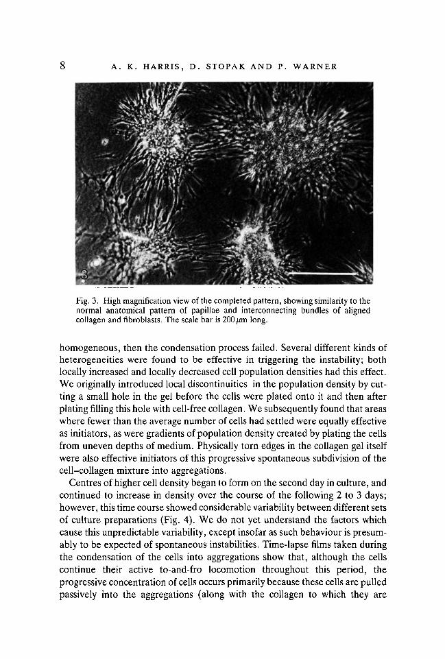
continuous restraint at the periphery, the cumulative effect of the fibroblasts'traction is to break up the initially homogeneous collagen gel into a series ofcompacted clumps. These clumps of cells and collagen are interconnected bystrands of stretched collagen fibres (see Fig. 2). At points distributed from 0-1to 0-8 mm apart through the matrix, the actively motile fibroblasts coalesce andaggregate gradually together. In time-lapse films, taken at high magnification,these aggregating cells can be seen to pull on the collagen fibres and to draw bothcollagen and other fibroblasts together into the developing aggregations. By thisprocess of fibroblast traction pulling on the collagen, the developing aggrega-tions progressively enlarge themselves, and collagen becomes further compactedinto and onto the accumulating masses of fibroblasts. These cells' traction drawsstill more collagen, and the additional fibroblasts attached to that collagen, intothe developing aggregations. This traction has the additional effect of aligningcollagen fibres into linear tracts running directly between adjacent concentra-tions of cells (Fig. 3).
The initiation of this process of cluster formation was difficult to record butseemed to require that the distribution of cells within the preparation be unevenin one or another respect. When the cells' population density was made perfectly
Fig. 2. Very low magnification views of 'doughnut' preparations, with reprecipitatedcollagen gels supported mechanically around their peripheries by attachment toglass-fibre filters. A millimeter scale is visible beside each culture. A. Punctatepattern of clumping which develops when fibroblasts are plated onto gels at an initialdensity of 4X10⁴ cells/cm2. B. Larger and more elongate clumps form whenfibroblasts are plated at a higher population density, 7 x 10⁴ cells/cm2.
A. K. HARRIS, D. STOPAK AND P. WARNER
'
Fig. 3. High magnification view of the completed pattern, showing similarity to thenormal anatomical pattern of papillae and interconnecting bundles of alignedcollagen and fibroblasts. The scale bar is 200 pm long.
homogeneous, then the condensation process failed. Several different kinds ofheterogeneities were found to be effective in triggering the instability; bothlocally increased and locally decreased cell population densities had this effect.We originally introduced local discontinuities in the population density by cut-ting a small hole in the gel before the cells were plated onto it and then afterplating filling this hole with cell-free collagen. We subsequently found that areaswhere fewer than the average number of cells had settled were equally effectiveas initiators, as were gradients of population density created by plating the cellsfrom uneven depths of medium. Physically torn edges in the collagen gel itselfwere also effective initiators of this progressive spontaneous subdivision of thecell-collagen mixture into aggregations.
Centres of higher cell density began to form on the second day in culture, andcontinued to increase in density over the course of the following 2 to 3 days;however, this time course showed considerable variability between different setsof culture preparations (Fig. 4). We do not yet understand the factors whichcause this unpredictable variability, except insofar as such behaviour is presum-ably to be expected of spontaneous instabilities. Time-lapse films taken duringthe condensation of the cells into aggregations show that, although the cellscontinue their active to-and-fro locomotion throughout this period, theprogressive concentration of cells occurs primarily because these cells are pulledpassively into the aggregations (along with the collagen to which they are
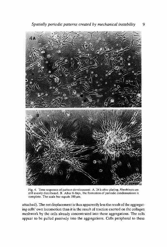
Spatially periodic patterns created by mechanical instability 9
J/ "I
^
:«*Fig. 4. Time sequence of pattern development. A. 24 h after plating, fibroblasts arestill evenly distributed. B. After 6 days, the formation of periodic condensations iscomplete. The scale bar equals 100 fim.
attached). The net displacement is thus apparently less the result of the aggregat-ing cells' own locomotion than it is the result of traction exerted on the collagenmeshwork by the cells already concentrated into these aggregations. The cellsappear to be pulled passively into the aggregations. Cells peripheral to these
10 A. K. HARRIS, D. STOPAK AND P. WARNER
growing aggregations were often drawn inward as a group, almost as if carriedon a conveyor belt.
Individual cells in these films could also be observed being passively stretchedand aligned between these aggregations. Cells attached to the matrix weremoved along with it as concentrations of cellular traction pulled the matrix fromplace to place (convection), and this occurred even when the locomotion of theseindividual cells happened to be propelling them in a different direction, relativeto the matrix itself. Periods of rapid inward convergence toward the aggregationsoften alternated with periods of relative stability, in which few additional cellswere added to these clusters. During these periods of relative quiescence, cellscould be seen to migrate to-and-fro, some toward and some away from thecentres of aggregation, until another period of matrix retraction toward thesecentres began.
A stable state was eventually reached (after 2-3 days), in which the tensilestrength of the aligned tracts of collagen resists the traction of the fibroblasts.The aligned collagen then remains tensely stretched between adjacent cellaggregations, balanced between them, instead of being drawn into either. Theend result is thus a tessellation or interlocking network of polygons. Thesepolygons are primarily rhombi (squares or parallelograms) and triangles (Fig.3). The dense self-aggregations of fibroblasts and compacted collagen make upthe vertices of these polygons, while the tracts of aligned collagen form thepolygons' sides. The polygon networks formed in this way by dissociatedfibroblasts plated out onto reprecipitated collagen gels, are generally less regularthan the networks of feather papillae and interconnecting collagen tracts whichare found in developing bird skin. The difference is often very marked. We havenot been able to determine why the resulting pattern is so much more regularin some culture preparations (Fig. 3), and in some parts of preparations, com-pared with others.
Condensations also formed spontaneously when embryonic skeletal muscle(rather than dermis, as in the rest of these experiments) was used as the sourceof dissociated cells. In this case, myoblasts present in the dissociated muscletissue fused and differentiated to form myotubes, and these even underwentirregular spasms of contraction. However, these muscle cells did not appear toparticipate directly in the formation of the condensations of fibroblasts andcollagen. The lengths of these myotubes was such that they typically spanned twoor more of the fibroblast aggregations. The tensile stress exerted by thefibroblasts in these aggregations caused stretching, elongation and eventuallytearing of these myotubes. As the fibroblast aggregations developed, myotubesstretched between adjacent aggregations gradually became narrowed in theirmiddles, before being torn in two. By the end of a week in culture, mostmyotubes had been disrupted by this tensile stress and had degenerated, ap-parently as a result of this mechanical fragmentation.

Spatially periodic patterns created by mechanical instability 11
Effects of cell population density on the pattern of condensation
The size and spacing of the aggregations (or condensations) varied as a func-tion of the original population density at which the cells had been plated out (Fig.5). At densities below 104 cells/cm2, cells spread and migrated on and throughthe collagen matrix, visibly distorting the gel in their immediate proximity. Butat these low population densities, the cells' collective strength was apparentlyinsufficient to produce the massive, permanent rearrangements of the matrix,which results in the formation of the condensations. Instead, only occasional,small and irregular aggregations were formed. At population densities of2 x 104 cells/cm2 small condensations of ten or fewer cells formed.
When the cell density was further increased, the numbers of cells composingeach resulting condensation likewise increased. For example, at a plating densityof 5 x 10⁴ cells/cm2, the condensations which formed had diameters averagingabout 500 fim. Each of these consisted of many hundreds of cells. At still higherpopulation levels, a different pattern of condensation arose, instead of the pun-ctate tessellation pattern. Between 7 x 10⁴ and 1 x 10⁵ cells per cm2, instead ofcondensing into more or less round aggregations as at lower densities, the cellscondensed into elongate columns which coursed irregularly through the matrix,roughly parallel to one another (Fig. 2B). Just as the rounded condensations
Fig. 5. Effects of differing initial population densities of fibroblasts on the resultingspatial pattern. Scale bar equals 400 jum. A. 2 x 10⁴ cells/cm2. B. 4 x 10⁴ cells/cm2.C. 7 x 10⁴ cells/cm2. D. 9 x 10⁴ cells/cm2.
12 A. K. HARRIS, D. STOPAK AND P. WARNER
formed at lower cell concentrations were reminiscent of the dermal papillae offeathers or hair, these elongate aggregations formed at higher population den-sities had shapes roughly similar to the scales on developing avian hindlimbs. Atinitial cell concentrations of 1 x 10⁵ or above a continuous monolayer of cells wasquickly established which was mechanically stable and did not break up intocondensations, but did develop small, irregular foci of locally increased celldensity.
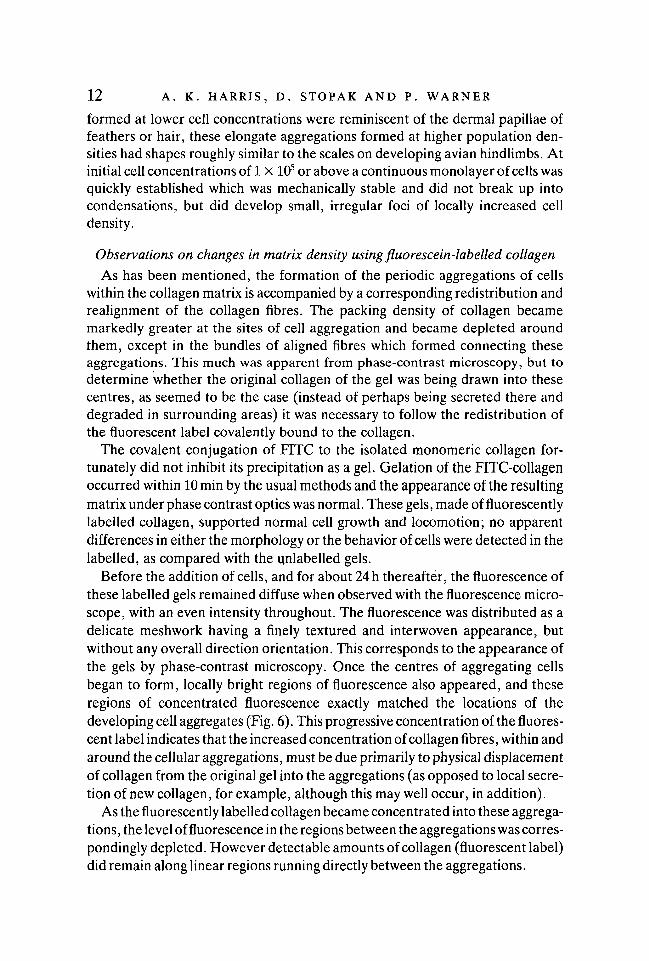
Observations on changes in matrix density using fluorescein-labelled collagen
As has been mentioned, the formation of the periodic aggregations of cellswithin the collagen matrix is accompanied by a corresponding redistribution andrealignment of the collagen fibres. The packing density of collagen becamemarkedly greater at the sites of cell aggregation and became depleted aroundthem, except in the bundles of aligned fibres which formed connecting theseaggregations. This much was apparent from phase-contrast microscopy, but todetermine whether the original collagen of the gel was being drawn into thesecentres, as seemed to be the case (instead of perhaps being secreted there anddegraded in surrounding areas) it was necessary to follow the redistribution ofthe fluorescent label covalently bound to the collagen.
The covalent conjugation of FITC to the isolated monomeric collagen for-tunately did not inhibit its precipitation as a gel. Gelation of the FITC-collagenoccurred within 10 min by the usual methods and the appearance of the resultingmatrix under phase contrast optics was normal. These gels, made of fluorescentlylabelled collagen, supported normal cell growth and locomotion; no apparentdifferences in either the morphology or the behavior of cells were detected in thelabelled, as compared with the unlabelled gels.
Before the addition of cells, and for about 24 h thereafter, the fluorescence ofthese labelled gels remained diffuse when observed with the fluorescence micro-scope, with an even intensity throughout. The fluorescence was distributed as adelicate meshwork having a finely textured and interwoven appearance, butwithout any overall direction orientation. This corresponds to the appearance ofthe gels by phase-contrast microscopy. Once the centres of aggregating cellsbegan to form, locally bright regions of fluorescence also appeared, and theseregions of concentrated fluorescence exactly matched the locations of thedeveloping cell aggregates (Fig. 6). This progressive concentration of the fluores-cent label indicates that the increased concentration of collagen fibres, within andaround the cellular aggregations, must be due primarily to physical displacementof collagen from the original gel into the aggregations (as opposed to local secre-tion of new collagen, for example, although this may well occur, in addition).
As the fluorescently labelled collagen became concentrated into these aggrega-tions , the level of fluorescence in the regions between the aggregations was corres-pondingly depleted. However detectable amounts of collagen (fluorescent label)did remain along linear regions running directly between the aggregations.

Spatially periodic patterns created by mechanical instability 13
t
Fig. 6. Accumulation of fluorescently labelled collagen into papilla-like clumps. Thescale bar equals 200jum. A. Phase-contrast view. B. Fluorescence excitation view.
DISCUSSION
Convection
The displacement of cells by being pulled along bodily with the extracellularmatrix is a type of morphogenetic cell movement which seems not previously tohave been reported or hypothesized in the embryological literature. We proposeto call this type of cell movement 'convection', as was suggested to us by GeorgeOster, based on the analogy with the carrying of heat or substances from placeby the flow of a liquid or gas. Not only is the etymology of the word convectionquite appropriate (Latin for 'carry along with') but the mathematical expressionsneeded to treat this class of cell displacements turn out to be the same as thoseused for the convection of heat by fluids (see Murray, Oster & Harris, 1983). Wewould also propose that whenever cells within embryos are found to bunchtogether into compact masses (as for example in the initial condensation ofskeletal rudiments), the possibility should be considered that these cells may alsohave been pulled along with extracellular matrices by a conveyer-belt-like effectof cell traction - rather than, for example, having become especially adhesive toone another. Note the similarity of convection to the phenomena aptly termed'passive locomotion' by Trinkaus (1982), in which cells are pulled along by thelocomotion of others to which they are attached.
The generation of spatial patterns by convection
Our observations on the (admittedly experimental) system of fibroblasts dis-persed on collagen gels suggest explanations for several aspects of normaldermal morphogenesis in birds. For one thing, these observations show thatneither papillae nor the aligned tracts of collagen between them need be thecause of the other's formation, and that neither class of structure requires achemical prepattern. Instead, our results suggest that both events (the aggrega-tion of fibroblasts into papillae, as well as the alignment of collagen fibres intolinear bundles interconnecting these aggregations) may, at least in principle, be

14 A. K. HARRIS, D. STOPAK AND P. WARNER
explained as parallel consequences of the same mechanical instability. This in-stability occurs when the initially homogeneous and isotropic dermal matrixyields plastically to fibroblast traction. The ability to generate patterns dependsupon the tendency of both cells and matrix to be displaced toward sites of locallygreater cell concentration (and thus of locally greater cell contractility). Thismovement of cells up local gradients of cell population density not only has theneeded autocatalytic effect of magnifying small or even random differences incell density (much as do the chemical reactions of Turing's model, though ofcourse for different reasons), it also entails a capacity for lateral propagation ofalternately concentrated and depleted cell population densities (Fig. 7).Propagation of the instability would occur because the aggregation of cells at onesite depletes the surrounding area below the prevailing population level of thearea beyond the depleted area. This depletion would thereby create additionalgradients of population density, up which cells will be pulled to form secondaryaggregations of cells at some distance from the first. These would then depletethe areas around themselves, and so on. The rectilinearity of the avian dermalpattern can be interpreted as a consequence of positive feedback between cellorientation by contact guidance along matrix fibres and matrix orientation by thetraction exerted by the oriented cells, with new condensations tending to formwhere the resulting fields of tension intersect (cf. Stuart et al. 1972).
Both in spacing and size, the aggregations formed in our cultures were lessregular than actual feather papillae, often very much so (see Figs 2, 5 & 6),although even these least regular cases compare favourably with patterns createdby computer simulation of Turing's mechanism (Turing, 1952; Bard & Lauder,1974). While we have not been able to determine the causes of this irregularity,or of its variation from case to case, the explanation may lie in the ability of thesepattern-generating mechanisms (both Turing's and the one proposed here) tocreate their peaks either by autocatalysis of random perturbations or secondarily,by propagation from previously formed peaks nearby. In the former case, thepattern generated is apt to partake of the randomness which triggered it (Bard &Lauder, 1974), but in the latter case the spacing is determined by the ranges andstrengths of the interactions involved and can therefore be very regular. Thus ifthe papillae in the skin are mostly secondary effects (of propagation), while in ourcultures they are mostly primary, the contrast in regularity would result. In simplecomputer simulations of our model, this was the case. These considerations sug-gest the possibility that embryos solve the problem of achieving regularity ofresults, despite using stochastic mechanisms, simply by developing sensitivity tothe instability progressively across a tissue field - so that most of the peaks will besecondary to pre-existing ones. Although we were unable to devise such a grada-tion of sensitivity in our cultures, we would suggest that if dermis could be causedto mature simultaneously over a broad area, and the papillae thus to form simul-taneously, then their spacing would be less regular. This may be the reason for thelesser regularity of hair (cf. Davidson, 1983).

Spatially periodic patterns created by mechanical instability 15
C D
sssssssssa.
Q CD CDO O ^ CD
Fig. 7. Schematic diagram of the type of mechanical instability responsible for re-arranging collagen matrices into the patterns described here. When cells move upgradients of their own concentration (as well as pulling collagen fibres up thesegradients), then even a small local deviation from the average population density willinitiate an accumulation of cells. The accumulation of cells into a growing peak willdeplete the surrounding area, thereby creating secondary gradients on the oppositeside of the depleted areas. Since cells also move up these secondary gradients, theinstability propagates itself. Thus the resulting pattern of alternating high and lowpopulation areas can develop either simultaneously over the field, if the system isexcited by widespread initiating irregularities, or sequentially when initiated in onearea from which the instability then propagates.
Several different factors may contribute to the antidiffusive tendency of cellsto move up gradients of cell and matrix density. One is haptotaxis - the migrationof cells up gradients of adhesiveness (Carter, 1967; Harris, 1973). As the cells areadhesive to sites on the collagen molecules, a gradient of collagen concentrationshould therefore be a gradient of adhesiveness for these cells (Murray et al.1983). Among other factors which should contribute to the autocatalytic

16 A. K. HARRIS, D. STOPAK AND P. WARNER
aggregation effect is the simple physical tearing of the collagen matrix (which isindeed the most apparent feature in our time-lapse films of the phenomenon),non-linear proportionality between stress and strain in collagen, the contactguidance effects mentioned above, as well as any cooperative effects or mutualstimulation of contractility by cells crowded together. The situation within thestressed matrix is rather like a many sided tug-of-war in which pulling an op-ponent (cell) close enough to oneself adds it to your team (aggregation). Theautocatalytic (or antidiffusive) consequence of such recruitment is obvious.
As to the possible operation of this pattern-generating mechanism in thedevelopment of bird skin, our observations on the effects of varying cell popula-tion density suggest that the formation of the bare areas ('apteria', see Lucas &Stettenheim, 1972) may be the result of insufficient traction (relative to theelastic resistance of the matrix) to pull fibroblasts together into aggregations;much as when too few fibroblasts are plated onto a collagen gel. This interpreta-tion is supported by the observation of Wessells (1965) that the dermis is indeedless dense in presumptive apterial regions, as well by the observations thatexperimental implants which cause feathers to form in normally apterial regionsfirst cause the appearance of a dense mesenchyme (Sengel & Kieny, 1967; Sen-gel, Dhouailly & Kieny, 1959). Also supporting this interpretation is the findingof Mauger (1972) that the induction of bare regions where feathers usually formis accompanied by the formation of a sparse dermis.
Likewise, the replacement of small, punctate aggregations by larger, elon-gated ones when cells are plated out at higher population densities, would seemto suggest an explanation for the replacement of feathers by the similarly broadscales (scuta and scutella) of avian feet. However, according to most workers,these scales lack well-organized papillae. Otherwise, we would suggest that thereplacement of scales by feathers (ptilopody) in certain mutants, or in embryostreated with retinoic acid (Dhouailly, Hardy & Sengel, 1980), might prove ex-plicable in terms of an inhibition of cell traction. In a similar way, the depen-dence of this type of instability on the maintenance of tensile stress may help toexplain the results of Novel (1973), in which early feather germs dispersed whenskin fragments were explanted. In addition, our observation of the ability of freeedges to initiate this instability suggests an explanation for another of Novel'sobservations: that explants subsequently develop new papillae in rows parallelto their long edges.
Mechanical pattern generation and non-diffusible morphogens
The implications of this mode of pattern generation are not confined to theskin, or to birds. For one thing, certain morphological similarities suggest thata very similar self-propagating mechanical instability may be responsible forgenerating the pattern of scales in bony fish, even though these scales are essenti-ally dermal bones, and as such not strictly homologous to the epidermal scalesof birds. Weiss (1959) has reported the observation by McMurray in developing

Spatially periodic patterns created by mechanical instability 17
teleost dermis of a pattern of collagen alignment nearly identical to thatsubsequently found in bird skin, but with the formation of bony scales (ratherthan papillae) at the fibre intersections. If one were to generalize the mechanismstill further to include the mechanical interactions among the epidermal cellsthemselves, it might be able to explain the remarkable splitting of the feathergerm itself to form its barb ridges.
Mechanical instabilities of the same basic kind may also help to explain certainfeatures of the spatial control of cartilage differentiation. Thorogood &Hinchliffe (1975) have described the transient compaction of the presumptivechondrocytes which immediately precedes their differentiation as cartilage. Thiscompaction could be causal, with the mechanical compression of cells and matrixlocally stimulating the cells' differentiation, since it is known that chondroblastdifferentiation is promoted by cell density (Holtzer, Abbot, Lash & Holtzer,1960) and by the concentration of extracellular matrix materials (Lash & Vasan,1978). Thus the morphogen or pre-pattern for cartilage formation could becompressive strain, an explanation which is further developed for the case of thelimb skeleton in a separate paper (Oster, Murray & Harris, 1984). This effect ofcompression may correspond to the second regulatory step in cartilage dif-ferentiation postulated by Solursh etal. (1982). The possibility is also temptingthat the segmentation of the somites represents the operation of such a mechan-ism along a one-dimensional cell column. However, we note that because thesegmental muscles and cartilages (vertebral arcualia) to which the somites giverise are spaced in alternation with each other (with the cartilage forming at theboundaries between somites and the muscles connecting these cartilages),therefore, if somite segmentation is mechanically analogous to the aggregationof dermal fibroblasts into papillae, then it would have to be the ends of thesomites (where the cartilages form) which are mechanically equivalent to thedermal papillae. The somites themselves would have to be equivalent to thealigned bundles of matrix between these papillae.
Mechanical versus chemical pattern generation
For the generation of anatomical patterns, mechanical instabilities, such asthose described here, differ in at least five important respects from chemicalinstabilities, like the Turing model. Several of these differences may prove usefulas criteria for distinguishing between the operations of the two classes of mechan-ism. The first and most obvious difference is that mechanical instabilities yieldactual physical structures, not just chemical 'blueprints' capable of guiding thesubsequent formation of these structures by separate and unspecified forces. Ifmechanical instabilities have as much (or more) ability to create regulargeometric patterns as chemical instabilities, why would evolution retain thelatter? Physical forces are always necessary for morphogenesis anyway.
The second difference is the longer distance over which mechanical forces can beeffective. Crick (1970) has estimated the maximum distance over which diffusion
18 A. K. HARRIS, D. STOPAK AND P. WARNER
gradients could be expected to stabilize within a reasonable period of time insidean embryo. His estimate was only about 1 mm, whereas the mechanically createdpatterns we have been encountering in organ culture have dimensions in thecentimetre range (Stopak & Harris, 1982), as do many actual embryonic patterns- such as those of the dermis. The third difference is closely related and is thegreater speed (in comparison with chemical diffusion) with which mechanicalforces can be propagated within developing tissues - and perhaps more impor-tant, the greater speed at which these forces can reach equilibrium (see Oster etal. 1984). A fourth difference is that mechanical systems are much less vulner-able than diffusion gradients to disruption by flows of liquids through thedeveloping field, as, for example, when the circulatory system begins to function.Conversely, pattern-generating systems which depend upon stress and strainshould be more susceptible to mechanical interference, and this difference mightbe made to provide an experimental criterion for their identification.
The fifth and last of these differences is that mechanical instabilities dependupon tensor properties (like stress, strain and curvature), which can differ invalue in different directions at each point in space. This is in contrast to chemicalconcentration, which as a scalar property has no inherent directionality, but onlya quantity at each point. While it is true that fields of scalar properties candetermine direction by their gradient (the gradient of a scalar field being a vectorfield), as in chemotaxis or in the determination of bristle alignment in the insectepidermis, the range of possible direction fields which can be determined in thisway turns out to be quite circumscribed. In particular, there are many vectorfields which are not the gradients of any possible scalar field. This is true of allvector fields whose curl is anywhere not equal to zero (Davis, 1961). These vectorfields are called non-conservative, or rotational, and the familiar spiral whorl ofhair alignment on the back of the human head (not to be confused with the curlof the hair itself!) is a good example of an actual anatomical pattern whose vectorfield has a non-zero curl. Thus, it is inherently incapable of being determined bya gradient of a morphogen concentration or of any other scalar property. Con-versely, if certain anatomical patterns were found persistently to possess zerocurl, then this could be taken as evidence of their determination by the gradientof some scalar property, such as chemical concentration.
This work was made possible by research grant No. 2 RO1 GM24251 from the NationalInstitutes of Health, Institute of General Medicine. The preparation of the manuscript wasaided by the helpful criticisms of the following: Danielle Dhouailly, Elizabeth Harris, JulianLewis, James Murray, George Oster, John Saunders, Peter Thorogood, J. P. Trinkaus andNorman Wessells. Fig. 7 was prepared by Susan Whitfield.
Our efforts to explain pattern generation in terms of mechanical interactions between cellshave been inspired by the example of our friend and colleague, H. Eugene Lehman, whosework on the generation of pigmentation bands in salamanders (J. of Experimental Zoology,vol. 135, pp. 355-86,1957) paralleled Turing's theory, in that it showed how certain combina-tions of mutual attraction and repulsion between cells could achieve much the same morpho-genetic results as equivalent combinations of chemical autocatalysis and inhibition.
Spatially periodic patterns created by mechanical instability 19
REFERENCESBARD, J. B. T. & LAUDER, I. (1974). How well does Turing's theory work? /. theor. Biol. 45,
501-531.BELL,E.,IVARSSON,B. & MERRILL, C. (1979). Production of a tissue-like structure by contrac-
tion of collagen lattices by human fibroblasts of different proliferative potential in vivo.Proc. natn. Acad. Sci. U.S.A. 76, 1274-1278.
CARTER, S. B. (1967). Haptotaxis and the mechanism of cell motility. Nature 213, 256-260.CRICK, F. (1970). Diffusion in embryogenesis. Nature 225, 420-422.DAVIDSON, D. (1983). The mechanism of feather pattern development in the chick. II. Control
of the sequence of pattern formation. /. Embryol. exp. Morph. 74, 261-273.DAVIS, H. F. (1961). Introduction to Vector Analysis. Boston: Allyn & Bacon.DHOUAILLY, D., HARDY, M. & SENGEL, P. (1980). Formation of feathers on chick foot scales:
a stage-dependent response to retinoic acid. /. Embryol. exp. Morph. 58, 63-78.ELSDALE, T. & BARD, J. (1973). Collagen substrata for studies on cell behavior. /. Cell Biol.
54, 626-637.EHRMAN, R. L. & GEY, G. O. (1956). The growth of cells on a transparent gel of reconstituted
rat-tail collagen. J. natn. Cancer lnst. 16, 1375-1403.HAMBURGER, V. & HAMILTON, H. L. (1951). A series of normal stages in the development of
the chick embryo. /. Morphology 88, 49-92.HARRIS, A. K. (1973). Behavior of cultured cells on substrata of variable adhesiveness. Exp.
Cell Res. 77,285-297.HARRIS, A. K.,STOPAK,D. & WILD, P. (1981). Fibroblast traction as a mechanism for collagen
morphogenesis. Nature 290, 249-251.HOLTZER, H., ABBOT, J., LASH, J. & HOLTZER, S. (1960). The loss of phenotypic traits by
differentiated cells in vitro. I. Redifferentiation of cartilage cells. Proc. natn. Acad. Sci.,U.S.A. 46, 1533-42.
LASH, J. W. & VASAN, N. S. (1978). Somite chondrogenesis in vitro. Stimulation by exogenousmatrix components, Devi Biol. 66, 151-171.
LOWRY, O. H., ROSENBROUGH, N. J., FARR, A. L. & RANDALL, R. J. (1951). Protein measure-ment with folin phenol reagent. /. biol. Chem. 193, 265-275.
LUCAS, A. M. & STETTENHEIM, P. R. (1972). Avian Anatomy; Integument, part 11. (Agricul-ture Handbook 362) Washington,D.C., U.S. Government Printing Office.
MAUGER, A. (1972). Role du tube neural dans le development du plumage dorsal de l'embryonde Poule. Wilhelm Roux Arch. EntwMech. Org. 170, 244-266.
MEINHARDT, H. (1982). Models of Biological Pattern Formation. New York: AcademicPress.
MURRAY, J. D., OSTER, G. F. & HARRIS, A. K. (1983). A model for mesenchyme morpho-genesis. /. Mathematical Biology 17, 125-129.
NAIRN, R. C. (1969). Fluorescent Protein Tracing. Edinburgh: E. & S. Livingston.NOVEL, G. (1973). Feather pattern stability and reorganization in cultured skin. /. Embryol.
exp. Morph. 30, 605-633.OSTER, G. F., MURRAY, J. D. & HARRIS, A. K. (1984). Mechanical aspects of mesenchymal
morphogenesis. /. Embryol. exp. Morph. 78.SENGEL, P. (1976). Morphogenesis of Skin. Cambridge: Cambridge Univ. Press.SENGEL, P., DHOUAILLY, D. & KIENY, M. (1969). Aptitude de constituants cutanes de l'apterie
medioventrale du Poulet a forme de plumes. Devi Biol. 19, 436-446.SENGEL, P. & KIENY, M. (1967). Productin d'une pteryle supplementaire chez l'embryon de
Poulet. II. Analyse experimentale. Devi Biol. 16, 532-563.SOLURSH,M., JENSEN, K. L., SINGLEY, C. T., LINSENMEYER,T. F. &REITER, R. S. (1982). Two
distinct regulatory steps in cartilage differentiation. Devi Biol. 94, 311-325.SPURR, O. K. & NEIGISCH, W. D. (1962). Stress crazing of some amorphous thermoplastics.
/. Applied Polymer Sci. 6, 585-599.STOPAK, D. & HARRIS, A. K. (1982). Connective tissue morphogenesis by fibroblast traction
1. Tissue culture observations. Devi Biol. 90, 383-398.

20 A. K. HARRIS, D. STOPAK AND P. WARNER
STUART, E. S., GARBER,B. &MOSCONA, A. A. (1972). An analysis of feather germ formationin the embryo and in vitro in normal development and in skin treated with hydrocortisone./. exp. Zool. 179, 97-118.
STUART, E. S. & MOSCONA, A. A. (1967). Embryonic morphogenesis: Role of fibrous latticein the development of feathers and feather patterns. Science 157, 947-948.
THOROGOOD, P. V. & HINCHLIFFE, J. R. (1975). An analysis of the condensation process duringchondrogenesis in the embryonic chick limb. /. Embryol. exp. Morph. 33, 581-606.
TRINKAUS, J. P. (1982). Some thoughts on directional cell movement during morphogenesis.In Cell Behavior (ed. R. Bellairs, A. Curtis & G. Dunn), pp. 471-498. Cambridge:Cambridge Univ. Press.
TURING, A. M. (1952). The chemical basis of morphogenesis. Phil. Trans. R. Soc. B 237,37-72.
WESSELLS, N. K. (1965). Morphology and proliferation during early feather development.Devi Biol. 12, 131-153.
WESSELLS, N. K. & EVANS, J. (1968). The ultrastructure of oriented cells and extracellularmaterials between developing feathers. Devi Biol. 18, 42-61.
WEISS, P. A. & MCMURRAY, V. (1959). Biological Organization: Cellular and Subcellular,UNESCO Symposium (ed. C. H. Waddington), pp. 12-13. New York: Pergamon.
WOLPERT, L. (1971). Positional information and pattern formation. Curr. Top. devl Biol. 6,183-224.
{Accepted 7 November 1983)


![A SPATIALLY PERIODIC KURAMOTO-SIVASHINSKY …2 H. UECKER, A. WIERSCHEM EJDE-2007/118 periodic stationary solution U s (Nusselt solution) is not known in closed form. In [15] an expansion](https://img.dokumen.tips/doc/110x75/60ee37dad72a27774c53b006/a-spatially-periodic-kuramoto-sivashinsky-2-h-uecker-a-wierschem-ejde-2007118.jpg)