Embed Size (px)
Citation preview

Submitted 27 May 2017Accepted 20 October 2017Published 13 November 2017
Corresponding authorPannamthip Pitaksajjakul,[email protected]
Academic editorMario Alberto Flores-Valdez
Additional Information andDeclarations can be found onpage 16
DOI 10.7717/peerj.4021
Copyright2017 Injampa et al.
Distributed underCreative Commons CC-BY 4.0
OPEN ACCESS
Generation and characterization of crossneutralizing human monoclonal antibodyagainst 4 serotypes of dengue viruswithout enhancing activitySubenya Injampa1,2, Nataya Muenngern1, Chonlatip Pipattanaboon1,Surachet Benjathummarak1, Khwanchit Boonha1, Hathairad Hananantachai2,Waranya Wongwit2, Pongrama Ramasoota1,2 and Pannamthip Pitaksajjakul1,2
1Center of Excellence for Antibody Reserach, Faculty of Tropical Medicine, Mahidol University, Bangkok,Thailand
2Department of Social and Environmental Medicine, Faculty of Tropical Medicine, Mahidol University,Bangkok, Thailand
ABSTRACTBackground. Dengue disease is a leading cause of illness and death in the tropics andsubtropics.Most severe cases occur among patients secondarily infected with a differentdengue virus (DENV) serotype compared with that from the first infection, resultingin antibody-dependent enhancement activity (ADE). Our previous study generatedthe neutralizing human monoclonal antibody, D23-1B3B9 (B3B9), targeting the firstdomain II of E protein, which showed strong neutralizing activity (NT) against all fourDENV serotypes. However, at sub-neutralizing concentrations, it showed ADE activityin vitro.Methods. In this study, we constructed a new expression plasmid using the existingIgG heavy chain plasmid as a template for Fc modification at position N297Q bysite-directed mutagenesis. The resulting plasmid was then co-transfected with alight chain plasmid to produce full recombinant IgG (rIgG) in mammalian cells(N297Q-B3B9). This rIgG was characterized for neutralizing and enhancing activityby using different FcγR bearing cells. To produce sufficient quantities of B3B9 rIgG forfurther characterization, CHO-K1 cells stably secreting N297Q-B3B9 rIgG were thenestablished.Results. The generated N297Q-B3B9 rIgG which targets the conserved N-terminalfusion loop ofDENVenvelope protein showed the same cross-neutralizing activity to allfour DENV serotypes as those of wild type rIgG. In both FcγRI- and RII-bearing THP-1 cells and FcγRII-bearing K562 cells, N297Q-B3B9 rIgG lacked ADE activity againstall DENV serotypes at sub-neutralizing concentrations. Fortunately, the N297Q-B3B9rIgG secreted from stable cells showed the same patterns of NT and ADE activities asthose of the N297Q-B3B9 rIgG obtained from transient expression against DENV2.Thus, the CHO-K1 stably expressing N297Q-B3B9 HuMAb can be developed as highproducer stable cells and used to produce sufficient amounts of antibody for furthercharacterization as a promising dengue therapeutic candidate.Discussion. Human monoclonal antibody, targeted to fusion loop of envelopedomainII (EDII), was generated and showed cross-neutralizing activity to 4 serotypesof DENV, but did not cause any viral enhancement activity in vitro. This HuMAb couldbe further developed as therapeutic candidates.
How to cite this article Injampa et al. (2017), Generation and characterization of cross neutralizing human monoclonal antibody against4 serotypes of dengue virus without enhancing activity. PeerJ 5:e4021; DOI 10.7717/peerj.4021

Subjects Biotechnology, Molecular Biology, Virology, Infectious DiseasesKeywords Therapeutic antibody, Dengue virus, Epitope mapping, Neutralization, Humanmonoclonal antibody
INTRODUCTIONDengue infection is a leading cause of illness and death in the tropics and subtropics. Morethan one-third of the world’s population lives in areas at risk (Natasha Evelyn, Mikkel &Annelies, 2013). DENV is a member of the Flavivirus genus in the Flaviviridae family. Thereare four antigenically different serotypes of DENV (DENV-1 to DENV-4) (Kuhn et al.,2002). Primary infection with one DENV serotype induces the production of homotypicneutralizing antibodies, which provides lifelong immunity against the infecting serotype.These homotypic neutralizing antibodies can cross-react but less specific against the otherthree DENV serotypes and persist for a period of several months to a few years (Guzman& Vazquez, 2010). Upon secondary infection with heterologous DENV serotypes, theseantibodies fail to neutralize virus resulting in an increase of infected cells and highervirus titers. This phenomenon is named antibody-dependent enhancement activity (ADE)(Halstead & O’Rourke, 1977). Primary infection typically causes asymptomatic or self-limited dengue fever (DF). Whereas patients in secondary infection usually have anincreased risk of developing severe disease, including life-threatening dengue hemorrhagicfever/dengue shock syndrome (DHF/DSS) (Murphy & Whitehead, 2011).
Currently, there is no available antiviral drug specific for all four DENV serotypes.Even though there are many studies of neutralizing antibodies against DENV, fully humanmonoclonal antibodies (HuMAbs) which are able to neutralize all four DENV serotypeswithout ADE activity are considered to be the main option for passive immune therapy(Marasco & Sui, 2007; Chan & Carter, 2010; Low, Ooi & Vasudevan, 2017).
Previously, we generated HuMAb clone D23-1B3B9 (B3B9) with strong in vitroneutralizing activity (NT) against DENV-1 to DENV-4 (Setthapramote et al., 2012).However, investigating the viral infection enhancing activity of this HuMAb on Fc-gammareceptor (FcγR)-bearing cells revealed an increase in DENV infection at sub-neutralizingconcentrations, which limits its application as a therapeutic candidate (Sasaki et al., 2013).To diminish enhancing activity, modifications at glycosylation site Asn297, which influencethe binding affinity between Fc region and FcγR presenting on immune effector cells, wereestablished (Hristodorov, Fischer & Linden, 2013; Chan & Carter, 2010; Balsitis et al., 2010).
We constructed plasmids expressing antibody light chain (LC) and modified the Fcregion of the heavy chain (HC) constant domain 2 at position N297Q by site-directedmutagenesis. The modified HC plasmid was co-transfected with the LC plasmid intoHEK293T mammalian cells to produce full recombinant IgG (rIgG). To evaluate theFc-modified rIgG as a potential therapeutic candidate for dengue treatment, NT andADE activity were determined in vitro and compared with those of wildtype rIgG. Wealso demonstrate the mimic binding epitope on envelope protein of dengue virus forunderstanding specificity and functionality of this HuMAb.
Injampa et al. (2017), PeerJ, DOI 10.7717/peerj.4021 2/20

MATERIALS AND METHODSCell lines and DENV strainsHEK293T cells were maintained in Dulbecco’s modified Eagle medium (Gibco, GrandIsland, NY, USA) with 10% fetal bovine serum (FBS). For the NT test, Vero cells werecultured in minimal essential medium (MEM) (GE Healthcare UK Ltd., Buckinghamshire,UK) with 10% fetal bovine serum. THP-1 and K562 cells, which were used in ADE assays,were cultured in RPMI 1640 medium (Gibco) with 10% FBS. The DENV strains used inthis study were the Mochizuki strain of DENV1, the 16,681 strain of DENV2, the H87strain of DENV3, and the H241 strain of DENV4. All DENVs were propagated in C6/36cells, which were maintained in Leibovitz’s L-15 medium (Gibco) supplemented with 10%FBS and 0.3% of BACTO Tryptose Phosphate Broth (TPB) (Sigma-Aldrich, St. Louis, MO,USA). CHO-K1 cells were maintained in MEM medium supplemented with 10% FBSand 1% non-essential amino acid (Gibco). All cell lines were kindly provided by ResearchInstitute for Microbial Diseases, Osaka University.
Generation of wild type and mutated human monoclonal antibodyclone B3B9Plasmid constructionVariable HC and LC sequences of B3B9 HuMAb were previously isolated from hybridomacells and used for construction of HC and LC expression plasmids producing B3B9rIgG (Pitaksajjakul et al., 2014). This HC plasmid was used as a template for N297Qmutagenesis by site-directed mutagenesis with the In-Fusion Cloning System (In-Fusion R©
HD Cloning Plus; Clontech Laboratories Inc., Shiga, Japan). Primers were designedaccording to the manufacturer’s instructions to mutate the amino acid at position 297from asparagine to glutamine. This system combines the action of the In-Fusion HDenzyme with inverse polymerase chain reaction (PCR), which generates linearized DNAfrom a plasmid template. The PCR reaction was composed of the CloneAmp Hifi PCRpremix, 300 nM each of forward and reverse primer, 5 ng of plasmid, and distilled waterto final volume of 25 µl. The amplification was performed with 35 cycles of 98 ◦C for10 s, 55 ◦C for 15 s, and 72 ◦C for 5 s. The inverse PCR products were gel-purified usinga PureLink R© Quick Gel Extraction Kit (Invitrogen, Carlsbad, CA, USA) following themanufacturer’s protocol. The linearized plasmids obtained from inverse PCR were thenfused by the In-Fusion enzyme, by following the manufacturer’s instructions. The reactionwas performed at 50 ◦C for 15 min. The in-fusion reaction was chemically transformedinto Stella chemical competent cells. Individual clones were randomly selected for DNAsequencing.
DNA sequencing and plasmid preparationThe DNA sequencing of the mutated HC plasmids were confirmed by Sanger sequencing(Macrogen Inc., Seoul, Korea). Potential N-glycosylation site of wild type and mutatedB3B9 HuMAb was identified using NetNGlyc 1.0 software (Gupta, Jung & Brunak, 2004).Plasmids that contained the target mutation were amplified in E. coli and isolated using a
Injampa et al. (2017), PeerJ, DOI 10.7717/peerj.4021 3/20

PurelinkTM plasmid Midiprep kit (Invitrogen) from 100 ml culture for further transfectionin mammalian cells.
Transient expression of N297Q-B3B9 rIgG in HEK293T cellsPlasmids expressing N297Q-B3B9 HC and LC were transfected into HEK293T cells toproduce whole rIgG with the N297Q mutation as previously described (Pitaksajjakulet al., 2014). The culture medium containing secreted N297Q-B3B9 rIgG was collectedand used in immunofluorescence assays (IFAs) to determine the DENV-binding activity.The N297Q-B3B9 rIgG was purified using a protein A affinity column and measured theconcentration of N297Q-B3B9 rIgG by BCA protein assay kit (Thermo Scientific,Waltham,MA, USA). The purity of purified antibody was determined by sodium dodecyl sulfatepolyacrylamide gel electrophoresis.
IFA for DENV-binding activityIFAs were used to determine the binding and specificity of N297Q-B3B9 rIgG for allfour DENV serotypes. IFA plates were prepared by infecting Vero cells with DENV at amultiplicity of infection of 0.1 as well as mocked infected. The plates were incubated for3 days before being fixed and permeabilized with 3.7% formaldehyde and 0.1% TritonX-100, respectively. Culture fluid from transfected cells was added, and the plates wereincubated at 37 ◦C for 1 h. AlexaFluor 488-conjugated anti-human IgG (1:1,000 dilution)(Invitrogen) was added as secondary antibody. The result was observed under fluorescencemicroscope (IX71, Olympus). For this assay, non-infected cells were used as a negativecontrol.
HuMAb isotypingThe IgG isotype of the N297Q-B3B9 rIgG was determined by PCR as previously described(Omokoko et al., 2014) using complementary DNA isolated from HuMAb clone B3B9hybridoma as a template. The gene-specific primers used to amplify IgG1, -2, -3, and -4are listed in Table S1. IgG1 and IgG3 were amplified using the same forward and reverseprimers. The primers were used to amplify the hinge region between the Fab and Fc partsthat produce different sized PCR products. IgG1 and IgG3 showed specific band at 211 and346 bp, respectively. With separate PCR reactions, IgG2 and IgG4 were amplified by thesame reverse and different forward primers. These IgG2 and IgG4 PCR reactions showed207 bp and 210 bp, respectively. The PCR reaction was composed of 0.5 µg of cDNA, 1mM Tris–HCl (pH 8.0), 5 mM KCl, 1.25 mM deoxyribonucleotide triphosphate (dNTP),1.25 units of ExTaq DNA polymerase (TAKARA, Shiga, Japan), and 5 mM of each primer,with distilled water to a final volume of 25 µl. The amplification of each primer pairs wasperformed in separate reaction with 35 cycles of 94 ◦C for 30 s, 65 ◦C for 30 s, and 72 ◦Cfor 30 s. The PCR products were separated by agarose gel electrophoresis and visualized bystaining with SYBR Safe DNA Gel stain (Invitrogen).
Foci reduction neutralization test (FRNT)FRNT assay was performed as previously described (Pitaksajjakul et al., 2014). DENVat MOI 0.01 were mixed with different concentrations of purified HuMAbs (B3B9 or
Injampa et al. (2017), PeerJ, DOI 10.7717/peerj.4021 4/20

N297Q-B3B9 rIgG) and incubated at 37 ◦C for 1 h. Each mixture was added to Vero cellsin 96-well cell culture plates and incubated for 2 h. The plates were then overlaid with 2%carboxymethyl cellulose (CMC) in MEM medium with 2% FBS and incubated for 2 daysfor DENV4 and for 3 days for DENV1, 2, and 3. After that, the cells were fixed with 3.7%paraformaldehyde/PBS and 0.1% Triton X-100/PBS. Immunostaining was performedby an incubation with anti-DENV human antibody, followed by Alexa-conjugated anti-human IgG (H +L) (1:1,000 dilution). Foci numbers were counted under a fluorescencemicroscope. The percent reduction was calculated by comparing the foci number foreach antibody concentration with the number of foci obtained from a virus–PBS mixture(negative control).
Antibody dependent enhancement (ADE) assay on K562 cellsThe ADE activity of the N297Q-B3B9 rIgG was also assessed on FcγRIIa-bearing K562 cells(Konishi, Tabuchi & Yamanaka, 2010). Antibodies at serial four-fold dilutions and viruseswere mixed in 10% FBS RPMI medium in 96-well poly-L-lysine-coated plates (CorningInc., New York, NY, USA) and incubated at 37 ◦C. After 2 h, 50 µl of 2×10⁶ cells/mlK562 cells were added. The cell–HuMAb–virus mixture was co-cultured at 37 ◦C under 5%CO2 for 2 days. The cells were fixed with an acetone/methanol fixing solution at −20 ◦C.Immunostaining was performed by adding anti-DENV human antibody and incubatingat 4 ◦C overnight. Horseradish peroxidase-conjugated anti-human IgG ( H+L) diluted in0.05% Tween and 1% FBS in PBS was then added, and the samples were incubated at 37 ◦Cfor 1 h. The signal was developed with a DAB substrate solution (KPL, Gathersburg, MD,USA). Infected cell counts obtained from the test were adjusted with the mean infected cellcounts obtained with the four negative controls set in the same experiment. The cut-offvalue for differentiating enhancing from non-enhancing activities was calculated from theaverage plus three times the SD of the percentages of infected cells obtained with the fournegative controls.
Antibody dependent enhancement (ADE) assay on THP-1 cellsDENV at a multiplicity of infection of 0.1 was incubated with serially diluted antibodiesin serum-free RPMI medium at 37 ◦C for 1 h. Then, 150 µl of 5×10⁵ cells/ml THP-1cells, which express both FcγRI and II on their surfaces, were added and the samples wereincubated at 37 ◦C under 5% CO2. After 2 h, RPMI medium with 4% FBS was added. Thecell–HuMAb–DENV solutionwas incubated for 3 days, afterwhichRNAwas extracted fromthe infected cells using TRIzol R© reagent (Invitrogen). The viral RNA was quantified by aone-step Realtime PCR using dengue virus specific primer and glyceraldehyde 3-phosphatedehydrogenase (GADPH) as an internal control (Sasaki et al., 2013). The result is presentedas the fold enhancement of virus copies compared with the virus–PBS mixture (control).
Epitope mapping by phage display of random peptide librariesPhage affinity selection (Biopanning)Human monoclonal antibody clone B3B9 that show cross-neutralizing activityto 4 serotypes of DENV was used for mapping the epitope (Rowley, O’Connor &Wijeyewickrema, 2004). Panning was performed by using Ph.D.-12 and Ph.D.-C7C Phage
Injampa et al. (2017), PeerJ, DOI 10.7717/peerj.4021 5/20

Display Peptide Library Kit (New England Biolabs Inc., Hitchin, UK) according to themanufacturer’s instructions. As described in the manufacturer lab manual, the dodeca-andloop-constrained hepta- peptide library consists of 1.2×10⁹ electroporated sequences.
Briefly, 50 µl of protein A/G magnetic beads were blocked with BSA by incubating atroom temperature for 1 h, and washed with 0.1% Tris Buffered Saline with Tween (TBST).In the meantime, Phages (5×10¹⁰ plaque forming units (PFU)) were incubated at roomtemperature for 30 min with purified B3B9 HuMAb to a final volume of 200 µl TBST.Phage—HuMAbmixturewas transferred to the tube containing the blockedmagnetic beadsand incubated for 20 min at room temperature. After incubation, nonbinding phages thatwere not captured on magnetic beads were washed with TBST. The bound phages wereeluted from magnetic beads with 1 ml glycine elution buffer (0.2 M Glycine-HCl, 1 mg/mlBSA, pH 2.2) and neutralized with 150 µl of 1M Tris–HCl (pH 9.1). Then, the boundphages were directly added to 20 ml early-log Escherichia coli (E. coli) ER2738 cultureand incubated at 37 ◦C with vigorous shaking for 4.5 h. After incubation, the culturewas transferred to a 50 ml centrifuge tube and spun at 5,000 rpm for 15 min at 4 ◦C.The phage-containing supernatant was transferred to a new 50 ml centrifuge tube. Theseamplified phages were precipitated with 20% PEG 8000/2.5 M NaCl at 4 ◦C overnight,and centrifuged at 12,000 g for 15 min. Phage pellets were then resuspended with 200 µlTBS and used in the next cycle. Three rounds of selection were performed. Specificity ofselected phage clones were confirmed by phage ELISA. Briefly, 96-well microtiter plateswere coated in triplicate well of 100µl B3B9HuMAb at 4 ◦C overnight. Then, the wells werewashed two times with 0.05% Phosphate Buffered Saline with Tween (PBST). After that,the wells were blocked with blocking buffer (3% BSA in TBST). Then, phage lysate of eachselected clones from panning step was added to two wells of coated antibody. At this step,wild type M13 phages were added to another HuMAb-coated well as a no fusion peptidecontrol. Consequently, anti-M13 antibody-HRP conjugated; dilute 1:5,000 in blockingbuffer were added. All incubations were performed in humidity chamber at 37 ◦C, for1 h. After washing, the reaction was developed using 3,3′,5,5′-Tetramethylbenzidine(TMB) substrate (Sigma-Aldrich), and terminated by adding 100 µl of 2.0 M H2SO4. Theabsorbance was measured at 450 nm using ELISA reader (TECAN). Plasmids from positiveclones of ELISAwere isolated and sequenced using -96 gIII sequencing primer 5′-TGAGCGGAT AAC AAT TTC AC-3′. This primer was used to amplify the fusion peptide region. Theinserted random 7 and 12 amino acids were identified and analyzed. All inserted sequenceswere aligned to determine the consensus sequence. The obtained consensus sequences werealigned with dengue viral genome to determine the mimic epitope.
Phage affinity binding by inhibition ELISAAfter sequence analysis, inhibition ELISA was used to confirm candidate phages fromeach group of inserted peptide sequences that had specifically bound to B3B9 HuMAb.Reciprocal dilution of each phage clones were firstly determined to use in inhibition assay.For inhibition ELISA, B3B9 HuMAb was serially 2-fold diluted started at 20 µg/ml. Then,the diluted antibody was incubated with equal volume of phage at specified dilution atroom temperature for 1 h. After incubation, the mixtures were transferred to HuMAb
Injampa et al. (2017), PeerJ, DOI 10.7717/peerj.4021 6/20

pre-coated plate, and incubated for another 1 h. Bound phages to the coated HuMAb weredetected by anti-M13-HRP, and signal was developed with TMB substrate. The absorbancevalues were measured at 450 nm using an ELISA reader (TECAN). Absorbance values ofeach concentration (A) of antibody in solution phase were divided by absorbance valuesin the absence of antibody (A0), yielding normalized values (A/A0) (Skottrup et al., 2012).Non-relevant anti-influenza antibody, which was tested as negative binding with ourselected dengue-specific phage clones, was used as a negative control.
Generation of stable, antibody-secreting CHO-K1 cellsThe HC-expressing constructed plasmid (pQCXIP-CH) contained a puromycin-resistantgene and the LC-expressing constructed plasmid (pQCXIH-CL) contained a hygromycin-resistant gene. After determining the optimal concentrations of the corresponding twoantibiotics, stable antibody-secreting CHO-K1 cells were selected using puromycin andhygromycin at 8 µg/ml and 800 µg/ml, respectively. For stable cell generation, briefly,CHO-K1 cells were seeded in a 6-well cell culture plate the day before transfection toobtain a cell density of 90–95% on the next day. The medium was replaced with Opti-MEM I reduced serum medium. Plasmid DNA was mixed with transfection reagent(Lipofectamine R© 2000) and added to the cells, which were then incubated for 5 h. Afterthat, the mediumwas replaced with culture medium (10% FBS, 1%NEAAMEM). The cellswere incubated at 37 ◦C with 5% CO2 for 24 h. After that, the medium was replaced withculture medium containing the two antibiotics at the concentrations specified above. Themedium was changed every 3–4 days until 60% of alive cells were observed. The transfectedcells were used for cell cloning by limiting dilution on 96 well-cell culture plates andincubated for 10–14 days with selection media. The positive clones that were able to secretethe target anti-DENV rIgG were selected by IFA using the culture supernatant of each wellcontaining a single stable clone. The cells were scaled up for further characterization. Thelevel of IgG contained in the culture fluid of each positive clone was roughly determinedby an IgG quantitation enzyme-linked immunosorbent assay (Bethyl Laboratories, Inc.,Montgomery, TX, USA). The functionalities of the purified N297Q rIgG secreted from thestable cells were confirmed by IFAs, NT and ADE assays.
RESULTSAglycosylated HuMAb clone B3B9 expressed by transient expressionsystem showed cross neutralizing activity to all serotypes of DENVwithout ADE activityWild type and mutated human monoclonal antibody clone B3B9 weregenerated and showed cross reactivity to all serotypes of DENVThe substitution of amino acid at position 297 from Asparagine to glutamine was createdand confirmed byDNA sequencing. Fivemg of wild type B3B9 and fourmg ofN297Q-B3B9HuMAb were obtained. The successful production of wild type and mutant (N297Q) rIgGwas verified by the capacity of binding activity observed via IFA. The N297Q-B3B9 rIgGdisplayed cross-reactivity to all four DENV serotypes similarly to the wild type rIgG(Figs. 1A–1D).
Injampa et al. (2017), PeerJ, DOI 10.7717/peerj.4021 7/20
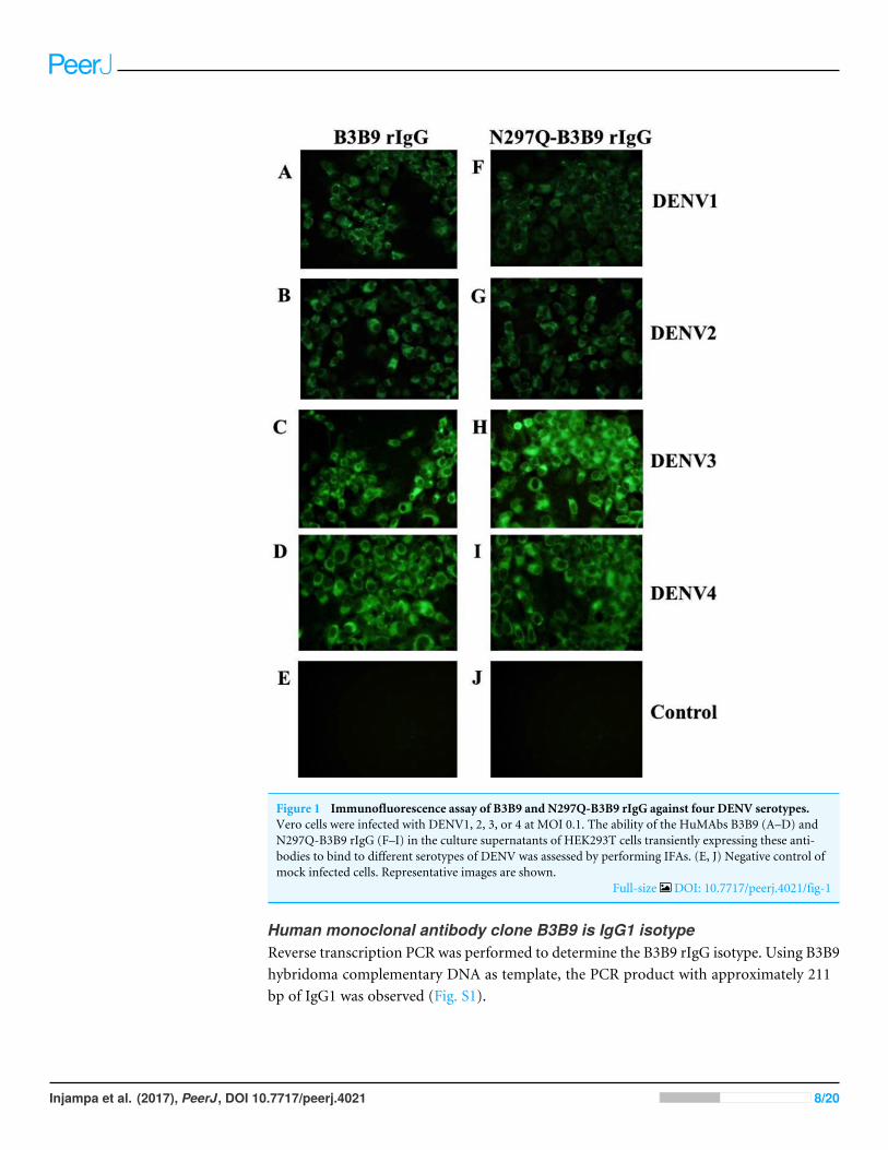
Figure 1 Immunofluorescence assay of B3B9 and N297Q-B3B9 rIgG against four DENV serotypes.Vero cells were infected with DENV1, 2, 3, or 4 at MOI 0.1. The ability of the HuMAbs B3B9 (A–D) andN297Q-B3B9 rIgG (F–I) in the culture supernatants of HEK293T cells transiently expressing these anti-bodies to bind to different serotypes of DENV was assessed by performing IFAs. (E, J) Negative control ofmock infected cells. Representative images are shown.
Full-size DOI: 10.7717/peerj.4021/fig-1
Human monoclonal antibody clone B3B9 is IgG1 isotypeReverse transcription PCR was performed to determine the B3B9 rIgG isotype. Using B3B9hybridoma complementary DNA as template, the PCR product with approximately 211bp of IgG1 was observed (Fig. S1).
Injampa et al. (2017), PeerJ, DOI 10.7717/peerj.4021 8/20
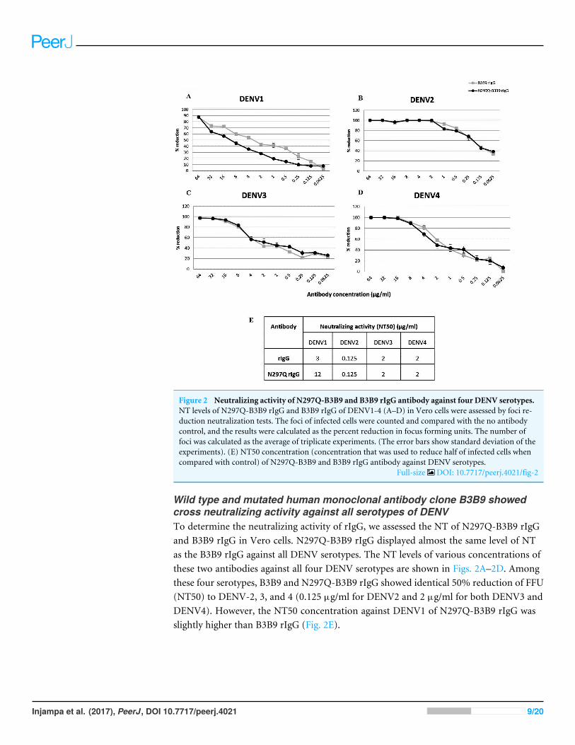
Figure 2 Neutralizing activity of N297Q-B3B9 and B3B9 rIgG antibody against four DENV serotypes.NT levels of N297Q-B3B9 rIgG and B3B9 rIgG of DENV1-4 (A–D) in Vero cells were assessed by foci re-duction neutralization tests. The foci of infected cells were counted and compared with the no antibodycontrol, and the results were calculated as the percent reduction in focus forming units. The number offoci was calculated as the average of triplicate experiments. (The error bars show standard deviation of theexperiments). (E) NT50 concentration (concentration that was used to reduce half of infected cells whencompared with control) of N297Q-B3B9 and B3B9 rIgG antibody against DENV serotypes.
Full-size DOI: 10.7717/peerj.4021/fig-2
Wild type and mutated human monoclonal antibody clone B3B9 showedcross neutralizing activity against all serotypes of DENVTo determine the neutralizing activity of rIgG, we assessed the NT of N297Q-B3B9 rIgGand B3B9 rIgG in Vero cells. N297Q-B3B9 rIgG displayed almost the same level of NTas the B3B9 rIgG against all DENV serotypes. The NT levels of various concentrations ofthese two antibodies against all four DENV serotypes are shown in Figs. 2A–2D. Amongthese four serotypes, B3B9 and N297Q-B3B9 rIgG showed identical 50% reduction of FFU(NT50) to DENV-2, 3, and 4 (0.125 µg/ml for DENV2 and 2 µg/ml for both DENV3 andDENV4). However, the NT50 concentration against DENV1 of N297Q-B3B9 rIgG wasslightly higher than B3B9 rIgG (Fig. 2E).
Injampa et al. (2017), PeerJ, DOI 10.7717/peerj.4021 9/20
Figure 3 ADE assays in K562 and THP-1 cells. (A–D) Enhancement activity against the four DENVserotypes in FcγRII-bearing K562 cells. The number of infected cells (log10) from each antibody concen-tration compared with that of the control without antibody is shown. The numbers of infected cells werederived from the average of the counted infected cells from three frames at 20×magnification multipliedby a surface area amplification factor to obtain the total number of cells in each well. Dotted lines indi-cate cut-off values for differentiating neutralizing and enhancing activity from the average plus three timesthe SD of the percentages of infected cells obtained with the four negative controls. (The error bars showstandard deviation of the repeated experiments.) (E–H) ADE assay on THP-1 cells. The antibody concen-tration was serially diluted ten-fold and are represented on the X-axis. The fold enhancement in the viruscopy number of the sample with each antibody was compared with that of the no antibody control and isrepresented on the Y -axis. The plotted values were obtained from the average of duplicates from two re-peated experiments.
Full-size DOI: 10.7717/peerj.4021/fig-3
Mutated human monoclonal antibody clone B3B9 eliminated ADE activityin K562 cells while wild type showed ADE activity against all types of DENVB3B9 rIgG was able to neutralize DENV2 and 3 at the minimum concentration, 0.39and 25 µg/ml respectively (Figs. 3B–3C), but not neutralize to DENV1 (Figs. 3A) and 4(Fig. 3D).Moreover, this antibody induced a virus infection enhancement at concentrations400–0.0015 µg/ml, 0.39–0.006 µg/ml, 100–0.0015 µg/ml, and 400–0.00038 µg/ml forDENV1, 2, 3, and 4, respectively (Figs. 3A–3D). To the contrary, N297Q-B3B9 rIgGshowed NT for all four DENV serotypes and showed no enhancing activity in any of thetested antibody concentrations.
Mutated human monoclonal antibody clone B3B9 showed no ADE activityin THP-1 cells while wild type showed ADE activity against all types ofDENVIn THP-1 cells, mutant IgG (N297Q-B3B9) that cannot bind to FcγR showed a completereduction of ADE activity in all tested antibody concentrations. In contrast, wild type rIgGinduced 9541, 43, 3061, and 2020 fold enhancement in the dengue virus RNA copy numbercompared with the control for DENV1, 2, 3, and 4, respectively (Figs. 3E–3H).
Epitope of B3B9 rIgG was within the highly conserved N-terminalfusion loop peptide of the E protein domain IIBiopanning of a phage display C7C and 12 random peptide library was performed usingthe affinity selection of purified B3B9 HuMAb. After biopanning, the number of outputphages obtained from Ph.D. C7C and Ph.D.12 increased from 6.9×10⁵ to 9.8×10⁸ PFU
Injampa et al. (2017), PeerJ, DOI 10.7717/peerj.4021 10/20

Figure 4 Epitope mapping by phage display of random peptide libraries. (A) Affinity selection ofphage-display. Ph.D.-12 and Ph.D.-C7C Phage Display Peptide Libraries were used in biopanning step.The constant units of phage (5× 10¹⁰ PFU) was used for three rounds of biopanning. Increasing percentyields of output phages represents the specific enrichment. (B) Alignment of phage-displayed peptidesequences selected by HuMAbs. Phage clones were shown to display a consensus sequences LXXXG (showin the box). (C) Comparison of the amino acid sequences of E proteins DENV1–4. In the box, DENV1–4shared the same amino acids at positions 107 (L), 108 (F), 109 (G), 110 (K), and 111 (G). (D) Phageinhibition ELISA of eight phage clones which matched to the motif of 107LXXXG111. Phage lysate ofselected clones bound with HuMAb in solution phase and the free phage clones were detected by ELISA.The absorbance values were measured at 450 nm using an ELISA reader (TECAN). Absorbance valuesof each concentration (A) were divided by absorbance values of no antibody (control) (A0), resulting innormalized values (A/A0). Anti-influenza antibody was used as negative control.
Full-size DOI: 10.7717/peerj.4021/fig-4
and 6.9×10⁵ to 7.7×10⁸ PFU respectively, which proposed that specific enrichment hadoccurred (Fig. 4A). Plasmids of phage clones that show binding activity with HuMAb byELISA were isolated for DNA sequencing. Inserted oligonucleotide sequences of phageDNAs were translated to peptide sequences. Peptide sequences were aligned using BioEditprogram 7.2.3 to analyze the epitopes and the binding motif of HuMAb. Phage clones wereshown to display a consensus sequence LXXXG (Fig. 4B). After comparing this peptidewith the amino acid sequences of E proteins of DENV1–4, it was found that this LXXXGmotif matched to 107LFGKG111 within the highly conserved N-terminal fusion looppeptide of the E protein domain II (Fig. 4C). To investigate the solution-phase binding ofeach candidate phage random peptide with target B3B9 HuMAb, eight candidate phageclones which matched to the motif of 107LXXXG111 were selected for this study (Fig. 4B,Table 1). From phage inhibition ELISA, it was found that the binding of all phage cloneswere inhibited by B3B9 HuMAb with dose-dependent manner, as shown in Fig. 4D.
Injampa et al. (2017), PeerJ, DOI 10.7717/peerj.4021 11/20

Table 1 The consensus peptide sequences of eight selected phage clones. These clones matched to themotif 107LXXXG111 of DENV genome.
Sequences No. Consensus sequences
1 LECGG2 LQSYG3 LSAYG4 LTSYG5 LGAYG6 LASYG7 LDAYG8 LERYG
N297Q-B3B9 rIgG producing from CHO-K1 stable cell lines showedneutralizing activity against DENV2 without enhancing activityFrom 125 stable clones that were screened, one showed the highest IgG secretion level(9,587 µg/ml). This clone was continually cultured to collect supernatant for purification.Preliminary study against DENV2 showed that all tested concentrations of the modifiedrIgG secreted from the stable cell line showed the same result of viral NT (Fig. 5A) withoutADE activity (Fig. 5B) as modified rIgG secreted from transient expression.
DISCUSSIONDue to the complexity of DENV infection and pathogenesis (Marasco & Sui, 2007), theideal therapeutic antibodies should be fully human-derived and capable of inhibiting allfour serotypes to reduce the risk of ADE causing more severe symptoms (Chan, Ong & Ooi,2013). We previously generated a cross-neutralizing HuMAb B3B9 (Setthapramote et al.,2012). Although this HuMAb showed strong NT to all DENV serotypes, at sub-neutralizingconcentrations, it promoted ADE activity (Sasaki et al., 2013). To overcome this problem,we generated rIgG with an engineered Fc to prohibit the binding between Fc portion andFc receptor.
The important receptor for ADE mechanism is FcγR (Fc gamma receptor) that, onceit binds with immune complex, can stimulate virus infection. The Fc region of antibodycontains asparagine residue (N297) which is a single N-linked glycosylation site in its CH2domain. The nature of this glycan can decisively influence the therapeutic performanceof a recombinant antibody, and their absence or modification can leads to changingof conformation and losing Fc effector functions (Hristodorov, Fischer & Linden, 2013;Subedi & Barb, 2015). The Fc fragment of antibody acquires the sugar moieties attachedat position N297 residues to maintain the structure of antibody. Thus, deletion of theN- glycan changes the structure of the Fc portion resulting in diminished binding to Fcγreceptor (Nimmerjahn & Ravetch, 2008).
Firstly, we evaluated the generated N297Q-B3B9 rIgG for its neutralizing activity inVero cells and found that the engineering antibody showed proper neutralizing activitycomparable to B3B9 rIgG against DENV2, -3, and -4 with some lesser degree of NT activityagainst DENV1 of N297Q-B3B9 comparing with B3B9 rIgG.
Injampa et al. (2017), PeerJ, DOI 10.7717/peerj.4021 12/20

Figure 5 The NT and ADE activity against DENV2 of N297Q-B3B9 rIgG derived from a stable andtransient CHO-K1 cell line. A line of CHO-K1 cells stably expressing N297Q-B3B9 rIgG was established.(A) The NT of N297Q-B3B9 rIgG from different expression system against DENV2. The X-axis representsthe concentration of antibodies, and the Y -axis represents the percent reduction in focus-forming unitscompared with the foci number of the control sample that lacked antibody. The number of foci was calcu-lated as the average of triplicate experiments. (B) An ADE assay of N297Q-B3B9 rIgG from different ex-pression system against DENV2 was performed in K562 cells. The infected cells were counted at each an-tibody concentration and compared with the control (DENV2-infected cells without antibody). Dottedlines indicate cut-off values for differentiating neutralizing and enhancing activity from the average plusthree times the SD of the percentages of infected cells obtained with the four negative controls. (The errorbars show standard deviation of the repeated experiments.)
Full-size DOI: 10.7717/peerj.4021/fig-5
Injampa et al. (2017), PeerJ, DOI 10.7717/peerj.4021 13/20

Since most of anti-dengue virus antibodies could showed enhancing activity at sub-neutralizing concentration (Sasaki et al., 2013; Schmidt, 2010). The study in the cell absentof FcγR may not represent the neutralizing status in vivo. We then studied the balance ofNT and ADE activity using K562 cells, which express FcγRII. In the simplified ADE assayusing K562 cells (Konishi, Tabuchi & Yamanaka, 2010), the virus infection enhancement isrepresented as the number of infected cells from each antibody concentration comparedwith those from a control performed in the absence of antibody. In this cell type, we foundthat B3B9 rIgG, which showed cross-neutralizing activity to 4 DENV serotypes in Verocell, only neutralized DENV 2 and 3, but not DENV1 and 4 (Figs. 3A–3D). In contrary,N297Q-B3B9 rIgG showed cross-neutralizing activity to 4 serotypes of DENV.
However, since K562 cell only expresses FcγRIIa, we further determined the ADEactivity in THP-1 cells that expressed both FcγRI and FcγRII. In accordance with ADEstudy in K562 cells, our N297Q-B3B9 rIgG showed clearly reduction of viral enhancementactivity in THP-1 cell. This result showed the potential of N297Q-B3B9 rIgG as a humanmonoclonal therapeutic antibody that cross neutralizes all serotypes of DENV withoutenhancing activity.
Many studies of Fc modification were explored for establishment of therapeuticantibodies candidates against dengue virus. In 1989, Tao & Morrison (1989) studied rolesof aglycosylated chimeric mouse-human IgG antibody by modifying at glycan positionsN297Q, N297H and N297K via site directed mutagenesis. These aglycosylated antibodiescannot bind to human FcγRI and not trigger C1q binding ability of complement system(Tao & Morrison, 1989). Balsitis et al. (2010) reported that a N297Q mutation of mouseand chimeric human-mouse IgG that efficiency reducing ADE in vitro could decreased themortality of DENV-infected mice (Balsitis et al., 2010).
The results of this study differ to the one carried out by Ramadhany et al. (2015), whichmodified human monoclonal antibody at N297A and showed NT activity to be the sameas its parental HuMAb. Nonetheless, this HuMAb still induced low levels of virus infectionenhancement (Ramadhany et al., 2015). One possible reason might be the type of mutatedamino acid used. Considering the amino acid structure, the substitution of Asparagine withGlutamine (conservative mutation) reduces the effects of functional properties becausethe side chains of these two amino acids differ by only one methylene group. Thus thesubstitution amino acid is one of the essential factors that affect to the efficiency of antibody.
Several studies have been approved for successful utilization of random peptide phagedisplay for finding specific epitope to several antiviral (Xue et al., 2012; Zhao et al., 2012),and anti-flavivirus monoclonal antibody (MAb) (Sun et al., 2011). This technique providesan economical and rapid approach for mapping antibody epitopes (Zhang et al., 2006;Chin et al., 2017). We mapped the epitopes of our HuMAb to LXXXG which correspondto 107LFGKG111 located in the conserved N-terminal fusion loop of envelope domainII (EDII). In accordance with our previous studies, this cross-neutralizing HuMAb B3B9also targeted to DII of envelope proteins residue 52–132, analyzed by western blot usingtruncated E protein (Sasaki et al., 2013). This is a major target epitope of human antibodiesfor NT and ADE activity (Costin et al., 2013; Deng et al., 2011). Comparing epitopes which
Injampa et al. (2017), PeerJ, DOI 10.7717/peerj.4021 14/20

targeted murine and humanMAb specific to dengue virus (Shrestha et al., 2010; Sukupolvi-Petty et al., 2010; Schieffelin et al., 2010; Beltramello et al., 2010; De Alwis et al., 2012), it wasfound that MAbs generated from mice are mostly serotype-specific that targeting DIII ofenvelope protein (Shrestha et al., 2010). However, most of the HuMAbs were targeted toDI-II of envelope proteins which is more cross-reactive (Beltramello et al., 2010; De Alwiset al., 2012).
As a promising therapeutic candidate, it was described byWilliams et al. (2013) that thechimeric N297Q MAbs targeting fusion loop and dimer interface on EDII, but not theA strand and C-C’ loop on EDIII, acted therapeutically by competing against enhancingantibodies in polyvalent serum that recognize the same or proximal epitopes. This kindof fusion loop specific N297Q MAb showed protective activity in vivo when administeredwith enhancing titer of polyserum (Williams et al., 2013). Thus, the identification of B cellepitope in this study is crucial to understand its function and antibody/epitope interaction.
For antibody production, in this study, we used HEK293T cell for transient expressionmainly because of its higher transfection efficiency and expression level, resulting inlower development costs when compare to stable cell line development (Zhang & Shen,2012). However, for further characterization of N297Q-B3B9 rIgG as a dengue therapeuticcandidate large quantities of antibody, with high stability in both production yield andquality, were required. Therefore, stable expression was used as an alternative solution forantibody production. CHO-K1 cells were used for stable cell line generation. This cell line isthe prominent system for bio-manufacturing of therapeutic products (Hossler, Khattak &Li, 2009) as 70%of the therapeutic protein was produced by this system (Croset et al., 2012).Characterization of this antibody in the other aspects such as stability, pharmacokineticsor phamacodynamics of antibody (Liu, 2015) was required because protein produced byHEK293T cells and CHO-K1 cells show different patterns of glycosylation. However, wefound no differentiation of NT and ADE activity in both cell types (Fig. 5).
Together, our results suggest that N297Q-B3B9 rIgG is a human monoclonal antibodythat can neutralize all four serotypes of DENV without viral enhancing activity. As afully human-derived monoclonal antibody, it avoids the problem of a human anti-mouseantibody response.
Interaction of Fc-FcγR on innate immune cells trigger immune effector functionssuch as antibody dependent cell-mediated cytotoxicity (ADCC), complement-dependentcytotoxicity (CDC) (Kellner et al., 2014), and antibody dependent cellular phagocytosis(ADCP) (Grevys et al., 2015). After thoroughly characterizing these functions, this B3B9rIgG can be a model testing for dengue therapeutic treatment.
ACKNOWLEDGEMENTSThe authors would like to thank Dr. Atsushi Yamanaka for his continuous advisement andencouragement.
Injampa et al. (2017), PeerJ, DOI 10.7717/peerj.4021 15/20

ADDITIONAL INFORMATION AND DECLARATIONS
FundingThis work was supported by the Faculty of Tropical Medicine, Mahidol University grantnumber 04/2556 and the National Research Council of Thailand grant number 32/2558.The Deutscher Akademischer Austauschdienst (DAAD, German Academic ExchangeService) fund for the student scholar grant number 577177105. There was no additionalexternal funding received for this study.
Grant DisclosuresThe following grant information was disclosed by the authors:Faculty of Tropical Medicine, Mahidol University: 04/2556.National Research Council of Thailand: 32/2558.The Deutscher Akademischer Austauschdienst: 577177105.
Competing InterestsThe authors declare there are no competing interests.
Author Contributions• Subenya Injampa conceived and designed the experiments, performed the experiments,analyzed the data, wrote the paper, prepared figures and/or tables.• Nataya Muenngern performed the experiments, analyzed the data, wrote the paper,prepared figures and/or tables.• Chonlatip Pipattanaboon conceived and designed the experiments, performed theexperiments.• Surachet Benjathummarak, Khwanchit Boonha and Hathairad Hananantachaiperformed the experiments.• Waranya Wongwit reviewed drafts of the paper.• Pongrama Ramasoota conceived and designed the experiments, contributedreagents/materials/analysis tools, reviewed drafts of the paper.• Pannamthip Pitaksajjakul conceived and designed the experiments, performed theexperiments, analyzed the data, contributed reagents/materials/analysis tools, wrote thepaper, reviewed drafts of the paper.
Data AvailabilityThe following information was supplied regarding data availability:
The raw data used to generate statistical analysis have been uploaded as SupplementalFiles.
Supplemental InformationSupplemental information for this article can be found online at http://dx.doi.org/10.7717/peerj.4021#supplemental-information.
Injampa et al. (2017), PeerJ, DOI 10.7717/peerj.4021 16/20

REFERENCESBalsitis SJ, Williams KL, Lachica R, Flores D, Kyle JL, Mehlhop E, Johnson S, Dia-
mondMS, Beatty PR, Harris E. 2010. Lethal antibody enhancement of denguedisease in mice is prevented by Fc modification. PLOS Pathogens 6:e1000790DOI 10.1371/journal.ppat.1000790.
Beltramello M,Williams KL, Simmons CP, Macagno A, Simonelli L, Quyen NT,Sukupolvi-Petty S, Navarro-Sanchez E, Young PR, De Silva AM, Rey FA, VaraniL, Whitehead SS, DiamondMS, Harris E, Lanzavecchia A, Sallusto F. 2010. Thehuman immune response to Dengue virus is dominated by highly cross-reactiveantibodies endowed with neutralizing and enhancing activity. Cell Host & Microbe8:271–283 DOI 10.1016/j.chom.2010.08.007.
Chan AC, Carter PJ. 2010. Therapeutic antibodies for autoimmunity and inflammation.Nature Reviews Immunology 10(5):301–316 DOI 10.1038/nri2761.
Chan KR, Ong EZ, Ooi EE. 2013. Therapeutic antibodies as a treatment option fordengue fever. Expert Review of Anti-Infective Therapy 11:1147–1157DOI 10.1586/14787210.2013.839941.
Chin CF, Lai JY, Choong YS, Anthony AA, Ismali A, Lim TS. 2017. Delineation of B-cell epitopes of Salmonella enterica serovar Typhi Haelysin E: potential antibodytherapeutic target. Scientific Report 7:2176 DOI 10.1038/s41598-017-01987-8.
Costin JM, Zaitseva E, Kahle KM, Nicholson CO, Rowe DK, Graham AS, Bazzone LE,Hogancamp G, Figueroa Sierra M, Fong RH, Yang ST, Lin L, Robinson JE, DoranzBJ, Chernomordik LV, Michael SF, Schieffelin JS, Isern S. 2013.Mechanisticstudy of broadly neutralizing human monoclonal antibodies. Journal of Virology87(1):52–66 DOI 10.1128/JVI.02273-12.
Croset A, Delafosse L, Gaudry JP, Arod C, Glez L, Losberger C, Begue D, Krstanovic A,Robert F, Vilbois F, Chevalet L, Antonsson B. 2012. Differences in the glycosylationof recombinant proteins expressed in HEK and CHO cells. Journal of Biotechnology161:336–348 DOI 10.1016/j.jbiotec.2012.06.038.
De Alwis R, Smith SA, Olivarez NP, MesserWB, Huynh JP,WahalaWM,WhiteLJ, DiamondMS, Baric RS, Crowe Jr JE, De Silva AM. 2012. Identification ofhuman neutralizing antibodies that bind to complex epitopes on dengue virions.Proceedings of the National Academy of Sciences of the United States of America109(19):7439–7444 DOI 10.1073/pnas.1200566109.
Deng YQ, Dai JX, Ji GH, Jiang T,Wang HJ, Yang HO. 2011. A broadly flavivirus cross-neutralizing monoclonal antibody that recognizes a novel epitope within the fusionloop of E protein. PLOS ONE 6(1):e16059 DOI 10.1371/journal.pone.0016059.
Grevys A, BernM, Foss S, Bratlie DB, Moen A, Gunnarsen KS, Aase A, Michaelsen TE,Sandlie I, Andersen JT. 2015. Fc engineering of human IgG1 for altered bindingto the neonatal Fc receptor affects Fc effector functions. Journal of Immunology194(11):5497–5508 DOI 10.4049/jimmunol.1401218.
Gupta R, Jung E, Brunak S. 2004. Prediction of N-glycosylation sites in human proteins.Available at http://www.cbs.dtu.dk/ services/NetNGlyc/ .
Injampa et al. (2017), PeerJ, DOI 10.7717/peerj.4021 17/20

GuzmanMG, Vazquez S. 2010. The complexity of antibody-dependent enhancement ofdengue virus infection. Viruses 2(12):2649–2662 DOI 10.3390/v2122649.
Halstead SB, O’Rourke EJ. 1977. Antibody-enhanced dengue virus infection in primateleukocytes. Nature 265:739–741 DOI 10.1038/265739a0.
Hossler P, Khattak SF, Li ZJ. 2009. Optimal and consistent protein glycosylation inmammalian cell culture. Glycobiology 19(9):936–949 DOI 10.1093/glycob/cwp079.
Hristodorov D, Fischer R, Linden L. 2013.With or without sugar? (A)glycosylation oftherapeutic antibodies.Molecular Biotechnology 54(3):1056–1068DOI 10.1007/s12033-012-9612-x.
Kellner C, Derer S, Valerius T, PeippM. 2014. Boosting ADCC and CDC activity by Fcengineering and evaluation of antibody effector functions.Methods 65(1):105–113DOI 10.1016/j.ymeth.2013.06.036.
Konishi E, Tabuchi Y, Yamanaka A. 2010. A simple assay system for infection-enhancing and neutralizing antibodies to dengue type 2 virus using layersof semi-adherent K562 cells. Journal of Virological Methods 163:360–367DOI 10.1016/j.jviromet.2009.10.026.
Kuhn RJ, ZhangW, RossmannMG, Pletnev SV, Corver J, Lenches E, Jones CT,Mukhopadhyay S, Chipman PR, Strauss EG, Baker TS, Strauss JH. 2002. Structureof dengue virus: implications for flavivirus organization, maturation, and fusion. Cell108(5):717–725 DOI 10.1016/S0092-8674(02)00660-8.
Liu L. 2015. Antibody glycosylation and its impact on the pharmacokinetics andpharmacodynamics of monoclonal antibodies and Fc-fusion proteins. Journal ofPharmaceutical Sciences 104(6):1866–1884 DOI 10.1002/jps.24444.
Low JG, Ooi EE, Vasudevan SG. 2017. Current status of dengue therapeutics re-search and development. Journal of Infectious Diseases 215(suppl_2):S96–S102DOI 10.1093/infdis/jiw423.
MarascoWA, Sui J. 2007. The growth and potential of human antiviral monoclonalantibody therapeutics. Nature Biotechnology 25:1421–1434 DOI 10.1038/nbt1363.
Murphy BR,Whitehead SS. 2011. Immune response to dengue virus and prospects for avaccine. Annual Review of Immunology 29:587–619DOI 10.1146/annurev-immunol-031210-101315.
Natasha Evelyn AM,Mikkel BQ, AnneliesWS. 2013. Epidemiology of dengue: past,present and future prospects. Clinical Epidemiology 5:299–309DOI 10.2147/CLEP.S34440.
Nimmerjahn F, Ravetch JV. 2008. Fc gamma receptors as regulators of immuneresponses. Nature Reviews Immunology 8(1):34–47 DOI 10.1038/nri2206.
OmokokoMD, Pambudi S, Phanthanawiboon S, Masrinoul P, Setthapramote C, SasakiT, KuharaM, Ramasoota P, Yamashita A, Hirai I, Ikuta K, Kurosu T. 2014. Ahighly conserved region between amino acids 221 and 266 of dengue virus non-structural protein 1 is a major epitope region in infected patients. American Journalof Tropical Medicine and Hygiene 91:146–155 DOI 10.4269/ajtmh.13-0624.
Pitaksajjakul P, Benjathummarak S, Pipattanaboon C,WongwitW, OkabayashiT, KuharaM,Misaki R, Fujiyama K, Ramasoota P. 2014. Antibody germline
Injampa et al. (2017), PeerJ, DOI 10.7717/peerj.4021 18/20

characterization of cross-neutralizing human IgGs against 4 serotypes ofdengue virus. Biochemical Biophysical Research Communication 446:475–480DOI 10.1016/j.bbrc.2014.02.131.
Ramadhany R, Hirai I, Sasaki T, Ono K, Ramasoota P, Ikuta K, Kurosu T. 2015.Antibody with an engineered Fc region as a therapeutic agent against dengue virusinfection. Antiviral Research 124:61–68 DOI 10.1016/j.antiviral.2015.10.012.
RowleyMJ, O’Connor K,Wijeyewickrema L. 2004. Phage display for epitope determi-nation: a paradigm for identifying receptor-ligand interactions. Biotechnology AnnualReview 10:151–188 DOI 10.1016/S1387-2656(04)10006-9.
Sasaki T, Setthapramote C, Kurosu T, NishimuraM, Asai A, OmokokoMD, Pipat-tanaboon C, Pitaksajjakul P, Limkittikul K, Subchareon A, Chaichana P, Ok-abayashi T, Hirai I, Leaungwutiwong P, Misaki R, Fujiyama K, Ono K, Okuno Y,Ramasoota P, Ikuta K. 2013. Dengue virus neutralization and antibody-dependentenhancement activities of human monoclonal antibodies derived from denguepatients at acute phase of secondary infection. Antiviral Research 98:423–431DOI 10.1016/j.antiviral.2013.03.018.
Schieffelin JS, Costin JM, Nicholson CO, Orgeron NM, Fontaine KA, Isern S, MichaelSF, Robinson JE. 2010. Neutralizing and non-neutralizing monoclonal antibodiesagainst dengue virus E protein derived from a naturally infected patients. VirologyJournal 7:28 DOI 10.1186/1743-422X-7-28.
Schmidt AC. 2010. Response to dengue fever—the good, the bad, and the ugly? NewEngland Journal of Medicin 363:484–487 DOI 10.1056/NEJMcibr1005904.
Setthapramote C, Sasaki T, PuipromO, Limkittikul K, Pitaksajjakul P, PipattanaboonC, SasayamaM, Leungwutiwong P, PhumratanaprapinW, Chamnachanan S,Kusolsuk T, Jittmittraphap A, Asai A, Arias JF, Hirai I, KuharaM, Okuno Y,Kurosu T, Ramasoota P, Ikuta K. 2012.Human monoclonal antibodies to neutralizeall dengue virus serotypes using lymphocytes from patients at acute phase of thesecondary infection. Biochemical Biophysical Research Communication 423:867–872DOI 10.1016/j.bbrc.2012.06.057.
Shrestha B, Brien JD, Sukupolvi-Petty S, Austin SK, EdelingMA, Kim T, O’Brien KM,Nelson CA, Johnson S, Fremont DH, DiamondMS. 2010. The development oftherapeutic antibodies that neutralize homologous and heterologous genotypes ofdengue virus type 1. PLOS Pathogens 6:e1000823 DOI 10.1371/journal.ppat.1000823.
Skottrup PD, Sørensen G, KsiazekM, Potempa J, Riise E. 2012. A phage displayselected 7-mer peptide inhibitor of the Tannerella forsythia metalloprotease-likeenzyme Karilysin can be truncated to Ser-Trp-Phe-Pro. PLOS ONE 7(10):e48537DOI 10.1371/journal.pone.0048537.
Subedi GP, Barb AW. 2015. The structural role of antibody N-glycosylation in receptorinteractions. Structure 23:1573–1583 DOI 10.1016/j.str.2015.06.015.
Sukupolvi-Petty S, Austin SK, Engle M, Brien JD, Dowd KA,Williams KL, Johnson S,Rico-Hesse R, Harris E, Pierson TC, Fremont DH, DiamondMS. 2010. Structureand function analysis of therapeutic monoclonal antibodies against dengue virus type2. Journal of Virology 84(18):9227–9239 DOI 10.1128/JVI.01087-10.
Injampa et al. (2017), PeerJ, DOI 10.7717/peerj.4021 19/20

Sun EC, Ma JN, Liu NH, Yang T, Zhao J, Geng HW,Wang LF, Qin YL, Bu ZG, Yang YH,Lunt RA,Wang LF,WuDL. 2011. Identification of two linear B-cell epitopes fromWest Nile virus NS1 by screening a phage-displayed random peptide library. BMCMicrobiology 11:160 DOI 10.1186/1471-2180-11-160.
TaoMH,Morrison SL. 1989. Studies of aglycosylated chimeric mouse-human IgG. Roleof carbohydrate in the structure and effector functions mediated by the human iggconstant region. Journal of Immunology 143:1595–1601.
Williams KL, Sukupolvi-Petty S, Beltramello M, Johnson S, Sallusto F, Lanzavecchia A.2013. Therapeutic efficacy of antibodies lacking Fcγ receptor binding against lethaldengue virus infection is due to neutralizing potency and blocking of enhancingantibodies. PLOS Pathogens 9(2):e1003157 DOI 10.1371/journal.ppat.1003157.
XueM, Shi X, Zhang J, Zhao Y, Cui H, Hu S, Gao H, Cui X,Wang YF. 2012. Identifi-cation of a conserved B-cell epitope on reticuloendotheliosis virus envelope proteinby screening a phage-displayed random peptide library. PLOS ONE 7(11):e49842DOI 10.1371/journal.pone.0049842.
Zhang F, YuM, Zhang N,Wang LF. 2006. Characterization of epitopes form neu-tralizing monoclonal antibodies to classical swine fever virus E2 and Erns us-ing phage-displayed random peptide library. Archives of Virology 151:37–54DOI 10.1007/s00705-005-0623-9.
Zhang RY, ShenWD. 2012.Monoclonal antibody expression in mammalian cells.Methods in Molecular Biology 907:341–358 DOI 10.1007/978-1-61779-974-7_20.
Zhao J, Sun EC, Liu NH, Yang T, Xu QY, Qin YL, Yang YH,WuDL. 2012. Phagedisplay identifies an eastern equine encephalitis virus glycoprotein E2-specificB cell epitope. Veterinary Immunology and Immunopathology 148:364–368DOI 10.1016/j.vetimm.2012.06.021.
Injampa et al. (2017), PeerJ, DOI 10.7717/peerj.4021 20/20