Embed Size (px)
Citation preview
T h e n e w e ngl a nd j o u r na l o f m e dic i n e
n engl j med 363;27 nejm.org december 30, 20102638
review article
Mechanisms of DiseaseRobert S. Schwartz, M.D., Editor
General Anesthesia, Sleep, and ComaEmery N. Brown, M.D., Ph.D., Ralph Lydic, Ph.D., and Nicholas D. Schiff, M.D.
From the Department of Anesthesia, Critical Care, and Pain Medicine, Massa-chusetts General Hospital and Harvard Medical School, Boston (E.N.B.); the MIT–Harvard Division of Health Scienc-es and Technology, Department of Brain and Cognitive Sciences, Massachusetts Institute of Technology, Cambridge, MA (E.N.B.); the Department of Anesthesiol-ogy, University of Michigan, Ann Arbor (R.L.); and the Department of Neurology and Neuroscience, Weill Cornell Medical College, New York (N.D.S.). Address re-print requests to Dr. Brown at the De-partment of Anesthesia, Critical Care, and Pain Medicine, Massachusetts General Hospital, 55 Fruit St., GRJ 4, Boston, MA 02114, or at [email protected].
N Engl J Med 2010;363:2638-50.Copyright © 2010 Massachusetts Medical Society.
In the United States, nearly 60,000 patients per day receive general anesthesia for surgery.1 General anesthesia is a drug-induced, reversible condi-tion that includes specific behavioral and physiological traits — unconscious-
ness, amnesia, analgesia, and akinesia — with concomitant stability of the auto-nomic, cardiovascular, respiratory, and thermoregulatory systems.2 General anesthesia produces distinct patterns on the electroencephalogram (EEG), the most common of which is a progressive increase in low-frequency, high-amplitude activity as the level of general anesthesia deepens3,4 (Fig. 1). How anesthetic drugs induce and maintain the behavioral states of general anesthesia is an important question in medicine and neuroscience.6 Substantial insights can be gained by considering the relationship of general anesthesia to sleep and to coma.
Humans spend approximately one third of their lives asleep. Sleep, a state of decreased arousal that is actively generated by nuclei in the hypothalamus, brain stem, and basal forebrain, is crucial for the maintenance of health.7,8 Normal hu-man sleep cycles between two states — rapid-eye-movement (REM) sleep and non-REM sleep — at approximately 90-minute intervals. REM sleep is characterized by rapid eye movements, dreaming, irregularities of respiration and heart rate, penile and clitoral erection, and airway and skeletal-muscle hypotonia.7 In REM sleep, the EEG shows active high-frequency, low-amplitude rhythms (Fig. 1). Non-REM sleep has three distinct EEG stages, with higher-amplitude, lower-frequency rhythms ac-companied by waxing and waning muscle tone, decreased body temperature, and decreased heart rate.
Coma is a state of profound unresponsiveness, usually the result of a severe brain injury.9 Comatose patients typically lie with eyes closed and cannot be roused to respond appropriately to vigorous stimulation. A comatose patient may grimace, move limbs, and have stereotypical withdrawal responses to painful stimuli yet make no localizing responses or discrete defensive movements. As the coma deepens, the patient’s responsiveness even to painful stimuli may diminish or disappear. Although the patterns of EEG activity observed in comatose patients depend on the extent of the brain injury, they frequently resemble the high–ampli-tude, low-frequency activity seen in patients under general anesthesia10 (Fig. 1). General anesthesia is, in fact, a reversible drug-induced coma. Nevertheless, anes-thesiologists refer to it as “sleep” to avoid disquieting patients. Unfortunately, anesthesiologists also use the word “sleep” in technical descriptions to refer to unconsciousness induced by anesthetic drugs.11 (For a glossary of terms com-monly used in the field of anesthesiology, see the Supplementary Appendix, avail-able with the full text of this article at NEJM.org.)
This review discusses the clinical and neurophysiological features of general anesthesia and their relationships to sleep and coma, focusing on the neural mech-anisms of unconsciousness induced by selected intravenous anesthetic drugs.
The New England Journal of Medicine Downloaded from nejm.org by LUIS HIGGINS GUERRA on December 30, 2010. For personal use only. No other uses without permission.
Copyright © 2010 Massachusetts Medical Society. All rights reserved.

mechanisms of disease
n engl j med 363;27 nejm.org december 30, 2010 2639
Clinic a l Signs a nd EEG Pat ter ns of Unconsciousness Induced
by Gener a l A nes thesi a
The clinical signs and EEG patterns of general anesthesia–induced unconsciousness can be de-scribed in relation to the three periods in which they appear: induction, maintenance, and emer-gence.
Induction Period
Before induction, the patient has a normal, active EEG, with prominent alpha activity (10 Hz) when the eyes are closed (Fig. 1). Administration of a small dose of a hypnotic drug such as propofol, a barbiturate, or etomidate, all of which act on
γ-aminobutyric acid type A (GABAA) receptors, induces a state of sedation in which the patient is calm and easily arousable, with the eyes generally closed.12 As the dose is slowly increased, the pa-tient may enter a state of paradoxical excitation,13 characterized by purposeless or defensive move-ments, incoherent speech, euphoria or dysphoria, and an increase in beta activity on the EEG (13 to 25 Hz).3,4,13-16 This state is termed paradoxical because the drug that is intended to induce un-consciousness induces excitation instead.
As more of the hypnotic agent is administered — typically as a bolus over a period of 10 to 15 seconds — an increasingly irregular respiratory pattern develops that progresses to apnea, at which point bag-mask ventilation must be initi-
Figure 1. Electroencephalographic (EEG) Patterns during the Awake State, General Anesthesia, and Sleep.
Panel A shows the EEG patterns when the patient is awake, with eyes open (left) and the alpha rhythm (10 Hz) with eyes closed (right). Panel B shows the EEG patterns during the states of general anesthesia: paradoxical excitation, phases 1 and 2, burst suppression, and the isoelectric tracing. Panel C shows the EEG patterns during the stages of sleep: rapid-eye-movement (REM) sleep; stage 1 non-REM sleep; stage 2 non-REM sleep, and stage 3 non-REM (slow-wave) sleep. The EEG patterns during recovery from coma — coma, vegeta-tive state, and minimally conscious state — resemble the patterns during general anesthesia, sleep, and the awake state. EEG tracings during sleep are from Watson et al.5
The New England Journal of Medicine Downloaded from nejm.org by LUIS HIGGINS GUERRA on December 30, 2010. For personal use only. No other uses without permission.
Copyright © 2010 Massachusetts Medical Society. All rights reserved.

T h e n e w e ngl a nd j o u r na l o f m e dic i n e
n engl j med 363;27 nejm.org december 30, 20102640
ated to support breathing. There is a concomitant loss of response to oral commands and skeletal-muscle tone. Loss of consciousness can be easily assessed by having patients follow the movement of the anesthesiologist’s finger with their eyes. As unconsciousness ensues, eye tracking stops, nystagmus may appear, and blinking increases. The oculocephalic, eyelash, and corneal reflexes are lost,17 yet the pupillary light reflex remains.18 There can be either an increase or a decrease in blood pressure, whereas the heart rate typically increases. Administering an opioid or a benzo-diazepine before or during induction may miti-gate the increased heart-rate response, and vaso-pressors may be given to maintain blood pressure. Tracheal intubation is usually performed at the end of induction, after the administration of a muscle relaxant.
Maintenance Period
General anesthesia is maintained by a combination of hypnotic agents, inhalational agents, opioids, muscle relaxants, sedatives, and cardiovascular drugs, along with ventilatory and thermoregula-tory support. During the maintenance period, changes in the heart rate and blood pressure are among the clinical signs used to monitor the level of general anesthesia (see the Supplemen-tary Appendix). When the state of general anes-thesia is inadequate for the level of nociceptive stimulation from surgery, the heart rate and blood pressure can increase dramatically, alerting the anesthesiologist to the possibility of increased nociception and arousal. Other indicators of in-adequate general anesthesia are perspiration, tearing, changes in pupil size, the return of mus-cle tone, movement,19 and changes in EEG mea-sures of brain activity.20 At levels appropriate for surgery, general anesthesia can functionally ap-proximate brain-stem death,21 because patients are unconscious, have depressed brain-stem reflex-es, do not respond to nociceptive stimuli, have no apneic drive, and require cardiorespiratory and thermoregulatory support.9
Four EEG patterns define the phases of the maintenance period (Fig. 1). Phase 1, a light state of general anesthesia, is characterized by a de-crease in EEG beta activity (13 to 30 Hz) and an increase in EEG alpha activity (8 to 12 Hz) and delta activity (0 to 4 Hz).22 During phase 2, the intermediate state, beta activity decreases and al-pha and delta activity increases, with so-called anteriorization — that is, an increase in alpha and
delta activity in the anterior EEG leads relative to the posterior leads.22,23 The EEG in phase 2 re-sembles that seen in stage 3, non-REM (or slow-wave) sleep. Phase 3 is a deeper state, in which the EEG is characterized by f lat periods inter-spersed with periods of alpha and beta activity — a pattern called burst suppression.15 As this state of general anesthesia deepens, the time be-tween the periods of alpha activity lengthens, and the amplitudes of the alpha and beta activity de-crease. Surgery is usually performed during phases 2 and 3. In phase 4, the most profound state of general anesthesia, the EEG is isoelectric (com-pletely flat). An isoelectric EEG may be purposely induced by the administration of a barbiturate or propofol to protect the brain during neurosur-gery24 or to stop generalized seizures.25,26
Emergence Period
Emergence from general anesthesia is a passive process that depends on the amounts of drugs administered; their sites of action, potency, and pharmacokinetics; the patient’s physiological char-acteristics; and the type and duration of the sur-gery. Recovery from general anesthesia is gener-ally assessed by monitoring physiological and behavioral signs. The return of spontaneous res-pirations is typically one of the first clinical signs observed once peripheral neuromuscular block-ade is decreased. This marks the patient’s return from a functional state that approximates brain-stem death (Table 1). The heart rate and blood pressure typically increase, provided that these responses are not pharmacologically blocked. Sal-ivation and tearing begin, followed by nonlocal-izing responses to painful stimulation, suggest-ing that the patient’s state is most similar to a vegetative state with the notable exception that the eyes remain closed. As skeletal-muscle tone returns, the patient begins to grimace, swallow, gag, and cough and make defensive movements, such as reaching for the endotracheal or naso-gastric tube. At this point the anesthesiologist will perform extubation, provided that there is suffi-cient return of brain-stem reflexes to maintain spontaneous respirations and airway protection, even if there is no response to oral commands. The eyes may still not open spontaneously. As the patient emerges from general anesthesia, the EEG patterns proceed in approximately reverse order from phases 2 or 3 of the maintenance period to an active EEG that is consistent with a fully awake state (Fig. 1). Between extubation and dis-
The New England Journal of Medicine Downloaded from nejm.org by LUIS HIGGINS GUERRA on December 30, 2010. For personal use only. No other uses without permission.
Copyright © 2010 Massachusetts Medical Society. All rights reserved.

mechanisms of disease
n engl j med 363;27 nejm.org december 30, 2010 2641
charge from the postanesthesia care unit, the pa-tient passes through a minimally conscious state.27 Functional responses that are beyond a minimally conscious state must be evident before the pa-tient is discharged from the postanesthesia care unit.27,28 The patient should be able to answer simple questions and to convey any discomfort, such as pain or nausea.
Mechanisms of Unconsciousness Induced by Gener al Anesthesia
Cortical Circuits and Altered Arousal
Observations from clinical practice and basic sci-ence indicate that anesthetic drugs induce uncon-sciousness by altering neurotransmission at mul-tiple sites in the cerebral cortex,29-35 brain stem, and thalamus. If a procedure does not require full general anesthesia, it is standard clinical practice to use low doses of a hypnotic or sedative drug to achieve sedation, which is defined as diminished cognitive function (cortical activity)35 with intact respiratory and cardiovascular (brain-stem) func-tion. Substantial decreases in neural activity in the cortex have been observed in a rodent model of general anesthesia.30 Similarly, positron-emission tomographic studies in humans under general an-esthesia revealed appreciable decreases in corti-cal metabolic activity.31,32 Functional magnetic resonance imaging33 and local-field-potential re-cordings34 in humans have provided additional evidence of cortical mechanisms of unconscious-ness induced by general anesthesia. In vivo and in vitro molecular pharmacologic studies have iden-tified GABAA and N-methyl-D-aspartate (NMDA) receptors in the cortex, thalamus, brain stem, and striatum as two of the important targets of hypnotic drugs.36,37 Because small numbers of inhibitory interneurons control large numbers of excitatory pyramidal neurons, the enhanced GABAA inhibition induced by general anesthesia can ef-ficiently inactivate large regions of the brain and contribute to unconsciousness (Fig. 2A).38,39
The Brain Stem, Sleep, and Altered Arousal
A hypnotic drug administered as a bolus during induction of general anesthesia rapidly reaches brain-stem arousal centers, where it contributes to unconsciousness.40 The clinical signs cited above are consistent with actions in the brain stem. Losses of the oculocephalic and corneal reflexes are nonspecific indicators of impaired brain-stem function due to the actions of the hypnotic agent
on the oculomotor, trochlear, abducens, trigemi-nal, and facial nuclei in the midbrain and pons.9
In a study in rodents, direct injection of a bar-biturate into the mesopontine tegmental area led to unconsciousness.41 Such observations confirm brain-injury studies showing that brain-stem un-consciousness typically involves the lateral dorsal tegmental areas of the pons and the midbrain paramedian region.42 Apnea can be explained, in part, by the actions of the hypnotic agent on GABAA interneurons in the respiratory control network in the ventral medulla and pons.43
The rapid atonia that occurs after bolus ad-ministration of propofol is most likely due to the actions of this drug in the spinal cord44 and in the pontine and medullary reticular nuclei that control the antigravity muscles.45 Certain obser-vations are consistent with this concept of pro-pofol’s mechanism of action — for example, the atonia that follows the inadvertent injection of local anesthetics into the subarachnoid space or basilar artery during an interscalene block46,47 or the inadvertent injection of alcohol into the cer-vical spinal cord during a facet block.9 Further-more, observations in patients who have pontine strokes and locked-in syndromes support this con-cept,9 as do observations with the muscle-relax-ing and soporific effects of the GABA agonist baclofen.48 Such observations may explain why near the end of surgery, small doses of propofol can be used to provide rapid muscle relaxation of short duration and why, in this case, propofol is preferable to muscle relaxants that have a slower onset and longer duration of action. In contrast to the drug-induced atonia described above, rigid-ity and spasticity are typically seen in patients who are in a coma or a vegetative state,9 and muscle tone is preserved during slow-wave sleep.7
Signs of the loss of brain-stem function (apnea, atonia, and losses of the oculocephalic and cor-neal49 reflexes) and hence unconsciousness reli-ably indicate when to initiate bag-mask ventila-tion or to place a laryngeal mask airway during induction of general anesthesia.
Neural circuits involved in sleep provide ad-ditional insights into basal forebrain, brain-stem, and hypothalamic mechanisms of unconscious-ness. During the awake state, the locus ceruleus provides norepinephrine-mediated inhibition of the ventrolateral preoptic nucleus in the hypo-thalamus.50,51 Therefore, GABAA-mediated and galanin-mediated inhibition of the ascending arousal circuits by the ventrolateral preoptic nu-
The New England Journal of Medicine Downloaded from nejm.org by LUIS HIGGINS GUERRA on December 30, 2010. For personal use only. No other uses without permission.
Copyright © 2010 Massachusetts Medical Society. All rights reserved.

T h e n e w e ngl a nd j o u r na l o f m e dic i n e
n engl j med 363;27 nejm.org december 30, 20102642
cleus is inhibited and the awake state is pro-moted.51 Adenosine, one of the brain’s princi-pal somnogens, accumulates from degradation of adenosine triphosphate during prolonged inter-vals of wakefulness.8 Binding of adenosine in the ventrolateral preoptic nucleus is associated with increased activity in this brain center.52 Adeno-sine binding and inhibition of the locus ceruleus lead to activation of the ventrolateral preoptic nucleus, which inhibits the ascending arousal cir-cuits and promotes non-REM sleep. The sedative dexmedetomidine is an α2-adrenergic agonist that inhibits the release of norepinephrine from the locus ceruleus and thus allows the ventrolateral preoptic nucleus to reduce arousal by inhibiting the ascending arousal circuits (Fig. 2B).53,54 The EEG patterns of dexmedetomidine-induced se-dation closely resemble those of non-REM sleep.55
Propofol also promotes unconsciousness, in part, by GABAA-mediated inhibition of release of the arousal-promoting neurotransmitter histamine in the cortex from the tuberomammillary nucleus in the hypothalamus.50
The active EEG patterns observed during REM sleep are due in part to strong cholinergic inputs from the lateral dorsal tegmental and pedunculo-pontine tegmental nuclei to the medial pontine reticular formation and thalamus and from the basal forebrain to the cortex.56 The synthetic opioid fentanyl decreases arousal by reducing acetylcholine in the medial pontine reticular formation, whereas morphine decreases arousal by inhibiting the neurons in the lateral dorsal teg-mental nucleus, medial pontine reticular forma-tion,56 and basal forebrain57 (Fig. 2C). Opioids further contribute to unconsciousness by bind-
Table 1. Emergence from General Anesthesia and Stages of Recovery from Coma.*
Emergence from General Anesthesia Recovery from Coma
General anesthesiaStable administration of anesthetic drugsArousal not possible, unresponsive; eyes closed, with reactive pupilsAnalgesia, akinesiaDrug-controlled blood pressure and heart rateMechanically controlled ventilationEEG patterns ranging from delta and alpha activity to burst
suppression
Brain-stem deathNo respiratory response to apneic oxygenation testTotal loss of brain-stem reflexesIsoelectric EEG patternComaStructural brain damage to both cerebral hemispheres, with or without
injuries to tegmental midbrain, rostral pons, or bothIsolated bilateral injuries to midline tegmental midbrain, rostral pons, or bothArousal not possible, unresponsiveFunctionally intact brain stem, normal arterial blood gasesEEG pattern of low-amplitude delta activity and intermittent bursts
of theta and alpha activity or possibly burst suppression
Emergence, phase 1Cessation of anesthetic drugsReversal of peripheral-muscle relaxation (akinesis)Transition from apnea to irregular breathing to regular breathingIncreased alpha and beta activity on EEG
Emergence, phase 2Increased heart rate and blood pressureReturn of autonomic responsivenessResponsiveness to painful stimulationSalivation (7th and 9th cranial nerve nuclei)Tearing (7th cranial nerve nuclei)Grimacing (5th and 7th cranial nerve nuclei)Swallowing, gagging, coughing (9th and 10th cranial nerve nuclei)Return of muscle tone (spinal cord, reticulospinal tract, basal ganglia,
and primary motor tracts)Defensive posturingFurther increase in alpha and beta activity on EEGExtubation possible
Vegetative stateSpontaneous cycling of eye opening and closingGrimacing and nonpurposeful movementsEEG pattern of high-amplitude delta and theta activityAbsence of EEG features of sleepUsually able to ventilate without mechanical support
Emergence, phase 3Eye openingResponses to some oral commandsAwake patterns on EEGExtubation possible
Minimally conscious statePurposeful guarding movements, eye trackingInconsistent communication, verbalizationsFollowing oral commandsReturn of sleep–wake cyclesRecovery of some EEG features of normal sleep–wake architecture
* EEG denotes electroencephalogram.
The New England Journal of Medicine Downloaded from nejm.org by LUIS HIGGINS GUERRA on December 30, 2010. For personal use only. No other uses without permission.
Copyright © 2010 Massachusetts Medical Society. All rights reserved.

mechanisms of disease
n engl j med 363;27 nejm.org december 30, 2010 2643
ing to opioid receptors in the periaqueductal gray,58 rostral ventral medulla,58,59 spinal cord,59 and possibly peripheral tissue60 to reduce noci-ceptive transmission in the central nervous sys-tem. That opioids act primarily in the nociceptive pathways rather than in the cortex to alter arousal and to partially alter cognition helps ex-plain the high incidence of postoperative aware-ness in patients undergoing cardiac surgery, for which high-dose opioids have, until recently, been the primary anesthetic.61,62
Clinical studies have shown that unconscious-ness induced by propofol can be reversed by the administration of the cholinomimetic agent phy-sostigmine.63 A combination of imaging,31 mo-lecular,37 and neurophysiological30 studies sug-gests that propofol acts, in part, by enhancing GABAA-mediated inhibition by interneurons of pyramidal neurons in the cortex and subcortical areas,50 whereas physostigmine counteracts this effect by enhancing cholinergic activity through-out the cortex (Fig. 2A). Physostigmine is a stan-dard treatment for emergence delirium,64,65 a state of confusion seen on emergence from general anesthesia.
The EEG patterns and other features of general anesthesia generally differ from those of sleep (Fig. 1). The highly active state of the cortex dur-ing REM sleep is mediated by a cholinergic drive emanating from the basal forebrain and from the lateral dorsal and pedunculopontine tegmental nuclei to the cortex via the thalamus.56 As dis-cussed below, paradoxical excitation under general anesthesia may represent GABA-mediated disin-hibition in striatothalamic pathways. The skele-tal-muscle hypotonia observed during REM sleep is partially due to cholinergic activation of pon-tomedullary networks, resulting in glycine-medi-ated inhibition of alpha motor neurons in the spinal cord,66 whereas during paradoxical exci-tation, motor tone is preserved.13
There is similarity between the EEG patterns seen in slow-wave sleep and those seen in phase 2 of the maintenance period of general anesthe-sia (Fig. 1). This period of general anesthesia is sufficiently deep to perform surgery. During sleep, the greatest decrease in pain perception occurs in the slow-wave stage. Arousal is possible during this stage, but it requires stronger stimulation than during other stages of sleep.67 Slow-wave sleep has been shown to represent the switch of the thalamus from its tonic to its bursting mode.68 The tonic mode favors transmission of somato-
sensory information through the thalamus, where-as the bursting mode inhibits transmission of such information. Phase 2 of the maintenance period of general anesthesia and slow-wave sleep rep-resent profound decreases in cortical activity achieved by pharmacologic means and by an endogenous circuit mechanism, respectively.
Central Thalamic Circuits and Control of Arousal
The central thalamus plays an important role in normal arousal regulation.69 The convergence in this area of ascending pathways from the brain stem and basal forebrain and descending path-ways from the frontal cortex helps regulate fore-brain arousal and maintain organized behavior during wakefulness. Both direct injury of the cen-tral thalamus and marked deafferentation of its neurons due to diffuse brain insults are associated with the severe impairment of arousal and of fore-brain functional integration observed in several brain disorders.70 Electrical stimulation of the cen-tral thalamus in a minimally conscious patient has been reported to improve cognitive function, mobility, and oral feeding.71 Similarly, uncon-sciousness induced by general anesthesia has been reversed experimentally by the direct injection of cholinergic agonists into the central thalamus.72
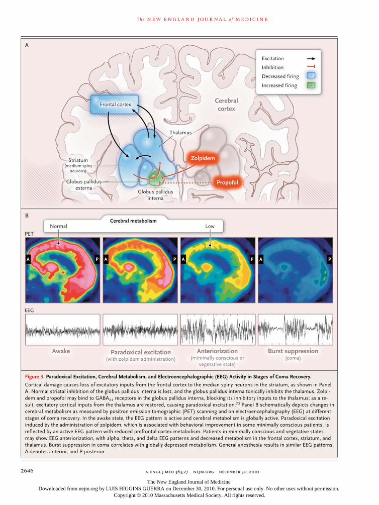
The central thalamus may mediate several of the phenomena seen in general anesthesia, as described above (Fig. 3). A possible explanation for the paradoxical excitation observed with a low dose of nearly every anesthetic drug3,4,13-16 is suggested by the circuit mechanism,69,73 which has been proposed to explain the paradoxical arousal of a patient from a minimally conscious state after administration of the GABAA1 agonist zolpidem.74 After administration of zolpidem, the marked downregulation of the anterior-fore-brain network that accompanied this patient’s brain injury was reversed, with notable functional improvement. The globus pallidus interna nor-mally provides tonic inhibitory inputs to the cen-tral thalamus that are opposed by striatal inhibi-tion of the pallidum. Binding of zolpidem in the GABAA1-receptor–rich globus pallidus interna may suppress this tonic inhibitory input to the thala-mus,75 fostering activation of thalamocortical and thalamostriatal circuits and, consequently, en-hancing arousal (Fig. 3A). For anesthetic drugs that promote GABAA inhibition, paradoxical ex-citation may be explained by a similar action in these circuits. This hypothesis could also account
The New England Journal of Medicine Downloaded from nejm.org by LUIS HIGGINS GUERRA on December 30, 2010. For personal use only. No other uses without permission.
Copyright © 2010 Massachusetts Medical Society. All rights reserved.

T h e n e w e ngl a nd j o u r na l o f m e dic i n e
n engl j med 363;27 nejm.org december 30, 20102644
for the paradoxical excitation observed during endoscopy and the delirium observed in inten-sive care units that is commonly associated with sedation induced with benzodiazepines, which are GABAA agonists.76,77 The fact that purpose-less movements are associated with paradoxical excitation is consistent with a possible basal ganglia mechanism.13
An imaging study in humans suggests a role for the corticobasal ganglia–thalamic circuit in propofol-induced unconsciousness.78 This circuit may also underlie anteriorization (Fig. 3B). The delta and alpha rhythms seen on the EEG during general anesthesia and sleep79 could arise from sustained hyperpolarization of pyramidal neurons in the lower output layers of the cortex owing to withdrawal (during sleep) or inhibition (during general anesthesia) of excitatory synaptic inputs.80 Initiation of these changes in the anterior fore-brain could be a consequence of active inhibition of the central thalamus that occurs when corti-cal inputs to the striatum are insufficient (Fig. 3A). A theoretical model has shown that as pro-pofol is increased to a dose beyond that which produces paradoxical excitation, its actions in the thalamocortical circuits lead to a coherent alpha oscillation between the thalamus and the ante-rior forebrain.81
Finally, burst suppression is believed to be a strong, synchronized outf low of thalamic dis-charges to a widely unresponsive cortex82 (Fig. 1). It is a deeper state of general anesthesia than is phase 2 of the maintenance period, which resem-bles the tonic bursting mode of the thalamus seen in slow-wave sleep. Bursts become more widely separated during burst suppression as the level of general anesthesia deepens. This suggests that a larger fraction of the cortex is inactive during burst suppression relative to phase 2 of general anesthesia or slow-wave sleep. Support-ing this hypothesis is the observation that burst suppression is also seen in coma due to diffuse anoxic damage,83 induced hypothermia,84 and epilepsy due to the Ohtahara syndrome.85 The absence of burst suppression during sleep is an important electrophysiological distinction between sleep and general anesthesia.
Ac ti v e Br a in S tates a nd Unconsciousness
In contrast to unconsciousness induced by most hypnotic agents, which is predominantly associ-
ated with slow EEG patterns, unconsciousness induced by the NMDA antagonist ketamine is as-sociated with active EEG patterns.86,87 Seizures are commonly associated with active, highly or-ganized EEG patterns. Unconsciousness due to seizures most likely results from organized, ab-errant brain activity that impedes the normal com-munications necessary to maintain arousal and cognition.88 Similarly, a highly active brain state most likely plays a role in unconsciousness in-duced by ketamine. Ketamine preferentially in-hibits NMDA-mediated glutamatergic inputs to GABAergic interneurons, leading to aberrant ex-citatory activity in the cortex, hippocampus, and limbic system and ultimately unconsciousness (Fig. 4).89,90 Hallucinations may result because the aberrant activation allows the association of information in a manner that is inconsistent in time and space. The hallucinations can be miti-gated by the concurrent administration of a ben-zodiazepine,91 which presumably acts to enhance
Figure 2 (facing page). Possible Neural-Circuit Mechanisms of Altered Arousal Induced by Anesthetic Agents.
Panel A shows a GABAergic inhibitory interneuron (orange) synapsing on a pyramidal neuron (gray) receiving excitatory inputs from ascending arousal pathways. The monoaminergic pathways arise from the locus ceruleus, which releases norepinephrine; the raphe, which releases serotonin; the tuberomammillary nucleus, which releases histamine; and the ventral teg-mental area, which releases dopamine. The cholinergic pathways, which release acetylcholine, arise from the basal forebrain, the lateral dorsal tegmental nuclei, and the pedunculopontine tegmental nuclei. Lateral hypo-thalamic neurons release orexin. Propofol binds post-synaptically and enhances GABAergic inhibition, counter-acting arousal inputs to the pyramidal neuron, decreasing its excitatory activity, and contributing to unconscious-ness. Dexmedetomidine binds to α2 receptors on neu-rons from the locus ceruleus, inhibiting norepineph-rine release (dashed line) in the ventrolateral preoptic nucleus, as shown in Panel B. The disinhibited ventro-lateral preoptic nucleus reduces arousal by means of GABAA-mediated and galanin-mediated inhibition of the midbrain, hypothalamic, and pontine arousal nuclei. As shown in Panel C, opioids reduce arousal by inhibit-ing the release of acetylcholine from neurons projecting from the lateral dorsal and pedunculopontine tegmen-tal nuclei to the medial pontine reticular formation and to the thalamus, by binding to opioid receptors in the periaqueductal gray and rostral ventral medulla, and by binding presynaptically and postsynaptically to spinal cord opioid receptors at the synapses between periph-eral afferent neurons in the dorsal-root ganglion and projecting neurons.
The New England Journal of Medicine Downloaded from nejm.org by LUIS HIGGINS GUERRA on December 30, 2010. For personal use only. No other uses without permission.
Copyright © 2010 Massachusetts Medical Society. All rights reserved.

mechanisms of disease
n engl j med 363;27 nejm.org december 30, 2010 2645
The New England Journal of Medicine Downloaded from nejm.org by LUIS HIGGINS GUERRA on December 30, 2010. For personal use only. No other uses without permission.
Copyright © 2010 Massachusetts Medical Society. All rights reserved.
T h e n e w e ngl a nd j o u r na l o f m e dic i n e
n engl j med 363;27 nejm.org december 30, 20102646
Figure 3. Paradoxical Excitation, Cerebral Metabolism, and Electroencephalographic (EEG) Activity in Stages of Coma Recovery.
Cortical damage causes loss of excitatory inputs from the frontal cortex to the median spiny neurons in the striatum, as shown in Panel A. Normal striatal inhibition of the globus pallidus interna is lost, and the globus pallidus interna tonically inhibits the thalamus. Zolpi-dem and propofol may bind to GABAA1 receptors in the globus pallidus interna, blocking its inhibitory inputs to the thalamus; as a re-sult, excitatory cortical inputs from the thalamus are restored, causing paradoxical excitation.73 Panel B schematically depicts changes in cerebral metabolism as measured by positron emission tomographic (PET) scanning and on electroencephalography (EEG) at different stages of coma recovery. In the awake state, the EEG pattern is active and cerebral metabolism is globally active. Paradoxical excitation induced by the administration of zolpidem, which is associated with behavioral improvement in some minimally conscious patients, is reflected by an active EEG pattern with reduced prefrontal cortex metabolism. Patients in minimally conscious and vegetative states may show EEG anteriorization, with alpha, theta, and delta EEG patterns and decreased metabolism in the frontal cortex, striatum, and thalamus. Burst suppression in coma correlates with globally depressed metabolism. General anesthesia results in similar EEG patterns. A denotes anterior, and P posterior.
The New England Journal of Medicine Downloaded from nejm.org by LUIS HIGGINS GUERRA on December 30, 2010. For personal use only. No other uses without permission.
Copyright © 2010 Massachusetts Medical Society. All rights reserved.

mechanisms of disease
n engl j med 363;27 nejm.org december 30, 2010 2647
GABAA-mediated activity of the interneurons and hence leads to sedation. The potent antinocicep-tive effects of ketamine on NMDA receptors in the spinal cord and its inhibition of acetylcholine release from the pons also contribute to uncon-sciousness (Fig. 4).92-94
Emergence from Gener a l A nes thesi a a nd R ecov er y
from Com a
Recovery from an initial coma — if it occurs — may require hours to years. In contrast, emer-gence from general anesthesia typically requires minutes. Nevertheless, it is useful to compare emergence from general anesthesia and recovery from coma (Table 1). The early clinical signs of emergence from general anesthesia — return of
regular breathing, salivation, tearing, swallow-ing, gagging, and grimacing — approximate the caudal–rostral progression in the brain stem of the return of sensory, motor, and autonomic function (Table 1). A later sign, such as response to oral commands, indicates the return of corti-cal function. The quantitative neurobehavioral metrics used to monitor recovery from coma could be used to track the emergence from gen-eral anesthesia from a functional state that can approximate brain-stem death to states similar to a vegetative state and, eventually, to a minimally conscious state.95 The fact that general anesthe-sia can be functionally equivalent to brain-stem death indicates how deeply general anesthesia can depress brain function and perhaps explains why some patients do not fully recover conscious-ness for several hours after general anesthesia
Figure 4. Unconsciousness and Active Brain States.
Ketamine binds preferentially to N-methyl-D-aspartate (NMDA) receptors on inhibitory interneurons in the cortex, limbic system (amyg-dala), and hippocampus, promoting an uncoordinated increase in neural activity, an active electroencephalographic pattern, and uncon-sciousness, as shown in Panel A. In the spinal cord, ketamine decreases arousal by blocking NMDA glutamate (Glu)–mediated nocicep-tive signals from peripheral afferent neurons in the dorsal-root ganglion to projecting neurons, as shown in Panel B.
The New England Journal of Medicine Downloaded from nejm.org by LUIS HIGGINS GUERRA on December 30, 2010. For personal use only. No other uses without permission.
Copyright © 2010 Massachusetts Medical Society. All rights reserved.

T h e n e w e ngl a nd j o u r na l o f m e dic i n e
n engl j med 363;27 nejm.org december 30, 20102648
and why postoperative cognitive dysfunction could persist in elderly patients for several months.96
In conclusion, a better understanding of sleep and coma may lead to new approaches to gen-eral anesthesia based on new ways to alter con-sciousness,29,97,98 provide analgesia,99,100 induce amnesia, and provide muscle relaxation.66
Supported by the Massachusetts General Hospital Department of Anesthesia, Critical Care, and Pain Medicine and by the National Institutes of Health (NIH) Director’s Pioneer Award (DP1OD003646, to Dr. Brown); by the University of Michigan, Department of Anes-thesiology, and by NIH grants (HL40881 and HL65272, to Dr. Lydic); and by grants from the James S. McDonnell Foundation (to Dr. Schiff) and the NIH (HD51912, to Dr. Schiff).
Disclosure forms provided by the authors are available with the full text of this article at NEJM.org.
References
1. Sentinel event alert: preventing, and managing the impact of, anesthesia awareness. Oakbrook Terrace, IL: The Joint Commission, 2004. (http://www.joint commission.org/sentinel_event_alert_ issue_32_preventing_and_managing_the_impact_of_anesthesia_awareness.)2. Evers A, Crowder M. Cellular and mo-lecular mechanisms of anesthesia. In: Barash PG, Cullen BF, Stoelting RK, Ca-halan M, Stock MC, eds. Clinical anesthe-sia. 6th ed. New York: Lippincott Wil-liams & Wilkins, 2006:95-114.3. Gibbs FA, Gibbs LE, Lennox WG. Ef-fects on the electroencephalogram of cer-tain drugs which influence nervous activ-ity. Arch Intern Med 1937;60:154-66.4. Kiersey DK, Bickford RG, Faulconer A Jr. Electro-encephalographic patterns pro-duced by thiopental sodium during surgi-cal operations; description and classifica-tion. Br J Anaesth 1951;23:141-52.5. Watson C, Bagdoyan H, Lydic R. A neu-rochemical perspective on states of con-sciousness. In: Hudetz AG, Pearce RA, eds. Suppressing the mind: anesthetic modula-tion of memory and consciousness. New York: Springer/Humana Press, 2010:33-80.6. Kennedy D, Norman C. What don’t we know? Science 2005;309:75.7. Kryger M, Roth T, Dement W. Princi-ples and practice of sleep medicine. 5th ed. New York: Elsevier Saunders, 2010.8. McCarley RW. Neurobiology of REM and NREM sleep. Sleep Med 2007;8:302-30.9. Posner J, Saper C, Schiff N, Plum F. Plum and Posner’s diagnosis of stupor and coma. New York: Oxford University Press, 2007.10. Young GB. The EEG in coma. J Clin Neurophysiol 2000;17:473-85.11. Gawande A, Denno DW, Truog RD, Waisel DM. Physicians and execution — highlights from a discussion of lethal in-jection. N Engl J Med 2008;358:448-51.12. Davis MH, Coleman MR, Absalom AR, et al. Dissociating speech perception and comprehension at reduced levels of awareness. Proc Natl Acad Sci U S A 2007;104:16032-7.13. Bevan JC, Veall GR, Macnab AJ, Ries CR, Marsland C. Midazolam premedica-tion delays recovery after propofol with-out modifying involuntary movements. Anesth Analg 1997;85:50-4.
14. McCarthy MM, Brown EN, Kopell N. Potential network mechanisms mediating electroencephalographic beta rhythm changes during propofol-induced para-doxical excitation. J Neurosci 2008;28: 13488-504.15. Clark DL, Rosner BS. Neurophysio-logic effects of general anesthetics. I. The electroencephalogram and sensory evoked responses in man. Anesthesiology 1973; 38:564-82.16. Rampil IJ. A primer for EEG signal processing in anesthesia. Anesthesiology 1998;89:980-1002.17. Coté CJ, Goudsouzian NG, Liu LM, Dedrick DF, Rosow CE. The dose re-sponse of intravenous thiopental for the induction of general anesthesia in unpre-medicated children. Anesthesiology 1981; 55:703-5.18. Gray AT, Krejci ST, Larson MD. Neu-romuscular blocking drugs do not alter the pupillary light reflex of anesthetized humans. Arch Neurol 1997;54:579-84.19. Prys-Roberts C. Anaesthesia: a practi-cal or impractical construct? Br J Anaesth 1987;59:1341-5.20. Palanca BJ, Mashour GA, Avidan MS. Processed electroencephalogram in depth of anesthesia monitoring. Curr Opin An-aesthesiol 2009;22:553-9.21. Quality Standards Subcommittee of the American Academy of Neurology. Practice parameters: determining brain death in adults. Neurology 1995;45:1012-4.22. Feshchenko VA, Veselis RA, Reinsel RA. Propofol-induced alpha rhythm. Neu-ropsychobiology 2004;50:257-66.23. Tinker JH, Sharbrough FW, Michen-felder JD. Anterior shift of the dominant EEG rhythm during anesthesia in the Java monkey: correlation with anesthetic po-tency. Anesthesiology 1977;46:252-9.24. Doyle PW, Matta BF. Burst suppres-sion or isoelectric encephalogram for ce-rebral protection: evidence from meta-bolic suppression studies. Br J Anaesth 1999;83:580-4.25. Bergey GK. Refractory status epilepti-cus: is EEG burst suppression an appro-priate treatment target during drug- induced coma? What is the Holy Grail? Epilepsy Curr 2006;6:119-20.26. Claassen J, Hirsch LJ, Emerson RG, Mayer SA. Treatment of refractory status
epilepticus with pentobarbital, propofol, or midazolam: a systematic review. Epi-lepsia 2002;43:146-53.27. Giacino JT, Ashwal S, Childs N, et al. The minimally conscious state: definition and diagnostic criteria. Neurology 2002;58:349-53.28. Aldrete JA. The post-anesthesia recov-ery score revisited. J Clin Anesth 1995;7: 89-91.29. Alkire MT, Hudetz AG, Tononi G. Consciousness and anesthesia. Science 2008;322:876-80.30. Angel A. The G.L. Brown lecture. Ad-ventures in anaesthesia. Exp Physiol 1991; 76:1-38.31. Alkire MT, Haier RJ, Barker SJ, Shah NK, Wu JC, Kao YJ. Cerebral metabolism during propofol anesthesia in humans studied with positron emission tomogra-phy. Anesthesiology 1995;82:393-403.32. Fiset P, Paus T, Daloze T, et al. Brain mechanisms of propofol-induced loss of consciousness in humans: a positron emis-sion tomographic study. J Neurosci 1999; 19:5506-13.33. Purdon PL, Pierce ET, Bonmassar G, et al. Simultaneous electroencephalogra-phy and functional magnetic resonance imaging of general anesthesia. Ann N Y Acad Sci 2009;1157:61-70.34. Velly LJ, Rey MF, Bruder NJ, et al. Dif-ferential dynamic of action on cortical and subcortical structures of anesthetic agents during induction of anesthesia. Anesthesiology 2007;107:202-12. [Erra-tum, Anesthesiology 2008;108:175.]35. Ferrarelli F, Massimini M, Sarasso S, et al. Breakdown in cortical effective con-nectivity during midazolam-induced loss of consciousness. Proc Natl Acad Sci U S A 2010;107:2681-6.36. Rudolph U, Antkowiak B. Molecular and neuronal substrates for general anaes-thetics. Nat Rev Neurosci 2004;5:709-20.37. Hemmings HC Jr, Akabas MH, Gold-stein PA, Trudell JR, Orser BA, Harrison NL. Emerging molecular mechanisms of general anesthetic action. Trends Phar-macol Sci 2005;26:503-10.38. Bai D, Pennefather PS, MacDonald JF, Orser BA. The general anesthetic propofol slows deactivation and desensitization of GABA(A) receptors. J Neurosci 1999;19: 10635-46.
The New England Journal of Medicine Downloaded from nejm.org by LUIS HIGGINS GUERRA on December 30, 2010. For personal use only. No other uses without permission.
Copyright © 2010 Massachusetts Medical Society. All rights reserved.

mechanisms of disease
n engl j med 363;27 nejm.org december 30, 2010 2649
39. Shepherd G. The synaptic organiza-tion of the brain. 4th ed. New York: Ox-ford University Press, 1998.40. Propofol. Bedford, OH: Bedford Labo-ratories, 2008 (package insert). (http://dailymed.nlm.nih.gov/dailymed/drugInfo .cfm?id=6337.)41. Devor M, Zalkind V. Reversible anal-gesia, atonia, and loss of consciousness on bilateral intracerebral microinjection of pentobarbital. Pain 2001;94:101-12.42. Parvizi J, Damasio AR. Neuroanatom-ical correlates of brainstem coma. Brain 2003;126:1524-36.43. Feldman JL, Del Negro CA. Looking for inspiration: new perspectives on respiratory rhythm. Nat Rev Neurosci 2006;7:232-42.44. Kungys G, Kim J, Jinks SL, Atherley RJ, Antognini JF. Propofol produces im-mobility via action in the ventral horn of the spinal cord by a GABAergic mecha-nism. Anesth Analg 2009;108:1531-7.45. Pontine (medial) and medullary (lat-eral) reticulospinal tracts. Laboratory 12: tract systems I. University of Pennsylvania School of Veterinary Medicine, 2006. (http:// cal.vet.upenn.edu/projects/neurology/lab12/ lab12_rspt.htm.)46. Dutton RP, Eckhardt WF III, Sunder N. Total spinal anesthesia after intersca-lene blockade of the brachial plexus. An-esthesiology 1994;80:939-41.47. Durrani Z, Winnie AP. Brainstem tox-icity with reversible locked-in syndrome after intrascalene brachial plexus block. Anesth Analg 1991;72:249-52.48. Penn RD, Savoy SM, Corcos D, et al. Intrathecal baclofen for severe spinal spasticity. N Engl J Med 1989;320:1517-21.49. Mourisse J, Gerrits W, Lerou J, van Eg-mond J, Zwarts MJ, Booij L. Electromyo-graphic assessment of blink and corneal reflexes during midazolam administra-tion: useful methods for assessing depth of anesthesia? Acta Anaesthesiol Scand 2003;47:593-600.50. Nelson LE, Guo TZ, Lu J, Saper CB, Franks NP, Maze M. The sedative compo-nent of anesthesia is mediated by GABA(A) receptors in an endogenous sleep pathway. Nat Neurosci 2002;5:979-84.51. Saper CB, Scammell TE, Lu J. Hypo-thalamic regulation of sleep and circadi-an rhythms. Nature 2005;437:1257-63.52. Morairty S, Rainnie D, McCarley R, Greene R. Disinhibition of ventrolateral preoptic area sleep-active neurons by ade-nosine: a new mechanism for sleep pro-motion. Neuroscience 2004;123:451-7.53. Correa-Sales C, Rabin BC, Maze M. A hypnotic response to dexmedetomi-dine, an alpha 2 agonist, is mediated in the locus coeruleus in rats. Anesthesiology 1992;76:948-52.54. Jorm CM, Stamford JA. Actions of the hypnotic anaesthetic, dexmedetomidine, on noradrenaline release and cell firing in
rat locus coeruleus slices. Br J Anaesth 1993;71:447-9.55. Huupponen E, Maksimow A, Lapin-lampi P, et al. Electroencephalogram spindle activity during dexmedetomidine sedation and physiological sleep. Acta Anaesthesiol Scand 2008;52:289-94.56. Lydic R, Baghdoyan HA. Sleep, anes-thesiology, and the neurobiology of arous-al state control. Anesthesiology 2005;103: 1268-95.57. Osman NI, Baghdoyan HA, Lydic R. Morphine inhibits acetylcholine release in rat prefrontal cortex when delivered sys-temically or by microdialysis to basal fore-brain. Anesthesiology 2005;103:779-87.58. Heinricher MM, Morgan MM. Supra-spinal mechanisms of opioid analgesia. In: Stein C, ed. Opioids in pain control: basic and clinical aspects. Cambridge, United Kingdom: Cambridge University Press, 1999:46-69.59. Cesselin FBJ, Bourgoin S, et al. Spinal mechanisms of analgesia. In: Stein C, ed. Opioids in pain control: basic and clinical aspects. Cambridge, United Kingdom: Cambridge University Press, 1999:70-95.60. Stein C, Machelska H, Binder W, Schafer M. Peripheral opioid analgesia. Curr Opin Pharmacol 2001;1:62-5.61. Hug CC Jr. Does opioid “anesthesia” exist? Anesthesiology 1990;73:1-4.62. Hilgenberg JC. Intraoperative aware-ness during high-dose fentanyl–oxygen anesthesia. Anesthesiology 1981;54:341-3.63. Meuret P, Backman SB, Bonhomme V, Plourde G, Fiset P. Physostigmine reverses propofol-induced unconsciousness and at-tenuation of the auditory steady state re-sponse and bispectral index in human vol-unteers. Anesthesiology 2000;93:708-17.64. Lepousé C, Lautner CA, Liu L, Gomis P, Leon A. Emergence delirium in adults in the post-anaesthesia care unit. Br J An-aesth 2006;96:747-53.65. Funk W, Hollnberger H, Geroldinger J. Physostigmine and anaesthesia emer-gence delirium in preschool children: a randomized blinded trial. Eur J Anaes-thesiol 2008;25:37-42.66. Chase MH. Confirmation of the con-sensus that glycinergic postsynaptic inhi-bition is responsible for the atonia of REM sleep. Sleep 2008;31:1487-91.67. Lavigne GJ, Sessle BJ, Choiniere M, Soja PJ, eds. Sleep and pain. Seattle: IASP Press, 2007.68. Steriade M, Timofeev I. Neuronal plas-ticity in thalamocortical networks during sleep and waking oscillations. Neuron 2003;37:563-76.69. Schiff ND. Central thalamic contribu-tions to arousal regulation and neurologi-cal disorders of consciousness. Ann N Y Acad Sci 2008;1129:105-18.70. Schiff ND, Plum F. The role of arousal and “gating” systems in the neurology of
impaired consciousness. J Clin Neuro-physiol 2000;17:438-52.71. Schiff ND, Giacino JT, Kalmar K, et al. Behavioural improvements with tha-lamic stimulation after severe traumatic brain injury. Nature 2007;448:600-3.72. Alkire MT, McReynolds JR, Hahn EL, Trivedi AN. Thalamic microinjection of nicotine reverses sevoflurane-induced loss of righting reflex in the rat. Anesthe-siology 2007;107:264-72.73. Schiff ND, Posner JB. Another “Awak-enings.” Ann Neurol 2007;62:5-7.74. Brefel-Courbon C, Payoux P, Ory F, et al. Clinical and imaging evidence of zol-pidem effect in hypoxic encephalopathy. Ann Neurol 2007;62:102-5.75. Chen L, Savio Chan C, Yung WH. Electrophysiological and behavioral ef-fects of zolpidem in rat globus pallidus. Exp Neurol 2004;186:212-20.76. Fulton SA, Mullen KD. Completion of upper endoscopic procedures despite par-adoxical reaction to midazolam: a role for flumazenil? Am J Gastroenterol 2000;95: 809-11.77. Tung A, Tadimeti L, Caruana-Montal-do B, et al. The relationship of sedation to deliberate self-extubation. J Clin Anesth 2001;13:24-9.78. Mhuircheartaigh RN, Rosenorn-Lanng D, Wise R, Jbabdi S, Rogers R, Tracey I. Cortical and subcortical connec-tivity changes during decreasing levels of consciousness in humans: a functional magnetic resonance imaging study using propofol. J Neurosci 2010;30:9095-102.79. Steriade M, Nuñez A, Amzica F. A novel slow (< 1 Hz) oscillation of neo-cortical neurons in vivo: depolarizing and hyperpolarizing components. J Neurosci 1993;13:3252-65.80. Steriade M, Contreras D, Curro Dossi R, Nuñez A. The slow (< 1 Hz) oscillation in reticular thalamic and thalamocortical neurons: scenario of sleep rhythm genera-tion in interacting thalamic and neocorti-cal networks. J Neurosci 1993;13:3284-99.81. Ching S, Cimenser A, Purdon P, Brown EN, Kopell NK. Thalamocortical model for a propofol-induced alpha rhythm associ-ated with loss of consciousness. Proc Natl Acad Sci U S A 2010 (in press).82. Steriade M, Amzica F, Contreras D. Cortical and thalamic cellular correlates of electroencephalographic burst-sup-pression. Electroencephalogr Clin Neuro-physiol 1994;90:1-16.83. Koenig MA, Kaplan PW, Thakor NV. Clinical neurophysiologic monitoring and brain injury from cardiac arrest. Neurol Clin 2006;24:89-106.84. Stecker MM, Cheung AT, Pochettino A, et al. Deep hypothermic circulatory ar-rest: I. Effects of cooling on electroen-cephalogram and evoked potentials. Ann Thorac Surg 2001;71:14-21.
The New England Journal of Medicine Downloaded from nejm.org by LUIS HIGGINS GUERRA on December 30, 2010. For personal use only. No other uses without permission.
Copyright © 2010 Massachusetts Medical Society. All rights reserved.

n engl j med 363;27 nejm.org december 30, 20102650
mechanisms of disease
85. Yamatogi Y, Ohtahara S. Early-infan-tile epileptic encephalopathy with sup-pression-bursts, Ohtahara syndrome: its overview referring to our 16 cases. Brain Dev 2002;24:13-23.86. Maksimow A, Särkelä M, Långsjö JW, et al. Increase in high frequency EEG ac-tivity explains the poor performance of EEG spectral entropy monitor during S-ketamine anesthesia. Clin Neurophysiol 2006;117:1660-8.87. Tsuda N, Hayashi K, Hagihira S, Sawa T. Ketamine, an NMDA-antagonist, in-creases the oscillatory frequencies of al-pha-peaks on the electroencephalograph-ic power spectrum. Acta Anaesthesiol Scand 2007;51:472-81.88. Blumenfeld H, Taylor J. Why do sei-zures cause loss of consciousness? Neuro-scientist 2003;9:301-10.89. Seamans J. Losing inhibition with ketamine. Nat Chem Biol 2008;4:91-3.90. Olney JW, Newcomer JW, Farber NB. NMDA receptor hypofunction model of
schizophrenia. J Psychiatr Res 1999;33: 523-33.91. Reboso Morales JA, González Miran-da F. Ketamine. Rev Esp Anestesiol Re-anim 1999;46:111-22. (In Spanish.)92. Oye I, Paulsen O, Maurset A. Effects of ketamine on sensory perception: evi-dence for a role of N-methyl-D-aspartate receptors. J Pharmacol Exp Ther 1992; 260:1209-13.93. Sinner B, Graf BM. Ketamine. Handb Exp Pharmacol 2008;182:313-33.94. Lydic R, Baghdoyan HA. Ketamine and MK-801 decrease acetylcholine re-lease in the pontine reticular formation, slow breathing, and disrupt sleep. Sleep 2002;25:617-22.95. Giacino JT, Kalmar K, Whyte J. The JFK Coma Recovery Scale–Revised: mea-surement characteristics and diagnostic utility. Arch Phys Med Rehabil 2004;85: 2020-9.96. Monk TG, Weldon BC, Garvan CW, et al. Predictors of cognitive dysfunction af-
ter major noncardiac surgery. Anesthesi-ology 2008;108:18-30.97. Kelz MB, Sun Y, Chen J, et al. An es-sential role for orexins in emergence from general anesthesia. Proc Natl Acad Sci U S A 2008;105:1309-14.98. Franks NP. General anaesthesia: from molecular targets to neuronal pathways of sleep and arousal. Nat Rev Neurosci 2008;9:370-86.99. Ren J, Ding X, Funk GD, Greer JJ. Am-pakine CX717 protects against fentanyl-induced respiratory depression and lethal apnea in rats. Anesthesiology 2009;110: 1364-70.100. Watson SL, Watson CJ, Baghdoyan HA, Lydic R. Thermal nociception is de-creased by hypocretin-1 and an adenosine A1 receptor agonist microinjected into the pontine reticular formation of Sprague Dawley rat. J Pain 2010;11:535-44.Copyright © 2010 Massachusetts Medical Society.
journal archive at nejm.org
Every issue of the Journal is now available at NEJM.org, beginning with the first article published in January 1812. The entire archive is fully searchable, and browsing of titles and tables of contents is easy and available to all. Individual subscribers are entitled to free 24-hour access to 50 archive articles per year. Access to content in the archive is available on a per-article basis and is also being provided through
many institutional subscriptions.
The New England Journal of Medicine Downloaded from nejm.org by LUIS HIGGINS GUERRA on December 30, 2010. For personal use only. No other uses without permission.
Copyright © 2010 Massachusetts Medical Society. All rights reserved.