Embed Size (px)
Citation preview
Knowing what to looK for Knowing where to looK and Knowing what it means
GeneDx Prenatal Targeted ArrayUSER’S GUIDE

3 www.genedx.com
Dear Colleague,
This guide has been created as an educational tool to assist with your discussion of Prenatal Targeted Array analysis with your patients. It explains the combined CGH and SNP array platform used at GeneDx for detection of copy number changes and uniparental disomy, respectively. It is designed such that one side of the booklet is intended as the patient view, and the opposing page contains information for the practitioner. Pages for the patient are designated with the word “Patient” at the top right corner of the page. This guide also contains a separate Practitioner’s Section including an overview of the prenatal testing services available at GeneDx, and a more detailed overview of the GeneDx Prenatal Targeted Array testing service.
We hope that this guide provides you and your patients with a better understanding of the GeneDx Prenatal Targeted Array.
Sincerely,The GeneDx Team
Information for the Patient
Information for the Patient
6 www.genedx.com
The GeneDx Prenatal Targeted Array screens for 100 clinically defined microdeletion and microduplication syndromes*, imbalances >1.5 Mb genome-wide, and uniparental disomy.
The sensitivity of array CGH is 10-15% higher than conventional cytogenetic testing in individuals with developmental delay, mental retardation, and/or multiple congenital anomalies (Miller et al., Am J Hum Genet 86:749-764, 2010). The sensitivity of array CGH in prenatal settings is also expected to be high, and the American College of Obstetrics & Gynecology has recommended targeted array CGH testing for fetuses with abnormal ultrasound findings (ACOG Committee Opinion No. 446. Obstet & Gynecol 114:1161-1163, 2009).
Below are indications for ordering a GeneDx Prenatal Targeted Array:
• Abnormal fetal ultrasound findings
• Ambiguous karyotype results
• Suspected deletion/duplication syndrome
• Suspected disorder caused by uniparental disomy (UPD)
• Family history of known or suspected chromosome imbalances
*For complete list of targeted regions, refer to page 37
7 www.genedx.com
Patient
There is a new improved test available for diagnosis of chromosome abnormalities known as the GeneDx Prenatal Targeted Array. This microarray analyzes genetic material for 100 syndromes that typically cannot be detected by standard chromosome analysis, loss or gains of chromosomal material from small changes to the length of an entire chromosome, as well as unusual inheritance of genetic material from parents.
Below are indications for ordering a GeneDx Prenatal Targeted Array:
• Abnormal ultrasound findings
• Ambiguous chromosomal structure
• Suspected loss or gain of genetic material in the fetus
• Suspected disorder caused by genetic material received from only one parent (uniparental disomy or UPD)
• Family history (or suspicion) of diseases caused by genetic anomalies
9 www.genedx.com
Patient
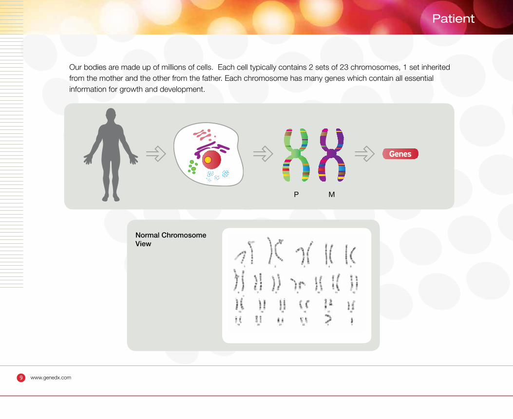
Our bodies are made up of millions of cells. Each cell typically contains 2 sets of 23 chromosomes, 1 set inherited from the mother and the other from the father. Each chromosome has many genes which contain all essential information for growth and development.
Genes
P M
Normal Chromosome View
11 www.genedx.com
Patient
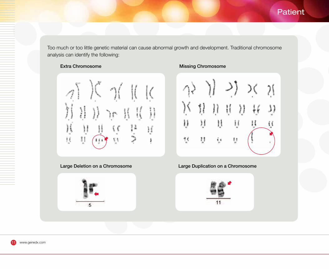
Too much or too little genetic material can cause abnormal growth and development. Traditional chromosome analysis can identify the following:
Extra Chromosome
Large Deletion on a Chromosome Large Duplication on a Chromosome
Missing Chromosome
13 www.genedx.com
Patient
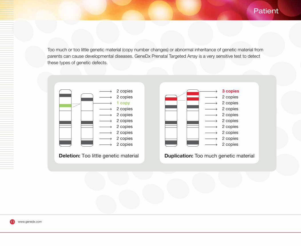
Too much or too little genetic material (copy number changes) or abnormal inheritance of genetic material from parents can cause developmental diseases. GeneDx Prenatal Targeted Array is a very sensitive test to detect these types of genetic defects.
3 copies2 copies2 copies2 copies2 copies2 copies2 copies2 copies2 copies2 copies
2 copies2 copies1 copy2 copies2 copies2 copies2 copies2 copies2 copies2 copies
Deletion: Too little genetic material Duplication: Too much genetic material
15 www.genedx.com
Patient
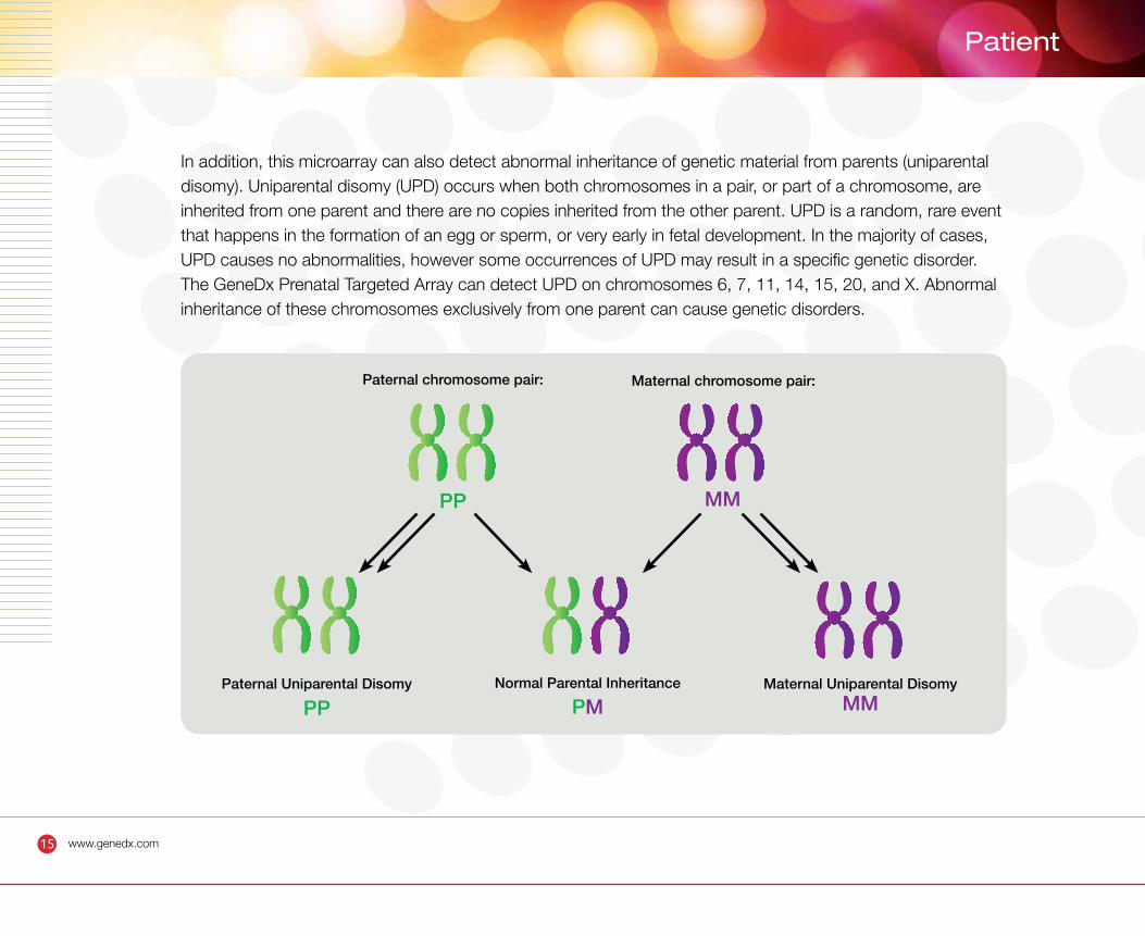
In addition, this microarray can also detect abnormal inheritance of genetic material from parents (uniparental disomy). Uniparental disomy (UPD) occurs when both chromosomes in a pair, or part of a chromosome, are inherited from one parent and there are no copies inherited from the other parent. UPD is a random, rare event that happens in the formation of an egg or sperm, or very early in fetal development. In the majority of cases, UPD causes no abnormalities, however some occurrences of UPD may result in a specific genetic disorder. The GeneDx Prenatal Targeted Array can detect UPD on chromosomes 6, 7, 11, 14, 15, 20, and X. Abnormal inheritance of these chromosomes exclusively from one parent can cause genetic disorders.
Paternal chromosome pair:
PP
PPPaternal Uniparental Disomy Normal Parental Inheritance
PMMaternal Uniparental Disomy
MM
MM
Maternal chromosome pair:
16 www.genedx.com
What can the GeneDx Prenatal Targeted Array find?
• Too few (monosomy) or too many (trisomy) chromosomes
• Deletions (500 bp - 100 kb in 100 targeted regions and 1.5 Mb or larger in the rest of the genome)
• Duplications (500 bp - 100 kb in 100 targeted regions and 1.5 Mb or larger in the rest of the genome)
• Small deletions or duplications of individual exons within select single genes (intragenic mutations)
• The chromosomal origin of ambiguous karyotype results (e.g. marker chromosomes, ring chromosomes, suspected chromosomal rearrangements)
• Accurate boundaries of deletions or duplications to help define which genes may be of clinical significance
• Uniparental disomy (UPD) on chromosomes 6, 7, 11, 14, 15, 20, and X.
• Evidence suggesting a close genetic relationship between the mother and father of the pregnancy being tested (consanguinity or shared ancestry)
What can the GeneDx Prenatal Targeted Array not find?
• Balanced rearrangements such as balanced robertsonian or reciprocal translocations, balanced insertions, and inversions (detectable by traditional karyotyping and FISH analysis only)
• Low-level mosaicism (<25%)
• Genomic imbalances in regions that are not represented on the array
• Small DNA mutations such as point mutations, small intragenic deletions or insertions (detectable by DNA sequencing only)
17 www.genedx.com
Patient
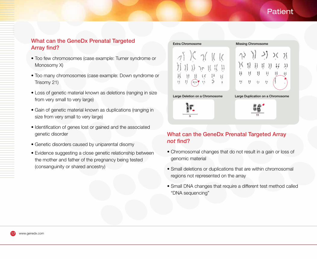
What can the GeneDx Prenatal Targeted Array find?
• Too few chromosomes (case example: Turner syndrome or Monosomy X)
• Too many chromosomes (case example: Down syndrome or Trisomy 21)
• Loss of genetic material known as deletions (ranging in size from very small to very large)
• Gain of genetic material known as duplications (ranging in size from very small to very large)
• Identification of genes lost or gained and the associated genetic disorder
• Genetic disorders caused by uniparental disomy
• Evidence suggesting a close genetic relationship between the mother and father of the pregnancy being tested (consanguinity or shared ancestry)
What can the GeneDx Prenatal Targeted Array not find?
• Chromosomal changes that do not result in a gain or loss of genomic material
• Small deletions or duplications that are within chromosomal regions not represented on the array
• Small DNA changes that require a different test method called “DNA sequencing”
Extra Chromosome
Large Deletion on a Chromosome Large Duplication on a Chromosome
Missing Chromosome
18 www.genedx.com
• Prenatal specimen is obtained through chorionic villi sampling (CVS), amniocentesis, or
percutaneous umbilical blood sampling (PUBS) procedure
• Fetal DNA is isolated from direct or cultured prenatal specimen
• Fetal DNA is tagged with red fluorescent dye
• Control DNA is tagged with green fluorescent dye
How Is It Done? Step 1
19 www.genedx.com
Patient
How Is It Done? Step 1
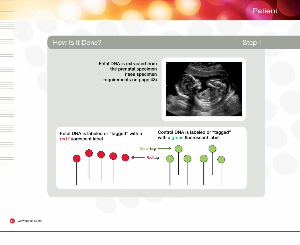
Fetal DNA is extracted from the prenatal specimen
(*see specimen requirements on page 43)
Red tag
Green tag
DNA is labeled or “tagged” with redfluorescent label
Control DNA is labeled or “tagged” with greenfluorescent label
FetalFetal DNA is labeled or “tagged” with a red fluorescent label
Control DNA is labeled or “tagged” with a green fluorescent label
20 www.genedx.com
• The fetal DNA is extracted and combined with the control DNA
• The combined DNA is hybridized to the GeneDx Prenatal Targeted Array
• The array is a glass slide coated with 42,000 specifically selected CGH oligonucleotide
probes placed throughout the genome and within 100 targeted common and novel
microdeletion/microduplication syndromes, and an additional 18,000 SNP probes covering
chromosomes 6, 7, 11, 14, 15, 20 and X.
• During hybridization, fluorescently “tagged” pieces of fetal DNA and the control DNA attach to
the probes with complementary sequences.
How Is It Done? Step 2
21 www.genedx.com
Patient
How Is It Done? Step 2
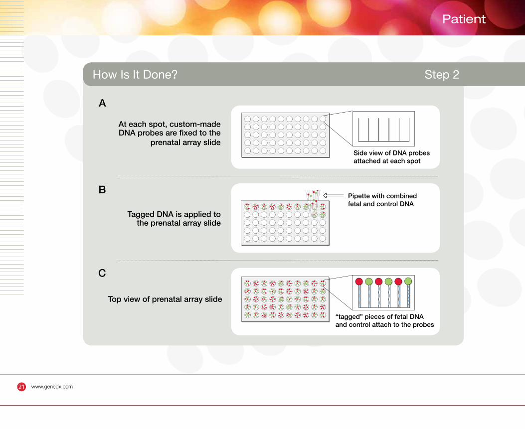
At each spot, custom-made DNA probes are fixed to the
prenatal array slide
Tagged DNA is applied tothe prenatal array slide
Top view of prenatal array slide
Pipette with combined fetal and control DNA
“tagged” pieces of fetal DNAand control attach to the probes
Side view of DNA probesattached at each spot
A
B
C
22 www.genedx.com
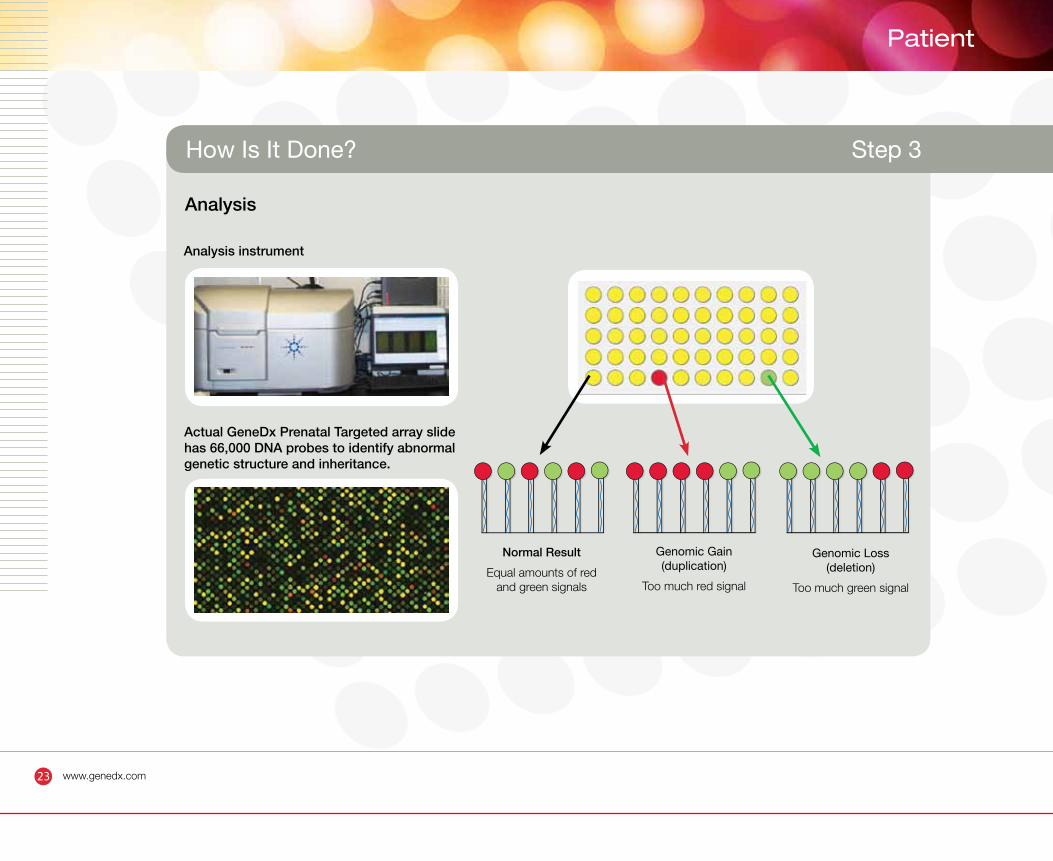
• After incubation, the analysis instrument determines how much red (fetal DNA) and green
(control DNA) is attached to each of the spots on the array
• The ratio of red to green signals are displayed
Yellow = Equal amounts of fetal and control DNA = Normal result
Red>>Green = Genomic gain in fetal DNA = duplication or trisomy
Red<<Green = Genomic loss in fetal DNA = deletion or monosomy
• In addition, at some spots on the array the intensity of the fluorescence signal can provide
information about the parental origin of chromosomal material (SNP analysis).
How Is It Done? Step 3
23 www.genedx.com
Patient
Actual GeneDx Prenatal Targeted array slide has 66,000 DNA probes to identify abnormal genetic structure and inheritance.
Analysis instrument
Normal Result
Equal amounts of red and green signals
Analysis
Genomic Gain (duplication)
Too much red signal
Genomic Loss (deletion)
Too much green signal
How Is It Done? Step 3
Possible Result Outcomes
Possible Result Outcomes
26 www.genedx.com
Negative result is illustrated by:
• Similar amount of fetal and control DNA present (yellow dots)
• Normal CGH result is represented by an even signal distributed on the baseline along the length of the entire chromosome; indicating no gain or loss of genetic material.
• Normal SNP result is represented by the presence of signals distributed into three lines along the length of the entire chromosome, reflecting normal inheritance of genetic material from both parents.
Negative Prenatal Targeted Array Results
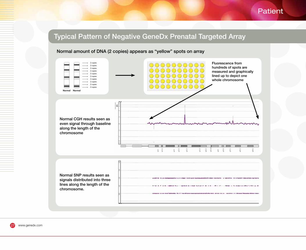
27 www.genedx.com
Patient
Normal amount of DNA (2 copies) appears as “yellow” spots on array
2 copies2 copies2 copies2 copies2 copies2 copies2 copies2 copies2 copies2 copies
NormalNormal
Fluorescence from hundreds of spots are measured and graphically lined up to depict one whole chromosome
Normal CGH results seen as even signal through baseline along the length of the chromosome
Typical Pattern of Negative GeneDx Prenatal Targeted Array
Normal SNP results seen as signals distributed into three lines along the length of the chromosome.
29 www.genedx.com
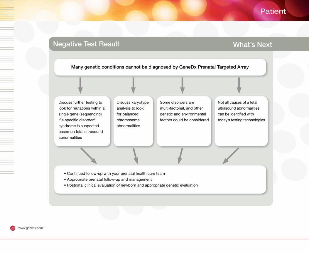
Patient
Negative Test Result What’s Next
Many genetic conditions cannot be diagnosed by GeneDx Prenatal Targeted Array
Discuss further testing to
look for mutations within a
single gene (sequencing)
if a specific disorder/
syndrome is suspected
based on fetal ultrasound
abnormalities
• Continued follow-up with your prenatal health care team
• Appropriate prenatal follow-up and management
• Postnatal clinical evaluation of newborn and appropriate genetic evaluation
Discuss karyotype
analysis to look
for balanced
chromosome
abnormalities
Some disorders are
multi-factorial, and other
genetic and environmental
factors could be considered
Not all causes of a fetal
ultrasound abnormalities
can be identified with
today’s testing technologies
30 www.genedx.com
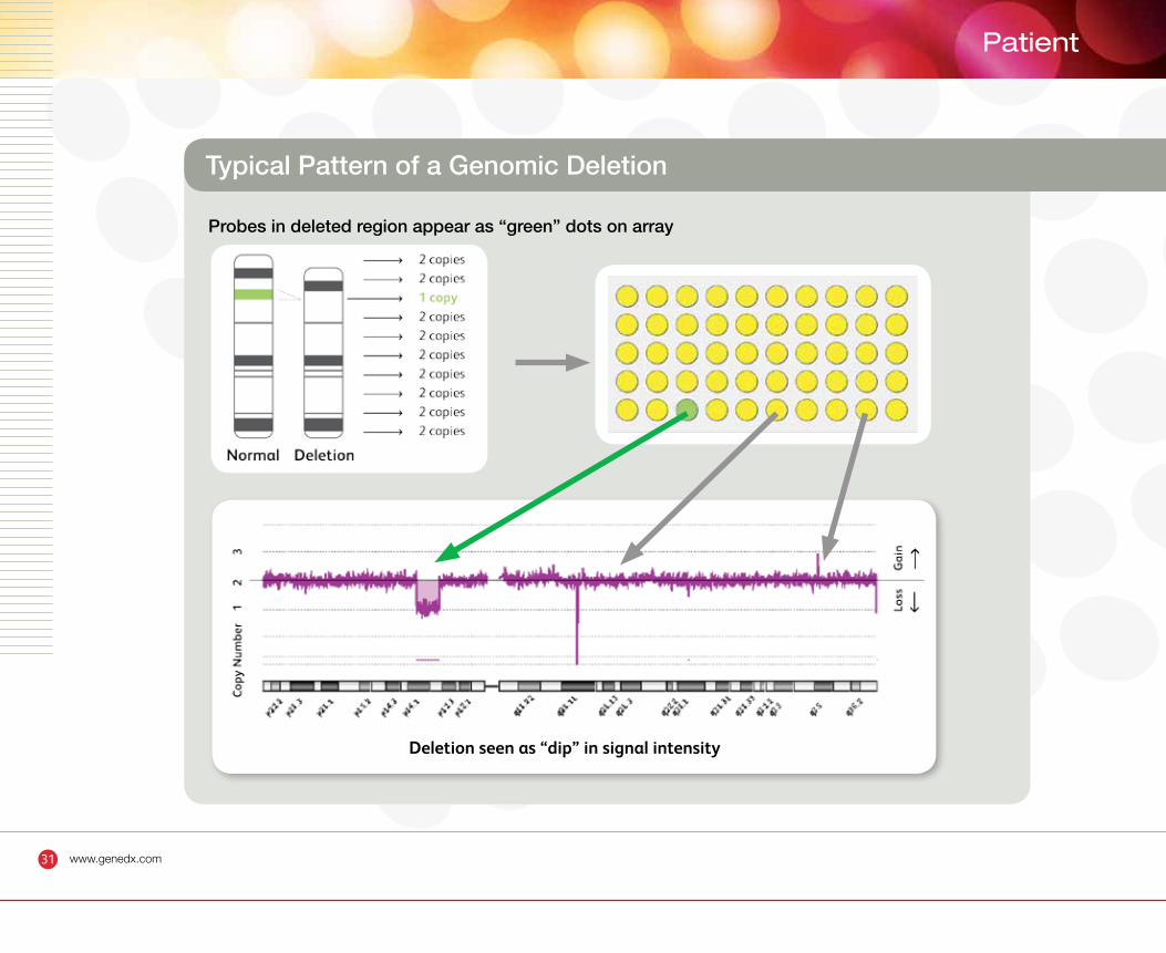
Typical Pattern of a Genomic Deletion
Genomic deletion is illustrated by:
• Loss of fetal DNA compared to the control
• Green dot on the array diagram
• “Dip” on the CGH chromosome array plot
31 www.genedx.com
Patient
Probes in deleted region appear as “green” dots on array
Typical Pattern of a Genomic Deletion
Deletion seen as “dip” in signal intensity
32 www.genedx.com
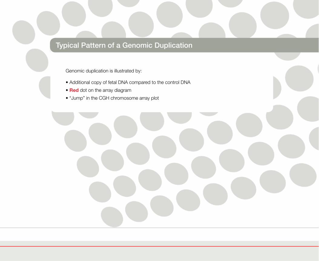
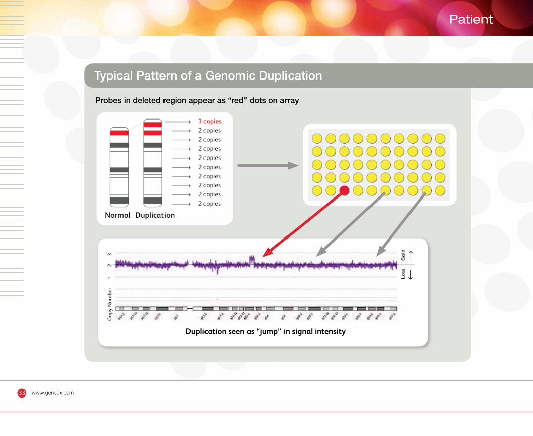
Typical Pattern of a Genomic Duplication
Genomic duplication is illustrated by:
• Additional copy of fetal DNA compared to the control DNA
• Red dot on the array diagram
• “Jump” in the CGH chromosome array plot
33 www.genedx.com
Patient
Duplication seen as “jump” in signal intensity
Typical Pattern of a Genomic Duplication
Probes in deleted region appear as “red” dots on array
34 www.genedx.com
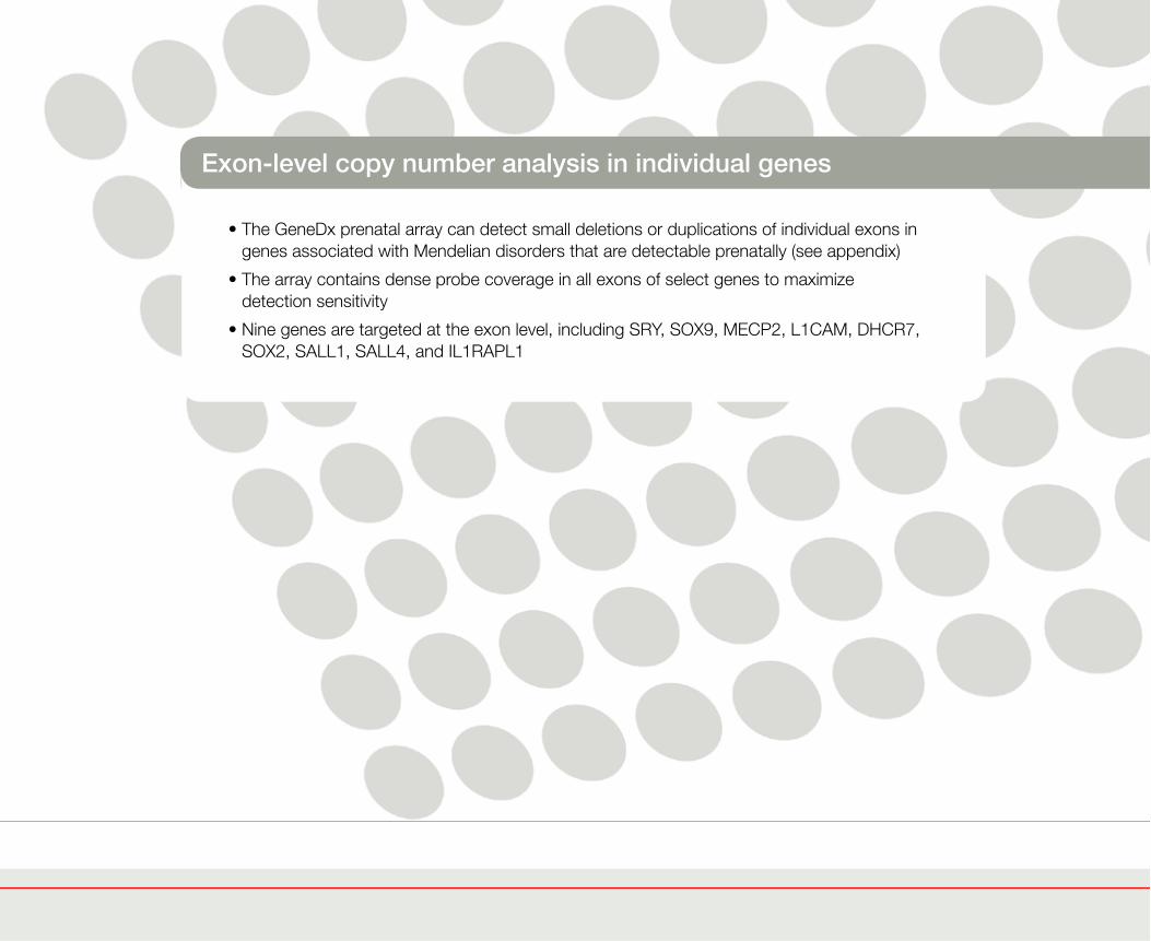
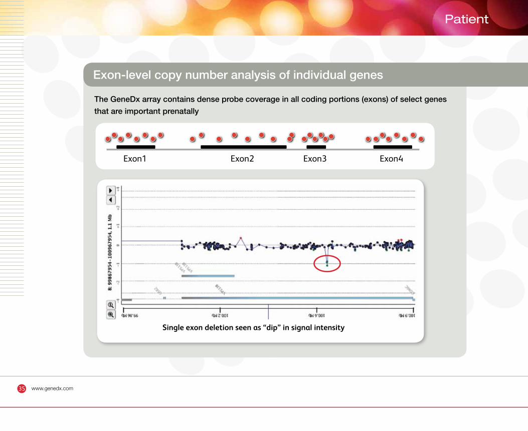
Exon-level copy number analysis in individual genes
• The GeneDx prenatal array can detect small deletions or duplications of individual exons in genes associated with Mendelian disorders that are detectable prenatally (see appendix)
• The array contains dense probe coverage in all exons of select genes to maximize detection sensitivity
• Nine genes are targeted at the exon level, including SRY, SOX9, MECP2, L1CAM, DHCR7, SOX2, SALL1, SALL4, and IL1RAPL1
35 www.genedx.com
Patient
Exon1 Exon2 Exon3 Exon4
Single exon deletion seen as “dip” in signal intensity
Exon-level copy number analysis of individual genes
The GeneDx array contains dense probe coverage in all coding portions (exons) of select genes
that are important prenatally
36 www.genedx.com
Typical Pattern of Uniparental Disomy
(Case example: Prader-Willi syndrome)
Segmental uniparental disomy for chromosome 15 is illustrated by:
• Normal copy number for chromosome 15 illustrated by an even signal along the entire chromosome 15 on the CGH array chromosome graph
• Abnormal SNP array chromosome graph showing absence of signal on middle line (absence of paternal contribution on part of chromosome 15), indicating abnormal parental inheritance.
37 www.genedx.com
Patient
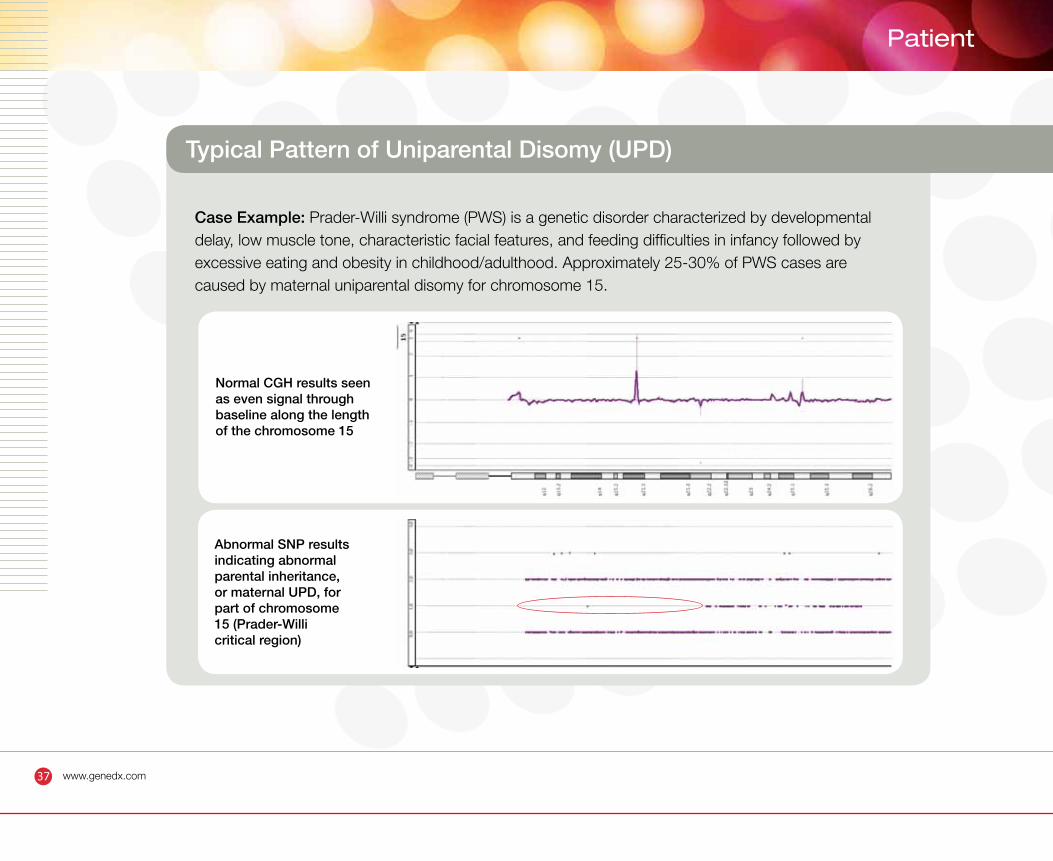
Typical Pattern of Uniparental Disomy (UPD)
Case Example: Prader-Willi syndrome (PWS) is a genetic disorder characterized by developmental delay, low muscle tone, characteristic facial features, and feeding difficulties in infancy followed by excessive eating and obesity in childhood/adulthood. Approximately 25-30% of PWS cases are caused by maternal uniparental disomy for chromosome 15.
Abnormal SNP results indicating abnormal parental inheritance, or maternal UPD, for part of chromosome 15 (Prader-Willi critical region)
Normal CGH results seen as even signal through baseline along the length of the chromosome 15
38 www.genedx.com
A positive or variant of unknown significance is identified What’s next?
• Result confirmation – the abnormal result is confirmed by FISH analysis, quantitative PCR (qPCR), or repeat array analysis
• FISH analysis is performed to determine the chromosomal mechanism for the imbalance, qPCR is used to confirm small aberrations (<1 Mb deletions or duplications), and repeat array analysis is used to confirm multiple chromosomal imbalances, UPD or consanguinity
• Parental samples (if available) are evaluated concurrently with the fetal sample, using the same method
• Abnormal test results are reported out directly to the ordering practitioner(s) by a GeneDx genetic counselor
39 www.genedx.com
Patient
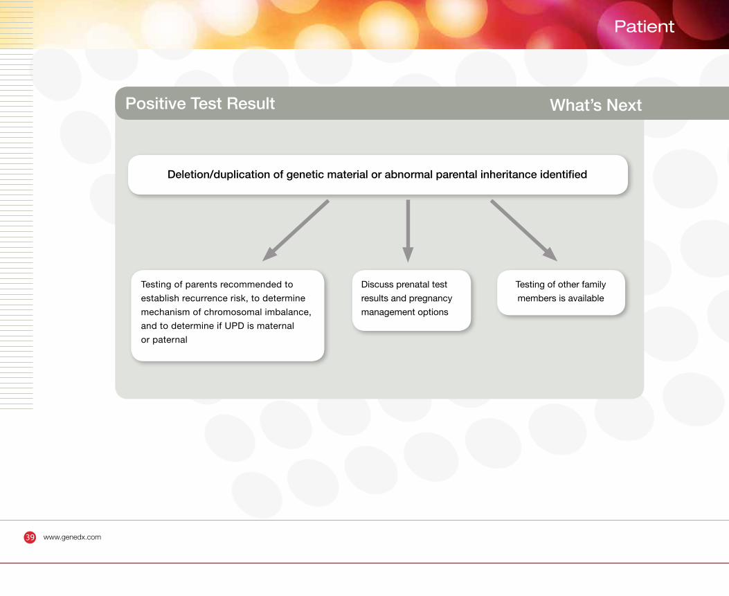
Positive Test Result What’s Next
Deletion/duplication of genetic material or abnormal parental inheritance identified
Testing of parents recommended to
establish recurrence risk, to determine
mechanism of chromosomal imbalance,
and to determine if UPD is maternal
or paternal
Discuss prenatal test
results and pregnancy
management options
Testing of other family
members is available
41 www.genedx.com
Patient
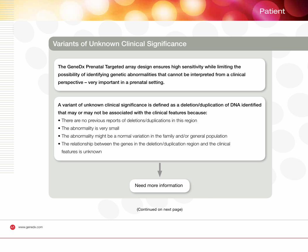
Variants of Unknown Clinical Significance
The GeneDx Prenatal Targeted array design ensures high sensitivity while limiting the
possibility of identifying genetic abnormalities that cannot be interpreted from a clinical
perspective – very important in a prenatal setting.
A variant of unknown clinical significance is defined as a deletion/duplication of DNA identified
that may or may not be associated with the clinical features because:
• There are no previous reports of deletions/duplications in this region
• The abnormality is very small
• The abnormality might be a normal variation in the family and/or general population
• The relationship between the genes in the deletion/duplication region and the clinical
features is unknown
Need more information
(Continued on next page)
43 www.genedx.com
Patient
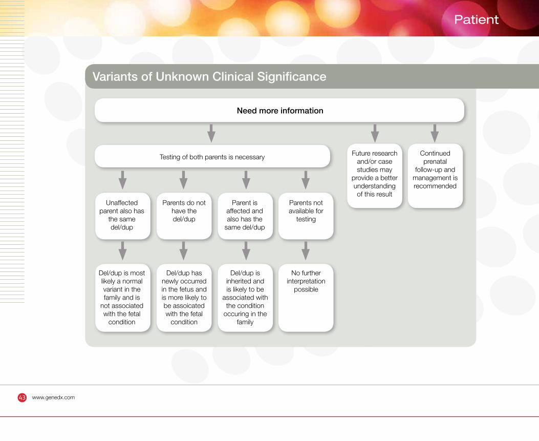
Variants of Unknown Clinical Significance
Need more information
Testing of both parents is necessary
Unaffected parent also has
the same del/dup
Del/dup is most likely a normal variant in the family and is
not associated with the fetal
condition
Del/dup has newly occurred in the fetus and is more likely to be assoicated with the fetal
condition
Del/dup is inherited and is likely to be
associated with the condition
occuring in the family
No further interpretation
possible
Parents do not have the del/dup
Parent is affected and also has the
same del/dup
Parents not available for
testing
Future research and/or case studies may
provide a better understanding of this result
Continued prenatal
follow-up and management is recommended
Other GeneDx Prenatal Tests
Other GeneDx Prenatal Tests
46 www.genedx.com
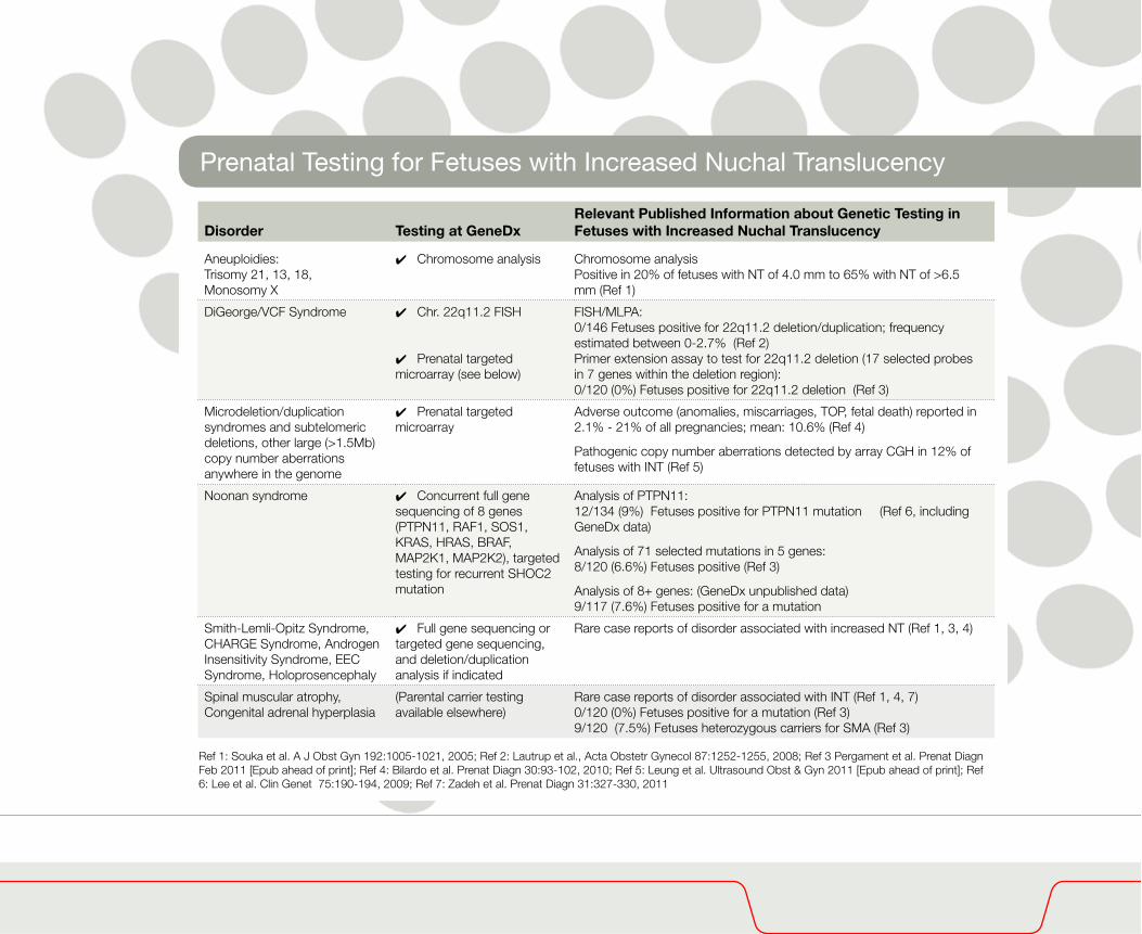
Prenatal Testing for Fetuses with Increased Nuchal Translucency
Disorder Testing at GeneDxRelevant Published Information about Genetic Testing in Fetuses with Increased Nuchal Translucency
Aneuploidies:Trisomy 21, 13, 18, Monosomy X
4 Chromosome analysis Chromosome analysis Positive in 20% of fetuses with NT of 4.0 mm to 65% with NT of >6.5 mm (Ref 1)
DiGeorge/VCF Syndrome 4 Chr. 22q11.2 FISH
4 Prenatal targeted microarray (see below)
FISH/MLPA: 0/146 Fetuses positive for 22q11.2 deletion/duplication; frequency estimated between 0-2.7% (Ref 2) Primer extension assay to test for 22q11.2 deletion (17 selected probes in 7 genes within the deletion region): 0/120 (0%) Fetuses positive for 22q11.2 deletion (Ref 3)
Microdeletion/duplication syndromes and subtelomeric deletions, other large (>1.5Mb) copy number aberrations anywhere in the genome
4 Prenatal targeted microarray
Adverse outcome (anomalies, miscarriages, TOP, fetal death) reported in 2.1% - 21% of all pregnancies; mean: 10.6% (Ref 4)
Pathogenic copy number aberrations detected by array CGH in 12% of fetuses with INT (Ref 5)
Noonan syndrome 4 Concurrent full gene sequencing of 8 genes (PTPN11, RAF1, SOS1, KRAS, HRAS, BRAF, MAP2K1, MAP2K2), targeted testing for recurrent SHOC2 mutation
Analysis of PTPN11:12/134 (9%) Fetuses positive for PTPN11 mutation (Ref 6, including GeneDx data)
Analysis of 71 selected mutations in 5 genes:8/120 (6.6%) Fetuses positive (Ref 3)
Analysis of 8+ genes: (GeneDx unpublished data)9/117 (7.6%) Fetuses positive for a mutation
Smith-Lemli-Opitz Syndrome, CHARGE Syndrome, Androgen Insensitivity Syndrome, EEC Syndrome, Holoprosencephaly
4 Full gene sequencing or targeted gene sequencing, and deletion/duplication analysis if indicated
Rare case reports of disorder associated with increased NT (Ref 1, 3, 4)
Spinal muscular atrophy,Congenital adrenal hyperplasia
(Parental carrier testing available elsewhere)
Rare case reports of disorder associated with INT (Ref 1, 4, 7)0/120 (0%) Fetuses positive for a mutation (Ref 3)9/120 (7.5%) Fetuses heterozygous carriers for SMA (Ref 3)
Ref 1: Souka et al. A J Obst Gyn 192:1005-1021, 2005; Ref 2: Lautrup et al., Acta Obstetr Gynecol 87:1252-1255, 2008; Ref 3 Pergament et al. Prenat Diagn Feb 2011 [Epub ahead of print]; Ref 4: Bilardo et al. Prenat Diagn 30:93-102, 2010; Ref 5: Leung et al. Ultrasound Obst & Gyn 2011 [Epub ahead of print]; Ref 6: Lee et al. Clin Genet 75:190-194, 2009; Ref 7: Zadeh et al. Prenat Diagn 31:327-330, 2011
47 www.genedx.com
Patient
Increased Nuchal Translucency
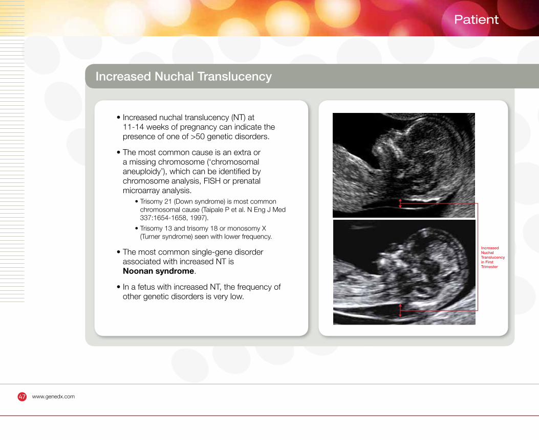
• Increased nuchal translucency (NT) at 11-14 weeks of pregnancy can indicate the presence of one of >50 genetic disorders.
• The most common cause is an extra or a missing chromosome (‘chromosomal aneuploidy’), which can be identified by chromosome analysis, FISH or prenatal microarray analysis.
• Trisomy 21 (Down syndrome) is most common chromosomal cause (Taipale P et al. N Eng J Med 337:1654-1658, 1997).
• Trisomy 13 and trisomy 18 or monosomy X (Turner syndrome) seen with lower frequency.
• The most common single-gene disorder associated with increased NT is Noonan syndrome.
• In a fetus with increased NT, the frequency of other genetic disorders is very low.
Increased Nuchal Translucencyin First Trimester
48 www.genedx.com
Prenatal Testing For Noonan Syndrome
• Autosomal dominant mutations in at least 9 different genes are known to cause Noonan syndrome or related disorders with overlapping features.
• The ‘Prenatal Noonan Panel’ at GeneDx simultaneously analyzes the sequence of eight genes (PTPN11, RAF1, SOS1, KRAS, HRAS, BRAF, MAP2K1, MAP2K2) and one recurrent mutation (SHOC2 ).
• Using this panel, 7.6% of fetuses with increased NT and/or other ultrasound abnormalities tested POSITIVE for a published mutation in these genes.
• 75% of fetuses (9/12) with a disease-causing mutation in PTPN11 had other ultrasound abnormalities with or without increased NT (Lee at al. Clin Genet 75:190-194, 2009)
• Prenatal Noonan syndrome testing is at present the only single-gene disorder test with significant diagnostic relevance for increased NT.
49 www.genedx.com
Patient
Prenatal Testing For Noonan Syndrome
• Noonan syndrome is an autosomal dominant disorder mainly characterized by short stature, congenital heart defects, characteristic facial features. The clinical features can be highly variable, even within a family.
• Certain fetal ultrasound findings, such as cystic hygroma, increased nuchal translucency and fetal hydrops, have been observed in individuals with Noonan syndrome.
• Autosomal dominant mutations in at least 9 different genes are known to cause Noonan syndrome or related disorders with overlapping features.
• The ‘Prenatal Noonan Panel’ at GeneDx simultaneously analyzes the sequence of eight genes (PTPN11, RAF1, SOS1, KRAS, HRAS, BRAF, MAP2K1, MAP2K2) and one recurrent mutation in the SHOC2 gene.
• Prenatal Noonan syndrome testing is at present the only single-gene disorder test with significant diagnostic relevance for increased NT.
50 www.genedx.com
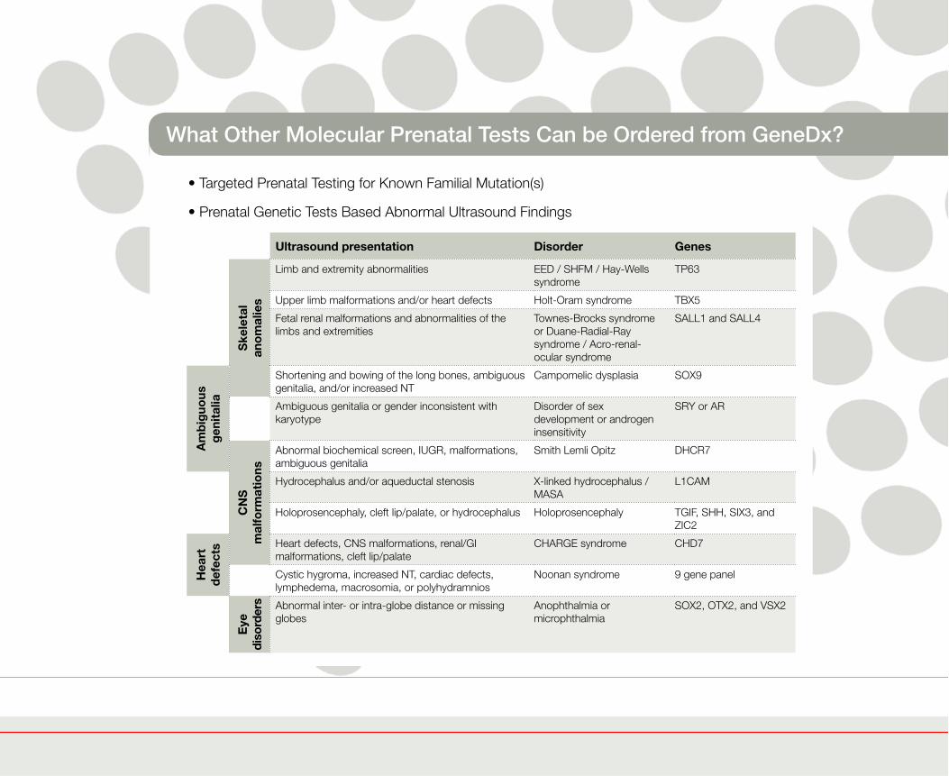
What Other Molecular Prenatal Tests Can be Ordered from GeneDx?
• Targeted Prenatal Testing for Known Familial Mutation(s)
• Prenatal Genetic Tests Based Abnormal Ultrasound Findings
Ultrasound presentation Disorder GenesS
kele
tal
ano
mal
ies
Limb and extremity abnormalities EED / SHFM / Hay-Wells syndrome
TP63
Upper limb malformations and/or heart defects Holt-Oram syndrome TBX5
Fetal renal malformations and abnormalities of the limbs and extremities
Townes-Brocks syndrome or Duane-Radial-Ray syndrome / Acro-renal-ocular syndrome
SALL1 and SALL4
Am
big
uous
g
enit
alia
Shortening and bowing of the long bones, ambiguous genitalia, and/or increased NT
Campomelic dysplasia SOX9
Ambiguous genitalia or gender inconsistent with karyotype
Disorder of sex development or androgen insensitivity
SRY or AR
CN
S
mal
form
atio
ns
Abnormal biochemical screen, IUGR, malformations, ambiguous genitalia
Smith Lemli Opitz DHCR7
Hydrocephalus and/or aqueductal stenosis X-linked hydrocephalus / MASA
L1CAM
Holoprosencephaly, cleft lip/palate, or hydrocephalus Holoprosencephaly TGIF, SHH, SIX3, and ZIC2
Hea
rt
def
ects Heart defects, CNS malformations, renal/GI
malformations, cleft lip/palateCHARGE syndrome CHD7
Cystic hygroma, increased NT, cardiac defects, lymphedema, macrosomia, or polyhydramnios
Noonan syndrome 9 gene panel
Eye
di
sord
ers Abnormal inter- or intra-globe distance or missing
globes
Anophthalmia or microphthalmia
SOX2, OTX2, and VSX2
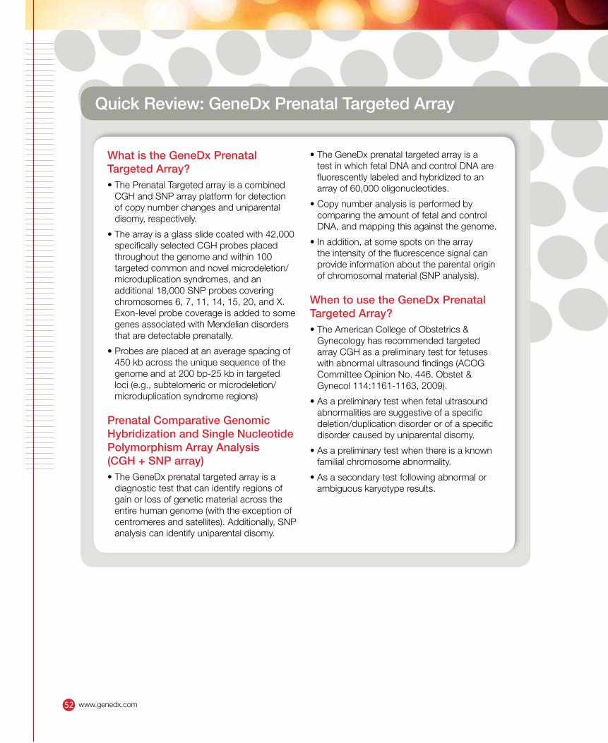
Quick Review: GeneDx Prenatal Targeted Array
What is the GeneDx Prenatal Targeted Array?• The Prenatal Targeted array is a combined
CGH and SNP array platform for detection of copy number changes and uniparental disomy, respectively.
• The array is a glass slide coated with 42,000 specifically selected CGH probes placed throughout the genome and within 100 targeted common and novel microdeletion/microduplication syndromes, and an additional 18,000 SNP probes covering chromosomes 6, 7, 11, 14, 15, 20, and X. Exon-level probe coverage is added to some genes associated with Mendelian disorders that are detectable prenatally.
• Probes are placed at an average spacing of 450 kb across the unique sequence of the genome and at 200 bp-25 kb in targeted loci (e.g., subtelomeric or microdeletion/microduplication syndrome regions)
Prenatal Comparative Genomic Hybridization and Single Nucleotide Polymorphism Array Analysis (CGH + SNP array)• The GeneDx prenatal targeted array is a
diagnostic test that can identify regions of gain or loss of genetic material across the entire human genome (with the exception of centromeres and satellites). Additionally, SNP analysis can identify uniparental disomy.
• The GeneDx prenatal targeted array is a test in which fetal DNA and control DNA are fluorescently labeled and hybridized to an array of 60,000 oligonucleotides.
• Copy number analysis is performed by comparing the amount of fetal and control DNA, and mapping this against the genome.
• In addition, at some spots on the array the intensity of the fluorescence signal can provide information about the parental origin of chromosomal material (SNP analysis).
When to use the GeneDx Prenatal Targeted Array?• The American College of Obstetrics &
Gynecology has recommended targeted array CGH as a preliminary test for fetuses with abnormal ultrasound findings (ACOG Committee Opinion No. 446. Obstet & Gynecol 114:1161-1163, 2009).
• As a preliminary test when fetal ultrasound abnormalities are suggestive of a specific deletion/duplication disorder or of a specific disorder caused by uniparental disomy.
• As a preliminary test when there is a known familial chromosome abnormality.
• As a secondary test following abnormal or ambiguous karyotype results.
www.genedx.com52
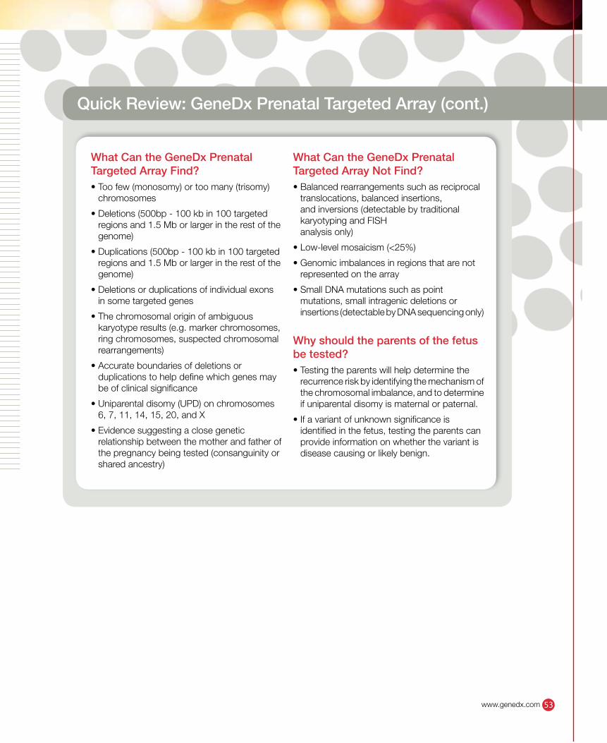
Quick Review: GeneDx Prenatal Targeted Array (cont.)
What Can the GeneDx Prenatal Targeted Array Find?• Too few (monosomy) or too many (trisomy)
chromosomes
• Deletions (500bp - 100 kb in 100 targeted regions and 1.5 Mb or larger in the rest of the genome)
• Duplications (500bp - 100 kb in 100 targeted regions and 1.5 Mb or larger in the rest of the genome)
• Deletions or duplications of individual exons in some targeted genes
• The chromosomal origin of ambiguous karyotype results (e.g. marker chromosomes, ring chromosomes, suspected chromosomal rearrangements)
• Accurate boundaries of deletions or duplications to help define which genes may be of clinical significance
• Uniparental disomy (UPD) on chromosomes 6, 7, 11, 14, 15, 20, and X
• Evidence suggesting a close genetic relationship between the mother and father of the pregnancy being tested (consanguinity or shared ancestry)
What Can the GeneDx Prenatal Targeted Array Not Find?• Balanced rearrangements such as reciprocal
translocations, balanced insertions, and inversions (detectable by traditional karyotyping and FISH analysis only)
• Low-level mosaicism (<25%)
• Genomic imbalances in regions that are not represented on the array
• Small DNA mutations such as point mutations, small intragenic deletions or insertions (detectable by DNA sequencing only)
Why should the parents of the fetus be tested?• Testing the parents will help determine the
recurrence risk by identifying the mechanism of the chromosomal imbalance, and to determine if uniparental disomy is maternal or paternal.
• If a variant of unknown significance is identified in the fetus, testing the parents can provide information on whether the variant is disease causing or likely benign.
53www.genedx.com
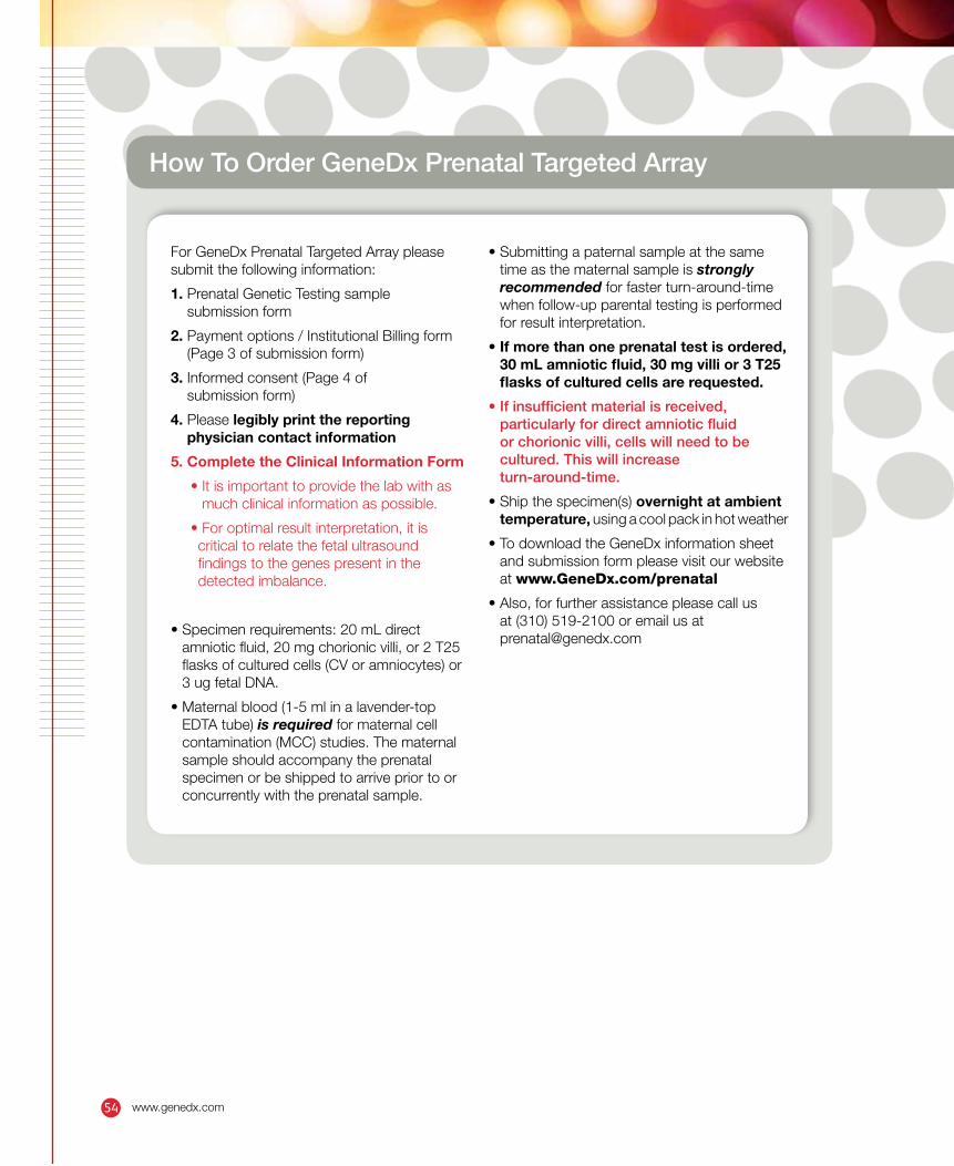
How To Order GeneDx Prenatal Targeted Array
For GeneDx Prenatal Targeted Array please submit the following information:
1. Prenatal Genetic Testing sample submission form
2. Payment options / Institutional Billing form (Page 3 of submission form)
3. Informed consent (Page 4 of submission form)
4. Please legibly print the reporting physician contact information
5. Complete the Clinical Information Form
• It is important to provide the lab with as much clinical information as possible.
• For optimal result interpretation, it is critical to relate the fetal ultrasound findings to the genes present in the detected imbalance.
• Specimen requirements: 20 mL direct amniotic fluid, 20 mg chorionic villi, or 2 T25 flasks of cultured cells (CV or amniocytes) or 3 ug fetal DNA.
• Maternal blood (1-5 ml in a lavender-top EDTA tube) is required for maternal cell contamination (MCC) studies. The maternal sample should accompany the prenatal specimen or be shipped to arrive prior to or concurrently with the prenatal sample.
• Submitting a paternal sample at the same time as the maternal sample is strongly recommended for faster turn-around-time when follow-up parental testing is performed for result interpretation.
•���If�more�than�one�prenatal�test�is�ordered,�30 mL amniotic fluid, 30 mg villi or 3 T25 flasks of cultured cells are requested.
•���If�insufficient�material�is�received,�particularly for direct amniotic fluid or chorionic villi, cells will need to be cultured. This will increase turn-around-time.
• Ship the specimen(s) overnight at ambient temperature, using a cool pack in hot weather
• To download the GeneDx information sheet and submission form please visit our website at www.GeneDx.com/prenatal
• Also, for further assistance please call us at (310) 519-2100 or email us at [email protected]
www.genedx.com54
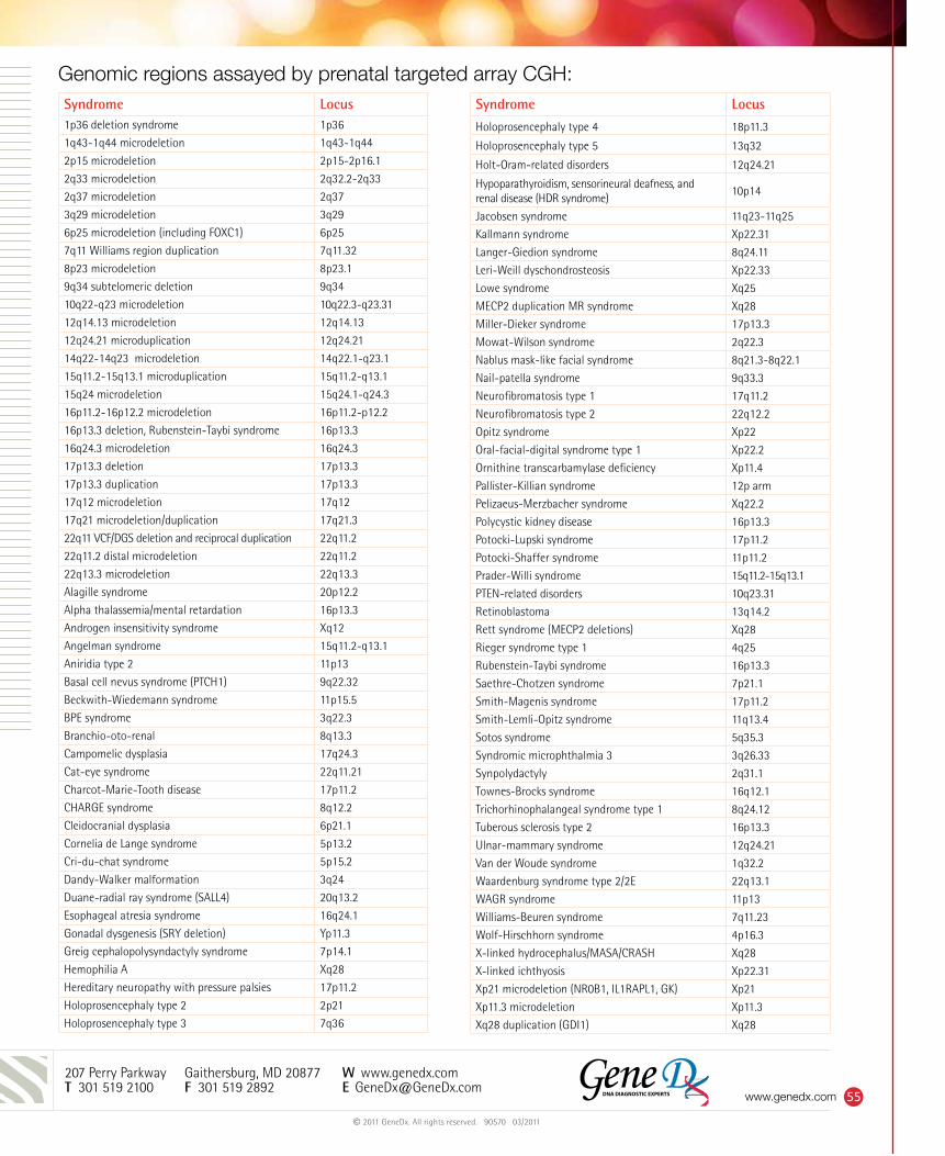
Genomic regions assayed by prenatal targeted array CGH:
207 Perry Parkway Gaithersburg, MD 20877 W www.genedx.com T 301 519 2100 F 301 519 2892 E [email protected]
Syndrome Locus1p36 deletion syndrome 1p36
1q43-1q44 microdeletion 1q43-1q44
2p15 microdeletion 2p15-2p16.1
2q33 microdeletion 2q32.2-2q33
2q37 microdeletion 2q37
3q29 microdeletion 3q29
6p25 microdeletion (including FOXC1) 6p25
7q11 Williams region duplication 7q11.32
8p23 microdeletion 8p23.1
9q34 subtelomeric deletion 9q34
10q22-q23 microdeletion 10q22.3-q23.31
12q14.13 microdeletion 12q14.13
12q24.21 microduplication 12q24.21
14q22-14q23 microdeletion 14q22.1-q23.1
15q11.2-15q13.1 microduplication 15q11.2-q13.1
15q24 microdeletion 15q24.1-q24.3
16p11.2-16p12.2 microdeletion 16p11.2-p12.2
16p13.3 deletion, Rubenstein-Taybi syndrome 16p13.3
16q24.3 microdeletion 16q24.3
17p13.3 deletion 17p13.3
17p13.3 duplication 17p13.3
17q12 microdeletion 17q12
17q21 microdeletion/duplication 17q21.3
22q11 VCF/DGS deletion and reciprocal duplication 22q11.2
22q11.2 distal microdeletion 22q11.2
22q13.3 microdeletion 22q13.3
Alagille syndrome 20p12.2
Alpha thalassemia/mental retardation 16p13.3
Androgen insensitivity syndrome Xq12
Angelman syndrome 15q11.2-q13.1
Aniridia type 2 11p13
Basal cell nevus syndrome (PTCH1) 9q22.32
Beckwith-Wiedemann syndrome 11p15.5
BPE syndrome 3q22.3
Branchio-oto-renal 8q13.3
Campomelic dysplasia 17q24.3
Cat-eye syndrome 22q11.21
Charcot-Marie-Tooth disease 17p11.2
CHARGE syndrome 8q12.2
Cleidocranial dysplasia 6p21.1
Cornelia de Lange syndrome 5p13.2
Cri-du-chat syndrome 5p15.2
Dandy-Walker malformation 3q24
Duane-radial ray syndrome (SALL4) 20q13.2
Esophageal atresia syndrome 16q24.1
Gonadal dysgenesis (SRY deletion) Yp11.3
Greig cephalopolysyndactyly syndrome 7p14.1
Hemophilia A Xq28
Hereditary neuropathy with pressure palsies 17p11.2
Holoprosencephaly type 2 2p21
Holoprosencephaly type 3 7q36
Syndrome Locus
Holoprosencephaly type 4 18p11.3
Holoprosencephaly type 5 13q32
Holt-Oram-related disorders 12q24.21
Hypoparathyroidism, sensorineural deafness, and renal disease (HDR syndrome)
10p14
Jacobsen syndrome 11q23-11q25
Kallmann syndrome Xp22.31
Langer-Giedion syndrome 8q24.11
Leri-Weill dyschondrosteosis Xp22.33
Lowe syndrome Xq25
MECP2 duplication MR syndrome Xq28
Miller-Dieker syndrome 17p13.3
Mowat-Wilson syndrome 2q22.3
Nablus mask-like facial syndrome 8q21.3-8q22.1
Nail-patella syndrome 9q33.3
Neurofibromatosis type 1 17q11.2
Neurofibromatosis type 2 22q12.2
Opitz syndrome Xp22
Oral-facial-digital syndrome type 1 Xp22.2
Ornithine transcarbamylase deficiency Xp11.4
Pallister-Killian syndrome 12p arm
Pelizaeus-Merzbacher syndrome Xq22.2
Polycystic kidney disease 16p13.3
Potocki-Lupski syndrome 17p11.2
Potocki-Shaffer syndrome 11p11.2
Prader-Willi syndrome 15q11.2-15q13.1
PTEN-related disorders 10q23.31
Retinoblastoma 13q14.2
Rett syndrome (MECP2 deletions) Xq28
Rieger syndrome type 1 4q25
Rubenstein-Taybi syndrome 16p13.3
Saethre-Chotzen syndrome 7p21.1
Smith-Magenis syndrome 17p11.2
Smith-Lemli-Opitz syndrome 11q13.4
Sotos syndrome 5q35.3
Syndromic microphthalmia 3 3q26.33
Synpolydactyly 2q31.1
Townes-Brocks syndrome 16q12.1
Trichorhinophalangeal syndrome type 1 8q24.12
Tuberous sclerosis type 2 16p13.3
Ulnar-mammary syndrome 12q24.21
Van der Woude syndrome 1q32.2
Waardenburg syndrome type 2/2E 22q13.1
WAGR syndrome 11p13
Williams-Beuren syndrome 7q11.23
Wolf-Hirschhorn syndrome 4p16.3
X-linked hydrocephalus/MASA/CRASH Xq28
X-linked ichthyosis Xp22.31
Xp21 microdeletion (NR0B1, IL1RAPL1, GK) Xp21
Xp11.3 microdeletion Xp11.3
Xq28 duplication (GDI1) Xq28
© 2011 GeneDx. All rights reserved. 90570 03/2011
55www.genedx.com