Embed Size (px)
Citation preview

Gel electrophoresis Ashti Mohammad Amin M.Sc. Molecular Biology
Medical Research Center Hawler Medical University- 2012

Gel electrophoresis
• Gel electrophoresis is a technique used for the separation of (DNA), (RNA), and protein molecules using an electric field applied to a gel matrix. DNA Gel electrophoresis is usually performed for analytical purposes, often after amplification of DNA via PCR.

used as a preparative technique prior to use of other methods such as mass spectrometry, RFLP, PCR, cloning, DNA sequencing, or Southern blotting for further characterization

The term "gel" in this instance refers to the matrix used to contain, then separate the target molecules. In most cases, the gel is a crosslinked polymer whose
composition and porosity is chosen based on the specific weight and composition of the target to be
analyzed.

Common applications of gel electrophoresis process
• Embryo screening for genetic diseases • Genotyping or genetic fingerprinting• Epidemological studies of pathogenic organisms • Cloning large plant DNA using YACs, Identifying RFLP.• Detection in vivo chromosomes from yeasts, fungi
and parasites ……..

Types of gel electrophoresis
• SDS-PAGE• Pulsed field gel electrophoresis (PFGE)• Agarose gel electrophoresis

SDS –PAGESodium dodecyl sulfate polyacrylamide gel
electrophoresis
• Separate proteins according to their electrophoretic mobility (a function of the length of a polypeptide chain and its charge ) .

SDS-PAGE
Tertiary
Negative charge
Positive charge

• Is a technique used for the separation of large DNA molecules like (fungi ,yeasts, parasites…..) by applying an electric field that periodically changes direction to a gel matrix .
Pulsed field gel electrophoresis (PFGE)

PFGE

•
Agarose gel electrophoresis
Agarose gel electrophoresis is the easiest and commonest way of separation and analysis DNA .
Using agarose gel electrophoresis to determine the presence and size of PCR products.

Casting tray Gel combs
Power supply
Gel tank Cover
Electrical leads
Electrophoresis EquipmentElectrophoresis Equipment

The Materials necessary for conducting agarose gel electrophoresis
• A garose • Electrophoresis buffer• Loading dye • Ethidium bromide• Transilluminator UV light.

• What is agarose ?Is a chain of sugar extracted from seaweed .
Agarose
DD-galactose-galactose
3,6-anhydroL-galactose3,6-anhydroL-galactose

Making an agarose gel .
An agarose gel is prepeard by combining agarose powder and a buffer solution
Use a flask that is several times larger than the volume of buffer, because the agarose boils over easily

Melting the Agarose
Agarose is insoluble at room temperature (left). The agarose solution is boiled until clear (right).
Leave it to cool on the bench for 5 minutes down to about 60 C.

Staining the Gelwith ethidium bromide
Ethidium bromide can be added to the gel and/or running buffer before the gel is run or the gel can be stained after it has run. Ethidium bromide binds to DNA and fluoresces under UV light, allowing the visualization of DNA on a Gel.

Gel casting tray & combs
A comb is put in the gel to create wells , each of the gel combs should be submerged in the melted agarose solution.

Carefully pour the melted agarose solution with ethedium bromide dye into the casting tray.
Avoid air bubbles.

When cooled, the agarose polymerizes, forming a flexible gel. It should appear lighter in color when completely cooled (30-45 minutes). Carefully remove the combs and tape.

Place the gel in the gel tank ,add enough electrophoresis buffer to cover the gel to a depth of at least 1 mm. Make sure each well is filled
with buffer.
buffer
Cathode(negative)
Anode(positive)
wells
DNA

with buffer.Sample Preparation
Mix the samples of DNA with the 6X sample loading buffer .This allows the samples to be seen when loading onto the gel, and increases the density of the samples, causing them to sink into the gel wells.
6X Loading Buffer: Bromophenol Blue (for color) Glycerol (for weight)
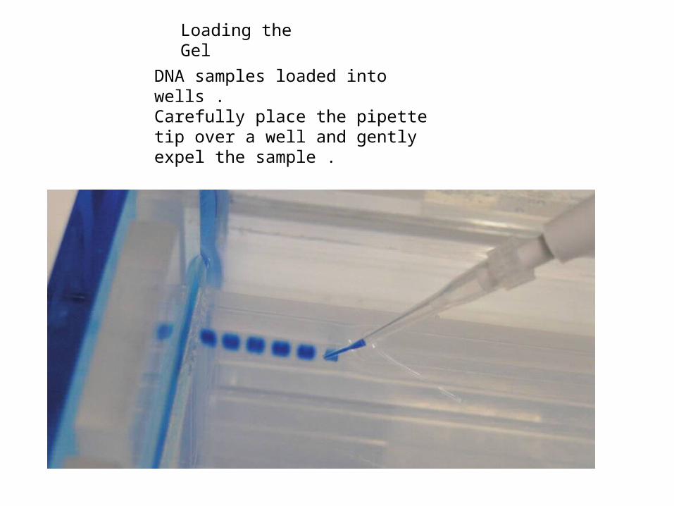
DNA samples loaded into wells .Carefully place the pipette tip over a well and gently expel the sample .
Loading the Gel

Running the GelRunning the Gel
Place the cover on the electrophoresis chamber, connecting the electrical leads. Connect the electrical leads to the power supply. Be sure the leads are attached correctly - DNA migrates toward the anode (red). When the power is turned on, bubbles should form on the electrodes in the electrophoresis chamber.

Cathode(-)
Anode
(+) DNA(-)
Anode(+)
Anode(+)
DNA(-)
DNA(-)
wellsAfter the current is applied, make sure the gel is running in the correct direction. Bromophenol blue will run in the same direction as the DNA.
Bromophenol Blue

Ethidium Bromide -stained gel requires an ultraviolet light source to visualize


Visualizing the DNA (QuikVI stain)
250
1,500 1,000
500 750
2,000 bp
DNA ladder
+ - - - - + + - - + - +
100 200 300
1,650
1,000
500
850
650
400
12,000 bp
5,000
2,000
DNA Ladder Standard
Inclusion of a DNA ladder (DNAs of know sizes) on the gel makes it easy to determine the sizes of unknown DNAs.
-
+
DNAmigration
bromophenol blue
bromophenol blue migrates at approximately the same rate as a 300 bp DNA molecule

DNA is negatively charged

How fast will the DNA migrate?Strength of the electrical field, density of agarose and size of the DNA (Small DNA move faster than large DNA)…gel electrophoresis separates DNA according to size
+-
Power
DNA
smalllarge
DNA
DNA

Visualizing the DNA (ethidium bromide))

Thank you

• Thank you


![[XLS]drtktopecollege.indrtktopecollege.in/pol/sites/default/files/detail... · Web viewANANDRAO GANGARAM GEDAM Arni SHIWAJIRAO SHIWRAMJI MOGHE Arvi DADARAO YADAVRAOJI KECHE Ashti](https://img.dokumen.tips/doc/110x75/5aa7ef1b7f8b9ad31c8c91fd/xls-viewanandrao-gangaram-gedam-arni-shiwajirao-shiwramji-moghe-arvi-dadarao-yadavraoji.jpg)