Embed Size (px)
Citation preview

Ashley Cullen, Michael Lerch, Marco Petasecca, Susanna Guatelli, Anatoly RosenfeldCentre for Medical Radiation Physics, University of Wollongong, Australia
Geant4 Workshop 2011Wollongong, Australia

Introduction to MRT
Dosimetry for MRT
The Silicon Microstrip Detector
Energy Dependence Problem
Monte Carlo Simulations
Conclusions

In the early days of radiotherapy Co-60 and kilovoltage x-ray units were the dominant radiotherapy modality.
Superficial doses were high, causing excessive skin burn
Discovery that iron grids pressed hard against the skin caused regions shielded from radiation healed adjacent burns
In 1960’s, experiments investigate the effect of 22.5 MeV 25μm deuteron microbeams on mice cerebrum
◦ Doses in excess of 3,000 Gy caused little damage
◦ Range of 1.5mm in tissue was of little clinical use
Microtomography experiments conducted with 30μm synchrotron pencil beams on mouse heads had little contrast at 10 Gy, so dose was increased to 200 Gy.
◦ A month later, a histopathological search failed to find any radiation damage.

A study investigated the effect of 25μm-wide planar 50-150 keV synchrotron x-rays on murine brains
◦ Thousands of Gy were survived
◦ Microbeam radiation therapy (MRT) was born
MRT under investigation as an experimental treatment for untreatable paediatric brain tumours.
A “white” synchrotron beam is used (ID17 @ ESRF, Grenoble)
A striated planar array of beams tens-of μmwide and hundreds-of μm centre-to-centre
Peak-to-valley dose ratio very important parameter
[1] E.A Siegbhan, et al., Dosimetry for synchrotron x-ray microbeam radiation therapy, ESRF[2] E. Braüer-Krisch, et al., Effects of pulsed, spatially fractionated, microscopic synchrotron X-ray beamson normal and tumoral brain tissue
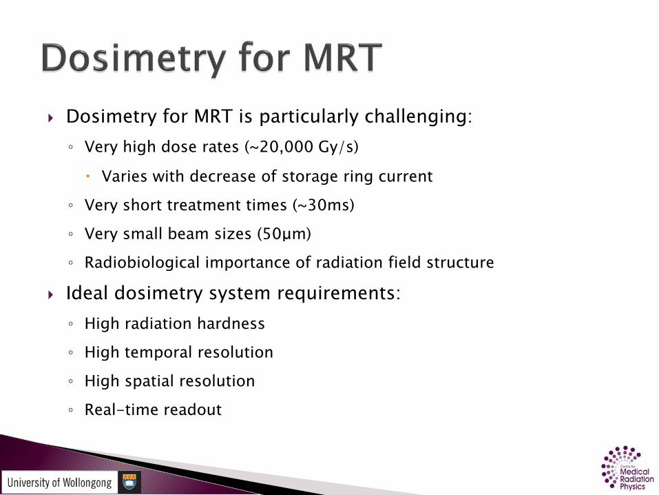
Dosimetry for MRT is particularly challenging:◦ Very high dose rates (~20,000 Gy/s)
Varies with decrease of storage ring current
◦ Very short treatment times (~30ms)
◦ Very small beam sizes (50μm)
◦ Radiobiological importance of radiation field structure
Ideal dosimetry system requirements:◦ High radiation hardness
◦ High temporal resolution
◦ High spatial resolution
◦ Real-time readout
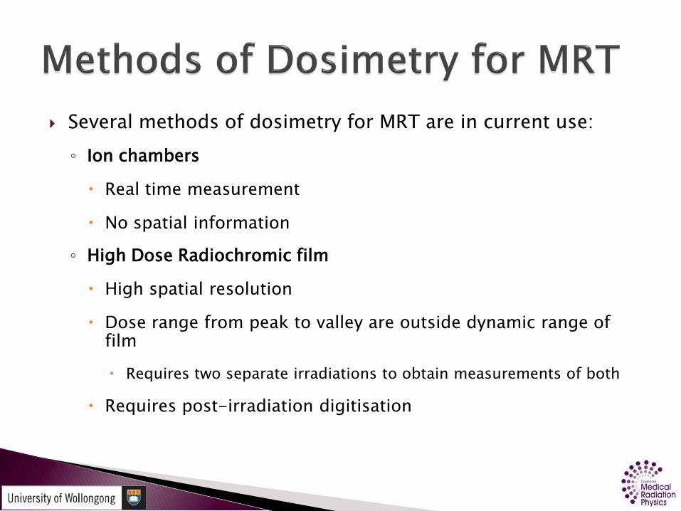
Several methods of dosimetry for MRT are in current use:◦ Ion chambers
Real time measurement
No spatial information
◦ High Dose Radiochromic film
High spatial resolution
Dose range from peak to valley are outside dynamic range of film
Requires two separate irradiations to obtain measurements of both
Requires post-irradiation digitisation
Experimental dosimetry methods:◦ MOSFETs Provides high-spatial resolution when scanned
through beam Not radiation hard to intense synchrotron radiation◦ Fibre-optic◦ OSL

Epitaxial construction◦ 26 μm thick epitaxial layer◦ On 370 μm Si substrate
Sensitive volume is 20 µm × 500 μm
Surrounded by a guard-ring

10 100 10000
1
2
3
4
5
6
7
8
Photon Energy (keV)
γ Mas
s En
ergy
Abs
. Coe
ff. R
atio
(Si/W
ater
)
0.0
0.2
0.4
0.6
0.8
1.0
ID17
Nor
mal
ised
Phot
on S
pect
rum

Monochromatic synchrotron x-rays
ID17 @ ESRF, Grenoble Produced by a double-crystal
monochromator Dose to microstrip compared
to tissue-eq. ionisationchamber results
Displays low energy over-response

0 10000 20000 300001E-5
1E-4
1E-3
0.01
0.1
1
Norm
alise
d In
tens
ity
Distance (arb units)
Left Right

The radiation transport simulation toolkit Geant4 (v9.3) was used for all Monte Carlo simulations described herein
The Low Energy Electromagnetic Physics Package was used◦ Provides accurate tracking of photons to energies of 250 eV
◦ Agrees with NIST data Amako, et al., Comparison of Geant4 Electromagnetic Physics Models Against the NIST Reference
Data, IEEE Trans. Nuc. Sci., Vol. 52, No. 4, August 2005
To improve simulation speed, beamline components were not modeled
Microbeams were assumed to have a homogeneous energy and fluence distribution
Polarisation not modeled◦ see G. Takacs, Photon polarisation, magnetic fields, and electron trajectories: the
irrelevance of the electron mean free path

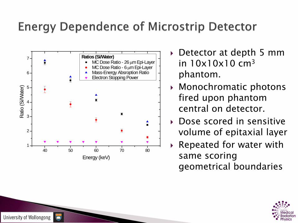
Detector at depth 5 mm in 10x10x10 cm3
phantom. Monochromatic photons
fired upon phantom central on detector.
Dose scored in sensitive volume of epitaxial layer
Repeated for water with same scoring geometrical boundaries
40 50 60 70 801
2
3
4
5
6
7
Ratios (Si/Water) MC Dose Ratio - 26 µm Epi-Layer MC Dose Ratio - 6 µm Epi-Layer Mass-Energy Absroption Ratio Electron Stopping Power
Ratio
(Si/W
ater
)
Energy (keV)

Microstrip placed at various depths in water phantom
Irradiated with the MRT spectrum in a homogeneous field
Dose to microstrip and water in same geometric boundaries scored
Water dose equivalence obtained from ratio

0 20 40 60 802
4
6
8
10
12
14
16
18
Dose
per
Prim
ary
Phot
on x
10-1
1 (Gy/
n)
Depth in Phantom (mm)
Microstrip Detector Water
0 20 40 60 80
1.45
1.50
1.55
1.60
1.65
1.70
1.75
Wat
er E
quiva
lenc
e Ra
tio (D
Si/D
w)Depth (mm)

A simulation was produced to determine the photon spectrum outside of microbeams
An infinitesimally thin pencil beam was fired into a 10×10×10 cm3 water phantom normal to a central surface
The spectrum of photons entering a 5×5×5 mm3
sensitive volume were binned in 1 keV increments.
Performed at various positions

1 10 1001
10
100
1000
10000
100000
Cou
nts
Photon Energy (keV)
Distance fromBeam Centre
0 mm 1 mm 5 mm 20 mm
20 25 30 35 40 45 501
10
100
1000
10000
100000
Counts
Photon Energy (keV)
Distance fromBeam Centre
0 mm 1 mm 5 mm 20 mm
Previous simulation repeated with monochromatic photons
To isolate which part of the input spectrum is most responsible
Energies:◦ 40 keV
◦ 80 keV
◦ 100 keV
◦ 400 keV

0 10 20 30 40 50 60 70 801
10
100
1000
10000
100000
1000000
1E7
Coun
ts
Photon Energy (keV)
Lateral Pos. 0 mm 1 mm 5 mm 20 mm
0 100 200 300 4001
10
100
1000
10000
100000
1000000
1E7
Coun
ts
Photon Energy (keV)
Lateral Pos. 0 mm 1 mm 5 mm 20 mm
0 10 20 30 Comptonmin.
401
10
100
1000
10000
100000
1000000
1E7
Coun
ts
Photon Energy (keV)
Lateral Pos. 0 mm 1 mm 5 mm 20 mm
0 20 40 60 80 1001
10
100
1000
10000
100000
1000000
1E7
Coun
ts
Photon Energy (keV)
Lateral Pos. 0 mm 1 mm 5 mm 20 mm
40 keV
400 keV
80 keV
100 keV
The photon spectrum out-of-field due to being as a result of Compton scatter
A 25-35 keV low-energy photon component is present which does not attenuate with distance◦ This is the region Si is most sensitive to
◦ Produced by Compton scattering of previously scattered photons.
80 – 100 keV primary photons most responsible◦ Requires more investigation

A new detector has been developed to reduce the low energy over response.
Silicon-on-Insulator construction
◦ 7μm Si detector on 2μm SiO2 insulator
◦ Constructed on 370μm Si substrate
Ion-beam-induced charge-collection studies have been performed, but yet to be analysed.
Will be tested with a synchrotron MRT field at ESRF, France in October
Microstrip detector is over sensitive to low energy photons
Response will vary with spectrum
◦ i.e. depth, in-field/out-of field
The use of Geant4 has been used to design a less energy-dependent detector
◦ Has yet to be experimentally tested