Embed Size (px)
Citation preview
Horizontal Gene Transfer Contributes to the Evolution
of Arthropod Herbivory
Nicky Wybouw,*,1 Yannick Pauchet,2 David G. Heckel,2 and Thomas Van Leeuwen*,1,3
1Department of Evolutionary Biology, Institute for Biodiversity and Ecosystem Dynamics, University of Amsterdam, Amsterdam, the Netherlands2Department of Entomology, Max Planck Institute for Chemical Ecology, Jena, Germany3Laboratory of Agrozoology, Department of Crop Protection, Faculty of Bioscience Engineering, Ghent University, Gent, Belgium
*Corresponding author: E-mail: [email protected]; [email protected].
Accepted: May 9, 2016
Abstract
Within animals, evolutionary transition toward herbivory is severely limited by the hostile characteristics of plants. Arthropods have
nonetheless counteracted many nutritional and defensive barriers imposed by plants and are currently considered as the most
successful animal herbivores in terrestrial ecosystems. We gather a body of evidence showing that genomes of various plant feeding
insects and mites possess genes whose presence can only be explained by horizontal gene transfer (HGT). HGT is the asexual
transmission of genetic information between reproductively isolated species. Although HGT is known to have great adaptive signif-
icance in prokaryotes, its impact on eukaryotic evolution remains obscure. Here, we show that laterally transferred genes into
arthropods underpin many adaptations to phytophagy, including efficient assimilation and detoxification of plant produced metab-
olites. Horizontally acquired genes and the traits they encode often functionally diversify within arthropod recipients, enabling the
colonization of more host plant species and organs. We demonstrate that HGT can drive metazoan evolution by uncovering its
prominent role in the adaptations of arthropods to exploit plants.
Key words: arthropods, horizontal gene transfer, herbivory.
Introduction
Plants have evolved a wide and complex set of adaptations to
survive the many stressors present in their environment. These
include abiotic factors such as drought and nutrition-poor
soils, but also herbivore and pathogen attacks. During their
evolution, plants have transformed themselves to such a re-
calcitrant and unfavorable food source that herbivory (or feed-
ing exclusively on plant material) has only evolved in roughly
one third of all animal species (Strong et al. 1984). Both the
nutritional and defensive qualities of plants are known to limit
the evolution of animal herbivory (Awmack and Leather
2002). As many plant species and tissues are characterized
by a relatively high carbohydrate content, but low protein
and vitamin levels (Mattson 1980; Strong et al. 1984;
Awmack and Leather 2002), herbivorous animals need to op-
timize the assimilation of the limited supply of nutrients in
plant tissues. In addition, plants have evolved multi-layered
defense strategies that negatively affect the reproductive per-
formance of herbivores. Plant resistance to herbivory can be
achieved by physical barriers such as trichomes and waxy
cuticles and/or by the production of chemical defenses
(Howe and Jander 2008). Defensive phytochemicals, or
plant allelochemicals, can repulse and poison herbivores or
interfere with the assimilation of plant nutrients inside the
herbivore’s gut (Whittaker and Feeny 1971).
Nevertheless, arthropods can overcome these nutritional
and defensive barriers and various lineages have successfully
adapted to a phytophagous lifestyle. The arthropod phylum is
now considered to harbor the most successful animal herbi-
vores in terrestrial ecosystems, both in terms of biomass and
species diversity (Strong et al. 1984; Labandeira 2002, 2006;
Schoonhoven et al. 2006) (fig. 1). Within arthropod–plant in-
teractions, there has been an evolutionary trend for phytoph-
agous arthropods to further specialize to a specific plant family
or even species by optimizing the assimilation and detoxifica-
tion processes (Bernays and Graham, 1988; Schoonhoven
et al. 2006). Using next-generation sequencing-based meth-
ods, many studies are currently investigating the genomic and
genetic innovations that are associated with these evolution-
ary transitions to arthropod herbivory and further host plant
GBE
� The Author 2016. Published by Oxford University Press on behalf of the Society for Molecular Biology and Evolution.
This is an Open Access article distributed under the terms of the Creative Commons Attribution Non-Commercial License (http://creativecommons.org/licenses/by-nc/4.0/), which permits
non-commercial re-use, distribution, and reproduction in any medium, provided the original work is properly cited. For commercial re-use, please contact [email protected]
Genome Biol. Evol. 8(6):1785–1801. doi:10.1093/gbe/evw119 Advance Access publication June 15, 2016 1785
specializations. Of the identified arthropod genes that code
for enzymes with a clear selective advantage to the phytoph-
agous lifestyle, a subset has been identified with two unusual
properties: high similarity to microbial genes, and absence in
the majority of other lineages within the Arthropoda phylum.
This unusual phylogenetic distribution has often prompted the
conclusion that these functional genes were introduced to the
arthropod genome by the process of horizontal gene transfer
(HGT), defined as the nonsexual transmission of genetic ma-
terial across species boundaries (Kidwell 1993; Li and Graur
1991). This entails that in contrast to the classical evolutionary
model where new genes arise by duplication of pre-existing
ancestral genes followed by mutation and then gradual selec-
tion for novel functions, HGT suddenly introduces new genes
that had already been subjected to natural selection in a
nonrelated species. Such inferences of HGT in animal ge-
nomes are often met with high skepticism, as the mechanisms
of HGT are only well documented within the prokaryotic
domain of life. HGT is accepted to be widespread and preva-
lent among prokaryotes and acknowledged as a major force
in their adaptive evolution (Koonin et al. 2001; Springael and
Top 2004; Pal et al. 2005). An additional argument against
HGT in animal genomes is that eukaryotic biology presents too
many barriers to HGT. Before a mobile functional genetic
element can be stably introduced in an animal genome, it
has to enter the nucleus within the isolated germ cell line,
and needs to be adjusted to the transcription machinery of
the recipient species. Despite these evolutionary barriers, an
increasing number of apparently horizontally transferred
genes are being reported in arthropod herbivores.
Moreover, many of these unique genes, while still coding
for functionally active enzymes after the HGT event, addition-
ally show signs of functional diversification. Here, we discuss
the different criteria that are used to establish that arthropod
herbivores are the recipients of functional foreign genetic in-
formation. We furthermore argue for an evolutionary trend in
arthropods where HGT facilitates adapting to the nutritive and
defensive barriers of plants, and we argue that HGT has
played a substantial role in the ecological success of arthropod
herbivory within several lineages.
Detecting HGT by ItsPhylogenetic Signatures
The direct observation of an individual HGT event that oc-
curred in the distant past is physically impossible. Therefore,
putative HGT events must be inferred from currently available
data, and the hypothesis that a specific HGT event has
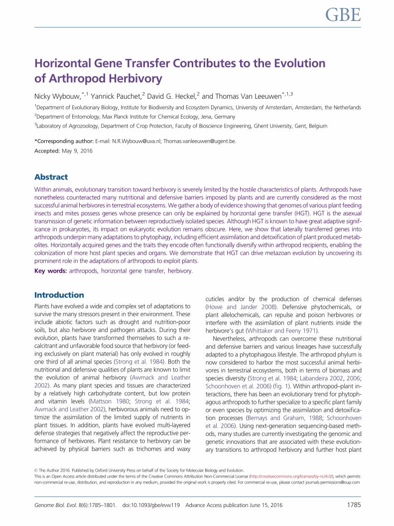
FIG. 1.—Arthropod phylogenetic tree showing documented HGT events in phytophagous chelicerate and hexapod lineages. The names of the clades
harboring herbivores are depicted in green. Horizontally transferred genes coding for traits involved in plant cell wall digestion, intracellular plant metabolite
assimilation, and overcoming plant defenses are depicted in blue, green, and red lines, respectively. Detailed lists of the methodologies that identified the
HGT events, the nature of the horizontally transferred genes and donor and recipient species can be found in table 1.
Wybouw et al. GBE
1786 Genome Biol. Evol. 8(6):1785–1801. doi:10.1093/gbe/evw119 Advance Access publication June 15, 2016
occurred can only be tested by probabilistic methods. As HGT
presents an unusually high degree of sequence similarity be-
tween the distantly related donor and recipient species for the
gene of interest, studies employ methodologies that analyze
different properties of the nucleotide and/or amino acid se-
quence to test the HGT hypothesis.
Molecular phylogenetics is the study of the evolutionary
history and relationships of genes across organisms by using
molecular data such as nucleotide and amino acid sequences.
A phylogenetic analysis generates a phylogenetic tree which is
characterized by a certain branching pattern or topology. This
topology mirrors the courses of inheritance of the genes of
interest over evolutionary time. The reliability of these topolo-
gies can be inferred by various computational techniques that
estimate the confidence level of each internal bifurcation. By
creating a consensus tree of numerous individual gene trees
(often on a genome-wide level), a species tree can be pro-
duced where the actual speciation events are mapped.
Molecular phylogenetic methods are the gold standard in ar-
thropod studies and suggest an HGT when the gene of inter-
est is grouped with homologues of nonrelated species by
strongly statistically supported branches (for instance with at
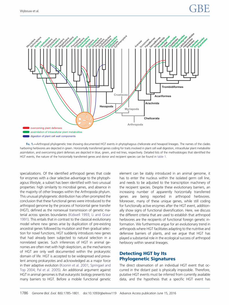
least 60% bootstrap support) (Efron et al. 1996) (fig. 2). In
order to be reliable, these phylogenetic incongruence meth-
ods require: (1) a well-established species phylogeny which is
needed as a reference topology and (2) an expansive set of
closely and step-wise more distantly related homologous
genes in order to avoid sampling bias. For many arthropod
studies, both requirements can be met as a significant part of
the arthropod phylogenomic tree is well mapped and keeps
improving due to the increasing number of sequenced arthro-
pod genomes and transcriptomes (Regier et al. 2010; Wheat
and Wahlberg 2013; Misof et al. 2014). These genomic se-
quences simultaneously offer an unbiased set of orthologues
spanning both short taxonomic distances (often within the
same genus) and large ones (between different subphyla). In
order to be convincing as a general methodology, phyloge-
netic incongruence methods must also be able to reject the
HGT hypothesis under appropriate conditions. For instance,
using this extensive sequence dataset, phylogenetic analysis
finally concluded that a much-debated putative horizontally
transferred gene in termites is instead much more likely to be a
typical vertically transmitted gene, present in various other
arthropod lineages (Lo et al. 2011). A potential shortcoming
of this type of test is that the putative horizontally transferred
gene and the set of available homologues may not contain a
strong enough phylogenetic signal (for instance due to short
sequence lengths). Alignments of such sequence sets typically
lead to various poorly supported branches in phylogenetic re-
constructions. When faced with nonoptimal tree support
values at critical bipartitions, studies often combine multiple
phylogenetic methods to minimize the chances of an errone-
ous identification of an HGT event in arthropods (Wybouw
et al. 2012; Husnik et al. 2013; Pauchet and Heckel 2013).
Doubts about the identification of an HGT event by phylo-
genetics often arise due to a misunderstanding of the ances-
tral distribution of the gene of interest. To address this
problem, it is useful to divide phylogenetic reconstructions
into three major scenarios, based on the occurrence of the
gene of interest across all lineages of the tree of life. First, HGT
can convey a completely novel gene to arthropods which is
normally absent in their entire phylogenetic lineage (fig. 2B,
left tree). Here, the simple presence of the gene in an arthro-
pod genome already strongly favors HGT, and HGT cannot be
ruled out even if the donor clade cannot be identified by
suboptimal phylogenetic support. Clear-cut examples of com-
pletely novel genes in plant feeding arthropods include genes
that code for carotenoid cyclases/synthases, chorismate
mutase (CM), and cyanase (Grbic et al. 2011; Novakova and
Moran 2012; Wybouw et al. 2012; Sloan et al. 2014). In the
second scenario, the arthropod species already harbors a re-
lated homologue to the potential laterally transferred
gene (fig. 2B, middle tree). Here, HGT is inferred when the
candidate transferred and the other paralogue(s) show
strongly divergent evolutionary histories. Spider mites of the
Tetranychidae family (Arthropoda: Chelicerata) and lepidop-
terans (Arthropoda: Hexapoda) possess a universal arthropod
cystathionine b-synthase (CBS) gene and a unique cysteine
synthase (or b-cyanoalanine synthase) (CAS) gene, part of
the same b-substituted alanine synthase gene family, but
with the latter normally restricted to bacterial and plant spe-
cies. Wybouw et al. (2014) showed phylogenetically that the
mite and lepidopteran CAS enzymes clustered together with
bacterial and plant enzymes, while their CBS enzymes were
embedded with high probability in a eukaryotic group of en-
zymes. In a third scenario (fig. 2B, right tree), the close ances-
tral homologue to the transmitted gene is lost in the recipient
species. Here, HGT is identified when the homologues of spe-
cies related to the recipient exhibit distinct evolutionary histo-
ries compared with the gene of interest. For instance, Ahn
et al. (2014) suggested by a phylogenetic reconstruction
that an ancestral chelicerate lost the arthropod UDP-glycosyl-
transferase (UGT) gene(s) and that tetranychid mites later
horizontally received novel UGTs, clearly different from the ver-
tically transmitted arthropod UGT sequences. Independent of
the ancestral distribution, as gene duplications, differential
gene losses and evolutionary convergence can also result in
a phylogenetic reconstruction where distant species lineages
cluster together, the likelihood of an HGT event versus alter-
native scenarios should always be weighed (Ku et al. 2015).
Detecting HGT by OtherProbabilistic Methods
To maximize the likelihood of correctly identifying an HGT
event, studies can incorporate additional, independent prob-
abilistic methods before concluding that a gene arose by HGT.
Many of these approaches take advantage of the high
HGT in Arthropod Herbivores GBE
Genome Biol. Evol. 8(6):1785–1801. doi:10.1093/gbe/evw119 Advance Access publication June 15, 2016 1787

number of sequenced genomes across the tree of life. After
phylogenetic analysis, one approach analyzes the codon con-
tent of the entire genome of the recipient and putative donor
species. As phylogenetic reconstruction usually does not un-
cover the exact donor species, the genomes of a broader
group of species are included in the codon content analysis.
Based on the theory that the codon usage pattern of all
coding sequences within a certain genome is similar and
that this genomic signature varies between species
(Lawrence and Ochman 1997), a horizontally transferred
gene candidate can be confirmed by detecting a different
codon content compared with the rest of the recipient
genome. However, this methodology should always be used
in combination with a phylogenetic analysis as it has several
imperfections. First, a true HGT event can be overlooked if the
nonrelated donor and recipient species have similar genomic
codon contents. Second, even if the donor and recipient ge-
nomes are characterized by distinctly different codon con-
tents, the hypothesis of an ancient HGT can still be rejected.
Once a gene is laterally incorporated, it is subjected to the
same mutational processes as the rest of the recipient’s
genome and will, over time, homogenize its codon content
with its genomic surroundings, in a process called ameliora-
tion (Lawrence and Ochman 1997). Indeed, in arthropods and
other animals, laterally acquired genes often exhibit nondis-
tinguishable codon contents from those of ancestral, innate
A
B
FIG. 2.—(A) Left: A species tree where the bifurcations represent speciation events. Species trees are based on the sequence information of multiple
genes (often genome-wide; phylogenomic trees) and depict how species are related. Here, horizontal transfer of a single gene is not expected to change the
phylogenomic tree. Right: the three known mechanisms of HGT in prokaryotes; transformation (direct uptake of foreign DNA), conjugation (plasmid-
mediated uptake) and transduction (virus-mediated uptake). (B) Phylogenetic analysis of a gene of interest can detect an HGT event by showing that a
particular species is embedded within a group of distantly related organisms. The implanted species and the surrounding clade are then considered to be the
recipient species and donor clade of the transferred gene, respectively. Three scenarios are depicted of how HGT can distort the phylogenetic reconstruction
of a single gene. These scenarios are characterized by a certain ancestral gene distribution across the tree of life. Left: The gene is originally restricted to a
certain phylogenetic clade, distantly related to the recipient species. As the horizontally transferred gene does not have related homologues within the clade
harboring the recipient species, it is unique to the recipient. Middle: The gene is present in all lineages, but a gene copy from a distant species supplements
an existing innate homologue in the recipient genome. Right: Here, a foreign gene replaces the original homologue in the recipient genome through
HGT. In each case however, the possibility of alternative scenarios to an HGT event should always be considered (for instance, differential gene loss across
the tree of life).
Wybouw et al. GBE
1788 Genome Biol. Evol. 8(6):1785–1801. doi:10.1093/gbe/evw119 Advance Access publication June 15, 2016
genes (Danchin et al. 2010; Wybouw et al. 2014). Therefore,
this methodology only gives further support when the HGT
was very recent and when the reproductively isolated species
have distinctly different genomic codon signatures. More and
more HGT cases with an arthropod recipient are being exam-
ined with this technique, in addition to the standard phyloge-
netic analysis (Sun et al. 2013; Wybouw et al. 2014; van Ohlen
et al. 2016).
Using the plethora of sequenced genomes, families of
orthologues can be systemically classified (Tatusov et al.
1997; Li et al. 2003). These comparative genomics techniques
allow for a new approach to examine the HGT hypothesis: the
analysis of the phyletic pattern within a certain orthologue
family. The phyletic pattern is the occurrence or absence of
species within these defined clusters of orthologues. For in-
stance, when a fungal lineage is detected within an otherwise
exclusively bacterial protein family, an HGT event can be in-
ferred. This methodology has confirmed multiple HGT events
to microorganisms (Koonin et al. 2001). After a BLASTP search
of a proteome under investigation against a large species cu-
rated sequence database, Clarke’s phylogenetic discordance
test identifies horizontally transferred genes that exhibit sta-
tistically significantly different similarity patterns compared
with the median similarity pattern shown by the proteome
(Clarke et al. 2002).These and other methodologies are not
yet well established in arthropod studies but show great po-
tential for future studies, despite the fact that each technique
is not without its potential limitations (Koonin et al. 2001;
Ragan 2001; Lawrence and Ochman 2002).
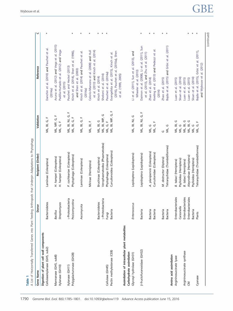
Incorporation of a Microbial Genein the Arthropod Genome
The first nucleotide and amino acid sequences of horizontally
transferred genes in plant feeding arthropods were mainly
uncovered by transcriptomic and proteomic studies (Pauchet
et al. 2008, 2009, 2010b; Kirsch et al. 2012). Although phy-
logenetic procedures strongly pointed toward an HGT sce-
nario with an arthropod recipient, skeptical reviewers
claimed that these genes and the enzymes they encode had
such unusual phylogenetic distributions because they origi-
nated from associated free-living or symbiotic microorganisms
and were not part of the arthropod genome. Indeed, arthro-
pods often host large bacterial communities in various organs,
including their digestive tract (Buchner 1965; Watanabe and
Tokuda 2010). Even though these genes exhibit strong se-
quence similarity to bacterial genes, they often possess typical
eukaryotic characteristics, strongly disfavoring the contamina-
tion hypothesis and supportive of an HGT event. These gene
attributes include eukaryotic signal peptides, polyadenylation
tails and spliceosomal introns (Acuna et al. 2012; Ahn et al.
2014; Luan et al. 2015). In addition, genomics now provide a
growing body of evidence showing that these bacterial genes
are physically incorporated into arthropod genomes (table 1).
For instance, after identifying potential laterally transferred
genes coding for xylanases in a midgut transcriptome of the
mustard leaf beetle (Phaedon cochleariae), Pauchet and
Heckel (2013) screened a genomic DNA fosmid library to
prove that two laterally transferred xylanase genes reside in
the beetle genome and are neighbored by insect-derived
transposable elements (TEs). Using next-generation sequenc-
ing technology, multiple genomic studies of various arthropod
lineages show that horizontally transferred genes are present
on large genomic scaffolds, where they are flanked by typical
arthropod genes (Grbic et al. 2011; Husnik et al. 2013; Vega
et al. 2015; Zhao et al. 2015). As genome assemblies can have
imperfect computational sequence filters (Baker 2012), an ap-
parent lateral acquisition can be an artifact due to the pres-
ence and incorrect concatenation of contaminating bacterial
sequences with other reads from the shotgun library
(Koutsovoulos et al. 2016). If a Bacterial Artificial
Chromosome library is available, this possibility can be ruled
out (Pauchet et al. 2014b). Potential genome assembly arti-
facts can also be ruled out by validating the genomic sequence
through PCR amplifications which often include geographi-
cally distinct arthropod populations. After identifying a hori-
zontally transferred gene, encoding for a mannanase, in the
genome of the coffee berry borer beetle (Hypothenemus
hampei), Acuna et al. (2012) showed that the transferred
gene is incorporated at the same genomic location in H.
hampei populations spanning Asia, Africa, and America. The
genomic location of the horizontally transferred CAS gene in
the spider mite species Tetranychus urticae was confirmed by
PCR amplification and shown to be identical in strains sampled
from different regions worldwide (Wybouw et al. 2014). If
speciation in the recipient group occurred after the HGT
event, the genomic region bracketing the laterally acquired
gene can also be analyzed in all descendent species. The ge-
nomic region bracketing the CAS gene exhibits clear synteny
between T. urticae and a closely related mite species,
Tetranychus evansi. Such approach was also followed by
Wheeler et al. (2013), who uncovered a single, ancient hori-
zontal transfer of a glycosyl hydrolase (GH) family 31 gene
from bacteria to the ancestor to all modern lepidopterans by
demonstrating microsynteny neighboring the laterally trans-
ferred gene in different lepidopteran genomes.
HGT and the Evolution ofArthropod Herbivory
Recent comparative studies show a high prevalence of HGT in
arthropod lineages, relative to other animal clades (Hotopp
et al. 2007; Hotopp 2011; Ramulu et al. 2012), which raises
the question of how these genes and the traits they encode
shape arthropod evolution. In this review, we will gather
strong indications that HGT from microbial species enhanced
the enzymatic repertoire of phytophagous arthropods, which
in turn seems to have facilitated adaptations toward herbivory
HGT in Arthropod Herbivores GBE
Genome Biol. Evol. 8(6):1785–1801. doi:10.1093/gbe/evw119 Advance Access publication June 15, 2016 1789
Tab
le1
ALi
stof
Horizo
nta
llyTr
ansf
erre
dG
enes
into
Plan
tFe
edin
gA
rthro
pods
that
Under
pin
Adap
tations
toPh
ytophag
y
Gen
eN
am
eD
on
or
Reci
pie
nt
(Ord
er)
Valid
ati
on
Refe
ren
ceC
Dig
est
ion
of
pla
nt
cell
wall
com
po
nen
ts
Cellu
lase
/xyl
an
ase
(GH
5,
sub
2)
Bact
ero
idete
sLa
miin
ae
(Co
leo
pte
ra)
ML,
BI,
G,
FD
an
chin
et
al.
(2010)
an
dPau
chet
et
al.
(2014a)
*
Man
nan
ase
(GH
5,
sub
8)
Baci
llus
H.
ham
pei
(Co
leo
pte
ra)
ML,
MP,
G,
FA
cun
aet
al.
(2012)
an
dV
eg
aet
al.
(2015)
Xyl
an
ase
(GH
10)
Stre
pto
myc
es
H.
ham
pei
(Co
leo
pte
ra)
ML,
G,
FPad
illa-H
urt
ad
oet
al.
(2012)
an
dV
eg
a
et
al.
(2015)
Xyl
an
ase
(GH
11)
g-Pro
teo
bact
eri
aP.
coch
leari
ae
(Co
leo
pte
ra)
ML,
BI,
NJ,
G,
FPau
chet
an
dH
eck
el
(2013)
Po
lyg
ala
ctu
ron
ase
(GH
28)
Pezi
zom
yco
tin
aPh
yto
ph
ag
a(C
ole
op
tera
)M
L,B
I,G
,F
Kir
sch
et
al.
(2014),
Shen
et
al.
(1996),
an
dSh
en
et
al.
(2003)
*
Asc
om
yco
taLa
miin
ae
(Co
leo
pte
ra)
ML,
BI,
G,
FK
irsc
het
al.
(2014)
an
dPau
chet
et
al.
(2014a)
*
Mir
inae
(Hem
ipte
ra)
ML,
BI,
FC
elo
rio
-Man
cera
et
al.
(2008)
an
dH
ull
et
al.
(2013)
an
dK
irsc
het
al.
(2014)
*
Bact
ero
idete
sB
ruch
inae
(Co
leo
pte
ra)
ML,
BI,
GK
irsc
het
al.
(2014)
*
g-Pro
teo
bact
eri
aV
ero
ph
asm
ato
dea
(Ph
asm
ato
dea)
ML,
BI,
MP,
GSh
elo
mi
et
al.
(2014)
*
Cellu
lase
(GH
45)
Fun
gi
Ph
yto
ph
ag
a(C
ole
op
tera
)M
L,G
,F
Pau
chet
et
al.
(2014a)
*
Pect
inm
eth
ylest
era
se(C
E8)
Bact
eri
aC
urc
ulio
no
idea
(Co
leo
pte
ra)
ML,
BI,
MP,
G,
FEva
ng
elis
taet
al.
(2015),
Kir
sch
et
al.
(2016),
Pau
chet
et
al.
(2010a),
Shen
et
al.
(1999,
2005)
Ass
imila
tio
no
fin
trace
llula
rp
lan
tm
eta
bo
lites
Carb
oh
yd
rate
ass
imila
tio
n
Gly
cosy
lh
ydro
lase
(GH
31)
En
tero
cocc
us
Lep
ido
pte
ra(L
ep
ido
pte
ra)
ML,
BI,
NJ,
GLi
et
al.
(2011),
Sun
et
al.
(2013),
an
d
Wh
eele
ret
al.
(2013)
b-fr
uct
ofu
ran
osi
dase
(GH
32)
Bact
eri
aLe
pid
op
tera
(Lep
ido
pte
ra)
ML,
BI,
NJ,
G,
FD
aim
on
et
al.
(2008),
Liet
al.
(2011),
Sun
et
al.
(2013),
an
dZh
uet
al.
(2011)
*
Bact
eri
aA
.p
lan
ipen
nis
(Co
leo
pte
ra)
ML,
GZh
ao
et
al.
(2014)
*
Bact
eri
aC
urc
ulio
nid
ae
(Co
leo
pte
ra)
ML,
G,
FK
eelin
get
al.
(2013)
an
dPed
ezz
iet
al.
(2014)
*
Bact
eri
aM
.d
est
ruct
or
(Dip
tera
)G
Zh
ao
et
al.
(2015)
*
Bact
eri
aTetr
an
ych
idae
(Tro
mb
idif
orm
es)
ML,
GB
ajd
aet
al.
(2015)
an
dG
rbic
et
al.
(2011)
*
Am
ino
aci
dass
imila
tio
n
Arg
inin
osu
ccin
ate
lyase
En
tero
bact
eri
ale
sB
.ta
baci
(Hem
ipte
ra)
ML,
BI,
GLu
an
et
al.
(2015)
Cars
on
ella
Psy
lloid
ea
(Hem
ipte
ra)
ML,
GSl
oan
et
al.
(2014)
Arg
inin
osu
ccin
ate
syn
thase
En
tero
bact
eri
ale
sB
.ta
baci
(Hem
ipte
ra)
ML,
BI,
GLu
an
et
al.
(2015)
CM
En
tero
bact
eri
ale
sA
leyr
od
inae
(Hem
ipte
ra)
ML,
BI,
GLu
an
et
al.
(2015)
*
Bact
eri
aPsy
lloid
ea
(Hem
ipte
ra)
ML,
GSl
oan
et
al.
(2014)
*
Cya
nase
Pla
nts
Tetr
an
ych
idae
(Tro
mb
idif
orm
es)
ML,
G,
FB
ajd
aet
al.
(2015),
Grb
icet
al.
(2011),
an
dW
ybo
uw
et
al.
(2012)
*
(continued
)
Wybouw et al. GBE
1790 Genome Biol. Evol. 8(6):1785–1801. doi:10.1093/gbe/evw119 Advance Access publication June 15, 2016
Tab
le1
Continued
Gen
eN
am
eD
on
or
Reci
pie
nt
(Ord
er)
Valid
ati
on
Refe
ren
ceC
Cys
tein
esy
nth
ase
Meth
ylo
bact
eri
aLe
pid
op
tera
(Lep
ido
pte
ra)
ML,
BI,
NJ,
GLi
et
al.
(2011),
Sun
et
al.
(2013),
Van
Oh
len
et
al.
(2016),
an
dW
ybo
uw
et
al.
(2014)
*
Meth
ylo
bact
eri
aTetr
an
ych
idae
(Tro
mb
idif
orm
es)
ML,
G,
FB
ajd
aet
al.
(2015)
an
dW
ybo
uw
et
al.
(2014)
*
g-Pro
teo
bact
eri
aP.
citr
i(H
em
ipte
ra)
ML,
BI,
GH
usn
iket
al.
(2013)
*
Dia
min
op
imela
teep
imera
seEn
tero
bact
eri
ale
sB
.ta
baci
(Hem
ipte
ra)
ML,
BI,
GLu
an
et
al.
(2015)
a-Pro
teo
bact
eri
aP.
citr
i(H
em
ipte
ra)
ML,
BI,
GH
usn
iket
al.
(2013)
Dia
min
op
imela
ted
eca
rbo
xyla
sePla
nct
om
ycete
sB
.ta
baci
(Hem
ipte
ra)
ML,
BI,
GLu
an
et
al.
(2015)
a-Pro
teo
bact
eri
aP.
citr
i(H
em
ipte
ra)
ML,
BI,
GH
usn
iket
al.
(2013)
4-H
ydro
xy-t
etr
ah
ydro
dip
ico
linate
red
uct
ase
Ric
kett
siale
sB
.ta
baci
(Hem
ipte
ra)
ML,
BI,
GLu
an
et
al.
(2015)
Kyn
ure
nin
ase
L.g
rayi
bact
eri
aLe
pid
op
tera
(Lep
ido
pte
ra)
ML,
BI,
NJ,
G,
FM
en
get
al.
(2009),
Liet
al.
(2011),
Sun
et
al.
(2013),
an
dZh
uet
al.
(2011)
meth
ion
ine
syn
thase
Bact
eri
aTetr
an
ych
idae
(Tro
mb
idif
orm
es)
ML,
GB
ajd
aet
al.
(2015)
an
dG
rbic
et
al.
(2011)
N-m
eth
yltr
ypto
ph
an
oxi
dase
Bact
eri
aB
.m
ori
(Lep
ido
pte
ra)
ML,
BI,
NJ,
GLi
et
al.
(2011),
Sun
et
al.
(2013),
an
dZh
u
et
al.
(2011)
Try
pto
ph
an
2-m
on
oo
xyg
en
ase
Pro
teo
bact
eri
aP.
citr
i(H
em
ipte
ra)
ML,
BI,
GH
usn
iket
al.
(2013)
*
Vit
am
inass
imila
tio
n
Ad
en
osy
lmeth
ion
ine
tran
sam
inase
Bact
eri
aA
leyr
od
inae
(Hem
ipte
ra)
ML,
BI,
GLu
an
et
al.
(2015)
a-Pro
teo
bact
eri
aP.
citr
i(H
em
ipte
ra)
ML,
BI,
GH
usn
iket
al.
(2013)
Bio
tin
syn
thase
Ric
kett
siale
sA
leyr
od
inae
(Hem
ipte
ra)
ML,
BI,
GLu
an
et
al.
(2015)
a-Pro
teo
bact
eri
aP.
citr
i(H
em
ipte
ra)
ML,
BI,
GH
usn
iket
al.
(2013)
Deth
iob
ioti
nsy
nth
ase
a-Pro
teo
bact
eri
aP.
citr
i(H
em
ipte
ra)
ML,
BI,
GH
usn
iket
al.
(2013)
GTP
cycl
oh
ydro
lase
g-Pro
teo
bact
eri
aP.
citr
i(H
em
ipte
ra)
ML,
BI,
GH
usn
iket
al.
(2013)
Rib
ofl
avi
nsy
nth
ase
Bact
eri
aPsy
lloid
ea
(Hem
ipte
ra)
ML,
GSl
oan
et
al.
(2014)
Fuse
dd
eam
inase
/red
uct
ase
a-Pro
teo
bact
eri
aP.
citr
i(H
em
ipte
ra)
ML,
BI,
GH
usn
iket
al.
(2013)
Caro
ten
oid
ass
imila
tio
n
Caro
ten
oid
desa
tura
seM
uco
rom
yco
tin
aA
ph
idid
ae
(Hem
ipte
ra)
ML,
BI,
G,
FM
ora
nan
dJa
rvik
(2010)
an
dN
ova
ko
va
an
dM
ora
n(2
012)
Ad
elg
idae
(Hem
ipte
ra)
ML,
BI
No
vako
vaan
dM
ora
n(2
012)
Zyg
om
yco
taTetr
an
ych
idae
(Tro
mb
idif
orm
es)
ML,
BI,
GA
ltin
cice
ket
al.
(2011),
Bajd
aet
al.
(2015),
an
dG
rbic
et
al.
(2011)
Mu
cora
les
Ceci
do
myi
idae
(Dip
tera
)M
L,B
I,G
Co
bb
set
al.
(2013)
Fuse
dca
rote
no
idcy
clase
/syn
thase
Fun
gi
Macr
osi
ph
ini
(Hem
ipte
ra)
ML,
GM
ora
nan
dJa
rvik
(2010)
Zyg
om
yco
taTetr
an
ych
idae
(Tro
mb
idif
orm
es)
ML,
BI,
GA
ltin
cice
ket
al.
(2012),
Bajd
aet
al.
(2015),
an
dG
rbic
et
al.
(2011)
Mu
cora
les
Ceci
do
myi
idae
(Dip
tera
)M
L,B
I,G
Co
bb
set
al.
(2013)
(continued
)
HGT in Arthropod Herbivores GBE
Genome Biol. Evol. 8(6):1785–1801. doi:10.1093/gbe/evw119 Advance Access publication June 15, 2016 1791

Tab
le1
Continued
Gen
eN
am
eD
on
or
Reci
pie
nt
(Ord
er)
Valid
ati
on
Refe
ren
ceC
Overc
om
ing
pla
nt
defe
nse
s
CA
SM
eth
ylo
bact
eri
aLe
pid
op
tera
(Lep
ido
pte
ra)
ML,
BI,
NJ,
G,
FLi
et
al.
(2011),
Sun
et
al.
(2013),
Van
Oh
len
et
al.
(2016),
an
dW
ybo
uw
et
al.
(2014)
*
Meth
ylo
bact
eri
aTetr
an
ych
idae
(Tro
mb
idif
orm
es)
ML,
G,
FB
ajd
aet
al.
(2015)
an
dW
ybo
uw
et
al.
(2014)
*
g-Pro
teo
bact
eri
aP.
citr
i(H
em
ipte
ra)
ML,
BI,
GH
usn
iket
al.
(2013)
*
CM
En
tero
bact
eri
ale
sA
leyr
od
inae
(Hem
ipte
ra)
ML,
BI,
GLu
an
et
al.
(2015)
*
Bact
eri
aPsy
lloid
ea
(Hem
ipte
ra)
ML,
GSl
oan
et
al.
(2014)
Cya
nase
Pla
nts
Tetr
an
ych
idae
(Tro
mb
idif
orm
es)
ML,
G,
FB
ajd
aet
al.
(2015),
Grb
icet
al.
(2011),
an
dW
ybo
uw
et
al.
(2012)
*
b-fr
uct
ofu
ran
osi
dase
Bact
eri
aB
.m
ori
(Lep
ido
pte
ra)
ML,
BI,
NJ,
G,
FD
aim
on
et
al.
(2008),
Liet
al.
(2011),
Sun
et
al.
(2013),
an
dZh
uet
al.
(2011)
*
Intr
ad
iol
rin
g-c
leavi
ng
dio
xyg
en
ase
Fun
gi
Tetr
an
ych
idae
(Tro
mb
idif
orm
es)
ML,
GB
ajd
aet
al.
(2015),
Derm
au
wet
al.
(2013),
an
dG
rbic
et
al.
(2011)
*
Kyn
ure
nin
ase
L.g
rayi
bact
eri
aLe
pid
op
tera
(Lep
ido
pte
ra)
ML,
BI,
NJ,
G,
FM
en
get
al.
(2009),
Liet
al.
(2011)�
Sun
et
al.
(2013),
an
dZh
uet
al.
(2011)
UD
P-g
lyco
sylt
ran
sfera
seB
act
eri
aTetr
an
ych
idae
(Tro
mb
idif
orm
es)
ML,
GA
hn
et
al.
(2014)
an
dB
ajd
aet
al.
(2015)
*
Tech
niq
ues
use
dto
exa
min
eeach
HG
Teve
nt
are
liste
din
the
Valid
ati
on
colu
mn
.Th
ep
hyl
og
en
eti
cm
eth
od
olo
gie
sare
ab
bre
viate
das
ML:
Maxi
mu
mLi
kelih
oo
dan
aly
sis
of
pro
tein
seq
uen
ces,
BI:
Baye
sian
Infe
ren
ce,
NJ:
Neig
hb
or-
Join
ing
an
dM
P:
Maxi
mu
mPars
imo
ny.
Furt
her
an
aly
sis
incl
ud
e;
G:
pro
of
of
ph
ysic
al
inco
rpo
rati
on
into
the
art
hro
po
dg
en
om
ean
dF:
en
zym
ep
rod
uct
isfu
nct
ion
al.
An
ast
eri
skw
ith
inco
lum
n‘C
’in
dic
ate
sw
heth
er
asi
mila
rg
en
eh
as
been
ind
ep
en
den
tly
ho
rizo
nta
llytr
an
sferr
ed
toa
ph
yto
ph
ag
ou
ssp
eci
es
wit
hin
the
Fun
gi,
Oo
myc
ota
,an
dN
em
ato
da
lineag
e.
Wybouw et al. GBE
1792 Genome Biol. Evol. 8(6):1785–1801. doi:10.1093/gbe/evw119 Advance Access publication June 15, 2016
and novel host plants. Here, the horizontally transferred genes
are categorized based on how their enzyme products interact
with the nutritional and defensive barriers of plants. The cat-
egories discussed are: (1) penetration and digestion of plant
cell walls, (2) assimilation of plant nutrients, and (3) overcom-
ing plant defenses (table 1).
Penetration and Digestion ofPlant Cell Walls
Various networks of complex composite fibers form the plant
cell wall and cuticle and enclose all cells of land plant species.
These plant structures are central to the biology of all terres-
trial plants as they provide the necessary structural integrity
and mechanical support for living on land. In some plant spe-
cies, these composite fibers form a thick rigid layer (for in-
stance in woody plants), and pose a barrier for arthropods
aiming to feed on plant nutrients (Carpita and Gibeaut
1993; Whetten and Sederoff 1995; Heredia 2003; Sorensen
et al. 2010). These complex matrices are mainly built from
polysaccharides and constitute the largest reservoir of organic
carbon on earth. As all animals, arthropods have an inherently
inadequate battery of enzymes that are able to cleave these
recalcitrant cell wall polysaccharides and access their stored
energy. Some specialized phytophagous arthropods, like
wood-feeding termites, have extensively proliferated a gene
family of ancestral animal cellulases to enzymatically cleave
certain cell wall components (Watanabe and Tokuda 2010),
while other arthropods seem to solely rely on microbial sym-
bionts in their gut (Breznak 1982). Microorganisms are able to
convert the complex fibers of the plant cell wall into simple
oligo- and monosaccharides by producing unique plant cell
wall degrading enzymes (PCWDEs). These microbial
PCWDEs are able to digest the rigid plant cell wall constituents
in the digestive tract and release their stored energy, benefit-
ing both the microorganisms and their arthropod hosts. Most
of the PCWDEs belong to various GH families that hydrolyze
glycosidic bonds either by a single or double displacement
mechanism. GHs are delineated into specific families and sub-
families through the curated Carbohydrate-Active database of
enzymes involved in carbohydrate metabolism (Cantarel et al.
2009). Although GH families are found across the whole tree
of life, most GH families coding for PCWDEs are restricted to
bacterial and fungal species (Rye and Withers 2000; Gilbert
2010).
Within the insect orders of the Coleoptera, Phasmatodea,
and Hemiptera (fig. 1), studies have indicated that genes
coding for PCWDEs have been laterally transferred to insect
genomes enabling the host to degrade the complex cell wall
polysaccharides themselves. Within Coleoptera, the most di-
verse monophyletic clade of herbivorous beetles is the
Phytophaga group (harboring ~80% of all plant feeding bee-
tles) and contains the superfamilies Chrysomeloidea and
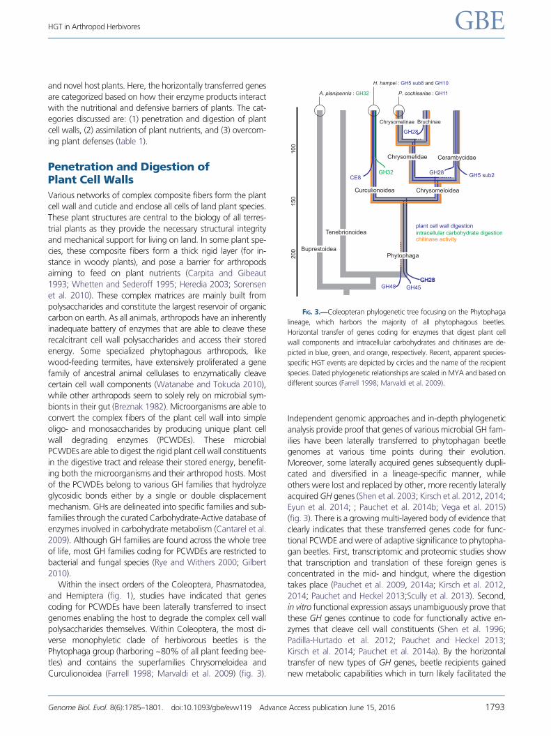
Curculionoidea (Farrell 1998; Marvaldi et al. 2009) (fig. 3).
Independent genomic approaches and in-depth phylogenetic
analysis provide proof that genes of various microbial GH fam-
ilies have been laterally transferred to phytophagan beetle
genomes at various time points during their evolution.
Moreover, some laterally acquired genes subsequently dupli-
cated and diversified in a lineage-specific manner, while
others were lost and replaced by other, more recently laterally
acquired GH genes (Shen et al. 2003; Kirsch et al. 2012, 2014;
Eyun et al. 2014; ; Pauchet et al. 2014b; Vega et al. 2015)
(fig. 3). There is a growing multi-layered body of evidence that
clearly indicates that these transferred genes code for func-
tional PCWDE and were of adaptive significance to phytopha-
gan beetles. First, transcriptomic and proteomic studies show
that transcription and translation of these foreign genes is
concentrated in the mid- and hindgut, where the digestion
takes place (Pauchet et al. 2009, 2014a; Kirsch et al. 2012,
2014; Pauchet and Heckel 2013;Scully et al. 2013). Second,
in vitro functional expression assays unambiguously prove that
these GH genes continue to code for functionally active en-
zymes that cleave cell wall constituents (Shen et al. 1996;
Padilla-Hurtado et al. 2012; Pauchet and Heckel 2013;
Kirsch et al. 2014; Pauchet et al. 2014a). By the horizontal
transfer of new types of GH genes, beetle recipients gained
new metabolic capabilities which in turn likely facilitated the
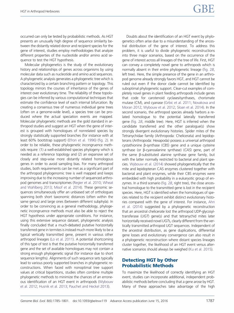
FIG. 3.—Coleopteran phylogenetic tree focusing on the Phytophaga
lineage, which harbors the majority of all phytophagous beetles.
Horizontal transfer of genes coding for enzymes that digest plant cell
wall components and intracellular carbohydrates and chitinases are de-
picted in blue, green, and orange, respectively. Recent, apparent species-
specific HGT events are depicted by circles and the name of the recipient
species. Dated phylogenetic relationships are scaled in MYA and based on
different sources (Farrell 1998; Marvaldi et al. 2009).
HGT in Arthropod Herbivores GBE
Genome Biol. Evol. 8(6):1785–1801. doi:10.1093/gbe/evw119 Advance Access publication June 15, 2016 1793
colonization of novel plant tissues and species. For instance,
cerambycid beetles are wood-feeders and can metabolize
xylan, a polysaccharide abundantly present in woody plant
tissues, by the enzymes coded by horizontally acquired sub-
family 2 genes of the extremely diverse GH5 family (Pauchet
et al. 2014a) (fig. 3). Enzyme kinetics also show that post-
transfer gene duplications have led to functional diversifica-
tion, potentially further expanding the host plant range of the
beetles (Sugimura et al. 2003; Lee et al. 2005; Pauchet and
Heckel 2013; Xia et al. 2013; Kirsch et al. 2014, 2016; Pauchet
et al. 2014a). Such functional diversification is shown by mi-
crobial GH48 genes, which no longer code for PCWDEs with
cellobiosidase activity, but have been co-opted by the beetles
to serve as chitinases (Fujita et al. 2006; Sukharnikov et al.
2012) (fig. 3).
In addition to Coleoptera, GH28 genes, which code for
polygalacturonases (fig. 3), were also laterally transferred
into the Phasmatodea and Hemiptera lineages and were sub-
sequently duplicated within their genomes (Allen and Mertens
2008; Celorio-Mancera et al. 2008; Shelomi et al. 2014) (fig.
1). In contrast to the recurrent and ongoing HGT events in
Coleoptera, phylogenetic analysis indicates a single ancient
HGT event within Phasmatodea. Although no GH28 enzyme
has been functionally characterized in this insect lineage yet,
the genes are also expressed in the gut and show differential
transcript levels along the phasmid digestive tract, strongly
indicative of a role in digestive physiology (Shelomi et al.
2014). Whereas Phasmatodea and Coleoptera disrupt plant
tissue by chewing, hemipteran insects pierce plant tissue using
highly specialized stylet-like mouthparts. Within Hemiptera,
the distribution of laterally acquired polygalacturonase genes
seems to be limited to species of the Mirinae, a subfamily
within the large and diverse Miridae family (Allen and
Mertens 2008; Celorio-Mancera et al. 2008; Hull et al.
2013; Shelomi et al. 2014). These mirid insects digest plant
material extra-orally by secreting enzymatically active saliva
through their stylet into the host (Miles and Taylor 1994). In
line with the in plantae digestion of this macerate-and-flush
feeding strategy, polygalacturonase genes in mirid plant bugs
are expressed in the salivary glands whereupon the protein
products are secreted into the plant (Celorio-Mancera et al.
2008).
In addition to their stalks and leaves, plants also accumulate
huge amounts of complex cell wall carbohydrates in their
seeds (Buckeridge et al. 2000). Many arthropods feed
exclusively on seeds and show HGT-mediated adaptations
to fully utilize the seed’s high carbohydrate content.
Homogalacturonan and xylogalacturonan are highly methyl-
ated polysaccharides that serve as a backbone in the complex
pectin network and can be abundantly present in seeds
(Zandleven et al. 2006). Methylation of the polysaccharide
backbone typically inhibits the pectin cleaving activity of poly-
galacturonase enzymes, including those that are produced by
weevils (Coleoptera: Curculionoidea) after an HGT event from
fungi (fig. 3). Rice weevil, a seed specialist, is however able to
demethylate pectin through an independent horizontal trans-
mission of bacterial genes of the carbohydrate esterase family
8 (Shen et al. 1999, 2005; Pauchet et al. 2010a; Kirsch et al.
2016). These genes code for functional pectin methylesterases
that remove the methyl-groups of both homogalacturonan
and xylogalacturonan. The two independently horizontally ac-
quired pectin methylesterase and polygalacturonase families
are shown to act synergistically in the rice weevil’s degradation
of plant pectin (Kirsch et al. 2016). Bacterial pectin methyles-
terase genes have been transferred to an ancestral species
within the Curculionoidea superfamily and could serve a sim-
ilar function in other descendent weevil species (Pauchet et al.
2010a; Evangelista et al. 2015; Kirsch et al. 2016).
Seeds of the coffee plant consist of 60% of recalcitrant
polymeric carbohydrates of which galactomannan is the major
storage product (Bradbury and Halliday 1990). Within the
Hypothenemus genus (Coleoptera: Curculionoidea), H.
hampei is a specialist and, in contrast to its close relatives,
can complete a full life cycle when feeding exclusively on
coffee beans. In search for the responsible digestive enzymes,
Acuna et al. (2012) discovered a very recent horizontal transfer
of a bacterial mannanase gene. The recombinant mannanase
enzyme exhibits endo-b-mannanase activity and is able to
metabolize galactomannan extracted from coffee beans, sug-
gesting that this HGT promoted the transition of the specialist
H. hampei to its current seed diet (fig. 3).
In conclusion, various phytopathogenic and symbiont mi-
crobial species provide a source of genetic diversity that allows
arthropod lineages to quickly adapt to recalcitrant plant cell
wall components.
Assimilation of IntracellularPlant Nutrients
Carbohydrate Metabolism
In addition to a polysaccharide-enriched extracellular cell wall,
plants are also characterized by a high intracellular carbohy-
drate content (Mattson 1980; Strong et al. 1984). For herbi-
vores, these high intracellular carbohydrate levels represent a
substantial amount of potential energy. Indeed, early studies
indicate that leaf-feeding caterpillars harvest this energy by a
dual a- and b-glycosidase activity in their midgut, which
breaks disaccharides down into monosaccharides (Santos
and Terra 1986; Sumida et al. 1994). As only microbial organ-
isms express b-fructofuranosidases (or also called invertases or
b-glucosidases) that are able to catalyze b-glucosidase reac-
tions, it was long postulated that the observed dual activity in
the lepidopteran midgut was a combined product of the
insect and its midgut microflora. The insect host benefits
from its microflora, as a- and b-glycosidases only hydrolyze
terminal a- and b-linked glucosyl residues, respectively, and
differ in substrate specificities (Terra and Ferreira 1994).
Wybouw et al. GBE
1794 Genome Biol. Evol. 8(6):1785–1801. doi:10.1093/gbe/evw119 Advance Access publication June 15, 2016

However, more recent studies started to indicate that these
digestive enzymes might be expressed by the insects them-
selves and not by associated symbionts (Carneiro et al. 2004;
Pauchet et al. 2008). The study of Daimon et al. (2008) shows
that a Bombyx mori gene of the GH32 family codes for a
functional b-fructofuranosidase and claims that this is ac-
quired by an HGT from a bacterial donor. Further phylogenetic
analysis and genome mining shows that close homologues of
the B. mori GH32 gene are present in all lepidopteran ge-
nomes sequenced so far, suggestive of a single ancient HGT
about 145-200 MYA (Li et al. 2011; Zhu et al. 2011; Sun et al.
2013). In addition, current comparative genomic studies
across the Arthropoda also uncovered the presence of b-fruc-
tofuranosidase genes in the genomes of beetles belonging to
the Curculionoidea and Buprestoidea superfamilies, the dip-
teran Mayetolia destructor and in the chelicerate spider mite T.
urticae, suggestive of multiple, independent HGT events
(Grbic et al. 2011; Keeling et al. 2013; Sun et al. 2013;
Pedezzi et al. 2014; Zhao et al. 2014, 2015) (figs. 1 and 3
and table 1). The alternative evolutionary scenario explaining
the presence of b-fructofuranosidase genes in the genomes of
these phytophagous arthropods and their absence in all other
sequenced arthropod genomes so far is that a gene copy was
present in an arthropod ancestor and subsequently lost in all
individual lineages across the whole arthropod phylogenetic
tree (fig. 1). For now, this is a far less parsimonious scenario
due to the extremely high prevalence of gene loss that would
be required. Moreover, phylogenetic analysis strongly sug-
gests independent horizontal transfers of the b-fructofurano-
sidase genes from different bacterial donor species to the
diverse phytophagous lineages (Keeling et al. 2013; Pedezzi
et al. 2014; Zhao et al. 2014). As within Lepidoptera, the
transferred gene into the Curculionoidea superfamily still
codes for a functional b-fructofuranosidase (Pedezzi et al.
2014).
In all sequenced lepidopteran genomes, horizontally trans-
ferred genes of a bacterial origin have been identified that
code for enzymes classified as GHs of the 31 family.
Although these genes are not yet functionally characterized,
GH31 genes often code for enzymes that cleave oligosaccha-
rides (Cantarel et al. 2009; Sun et al. 2013; Wheeler et al.
2013).
These studies suggest that horizontally transferred genes
from various sources offer selective advantages to phytopha-
gous arthropods by enabling them to fully harvest the carbo-
hydrate-rich compositions of plant tissue (table 1).
Amino Acid, Vitamin, and Carotenoid Metabolism
Compared with animals, bacteria and fungi exhibit an ex-
tremely high diversity of biosynthetic metabolic networks.
This rich enzymatic repertoire enables microorganisms to
uniquely synthesize certain compounds, some of which have
been defined as vitamins (Karlin et al. 2001; LeBlanc et al.
2011). Arthropod herbivores again have taken advantage of
the microbial metabolic richness and harbor horizontally trans-
ferred genes that code for enzymes with key roles in de novo
nutrient synthesis. Both the functional replacement of pre-
existing metabolic traits as well as the acquisition of comple-
tely new ones by HGT have occurred in phytophagous arthro-
pod lineages (Meng et al. 2009; Moran and Jarvik 2010; Grbic
et al. 2011; Sun et al. 2013; Luan et al. 2015). For instance,
carotenoid biosynthesis genes detected in dipteran, hemip-
teran, and tetranychid genomes, but nowhere else in the
animal kingdom, all have a high similarity to fungal homo-
logues, leading multiple studies to conclude that these were
laterally acquired from fungal donors. These genes have been
proven to still code for functional enzymes and likely offer
phytophagous arthropods the unique ability to endogenously
produce carotenoids, which are conjugated isoprenoid mole-
cules (Moran and Jarvik 2010; Grbic et al. 2011; Altincicek
et al. 2012; Novakova and Moran 2012; Bryon et al. 2013;
Cobbs et al. 2013).
Relative to other plant tissues, phloem, which translocates
photosynthate, is especially low in essential amino acids and
vitamins (Sandstrom and Pettersson 1994; Sandstrom and
Moran 1999). Hemipteran insects are nonetheless able to
feed exclusively on phloem sap. At first glance, HGT would
not seem to be required, as hemipterans supplement their
phloem diet with nutrients by engaging in intimate symbiotic
associations with bacteria. Hemipterans form an organ, the
bacteriome, where bacteria are sequestered into specialized
host cells called bacteriocytes (Buchner 1965). This symbiosis
benefits both insect and endosymbiont, since the bacteria re-
ceive a steady flow of nutrients and in return provide the sap-
feeding hemipteran host with essential amino acids and vita-
mins. After the original bacterial inoculation of the bacteriome
organ, these symbiotic bacteria go through extreme popula-
tion bottlenecking, facilitating the fixation of non-functional
mutations by genetic drift and resulting in progressive geno-
mic decay. As the bacterial genome deteriorates, the symbi-
onts become more and more dependent on the metabolism
of their insect hosts, mirroring the evolution of organelles
(Mccutcheon and Moran 2012). Gene losses in vitamin,
amino acid and other synthetic pathways unique to bacteria
have been observed, endangering the persistence of the mu-
tualistic symbiosis as the insect host does not produce the
necessary battery of enzymes. By sequencing the genomes
of different hemipteran insects, a convergent evolutionary
trend emerged in that independent HGT events from other,
free-living bacteria to the insect genome counteract the ge-
nomic deterioration of unique bacterial pathways within the
endosymbionts (Husnik et al. 2013; Sloan et al. 2014; Luan
et al. 2015). For instance, both the citrus mealybug and silver-
leaf whitefly possess multiple horizontally acquired genes that
function within the microbial lysine synthesis pathway (Luan
et al. 2015). Genome analysis of various hemipteran hosts and
their symbionts revealed that several gene losses within the
HGT in Arthropod Herbivores GBE
Genome Biol. Evol. 8(6):1785–1801. doi:10.1093/gbe/evw119 Advance Access publication June 15, 2016 1795
bacterial biosynthesis pathways of the vitamins biotin and ri-
boflavin have been compensated by multiple, independent
HGT events to the host (table 1) (Husnik et al. 2013; Sloan
et al. 2014; Luan et al. 2015). The transcription of these for-
eign genes is often concentrated in the bacteriocytes where
the enzyme products can readily interact with the intermedi-
ate substrates within the bacterial synthetic pathways (Husnik
et al. 2013; Sloan et al. 2014; Luan et al. 2015).
Overcoming Plant Defenses
In response to the selection pressure of herbivore attacks,
plants have developed numerous anti-herbivore physical and
chemical defenses. Plant toxins and their mode of action vary
widely across the plant kingdom (Whittaker and Feeny 1971;
Howe and Jander 2008), and HGT events have been impli-
cated in arthropod adaptations to some of them.
Mulberry plants defend themselves by accumulating alka-
loid compounds in their foliar latex layer. These alkaloids deter
herbivores from feeding by strongly inhibiting their a-glycosi-
dase activity. Functional characterization of the B. mori b-fruc-
tofuranosidase, coded by the horizontally transferred GH32
gene, showed that the enzyme is not only active but also
insensitive to these inhibitory alkaloids. Therefore, Daimon
et al. (2008) claimed that after HGT to an ancestor and sub-
sequent natural selection, B. mori was able to specialize on
mulberry plants by co-opting this carbohydrate metabolizing
enzyme in its defenses.
Cyanogenic plants release poisonous cyanide from non-
toxic precursor compounds upon herbivore attack
(Zagrobelny et al. 2008). Like plants, bacteria are also able
to biosynthesize cyanide and both prevent auto-toxicity by
producing a CAS enzyme. CAS efficiently detoxifies cyanide
by incorporating it into cysteine, hereby releasing the amino
acid derivative b-cyanoalanine (Miller and Conn 1980; Ebbs
2004). In contrast, animals have less efficient cyanide detoxi-
fication pathways, making cyanide an effective allelochemical
(Beesley et al. 1985; Zagrobelny et al. 2008). In-depth phylo-
genetic analysis from different studies indicates that at differ-
ent times in arthropod evolution, spider mites and Lepidoptera
independently acquired genes related to characterized CAS
genes from a common bacterial clade often associated with
the phyllosphere. Functional characterization of both mite and
lepidopteran recombinant CAS enzymes shows that they con-
vert toxic cyanide rapidly into b-cyanoalanine (Wybouw et al.
2014; Van Ohlen et al. 2016). Furthermore, it is shown in
caterpillars that CAS activity is widespread and inducible
upon exposure to dietary cyanide (Witthohn and Naumann,
1987; Meyers and Ahmad 1991; Stauber et al. 2012). By en-
dogenously producing this cyanide detoxifying enzyme, some
Lepidoptera could not only colonize a whole new range of
host plants, but also develop their own cyanogenic defenses
to deter predators (Witthohn and Naumann 1987; Zagrobelny
et al. 2008). These studies provide strong evidence that these
two arthropod lineages co-opted this bacterial gene to serve in
their xenobiotic metabolism with significant ecological
consequences.
In addition to a CAS gene, spider mites also laterally ac-
quired a gene that codes for a functional cyanase enzyme. As
cyanase metabolizes noxious cyanate, a common oxidation
product of cyanide, into nontoxic products, it may represent
a second, alternative route of cyanide detoxification in phy-
tophagous spider mites. Indeed, cyanase gene-expression was
induced in mites feeding on cyanogenic bean, compared to an
acyanogenic bean cultivar. However, transcription analysis
across a wider range of host plants led Wybouw et al.
(2012) to conclude that it may also serve other biological func-
tions (such as regulation of amino acid metabolism).
Aromatic ring structures are commonly found in plant
toxins and give these compounds an exceptionally high stabil-
ity due to their resonance structure. Phytopathogenic fungi
nevertheless metabolize complex catecholic metabolites,
such as procyanidins, by secreting intradiol ring-cleaving
dioxygenases. This family of dioxygenases, restricted to
microorganisms, catalyze the oxygenolytic fission of aromatic
structures between adjacent hydroxyl groups (Vaillancourt
et al. 2006; Roopesh et al. 2010, 2012; Yang et al. 2012).
An intradiol ring-cleaving dioxygenase has been horizontally
transferred into the plant feeding Tetranychidae mite family
with secreted fungal enzymes as its closest homologues. Here,
the dioxygenase gene duplicated and currently forms multi-
gene families in descendent mite species (for instance, the T.
urticae genome harbors 17 intradiol dioxygenase genes).
Previous studies uncovered a high transcriptional response of
this unique gene family to various chemical stresses, including
host plants and acaricides (Grbic et al. 2011; Dermauw et al.
2013; Bajda et al. 2015; Van Leeuwen and Dermauw 2016).
Although this dioxygenase gene family awaits functional char-
acterization, current data strongly suggest that mites use
these laterally acquired genes in their detoxification pathways.
As well as detoxifying, some arthropod herbivores also sup-
press toxin production to avoid the negative effects on their
reproductive performance. Such manipulation of plant physi-
ology by arthropods (and other herbivores) is achieved by se-
creting salivary compounds into the plant where these
interfere with plant pathways (Kant et al. 2015; Zhao et al.
2015; Villarroel et al. 2016). Aromatic allelochemicals (includ-
ing flavonoids) are produced by enzymes of the conserved
shikimate pathway, restricted to plants and bacteria
(Herrmann 1995). Specifically, the aromatic toxins are derived
from chorismate, the end product of the shikimate pathway
(Strack 1997). CM is a key branch-point enzyme that converts
chorismate to prephenate (Romero et al. 1995). In plant par-
asitic nematodes, it has been proposed that by horizontal
transfer of a CM gene, nematodes are able to modulate the
plant shikimate pathway to their own benefit (Lambert et al.
1999; Vanholme et al. 2009; Haegeman et al. 2011). The
discovery of a horizontally transferred CM gene in multiple
Wybouw et al. GBE
1796 Genome Biol. Evol. 8(6):1785–1801. doi:10.1093/gbe/evw119 Advance Access publication June 15, 2016
hemipteran lineages now raises the question whether it serves
a similar function there (Husnik et al. 2013; Luan et al. 2015)
(table 1).
These examples show that arthropods have evolved various
counter adaptations to their host plants’ defenses by hijacking
pre-evolved genes from bacteria and fungi, some of which
share the same metabolic pathways as plants.
Parallel Roles for HGT in OtherEukaryotic Linages
Moreover, a role of HGT in the evolutionary transition towards
exploiting plants, whether by herbivory or a phytopathogenic
strategy, has also been suggested in three other eukaryotic
lineages, namely Fungi, Oomycota (Heterokontophyta) and
the animal phylum of Nematoda. The genomic innovations
due to HGT have shaped the penetration, assimilation and
detoxification pathways of plant tissue independently and in
parallel within these phylogenetically diverse plant parasites
(Danchin et al. 2010; Haegeman et al. 2011; Soanes and
Richards 2014). Comparing these studies with the data re-
viewed here, it is apparent that a significant number of
genes coding for enzymes with identical catalytic properties
were horizontally transferred to these highly divergent line-
ages where they play similar roles in the herbivorous lifestyle
(table 1). For instance, laterally acquired PCWDEs and b-fruc-
tofuranosidases are also found in a wide range of plant par-
asitic nematodes and are responsible for the digestion of
recalcitrant plant cell wall components and host-derived su-
crose, respectively (Danchin et al. 2010, 2016). Moreover,
horizontally transferred genes have a tendency to duplicate
and form novel multi-gene families across the four lineages,
strongly indicative of functional diversification (Danchin et al.
2010; Dermauw et al. 2013; Ahn et al. 2014; Kirsch et al.
2014).
The strong parallel role of HGT in the evolution of plant
exploitation across different eukaryotic linages indicates that
the genomes of non-related microorganisms already adapted
to living on and from plants provide a great adaptive potential
for eukaryotes trying to colonize the same niche.
Potential Mechanisms of HGT toArthropod Herbivores
Because HGT to a eukaryotic recipient has only been detected
after the fact, the exact mechanisms of the transfer remain
elusive. HGT must be a multi-step process whereby a genetic
element is excised from a donor genome, transmitted and
introduced into a reproductively isolated recipient genome.
Multiple facets of eukaryotic biology seem to lower the likeli-
hood of successfully completing this multi-step process. First,
before genetic information can be integrated and expressed in
a eukaryotic genome, it first has to pass through the selective
double membrane of the nucleus. Second, if it concerns a
bacterial donor, the mobile genetic element needs to be ex-
pressed by a functionally appropriate promoter and adjusted
to the eukaryotic transcription machinery (polyadenylation,
codon compositions, and binding site modifications). Last, in
multicellular eukaryotes, a new laterally acquired genetic ele-
ment can only be transmitted to the following generation if it
is incorporated within the isolated germ cell line. Arthropods
have widespread and intimate relationships with bacterial
communities which consist of free-living bacterial species
and/or of symbiotic bacteria that permanently reside within
certain arthropod organs (Buchner 1965; Hotopp et al. 2007;
Hotopp 2011; Li et al. 2011). Even the reproductive tissues of
arthropods are commonly infected with intracellular endosym-
biotic bacteria. Some endosymbionts are extremely closely as-
sociated with germ line tissues as they are able to interfere
with the reproductive processes of their arthropod hosts in
order to increase their vertical transmission to the next gener-
ation (Stouthamer et al. 1999; Hotopp et al. 2007; Hotopp,
2011). Although the barriers of HGT to a eukaryote are high,
the close proximity of dense and complex bacterial popula-
tions around and within arthropods at least facilitates HGT to
the germ cell line.
In prokaryotes, HGT is known to operate through three
mechanisms, namely (1) transformation—the direct uptake
of exogenous DNA, (2) conjugation—the plasmid-mediated
uptake of foreign DNA through cell-to-cell contact, and (3)
transduction—the virus-mediated uptake of foreign DNA
(fig. 2A). Studies have found that some laterally transferred
genes in animal recipients are linked to genomic regions en-
riched in TEs (Acuna et al. 2012; Paganini et al. 2012; Flot et al.
2013; Pauchet and Heckel 2013; Gasmi et al. 2015). TEs are
mobile and can jump within and between genomes. Other
genes close to these TEs could potentially hitchhike on such a
transposition event between close physical interacting organ-
isms (through endosymbiosis, husbandry, parasitism, preda-
torism or other forms of contact). Moreover, studies indicate
that viruses can act as a shuttle for TE-mediated HGT (Liu et al.
2010; Gilbert et al. 2014; Gasmi et al. 2015). For instance,
parasitoid wasps inject bracoviral particles into their lepidop-
teran hosts to aid in the development of their offspring.
Genome analysis revealed that wasp genes have been stably
incorporated into lepidopteran species through the bracoviral
integration mechanism (Gasmi et al. 2015). As viruses can act
as gene transfer agents, the donor and recipient species do
not necessarily have to be in direct physical contact, further
increasing the potential source of alien genetic information
(Houck et al. 1991; Dimmock et al. 2007; Liu et al. 2010;
Gilbert et al. 2014; Gasmi et al. 2015). Extracellular vesicles,
such as exosomes, could also be a potential route of gene
transfer into eukaryotic species. These vesicles function in
intercellular communication and are produced by organisms
throughout the domains of life. They contain various com-
pounds, including DNA and RNA molecules. Notably, within
bacteria, genetic information is known to be transferred not
HGT in Arthropod Herbivores GBE
Genome Biol. Evol. 8(6):1785–1801. doi:10.1093/gbe/evw119 Advance Access publication June 15, 2016 1797

only between the cells of the same organism but also across
species boundaries (Yaron et al. 2000; Mashburn-Warren and
Whiteley 2006).
Concluding Remarks
Arthropods show great adaptability toward the biochemical
challenges posed by herbivory. Various independent tech-
niques show that a plethora of horizontally transferred
genes are embedded within the genomes of phytophagous
insects and mites. Functional characterization shows that
these genes code for active enzymes and are co-opted by
the arthropod recipient to serve in their detoxification and
assimilation pathways of plant compounds. Beetles from the
Phytophaga clade exhibit a complex evolutionary sequence of
multiple lateral acquisitions and subsequent duplication and
loss events of genes that code for active PCWDEs. This ac-
quired metabolic repertoire enabled them to specialize to var-
ious new plant organs, including seeds and wooden tissue.
More studies are however needed to fully map the intricate
evolutionary history of various phytophagan-specific PCWDE
families, such as hemicellulases and pectate lyases. Insects of
the Hemiptera order independently integrated various micro-
bial genes in their genome of which the enzyme products
seem to play a role in their endosymbiont-mediated assimila-
tion of plant metabolites. Future studies should be directed
towards functionally expressing these genes and gaining ad-
ditional evidence for their function in the symbiotic relation-
ship. Within the Hexapoda subphylum, numerous sequenced
genomes and transcriptomes have allowed for a greater un-
derstanding of how HGT facilitates insect herbivory. The exact
role of HGT across the other subphyla (Myriapoda, Crustacea,
and Chelicerata) largely remains to be discovered when more
sequence data will become available (fig. 1). However, the first
sequenced genome of a plant feeding mite revealed an un-
usually high number of horizontally transferred genes,
strongly indicating that HGT might be a driving force shaping
many unique and unprecedented metabolic capabilities within
Chelicerata.
Funding
N.W. was supported by a Marie Sklodowska-Curie Action
(MSCA) Individual fellowship (658795-DOGMITE) of Horizon
2020. Y.P. and D.G.H. are supported by the Max-Planck-
Gesellschaft. T.V.L. was supported by the European
Commission (EC contract 618105) via FACCE ERA-NET Plus
and FACCE-JP (Genomite, project ID 137) and the Fund for
Scientific Research Flanders (FWO) (grant G009312N and
G053815N).
Literature CitedAcuna R. 2012. Adaptive horizontal transfer of a bacterial gene to
an invasive insect pest of coffee. Proc Natl Acade Sci U S A.
109:4197–4202.
Ahn SJ, Dermauw W, Wybouw N, Heckel DG, Van Leeuwen T. 2014.
Bacterial origin of a diverse family of UDP-glycosyltransferase genes
in the Tetranychus urticae genome. Insect Biochem Mol Biol.
50:43–57.
Allen ML, Mertens JA. 2008. Molecular cloning and expression of three
polygalacturonase cDNAs from the tarnished plant bug, Lygus line-
olaris. J Insect Sci. 8:27.
Altincicek B, Kovacs JL, Gerardo NM. 2012. Horizontally transferred fungal
carotenoid genes in the two-spotted spider mite Tetranychus urticae.
Biol Lett. 8:253–257.
Awmack CS, Leather SR. 2002. Host plant quality and fecundity in herbiv-
orous insects. Annu Rev Entomol. 47:817–844.
Bajda S, et al. 2015. Transcriptome profiling of a spirodiclofen susceptible
and resistant strain of the European red mite Panonychus ulmi using
strand-specific RNA-seq. BMC Genomics 16. doi: 10.1186/s12864-
015-2157-1.
Baker M. 2012. De novo genome assembly: what every biologist should
know. Nat Methods 9:333–337.
Beesley SG, Compton SG, Jones DA. 1985. Rhodanese in Insects. J Chem
Ecol. 11:45–50.
Bernays E, Graham M. 1988. On the evolution of host specificity in phy-
tophagous arthropods. Ecology 69:886–892.
Bradbury AGW, Halliday DJ. 1990. Chemical structures of green coffee
bean polysaccharides. J Agric Food Chem. 38:389–392.
Breznak JA. 1982. Intestinal microbiota of termites and other xylophagous
insects. Annu Rev Microbiol. 36:323–343.
Bryon A, et al. 2013. Genome wide gene-expression analysis of facultative
reproductive diapause in the two-spotted spider mite Tetranychus
urticae. BMC Genomics 14:815.
Buchner P. 1965. Endosymbiosis of animals with plant microorganisms.
New York: John Wiley & Sons.
Buckeridge MS, Dos Santos HP, Tine MaS. 2000. Mobilisation of
storage cell wall polysaccharides in seeds. Plant Physiol
Biochem. 38:141–156.
Cantarel BL, et al. 2009. The carbohydrate-active enzymes database
(CAZy): an expert resource for glycogenomics. Nucleic Acids Res.
37:D233–D238.
Carneiro CNB, Isejima EM, Samuels RI, Silva CP. 2004. Sucrose hydrolases
from the midgut of the sugarcane stalk borer Diatraea saccharalis.
J Insect Physiol. 50:1093–1101.
Carpita NC, Gibeaut DM. 1993. Structural models of primary cell walls in
flowering plants—constistency of molecular structure with the physi-
cal properties of the walls during growth. Plant J. 3:1–30.
Celorio-Mancera MDLP, et al. 2008. Polygalacturonase causes lygus-like
damage on plants: cloning and identification of western tarnished
plant bug (Lygus hesperus) polygalacturonases secreted during feed-
ing. Arthropod-Plant Interact. 2:215–225.
Clarke GDP, Beiko RG, Ragan MA, Charlebois RL. 2002. Inferring genome
trees by using a filter to eliminate phylogenetically discordant se-
quences and a distance matrix based on mean normalized BLASTP
scores. J Bacteriol. 184:2072–2080.
Cobbs C, Heath J, Stireman JO, III, Abbot P. 2013. Carotenoids in unex-
pected places: Gall midges, lateral gene transfer, and carotenoid bio-
synthesis in animals. Mol Phylogenet Evol. 68:221–228.
Daimon T, et al. 2008. beta-fructofuranosidase genes of the silkworm,
Bombyx mori—insights into enzymatic adaptation of B. mori to toxic
alkaloids in mulberry latex. J Biol Chem. 283:15271–15279.
Danchin EGJ, et al. 2010. Multiple lateral gene transfers and duplications
have promoted plant parasitism ability in nematodes. Proc Natl Acad
Sci U S A. 107:17651–17656.
Danchin EGJ, Guzeeva EA, Mantelin S, Berepiki A, Jones JT. 2016.
Horizontal gene transfer from bacteria has enabled the plant-
parasitic nematode Globodera pallida to feed on host-derived sucrose.
Mol Biol Evol. 33:1571–1579.
Wybouw et al. GBE
1798 Genome Biol. Evol. 8(6):1785–1801. doi:10.1093/gbe/evw119 Advance Access publication June 15, 2016
Dermauw W, et al. 2013. A link between host plant adaptation and pes-
ticide resistance in the polyphagous spider mite Tetranychus urticae.
Proc Natl Acad Sci U S A. 110:E113–E122.
Dimmock NJ, Easton AJ, Leppard KN. (2007) Introduction to modern vi-
rology. Malden (MA): Blackwell Publishing.
Ebbs S. 2004. Biological degradation of cyanide compounds. Curr Opin
Biotechnol. 15:231–236.
Efron B, Halloran E, Holmes S. 1996. Bootstrap confidence levels for phy-
logenetic trees. (vol 93, pg 7085, 1996). Proc Natl Acad Sci U S A.
93:13429–13434.
Evangelista DE, Pereira De Paula FF, Rodrigues A, Henrique-Silva F. 2015.
Pectinases from Sphenophorus levis Vaurie, 1978 (Coleoptera:
Curculionidae): putative accessory digestive enzymes. J Insect Sci. 15:5.
Eyun SI, et al. 2014. Molecular evolution of glycoside hydrolase genes in
the western corn rootworm (Diabrotica virgifera virgifera). PLoS One
9:e94052.
Farrell BD. 1998. “Inordinate fondness” explained: why are there so many
beetles? Science 281:555–559.
Flot JF, et al. 2013. Genomic evidence for ameiotic evolution in the bdelloid
rotifer Adineta vaga. Nature 500:453–457.
Fujita K, Shimomura K, Yamamoto K, Yamashita T, Suzuki K. 2006. A
chitinase structurally related to the glycoside hydrolase family 48 is
indispensable for the hormonally induced diapause termination in a
beetle. Biochem Biophys Res Commun. 345:502–507.
Gasmi L, et al. 2015. Recurrent domestication by lepidoptera of genes from
their parasites mediated by bracoviruses. PLoS Genet. 11:e1005470.
Gilbert C, et al. 2014. Population genomics supports baculoviruses as
vectors of horizontal transfer of insect transposons. Nat Commun. 5.
Gilbert HJ. 2010. The biochemistry and structural biology of plant cell wall
deconstruction. Plant Physiol. 153:444–455.
Grbic M, et al. 2011. The genome of Tetranychus urticae reveals herbivo-
rous pest adaptations. Nature 479:487–492.
Haegeman A, Jones JT, Danchin EGJ. 2011. Horizontal gene transfer in
nematodes: a catalyst for plant parasitism? Mol Plant-Microbe Interact.
24:879–887.
Heredia A. 2003. Biophysical and biochemical characteristics of cutin, a
plant barrier biopolymer. Biochimica Et Biophysica Acta-General
Subjects 1620:1–7.
Herrmann KM. 1995. The shikimate pathway as an entry to aromatic
secondary metabolism. Plant Physiol. 107:7–12.
Hotopp JCD, et al. 2007. Widespread lateral gene transfer from intracel-
lular bacteria to multicellular eukaryotes. Science 317:1753–1756.
Hotopp JCD. 2011. Horizontal gene transfer between bacteria and ani-
mals. Trends Genet. 27:157–163.
Houck MA, Clark JB, Peterson KR, Kidwell MG. 1991. Possible horizontal
transfer of Drosophila genes by the mite Proctolaelaps regalis. Science
253:1125–1129.
Howe GA, Jander G. 2008. Plant immunity to insect herbivores. Annu Rev
Plant Biol. 59:41–66.
Hull JJ, Geib SM, Fabrick JA, Brent CS. 2013. Sequencing and de novo
assembly of the western tarnished plant bug (Lygus hesperus) tran-
scriptome. PLoS One 8:e55105.
Husnik F, et al. 2013. Horizontal gene transfer from diverse bacteria to an
insect genome enables a tripartite nested mealybug symbiosis. Cell
153:1567–1578.
Kant MR, et al. 2015. Mechanisms and ecological consequences of plant
defence induction and suppression in herbivore communities. Ann
Bot. 115:1015–1051.
Karlin S, Mrazek J, Campbell A, Kaiser D. 2001. Characterizations of
highly expressed genes of four fast-growing bacteria. J Bacteriol.
183:5025–5040.
Keeling CI, et al. 2013. Draft genome of the mountain pine beetle,
Dendroctonus ponderosae Hopkins, a major forest pest. Genome
Biol. 14:1–20.
Kidwell MG. 1993. Lateral transfer in natural populations of eukaryotes.
Annu Rev Genet. 27:235–256.
Kirsch R, et al. 2012. Combining proteomics and transcriptome sequenc-
ing to identify active plant-cell-wall-degrading enzymes in a leaf
beetle. BMC Genomics 13:587.
Kirsch R, et al. 2014. Horizontal gene transfer and functional diversification
of plant cell wall degrading polygalacturonases: key events in the evo-
lution of herbivory in beetles. Insect Biochem Mol Biol. 52:33–50.
Kirsch R, Heckel DG, Pauchet Y. 2016. How the rice weevil breaks down
the pectin network: enzymatic synergism and sub-functionalization.
Insect Biochem Mol Biol. 71:72–82.
Koonin EV, Makarova KS, Aravind L. 2001. Horizontal gene transfer in
prokaryotes: quantification and classification. Annu Rev Microbiol.
55:709–742.
Koutsovoulos G, et al. 2016. No evidence for extensive horizontal gene
transfer in the genome of the tardigrade Hypsibius dujardini. Proc Natl
Acad Sci U S A. 113:5053–5058.
Ku C, et al. 2015. Endosymbiotic origin and differential loss of eukaryotic
genes. Nature 524:427.
Labandeira CC. 2002. The history of associations between plants and ani-
mals. In: Herrera CM, Pellmyr O, editors. Plant-Animal Interactions An
Evolutionary Approach. West Sussex (United Kingdom): Blackwell-
Wiley.
Labandeira CC. 2006. The four phases of plant-arthropod associations in
deep time. Geologica Acta. 4:409–438.
Lambert KN, Allen KD, Sussex IM. 1999. Cloning and characterization of
an esophageal-gland-specific chorismate mutase from the phytopar-
asitic nematode Meloidogyne javanica. Mol Plant-Microbe Interact.
12:328–336.
Lawrence JG, Ochman H. 1997. Amelioration of bacterial genomes: rates
of change and exchange. J Mol Evol. 44:383–397.
Lawrence JG, Ochman H. 2002. Reconciling the many faces of lateral gene
transfer. Trends Microbiol. 10:1–4.
Leblanc JG, et al. 2011. B-group vitamin production by lactic acid bacte-
ria—current knowledge and potential applications. J Appl Microbiol.
111:1297–1309.
Lee SJ, et al. 2005. A novel cellulase gene from the mulberry longi-
corn beetle, Apriona germari: gene structure, expression, and
enzymatic activity. Comp Biochem Physiol B-Biochem Mol Biol.
140:551–560.
Li WH, Graur D. 1991. Fundamentals of molecular evolution. 23 Plumtree
Road, Sunderland, USA: Sinauer Associates.
Li ZW, Shen YH, Xiang ZH, Zhang Z. 2011. Pathogen-origin horizontally
transferred genes contribute to the evolution of Lepidopteran insects.
BMC Evol Biol. 11.
Li L, Stoeckert CJ, Roos DS. 2003. OrthoMCL: identification of ortholog
groups for eukaryotic genomes. Genome Res. 13:2178–2189.
Liu H, et al. 2010. Widespread horizontal gene transfer from double-
stranded RNA viruses to eukaryotic nuclear genomes. J Virol.
84:11876–11887.
Lo N, Tokuda G, Watanabe H. 2011. Evolution and function of endoge-
nous termite cellulases. Biology of termites: a modern synthesis.
Netherlands: Springer. p. 51–67.
Luan JB, et al. 2015. Metabolic coevolution in the bacterial symbiosis of
whiteflies and related plant sap-feeding insects. Genome Biol Evol.
7:2635–2647.
Marvaldi AE, Duckett CN, Kjer KM, Gillespie JJ. 2009. Structural alignment
of 18S and 28S rDNA sequences provides insights into phylogeny of
Phytophaga (Coleoptera: Curculionoidea and Chrysomeloidea).
Zoologica Scripta 38:63–77.
Mashburn-Warren LM, Whiteley M. 2006. Special delivery: vesicle traffick-
ing in prokaryotes. Mol Microbiol. 61:839–846.
Mattson WJ. 1980. Herbivory in relation to plant nitrogen content. Annu
Rev Ecol Syst. 11:119–161.
HGT in Arthropod Herbivores GBE
Genome Biol. Evol. 8(6):1785–1801. doi:10.1093/gbe/evw119 Advance Access publication June 15, 2016 1799
Mccutcheon JP, Moran NA. 2012. Extreme genome reduction in symbiotic
bacteria. Nat Rev Microbiol. 10:13–26.
Meng Y, Katsuma S, Mita K, Shimada T. 2009. Abnormal red body col-
oration of the silkworm, Bombyx mori, is caused by a mutation in a
novel kynureninase. Genes Cells 14:129–140.
Meyers DM, Ahmad S. 1991. Link between L-3-cyanoalanine synthase
activity and differential cyanide sensitivity of insects. Biochimica Et
Biophysica Acta. 1075:195–197.
Miles PW, Taylor GS. 1994. Osmotic pump feeding by Coreids.
Entomologia Experimentalis Et Applicata 73:163–173.
Miller JM, Conn EE. 1980. Metabolism of hydrogen cyanide in higher
plants. Plant Physiol. 65:1199–1202.
Misof B, et al. 2014. Phylogenomics resolves the timing and pattern of
insect evolution. Science 346:763–767.
Moran NA, Jarvik T. 2010. Lateral transfer of genes from fungi underlies
carotenoid production in aphids. Science 328:624–627.
Novakova E, Moran NA. 2012. Diversification of genes for carotenoid
biosynthesis in aphids following an ancient transfer from a fungus.
Mol Biol Evol. 29:313–323.
Padilla-Hurtado B, et al. 2012. Cloning and expression of an endo-
1,4-beta-xylanase from the coffee berry borer, Hypothenemus
hampei. BMC res Notes 5:23.
Paganini J, et al. 2012. Contribution of lateral gene transfers to the
genome composition and parasitic ability of root-knot nematodes.
PLoS One 7:e50875.
Pal C, Papp B, Lercher MJ. 2005. Adaptive evolution of bacterial
metabolic networks by horizontal gene transfer. Nat Genet.
37:1372–1375.
Pauchet Y, Muck A, Svatos A, Heckel DG, Preiss S. 2008. Mapping the
larval midgut lumen proteome of Helicoverpa armigera, a generalist
herbivorous insect. J Proteome Res. 7:1629–1639.
Pauchet Y, et al. 2009. Pyrosequencing of the midgut transcriptome of the
poplar leaf beetle Chrysomela tremulae reveals new gene families in
Coleoptera. Insect Biochem Mol Biol. 39:403–413.
Pauchet Y, Wilkinson P, Chauhan R, Ffrench-Constant RH. 2010a.
Diversity of beetle genes encoding novel plant cell wall degrading
enzymes. PLoS One 5:e15635.
Pauchet Y, et al. 2010b. Pyrosequencing the Manduca sexta larval midgut
transcriptome: messages for digestion, detoxification and defence.
Insect Mol Biol. 19:61–75.
Pauchet Y, Heckel DG. 2013. The genome of the mustard leaf beetle
encodes two active xylanases originally acquired from bacteria through
horizontal gene transfer. Proc R Soc B-Biol Sci. 280.
Pauchet Y, Kirsch R, Giraud S, Vogel H, Heckel DG. 2014a. Identification
and characterization of plant cell wall degrading enzymes from three
glycoside hydrolase families in the cerambycid beetle Apriona japonica.
Insect Biochem Mol Biol. 49:1–13.
Pauchet Y, et al. 2014b. Studying the organization of genes
encoding plant cell wall degrading enzymes in Chrysomela tremula
provides insights into a leaf beetle genome. Insect Mol Biol.
23:286–300.
Pedezzi R, et al. 2014. A novel b-fructofuranosidase in Coleoptera: char-
acterization of a b-fructofuranosidase from the sugarcane weevil,
Sphenophorus levis. Insect Biochem Mol Biol. 55:31–38.
Ragan MA. 2001. On surrogate methods for detecting lateral gene trans-
fer. FEMS Microbiol Lett. 201:187–191.
Ramulu HG, Raoult D, Pontarotti P. 2012. The rhizome of life: what about
metazoa? Front Cell Infect Microbiol. 2.
Regier JC, et al. 2010. Arthropod relationships revealed by phyloge-
nomic analysis of nuclear protein-coding sequences. Nature
463:1079–1098.
Romero RM, Roberts MF, Phillipson JD. 1995. Chorismate mutase in mi-
croorganisms and plants. Phytochemistry 40:1015–1025.
Roopesh K, et al. 2010. Biotransformation of procyanidins by a purified
fungal dioxygenase: identification and characterization of the products
using mass spectrometry. Process Biochem. 45:904–913.
Roopesh K, et al. 2012. Dioxygenase from Aspergillus fumigatus MC8:
molecular modelling and in silico studies on enzyme-substrate inter-
actions. Mol Simulat. 38:144–151.
Rye CS, Withers SG. 2000. Glycosidase mechanisms. Curr Opin Chem Biol.
4:573–580.
Sandstrom J, Moran N. 1999. How nutritionally imbalanced is phloem sap
for aphids? Entomologia Experimentalis Et Applicata 91:203–210.
Sandstrom J, Pettersson J. 1994. Amino-acid composition of phloem sap
and the relation to intraspecific variation in pea aphid (Acyrthosiphon
pisum) performance. J Insect Physiol. 40:947–955.
Santos CD, Terra WR. 1986. Midgut alpha-glucosidase and beta-fructosi-
dase from Erinnyis ello larvae and imagoes—physical and kinetic prop-
erties. Insect Biochem. 16:819–824.
Schoonhoven LM, Van Loon JJA, Dicke M. 2006. Insect–plant biology.
Great Clarendon Street, Oxford, UK: Oxford University Press.
Scully ED, Hoover K, Carlson JE, Tien M, Geib SM. 2013. Midgut tran-
scriptome profiling of Anoplophora glabripennis, a lignocellulose
degrading cerambycid beetle. Bmc Genomics 14.
Shelomi M, Jasper WC, Atallah J, Kimsey LS, Johnson BR. 2014. Differential
expression of endogenous plant cell wall degrading enzyme genes in
the stick insect (Phasmatodea) midgut. BMC Genomics 15:21.
Shen ZC, Reese JC, Reeck GR. 1996. Purification and characterization of
polygalacturonase from the rice weevil, Sitophilus oryzae (Coleoptera:
Curculionidae). Insect Biochem Mol Biol. 26:427–433.
Shen ZC, Manning G, Reese JC, Reeck GR. 1999. Pectin methylesterase
from the rice weevil, Sitophilus oryzae (L.) (Coleoptera: Curculionidae):
purification and characterization. Insect Biochem Mol Biol.
29:209–214.
Shen Z, et al. 2003. Polygalacturonase from Sitophilus oryzae: possible
horizontal transfer of a pectinase gene from fungi to weevils.
J Insect Sci. (Online) 3:24–24.
Shen ZC, et al. 2005. Pectinmethylesterase from the rice weevil, Sitophilus
oryzae: cDNA isolation and sequencing, genetic origin, and expression
of the recombinant enzyme. J Insect Sci. 5:21.
Sloan DB, et al. 2014. Parallel histories of horizontal gene transfer facili-
tated extreme reduction of endosymbiont genomes in sap-feeding
insects. Mol Biol Evol. 31:857–871.
Soanes D, Richards TA. 2014. Horizontal gene transfer in eukaryotic plant
pathogens. Annu Rev Phytopathol. 52:583–614.
Sorensen I, Domozych D, Willats WGT. 2010. How have plant cell walls
evolved? Plant Physiol. 153:366–372.
Springael D, Top EM. 2004. Horizontal gene transfer and micro-
bial adaptation to xenobiotics: new types of mobile genetic
elements and lessons from ecological studies. Trends Microbiol.
12:53–58.
Stauber EJ, et al. 2012. Turning the ‘Mustard Oil Bomb’ into a ‘Cyanide
Bomb’: aromatic glucosinolate metabolism in a specialist insect herbi-
vore. PLoS One 7:e35545.
Stouthamer R, Breeuwer JAJ, Hurst GDD. 1999. Wolbachia pipientis: mi-
crobial manipulator of arthropod reproduction. Annu Rev Microbiol.
53:71–102.
Strack D. 1997. Phenolic metabolism. In: Dey PM, Harborne, JB, editors.
Plant biochemistry. New York: Academic Press.
Strong DR, Lawton JH, Southwood R. 1984. Insects on plants. Community
patterns and mechanisms. Osney Mead, Oxford, UK: Blackwell
Scientific Publications.
Sugimura M, Watanabe H, Lo N, Saito H. 2003. Purification, characteri-
zation, cDNA cloning and nucleotide sequencing of a cellulase from
the yellow-spotted longicorn beetle, Psacothea hilaris. Eur J Biochem.
270:3455–3460.
Wybouw et al. GBE
1800 Genome Biol. Evol. 8(6):1785–1801. doi:10.1093/gbe/evw119 Advance Access publication June 15, 2016

Sukharnikov LO, et al. 2012. Sequence, structure, and evolution of
cellulases in glycoside hydrolase family 48. J Biol Chem.
287:41068–41077.
Sumida M, Yuan XL, Matsubara F. 1994. Purification and some properties
of soluble beta-fructofuranosidase from larval midgut of the
silkworm, Bombyx mori. Comp Biochem Physiol B-Biochem Mol Biol.
107:273–284.
Sun BF, et al. 2013. Multiple ancient horizontal gene transfers and dupli-
cations in lepidopteran species. Insect Mol Biol. 22:72–87.
Tatusov RL, Koonin EV, Lipman DJ. 1997. A genomic perspective on pro-
tein families. Science 278:631–637.
Terra WR, Ferreira C. 1994. Insect digestive enzymes—properties, com-
partmentalization and function. Comp Biochem Physiol B-Biochem
Mol Biol. 109:1–62.
Vaillancourt FH, Bolin JT, Eltis LD. 2006. The ins and outs of ring-cleaving
dioxygenases. Crit Rev Biochem Mol Biol. 41:241–267.
Van Leeuwen T, Dermauw W. 2016. The molecular evolution of xenobi-
otic metabolism and resistance in chelicerate mites. Annu Rev
Entomol. 61:475–498.
Van Ohlen M, Herfurth AM, Kerbstadt H, Wittstock U. 2016.
Cyanide detoxification in an insect herbivore: molecular identification
of b-cyanoalanine synthases from Pieris rapae. Insect Biochem Mol
Biol. 70:99–110.
Vanholme B, et al. 2009. Structural and functional investigation of a se-
creted chorismate mutase from the plant-parasitic nematode
Heterodera schachtii in the context of related enzymes from diverse
origins. Mol Plant Pathol. 10:189–200.
Vega FE, et al. 2015. Draft genome of the most devastating insect pest of
coffee worldwide: the coffee berry borer, Hypothenemus hampei. Sci
Rep. 5.
Villarroel CA, et al. 2016. Salivary proteins of spider mites suppress
defenses in Nicotiana benthamiana and promote mite reproduction.
Plant Journal 86:119–131.
Watanabe H, Tokuda G. 2010. Cellulolytic systems in insects. Annu Rev
Entomol. 55:609–632.
Wheat CW, Wahlberg N. 2013. Phylogenomic insights into the cambrian
explosion, the colonization of land and the evolution of flight in
Arthropoda. Syst Biol. 62:93–109.
Wheeler D, Redding AJ, Werren JH. 2013. Characterization of an ancient
lepidopteran lateral gene transfer. PLoS One 8:e59262.
Whetten R, Sederoff R. 1995. Lignin biosynthesis. Plant Cell 7:1001–1013.
Whittaker RH, Feeny PP. 1971. Allelochemics: chemical interactions be-
tween species. Science 171:757–770.
Witthohn K, Naumann CM. 1987. Cyanogenesis—a general phenome-
non in the Lepidoptera. J Chem Ecol. 13:1789–1809.
Wybouw N, et al. 2012. A horizontally transferred cyanase gene in the
spider mite Tetranychus urticae is involved in cyanate metabolism and
is differentially expressed upon host plant change. Insect Biochem Mol
Biol. 42:881–889.
Wybouw N, et al. 2014. A gene horizontally transferred from bacteria
protects arthropods from host plant cyanide poisoning. Elife 3.
Xia D, et al. 2013. cDNA cloning, expression, and enzymatic activity of a
novel endogenous cellulase from the beetle Batocera horsfieldi. Gene
514:62–68.
Yang F, et al. 2012. Secretomics identifies Fusarium graminearum proteins
involved in the interaction with barley and wheat. Mol Plant Pathol.
13:445–453.
Yaron S, Kolling GL, Simon L, Matthews KR. 2000. Vesicle-mediated trans-
fer of virulence genes from Escherichia coli O157: H7 to other enteric
bacteria. Appl Environ Microbiol. 66:4414–4420.
Zagrobelny M, Bak S, Moller BL. 2008. Cyanogenesis in plants and arthro-
pods. Phytochemistry 69:1457–1468.
Zandleven J, Beldman G, Bosveld M, Schols HA, Voragen AGJ. 2006.
Enzymatic degradation studies of xylogalacturonans from apple and po-
tato, using xylogalacturonan hydrolase. Carbohyd Polym. 65:495–503.
Zhao C, Doucet D, Mittapalli O. 2014. Characterization of horizontally
transferred beta-fructofuranosidase (ScrB) genes in Agrilus planipen-
nis. Insect Mol Biol. 23:821–832.
Zhao C, et al. 2015. A massive expansion of effector genes underlies gall-
formation in the wheat pest Mayetiola destructor. Curr Biol: CB.
25:613–620.
Zhu B, et al. 2011. Horizontal gene transfer in silkworm, Bombyx mori.
BMC Genomics 12.
Associate editor: Kateryna Makova
HGT in Arthropod Herbivores GBE
Genome Biol. Evol. 8(6):1785–1801. doi:10.1093/gbe/evw119 Advance Access publication June 15, 2016 1801