Embed Size (px)
Citation preview
© 2014 The Korean Academy of Medical Sciences.This is an Open Access article distributed under the terms of the Creative Commons Attribution Non-Commercial License (http://creativecommons.org/licenses/by-nc/3.0) which permits unrestricted non-commercial use, distribution, and reproduction in any medium, provided the original work is properly cited.
pISSN 1011-8934eISSN 1598-6357
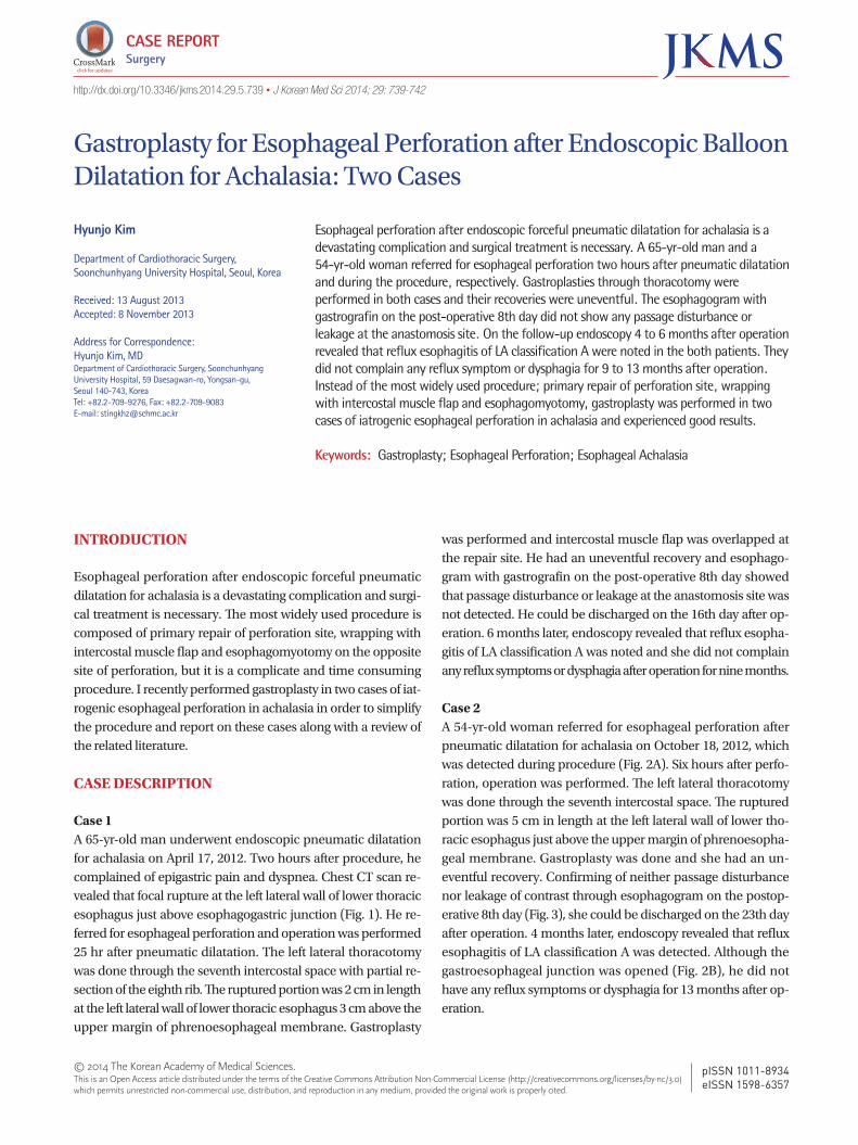
Gastroplasty for Esophageal Perforation after Endoscopic Balloon Dilatation for Achalasia: Two Cases
Esophageal perforation after endoscopic forceful pneumatic dilatation for achalasia is a devastating complication and surgical treatment is necessary. A 65-yr-old man and a 54-yr-old woman referred for esophageal perforation two hours after pneumatic dilatation and during the procedure, respectively. Gastroplasties through thoracotomy were performed in both cases and their recoveries were uneventful. The esophagogram with gastrografin on the post-operative 8th day did not show any passage disturbance or leakage at the anastomosis site. On the follow-up endoscopy 4 to 6 months after operation revealed that reflux esophagitis of LA classification A were noted in the both patients. They did not complain any reflux symptom or dysphagia for 9 to 13 months after operation. Instead of the most widely used procedure; primary repair of perforation site, wrapping with intercostal muscle flap and esophagomyotomy, gastroplasty was performed in two cases of iatrogenic esophageal perforation in achalasia and experienced good results.
Keywords: Gastroplasty; Esophageal Perforation; Esophageal Achalasia
Hyunjo Kim
Department of Cardiothoracic Surgery, Soonchunhyang University Hospital, Seoul, Korea
Received: 13 August 2013Accepted: 8 November 2013
Address for Correspondence:Hyunjo Kim, MDDepartment of Cardiothoracic Surgery, Soonchunhyang University Hospital, 59 Daesagwan-ro, Yongsan-gu, Seoul 140-743, KoreaTel: +82.2-709-9276, Fax: +82.2-709-9083 E-mail: [email protected]
http://dx.doi.org/10.3346/jkms.2014.29.5.739 • J Korean Med Sci 2014; 29: 739-742
INTRODUCTION
Esophageal perforation after endoscopic forceful pneumatic dilatation for achalasia is a devastating complication and surgi-cal treatment is necessary. The most widely used procedure is composed of primary repair of perforation site, wrapping with intercostal muscle flap and esophagomyotomy on the opposite site of perforation, but it is a complicate and time consuming procedure. I recently performed gastroplasty in two cases of iat-rogenic esophageal perforation in achalasia in order to simplify the procedure and report on these cases along with a review of the related literature.
CASE DESCRIPTION
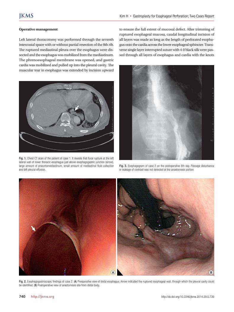
Case 1A 65-yr-old man underwent endoscopic pneumatic dilatation for achalasia on April 17, 2012. Two hours after procedure, he complained of epigastric pain and dyspnea. Chest CT scan re-vealed that focal rupture at the left lateral wall of lower thoracic esophagus just above esophagogastric junction (Fig. 1). He re-ferred for esophageal perforation and operation was performed 25 hr after pneumatic dilatation. The left lateral thoracotomy was done through the seventh intercostal space with partial re-section of the eighth rib. The ruptured portion was 2 cm in length at the left lateral wall of lower thoracic esophagus 3 cm above the upper margin of phrenoesophageal membrane. Gastroplasty
was performed and intercostal muscle flap was overlapped at the repair site. He had an uneventful recovery and esophago-gram with gastrografin on the post-operative 8th day showed that passage disturbance or leakage at the anastomosis site was not detected. He could be discharged on the 16th day after op-eration. 6 months later, endoscopy revealed that reflux esopha-gitis of LA classification A was noted and she did not complain any reflux symptoms or dysphagia after operation for nine months.
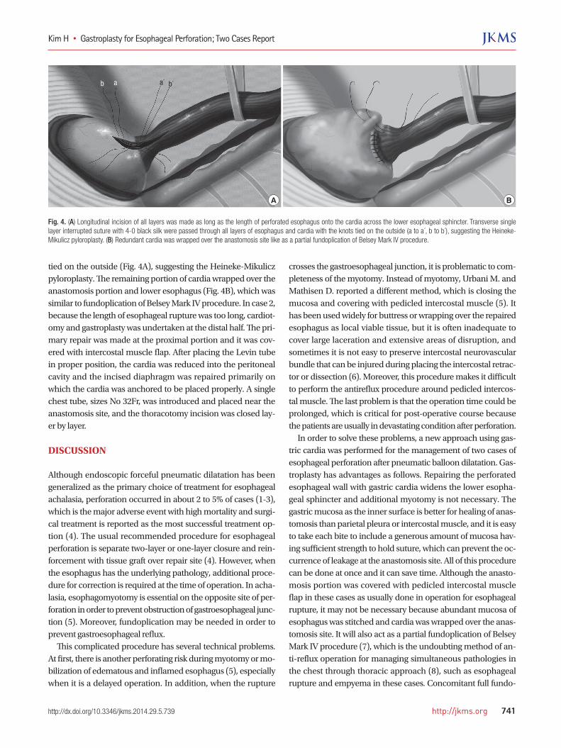
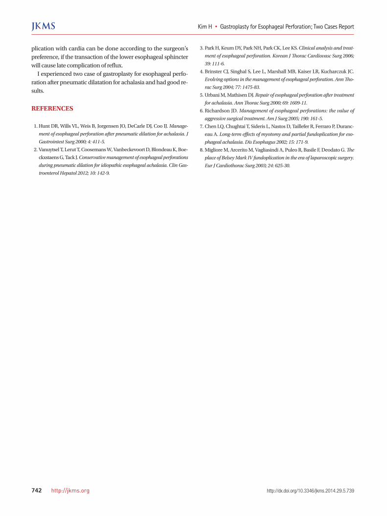
Case 2A 54-yr-old woman referred for esophageal perforation after pneumatic dilatation for achalasia on October 18, 2012, which was detected during procedure (Fig. 2A). Six hours after perfo-ration, operation was performed. The left lateral thoracotomy was done through the seventh intercostal space. The ruptured portion was 5 cm in length at the left lateral wall of lower tho-racic esophagus just above the upper margin of phrenoesopha-geal membrane. Gastroplasty was done and she had an un-eventful recovery. Confirming of neither passage disturbance nor leakage of contrast through esophagogram on the postop-erative 8th day (Fig. 3), she could be discharged on the 23th day after operation. 4 months later, endoscopy revealed that reflux esophagitis of LA classification A was detected. Although the gastroesophageal junction was opened (Fig. 2B), he did not have any reflux symptoms or dysphagia for 13 months after op-eration.
CASE REPORTSurgery
Kim H • Gastroplasty for Esophageal Perforation; Two Cases Report
740 http://jkms.org http://dx.doi.org/10.3346/jkms.2014.29.5.739
Fig. 1. Chest CT scan of the patient of case 1. It reveals that focal rupture at the left lateral wall of lower thoracic esophagus just above esophagogastric junction (arrow), large amount of pneumomediastinum, small amount of mediastinal fluid collection and left pleural effusion.
Fig. 3. Esophagogram of case 2 on the postoperative 8th day. Passage disturbance or leakage of contrast was not detected at the anastomosis portion.
Fig. 2. Esophagogastroscopic findings of case 2. (A) Preoperative view of distal esophagus. Arrow indicated the ruptured esophageal wall, through which the pleural cavity could be identified. (B) Postoperative view of anastomosis site from distal body.
A B
Operative management
Left lateral thoracotomy was performed through the seventh intercostal space with or without partial resection of the 8th rib. The ruptured mediastinal pleura over the esophagus were dis-sected and the esophagus was mobilized from the mediastinum. The phrenoesophageal membrane was opened, and gastric cardia was mobilized and pulled up into the pleural cavity. The muscular tear in esophagus was extended by incision upward
to ensure the full extent of mucosal defect. After trimming of ruptured esophageal mucosa, caudal longitudinal incision of all layers was made as long as the length of perforated esopha-gus onto the cardia across the lower esophageal sphincter. Trans-verse single layer interrupted suture with 4-0 black silk were pas-sed through all layers of esophagus and cardia with the knots
Kim H • Gastroplasty for Esophageal Perforation; Two Cases Report
http://jkms.org 741http://dx.doi.org/10.3346/jkms.2014.29.5.739
Fig. 4. (A) Longitudinal incision of all layers was made as long as the length of perforated esophagus onto the cardia across the lower esophageal sphincter. Transverse single layer interrupted suture with 4-0 black silk were passed through all layers of esophagus and cardia with the knots tied on the outside (a to a´, b to b´), suggesting the Heineke-Mikulicz pyloroplasty. (B) Redundant cardia was wrapped over the anastomosis site like as a partial fundoplication of Belsey Mark IV procedure.
A B
b a a´ b´
tied on the outside (Fig. 4A), suggesting the Heineke-Mikulicz pyloroplasty. The remaining portion of cardia wrapped over the anastomosis portion and lower esophagus (Fig. 4B), which was similar to fundoplication of Belsey Mark IV procedure. In case 2, because the length of esophageal rupture was too long, cardiot-omy and gastroplasty was undertaken at the distal half. The pri-mary repair was made at the proximal portion and it was cov-ered with intercostal muscle flap. After placing the Levin tube in proper position, the cardia was reduced into the peritoneal cavity and the incised diaphragm was repaired primarily on which the cardia was anchored to be placed properly. A single chest tube, sizes No 32Fr, was introduced and placed near the anastomosis site, and the thoracotomy incision was closed lay-er by layer.
DISCUSSION
Although endoscopic forceful pneumatic dilatation has been generalized as the primary choice of treatment for esophageal achalasia, perforation occurred in about 2 to 5% of cases (1-3), which is the major adverse event with high mortality and surgi-cal treatment is reported as the most successful treatment op-tion (4). The usual recommended procedure for esophageal perforation is separate two-layer or one-layer closure and rein-forcement with tissue graft over repair site (4). However, when the esophagus has the underlying pathology, additional proce-dure for correction is required at the time of operation. In acha-lasia, esophagomyotomy is essential on the opposite site of per-foration in order to prevent obstruction of gastroesophageal junc-tion (5). Moreover, fundoplication may be needed in order to prevent gastroesophageal reflux. This complicated procedure has several technical problems. At first, there is another perforating risk during myotomy or mo-bilization of edematous and inflamed esophagus (5), especially when it is a delayed operation. In addition, when the rupture
crosses the gastroesophageal junction, it is problematic to com-pleteness of the myotomy. Instead of myotomy, Urbani M. and Mathisen D. reported a different method, which is closing the mucosa and covering with pedicled intercostal muscle (5). It has been used widely for buttress or wrapping over the repaired esophagus as local viable tissue, but it is often inadequate to cover large laceration and extensive areas of disruption, and sometimes it is not easy to preserve intercostal neurovascular bundle that can be injured during placing the intercostal retrac-tor or dissection (6). Moreover, this procedure makes it difficult to perform the antireflux procedure around pedicled intercos-tal muscle. The last problem is that the operation time could be prolonged, which is critical for post-operative course because the patients are usually in devastating condition after perforation. In order to solve these problems, a new approach using gas-tric cardia was performed for the management of two cases of esophageal perforation after pneumatic balloon dilatation. Gas-troplasty has advantages as follows. Repairing the perforated esophageal wall with gastric cardia widens the lower esopha-geal sphincter and additional myotomy is not necessary. The gastric mucosa as the inner surface is better for healing of anas-tomosis than parietal pleura or intercostal muscle, and it is easy to take each bite to include a generous amount of mucosa hav-ing sufficient strength to hold suture, which can prevent the oc-currence of leakage at the anastomosis site. All of this procedure can be done at once and it can save time. Although the anasto-mosis portion was covered with pedicled intercostal muscle flap in these cases as usually done in operation for esophageal rupture, it may not be necessary because abundant mucosa of esophagus was stitched and cardia was wrapped over the anas-tomosis site. It will also act as a partial fundoplication of Belsey Mark IV procedure (7), which is the undoubting method of an-ti-reflux operation for managing simultaneous pathologies in the chest through thoracic approach (8), such as esophageal rupture and empyema in these cases. Concomitant full fundo-
Kim H • Gastroplasty for Esophageal Perforation; Two Cases Report
742 http://jkms.org http://dx.doi.org/10.3346/jkms.2014.29.5.739
plication with cardia can be done according to the surgeon’s preference, if the transaction of the lower esophageal sphincter will cause late complication of reflux. I experienced two case of gastroplasty for esophageal perfo-ration after pneumatic dilatation for achalasia and had good re-sults.
REFERENCES
1. Hunt DR, Wills VL, Weis B, Jorgensen JO, DeCarle DJ, Coo IJ. Manage-
ment of esophageal perforation after pneumatic dilation for achalasia. J
Gastrointest Surg 2000; 4: 411-5.
2. Vanuytsel T, Lerut T, Coosemans W, Vanbeckevoort D, Blondeau K, Boe-
ckxstaens G, Tack J. Conservative management of esophageal perforations
during pneumatic dilation for idiopathic esophageal achalasia. Clin Gas-
troenterol Hepatol 2012; 10: 142-9.
3. Park H, Keum DY, Park NH, Park CK, Lee KS. Clinical analysis and treat-
ment of esophageal perforation. Korean J Thorac Cardiovasc Surg 2006;
39: 111-6.
4. Brinster CJ, Singhal S, Lee L, Marshall MB, Kaiser LR, Kucharczuk JC.
Evolving options in the management of esophageal perforation. Ann Tho-
rac Surg 2004; 77: 1475-83.
5. Urbani M, Mathisen DJ. Repair of esophageal perforation after treatment
for achalasia. Ann Thorac Surg 2000; 69: 1609-11.
6. Richardson JD. Management of esophageal perforations: the value of
aggressive surgical treatment. Am J Surg 2005; 190: 161-5.
7. Chen LQ, Chughtai T, Sideris L, Nastos D, Taillefer R, Ferraro P, Duranc-
eau A. Long-term effects of myotomy and partial fundoplication for eso-
phageal achalasia. Dis Esophagus 2002; 15: 171-9.
8. Migliore M, Arcerito M, Vagliasindi A, Puleo R, Basile F, Deodato G. The
place of Belsey Mark IV fundoplication in the era of laparoscopic surgery.
Eur J Cardiothorac Surg 2003; 24: 625-30.