Embed Size (px)
Citation preview

Korean J Radiol 9(5), October 2008 449
Gastrointestinal Complications FollowingHematopoietic Stem Cell Transplantationin Children
Gastrointestinal system involvement is one of the principal complications seenin the recipients of hematopoietic stem cell transplantation (HSCT), and it is alsoa major cause of morbidity and death in these patients. The major gastrointestinalcomplications include typhlitis (neutropenic enterocolitis), pseudomembranousenterocolitis, viral enteritis, graft-versus-host disease, benign pneumatosis intesti-nalis, intestinal thrombotic microangiopathy, and post-transplantation lymphopro-liferative disease. As these patients present with nonspecific abdominal symp-toms, evaluation with using such imaging modalities as ultrasonography and CTis essential in order to assess the extent of gastrointestinal involvement and todiagnose these complications. We present here a pictorial review of the imagingfeatures and other factors involved in the diagnosis of these gastrointestinal com-plications in pediatric HSCT recipients.
ematopoietic stem cell transplantation (HSCT) is increasingly being usedto treat many disorders ranging from hematologic malignancies to solidorgan malignancies (1). However, all the recipients are at risk for a
variety of post-transplant complications. Gastrointestinal tract involvement is afrequent complication that causes nonspecific symptoms (2, 3). The timing of complica-tions that occur following transplantation is divided into three phases (4). The pre-engraftment phase lasts 15-30 days and it is characterized by marrow aplasia. Thehost defense barriers may be weakened during this pancytopenic period. The broad-spectrum antimicrobial agents that are administered during this period are also acontributing factor for infection (3). The early post-engraftment period, i.e., from 30 to100 days after transplantation, is characterized by the restoration of hematopoiesis.However, as lymphocyte recovery occurs more slowly, there is a continued deficiencyof both cellular and humoral immunity during this period. The late, post-engraftmentperiod begins only months or years after transplantation (4).
The complications that arise shortly after transplantation often result from themucosal damage that’s secondary to the pre-transplantation chemotherapy andradiation therapy regimens, together with the immunosuppression of the pre-engraft-ment period and the infection that’s secondary to neutropenia. (4). The complicationsthat occur later during the post-transplantation phase include chronic graft-versus-hostdisease (GVHD) and proliferative diseases such as post-transplantation lymphoprolif-erative disease (PTLD) or secondary malignant neoplasm (1, 4).
Despite the fact that gastrointestinal complications remain a major cause of morbid-ity and mortality in pediatric HSCT recipients, there is only limited data regarding theimaging features of the gastrointestinal complications experienced by these patients(3). Careful evaluation of the imaging findings of these gastrointestinal complications,
Ji-Hye Lee, MD1
Gye-Yeon Lim, MD1
Soo Ah Im, MD1
Nak-Gyun Chung, MD2
Seung-Tae Hahn, MD1
Index terms:Hematopoietic stem cell
transplantation PediatricComplicationsGastrointestinal tract
DOI:10.3348/kjr.2008.9.5.449
Korean J Radiol 2008;9:449-457Received November 12, 2007; accepted after revision March 6, 2008.
Departments of 1Radiology and2Pediatrics, St. Mary’s Hospital, TheCatholic University of Korea, Seoul 150-713, Korea
Address reprint requests to:Gye-Yeon Lim, MD, Department ofRadiology, St. Mary’s Hospital, TheCatholic University of Korea, 62, St.Mary’s Hospital, Youido-dong,Yongdungpo-gu, Seoul 150-713, Korea.Tel. (822) 3779-2168Fax. (822) 783-5188e-mail: [email protected]
Abbreviations:CMV = cytomegalovirusCsA = cyclosporine AGI = gastrointestinalGVHD = graft-versus-host diseaseHSCT = hematopoietic stem cell
transplantationLDH = lactate dehydrogenasePMC = pseudomembranous colitisPTLD = post-transplantation
lymphoproliferative diseaseTMA = thrombotic microangiopathy
H

together with the patient’s clinical history, allows radiolo-gists to arrive at a more probable differential diagnosis.
Complications arising during the Pre-EngraftmentPeriod (Days 0-30)
Typhlitis (Neutropenic Colitis)Neutropenic colitis is also termed typhlitis (greek:
typhlon = cecum), and this is a complication of severeneutropenia in patients who have undergone chemother-apy. Bacterial infection of the chemotherapy-related,damaged mucosa, and often this is superinfection, e.g.,Clostridia, may lead to necrotizing inflammatory disease ofthe ileocecal region (1). The clinical manifestations are atriad of abdominal pain or tenderness, fever and neutrope-nia, and this all usually improves after neutrophil recovery.
The imaging feature of typhlitis on plain radiography is apaucity of gas in the right side of the abdomen. Dilatationof a small bowel loop suggests mechanical ileus caused bymarked ileocecal wall thickening and hypervascularity ofthe thickened wall that’s caused by a transmural inflamma-
tory reaction (4) (Figs. 1A, B). The CT images maydemonstrate marked hypodense ileocecal wall thickening,increased mucosal enhancement and inflammatory strand-ing in the pericecal fat (Fig. 1C). These abnormalities arelocalized in the cecum and ascending colon, appendix andterminal ileum. Free abdominal gas and fluid collectionsmay occur in those patients with perforations and severetyphlitis (1, 4).
Acute or chronic inflammatory disease of the ileocecalarea and the right colon, including cytomegalovirusinfection, bacterial ileocecitis, pseudomenbranous entero-colitis and appendicitis, should be considered when makingthe differential diagnosis. The appropriate microbiologicaltests for bacteria and viruses include cytomegalovirus(CMV) polymerase chain reaction and the CMV earlyantigen tests. The endoscopic approach during pancytope-nia is usually contraindicated. The finding of a thickenedbowel wall involving the ileocecal region, which is associ-ated with the clinical triad of neutropenic patients whohave undergone intensive chemotherapy, suggests thisdiagnosis.
Lee et al.
450 Korean J Radiol 9(5), October 2008
A B
Fig. 1. Neutropenic colitis in 10-year-old boy, and this developed on 23rd dayafter bench mark test for treating his leukemia.A, B. US of right lower guadrant abdomen shows marked asymmetricechogenic wall thickening and abundant vascular flow of cecum (A) andterminal ileum (B).C. CT scan shows thickening of cecal wall with associated luminal narrowingand stranding in pericecal fat (arrow).
C

Pseudomembranous Colitis (PMC)Bacterial pathogens are the predominant cause of
infection during the first month following HSCT. The use
of antimicrobial agents during this period provideseffective preventative treatment, but these drugs can alterthe microflora of the colon and cause overgrowth of
Gastrointestinal Complications Following Hematopoietic Stem Cell Transplantation in Children
Korean J Radiol 9(5), October 2008 451
Fig. 3. Cytomegalovirus enteritis in 15-year-old boy, and this developed during second month after bench mark test for treating hisaplastic anemia. A, B. US of abdomen shows multiple sites of segmental hypoechoic bowel wall thickening that involves terminal ileum (A), and there areabundant Doppler signals with ascites (B).
A B
A
B
Fig. 2. Pseudomembranous colitis in 9-year-old boy, and this developed on23rd day after bench mark test for treating his leukemia. A. Longitudinal US of ascending colon shows rather striking diffuse thicken-ing of colonic wall (arrows). Exaggerated haustral markings and inhomoge-nously thickened submucosa with apposition of muscosal surfaces ofthickened wall are noted.B, C. Axial contrast enhanced CT shows pancolitis involving ascending,transverse, descending (B) and rectosigmoid colon (C). Note the hyperemicenhancing mucosa surrounded by thickened hypodense submucosa edema,which forms accordion pattern (arrows).
C

Clostridium difficile. PMC almost always occurs in childrenwith neutropenia and antibiotic-related diarrhea (4).
The US features of PMC include exaggerated and
inhomogenously thickened submucosa with apposition ofthe mucosal surfaces of the thickened wall (Fig. 2A). Thecharacteristic CT feature of PMC is diffuse colonic wall
Lee et al.
452 Korean J Radiol 9(5), October 2008
Fig. 4. Acute graft-versus-host disease in 12-year-old boy, and this developed on 32nd day after bench mark test for treating his leukemia. A, B. US of abdomen shows slightly thickened echogenic wall (arrows) (A) and fluid (*) distended small bowel loops with ascites (B).
A B
Fig. 5. Acute graft-versus-host disease in 11-year-old-boy, and thisdeveloped on 40th day after bench mark test for treating hisleukemia. A, B. Axial contrast-enhanced CT scans show ascites and diffusewall thickening with mucosal enhancement involving small bowel(long arrows) and large bowel (short arrows). C. Coronal MPR contrast-enhanced CT demonstrates morepronounced engorgement of vasa recta (arrow) adjacent to thickenedbowel wall segment, as well as multiple, fluid-filled, dilated loops ofcolon without wall thickening in sigmoid colon (*). Abnormal enhance-ment of gall bladder and urinary bladder is also present, and this issimilar to that seen within bowel wall.
A
B
C

thickening with low attenuation and trapped contrastmedia between the thickened haustral folds, and this isreferred to as the “accordion sign” (Figs. 2B, C).Pancolonic involvement is common in patients with PMC,but this involvement may be focal or restricted to the rightor rectosigmoid colon.
Pseudomembranous colitis can be confirmed by eitherendoscopic demonstration of the characteristic yellowplaque (pseudomembrane) on the rectal or colonic mucosaor by serologic documentation of Clostridium difficile inthe feces (5).
Complications arising during the Early Post-Engraftment Period (days 31-100)
Cytomegalovirus (CMV) GastroenteritisViruses such as rotavirus, adenovirus and CMV are well
recognized causes of the diarrhea in HSCT recipientsduring the early post-transplantation period, and CMV isthe leading cause of intra-abdominal infectious complica-tions. CMV gastroenteritis and hepatitis are major causesof infection-associated mortality following transplantation(4). The imaging findings include non-specific bowel wallthickening, ascites and adjacent inflammatory change,especially in the ileocecal region (Fig. 3).
Graft-versus-Host Disease (GVHD)Graft-versus-host disease results from the damage that
donor lymphocytes do to the recipient’s target organs. The
most commonly affected organs are the skin, liver andgastrointestinal tract. GVHD develops in 55% of theallogenic marrow recipients. The degree of histocompati-bility between the stem cell donor and the recipientsaffects the risk for developing GVHD.
Graft-versus-host disease is not only a primary complica-tion, but it also acts as a predisposing factor for othercomplications. Because the GVHD-involved gut mucosaimpairs the mucosal lymphoid intestinal immunity, patientswith GVHD are also susceptible to concomitant gastroin-testinal infections.
Acute GVHD usually develops 3-5 weeks aftertransplantation and it is most commonly diagnosed inconjunction with or it is usually heralded by such skinmanifestations as maculopapular rash and pruritis. ChronicGVHD can develop following acute GVHD and it can ariseseveral months after allogenic transplantation (6, 7). It israre to diagnose GVHD when there is only a single site ofinvolvement such as isolated intestinal tract involvement.Symptoms of intestinal GVHD can also occur, includingsecretory diarrhea, fever, nausea, vomiting, abdominalpain and intestinal hemorrhage (8).
The plain radiographic findings of intestinal GVHDinclude air fluid levels, bowel wall thickening, a gaslessabdomen, bowel dilatation and ascites. US shows multiple,fluid-filled, dilated bowel loops (Fig. 4). The CT findingsthat suggest acute GVHD include abnormal bowel wallenhancement, and this correlates histopathologically withmucosal destruction and its replacement by a thin layer of
Gastrointestinal Complications Following Hematopoietic Stem Cell Transplantation in Children
Korean J Radiol 9(5), October 2008 453
Fig. 6. Benign pneumatosis intestinalis in 15-year-old asymptomatic boy, and this developed on 75th day after BMT for treating hisleukemia.A. Plain film of abdomen shows diffuse linear and bubbly intramural air along entire colon, as well as presence of subhepatic gas. B. CT scan shows diffuse intramural air densities in entire colon with involvement of retroperitoneal (arrows) and intraperitoneal cavities.
BA

granulation tissue (Figs. 5A, B) (7, 8). As oral contrastmaterial, the use of a negative contrast agent, such aswater, may be advantageous for identifying mucosalenhancement. Bowel wall thickening may involve any partof the small or large bowel, but this is less severe than thatfound in patients with typhlitis or pseudomembranouscolitis (4). The common extraintestinal findings of intestinalGVHD include ascites and engorgement of the mesentericvessels; these findings are more pronounced adjacent tothe thickened bowel loops (Figs. 5A, B). The abnormalfindings that represent periportal edema and abnormalenhancement of the gall bladder and urinary bladder wallmay also occur (Fig. 5C).
Because differentiation of intestinal GVHD frominfective enteritis is critical for proper patient treatment,tissue sampling via biopsy is often required for confirma-tion. Microbiological tests for bacteria and viruses, such asthose performed on rectal biopsies, are useful for makingthe diagnosis of intestinal GVHD. A diagnostic biopsy fromskin, liver or rectum is required.
To diagnose the symptoms of intestinal GVHD, as wellas the other typical clinical findings of GVHD.
Pneumatosis IntestinalisThe factors contributing to the development of
pneumatosis intestinalis include pre-transplantationchemotherapy and radiotherapy, steroid therapy,infectious colitis, GVHD and septic shock (9). Steroidtherapy appears to be a significant factor for the develop-ment of pneumatosis in patients who suffer with HSCT. Ithas been postulated that steroid administration inducesatropy of the lymphoid aggregates (Peyer patches) in thegastrointestinal (GI) tract and that the resultant mucosaldefects allow dissection of intraluminal air into thesubmucosal or subserosal regions (2). If this is detected inan asymptomatic patient after steroid therapy, it is usuallynot associated with bowel necrosis and it can be resolvedwith conservative management. In these benign cases,even the subsequent development of retroperitoneal air orpneumoperitoneum does not necessitate surgery or
Lee et al.
454 Korean J Radiol 9(5), October 2008
Fig. 7. Intestinal thrombotic microangiopathy followed by graft-versus-host disease in 13-year-old girl, and this developed on 28th day afterperipheral blood stem cell transplantation for treating her leukemia.A, B. US shows multiple distended small bowel loops with wall thicken-ing. Medium echogenic debris with floating echogenicities that filldistended lumen are noted in some of these loops (*).C. Unenhanced coronal multiplanar reconstruction CT image showsincreased intraluminal attenuation, which is consistent with intraluminalbleeding (*), and submucosal zone of decreased attenuation parallel tolumen (arrowhead). Ascites and mesenteric thickening are also noted.
A
B
C
*
*
indicate a grave prognosis (Fig. 6). However, pneumatosisintestinalis associated with neutropenic colitis is veryworrisome as it implies imminent bowel perforation.
The imaging features of pneumatosis intestinalis includebubbly and linear intraluminal lucencies that representsubmucosal and subserosal gas, respectively (Fig. 6).Usually, the right side of the colon is predominantlyinvolved.
Thrombotic Microangiopathy (TMA) afterTransplantation
Thrombotic microangiopathy represents intimal injury ofthe microvasculature followed by pathophysiologicformation of microthrombi. As intestinal TMA after HSCTis a rare, but fatal complication that occurs in patients whoundergo allogenic or autologous BMT (bench mark test), itsearly diagnosis is very important. The risk factors such asacute GVHD, cyclosporine A (CsA), tacrolimus (FK 506)and total body irradiation may contribute to its occurrence,but the pre-transplant conditioning regimen and thepresence of infection may also have a role in the pathogen-esis of TMA (10). The intestinal tract is a prevalent site forpost-HSCT TMA, and ischemic enterocolitis caused bymicroangiopathy is another mimic of gut GVHD. Although
the clinical symptoms and imaging features of intestinalTMA are similar to those of intestinal GVHD, differentiat-ing TMA from acute GVHD is important for patientssuffering from severe and refractory diarrhea after HSCT(10). Because of the difficulty in making a definitivediagnosis of TMA in HSCT recipients, this condition isusually diagnosed based on the clinical and laboratoryfindings such as the serum lactate dehydrogenase (LDH)levels and the percentage of fragmented erythrocytes.However, as these findings are frequently non-specific,pathologic examination of the gastrointestinal tract may beessential in order to make a definite diagnosis of TMA inHSCT recipients with refractory diarrhea (11). In ourseries, a 13-year-old girl developed intestinal TMAfollowed by GVHD. Despite that her skin-GVHDimproved with intensive treatment, her intestinal symptomof severe bloody refractory diarrhea progressed. Thispatient also experienced subsequent hyperammonemicencephalopathy and an elevated level of LDH withfragmented erythrocytes. On the basis of the clinical andlaboratory data, this patient was diagnosed with CsA-associated neurotoxicity with microangiopathic hemolyticanemia. To the best of our knowledge, little attention hasbeen given to the imaging features of intestinal TMA after
Gastrointestinal Complications Following Hematopoietic Stem Cell Transplantation in Children
Korean J Radiol 9(5), October 2008 455
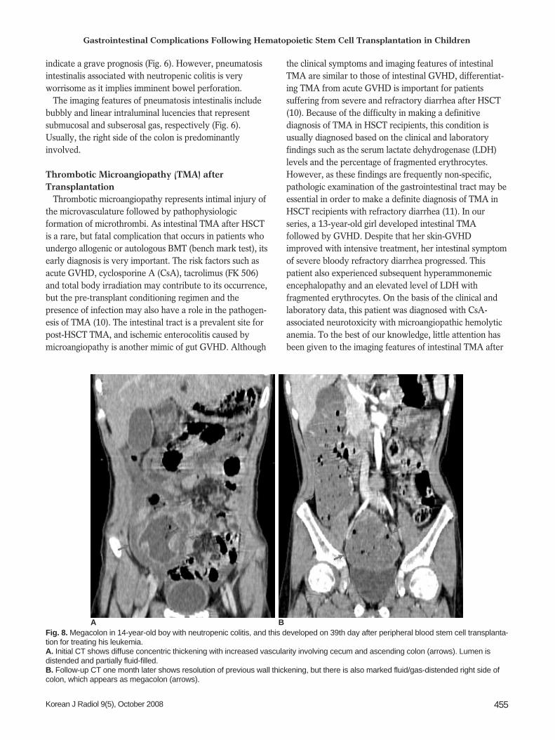
Fig. 8. Megacolon in 14-year-old boy with neutropenic colitis, and this developed on 39th day after peripheral blood stem cell transplanta-tion for treating his leukemia.A. Initial CT shows diffuse concentric thickening with increased vascularity involving cecum and ascending colon (arrows). Lumen isdistended and partially fluid-filled. B. Follow-up CT one month later shows resolution of previous wall thickening, but there is also marked fluid/gas-distended right side ofcolon, which appears as megacolon (arrows).
BA

transplantation and although they do not differ from thoseof GVHD, intraluminal or intramucosal hemorrhage isfrequently present in the TMA patients who present withsevere bloody diarrhea (Fig. 7).
MegacolonMegacolon may be caused by motility problems that are
related to the complications of systemic chemotherapy andintestinal or intrabdominal infection or a combination ofthese factors (1) (Fig. 8).
Complications arising during the Late Post-Engraftment Period
Post-Transplantation Lymphoproliferative Disease(PTLD)
Post-transplantation lymphoproliferative disease iscaused by Epstein-Barr virus, which induces uncontrolledproliferation of engrafted B lymphocytes in organtransplant recipients. The histologic subtypes of PTLDrange from polyclonal lymphoid hyperplasia to malignantmonoclonal lymphoma (12). GI tract involvement byPTLD frequently occurs, but this is rare following HSCT,with a prevalence of 0.34% and 1.4% in patients withmatched allogenic grafts and unmatched grafts, respec-tively (13).
The imaging features of PTLD are similar those of non-Hodgkin’s lymphoma. PTLD involving the GI tract maybe infiltrative, and so it causes circumferential mural
infiltration with luminal excavation or there is a discreteeccentric mass that causes luminal narrowing with intussus-ception (12) (Fig. 9).
Reduced immunosuppression in the case of polyclonalhyperplasia leads to regression of the PTLD, whereasaggressive chemotherapy is required for monoclonallymphoma. Unlike the PTLD in solid organ recipients, thePTLD seen in HSCT recipients has a poor prognosisbecause decreasing these patients’ immunosuppression isnot a feasible therapeutic option.
CONCLUSION
Hematopoietic stem cell transplantation has resulted inthe prolonged survival of children with hematologicmalignancy. However, gastrointestinal complicationsfollowing transplantation are now seen as a major cause ofmorbidity and mortality in pediatric HSCT recipients.Therefore, it is important to recognize the imaging findingsof these complications in those patients who undergohematopoietic stem cell transplantation in order to identifyand properly treat them.
References1. Benya EC, Sivit CJ, Quinones RR. Abdominal complications
after bone marrow transplantation in children: sonographic andCT findings. AJR Am J Roentgenol 1993;161:1023-1027
2. Jones B, Fishman EK, Kramer SS, Siegelman SS, Saral R,Beschorner WE, et al. Computed tomography of gastrointestinalinflammation after bone marrow transplantation. AJR Am J
Lee et al.
456 Korean J Radiol 9(5), October 2008
Fig. 9. Post-transplantation lymphoproliferative disease involving small bowel infive-year-old boy, and this developed during third year following bench mark test fortreating his leukemia.A. Longitudinal US of left mild abdomen shows short area of segmental severeconcentric hypoechoic mural thickening (*), and this causes luminal narrowing injejunum.B. CT scan shows short area of segmental concentric wall thickening with homoge-nous enhancement of small bowel loop in left side of abdomen (arrow).
A
B

Roentgenol 1986;146:691-6953. Barker CC, Anderson RA, Sauve RS, Butzner JD. GI complica-
tions in pediatric patients post-BMT. Bone Marrow Transplant2005;36:51-58
4. Coy DL, Ormazabal A, Godwin JD, Lalani T. Imaging evalua-tion of pulmonary and abdominal complications followinghematopoietic stem cell transplantation. Radiographics2005;25:305-317
5. Bolton RP, Thomas DF. Pseudomembranous colitis in childrenand adults. Br J Hosp Med 1086;35:37-42
6. Donnelly LF, Morris CL. Acute graft-versus-host disease inchildren: abdominal CT findings. Radiology 1996;199:265-268
7. Mentzel HJ, Kentouche K, Kosmehl H, Gruhn B, Vogt S,Sauerbrey A, et al. US and MRI of gastrointestinal graft-versus-host disease. Pediatr Radiol 2002;32:195-198
8. Fisk JD, Shulman HM, Greening RR, McDonald GB, Sale GE,Thomas ED. Gastrointestinal radiographic features of humangraft-vs.-host disease. AJR Am J Roentgenol 1981;136:329-336
9. Day DL, Ramsay NK, Letourneau JG. Pneumatosis intestinalis
after bone marrow transplantation. AJR Am J Roentgenol1988;151:85-87
10. Nishida T, Hamaguchi M, Hirabayashi N, Haneda M, TerakuraS, Atsuta Y, et al. Intestinal thrombotic microangiopathy afterallogeneic bone marrow trasplanatation: a clinical imitator ofacute enteric graft-versus-host disease. Bone MarrowTransplant 2004;33:1143-1150
11. Narimatsu H, Kami M, Hara S, Matsumura T, Miyakoshi S,Kusumi E, et al. Intestinal thrombotic microangiopathy follow-ing reduced-intensity umbilical cord blood transplantation. BoneMarrow Transplant 2005;36:517-523
12. Lim GY, Newman B, Kurland G, Webber SA. Post transplanta-tion lymphoproliferative disorder: manifestations in pediatricthoracic organ recipients. Radiology 2002;222:699-708
13. Lones MA, Kirov I, Said JW, Shintaku IP, Neudorf S. Post-transplant lymphoproliferative disorder after autologous periph-eral stem cell transplantation in a pediatric patient. BoneMarrow Transplant 2000;26:1021-1024
Gastrointestinal Complications Following Hematopoietic Stem Cell Transplantation in Children
Korean J Radiol 9(5), October 2008 457