Embed Size (px)
Citation preview
Anaerobic infection
Gas gangrene
General surgery department of SGMU
Lecturer –ass. Khilgiyaev R.H.
General surgery department of SGMU
Lecturer –ass. Khilgiyaev R.H.
Anaerobic bacteria
Anaerobic bacteria are the most numerous inhabitants of the normal gastrointestinal tract, including the mouth
Bacteroides fragilis and Clostridium
The most common anaerobic isolate from surgical infections is Bacteroides fragilis

General surgery department of SGMU
Lecturer –ass. Khilgiyaev R.H.
Bacteroides fragilis:
have significant resistance to many
β-lactam antibiotics
most effective antibiotics against
these species are metronidazole,
clindamycin, meropenem and
sulperazon

General surgery department of SGMU
Lecturer –ass. Khilgiyaev R.H.
.
Clostridium:
• The Clostridium species are all gram-positive, spore-forming encapsulated rods.
• C. Perfringens
• C. Septicum
• C. histolyticum
• These strains grow only in settings with a low oxidation-reduction potential.
• Thus, the recovery of anaerobes from a soft tissue infection or even from the blood implies their growth and multiplication in a focus of dead tissue.

General surgery department of SGMU
Lecturer –ass. Khilgiyaev R.H.
Necrotizing
Fasciitis
• This disease usually occurs in men in the lower extremities
after only minimal local trauma.
• Although a monobacterial etiology, typically Clostridium and
Streptococcus group A, can be found, the infections are
usually produced by mixed flora, both aerobes and
anaerobes.

General surgery department of SGMU
Lecturer –ass. Khilgiyaev R.H.
A substance in the cell wall of streptococci causes:
• separation of the dermal connective tissue, resulting in continued
inflammation and necrosis.
• tissue ischemia by widespread occlusion of small subcutaneous
vessels.
• Vessel occlusion results in skin infarction and necrosis, which
facilitates the growth of obligate anaerobes while promoting
anaerobic metabolism by facultative organisms (eg, Escherichia
coli), resulting in gangrene.
• Anaerobic metabolism produces hydrogen and nitrogen,
relatively insoluble gases that may accumulate in subcutaneous
tissues
Necrotizing Fasciitis
Pathogenesis
General surgery department of SGMU
Lecturer –ass. Khilgiyaev R.H.
Necrotizing Fasciitis
Signs
Streptococcal necrotizing fasciitis is frequently
associated with streptococcal toxic shock
syndrome.
Early clinical findings are similar to those with
most infected wounds, but the involved site
quickly becomes erythematous, tender, and
edematous; fever is usually present.
Deep pain is often out of proportion to the
physical findings.

General surgery department of SGMU
Lecturer –ass. Khilgiyaev R.H.
Necrotizing Fasciitis
Signs
Bullae, crepitus (from soft-tissue gas), and
gangrene may develop.
Subcutaneous tissues (including adjacent fascia)
necrose. Deep structures and muscles are not
involved.
Hypotension, tachycardia, leukocytosis, with
systemic toxicity out of proportion to the clinical
findings.

General surgery department of SGMU
Lecturer –ass. Khilgiyaev R.H.
Treatment
Treatment involves antibiotics and surgical debridement.
The initial incision should be extended until an instrument or finger can no longer separate the skin and subcutaneous tissue from the deep fascia.
The most common error is insufficient surgical intervention
Amputation of an extremity may be necessary. Fluids may be needed in large volumes before and after
surgery. Antibiotic choices should be reviewed based on Gram
stain and culture of tissues obtained during surgery. Hyperbaric O2 therapy may also be of benefit
Prognosis is poor without early, aggressive treatment.

General surgery department of SGMU
Lecturer –ass. Khilgiyaev R.H.
Treatment involves antibiotics and surgical debridement.
The initial incision should be extended until an instrument or finger can no longer separate the skin and subcutaneous tissue from the deep fascia.
The most common error is insufficient surgical intervention
Amputation of an extremity may be necessary. Fluids may be needed in large volumes before and after
surgery. Antibiotic choices should be reviewed based on Gram
stain and culture of tissues obtained during surgery. Hyperbaric O2 therapy may also be of benefit
Prognosis is poor without early, aggressive treatment.
Treatment of necrotizing fasciitis

General surgery department of SGMU
Lecturer –ass. Khilgiyaev R.H.
Cellulitis
Nonclostridial
Clostridial
General surgery department of SGMU
Lecturer –ass. Khilgiyaev R.H.
Cellulitis Etiology
Cellulitis is most often caused by aerobic and anaerobic coliforms such as E. coli, Klebsiella, Enterobacter, Peptostreptococcus, Peptococcus, and B. fragilis, group A β-hemolytic streptococci or Staphylococcus aureus.
Streptococci cause diffuse, rapidly spreading infection because enzymes produced by the organism (streptokinase, hyaluronidase) break down cellular components.
Staphylococcal cellulitis is typically more localized and usually occurs in open wounds or cutaneous abscesses.

General surgery department of SGMU
Lecturer –ass. Khilgiyaev R.H.
Cellulitis Etiology
Recently, methicillin-resistant S. aureus (MRSA) has become more common in the community.
Historically, MRSA was typically confined to patients who were exposed to the organism in a hospital or nursing facility.
MRSA infection should now be considered in patients with community-acquired cellulitis, particularly in those with cellulitis that is recurrent or unresponsive to monotherapy.

General surgery department of SGMU
Lecturer –ass. Khilgiyaev R.H.
Symptoms and Signs
• Infection is most common in the lower extremities. Cellulitis is typically unilateral.
• The major findings are local erythema and tenderness, frequently with lymphangitis and regional lymphadenopathy.
• The skin is hot, red, and edematous, often with surface appearance resembling the skin of an orange.
• The borders are usually indistinct.
• Vesicles and bullae may develop with necrosis of the involved skin.
• Fever, chills, tachycardia, headache, hypotension, and delirium may precede cutaneous findings by several hours, but many patients do not appear ill.
• Leukocytosis is common.
General surgery department of SGMU
Lecturer –ass. Khilgiyaev R.H.
Cellulitis treatment
Risk factors include skin abnormalities (eg,
trauma, ulceration, fungal infection, other skin
barrier compromise due to preexisting skin
disease), which are common in patients with
chronic venous insufficiency or lymphedema
These infections tend to progress from a fasciitis
to a myositis.
The treatment is broad-spectrum antibiotics and
close observation and debridement

General surgery department of SGMU
Lecturer –ass. Khilgiyaev R.H.
• Clostridial cellulitis (anaerobic cellulitis, local gas gangrene) is a gas-
forming infection of the skin and subcutaneous tissue that spreads through
intrafascial planes.
• Healthy muscle is not involved. It results from superinfection of
previously traumatized or necrotic tissue.
• Gas distributes in large bubbles in the fascial plane but not the muscle.
• Patients show signs of systemic toxicity: fever, tachycardia, edema of the
affected part, and pain.
• Incision and debridement of involved tissue and blebs are necessary.
Clostridial cellulitis

General surgery department of SGMU
Lecturer –ass. Khilgiyaev R.H.
Fournier’s syndrome
is a necrotizing subcutaneous infection of the perineum that occurs primarily in men, usually involving scrotum.
Pain or itching in the genitalia is followed by fever, chills, and impressive perineal swelling, which may simulate a strangulated hernia.
The inflammation may involve the entire abdomen, back, and thighs.
There is frequently crepitance on palpation, indicating subcutaneous gas.
Systemic symptoms include nausea and vomiting
The most common causal factors are infection or trauma to the perianal area, including anal intercourse, scratches, chemical or thermal injury, and diabetes.

General surgery department of SGMU
Lecturer –ass. Khilgiyaev R.H.
Fournier’s syndrome Management
wide incision and drainage of the area to remove all the necrotic tissue.
Gram’s stain and culturing of the wound, antibiotic therapy against anaerobes and gram-negative enterics

General surgery department of SGMU
Lecturer –ass. Khilgiyaev R.H.
Clostridial myonecrosis,
or gas gangrene
Myonecrosis, is a deep soft tissue infection with death of muscle and a variable degree of inflammation of the overlying tissues.
The skin may show minimal erythema, but usually the infection is associated with massive edema, with gas formation.
It is usually a result of trauma or recent surgical wounds.

General surgery department of SGMU
Lecturer –ass. Khilgiyaev R.H.
Pathogenesis
Pathogenesis includes the elaboration of exotoxins by Clostridial bacilli.
Clostridia produce a toxin that damages and kills muscle, setting up the anaerobic environment that promotes further growth of the bacilli.
The incubation period is 1 to 4 days.
The patient appears pale and anxious, with a rapid progression to toxemia and shock.
General surgery department of SGMU
Lecturer –ass. Khilgiyaev R.H.
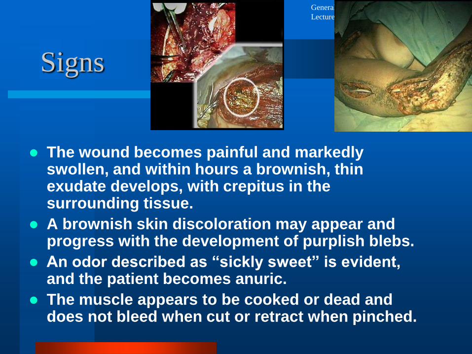
Signs
The wound becomes painful and markedly swollen, and within hours a brownish, thin exudate develops, with crepitus in the surrounding tissue.
A brownish skin discoloration may appear and progress with the development of purplish blebs.
An odor described as “sickly sweet” is evident, and the patient becomes anuric.
The muscle appears to be cooked or dead and does not bleed when cut or retract when pinched.

General surgery department of SGMU
Lecturer –ass. Khilgiyaev R.H.
Clostridial myonecrosis
Specially placed tight control
bandage leaves a mark on the skin
after removal and the sutures seem
to be «cutting through» the skin

General surgery department of SGMU
Lecturer –ass. Khilgiyaev R.H.
Clostridial myonecrosis Diagnostic
Gram’s stain smears of the area
show large gram-positive rods.
Radiographs may reveal gas.
General surgery department of SGMU
Lecturer –ass. Khilgiyaev R.H.
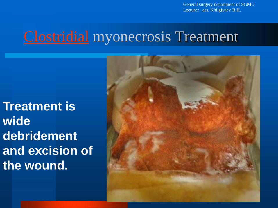
Clostridial myonecrosis Treatment
Treatment is
wide
debridement
and excision of
the wound.

General surgery department of SGMU
Lecturer –ass. Khilgiyaev R.H.
antibiotic
• Parenteral antibiotics should be
given to cover anaerobes and
enterics:
• a cephalosporin, carbopenem,
vancomycin, metronidazol and
dioxidin is indicated.
• The mortality rate is high

General surgery department of SGMU
Lecturer –ass. Khilgiyaev R.H.
Hyperbaric oxygen
therapy
Abacterial Atmosphere and
Hyperbaric oxygen therapy (HBO)
may be effective very early in this
disease.
HBO does not kill clostridia; it has a
bacteriostatic effect, and oxygen will
inhibit α-toxin production.

General surgery department of SGMU
Lecturer –ass. Khilgiyaev R.H.
After operation