Embed Size (px)
Citation preview

J Neurosurg 113:128–135, 2010
128 J Neurosurg / Volume 113 / December 2010
It is generally accepted that the majority (more than 90%) of “acoustic neuromas” are indeed VSs–mostly from the inferior branch–originating in the “fibrous
cone” where the oligodendroglial sheathing is substituted by Schwann cell coverage.6,14,23 A strict minority (3–10%) seem to belong to the cochlear nerve, the Scarpa gan-glion, the vestibule, and the fundus.5,8,25 This probably ex-plains the relevance of balance impairment and hearing disturbances in patients with these lesions as well as the
predominance of deficits following the surgical removal of such tumors. In small-to-medium VSs, GKS currently represents a major therapeutic option, basically because of the highly conformal and selective dose planning, ex-cellent rate of tumor control, and minimal morbidity in terms of cranial neuropathy.2,9,11,15–17,20,21,24,26 The increas-ing role of such a peculiar approach has been further con-firmed by a recent Acoustic Neuroma Association survey of US patients with acoustic neuromas (Table 1).1
In these patients, once again, GKS treatments have been followed by high levels of TGC, maintaining an undoubtedly low overall incidence of side effects (Table 2) basically confined to the cochlear and vestibular com-ponents, with extremely rare involvement of the facial nerve.3,11,16,17,20,26 These results were similar to our own
Gamma Knife surgery in vestibular schwannomas: impact on the anterior and posterior labyrinth
Clinical articleMassiMo Gerosa, M.D.,1 NazareNa MesiaNo, M.D.,2 Michele loNGhi, M.D.,1 aNtoNio De siMoNe, Ph.D.,1 roberto ForoNi, Ph.D.,1 aNGela Verlicchi, M.D.,1 bruNo zaNotti, M.D.,3 aND aNtoNio Nicolato, M.D.1
Departments of 1Neurosurgery and 2ENT Surgery, “Borgo Trento” University Hospital, Verona; and 3Unit of Neurosurgery, “S. Maria della Misericordia” University Hospital, Udine, Italy
Object. During the past decades, in small-to-medium size vestibular schwannomas, Gamma Knife surgery (GKS) has become a reliable therapeutic option because of either excellent local tumor control or minimal morbid-ity, with cranial neuropathy becoming increasingly rare. Although still insufficiently analyzed in larger cohorts of patients with long-term follow-ups, adequate chances of hearing preservation and vestibular sparing seem clinically guaranteed. However, deeper investigations are needed in this regard, expanding the number of cases and the follow-up period.
Methods. A small group of patients with vestibular schwannomas (74 patients, including 41 men and 33 women) treated between 2003 and 2009 using GKS at the authors’ institution were analyzed—both before and after GKS—with computerized static stabilometry and electronystagmography for balance disorders, vertigo, and ataxia on 1 side and pure tone average, vocal speech discrimination score, auditory brainstem response, and so forth for hearing impairment and tinnitus on the other side. Eligibility criteria for this prospective study included previously untreated unilateral lesions and a Gardner-Robertson hearing class of I–III. Dosimetry plans had been programmed at the lower effective dosages for these tumors (median surface dose 12.4 Gy, range 10–13 Gy), carefully avoiding even minimal toxic dosages on the most vulnerable targets: the cochlea (never > 6 Gy) and the vestibular canals (< 7.5 Gy).
Results. To date, tumor growth control rates remain satisfactory; at a mean follow-up of 50 months, the rate was 96%. The overall level of hearing preservation was 72%, with 81% having Gardner-Robertson Class I hearing. Tin-nitus decreased, from 52% to 28% of patients (p < 0.01). Significant improvements were also observed in vestibular symptoms, with computerized static stabilometry abnormalities decreasing from 62% to 32% (p < 0.001) and elect-ronystagmography abnormalities reducing from 48% to 14% (p < 0.001).
Conclusions. Using appropriate radiodosimetry planning, GKS seems to guarantee not only adequate tumor growth control rates, but also better levels of hearing preservation, with a documented, long-lasting improvement in vestibular functions. (DOI: 10.3171/2010.8.GKS101089)
Key WorDs • radiosurgery • Gamma Knife • vestibular schwannoma • labyrinth
Abbreviations used in this paper: CSS = computerized static sta-bilometry; ENG = electronystagmography; GKS = Gamma Knife surgery; G-R = Gardner-Robertson; MFU = mean follow-up; PTA = pure tone audiometry; SDS = speech discrimination score; TGC = tumor growth control; VS = vestibular schwannoma.
J Neurosurg / Volume 113 / December 2010
Gamma Knife surgery in vestibular schwannomas
129
experience with GKS for VS: from the very beginning, a negligible percentage (2%–3%) of facial neuropathy was associated with a 36% rate of acoustic, and a 28% rate of balance, deterioration.9
Consequently, some of the newer goals of a refined technique are strictly related to the cochlear and vestib-ular structures of the involved side, aiming to improve functional results for both. Briefly, accurate dose plan-ning may enhance hearing preservation, adequately spar-ing the cochlear region, the most vulnerable of the acous-tic “topography.” The process must be monitored with appropriate and repeated audiometric staging. Vestibular protection can be achieved first with accurate monitor-ing of the related functions by using ENG and CSS,28,29
followed by carefully reduced radiation exposure of the semicircular canals. Such a strategy and the relevant follow-up analyses must separately consider all the vari-ous specific segments: receptor, nerve fiber, nuclear, and ganglionic regions. This study was planned to evaluate
the impact of this approach on both the anterior and the posterior labyrinth.
MethodsBetween February 1993 and April 2010, 602 pa-
tients harboring symptomatic VSs were treated with GKS (Model C and Perfexion, Elekta AB) at our institution. A smaller group of 74 patients (those since 2003) consisting of 41 men and 33 women, with a mean age of 59 years (range 24–77 years), was considered for the purpose of this research.
Eligibility criteria included the following: unilateral VS, no previous surgical or radiation treatment, no con-comitant hydrocephalus, no systemic neurological dis-orders, and, finally, a G-R hearing class of I–III, that is, beyond the traditional threshold of “serviceable hearing” (G-R Class I–II). This criterion was decided to explore minimal variations in residual cochlear function or to analyze the potential role of acoustic devices under these conditions.3,4,7,10,19,22,24
TABLE 1: Acoustic Neuroma Association US 2007–2008 survey of over 2000 evaluable forms*
% Patients Parameter Microsurgical Treatment Stereotactic Radiosurgery Treatment Wait & Watch
survery year 1983 100 0 0 1998 85 5 10 2008 61 20 19sequelae Preop† Postop† Preop Postop hearing loss G-R Class I–III 67 16 56 23 G-R Class IV–V 33 84 44 77 balance deficit 34 61 11 25 facial deficit H-B Grade I–II 84 43 96 79 H-B Grade V–VI 11 16 0 2
* H-B = House-Brackmann.† Nontranslabyrinthine approach.
TABLE 2: Literature summary of studies on GKS for VS: results in terms of TGC
Authors & Year No. of Patients MFU (mos) TGC (%)
Norèn, 1998 250 36 95Kondziolka et al., 2000 162 60 98Unger et al., 2002 100 76 96Gerosa et al., 2002 112 49 93Iwai et al., 2003 51 60 86Litvack et al., 2003 121 32 96.7Régis et al., 2004 1000 84 97Beegle et al., 2007 390 60 91Arthurs et al., 2010 70 26 94present study 74 50 96
TABLE 3: Treatment planning parameters in 74 patients*
Parameter Value
ATV in cm3 (range) 2.7 (0.06–10.4)mean peripheral isodose in % (range) 51.7 (50–60)median surface dose in Gy (range) 12.4 (10–13)mean no. of isocenters 14.6 (2–23)mean conformity index (range) 1.28 (1–2.35)max receptor exposure (Gy)† cochlea 5 vestibular channel 7.2
* ATV = average tumor volume.† Max receptor exposure is the maximum dose reaching 3 mm3 of the receptor volume.

M. Gerosa et al.
130 J Neurosurg / Volume 113 / December 2010
A clinicoradiological diagnosis was routinely based on CT (including bone algorithm) and accurately selected MR imaging sequences (volume acquisition, constructive interference for steady state [CISS], fat suppression, and so forth). Patient quality of life was assessed according to the Karnofsky Performance Scale, starting with an aver-age of 73 before radiosurgery.
Cochlear and vestibular functions were assessed on 1 side before and after GKS by using pure tone audiometry, auditory brainstem response, and vocal SDS; on the other side, by testing reflectivity according to the Hallpike-
Fitzgerald method, “corticospinal balance” using CSS, and vertigo using ENG.
On admission, 50% of the patients reported slowly pro gres sive hypacusia, 4% reported a sudden deficit, and 6% reported a variable alternating trouble. Sixteen (21.6%) of 74 patients had G-R Class I hearing; 20 (27%) had G-R Class II; and 38 (51.4%) had G-R Class III.
Tinnitus was present in 56.7% of the cases.Regarding vestibular symptoms, balance disorders
(mainly ataxia) were present in 63.5% and vertigo in 46% of patients.
Dose PlanningDose planning was done in consideration of the tu-
mor size—although as low a dose as possible was select-ed—tumor location, and projected radiobiological risk to adjacent brainstem and cranial nerve. The mean periph-eral dose was 12.4 Gy (range 10–13 Gy), and the mean number of isocenters was 14.6 (range 2–23 isocenters). The main parameters are summarized in Table 3.
Patient Follow-Up Patients were regularly monitored at 6 and 12 months
Fig. 1. Coronal MR images with contrast medium showing a VS 7 (left) and 41 (right) months after GKS, with evident tumor shrinkage.
Fig. 2. Magnetic resonance images with contrast medium (A and C) and graphs (B and D) featuring fluctuating audiometric recovery. Left VS 6 (A and B) and 18 months (C and D) after GKS. The progressive tumor shrinkage is associated with fluctuat-ing audiometric recovery.
J Neurosurg / Volume 113 / December 2010
Gamma Knife surgery in vestibular schwannomas
131
after GKS and subsequently every 12–18 months, repeat-ing the same MR imaging sequences and otologic and neurootologic tests, that is, pure tone audiometry, audi-tory brainstem response, vocal SDS, CSS, and ENG with bithermal caloric tests. Data collection in this study was performed at an MFU of 50 months (12–74 months).
Statistical AnalysisStatistical analysis was performed using the general
Student t-test. On the basis of criteria usually accepted internationally, values of p ≤ 0.05 were considered statis-tically significant.
ResultsDespite the slightly reduced targeting dose, TGC was
achieved in 96% (71 of 74) of these patients in terms of either growth arrest with no significant reduction in tu-mor volume or evident tumor shrinkage (> 20% decrease in size; Fig. 1). In 3 (4%) of 74 cases, a mild increase in tumor volume was observed and is now being monitored for possible surgery. In all 3 of these patients–considering the time interval, the MR image, and so forth—it might still be a transient phenomenon.2–4,13,19,21,22 Nonetheless, this kind of neoplastic “progression” was invariably as-sociated with audiometric worsening.
In patients with G-R Class I hearing, the preserva-tion rate reached 81.2% (13 of 16 patients), 60% (12 of 20 patients) in those with G-R Class II, and 79% (30 of 38 patients) in those with G-R Class III. The SDS did not change during the 1st year in patients with G-R Class I or III hearing, although the overall rate of hearing preserva-tion was 72% (p ≤ 0.001) at the final follow-up. No patient has reported anacusia thus far. In addition, a small cohort of patients showed audiometric patterns of “fluctuating Ménière-like hypacusia,” that is, an early deficit followed by functional recovery, sometimes in a repeated sequence within a few months. Such an event was eventually cor-related with tumor shrinkage (Fig. 2).
Moreover, in the mid- to long-term follow-up, most patients experienced a marked reduction in or disap-pearance of tinnitus, with a nonnegligible statistical de-crease from 52% before GKS to 28% at the follow-up (p ≤ 0.01).
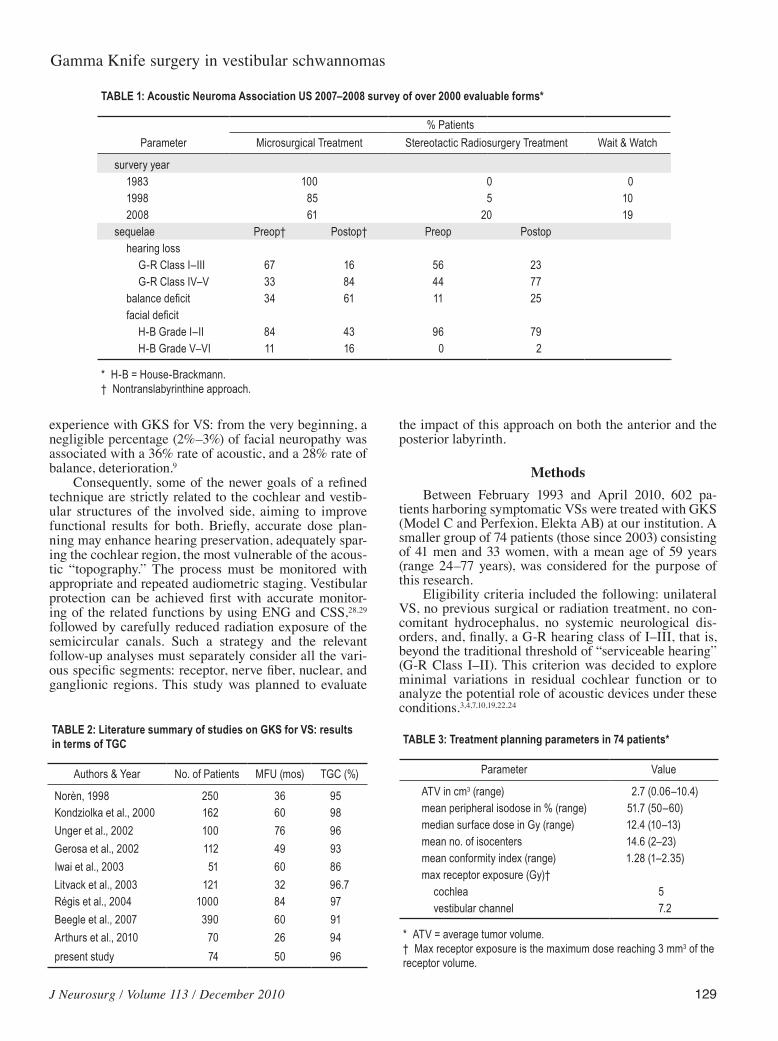
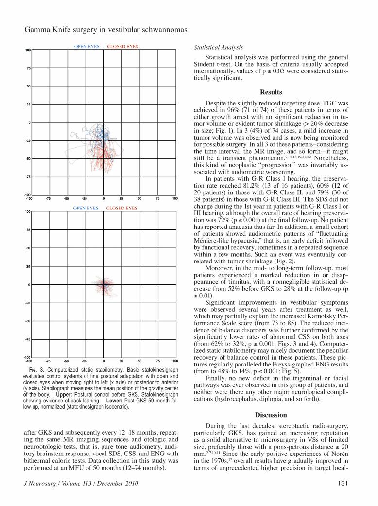
Significant improvements in vestibular symptoms were observed several years after treatment as well, which may partially explain the increased Karnofsky Per-formance Scale score (from 73 to 85). The reduced inci-dence of balance disorders was further confirmed by the significantly lower rates of abnormal CSS on both axes (from 62% to 32%, p ≤ 0.001; Figs. 3 and 4). Computer-ized static stabilometry may nicely document the peculiar recovery of balance control in these patients. These pic-tures regularly paralleled the Freyss-graphed ENG results (from to 48% to 14%, p ≤ 0.001; Fig. 5).
Finally, no new deficit in the trigeminal or facial pathways was ever observed in this group of patients, and neither were there any other major neurological compli-cations (hydrocephalus, diplopia, and so forth).
DiscussionDuring the last decades, stereotactic radiosurgery,
particularly GKS, has gained an increasing reputation as a solid alternative to microsurgery in VSs of limited size, preferably those with a pons-petrous distance ≤ 20 mm.2,7,10,11 Since the early positive experiences of Norén in the 1970s,17 overall results have gradually improved in terms of unprecedented higher precision in target local-
Fig. 3. Computerized static stabilometry. Basic statokinesigraph evaluates control systems of fine postural adaptation with open and closed eyes when moving right to left (x axis) or posterior to anterior (y axis). Stabilograph measures the mean position of the gravity center of the body. Upper: Postural control before GKS. Statokinesigraph showing evidence of back leaning. Lower: Post-GKS 59-month fol-low-up, normalized (statokinesigraph isocentric).

M. Gerosa et al.
132 J Neurosurg / Volume 113 / December 2010
ization—basically due to advances in CT, MR imaging, and stereoimaging—and in terms of novel algorithms in-troduced in computerized dose planning and automatic positioning system devices. The technique has reached extremely rewarding conformity and selectivity indexes, thanks also to the use of microshots in highly multifocal treatment plans.2,3,9,11,19–21
Furthermore, radiotoxicity thresholds for cranial neu-ropathy have been extensively analyzed, and their “radio-vulnerability grading” covers a wide spectrum of dosag-es, with pronounced inhomogeneity among the different cranial nerves and the various nerve regions, given that radiation sensitivity may vary quite significantly from the receptor to the ganglion and from the nucleus to the nerve fibers of the same cranial nerve.3–6,12,13,15,18,21
These aspects are particularly emphasized in cranial nerves VII and VIII. The most critical segments are prob-ably represented by the geniculate ganglion along the fa-cial pathways, as well as by the cochlear region among the acoustic pathways and structures.13,21,22,26 For both of
these structures, dose-volume analysis is not feasible be-cause of their small sizes.4 At these levels, the therapeutic dose must be drastically reduced or, if necessary, split for a staged procedure given that the internationally accepted radiotoxic thresholds are approximately 4–5 Gy for the cochlea, 7–8 Gy for the semicircular canals, and 9–10 Gy for the geniculate ganglion.7,10,16,22,24,26 However, no benefit from fractionation should be expected for VSs with a low alpha/beta ratio.27
Dose exposures of the various temporal bone struc-tures during routine GKS for VSs have been carefully investigated,12 thereby providing a basic “topographic” dosimetry in patients (Table 4). As a consequence, alter-native radiosurgical strategies have been proposed: first, to decrease treatment dose levels with the aim of reduc-ing the incidence and relevance of side effects, without eventually altering the excellent TGC rates; and second, to refine and model the matching isodose to obtain the minimal possible exposure of the most susceptible struc-tures, that is, the cochlea and the vestibular canals.
Fig. 4. Same case featured in Fig 3. Upper: Stabilogram obtained at GKS. Tilting of the body axis along the x and y coordi-nates in the time domain. Lower: Stabilogram obtained 59 months later (normal).

J Neurosurg / Volume 113 / December 2010
Gamma Knife surgery in vestibular schwannomas
133
Preliminary reports of adequate series of patients with more than 3 years of MFU seem to confirm that treatment planning based on slightly reduced edge doses (between 11 and 12.5 Gy) does not alter the overall TGC7,16,20,21,26 and may even spare or possibly enhance cranial nerve VIII function (Table 5).
Our results seem to validate this approach, first of all confirming at lower dosages the maintenance of TGC lev-els (96% at a 4-year MFU) typical of this technique, with excellent protection of hearing function (72% at a 4-year MFU). Moreover, there is additional evidence of the well-known phenomenon of fluctuating hypacusia, presumably linked to mechanical compression (edema) of the nerve fibers. Indeed, in the natural history of VSs, the majority
TABLE 4: Dose exposure of normal temporal bone structures during routine GKS in 54 patients*
Region Dose
intratemporal facial nerve 16% received > surface dosecochlea (basal turn, nerve modiolus) 11% exposed to > 10 Gyvestibular labyrinth (ends dilated as ampulla, lat & pst semicircular canals)
7.5% exposed to > 12 Gy
* Median surface dose was 13 Gy. Abbreviation: pst = posterior.
Fig. 5. Freyss graphs before and after GKS. Upper: Left VS ENG at the thermic stimulation before GKS. Note the reduction on the left side compared with the other (left vestibular hypofunction). Lower: Vestibular schwannoma Freyss graph at the 40-month follow-up showing normalization.

M. Gerosa et al.
134 J Neurosurg / Volume 113 / December 2010
of patients with these lesions have hearing loss progres-sion of 6–13 dB per year.15,18,24
Regarding single-stage radiosurgical treatments, most believe that to spare cochlear function, the tumor edge dose should never exceed the threshold of 13 Gy (Table 5).4,7,10,13,15,18,22,24,26 According to Régis and colleagues,21,22 under these conditions, patients with G-R Class I hearing who are treated with GKS have a 78% chance of hearing preservation.
Finally, this study provides additional evidence that lower dosages with extremely fitting isodoses sparing the vestibular labyrinth may not only protect balance and co-ordination (one-half to one-third of the patients clinically improved) but also provide functional recovery, with nor-malization of the statokinesigraph and the Freyss pro-file.28,29
ConclusionsData in this cohort of patients seem to confirm that
newer treatment planning strategies may improve radio-surgical results in VSs in terms of hearing preservation and vestibular protection. In short, for the best results, use 1) accurate targeting of the cochlea and the semicircular canals with MR imaging/CT bone algorithm fusion, 2) reduced radiation dosages, and 3) absolute sparing of the receptor sites.
Disclosure
The authors report no conflict of interest concerning the mate-rials or methods used in this study or the findings specified in this paper.
Author contributions to the study and manuscript preparation include the following. Conception and design: Gerosa. Acquisition of data: Gerosa, Mesiano, Longhi, De Simone, Foroni, Nicolato. Analysis and interpretation of data: all authors. Drafting the article: Gerosa, Mesiano, Nicolato. Critically revising the article: Gerosa, Longhi, De Simone, Foroni, Verlicchi, Zanotti, Nicolato. Reviewed final version of the manuscript and approved it for submission: all authors. Statistical analysis: Mesiano. Administrative/technical/material support: Verlicchi, Zanotti. Study supervision: Gerosa, Nicolato.
References
1. Acoustic Neuroma Association: 2007–2008 Patient Survey. Cumming, GA: Acoustic Neuroma Association, 2008 (http://www.anausa.org/patient_survey.shtml) [Accessed August 26, 2010]
2. Arthurs BJ, Lamoreaux WT, Mackay AR, Demakas JJ, Gid-dings NA, Fairbanks RK, et al: Gamma knife radiosurgery for vestibular schwannomas: tumor control and functional preser-vation in 70 patients. Am J Clin Oncol [epub ahead of print May 21, 2010]
3. Beegle RD, Friedman WA, Bova FJ: Effect of treatment plan quality on outcomes after radiosurgery for vestibular schwan-noma. J Neurosurg 107:913–916, 2007
4. Bhandare N, Jackson A, Eisbruch A, Pan CC, Flickinger JC, Antonelli P, et al: Radiation therapy and hearing loss. Int J Radiat Oncol Biol Phys 76 (3 Suppl):S50–S57, 2010
5. Boutin P, Guth A, Bouccara D, el Garem H, Rey A, Sterkers O: [Intra-labyrinthine schwannomas: a report of two cases.] Ann Otolaryngol Chir Cervicofac 115:35–41, 1998 (Fr)
6. Bridger MW, Farkashidy J: The distribution of neuroglia and schwann cells in the 8th nerve of man. J Laryngol Otol 94: 1353–1362, 1980
7. Flickinger JC, Kondziolka D, Niranjan A, Maitz A, Voynov G, Lunsford LD: Acoustic neuroma radiosurgery with marginal tumor doses of 12 to 13 Gy. Int J Radiat Oncol Biol Phys 60: 225–230, 2004
8. Foncin JF, Sterkers JM, Perre J, Corlieu P: [The origin of acoustic neurionoma. An ultrastructural study of operated neurinoma incipiens (author’s transl).] Ann Otolaryngol Chir Cervicofac 96:11–22, 1979 (Fr)
9. Gerosa M, Nicolato A, Foroni R, Bricolo A: Gamma knife ra-diosurgery in vestibular schwannoma: clinical and radiologi-cal impact on the tumor course, in Kanzaki J, Tos M, Sanna M, et al (eds): Acoustic Neuroma. Consensus on Systems for Reporting Results. Berlin: Springer Verlag, 2002, pp 129–138
10. Iwai Y, Yamanaka K, Shiotani M, Uyama T: Radiosurgery for acoustic neuromas: results of low-dose treatment. Neurosur-gery 53:282–288, 2003
11. Kondziolka D, Lunsford LD, Flickinger JC: Gamma knife ra-diosurgery for vestibular schwannomas. Neurosurg Clin N Am 11:651–658, 2000
12. Linskey ME, Johnstone PA, O’Leary M, Goetsch S: Radiation exposure of normal temporal bone structures during stereot-actically guided gamma knife surgery for vestibular schwan-nomas. J Neurosurg 98:800–806, 2003
13. Litvack ZN, Norén G, Chougule PB, Zheng Z: Preservation of functional hearing after gamma knife surgery for vestibular schwannoma. Neurosurg Focus 14(5):e3, 2003
14. Nedzelski JM, Canter RJ, Kassel EE, Rowed DW, Tator CH: Is no treatment good treatment in the management of acoustic neuromas in the elderly? Laryngoscope 96:825–829, 1986
15. Neely JG, Britton BH, Greenberg SD: Microscopic charac-teristics of the acoustic tumor in relationship of its nerve of origin. Laryngoscope 86:984–991, 1976
16. Niranjan A, Mathieu D, Kondziolka D, Flickinger JC, Lunsford LD: Radiosurgery for intracanalicular vestibular schwanno-mas. Prog Neurol Surg 21:192–199, 2008
TABLE 5: Literature survey of studies featuring a comparison between hearing preservation and the tumor margin dose in patients with normal hearing before radiosurgery
Authors & YearNo. of
Patients No. of Patients w/ G-R Class I FU (yrs) Median Surface Dose (Gy) Useful Hearing (%)
Norén, 1998 254 — 3 13.6 60 (at 2 yrs)Kondziolka et al., 2000 162 — 5–10 16 47Prasad et al., 2000 153 96 4.27 13 58Flickinger et al., 2004 198 — 2.5 13 71Unger et al., 2002 60 29 1–8 13 55Régis et al., 2004 1000 175 7 12.74 60 (at 3 yrs)
J Neurosurg / Volume 113 / December 2010
Gamma Knife surgery in vestibular schwannomas
135
17. Norén G: Long-term complications following gamma knife radiosurgery of vestibular schwannomas. Stereotact Funct Neurosurg 70 (Suppl 1):65–73, 1998
18. Ogawa K, Kanzaki J, Ogawa S, Tsuchihashi N, Ikeda S: Pro-gression of hearing loss in acoustic neuromas. Acta Otolar-yngol Suppl 487:133–137, 1991
19. Pollock BE: Management of vestibular schwannomas that en-large after stereotactic radiosurgery: treatment recommenda-tions based on a 15 year experience. Neurosurgery 58:241–248, 2006
20. Prasad D, Steiner M, Steiner L: Gamma surgery for vestibular schwannoma. J Neurosurg 92:745–759, 2000
21. Régis J, Delsanti C, Roche P, Soumare O, Dufour H, Porcheron D, et al: [Preservation of hearing function in the radiosurgical treatment of unilateral vestibular schwannomas. Preliminary results.] Neurochirurgie 48:471–478, 2002 (Fr)
22. Régis J, Delsanti C, Roche PH, Thomassin JM, Pellet W: [Functional outcomes of radiosurgical treatment of vestibular schwannomas: 1000 successive cases and review of the litera-ture.] Neurochirurgie 50:301–311, 2004 (Fr)
23. Roche PH, Bouvier C, Chinot O, Figarella-Branger D: Genesis and biology of vestibular schwannomas. Prog Neurol Surg 21:24–31, 2008
24. Thomsen J, Terkildsen K, Tos M: Acoustic neuromas. Pro-gression of hearing impairment and function of the eighth cranial nerve. Am J Otol 5:20–33, 1983
25. Tos M, Drozdziewicz D, Thomsen J: Medial acoustic neu-romas. A new clinical entity. Arch Otolaryngol Head Neck Surg 118:127–133, 1992
26. Unger F, Walch C, Schröttner O, Eustacchio S, Sutter B, Pendl G: Cranial nerve preservation after radiosurgery of vestibular schwannomas. Acta Neurochir Suppl 84:77–83, 2002
27. Vernimmen FJ, Slabbert JP: Assessment of the alpha/beta ra-tios for arteriovenous malformations, meningiomas, acoustic neuromas, and the optic chiasma. Int J Radiat Biol 86:486–498, 2010
28. Weber B: Strategiè et tactiques du système postural fin, in Gagey PM, Weber B (eds): Entrées du système postural fin. Paris: Masson, 1995, pp 139–146
29. Woollacot MH: Gait and postural control in the aging adult, in Bles W, Brandt Th (eds): Disorders of Posture and Gait. Amsterdam: Elsevier, 1986, pp 325–336
Manuscript submitted June 24, 2010.Accepted August 19, 2010.Address correspondence to: Massimo Gerosa, M.D., Depart-
ment of Neurosurgery, University Hospital Verona, Piazzale Stefani 1, 37126 Verona VR, Italy. email: massimo.gerosa@ ospedaleuniverona.it.