Embed Size (px)
Citation preview

INFECTION AND IMMUNITY, Apr. 1993, p. 1468-14730019-9567/93/041468-06$02.00/0Copyright © 1993, American Society for Microbiology
Gamma Interferon Cooperates with Lipopolysaccharide ToActivate Mouse Splenic Macrophages to an
Antihistoplasma StateTHOMAS E. LANE, BEITY A. WU-HSIEH, AND DEXTER H. HOWARD*
Department of Microbiology and Immunology, University of California atLos Angeles School ofMedicine, Los Angeles, California 90024
Received 21 September 1992/Accepted 21 January 1993
Inhibition of the intracellular growth of Histoplasma capsulatum by murine resident red pulp splenicmacrophages was examined. Splenic macrophages, unlike resident peritoneal macrophages, required a
prolonged preincubation (18 h) with recombinant murine gamma interferon (rMuIFN--y) for activation. To befully activated, the splenic macrophages required incubation with rMuIFN-'y in combination with 0.1 ,ug oflipopolysaccharide (LPS) per ml. Splenic macrophages stimulated with rMuIFN-y, LPS, or rMuIFN--y andLPS produced tumor necrosis factor alpha (TNF-a), but recombinant murine TNF-a (rMuTNF-a) did notactivate macrophages when used alone or as a second signal with rMuIFN-'y. Anti-TNF-a antibody did notblock IFN-y-LPS activation of splenic macrophages to any significant extent. One hundred micromolar ferroussulfate antagonized IFN--y-LPS activation of splenic macrophages, indicating that iron was involved in thefungistatic activity of cytokine-stimulated phagocytes. Our results indicate that (i) splenic macrophages differsignificantly from peritoneal macrophages in their requirements for activation and (ii) the mechanism by whichsplenic macrophages exert their antifungal effects involves iron.
The zoopathogenic fungus Histoplasma capsulatum is afacultative intracellular pathogen that invades the mononu-clear phagocyte system of animals. Cell-mediated immunityappears to play a major role in host defense against H.capsulatum (8, 30, 31). CD4+ T cells isolated from immunemice transfer immunity against infection by H. capsulatumto naive mice (8, 24, 26). In vitro studies have shown thatrecombinant murine gamma interferon (rMuIFN--y) activatesnormal peritoneal macrophages to inhibit the intracellulargrowth of the fungus (30).
Studies on the interaction between murine macrophagesand yeast cells of H. capsulatum have been conductedprimarily with peritoneal macrophages (15, 27-30). How-ever, peritoneal macrophages do not represent the type ofphagocyte infected upon dissemination of the organismwithin the host. Following intravenous injection of H. cap-sulatum, yeast cells circulate through organs of the reticu-loendothelial system such as the liver and the spleen (1) andcome to reside in the macrophages of those organs. Splenicmacrophages may play a key role in clearance of the funguswithin the infected host. However, the interaction of splenicmacrophages with H. capsulatum yeast cells has not beenexamined. Resident red pulp splenic macrophages sharecharacteristics with peritoneal macrophages, such as expres-sion of Fc receptors, la antigen, and the macrophage-specificantigen F4/80 (19). Therefore, we set out to determine ifsplenic macrophages could be activated to either kill orinhibit the intracellular growth of H. capsulatum.Our results indicate that resident splenic macrophages are
markedly different from peritoneal macrophages in theirrequirements for activation. Splenic macrophages require a
prolonged preincubation with rMuIFN-,y to be activated, andin order to become fully activated the macrophages require asecond signal, e.g., lipopolysaccharide (LPS) in the studies
* Corresponding author.
to be reported here. Splenic macrophages are stimulated byLPS to produce tumor necrosis factor alpha (TNF-a), butrecombinant murine TNF-ao (rMuTNF-cx) did not combinewith rMuIFN--y to activate macrophages in vitro to inhibit H.capsulatum. Inclusion of 100 p1M ferrous sulfate antagonizedrMuIFN--y-LPS-induced activation of splenic macrophages,indicating that iron is involved in the antifungal effectsinduced in cytokine-stimulated macrophages.
MATERIALS AND METHODS
Mice. Inbred male C57BL/6 mice were purchased fromJackson Laboratory, Bar Harbor, Maine. Age-matched 6- to8-week-old mice were used for all experiments.Fungus. H. capsulatum 505, which has been used in
experiments previously reported (15, 29-31), was employedin these studies. Yeast cells were grown on blood-cysteine-glucose agar slants at 37°C for 48 to 72 h prior to use.
Reagents and media. Splenic and peritoneal macrophagemonolayers were cultured in modified Eagle's medium(MEM) (GIBCO Laboratories, Grand Island, N.Y.) contain-ing 10% heat-inactivated defined fetal bovine serum (FBS)(Hyclone Laboratories Inc., Logan, Utah) and supple-mented with 15 mM HEPES (N-2-hydroxyethylpiperazine-N'-2-ethanesulfonic acid) 15 mM glucose, 100 U of penicillinper ml, and 100 ig of streptomycin per ml (SMEM). Phenol-extracted LPS from Escherichia coli serotype O11:B4 andferrous sulfate were purchased from Sigma Chemical Co.,St. Louis, Mo. The rMuIFN--y was supplied by GenentechInc., South San Francisco, Calif. The rMuTNF-ao and rabbitanti-mouse polyclonal anti-TNF-a antibody were purchasedfrom Genzyme Corp., Cambridge, Mass. Possible contami-nation by extraneous endotoxin was monitored as follows.Each experiment contained a medium control without addedstimulators, and activation of macrophages in these controlsituations was not observed. The FBS had 0.8 ng of endo-
1468
Vol. 61, No. 4
on May 13, 2018 by guest
http://iai.asm.org/
Dow
nloaded from

ANTIHISTOPLASMA ACTIVITY OF SPLENIC MACROPHAGES 1469
toxin per ml (supplier's assay), and the rMuIFN--y had <10pg of endotoxin per ml (supplier's assay).
Macrophages. Splenic macrophages were isolated by themethod described by Nusrat et al. (19). Briefly, spleens wereremoved from mice and perfused with collagenase (170U/ml, type D; Boehringer Mannheim, Indianapolis, Ind.)and DNase (20 ,ug/ml, type I; Boehringer Mannheim) dis-solved in prewarmed (37°C) Dulbecco's phosphate-bufferedsaline (DPBS) supplemented with 0.9 mM CaCl2-5.5 mMD-glucose (DPBSG) and adjusted to pH 7.3 (19). Followingperfusion, the cell suspension was added to a 50-ml Teflonbeaker and mixed by pipette for 8 min at 37°C. The cellsuspension was passed through a 40-,um-pore-size nylonmesh, spun at 250 x g for 10 min at 4°C, and resuspended ina 63.6% isotonic Percoll gradient (Pharmacia, Uppsala,Sweden) in Ca2+-Mg2+-free DPBS containing 9% heat-inactivated defined FBS and 36 ,ug of DNase I per ml. Thegradient was overlaid with 1.5 ml of DPBS and centrifuged at1,800 x g for 15 min at 4°C. The interface was collected andwashed twice with DPBSG containing 1% FBS, and the cellswere resuspended in medium at a final concentration of 4 x10⁷ per ml. One hundred microliters of the suspension wasseeded onto 15-mm-diameter Formvar-coated glass cover-slips (Bellco Glass Inc., Vineland, N.J.) in 24-well tissueculture plates (Corning Glass Works, Corning, N.Y.) andincubated at 37°C in a 5% CO2 atmosphere for 1 h. Theprocess was repeated once more for a total of two seedingsfor each coverslip. The coverslips were then vigorouslywashed with prewarmed Hanks' balanced salt solution toremove nonadherent cells. Greater than 80% of adherentcells expressed the macrophage-specific F4/80 antigen (13,19) as measured by immunofluorescence (data not shown).
Peritoneal cells were harvested from the peritoneal cavi-ties of mice as previously described (15, 29, 30). Theperitoneal cells were then subjected to the same isolationtechnique as described for splenic macrophages. The perito-neal cells were suspended in medium at a concentration of106 per ml. One hundred microliters of the cell suspensionwas seeded onto 15-mm-diameter Formvar-coated glass cov-erslips in 24-well tissue culture plates. Only a single seedingwas done for peritoneal cells. The cells were incubated at37°C in a 5% CO2 atmosphere for 2 h and vigorously washedto remove nonadherent cells. This technique resulted in aconfluent monolayer of peritoneal macrophages.Assay for macrophage activation. Splenic and peritoneal
macrophages were isolated and seeded onto Formvar-coatedglass coverslips as described above and used immediatelyfor the macrophage assay. In brief, macrophage monolayerswere incubated in 1 ml of SMEM with or without theactivating factors (rMuIFN--y, rMuTNF-a, and LPS) foreither a 3.5- or 18-h preincubation. At the completion of thepreincubation, yeast cells of H. capsulatum were washedand approximately 2 x 10⁵ to 3 x 10⁵ cells were added to themacrophage monolayer. Two hours were allowed for phago-cytosis, and then extracellular yeasts were washed away.Two coverslips that were not exposed to any activatingagent(s) were fixed in methanol in order to determine thenumber of yeast cells per macrophage at the start of incuba-tion. Control studies showed that preexposure to cytokine orLPS did not increase phagocytosis, and macrophages, in thepresence of activating agents, had nearly the same number ofyeast cells per macrophage as did those unexposed toactivating agents. The remaining monolayers were overlaidwith either medium alone or medium containing the activat-ing agent(s) corresponding to the preincubation protocol andincubated for an additional 18 h at 37°C. After the second
incubation period the monolayers were washed and fixed inmethanol. Monolayers were stained with periodic acid-Schiffstain, and the mean number of yeast cells within infectedmacrophages was determined by counting the yeast cells ina minimum of 100 infected cells. The net growth wascalculated by subtracting the mean number of yeast cells perinfected macrophage at zero time from the mean number ofyeast cells per macrophage at the conclusion of the experi-ment. The percent inhibition of growth was calculated fromthe following formula: [1 - (growth within macrophages invarious test media/growth within macrophages in controlmedium)] x 100.Assay for antagonistic effect of iron on activated splenic
macrophages. Macrophage monolayers were incubated asdescribed for the macrophage activation assay except thatthe monolayers treated with IFN-y-LPS were exposed to100 ,uM FeSO4 prior to and following exposure to yeast cellsof H. capsulatum. The antagonistic effect of iron on IFN--y-LPS-induced activation of splenic macrophages was deter-mined by counting the number of intracellular yeast cells asdescribed above.TNF-a production and assay. Splenic macrophages were
seeded onto Formvar-coated coverslips and incubated withIFN--y (2,000 U/ml), LPS (0.1 jig/ml), or both in 1 ml ofSMEM for 36 h. Yeast cells were added following an 18-hincubation period. Two hours were allowed for phagocyto-sis, and then unphagocytized yeast cells were washed away.Macrophage monolayers were incubated for an additional 18h in medium containing activating factors to which the cellswere previously exposed. Cell-free supernatants from wellswere collected at 4, 8, 18, 20, and 36 h following adherenceto coverslips. Supernatants were assayed for TNF-cx accord-ing to the method described by Merrill et al. (18). The murineL929 cell line (American Type Culture Collection), retrievedin log growth phase by trypsinization, was washed severaltimes, labelled with chromium in a total volume of 1.0 mlwith 0.25 mCi of 51Cr (Amersham International) for 1 h, andwashed three times in medium. Labelled target cells wereplated with test sample dilutions in 96-well U-bottom plates(Linbro Titertek; Flow Laboratories, McLean, Va.) at aconcentration of 5 x 10⁵ cells per ml with 5 jig of actinomy-cin D per ml. Eighteen hours later, plates were centrifuged at600 x g, 50 ,ul of supernatant was aspirated, and radioactiv-ity was counted in a gamma counter. rMuTNF-ao standardswere the generous gift of Cetus Corp. (Norwalk, Conn.).These were titrated from 104 U/ml (25 nM) to 10-3U/ml (2.5fM).
Statistical analyses. Statistical analyses were performed bythe Student t test.
RESULTS
IFN-y alone fails to fully activate splenic macrophages to anantihistoplasma state. Preexposure to rMuIFN--y is essentialfor activation of normal peritoneal macrophages to an anti-histoplasma state (30). We first studied whether residentsplenic macrophages had the same requirements as those ofperitoneal macrophages for activation to an antihistoplasmastate. Peritoneal and splenic macrophages were isolatedfrom the same mice and incubated with either 10 or 2,000 Uof rMuIFN-y per ml for either 3.5 or 18 h prior to theaddition of H. capsulatum. Following exposure to yeastcells, the macrophages were incubated for an additional 18 hin the presence of rMuIFN--y and their activated state wasmeasured as described in Materials and Methods. The re-sults recorded in Table 1 indicate that a preexposure to
VOL. 61, 1993
on May 13, 2018 by guest
http://iai.asm.org/
Dow
nloaded from
1470 LANE ET AL.
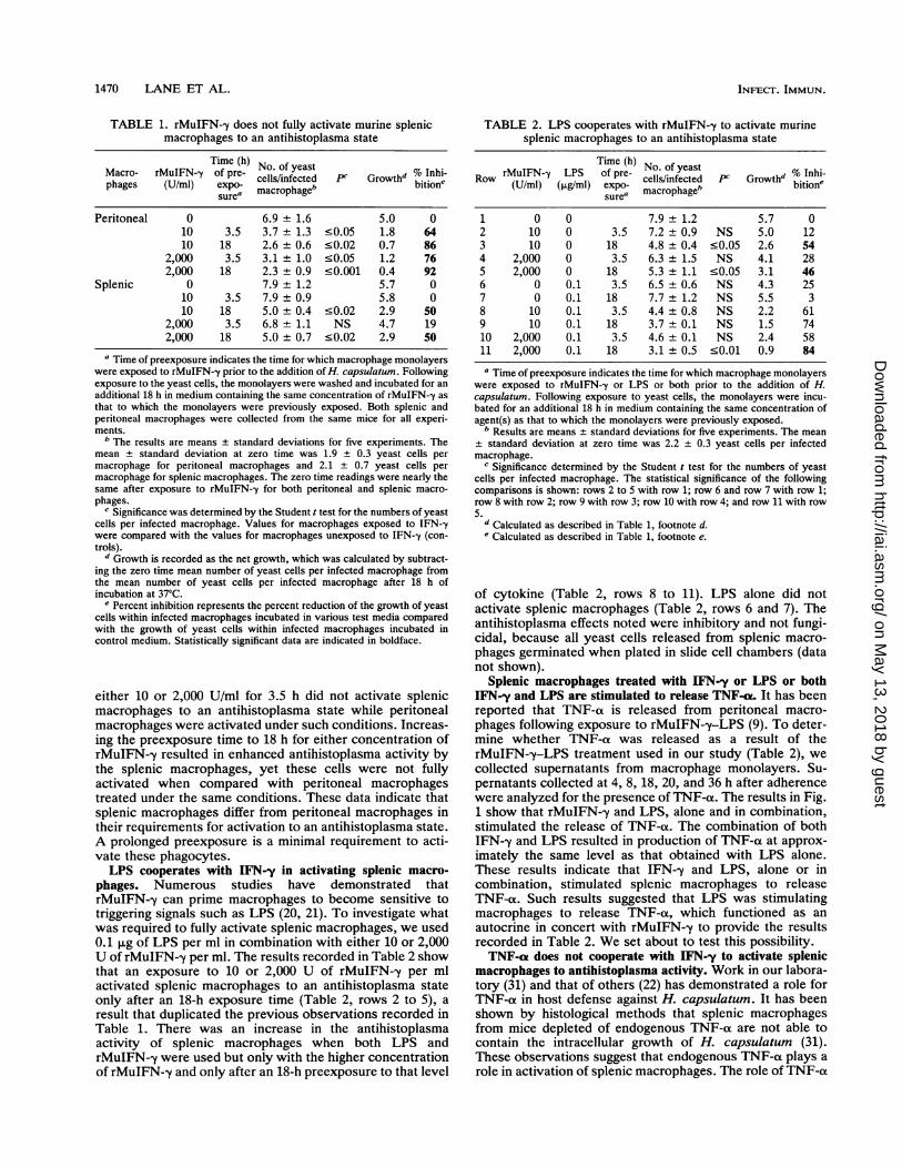
TABLE 1. rMuIFN-,y does not fully activate murine splenicmacrophages to an antihistoplasma state
Time (h) No. of yeast % Inhi-Macro- rMuIFN--y of pre- cells/infected P1 Growth b.Itiophages (U/ml) expo- macrophageb bton
sure'
Peritoneal 0 6.9 ± 1.6 5.0 010 3.5 3.7 ± 1.3 .0.05 1.8 6410 18 2.6 ± 0.6 .0.02 0.7 86
2,000 3.5 3.1 ± 1.0 .0.05 1.2 762,000 18 2.3 ± 0.9 .0.001 0.4 92
Splenic 0 7.9 ± 1.2 5.7 010 3.5 7.9 ± 0.9 5.8 010 18 5.0 + 0.4 <0.02 2.9 50
2,000 3.5 6.8 ± 1.1 NS 4.7 192,000 18 5.0 + 0.7 .0.02 2.9 50
a Time of preexposure indicates the time for which macrophage monolayerswere exposed to rMuIFN--y prior to the addition of H. capsulatum. Followingexposure to the yeast cells, the monolayers were washed and incubated for anadditional 18 h in medium containing the same concentration of rMuIFN--y asthat to which the monolayers were previously exposed. Both splenic andperitoneal macrophages were collected from the same mice for all experi-ments.
b The results are means ± standard deviations for five experiments. Themean ± standard deviation at zero time was 1.9 + 0.3 yeast cells permacrophage for peritoneal macrophages and 2.1 ± 0.7 yeast cells permacrophage for splenic macrophages. The zero time readings were nearly thesame after exposure to rMuIFN-y for both peritoneal and splenic macro-phages.
c Significance was determined by the Student t test for the numbers of yeastcells per infected macrophage. Values for macrophages exposed to IFN--ywere compared with the values for macrophages unexposed to IFN-,y (con-trols).
d Growth is recorded as the net growth, which was calculated by subtract-ing the zero time mean number of yeast cells per infected macrophage fromthe mean number of yeast cells per infected macrophage after 18 h ofincubation at 37°C.
e Percent inhibition represents the percent reduction of the growth of yeastcells within infected macrophages incubated in various test media comparedwith the growth of yeast cells within infected macrophages incubated incontrol medium. Statistically significant data are indicated in boldface.
either 10 or 2,000 U/ml for 3.5 h did not activate splenicmacrophages to an antihistoplasma state while peritonealmacrophages were activated under such conditions. Increas-ing the preexposure time to 18 h for either concentration ofrMuIFN--y resulted in enhanced antihistoplasma activity bythe splenic macrophages, yet these cells were not fullyactivated when compared with peritoneal macrophagestreated under the same conditions. These data indicate thatsplenic macrophages differ from peritoneal macrophages intheir requirements for activation to an antihistoplasma state.A prolonged preexposure is a minimal requirement to acti-vate these phagocytes.LPS cooperates with IFN-'y in activating splenic macro-
phages. Numerous studies have demonstrated thatrMuIFN--y can prime macrophages to become sensitive totriggering signals such as LPS (20, 21). To investigate whatwas required to fully activate splenic macrophages, we used0.1 ,ug of LPS per ml in combination with either 10 or 2,000U of rMuIFN-y per ml. The results recorded in Table 2 showthat an exposure to 10 or 2,000 U of rMuIFN--y per mlactivated splenic macrophages to an antihistoplasma stateonly after an 18-h exposure time (Table 2, rows 2 to 5), aresult that duplicated the previous observations recorded inTable 1. There was an increase in the antihistoplasmaactivity of splenic macrophages when both LPS andrMuIFN--y were used but only with the higher concentrationof rMuIFN--y and only after an 18-h preexposure to that level
TABLE 2. LPS cooperates with rMuIFN--y to activate murinesplenic macrophages to an antihistoplasma state
Time (h) No. ofyeast%IniRow rMuIFN--y LPS of pre- cells/infected P Growthd % Inhi-
(U/ml) (jig/ml) expo- macrophageb bition'sure'
1 0 0 7.9 1.2 5.7 02 10 0 3.5 7.2 0.9 NS 5.0 123 10 0 18 4.8 ± 0.4 <0.05 2.6 544 2,000 0 3.5 6.3 ± 1.5 NS 4.1 285 2,000 0 18 5.3 ± 1.1 <0.05 3.1 466 0 0.1 3.5 6.5 ±0.6 NS 4.3 257 0 0.1 18 7.7 1.2 NS 5.5 38 10 0.1 3.5 4.4 ± 0.8 NS 2.2 619 10 0.1 18 3.7 0.1 NS 1.5 7410 2,000 0.1 3.5 4.6 ± 0.1 NS 2.4 5811 2,000 0.1 18 3.1 ± 0.5 .0.01 0.9 84
a Time of preexposure indicates the time for which macrophage monolayerswere exposed to rMuIFN-y or LPS or both prior to the addition of H.capsulatum. Following exposure to yeast cells, the monolayers were incu-bated for an additional 18 h in medium containing the same concentration ofagent(s) as that to which the monolayers were previously exposed.
b Results are means + standard deviations for five experiments. The mean+ standard deviation at zero time was 2.2 ± 0.3 yeast cells per infectedmacrophage.
c Significance determined by the Student t test for the numbers of yeastcells per infected macrophage. The statistical significance of the followingcomparisons is shown: rows 2 to 5 with row 1; row 6 and row 7 with row 1;row 8 with row 2; row 9 with row 3; row 10 with row 4; and row 11 with row5.dCalculated as described in Table 1, footnote d.Calculated as described in Table 1, footnote e.
of cytokine (Table 2, rows 8 to 11). LPS alone did notactivate splenic macrophages (Table 2, rows 6 and 7). Theantihistoplasma effects noted were inhibitory and not fungi-cidal, because all yeast cells released from splenic macro-phages germinated when plated in slide cell chambers (datanot shown).
Splenic macrophages treated with IFN-y or LPS or bothIFN-y and LPS are stimulated to release TNF-s. It has beenreported that TNF-ot is released from peritoneal macro-phages following exposure to rMuIFN-y-LPS (9). To deter-mine whether TNF-ot was released as a result of therMuIFN-y-LPS treatment used in our study (Table 2), wecollected supernatants from macrophage monolayers. Su-pernatants collected at 4, 8, 18, 20, and 36 h after adherencewere analyzed for the presence of TNF-a. The results in Fig.1 show that rMuIFN-y and LPS, alone and in combination,stimulated the release of TNF-a. The combination of bothIFN--y and LPS resulted in production of TNF-a at approx-imately the same level as that obtained with LPS alone.These results indicate that IFN--y and LPS, alone or incombination, stimulated splenic macrophages to releaseTNF-ot. Such results suggested that LPS was stimulatingmacrophages to release TNF-ot, which functioned as anautocrine in concert with rMuIFN--y to provide the resultsrecorded in Table 2. We set about to test this possibility.TNF-a does not cooperate with IFN-'y to activate splenic
macrophages to antihistoplasma activity. Work in our labora-tory (31) and that of others (22) has demonstrated a role forTNF-ax in host defense against H. capsulatum. It has beenshown by histological methods that splenic macrophagesfrom mice depleted of endogenous TNF-aL are not able tocontain the intracellular growth of H. capsulatum (31).These observations suggest that endogenous TNF-a plays arole in activation of splenic macrophages. The role of TNF-ox
INFECT. IMMUN.
on May 13, 2018 by guest
http://iai.asm.org/
Dow
nloaded from

ANTIHISTOPLASMA ACTIVITY OF SPLENIC MACROPHAGES 1471
20000
10000
E
LLz
0 10 20 30 40
Time of Exposure (hours)FIG. 1. Splenic macrophages treated with rMuIFN--y, LPS, or both are stimulated to release TNF-ax. Supernatants from treated wells were
collected at 4, 8, 18, 20, and 36 h following adherence to coverslips and analyzed for TNF-a as described in Materials and Methods. Threeexperiments were performed. The data shown are from a representative experiment.
in activation of splenic macrophages was tested in vitro.rMuTNF-ao alone at 2,000 U/ml did not activate splenicmacrophages and combined with rMuIFN--y did not activatesplenic macrophages to any significant degree (data notshown). In further experiments the presence of 200 neutral-izing units of anti-TNF-a did not significantly block activa-tion of splenic macrophages by the combination ofrMuIFN-,y and LPS (data not shown). Increasing the con-centration of antibody did not alter the result.
Iron reverses the activation of splenic macrophages byIFN-y-LPS. We have previously shown that iron limitationis one of the bases for the IFN--y-induced antihistoplasmaeffect of mouse peritoneal macrophages (15). The results inTable 3 show that addition of 100 ,uM ferrous sulfate tocultures of IFN--y-LPS-treated splenic macrophages par-tially abrogated activation by 2,000 U of rMuIFN--y per mltogether with 0.1 ,ug of LPS per ml. The addition of thereducing agent sodium thiosulfate (6) did not enhance theeffect of iron on IFN--y-LPS-treated macrophages (data notshown). In addition, increasing the concentration of ferrous
TABLE 3. Iron antagonizes activation of splenic macrophages
No. of yeastTreatment cells/infected Growthb % Inhibition'
macrophagea
SMEM 7.7 ± 1.0 5.6 0IFN--y (2,000 U/ml) + 2.8 ± 0.7 0.7 88LPS (0.1 ,ug/ml)
IFN--y (2,000 U/ml) + 5.0 ± 0.7 2.9 48dLPS (0.1 ,ug/ml) +100 ,M FeSO4a Results are means + standard deviations for three experiments. The mean
+ standard deviation at zero time was 2.1 + 0.1 yeast cells per infectedmacrophage.
b Calculated as described in Table 1, footnote d.Calculated as described in Table 1, footnote e.d p < 0.001.
sulfate did not result in a further reduction of percentintracellular growth induced by IFN-y-LPS (data notshown). These results suggest that the mechanism by whichthe IFN--y-LPS combination stimulates splenic macrophagesto an antihistoplasma state involves the availability of iron(10, 15).
DISCUSSIONThe spleen represents a site of infection upon dissemina-
tion of H. capsulatum within the host (1, 31). Clearance ofthe organism from an infected animal often correlates withclearance from the spleen, suggesting that splenic macro-phages may have a profound influence on the course of thedisease. Identifying the factors required for activatingsplenic macrophages to an antihistoplasma state would offera better understanding of how these phagocytes cope withinfection. Unlike peritoneal macrophages, treatment ofsplenic macrophages with rMuIFN--y alone does not fullyactivate the cells to an antihistoplasma state. A secondsignal, LPS in this study, which can cooperate withrMuIFN-y to activate splenic macrophages is required. Suchsynergism between cytokines has been reported by others.For example, Hockertz et al. (11) demonstrated that acombination of rMuIFN-y and muramyltripeptide activatedsplenic macrophages in vitro to kill Leishmania donovanipromastigotes. Treatment of splenic macrophages withIFN-y alone resulted in limited antimicrobial activity, whileexposure to muramyltripeptide alone did not activate themacrophages. These results support our observation thatmurine resident splenic macrophages require a second signalin addition to rMuIFN-,y for activation to an antimicrobialstate.
Ingestion of erythrocytes (RBC) has been shown to inhibitmacrophage-mediated cytotoxicity against tumor targets(25). The splenic macrophage isolation protocol describedresults in the sedimentation of RBC following passage of thespleen cells through the Percoll gradient (19). Thus, it is
VOL. 61, 1993
on May 13, 2018 by guest
http://iai.asm.org/
Dow
nloaded from

1472 LANE ET AL.
unlikely that the deficient antihistoplasma response of thesplenic macrophages to single biological response modifiers(i.e., rMuIFN--y, LPS, or rMuTNF-ao) is due to phagocytosisof RBC during the isolation procedure.Our work (31) as well as that by others (22) has demon-
strated a role for TNF-ax in host defense against H. capsu-latum. TNF-ao is produced in spleens of mice infected withH. capsulatum (31). Mice infected with a sublethal dose ofH. capsulatum and injected with a polyclonal anti-TNF-aoantibody had increased mortality rates and significantlyhigher fungous counts in various tissues than did mice whichdid not receive the antibody (31). We have shown that IFN--yand LPS alone or together induce the release of TNF-ao bysplenic macrophages. This suggested that TNF-ao producedupon IFN--y-LPS treatment may be acting in an autocrinemanner to activate splenic macrophages (12). But evidencefor this presumption was not obtained. Drapier et al. (7) havereported that anti-TNF-ao antibody did not completely sup-press nitrite production or the antitumor properties of IFN-y-LPS-treated mouse peritoneal macrophages. It is possiblethat following IFN-,y-LPS treatment of splenic macrophagesthere is an increase not only of secreted TNF-a but also ofplasma membrane-associated TNF-a, which may have a rolein activation (2). The anti-TNF-ac antibody may not com-pletely block activation of splenic macrophages because it isunable to effectively bind membrane-associated TNF-ao orhas limited access to it because LPS causes rapid internal-ization of TNF-ao receptors (4, 5, 23). Additionally, it ispossible that intracellular precursors of TNF-a may have amajor role in triggering the macrophage-mediated antihisto-plasma mechanism. Another possibility is that monokinesother than TNF-ao are produced following IFN--y-LPS treat-ment which are capable of triggering the macrophage re-sponse (14, 21). This suggestion is likely on the basis of someof our recent work that shows that splenic macrophagesfrom Histoplasma-infected mice are fully activated (unpub-lished observation). Further work needs to be done in orderto resolve the cytokines involved in splenic macrophageactivation.Lepay et al. (17) have demonstrated that following a
sublethal infection of mice with Listeria monocytogenesthere is an influx of immigrant macrophages to the liver.Immigrant macrophages, unlike Kupffer cells, producedsubstantial levels of H202, suggesting that these cells, notthe Kupffer cells, were functioning in host defense (16, 17).A similar phenomenon occurs in the spleens of mice infectedwith L. monocytogenes (19). Resident splenic macrophagesare unable to produce reactive oxygen intermediates follow-ing exposure to IFN--y, which suggests that these cells mayhave an impaired antimicrobial defense system (19). How-ever, following infection of mice with L. monocytogenes,splenic inflammatory macrophages actively released H202(19). These observations suggest that resident tissue macro-phages and immigrant macrophages have distinct functions(17). Ding and Nathan (3) have suggested that the defect inthe respiratory burst by Kupffer cells is the result of frequentendocytic encounters with RBC, immune complexes, andvarious other ligands. These events allow the Kupffer cell toact as an effective scavenger without producing oxidativemetabolites which may be damaging to bystander cells (3). Asimilar sequence of events may occur in the spleen. Follow-ing exposure to a sublethal infection of H. capsulatum, thereis a massive influx of macrophages to the spleen (1, 31). Wolfet al. have demonstrated that IFN-y-treated mouse perito-neal macrophages exhibit antifungal activity yet do notproduce reactive oxygen intermediates despite extensive
phagocytosis of the organism (27, 28). This fact suggests thatthe macrophage oxidative burst does not play a role inmacrophage defense against intracellular H. capsulatum (27,28). It is possible that resident splenic macrophages, likeKupffer cells, function as scavengers in removing debrisfrom the spleen and that immigrant macrophages in thespleen may be the effector cells in host defense against H.capsulatum. We are currently investigating this possibility.
ACKNOWLEDGMENTS
These studies were supported by funds from Public HealthService grants AI-22963 and AI-25134 from the National Institute ofAllergy and Infectious Diseases. We are grateful for the rMuIFN-ysupplied by Genentech, Inc., South San Francisco, Calif., and forthe rTNF-ot standards supplied by Cetus Corp.We are grateful to A. Roomi Nusrat for helpful discussions. We
thank the laboratory of Jean Merrill, Department of Neurology,UCLA School of Medicine, for performing the TNF-a assay. Wethank Marcia Trylch for preparation of the manuscript.
REFERENCES1. Artz, R. P., and W. E. Bulloclk 1979. Immunoregulatory re-
sponses in experimental disseminated histoplasmosis: lymphoidorgan histopathology and serological studies. Infect. Immun.23:884-892.
2. Decker, T., M. L. Lohmann-Mathes, and G. E. Gifford. 1987.Cell-associated tumor necrosis factor (TNF) as a killing mech-anism of activated cytotoxic macrophages. J. Immunol. 138:957-962.
3. Ding, A., and C. Nathan. 1988. Analysis of the nonfunctionalrespiratory burst in murine Kupffer cells. J. Exp. Med. 167:1154-1170.
4. Ding, A. H., F. Porteu, E. Sanchez, and C. F. Nathan. 1990.Shared action of endotoxin and Taxol on TNF receptors andTNF release. Science 248:370-372.
5. Ding, A. H., E. Sanchez, S. Srimal, and C. F. Nathan. 1989.Macrophages rapidly internalize their tumor necrosis factorreceptors in response to bacterial lipopolysaccharide. J. Biol.Chem. 264:3924-3929.
6. Drapier, J. C., and J. B. Hibbs, Jr. 1986. Murine cytotoxicactivated macrophages inhibit aconitase in tumor cells: Inhibi-tion involves the iron-sulfur prosthetic group and is reversible.J. Clin. Invest. 78:790-797.
7. Drapier, J. C., J. Wietzerbin, and J. B. Hibbs, Jr. 1988.Interferon-gamma and tumor necrosis factor induce the L-argi-nine-dependent cytotoxic effector mechanism in murine macro-phages. Eur. J. Immunol. 18:1587-1592.
8. Gomez, A. M., W. E. Bullock, C. L. Taylor, and G. S. Deepe, Jr.1988. Role of L3T4+ T cells in host defense against Histoplasmacapsulatum. Infect. Immun. 56:1685-1691.
9. Green, S. J., R. M. Crawford, J. T. Hockmeyer, M. S. Meltzer,and C. A. Nacy. 1990. Leishmania major amastigotes initiate theL-arginine dependent killing mechanism in IFN-gamma stimu-lated macrophages by induction of TNF-a. J. Immunol. 145:4290-4297.
10. Hamilton, T. A., J. E. Weiel, and D. 0. Adams. 1984. Expres-sion of the transferrin receptor on murine peritoneal macro-phages is modulated by in vitro treatment with interferongamma. Cell. Immunol. 89:478-488.
11. Hockertz, S., G. Franke, E. Kniep, and M. L. Lohmann-Mathes.1989. Mouse interferon-gamma in liposomes: pharmacokinetics,organ-distribution, and activation of spleen and liver macro-phages in vivo. J. Interferon Res. 9:591-602.
12. Hori, K., M. J. Ehrke, K. Mace, and E. Mihich. 1987. Effect ofrecombinant tumor necrosis factor on tumoricidal activation ofmurine macrophages: synergism between tumor necrosis factorand interferon gamma. Cancer Res. 47:5868-5873.
13. Hume, D. A., A. P. Robinson, G. G. MacPherson, and S.Gordon. 1983. The mononuclear phagocyte system of the mousedefined by immunohistochemical localization of antigen F4/80:relationship between macrophages, Langerhans cells, reticular
INFECT. IMMUN.
on May 13, 2018 by guest
http://iai.asm.org/
Dow
nloaded from

ANTIHISTOPLASMA ACTIVITY OF SPLENIC MACROPHAGES 1473
cells and dendritic cells in lymphoid and hematopoietic organs.J. Exp. Med. 158:1522-1536.
14. Jayaraman, S., C. A. Martin, and M. E. Dorf. 1990. Enhance-ment of in vivo cell-mediated immune responses by threedistinct cytokines. J. Immunol. 144:942-951.
15. Lane, T. E., B. A. Wu-Hsieh, and D. H. Howard. 1991. Ironlimitation and the gamma interferon-mediated antihistoplasmastate of murine macrophages. Infect. Immun. 59:2274-2278.
16. Lepay, D. A., C. F. Nathan, R. M. Steinman, H. W. Murray, andZ. A. Cohn. 1985. Murine Kupffer cells: mononuclear phago-cytes deficient in the generation of reactive oxygen intermedi-ates. J. Exp. Med. 161:1079-1096.
17. Lepay, D. A., R. M. Steinman, C. F. Nathan, H. W. Murray, andZ. A. Cohn. 1985. Liver macrophages in murine listeriosis:cell-mediated immunity is correlated with an influx of macro-phages capable of generating reactive oxygen intermediates. J.Exp. Med. 161:1503-1512.
18. Merrill, J. E., S. R. Strom, G. W. Ellison, and L. W. Myers.1989. In vitro study of mediators of inflammation in multiplesclerosis. J. Clin. Immunol. 9:84-96.
19. Nusrat, A. R., S. D. Wright, A. A. Aderem, R. M. Steinman, andZ. A. Cohn. 1988. Properties of isolated red pulp macrophagesfrom mouse spleen. J. Exp. Med. 168:1505-1510.
20. Pace, J. L., S. W. Russell, B. A. Torres, H. M. Johnson, andP. W. Gray. 1983. Recombinant mouse gamma interferon in-duces the priming step in macrophage activation for tumor cellkilling. J. Immunol. 130:2011-2013.
21. Schultz, R. M., and W. J. Kleinschmidt. 1983. Functionalidentity between murine gamma interferon and macrophageactivating factor. Nature (London) 305:239-240.
22. Smith, J. G., D. M. Magee, D. M. Williams, and J. R. Graybill.1990. Tumor necrosis factor-alpha plays a role in host defenseagainst Histoplasma capsulatum. J. Infect. Dis. 162:1349-1353.
23. Smith, M. R., W. E. Munger, H. F. Kung, L. Takacs, and S. K.Durum. 1990. Direct evidence for an intracellular role for tumornecrosis factor-alpha: microinjection of tumor necrosis factorkills target cells. J. Immunol. 144:162-169.
24. Tewari, R. P., D. Sharma, M. Solotorovsky, R. Lafemina, and J.Balint. 1977. Adoptive transfer of immunity from mice immu-nized with ribosomes or live cells of Histoplasma capsulatum.Infect. Immun. 15:789-795.
25. Weinberg, J. B., and J. B. Hibbs, Jr. 1977. Endocytosis of redblood cells or haemoglobin by activated macrophages inhibitstheir tumoricidal effect. Nature (London) 269:245-247.
26. Williams, D. M., J. R. Graybill, and D. J. Drutz. 1981. Adoptivetransfer of immunity to Histoplasma capsulatum in athymicnude mice. Sabouraudia 19:39-48.
27. Wolf, J. E., A. L. Abegg, S. J. Travis, G. S. Kobayashi, and J. R.Little. 1989. Effects of Histoplasma capsulatum on murinemacrophage function: inhibition of macrophage priming, oxida-tive burst, and antifungal activities. Infect. Immun. 57:513-519.
28. Wolf, J. E., V. Kerchberger, G. S. Kobayashi, and J. R. Little.1987. Modulation of the macrophage oxidative burst by His-toplasma capsulatum. J. Immunol. 138:582-586.
29. Wu-Hsieh, B., and D. H. Howard. 1984. Inhibition of growth ofHistoplasma capsulatum by lymphokine-stimulated macro-phages. J. Immunol. 132:2593-2597.
30. Wu-Hsieh, B., and D. H. Howard. 1987. Inhibition of theintracellular growth of Histoplasma capsulatum by recombinantmurine gamma interferon. Infect. Immun. 55:1014-1016.
31. Wu-Hsieh, B. A., G.-S. Lee, M. Franco, and F. M. Hofman.1992. Early activation of splenic macrophages by tumor necro-sis factor alpha is important in determining the outcome ofexperimental histoplasmosis in mice. Infect. Immun. 60:4230-4238.
VOL. 61, 1993
on May 13, 2018 by guest
http://iai.asm.org/
Dow
nloaded from