Embed Size (px)
Citation preview

1
Gamma-hydroxybutyrate (GHB) reduces MAP kinase phosphorylation
via GABAB receptor activation in mouse frontal cortex and
hippocampus
Xiuhai Ren and Istvan Mody*
Departments of Neurology and Physiology, The David Geffen School of Medicine at
UCLA, RNRC 3-155, 710 Westwood Plaza, Los Angeles 90095, USA
Running Title: GHB reduces MAP kinase phosphorylation via GABAB receptors
*Correspondence to: Istvan Mody, PhD Dept of Neurology RNRC 3-155 The David Geffen School of Medicine at UCLA 710 Westwood Plaza Los Angeles 90095, USA Telephone: (+1) 310-206-4481 Fax: (+1) 310-825-0033 Email: [email protected]
Copyright 2003 by The American Society for Biochemistry and Molecular Biology, Inc.
JBC Papers in Press. Published on August 15, 2003 as Manuscript M304238200 by guest on M
ay 21, 2018http://w
ww
.jbc.org/D
ownloaded from

2
SUMMARY
Gamma-hydroxybutyrate (GHB) naturally occurs in the brain, but its exogenous
administration induces profound effects on the central nervous system in animals and
humans. The intracellular signaling mechanisms underlying its actions remain unclear. In
the present study, the effects of GHB on the activation (phosphorylation) of mitogen-
activated protein kinases (MAP kinases), extracellular signal-regulated kinase 1 and 2
(ERK1/2), were investigated. Acute administration of GHB (500 mg/kg, i.p.) induced a
fast and long-lasting inhibition of MAP kinase phosphorylation in both frontal cortex and
hippocampus. The reduced MAP kinase phosphorylation was observed in the CA1 and
CA3 areas but not in the dentate gyrus. Pretreatment with the specific GABAB receptor
antagonist CGP56999A (20 mg/kg, i.p.) prevented the action of GHB, and the effect of
GHB was mimicked by baclofen, a selective GABAB receptor agonist, whereas the high
affinity GHB receptor antagonist NCS-382 (200 mg/kg, i.p.) had no effect on GHB-
inhibited MAP kinase phosphorylation. Moreover, the GHB dehydrogenase inhibitor
valproate (500 mg/kg, i.p.), which inhibits the conversion of GHB into GABA, failed to
block the effect of GHB on MAP kinase phosphorylation. Altogether, these data suggest
that GHB, administered in vivo, reduces MAP kinase phosphorylation via a direct
activation of GABAB receptors by GHB. In contrast, GHB (10 mM for 15 min) was
found ineffective on MAP kinase phosphorylation in brain slices, indicating important
differences in the conditions required for the second messenger activating action of GHB.
Key words: Gamma-hydroxybutyrate, GABAB receptors, NCS-382, valproate, MAP
kinase, phosphorylation
by guest on May 21, 2018
http://ww
w.jbc.org/
Dow
nloaded from
3
INTRODUCTION
Gamma-hydroxybutyrate (GHB) 1 is a natural constituent of the mammalian brain
derived from the metabolism of γ-aminobutyric acid (GABA) (1). Peripherally
administered GHB can readily cross the blood-brain barrier and produces significant
behavioral, electrophysiological and biochemical effects. Clinically, GHB has been used
as an anesthetic agent (2), in the treatment of alcohol withdrawal and dependence (3),
opiate withdrawal (4), and in sleep disorders (5). Recently GHB has emerged as a major
recreational drug of abuse, and its abuse or intoxication has been associated with a mild
euphoria, respiratory depression, headache, vomiting, agitation, seizures, and even death
(6-8).
1 The abbreviations used are: GHB, γ-hydroxybutyrate; GABA, gamma-aminobutyric
acid; GABAB receptors, GABA type B receptors; MAP kinase, mitogen-activated protein
kinase; ERK1/2, extracellular signal regulated protein kinase 1 and 2; i.p.,
intraperitoneally; aCSF, artificial cerebrospinal fluid; DTT, dithiothreitol; SDS, sodium
dodecyl sulfate; DG, dentate gyrus; CSF, cerebrospinal fluid; HP, hydrogen peroxide;
cAMP, cyclic AMP; PKA, cAMP-dependent protein kinase A; PKC, protein kinase C;
PKB, protein kinase B; PI3K, phosphotidylinositol-3-kinase; MKP, MAP kinase
phosphatase.
by guest on May 21, 2018
http://ww
w.jbc.org/
Dow
nloaded from

4
GHB has been suggested to play a role as a neurotransmitter or neuromodulator in
brain (9-11) and was shown to modulate neuronal excitability and the release of some
neurotransmitters in the different brain regions (12-16). Although the precise
mechanisms underlying GHB actions have not been established, accumulating evidence
supports that GHB acts via GABAB receptors. For instance, the sedative/hypnotic effect
of GHB is entirely mediated by the stimulation of GABAB receptors (17), and GHB can
increase the concentrations of neurosteroids in the brain via a GABAB receptor-mediated
mechanism (18). Despite these findings, little is known about intracellular signaling
pathways possibly regulated by GHB in the central nervous system.
The extracellular signal-regulated protein kinase (ERK, also known as mitogen-
activated protein kinase, MAP kinase) pathway converts extracellular stimuli at many cell
surface receptors including G-protein coupled receptors into intracellular signals
controlling nuclear events and thus plays a crucial role in various physiological and
pathological processes (19, 20). Recent studies have implicated the ERK pathway in the
mechanisms underlying the actions of many types of substances of abuse (21, 22)
including opioid (23), ∆9-tetrahydrocannabinol (24), amphetamine (25, 26), cocaine (26),
phencyclidine (27), nicotine (28) and ethanol (29). Interestingly, GABAB receptor
agonists such as baclofen have been shown to promote abstinence and to reduce the use
of cocaine, heroin, alcohol and nicotine (7, 30). These findings, together with the use of
GHB in treating alcohol dependence and opioid withdrawal (3, 4, 9), suggest a possible
interaction between the intracellular signaling cascades induced by GABAB receptor
activation and some psychoactive drugs. In a previous study, we have demonstrated that
by guest on May 21, 2018
http://ww
w.jbc.org/
Dow
nloaded from

5
GHB reduces neuronal excitability and synaptic activity in neocortical and hippocampal
neurons via GABAB receptor activation (31). In the present study, we have examined the
actions of GHB on ERK 1 and 2 (ERK1/2) following acute GHB administration to mice
at a high dose (500 mg/kg). We present the first evidence for GHB significantly reducing
MAP kinase phosphorylation in mouse cortex and hippocampus specifically via the
activation of GABAB receptors.
EXPERIMENTAL PROCEDURES
In vivo treatment and slice preparation - C57Black6 mice, purchased from Harlan
(Indianapolis, IN), were maintained in the UCLA DLAM vivarium facilities on a 12-h
light/12-h dark cycle. The mice had free access to food and water. Six- to ten-weeks old
mice were used in the present experiments. All our experiments with mice were approved
by the UCLA Chancellor’s Animal Research Committee in accordance with NIH
guidelines.
The dose of GHB (purchased from Sigma), 500 mg/kg intraperitoneally (i.p.), was
chosen based on previous reports (11, 17, 18) and data from GHB abuse in humans. For
abuse purposes, subjects can consume tens of grams of GHB, which would give rise to
millimolar concentrations in the volume of distribution for GHB (32). The doses of (±)-
baclofen (purchased from Sigma), 20 mg/kg (i.p.), the highly specific GABAB receptor
antagonist CGP56999A (kindly provided by Dr Wolfgang Froestl, Novartis, Basel,
Switzerland), 20 mg/kg (i.p.), the GHB receptor antagonist NCS-382 (purchased from
by guest on May 21, 2018
http://ww
w.jbc.org/
Dow
nloaded from

6
Sigma), 200 mg/kg (i.p.), and GABA dehydrogenase inhibitor valproate (purchased from
Sigma), 500 mg/kg (i.p.), were chosen based on previous reports (9, 11, 17, 18) and on
our preliminary experiments. CGP56999A, NCS-382 and valproate were administered
(i.p.) 10 min prior to GHB administration. All drugs used for in vivo injections were
dissolved in sterile saline. At various times following the injections, the mice were
anesthetized by halothane and subsequently killed by decapitation. The brains were
rapidly dissected, frozen on dry ice, and kept at -80 °C until use for Western blot
analysis.
For slice preparation, the brain was prepared as above and placed into a cold artificial
cerebrospinal fluid (aCSF) containing (mM): 126 NaCl, 2.5 KCl, 2 CaCl2, 2 MgCl2, 1.25
NaH2PO4, 26 NaHCO3 and 10 D-glucose, pH 7.3, while bubbled with 95% O2 and 5%
CO2. The brain was glued to a platform, and 600-µm-thick (for hippocampal subfield
dissections after in vivo drug administrations) or 350-µm-thick coronal slices (for the in
vitro drug experiments) were cut with a Leica VT1000S vibratome. The slices were
incubated for 1 hour at 32 oC in bubbled aCSF before further treatment with 10 mM GHB
for 15 min (31) or 10 mM hydrogen peroxide (H2O2) for 10 min, which was used as a
positive control (33). The slices were then microdissected into the respective regions
under a microscope and/or directly frozen by putting on dry ice and finally kept at -80 °C
for protein analysis.
Western blot analysis - The frontal cortex and hippocampus, or the subregions of the
hippocampus (CA1, CA3 and dentate gyrus), were homogenized at 4 °C in a lysis buffer
by guest on May 21, 2018
http://ww
w.jbc.org/
Dow
nloaded from
7
composed of 50 mM Tris-HCl, pH 7.5, 150 mM NaCl, 10 mM NaF, 10 mM EDTA, 0.5
mM dithiothreitol (DTT), 1 mM sodium orthovanadate, 0.2 mM phenylmethylsulfonyl
fluoride, 4 µg/ml aprotinin, 4 µg/ml leupeptin and 4 µg/ml pepstatin A. The homogenate
was centrifuged at 12,000 g for 25 min and the supernatant was used for the measurement
of protein concentration and phosphorylation analysis. The protein concentration was
determined with a Dc Protein Assay kit (Bio-Rad).
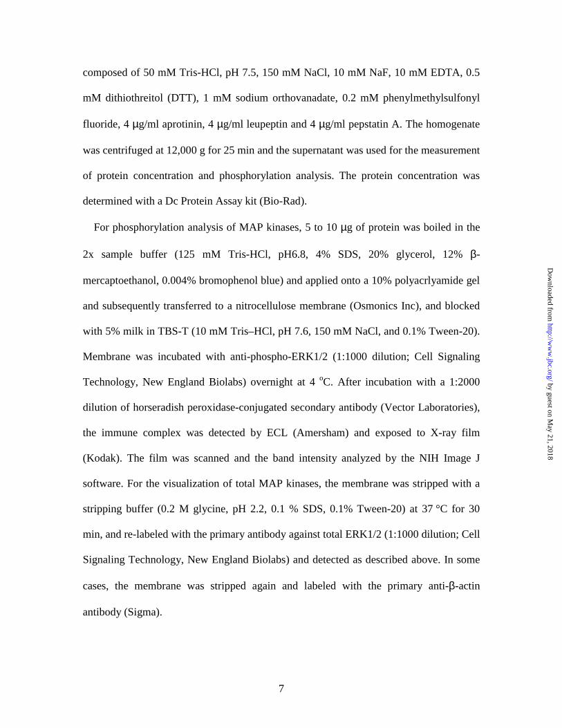
For phosphorylation analysis of MAP kinases, 5 to 10 µg of protein was boiled in the
2x sample buffer (125 mM Tris-HCl, pH6.8, 4% SDS, 20% glycerol, 12% β-
mercaptoethanol, 0.004% bromophenol blue) and applied onto a 10% polyacrlyamide gel
and subsequently transferred to a nitrocellulose membrane (Osmonics Inc), and blocked
with 5% milk in TBS-T (10 mM Tris–HCl, pH 7.6, 150 mM NaCl, and 0.1% Tween-20).
Membrane was incubated with anti-phospho-ERK1/2 (1:1000 dilution; Cell Signaling
Technology, New England Biolabs) overnight at 4 oC. After incubation with a 1:2000
dilution of horseradish peroxidase-conjugated secondary antibody (Vector Laboratories),
the immune complex was detected by ECL (Amersham) and exposed to X-ray film
(Kodak). The film was scanned and the band intensity analyzed by the NIH Image J
software. For the visualization of total MAP kinases, the membrane was stripped with a
stripping buffer (0.2 M glycine, pH 2.2, 0.1 % SDS, 0.1% Tween-20) at 37 °C for 30
min, and re-labeled with the primary antibody against total ERK1/2 (1:1000 dilution; Cell
Signaling Technology, New England Biolabs) and detected as described above. In some
cases, the membrane was stripped again and labeled with the primary anti-β-actin
antibody (Sigma).
by guest on May 21, 2018
http://ww
w.jbc.org/
Dow
nloaded from

8
Data analysis - Paired and unpaired t-tests were performed. Data are expressed as
means ± SEM, with n indicating the number of animals or slices. Significance level was
set at P < 0.05 (two-tailed test).
RESULTS
GHB reduces MAP kinase phosphorylation in frontal cortex and hippocampus -
Acute administration of a high dose of GHB in rodents has been shown to induce marked
decreases in locomotor activity and body temperature (34-36). We have observed similar
behavioral effects of GHB in the mice used in our current experiments (data not shown),
but our study focused on the effects of GHB (500 mg/kg) on the regulation of MAP
kinase activation by using Western blot analysis. All of our measurements are expressed
as the ratio (p/t) of phospho-MAP kinase (p) to total MAP kinase (t). No difference in the
total MAP kinase levels was detected in mouse cortex or hippocampus at different times
following GHB administration (as compared with β-actin, data not shown).
First, we determined the time course of ERK1/2 activation (phosphorylation) after an
acute i.p. injection of GHB (500 mg/kg body weight). Control mice were injected with
saline and their brains were prepared at 30 min, 60 min and 120 min respectively
following the injection. As there was no difference in the level of ERK1/2
phosphorylation between the control mice at the different time points, all control data
were pooled. As shown in Fig. 1, a significantly reduced MAP kinase phosphorylation
was observed in both frontal cortex and hippocampus (n = 5-7 for each time point of
by guest on May 21, 2018
http://ww
w.jbc.org/
Dow
nloaded from

9
GHB treatment, and a total n = 16 for control). A significant effect of GHB on the MAP
kinase phosphorylation in frontal cortex was detected as early as 15 min following GHB
injection. Thirty minutes following GHB administration, the phosphorylation levels of
MAP kinases were reduced to 52 ± 4% of control in frontal cortex (n = 7) and to 65 ± 6%
of control in hippocampus (n = 7). We were able to observe a significant down-regulation
of MAP kinase phosphorylation in frontal cortex and hippocampus (to 77 ± 9% and 67 ±
6% of control respectively, n = 5-7) lasting for at least 2 hours following GHB injection
(Fig. 1).
Possible regional differences in the GHB-dependent regulation of MAP kinase
phosphorylation specific to various cortical and hippocampal subfields were identified by
microdissecting the 600-µm-thick slices in ice-cold solution into the respective brain
regions. The brain regions, including somatosensory cortex and CA1, CA3 and dentate
gyrus (DG) of hippocampus, were then subjected to Western blot analysis. As shown in
Fig. 2, GHB-induced down-regulation of MAP kinase phosphorylation was detected only
in CA1 (to 30 ± 3% of control, n = 6 - 8) and CA3 (to 45 ± 7% of control, n = 6 - 8) but
not in DG (to 102 ± 10% of the control, n = 4 - 6). A clear GHB-induced decrease (to 70
± 5% of control, n = 4 - 7) in MAP kinase phosphorylation was also detected in the
somatosensory cortex.
The effect of GHB on MAP kinase phosphorylation is not mediated through the high
affinity GHB receptor - Since certain effects of GHB have been proposed to occur via
high affinity GHB receptors (9, 37, 38), we addressed this point by using the specific
by guest on May 21, 2018
http://ww
w.jbc.org/
Dow
nloaded from

10
GHB receptor antagonist NCS-382 in a separate set of experiments. We found that the
phosphorylation levels of MAP kinases in frontal cortex and hippocampus of animals
treated with GHB (500 mg/kg for 30 min) were 57 ± 8% of control and 60 ± 21% of
control respectively, while the phosphorylation levels of MAP kinases in frontal cortex
and hippocampus of animals pre-treated with NCS-382 (200 mg/kg) were 50 ± 8% of
control and 50 ± 13% of control (n= 5-7). There is no statistically significant difference
between the two groups. By itself, NCS-382 (200 mg/kg) did not significantly affect the
phosphorylation of MAP kinase in the frontal cortex (85 ± 15 % of control) or
hippocampus (82 ± 10% of control; n = 5). Since pretreatment with NCS-382 (200
mg/kg) failed to reverse GHB-induced inhibition of MAP kinase phosphorylation, the
proposed high affinity GHB receptors appears to play no role in mediating the GHB-
induced inhibition of MAP kinase phosphorylation.
GHB affects MAP kinase phosphorylation by acting GABAB receptors - We next
examined the role of GABAB receptors in the GHB-induced inhibition of MAP kinase
phosphorylation. As shown in Fig. 3, a pretreatment with the specific GABAB receptor
antagonist CGP56999A (20 mg/kg) administered 10 min before GHB prevented the
inhibitory effect of GHB (500 mg/kg) on MAP kinase phosphorylation both in cortex and
hippocampus, demonstrating that GABAB receptors are involved in diminishing MAP
kinase phosphorylation by GHB. In addition, as shown in Fig. 3, we also found that
CGP56999A alone, at the dose used (20 mg/kg, i.p.), significantly up-regulated the
phosphorylation of MAP kinases in the hippocampus (170 ± 14.4% of control, n = 5) but
not in the cortex (102 ± 17% of control, n = 5)
by guest on May 21, 2018
http://ww
w.jbc.org/
Dow
nloaded from

11
We also used baclofen, a well-known selective GABAB receptor agonist, as a positive
control for GABAB receptor activation. Similar to GHB, baclofen (20 mg/kg, i.p.)
significantly decreased the phosphorylation of MAP kinases in the cortex (to 33 ± 3% of
control, n = 5) and hippocampus (to 53 ± 12% of control, n = 5), further confirming the
role of activation of GABAB receptors in MAP kinase signaling (Fig. 3).
GABA is not an intermediate in the GHB-induced inhibition of MAP kinase
phosphorylation - We sought to determine whether the inhibitory effect of GHB on MAP
kinase phosphorylation was due to the activation of GABAB receptors by GABA that was
converted from the exogenously administered GHB. We used valproate, a compound
known to inhibit GHB dehydrogenase, hence preventing the conversion of GHB into
GABA (9, 39). The phosphorylation levels in GHB-treated animals were 57 ± 8% of
control and 60 ± 21% of control in frontal cortex and hippocampus respectively (n = 5-7),
while the phosphorylation levels in valproate plus GHB-treated animals were 40 ± 9% of
control and 48 ± 29% of control in frontal cortex and hippocampus respectively (n = 5-7),
indicating that blocking the conversion of GHB to GABA by valproate failed to prevent
GHB-reduced MAP kinase phosphorylation. Thus, GHB-induced inhibition of MAP
kinase phosphorylation most likely results from the direct activation of GABAB receptors
by GHB. In addition, a significant inhibitory effect of valproate (n = 5) on MAP kinase
phosphorylation was observed in the frontal cortex (to 52 ± 7% of control) and
hippocampus (to 62 ± 13% of control).
by guest on May 21, 2018
http://ww
w.jbc.org/
Dow
nloaded from

12
GHB is ineffective in brain slices - We finally compared the effects of GHB
administration in vivo and in vitro (brain slices), since we have previously shown in brain
slices that GHB activates GABAB receptors in neocortical and hippocampal neurons (31).
Although the inhibitory effect of GHB on MAP kinase phosphorylation was remarkable
in vivo (Figs. 1 to 3), GHB (10 mM for 15 min) caused no obvious change in MAP kinase
phosphorylation in cortical and hippocampal slices (Fig. 4). To address whether MAP
kinases could be phosphorylated in brain slices, we also included hydrogen peroxide as a
positive control. We confirmed a previous report that this compound induced a
significant increase in MAP kinase phosphorylation in brain slices (33).
We also wanted to find out whether the altered phosphorylation of MAP kinase could
be maintained in vitro. The effect of GHB on MAP kinase phosphorylation in vivo was
long lasting, as we were able to detect a significant down-regulation of MAP kinase
phosphorylation in cortex and hippocampus (to 77 ± 9% and 67 ± 6% of control
respectively, n = 5-7) even 2 hours following GHB injection (Fig. 1 and Fig. 5). In
contrast to our in vivo findings, in vitro incubation in aCSF of brain slices prepared 30
min after animals were acutely treated with GHB (500 mg/kg) resulted in the recovery of
the MAP kinase phosphorylation to control levels (Fig. 5). In addition, we also examined
whether slice cutting procedure itself would affect MAP kinase phosphorylation. One
whole brain was cut into two equal halves, one half of which was immediately put on dry
ice and the other half was cut into slices in cold aCSF. No differences were observed in
the level of MAP kinase phosphorylation between the intact and sliced brain (data not
shown).
by guest on May 21, 2018
http://ww
w.jbc.org/
Dow
nloaded from

13
DISCUSSION
Acute GHB administration in humans or animals induces profound physiological
effects. Previously we have demonstrated that GHB, at millimolar concentration in vitro,
reduces neuronal excitability and synaptic activity in both neocortical and hippocampal
neurons mediated via GABAB receptors (31). In our current experiments, we chose a high
of dose of GHB (500mg/kg), since the concentration of GHB in cerebrospinal fluid (CSF)
or brain in animals and in humans, after an exogenous administration at this dose,
reached millimolar levels (14, 40-42), compatible with the affinity of GABAB receptors
for GHB (43). Acute administration of GHB (500 mg/kg) in vivo caused a rapid and long-
lasting inhibition on MAP kinase phosphorylation in both frontal cortex and
hippocampus (Fig. 1).
The mechanisms whether GHB exerts its effects in the central nervous system are still
poorly understood. In general, two modes of GHB action have been proposed: through
GABAB receptors or via high affinity GHB receptors that have been suggested to exist in
the brain (9, 37, 38). According to binding studies, some actions of GHB may be
mediated via highly specific membrane-binding sites (9) that appear to have a different
regional distribution than GABAB-binding sites in rodent brain (37). More recently the
high affinity GHB receptor has been suggested to be a member of the G protein-coupled
receptor family (38). The overwhelming evidence favors GABAB-receptor-mediated
actions of GHB, as many effects of GHB on the central nervous system show striking
similarities to the effects produced by the selective GABAB-receptor agonist baclofen,
by guest on May 21, 2018
http://ww
w.jbc.org/
Dow
nloaded from
14
and can be attenuated by specific GABAB-receptor antagonists (11-13, 15, 17, 18, 44-47).
Clearly, the binding characteristics, reported by Bernasconi et al. (1) and Mathivet et al.
(47), also strongly support GHB as a selective GABAB receptor agonist.
To address whether the high affinity GHB receptors or GABAB receptors mediate the
GHB-induced inhibition of MAP kinase phosphorylation in our experiments, we tested
the effects of NCS-382, a specific GHB receptor antagonist shown to reverse some of
GHB’s effects (9, 38). Even at the extremely high dose of NCS-382 (200 mg/kg) used in
our experiments, NCS-382 was ineffective in reversing the suppressant effect of GHB on
MAP kinase phosphorylation. Therefore, the high affinity GHB receptors do not appear
to mediate this action of GHB. In contrast, CGP56999A, a specific GABAB receptor
antagonist completely antagonized the inhibitory effect of GHB on MAP kinase
phosphorylation. The antagonism of GHB action by CGP56999A strongly implicates a
role of GABAB receptors in mediating the GHB-induced inhibition of MAP kinase
phosphorylation. This was further confirmed by the selective GABAB receptor agonist
baclofen mimicking the effect of GHB on MAP kinase phosphorylation (Fig. 3).
Interestingly, the treatment with CGP56999A alone increased MAP kinase
phosphorylation in the hippocampus. As GABAB receptor activation by spontaneous
GABA release has been detected in the hippocampus (48), the activation of GABAB
receptors by ambient GABA levels appears to be sufficient to produce an inhibitory
effect on the phosphorylation of MAP kinases in the hippocampus. The GABAB receptor
antagonist CGP56999A may have blocked the tonic activation of GABAB receptors by
GABA, consequently increasing MAP kinase phosphorylation. Interestingly, CGP
by guest on May 21, 2018
http://ww
w.jbc.org/
Dow
nloaded from

15
56999A alone caused no upregulation of MAP kinase phosphorylation in the frontal
cortex. This could be due to differences in the two brain regions regardings the levels of
ambient GABA, or nature of the second messenger systems linked to the tonically
activated GABAB receptors.
We then asked whether the GHB-induced decrease in MAP kinase phosphorylation
was due to an indirect activation of GABAB receptors by GABA converted from the high
concentration of exogenously administered GHB through the combined actions of GHB
dehydrogenase and GABA transaminase. We used the short-chain fatty acid valproate
known to inhibit the cytosolic GHB dehydrogenase, thus preventing GHB degradation
into GABA (9, 39). As valproate failed to block the GHB-induced decrease in MAP
kinase phosphorylation, this effect of GHB is due to the direct activation of GABAB
receptors by GHB and not by GHB-derived GABA acting at GABAB receptors. In
addition, valproate per se also inhibited MAP kinase phosphorylation in both frontal
cortex and hippocampus. Considering that acute valproate administration (200 mg/kg -
600 mg/kg) induces significant increases in brain GHB levels (9, 49), it is very likely that
the elevated GHB concentration following valproate administration is responsible for
suppressing MAP kinase phosphorylation. However, the possible effects of valproate on
other signaling mechanisms cannot be ruled out. Our findings with valproate are
inconsistent with the recent report by Yuan et al (50), in which valproate was found to
increase MAP kinase phosphorylation in a cultured cell line (human neuroblastoma SH-
SY5Y).
by guest on May 21, 2018
http://ww
w.jbc.org/
Dow
nloaded from

16
Our experiments strongly support an acute reduction in MAP kinase phosphorylation
by GHB acting via direct activation of GABAB receptors in mouse frontal cortex and
hippocampus. This finding has important implications for the role of GABAB receptors
and of MAP kinase signaling pathways in the physiological function and regulation.
GHB levels in the cerebrospinal fluid (CSF) remain consistently high (millimolar range)
for more than 4 hours in mammals after one acute GHB (500 mg/kg) administration (40-
42), consequently, even one administration of a high dose of GHB, as often is the case
during the abuse in humans, might cause lasting influences in the central nervous system.
The GHB-induced down-regulation of MAP kinase phosphorylation, mediated via
GABAB receptors, may represent an important cellular mechanism underlying the effects
of GHB on the central nervous system.
The specific mechanism of MAP kinase phosphorylation suppression by an acute GHB
administration remains an open question. The most parsimonious explanation is a
decrease in cAMP levels and, consequently, in cAMP-dependent protein kinase A (PKA)
activity. The stimulation of GABAB receptors has been linked to the inhibition of
adenylyl cyclase, thus decreasing cAMP levels (51) and it is well documented that cAMP
level, through its effect on the PKA, correlates with the activation (phosphorylation) of
MAP kinase (52). Another possibility may be a change in protein kinase C (PKC) activity,
resulting from the activation of GABAB receptors leading to the inhibition of voltage
gated Ca 2+ channels (51). In support of a role of PKA and/or PKC in the GHB-induced
inhibition of MAP kinase phosphorylation, the GABAB receptor agonist baclofen has
been shown to reduce forskolin-stimulated increase in cAMP both in vivo and in vitro
by guest on May 21, 2018
http://ww
w.jbc.org/
Dow
nloaded from

17
(51), while baclofen and GABA have been shown to modulate PKC activity (53). In
addition, GHB has been also reported to modulate adenylyl cyclase activity and
intracellular Ca 2+ concentrations (38, 54). The involvement of other protein kinase
pathways in the negative regulation of MAP kinase signaling cascade cannot be
excluded. For example, several recent studies have identified Akt (also termed protein
kinase B, PKB), a main downstream effector of phosphotidylinositol-3-kinase (PI3K)
pathway, as a potent negative regulator of MAP kinase pathway (55, 56), while PI3K has
been shown to mediate certain G protein-coupled receptor activation of MAP kinase
signaling pathway (57), probably through the regulation of PI3K catalytic activity by the
α and/or βγ subunits of heterotrimeric G proteins, Ras and calmodulin (58, 59).
Calmodulin can also directly modulate the ERK pathway (52, 60).
Recently much attention has been devoted to the role of the family of dual specificity
MAP kinase phosphatases (MKPs), of which MKP-3 is selective for the inactivation of
ERKs (61). A significant up-regulation in its expression level in rat hippocampus has
been detected after acute amphetamine (62). However, by using a specific antibody
against MKP-3 (C-20) (sc-8599, Santa Cruz Biotechnology), we found that the
expression level of MKP-3 not to be significantly affected in frontal cortex and
hippocampus of mice subjected to acute GHB injection (the expression levels of MKP-3
in frontal cortex and hippocampus of mice 30 min following GHB injection were 91 ±
12% and 109 ± 10% respectively, n = 3). Therefore, it appears unlikely that the reduced
MAP kinase phosphorylation is due to increased activation of MAP kinase phosphatases
by guest on May 21, 2018
http://ww
w.jbc.org/
Dow
nloaded from

18
like MKP-3. The GHB-induced inhibition in MAP kinase phosphorylation more likely
represents the GABAB receptor-mediated modulation of MAP kinase (ERK1/2) by
different signaling cascades. The exact interactions between the different signaling
pathways will require further clarification.
In contrast to the GHB-induced inhibition of MAP kinases after in vivo GHB
administration, our experiments with brain slices showed that GHB (10 mM for 15 min)
did not alter the phosphorylation level of MAP kinases in slices. Although injury itself
was reported to strongly activate MAP kinases (63), our careful dissections (hippocampus
were kept intact) and the immediate freezing of brain tissues on dry ice make injury less
likely of a factor affecting the MAP kinase phosphorylation in these tissues, especially
hippocampus. Another intriguing observation of our study is that, after a 90 min of in
vitro incubation, the phosphorylation level of MAP kinases in slices from the brains of
animals acutely treated with GHB (500 mg/kg for 30 min) recovered to control levels
(Fig. 5), while a significantly reduced MAP kinase phosphorylation was still observed
even after 120 min following one GHB injection (500 mg/kg) in vivo (Fig.1 and Fig. 5).
One explanation for this could be that GHB levels in vivo remained consistently elevated
causing a persistent inactivation of MAP kinases, whereas the in vitro recovery of MAP
kinase phosphorylation level was due to the incubation of brain slices without GHB
stimulation. However, this seems unlikely since GHB (10 mM for 15 min) did not alter
the phosphorylation level of MAP kinases in cortical and hippocampal slices. The exact
reasons for the discrepancy between the in vivo and in vitro data remain to be further
investigated. In contrast to our findings, a recent report (64) showed that the GABAB
by guest on May 21, 2018
http://ww
w.jbc.org/
Dow
nloaded from

19
receptor agonist baclofen increased MAP kinase (ERK2) phosphorylation in the CA1
area of hippocampal slices. The inconsistence between these results probably might be
due to the experimental situations such as incubation temperature (these authors used an
incubation temperature of 26 - 28 oC). However, it is also possible that GHB may exert
some effects distinct from those induced by baclofen.
In summary, our findings represent the first demonstration that GHB markedly inhibits
MAP kinase activation (phosphorylation) via a GABAB receptor-mediated mechanism.
These findings have revealed a novel action of GHB and of GABAB receptors through
ERK MAP kinases in intracellular signaling cascades and thus provide new insights into
the intracellular mechanisms underlying the various effects of GHB on the central
nervous system (31, 65). Since MAP kinases have been found to play a crucial role in
mediating a number of physiological and pathological changes in cell function (19, 20),
the down-regulation of this pathway may be relevant to the pathological changes during
acute GHB intoxication. Given the role of MAP kinases in the induction of long-term
functional changes, the neuroadaptative changes (tolerance and dependence) following
repeated uses of GHB in rodents and humans (6-8) may involve this mechanism of
action. Our present findings may also open novel means for the treatment of acute GHB
intoxication by modulating the ERK signaling pathway. It will be important to elucidate
the precise mechanisms whereby GHB inhibits MAP kinase phosphorylation and the
possible correlation between the deactivation of MAP kinases specific to certain brain
regions and the behavioral effects associated with acute and repeated GHB
administrations.
by guest on May 21, 2018
http://ww
w.jbc.org/
Dow
nloaded from

20
Acknowledgements-The authors thank Dr. W. Froestl for kindly providing
CGP56999A. We are also grateful to Yijun Cui, Nils Ole Dalby, Brian Oyama, Mahsan
Rafizadeh and Weizheng Wei for assistance and help during experiments. This work was
supported by NIH grant DA14947 to I.M..
REFERENCES
1. Bernasconi, R., Mathivet, P., Bischoff, S., and Marescaux, C. (1999) Trends
Pharmacol. Sci. 20, 135-141
2. Kleinschmidt, S., Schellhase, C., and Mertzlufft, F. (1999) Eur. J. Anaesthesiol. 16,
23-30
3. Gallimberti, L., Canton, G., Gentile, N., Ferri, M., Cibin, M., Ferrara, S.D., Fadda,
F., and Gessa, G.L. (1989) Lancet 2, 787-789
4. Rosen, M.I., Pearsall, H.R., Woods, S.W., and Kosten, T.R. (1996)
Neuropsychopharmacology 14, 187-193
5. Mamelak, M., Scharf, M.B., and Woods, M. (1986) Sleep 9, 285-289
6. Timby, N., Eriksson, A., and Bostrom, K. (2000) Am. J. Med. 108, 518-519
7. Okun, M.S., Boothby, L.A., Bartfield, R.B., and Doering, P.L. (2001) J. Pharm.
Pharm. Sci. 4, 167-175
8. Nicholson, K.L., and Balster, R.L. (2001) Drug Alcohol Depend. 63, 1-22
9. Maitre, M. (1997) Prog. Neurobiol. 51, 337-361
10. Cash, C.D., Gobaille, S., Kemmel, V., Andriamampandry, C., and Maitre, M. (1999)
Biochem. Pharmacol. 58, 1815-1819
by guest on May 21, 2018
http://ww
w.jbc.org/
Dow
nloaded from

21
11. Nava, F., Carta, G., Bortolato, M., and Gessa, G.L. (2001) Eur. J. Pharmacol. 430,
261-263
12. Xie, X., and Smart, T.G. (1992) Eur. J. Pharmacol. 223, 193-196
13. Xie, X., and Smart, T.G. (1992) Eur. J. Pharmacol. 212, 291-294
14. Gobaille, S., Hechler, V., Andriamampandry, C., Kemmel, V., and Maitre, M.
(1999) J. Pharmacol. Exp. Ther. 290, 303-309
15. Hu, R.Q., Banerjee, P.K., and Snead, O.C. 3rd. (2000) Neuropharmacology 39, 427-
439
16. Ferraro, L., Tanganelli, S., O'Connor, W.T., Francesconi, W., Loche, A., Gessa,
G.L., and Antonelli, T. (2001) J. Neurochem. 78, 929-939
17. Carai, M.A., Colombo, G., Brunetti, G., Melis, S., Serra, S., Vacca, G., Mastinu, S.,
Pistuddi, A.M., Solinas, C., Cignarella, G., Minardi, G., and Gessa, G.L. (2001)
Eur. J. Pharmacol. 428, 315-321
18. Barbaccia, M.L., Colombo, G., Affricano, D., Carai, M.A., Vacca, G., Melis, S.,
Purdy, R.H., and Gessa, G.L. (2002) Neuropharmacology 42, 782-791
19. Sweatt, J.D. (2001) J. Neurochem. 76, 1-10
20. Johnson, G.L., and Lapadat, R. (2002) Science 298, 1911-1912
21. Nestler, E.J. (2002) Nat. Neurosci. 5(Suppl), 1076-1079
22. Koob, G.F., Sanna, P.P., and Bloom, F.E. (1998) Neuron 21, 467-476
23. Schulz, S., and Höllt, V. (1998) Eur. J. Neurosci. 10, 1196-1201
24. Derkinderen, P., Valjent, E., Toutant, M., Corvol, J.C., Enslen, H., Ledent, C.,
Trzaskos, J., Caboche, J., and Girault, J.A. (2003) J. Neurosci. 23, 2371-2382
25. Choe, E.S., Chung, K.T., Mao, L., and Wang, J.Q. (2002)
by guest on May 21, 2018
http://ww
w.jbc.org/
Dow
nloaded from

22
Neuropsychopharmacology 27, 565-575
26. Licata, S.C., and Pierce, R.C. (2003) J. Neurochem. 85, 14-22
27. Kyosseva, S.V., Owens, S.M., Elbein, A.D., and Karson, C.N. (2001)
Neuropsychopharmacology 24, 267-277
28. Brunzell, D.H., Russell, D.S., and Picciotto, M.R. (2003) J. Neurochem. 84, 1431-
1441
29. Kalluri, H.S., and Ticku, M.K. (2002) Eur. J. Pharmacol. 439, 53-58
30. Cousins, M.S., Roberts, D.C., and de Wit, H. (2002) Drug Alcohol Depend. 65, 209-
220
31. Jensen, K., and Mody, I. (2001) Cereb. Cortex 11, 424-429
32. Galloway, G.P., Frederick, S.L., Staggers, F.E. Jr, Gonzales, M., Stalcup, S.A., and
Smith, D.E. (1997) Addiction 92, 89-96
33. Kanterewicz, B.I., Knapp, L.T., and Klann, E. (1998) J. Neurochem. 70, 1009-1016
34. Kaufman, E.E., Porrino, L.J., and Nelson, T. (1990) Biochem. Pharmacol. 40, 2637-
2640
35. Nissbrandt, H. and Engberg, G. (1996) J. Neural. Transm.103, 1255-1263
36. Cook, C.D., Aceto, M.D., Coop, A., and Beardsley, P.M. (2002)
Psychopharmacology 160, 99-106
37. Snead, O.C. 3rd. (1994) Brain Res. 659, 147-156
38. Snead, O.C. 3rd. (2000) J. Neurochem. 75, 1986-1996
39. Hechler, V., Ratomponirina, C., and Maitre, M. (1997) J. Pharmacol. Exp. Ther.
281, 753-760
40. Snead, O.C. 3rd, Yu, P.K., and Huttenlocher, P.R. (1976) Neurology 26, 51-56
by guest on May 21, 2018
http://ww
w.jbc.org/
Dow
nloaded from

23
41. Snead, O.C. 3rd. (1978) Neurology 28, 636-642
42. Shumate, J.S., and Snead, O.C. 3rd. (1979) Res. Commun. Chem. Pathol.
Pharmacol. 25, 241-256
43. Berton, F., Brancucci, A., Beghe, F., Cammalleri, M., Demuro, A., Francesconi, W.,
and Gessa, G.L. (1999) Eur. J. Pharmacol. 380, 109-116
44. Engberg, G., and Nissbrandt, H. (1993) Naunyn Schmiedebergs Arch. Pharmacol.
348, 491- 497
45. Madden, T.E., and Johnson, S.W. (1998) J. Pharmacol. Exp. Ther. 287, 261-265
46. Erhardt, S., Andersson, B., Nissbrandt, H., and Engberg, G. (1998) Naunyn
Schmiedebergs Arch. Pharmacol. 357, 611-619
47. Mathivet, P., Bernasconi, R., De Barry, J., Marescaux, C., and Bittiger, H. (1997)
Eur. J. Pharmacol. 321, 67-75
48. McLean, H.A, Caillard, O., Khazipov, R., Ben-Ari, Y., and Gaiarsa, J.L. (1996) J.
Neurophysiol. 76, 1036-1046
49. Snead, O.C. 3rd, Bearden, L.J., and Pegram, V. (1980) Neuropharmacology 19, 47-
52 50. Yuan, P.X., Huang, L.D., Jiang, Y.M., Gutkind, J.S., Manji, H.K., and Chen, G.
(2001) J. Biol. Chem. 276, 31674-31683
51. Bowery, N.G., Bettler, B., Froestl, W., Gallagher, J.P., Marshall, F., Raiteri, M.,
Bonner, T.I., and Enna, S.J. (2002) Pharmacol. Rev. 54, 247-264
52. Grewal, S.S., York, R.D., and Stork, P.J. (1999) Curr. Opin. Neurobiol. 9, 544-553
53. Tremblay, E., Ben-Ari,Y., and Roisin, M.P. (1995) J. Neurochem. 65, 863-670
54. Ito, Y., Ishige, K., Zaitsu, E., Anzai, K., and Fukuda, H. (1995) J. Neurochem. 65,
75-83
by guest on May 21, 2018
http://ww
w.jbc.org/
Dow
nloaded from

24
55. Rommel, C., Clarke, B.A., Zimmermann, S., Nunez, L., Rossman, R., Reid, K.,
Moelling, K., Yancopoulos, G.D., and Glass, D.J. (1999) Science 286, 1738-1741
56. Guan, K.L., Figueroa, C., Brtva, T.R., Zhu, T., Taylor, J., Barber, T.D., and Vojtek,
A.B. (2000) J. Biol. Chem. 275, 27354-27359
57. Lopez-Ilasaca, M., Crespo, P., Pellici, P.G., Gutkind, J.S., and Wetzker, R. (1997)
Science 275, 394-397
58. Joyal, J.L., Burks, D.J., Pons, S., Matter, W.F., Vlahos, C.J., White, M.F., and Sacks,
D.B. (1997) J. Biol. Chem. 272, 28183-28186
59. Wymann, M.P., and Pirola, L. (1998) Biochim. Biophys. Acta 1436, 127-150
60. Agell, N., Bachs, O., Rocamora, N., and Villalonga, P. (2002) Cell Signal. 14, 649-
654
61. Hafen, E. (1998) Science 280, 1212-1213
62. Takaki, M., Ujike, H., Kodama, M., Takehisa, Y., Nakata, K., and Kuroda, S. (2001)
J. Neurochem. 79, 679-688
63. Martin, K.C., Michael, D., Rose, J.C., Barad, M., Casadio, A., Zhu, H., and Kandel,
E.R. Neuron 18, 899-912
64. Vanhoose, A.M., Emery, M., Jimenez, L., and Winder, D.G. (2002) J. Biol. Chem.
277, 9049- 9053
65. Banerjee, P.K., Hirsch, E., and Snead, O.C. 3rd. (1993) Neuroscience 56, 11-21
by guest on May 21, 2018
http://ww
w.jbc.org/
Dow
nloaded from

25
FIG. 1. Acute GHB administration significantly reduces MAP kinase
phosphorylation in mouse frontal cortex and hippocampus. Measurements of
normalized phospho-ERK1/2 levels were done at the indicated times (15 min, 30 min, 60
min and 120 min) following an acute injection of GHB (500 mg/kg, i.p.). Control mice
were injected with saline tissue was collected at 30 min, 60 min and 120 min respectively
after the injection. Since there was no difference between these time points in controls, all
control data were combined (designated as 0 min after GHB injection). *, *** P < 0.05 or
0.001 vs. control respectively (n = 5 - 7 for each GHB treatment group, n = 16 for
control).
FIG. 2. GHB-induced decrease of MAP kinase phosphorylation in areas CA1 and
CA3 of hippocampus. (A): A representative Western blot detected with anti-phospho-
ERK1/2 or anti-total ERK1/2 antibody. (B): Normalized phospho-ERK1/2 levels were
measured in tissue obtained 30 min following the injection of GHB (500 mg/kg, i.p.). A
clear reduction of MAP kinase phosphorylation was observed in somatosensory cortex
and in CA1 or CA3 but not in dentate gyrus (DG) of hippocampus. *, **, *** P < 0.05,
0.01, or 0.001 vs. saline respectively (n = 4 - 8. DG = dentate gyrus, S = saline, G =
GHB).
FIG. 3. The specific GABAB receptor antagonist CGP 56999A reverses, and the
GABAB receptor agonist baclofen mimics the effect of GHB on MAP kinase
by guest on May 21, 2018
http://ww
w.jbc.org/
Dow
nloaded from

26
phosphorylation. (A): A representative Western blot detected with anti-phospho-
ERK1/2 or anti-total ERK1/2 antibody. (B): Normalized phospho-ERK1/2 levels.
CGP56999A (20 mg/kg, i.p.) was given 10 min prior to GHB injection. The tissue was
collected 30 min following one injection of GHB (500 mg/kg, i.p.) or baclofen (20
mg/kg, i.p.). The cortices and hippocampi of brains from the mice subjected to various
treatments were then dissected out for Western blot analysis. *, **, *** P < 0.05, 0.01 or
0.001 vs. saline, #, ## P < 0.05 or 0.01 vs. C+G group respectively (n = 5. S = saline, C =
CGP56999A, G = GHB, C+G = CGP56999A plus GHB, B = baclofen).
FIG. 4. GHB has no effect on the MAP kinase phosphorylation in brain slices. (A): A
representative Western blot detected with anti-phospho-ERK1/2 or anti-total ERK1/2
antibody. (B): Normalized phospho-ERK1/2 levels. 350-µm-thick coronal brain slices
removed from after decapitation and placed into an ice-cold artificial cerebrospinal fluid
(aCSF) bubbled with 95% O2 and 5% CO2. The slices were incubated for 1 hour at 32 oC
in oxygenated aCSF before further treatment with 10 mM GHB for 15 min or 10 mM
hydrogen peroxide (H2O2) for 10 min (33). *** P < 0.001 vs. control (n = 5 - 7 for cortex
and 8-19 for hippocampus. S = saline, G = GHB, HP = hydrogen peroxide).
FIG. 5. A comparison of GHB effects on MAP kinase phosphorylations between in
vivo and in vitro conditions. MAP kinase phosphorylation was significantly inhibited in
frontal cortex and hippocampus in vivo 30 min following an acute administration of
by guest on May 21, 2018
http://ww
w.jbc.org/
Dow
nloaded from

27
GHB, and this effect of GHB was still observed in vivo 120 min after GHB injection. A
90 min of in vitro incubation of the slices from the mice acutely administrated with GHB
(500 mg/kg for 30 min) in aCSF led to the recovery of phospho-MAP kinase to control
levels. The brains were directly used for Western blot analysis, or cut into 350-µm-thick
coronal slices, which were further incubated for 90 min at 32 oC in aCSF before they
were used for Western blot analysis. The third group of mice were injected GHB (500
mg/kg, i.p.), and 120 min later, the brains were removed and dissected out for Western
blot analysis. *, *** P < 0.05 or 0.001 vs. control respectively (n = 5 - 7).
by guest on May 21, 2018
http://ww
w.jbc.org/
Dow
nloaded from

Ren and Mody, Fig. 1
0 30 60 90 1200.00
0.25
0.50
0.75
1.00
1.25
*
******
***
*********
Rela
tive
leve
l of p
hosp
ho-E
RK1/
2
Time (min) after GHB injection
Cortex Hippocampus
by guest on May 21, 2018
http://ww
w.jbc.org/
Dow
nloaded from

Cortex CA1 CA3 DG S G S G S G S G
Phospho-ERK1/2
Total ERK1/2
A
B
Cortex CA1 CA3 DG0.00
0.50
1.00
1.50
**
***
*
Rela
tive l
evel
of p
hosp
ho-E
RK1/2 Saline
GHB
ERK1ERK2ERK1ERK2
Ren and Mody, Fig. 2
by guest on May 21, 2018
http://ww
w.jbc.org/
Dow
nloaded from

Cortex Hippocampus S C G C+G B S C G C+G B
Phospho-ERK1/2
Total ERK1/2
A
B
Cortex Hippocampus0.00
0.50
1.00
1.50
2.00
*
#*
**
***
##***
Relat
ive le
vel o
f pho
spho
-ERK
1/2 Saline CGP GHB CGP + GHB Baclofen
ERK1ERK2
ERK1ERK2
Ren and Mody, Fig. 3
by guest on May 21, 2018
http://ww
w.jbc.org/
Dow
nloaded from

Ren and Mody, Fig. 4
A Cortex Hippocampus C G HP C G HP
Phospho-ERK1/2
Total ERK1/2
ERK1ERK2
ERK1ERK2
B
Cortex Hippocampus 0.00
0.50
1.00
1.50
2.00
2.50
3.00
***
***
Rela
tive
leve
l of p
hosp
ho-E
RK1/
2
Control GHB HP
by guest on May 21, 2018
http://ww
w.jbc.org/
Dow
nloaded from

Ren and Mody, Fig. 5
Cortex Hippocampus0.00
0.50
1.00
1.50
******
*
***
Rela
tive
leve
l of p
hosp
ho-E
RK1/
2 Control GHB in vivo 30 min GHB in vivo 120 min GHB in vivo 30 min + in vitro 90min
by guest on May 21, 2018
http://ww
w.jbc.org/
Dow
nloaded from

Xiuhai Ren and Istvan Modyreceptor activation in mouse frontal cortex and hippocampus
Gamma-hydroxybutyrate (GHB) reduces MAP kinase phosphorylation via GABA
published online August 15, 2003J. Biol. Chem.
10.1074/jbc.M304238200Access the most updated version of this article at doi:
Alerts:
When a correction for this article is posted•
When this article is cited•
to choose from all of JBC's e-mail alertsClick here
by guest on May 21, 2018
http://ww
w.jbc.org/
Dow
nloaded from