Embed Size (px)
Citation preview

International Journal of Scientific & Engineering Research Volume 10, Issue 11, November-2019 912 ISSN 2229-5518
IJSER © 2019
http://www.ijser.org
Gall Bladder Perforation in a Young Male of 17 years due to Cholelithiasis: A Case
Report 1Dr. Unsa Athar*, 2Dr. Muhammad Moiz Tahir, 3Dr.Taimoor Jamil, 4Dr.Muhammad Tahir Azeem
*Corresponding Author
Abstract:
all bladder perforations are reported rarely. They tend to have a high mortality rate as
they are not diagnosed in a timely manner. One of the reasons behind this is the fact
that the clinical signs and symptoms are vague. Investigation and imaging are also the
least helpful in such situations because of non-specific findings. Since it is an emergency,
a prompt decision to open the abdomen can be life-saving. Due the rare occurrence, gall
bladder perforations are skipped in the differential diagnosis. A few cases have been
reported world-wide, but they usually involve aged men and women. We present here a
case of a 17-year-old boy with post-operative diagnosis of gall bladder perforation without
any evident underlying cause.
Index Terms: Gall Bladder, Spontaneous Perforation, Cholecystitis, Sonographic,
Cholelithiasis.
Introduction:
In a study done retrospectively on 332 patients between 1997 to 2006 reported 16 cases
of gall bladder perforation. [1] Niemeyer’s classification be used to describe types of gall
bladder perforation based on their gross pathological picture. Our case can be classified
as type II perforation. [2] Gall bladder perforation is a life-threatening condition leading to
acute peritonitis. [3] It can be caused by both calculous and acalculous cholecystitis.
Typically, cholecystitis has a higher incidence in females, but gall bladder perforations are
more common in males. [4] There is a lack of definite knowledge and resources concerning
its diagnosis and treatment [5] Gall bladder perforation is uncommon and is mostly seen in
immune compromised patients. [6] It is the need of the hour to come up with new
knowledge to help is promptly diagnose and treat the condition. Here we present a case of
gall bladder perforation a very young age of 17, something that is rarely seen in the world.
Case Presentation:
A 17-year-old male presented to us in emergency department with the complaints of fever
for 6 days along with nausea and abdominal pain 3 days. The abdominal pain began in the
G
IJSER
International Journal of Scientific & Engineering Research Volume 10, Issue 11, November-2019 913 ISSN 2229-5518
IJSER © 2019
http://www.ijser.org
epigastrium and later became generalized. It was associated with nausea but no complaint
of vomiting/burning micturition/urinary retention/bowel disturbance. His history was
significant for an acute episode of hepatitis B two years ago which resolved on its own. On
examination, his vitals were stable. His abdomen exhibited board like rigidity. His total
leukocytes were slightly raised (14.7) along with aspartate aminotransferas and alanine
aminotransferase (64 and 40IU/L respectively) and Lactate dehydrogenase 325 IU/L. His
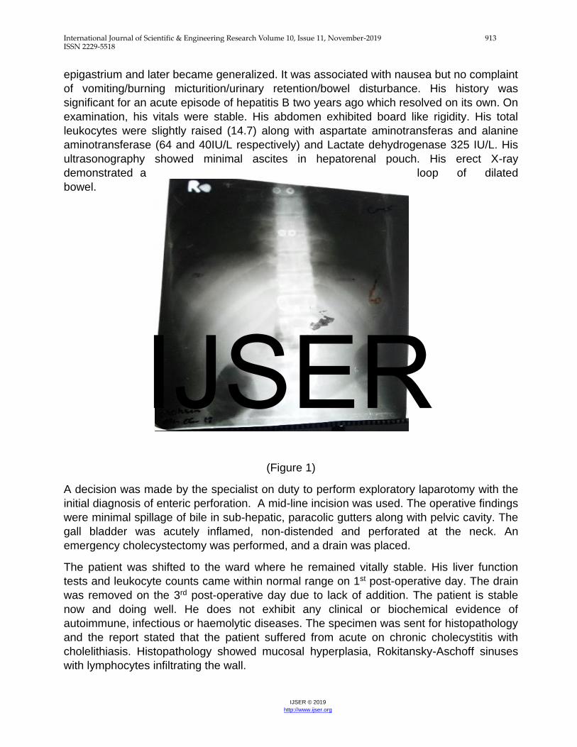
ultrasonography showed minimal ascites in hepatorenal pouch. His erect X-ray
demonstrated a loop of dilated
bowel.
(Figure 1)
A decision was made by the specialist on duty to perform exploratory laparotomy with the
initial diagnosis of enteric perforation. A mid-line incision was used. The operative findings
were minimal spillage of bile in sub-hepatic, paracolic gutters along with pelvic cavity. The
gall bladder was acutely inflamed, non-distended and perforated at the neck. An
emergency cholecystectomy was performed, and a drain was placed.
The patient was shifted to the ward where he remained vitally stable. His liver function
tests and leukocyte counts came within normal range on 1st post-operative day. The drain
was removed on the 3rd post-operative day due to lack of addition. The patient is stable
now and doing well. He does not exhibit any clinical or biochemical evidence of
autoimmune, infectious or haemolytic diseases. The specimen was sent for histopathology
and the report stated that the patient suffered from acute on chronic cholecystitis with
cholelithiasis. Histopathology showed mucosal hyperplasia, Rokitansky-Aschoff sinuses
with lymphocytes infiltrating the wall.
IJSER

International Journal of Scientific & Engineering Research Volume 10, Issue 11, November-2019 914 ISSN 2229-5518
IJSER © 2019
http://www.ijser.org
(Figure 2)
IJSER

International Journal of Scientific & Engineering Research Volume 10, Issue 11, November-2019 915 ISSN 2229-5518
IJSER © 2019
http://www.ijser.org
Discussion:
Derci and Dogan stated that in most of the cases presenting with a perforated gall bladder,
the underlying cause is mostly considered to be acute calcoulous cholecystitis. [7] .
According to Niemeier, gall bladder perforations can be of III types.[2] Type I shows acute
free rupture into the peritoneal cavity. Type II has pericholecystic abscess while Type III
shows chronic perforation. Our case came under type I classification.
In a review conducted over the course of 6 years by Felic et all, it was concluded that 40%
of the subjects that presented with perforated gall bladder did not have a typical history of
acute calcoulous cholecystitis.[3] Simmons TC found out that among patients with gall
stones 10% show no symptoms. Among these patients, 2-11% suffer from gall bladder
perforation.[4] The pathophysiology of perforation is shown to be ischemia of the gall
bladder wall, as proven by histopathology.[8] A gall bladder perforation cannot be foreseen.
It is necessary for clinicians to make a prompt and timely diagnosis based on clinical
history and examination. The lack of typical features of a perforation are usually not seen
on radiographic images in such instances. [10]
Few cases of gall bladder perforation that have been reported almost always involve old
men and women presenting with peritonitis of unknown origin [9] But there was a report
presented by Sharma R of perforations occurring as early as infancy, which was
diagnosed only with the help of imaging. [11] This case report and the one we have
presented clearly indicate that gall bladder perforation is a diagnosis that must be kept in
the differentials of acute peritonitis, despite the patient’s age.
It is not easily possible for surgeons to diagnose a perforation in a gall bladder before
surgical intervention is made[12] In our case the pre-operative diagnosis was enteric
perforation and in another similar study acute appendicitis was the working diagnosis
before exploratory laparotomy[6] Failure to diagnose this condition within the required time
frame leads to a higher morbidity and mortality.[3] The mortality for perforated gall bladder
cases is between 19-24%.[13]So we must be aware of the symptomatology and other
relevant information. The symptoms in most of the reported cases as well as are vague
and non-specific. Most of the patients share the common complaint of a week old
abdominal pain along with nausea as did our patient. [14] A study conducted by Kim PN
also indicated the presence of fever in 56% cases and a high leukocyte count in 59% of
them. [15] Sonographic hole sign is a reliable finding that confirms the diagnosis of a
perforation in gall bladder but it is not visualized in all cases as shown by a study. Kim et al
results stated that none of their subjects gave a positive sign on sonography. Computed
topography does provide more definite findings [10] The outcome in a case like this can be
positive as in our case and a similar study if a timely decision is made. Keeping a
perforated gall bladder in the differential while handling such a case and a timely decision
to perform a surgical intervention can help us combat the high death rate.
IJSER

International Journal of Scientific & Engineering Research Volume 10, Issue 11, November-2019 916 ISSN 2229-5518
IJSER © 2019
http://www.ijser.org
Conclusion:
A very few cases of gall bladder perforations have been reported internationally, most of
them concerning men and women over the age of 50. We have reported a case of a 17-
year-old young man presenting to us with gall bladder perforation. This condition can be
life threatening if not diagnosed and treated with cholecystectomy within a reasonable time
frame. So, it is the need of the hour to perform further research to help diagnose the
disease timely. And to keep this condition in the lift of differentials of acute abdomen.
REFERENCES:
1. Derici H, Kara C, Dogan A. et all Diagnosis and treatment of gallbladder perforation.
World Journal of Gastroenterology. 2006;12(48):7832.
2. Niemeier OW. Acute Free Perforation of the Gall-Bladder. Ann Surg. 1934;99(6):922–924.
doi: 10.1097/00000658-193499060-00005
3. Felice PR, Trowbridge PE, Ferrara JJ. Evolving changes in the pathogenesis and treatment
of the perforated gallbladder a combined hospital study. Am J Surg. 1985;149:466–473
4. Simmons TC, Miller C, Weaver R. Spontaneous gallbladder perforation. Am
Surg. 1989;55:311–313.
5. Kwon KH, Hong SJ, Park CW, Song DH, Lee JS, Lee MS, Cho SW, Shim CS. A case of
gallbladder perforation treated by percutaneous transhepatic chotecystic drainage and
percutaneous peritoneal drainage. Korean J Gastrointesti Endosc. 1994;14:482–488.
6. Jethwani U, Singh G, Mohil RS, Saroha R, Chouhan J, Bansal N. Gall bladder perforation:
report of two cases. OA Case Reports 2013 Jul 12;2(5):50.
7. Roslyn J, Busuttil RW. Perforation of the gallbladder: a frequently mismanaged
condition. Am J Surg. 1979;137:307–312.
8. Glenn F, Moore SW. Gangrene and perforation of the wall of the gall bladder. Arch Surg
1942;44677-86.
9. Kim HJ, Park SJ, Lee SB, Lee JK, Jung HS, Choi CK, et al. A Case of Spontaneous
Gallbladder Perforation [Internet]. Advances in pediatrics. U.S. National Library of
Medicine; 2004 [cited 2018Jun6]. Available from:
https://www.ncbi.nlm.nih.gov/pmc/articles/PMC4531587/#b4-kjim-19-2-128-11
10. Sood BP, Kalra N, Gupta S, Sidhu R, Gulati M, Khandelwal N, Suri S. Role of sonography
in the diagnosis of gallbladder perforation. J Clin Ultrasound. 2002;30:270–274
11. Sharma R, Mondal A, Sen IB, Sawroop K, Ravishanker L, Kashyap R. Spontaneous
perforation of the gallbladder during infancy diagnosed on hepatobiliary imaging. Clin Nucl
Med. 1997;22:759–761
12. Ong CL, Wong TH, Rauff A. Acute gallbladder perforation: a dilemma in early
diagnosis. Gut. 1991;32:956–958.
13. Bakalakos EA, Melvin WS, Kirkpatrick R: Liver abscess secondary tointrahepatic
perforation of the gallbladder, presenting as a liver mass.Am J Gastroenterol 1996, 91:1644-
1646.
IJSER

International Journal of Scientific & Engineering Research Volume 10, Issue 11, November-2019 917 ISSN 2229-5518
IJSER © 2019
http://www.ijser.org
14. Doko M, Zovak M, Kopljar M, Glavan E, Ljubicic N, Hoch-stadter H. Comparison of
surgical treatments of gallstone ileus: preliminary report.World J Surg2003; 27: 400-404
15. 13. Kim PN, Lee KS, Kim IY, Bae WK, Lee BH. Gallbladder per-foration: comparison of
US findings with CT.Abdominal Imaging1994; 19: 239-242
Author’s Profile
Dr. Unsa Athar Working
as House Officer (HO) at
Mayo Hospital, Lahore,
Pakistan.
Dr. Muhammad Moiz
Tahir Working as House
Officer (HO) at Mayo
Hospital, Lahore,
Pakistan.
Dr.Taimoor Jamil
Working as House Officer
(HO) at Mayo Hospital,
Lahore, Pakistan.
om
Dr. Muhammad Tahir
Azeem Doctor of
Physiotherapist from
Sargodha Medical
College and currently
working as a Senior
Lecturer at Afro Asian
Medical Institute,
Lahore, Pakistan.
m
IJSER


![Technique Doppler on Gall Bladder[1]](https://img.dokumen.tips/doc/110x75/577cd3401a28ab9e7896ff10/technique-doppler-on-gall-bladder1.jpg)


