Embed Size (px)
DESCRIPTION
Edge detection has beneficial applications in the fields such asmachine vision, pattern recognitionand biomedical imaging etc. Edge detection highlights high frequency components inthe image. Edge detection is a challenging task. It becomes more arduous when it comes tonoisy images. This study focuses on fuzzy logic based edge detection in smooth and noisyclinical images. The proposed method (in noisy images) employs a 3×3 mask guided by fuzzyrule set. Moreover, in case of smooth clinical images, an extra mask of contrast adjustment isintegrated with edge detectionmask to intensify the smooth images. The developed methodwas tested on noise-free, smooth and noisy images. The results were compared with otherestablished edge detection techniques like Sobel, Prewitt, Laplacian of Gaussian (LOG), Robertsand Canny.When the developed edge detection technique was applied to a smooth clinicalimage of size 270×290 pixels having 24 dB ‘salt and pepper’ noise, it detected very few(22) false edge pixels, compared to Sobel (1931), Prewitt (2741), LOG (3102), Roberts (1451)and Canny (1045) false edge pixels. Therefore it is evident that the developedmethod offersimproved solution to the edge detection problem in smooth and noisy clinical images.
Citation preview

RESEARCH ARTICLE
Fuzzy Logic Based Edge Detection in Smoothand Noisy Clinical ImagesIzhar Haq1*, Shahzad Anwar1☯, Kamran Shah1☯, Muhammad Tahir Khan1☯, ShaukatAli Shah2☯
1 Institute of Mechatronics Engineering, University of Engineering and Technology, Peshawar, Pakistan,2 Department of Mechanical Engineering, University of Engineering and Technology, Peshawar, Pakistan
☯ These authors contributed equally to this work.* [email protected]
AbstractEdge detection has beneficial applications in the fields such asmachine vision, pattern recog-
nition and biomedical imaging etc. Edge detection highlights high frequency components in
the image. Edge detection is a challenging task. It becomesmore arduous when it comes to
noisy images. This study focuses on fuzzy logic based edge detection in smooth and noisy
clinical images. The proposed method (in noisy images) employs a 3×3mask guided by fuzzy
rule set. Moreover, in case of smooth clinical images, an extra mask of contrast adjustment is
integrated with edge detection mask to intensify the smooth images. The developed method
was tested on noise-free, smooth and noisy images. The results were compared with other
established edge detection techniques like Sobel, Prewitt, Laplacian of Gaussian (LOG), Rob-
erts and Canny.When the developed edge detection technique was applied to a smooth clini-
cal image of size 270×290 pixels having 24 dB ‘salt and pepper’ noise, it detected very few
(22) false edge pixels, compared to Sobel (1931), Prewitt (2741), LOG (3102), Roberts (1451)and Canny (1045) false edge pixels. Therefore it is evident that the developedmethod offers
improved solution to the edge detection problem in smooth and noisy clinical images.
IntroductionEdges in an image are contours generated as a result of sudden or abrupt change in any of the(multiple) characteristics at pixel level. These changes could be observed due to alteration incolour, texture, shade or light absorption. These characteristics could further lead in estimatingthe orientation, size, depth and surface features in an image [1]. Edge detection has numerousapplications in the field of robotics [2], medical image analysis [3], geographical science [4],pattern recognition [5], and military technology [6] etc. In medical images the role of edgedetection is significant and has extensively been employed for the detection of structures andanomalies in computerized tomography (CT) scans, positron emission tomography (PET)scans and magnetic resonance images (MRI) [7]. It is often the case that these images embodyhigh frequency noise or irrelevant data which inhibits the detection of continuous edge points[8], since edge itself is a composition of high frequency data. The noise generates false flags asthey often mislead the algorithms for an edge.
PLOSONE | DOI:10.1371/journal.pone.0138712 September 25, 2015 1 / 17
OPEN ACCESS
Citation: Haq I, Anwar S, Shah K, Khan MT, ShahSA (2015) Fuzzy Logic Based Edge Detection inSmooth and Noisy Clinical Images. PLoS ONE 10(9):e0138712. doi:10.1371/journal.pone.0138712
Editor: Jonathan A Coles, Glasgow University,UNITED KINGDOM
Received: February 13, 2015
Accepted: September 2, 2015
Published: September 25, 2015
Copyright: © 2015 Haq et al. This is an open accessarticle distributed under the terms of the CreativeCommons Attribution License, which permitsunrestricted use, distribution, and reproduction in anymedium, provided the original author and source arecredited.
Data Availability Statement: All the data underlyingthe findings described in the manuscript is freelyavailable to other researchers in the body of themanuscript.
Funding: These authors have no support or fundingto report.
Competing Interests: The authors have declaredthat no competing interests exist.

Many techniques have been employed for the development of an optimum edge detectionalgorithm [9–14]. Each effort is guided by the motivation to overcome the limitations in previ-ous methodologies. The conventional techniques incorporate the use of linear time invariantfilters. These filters recognize an edge as an abrupt change of grey scale pixel intensities. Thetechniques are well established and computationally efficient. Canny [9], Sobel [10], Robert[11], Kirsch [12], Prewitt [13] and LOG [14], are based on the concept of spatial differential fil-ters utilizing local gradient. These filters process the data in a relatively short time and are com-putationally optimized, however, they are susceptible to noise.
Jiange and Bunke [15] proposed an approximation of scan lines method for edge detection.The results achieved were considerably accurate and substantial in comparison to other seg-mentation techniques. A 5×5 kernel was developed by Genming and Bouzong [16] for thedetection of edges in an image based on a fixed threshold level. However, their limitation wastheir inadaptability to regions with varying greyscale due to a fixed threshold point. Recenttechniques incorporates methods developed for artificial neural networks [17], ant colony opti-mization [18], and genetic algorithms with particle swarm optimization [19].
Fuzzy Set theory is another technique that has been employed for edge detection [20–21].The method performs mathematical and logical reasoning based on approximations ratherthan crisp values. Therefore the technique significantly reduces the complexity of problemswhere fixed values cannot be attained or predicted. Kim et al. [22] proposed a methodologyemploying the use of a 3×3 kernel and a look up table. However, the technique could not adaptto challenging tests as it required manual tuning and configuration for each test. Sixteen fuzzyrules were defined for edge detection in a study conducted by Kaur et al. [23]. The results foredge detection were appreciable in images (with no noise) but performed poorly when noisewas introduced. Further studies have been conducted in higher form of fuzzy logic especiallyfuzzy type-2 to accommodate greater uncertainties [24–26]. A theoretical perception suggeststhat higher order fuzzy rules set would compensate other limitations and effectively representuncertainties. Unfortunately, the complexity of representation of model in fuzzy type-2increases multi-folds.
To address these concerns this study is to develop a methodology that is able to detect edgeseffectively in smooth and noisy clinical images. Our technique employs a 3×3mask guided byfuzzy rule set for edge detection in noisy images. Moreover, for smooth clinical images an extramask of contrast adjustment is integrated with the edge detection mask based on fuzzy logic tointensify the smooth images. A robust filter was achieved as a result which is convenient toapply (invariant to noise and achieves optimal results).
The remaining article is organized in the following sections. Section 2 presents the devel-oped methodology for edge detection followed by simulation results and discussion in section3. Finally conclusions are drawn in section 4.
Proposed MethodologyThe Lady Reading Hospital (LRH) Peshawar, Pakistan medical staff explain the Magnetic Res-onance Imaging (MRI) procedure to the patient. Subsequently, verbal consent was acquired(from the patient) prior to the MRI, and this was documented and added to the patient record.The data employed in this study provided by the LRH was completely anonymous and uniden-tified. Since the data is unidentified therefore the ethics committee of the LRH approved thestudy protocol and the method of consent.
The proposed edge detection algorithm for noisy and clinical images is based on a fuzzyinference system. A two mask technique was used to detect edges in greyscale images. Fordetection of edges in noisy images only one mask (edge detection mask) was used. However,
Noisy Edge Detection
PLOS ONE | DOI:10.1371/journal.pone.0138712 September 25, 2015 2 / 17

for smooth clinical images an extra mask of contrast adjustment was integrated with the edgedetection mask to intensify the image based on fuzzy logic. The workflow of the proposedmethodology is shown in Fig 1.
Edge DetectionThe developed edge detection technique for noisy images is based on fuzzy logic. A 3x3 windowmask was designed to take the greyscale values of neighborhood pixels from the input image.The greyscale values of the neighborhood pixels obtained from the mask were pre-processed
Fig 1. Work flow of the proposed edge detection technique.
doi:10.1371/journal.pone.0138712.g001
Noisy Edge Detection
PLOS ONE | DOI:10.1371/journal.pone.0138712 September 25, 2015 3 / 17

prior to the fuzzy inference system. A fuzzy inference system was designed to take the pro-cessed values as an input. These values were subsequently converted into the fuzzy plane. Afuzzy rule base was defined to determine and show the edge pixels’ in the output image. Theoutput of the system was calculated by the centroid method and defuzzification was performedbased on Mamdani inference. The block diagram of the proposed fuzzy edge detection isshown in Fig 2.
WindowMaskA 3x3 window mask was designed for scanning the image, in the proposed approach as shownin Fig 3(A). The mask took the greyscale values, Pj of eight neighborhood pixels with the cen-tral pixel, P as the out pixel. The greyscale values obtained from the mask were pre-processed.Fig 3(B) shows the processed mask, where ΔPj = |Pj − P| for j = 1, 2, 3. . . 8.
Fuzzy membership functionsIn fuzzy inference system, membership functions (MFs) play a key role. In the fuzzy set, fuzzi-ness is measured using MFs as they are the key constituents of the fuzzy set theory. The typeand shape of the MF should carefully be selected as they have effects on the fuzzy inference sys-tem. Trapezoidal MFs were used for the input data, because they exhibit reasonably improvedresults in comparison to other MFs [27–28]. Whereas, Gaussian MFs were used for the outputdata, because they are smooth and non-zero at all points. The standard trapezoidal
Fig 2. Block diagram of the developed edge detection approach through fuzzy logic.
doi:10.1371/journal.pone.0138712.g002
Fig 3. (a) Windowmask, (b) Processed windowmask.
doi:10.1371/journal.pone.0138712.g003
Noisy Edge Detection
PLOS ONE | DOI:10.1371/journal.pone.0138712 September 25, 2015 4 / 17

membership function TrzF [29] is expressed as:
TrzF ðw; r; s; t; uÞ ¼
0 ðw < rÞorðw > uÞz � rs� r
r � w � s
1 s � w � tu� zu� t
t � w � u
ð1Þ
8>>>>>><>>>>>>:
Where ‘r’ ‘s’‘t’,’ and u’, are the various parameters of trapezoidal MF, and its details aredepicted in Fig 4(A).
While the Gaussian MF [30] is expressed as
GFðw;m; dÞ ¼ e�ðw�mÞ2
2d2 : ð2Þ
Fig 4. MF plots (a) Trapezoidal, (b) Gaussian.
doi:10.1371/journal.pone.0138712.g004
Noisy Edge Detection
PLOS ONE | DOI:10.1371/journal.pone.0138712 September 25, 2015 5 / 17

Where ‘m’ and ‘d’ are the different parameters of the Gaussian MF and its details are shown inFig 4(B).
Fuzzy SetsEach input, ‘ΔPj’ to fuzzy inference system was divided into two fuzzy sets; lower and higher.The output (pixel), ‘P’ from the fuzzy inference system was divided into two fuzzy sets; non-edge and edge. The associated MFs with the input and output fuzzy set are shown in s Figs 5and 6, respectively.
Table 1 lists the various terminologies and parameters of both the input and output fuzzysets.
Fig 5. MFs of the input variable ΔPj.
doi:10.1371/journal.pone.0138712.g005
Fig 6. MFs of the output pixel P.
doi:10.1371/journal.pone.0138712.g006
Noisy Edge Detection
PLOS ONE | DOI:10.1371/journal.pone.0138712 September 25, 2015 6 / 17

Fuzzy Knowledge BaseFuzzy knowledge base or rule base in fuzzy inference system is a set of linguistic descriptions[31]. Fuzzy rule base plays a key role in fuzzy inference system as it makes conclusions related toeither classifying an input or stabilizing and adjusting the output. Fuzzy rule base for the proposededge detection algorithm consists of the following linguistic descriptions as listed in Table 2.
De-fuzzificationDe-fuzzification is the final step involved in fuzzy inference system and is a significant as fuzzi-fication of data set. The membership degrees corresponding to input parameters were attainedthrough fuzzy rule sets and membership functions (MFs). This fuzzy information was quanti-fied into numerical data in this step. There are multiple techniques available for de-fuzzifica-tion such as middle of maximum (MOM), center of area (COA), weighted fuzzy mean (WFM),random choice of maximum (RCOM), indexed center of gravity (ICOG), and centre of gravity(COG) etc. Our method employs centroid de-fuzzification (COD), since COD is one of themost accurate, effective and efficient in its applications [32]. The calculated output is as
Table 1. Parameters and terminologies of input and output fuzzy sets.
Linguistic Variable Parameter Range MF Type
Fuzzy Input ΔP1
Lower [0 0 25 75] [0 255] TMF a
Higher [25 75 255 255] [0 255] TMF a
Fuzzy Input ΔP2
Lower [0 0 25 75] [0 255] TMF a
Higher [25 75 255 255] [0 255] TMF a
Fuzzy Input ΔP3
Lower [0 0 25 75] [0 255] TMF a
Higher [25 75 255 255] [0 255] TMF a
Fuzzy Input ΔP4
Lower [0 0 25 75] [0 255] TMF a
Higher [25 75 255 255] [0 255] TMF a
Fuzzy Input ΔP5
Lower [0 0 25 75] [0 255] TMF a
Higher [25 75 255 255] [0 255] TMF a
Fuzzy Input ΔP6
Lower [0 0 25 75] [0 255] TMF a
Higher [25 75 255 255] [0 255] TMF a
Fuzzy Input ΔP7
Lower [0 0 25 75] [0 255] TMF a
Higher [25 75 255 255] [0 255] TMF a
Fuzzy Input ΔP8
Lower [0 0 25 75] [0 255] TMF a
Higher [25 75 255 255] [0 255] TMF a
Fuzzy Output P
Non-Edge [3.5 10] [0 255] TMF a
Edge [3.5 245] [0 255] TMF a
a Trapezoidal MF
doi:10.1371/journal.pone.0138712.t001
Noisy Edge Detection
PLOS ONE | DOI:10.1371/journal.pone.0138712 September 25, 2015 7 / 17

following:
c ¼XN
x¼1qxzxXN
x¼1qx
: ð3Þ
Where N is the number of quantized RPN conclusions, ‘zx’ is the support value at which the‘xth’MF touches its extreme value (it is considered as the centre of maximum range in case oftrapezoidal MFs), ‘qx’ is the degree of the truth of the ‘xth’MF, and centre of gravity conclusionsis indicated by ‘c’.
Contrast AdjustmentContrast adjustment was performed before edge detection in the smooth grey (color) clinicalimages in order to enhance and intensify the edge pixels. The proposed contrast adjustmentwas based on fuzzy logic. The corresponding MFs for input data (pixels) and output pixels areshown in Fig 7.
The fuzzy rule base for the proposed contrast adjustment is summarized in Table 3.
Simulation Results and DiscussionThe developed edge detection technique was tested on a number of greyscale images includingnoise free, noisy and smooth images. For noise free and noisy images, only one mask (Edgedetection) was employed. However, for smooth clinical images contrast adjustment mask wascollectively used, with edge detection mask.
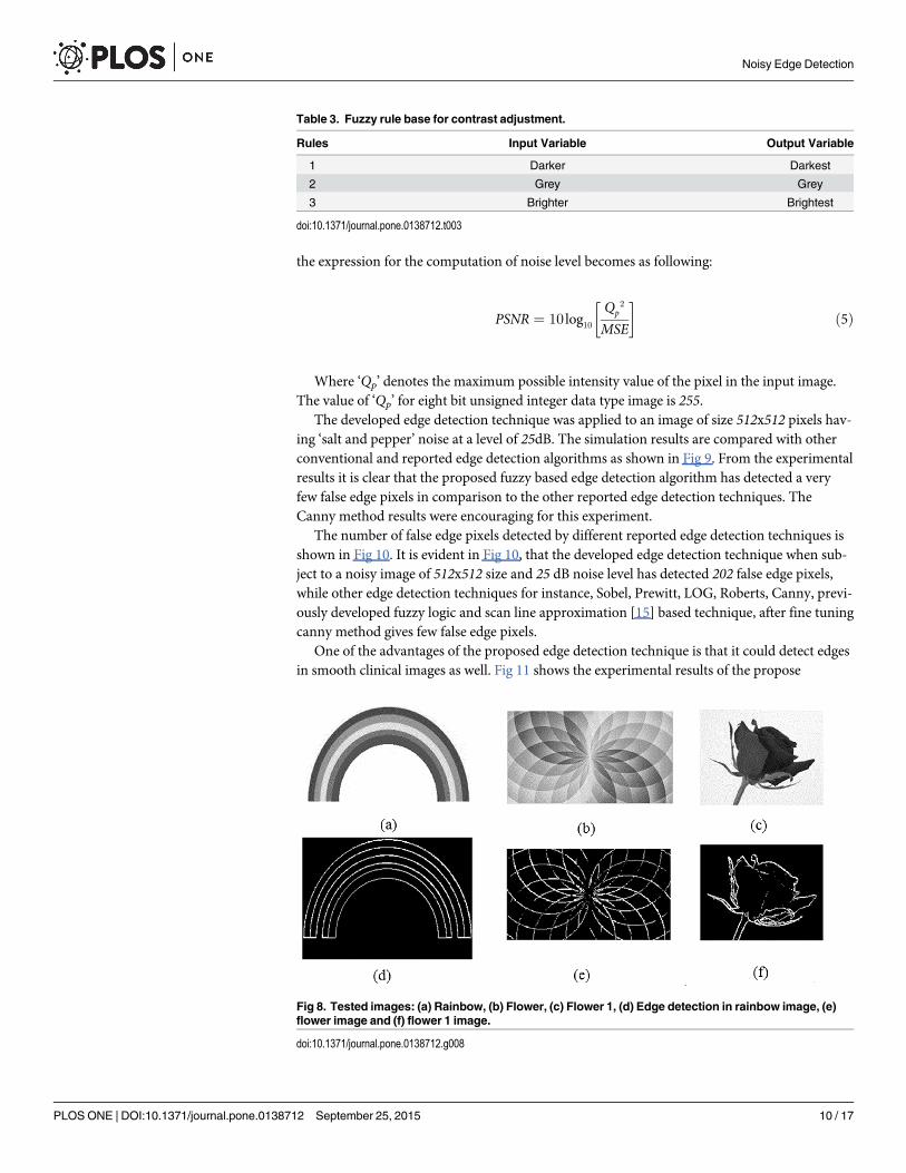
In noise free greyscale images, the developed technique has successfully detected all type ofedges as shown in Fig 8. The greyscale rainbow image of size 314x192 pixels having five differ-ent regions covered by six boundary lines is shown in Fig 8(A). The proposed technique foredge detection have detected these six boundary lines (edges) successfully as shown in Fig 8(D). Similarly, the proposed method has successfully detected edges in the greyscale (flower)images as shown in Fig 8E & 8F.
The developed edge detection technique has the advantage of detecting edges in the noisyimages as previously discussed (in the introduction). This was verified by detecting edges in animage having 25 dB ‘salt and pepper’ noise. To compute the noise level in an image through
Table 2. Fuzzy knowledge base for the developed edge detection technique.
Rules Input Variables Output Variable
ΔP1 ΔP2 ΔP3 ΔP4 ΔP5 ΔP6 ΔP7 ΔP8 P
1 Higher Higher None None None None None Lower Edge
2 Higher None None High None None None Lower Edge
3 None Higher Higher None None None None Lower Edge
4 None None None Higher None Higher None Lower Edge
5 Higher Higher None None None None Lower None Edge
6 Higher None None Higher None None Lower None Edge
7 None Higher Higher None None None Lower None Edge
8 None None None Higher None Higher Lower None Edge
9 Higher Higher None None Lower None None None Edge
10 Higher None None Higher Lower None None None Edge
11 None Higher Higher None Lower None None None Edge
12 None None None Higher Lower Higher None None Edge
doi:10.1371/journal.pone.0138712.t002
Noisy Edge Detection
PLOS ONE | DOI:10.1371/journal.pone.0138712 September 25, 2015 8 / 17

peak signal to noise ratio (PSNR) [33–34], the mean square error (MSE) was first computed as:
MSE ¼ 1
mn
Xn
v¼1
Xmu¼1
½G1ðu; vÞ � G2ðu; vÞ� ð4Þ
Where’ G1’ and ‘G2’ represents the input noise free and noisy images respectively. While 'm'and 'n' indicates the total number of rows and columns of the input images respectively. Finally
Fig 7. (a) MFs for the intensity value of input pixel (b) MFs for the intensity value of output pixel.
doi:10.1371/journal.pone.0138712.g007
Noisy Edge Detection
PLOS ONE | DOI:10.1371/journal.pone.0138712 September 25, 2015 9 / 17
the expression for the computation of noise level becomes as following:
PSNR ¼ 10 log10Qp
2
MSE
� �ð5Þ
Where ‘Qp’ denotes the maximum possible intensity value of the pixel in the input image.The value of ‘Qp’ for eight bit unsigned integer data type image is 255.
The developed edge detection technique was applied to an image of size 512x512 pixels hav-ing ‘salt and pepper’ noise at a level of 25dB. The simulation results are compared with otherconventional and reported edge detection algorithms as shown in Fig 9. From the experimentalresults it is clear that the proposed fuzzy based edge detection algorithm has detected a veryfew false edge pixels in comparison to the other reported edge detection techniques. TheCanny method results were encouraging for this experiment.
The number of false edge pixels detected by different reported edge detection techniques isshown in Fig 10. It is evident in Fig 10, that the developed edge detection technique when sub-ject to a noisy image of 512x512 size and 25 dB noise level has detected 202 false edge pixels,while other edge detection techniques for instance, Sobel, Prewitt, LOG, Roberts, Canny, previ-ously developed fuzzy logic and scan line approximation [15] based technique, after fine tuningcanny method gives few false edge pixels.
One of the advantages of the proposed edge detection technique is that it could detect edgesin smooth clinical images as well. Fig 11 shows the experimental results of the propose
Table 3. Fuzzy rule base for contrast adjustment.
Rules Input Variable Output Variable
1 Darker Darkest
2 Grey Grey
3 Brighter Brightest
doi:10.1371/journal.pone.0138712.t003
Fig 8. Tested images: (a) Rainbow, (b) Flower, (c) Flower 1, (d) Edge detection in rainbow image, (e)flower image and (f) flower 1 image.
doi:10.1371/journal.pone.0138712.g008
Noisy Edge Detection
PLOS ONE | DOI:10.1371/journal.pone.0138712 September 25, 2015 10 / 17

Fig 9. Comparison of experimental results in noisy image: (a) Original image, (b) Noisy image, (c)Sobel edge detection (d) Prewitt edge detection, (e) LoG edge detection, (f) Robert edge detection (g)Previously developed fuzzy based edge detection technique [22], (h) Canny edge detection and (i) Thedevelopedmethod. All the experimentation was performed on image b.
doi:10.1371/journal.pone.0138712.g009
Noisy Edge Detection
PLOS ONE | DOI:10.1371/journal.pone.0138712 September 25, 2015 11 / 17

algorithm when applied to smooth clinical MRI images. It is evident in Fig 11 that the devel-oped algorithm has successfully detected edges in the smooth clinical images.
Finally the developed edge detection technique was applied to the smooth clinical image ofsize 270x290 pixels having 24 dB ‘salt and pepper’ noise. The experimental results were com-pared with other conventional edge detection techniques like Sobel, Prewitt, LOG, Roberts,Canny and scan line approximation based technique [15] as shown in Fig 12.
It is clear from results that the developed technique shows excellent results compared to theestablished edge detection techniques. The number of false edge pixels detected by variousedge detection techniques, when subject to images having different PSNR values is shown inFig 13. It obvious that from Fig 13 that the number of false edge pixels detected by varioustechniques increases as we increase the noise level in the images. Furthermore, it is clear fromFig 13 that the developed technique when (subjected to image having 24 dB PSNR), hasdetected very few false edge pixels (22) in comparison to the other established edge detectiontechniques like Sobel (1931), Prewitt (2741), LOG (3102), Roberts (1451), Canny (1045) andscan line approximation based technique [15] (225). Further, Table 4 present statistical analysissuch as sensitivity and specificity of the proposed technique with Sobel, Canny and scan lineapproximation [15]. It is evident from the table that proposed technique has higher value forsensitivity and specificity among the previously established techniques. The proposed tech-nique has potential applications in many disciplines ranging from medical (MRI images, bonesdefects/cracks) to industrial (surface inspection, crack detection, rust detection) and in agricul-ture (identification of deforestation, crop yield production, identification of nutritionaldeficiencies).
Conclusion and Future WorkThis paper proposes and demonstrates a fuzzy logic based edge detection algorithm for smoothand noisy images. The developed technique employs a 3×3mask guided by fuzzy rule set foredge detection in noisy images. Furthermore, for smooth clinical images an extra mask of con-trast adjustment is integrated with the edge detection mask based on fuzzy logic to intensify thesmooth images. The developed technique has successfully detected all the edge pixels in noise
Fig 10. False edge detected pixels in a standard image of 512x512 pixels with 25 dB noise level: Acomparison.
doi:10.1371/journal.pone.0138712.g010
Noisy Edge Detection
PLOS ONE | DOI:10.1371/journal.pone.0138712 September 25, 2015 12 / 17

free, noisy and smooth images. The developed algorithm is also compared with other conven-tional and previously developed fuzzy logic based edge detection techniques. The developededge detection algorithm when subjected to a 512 x 512 size greyscale image having 25 dB ‘saltand pepper’ noise has detected very few false edge pixels (202), while the reported edge detec-tion techniques like Sobel, Prewitt, LOG, Roberts, Canny and previously developed fuzzy logichave detected 6673, 9395, 1241, 4792, 172 and 5362 respectively. When the developed techniquewas applied to a smooth clinical image of 270 x 290 size having 24 dB ‘salt and pepper’ noise, itdetected 22 false edge pixels, while the reported edge detection techniques like Sobel, Prewitt,LOG, Roberts and Canny have respectively detected 1931, 2741, 3102, 1451 and 1045 false edgepixels.It is obvious from the experimental results that in case of smooth and noisy images thedeveloped technique provides better results.
In future work, an investigation on how to incorporate Artificial Immune System andGenetic algorithm with fuzzy logic to develop a hybrid technique for edge detection is underconsideration.
Fig 11. The results of the developed edge detection technique: (a) Greyscale clinical image 1, (b)Greyscale clinical image 2, (c) Edge detection in clinical image1, and (d) Edge detection in clinicalimage 2.
doi:10.1371/journal.pone.0138712.g011
Noisy Edge Detection
PLOS ONE | DOI:10.1371/journal.pone.0138712 September 25, 2015 13 / 17

Noisy Edge Detection
PLOS ONE | DOI:10.1371/journal.pone.0138712 September 25, 2015 14 / 17

Author ContributionsConceived and designed the experiments: SA IH. Performed the experiments: IH SA. Analyzedthe data: SA MTK. Contributed reagents/materials/analysis tools: SAS KS. Wrote the paper: IHSA. Paper revision and addressing reviewers' comments: IH SA.
References1. Torre V, Poggio TA. On Edge Detection. Pattern Analysis and Machine Intelligence, IEEE Transactions
on. 1986; 8(2):147–163.
2. Bruno S, Lorenzo S, Luigi V, Giuseppe O. Robotics: Modelling, Planning and Control: Springer-VerlagLondon; 2008.
Fig 12. Comparison of the experimental results in noisy clinical image, (a) Original image, (b) Noisyimage, (c) Sobel edge detection (d) Prewitt edge detection, (e) LoG edge detection, (f) Robert edgedetection (g) Canny edge detection and (h) The developedmethod.
doi:10.1371/journal.pone.0138712.g012
Fig 13. False edge detection in a smooth clinical image of 270x290 pixels with 24 dB noise level.
doi:10.1371/journal.pone.0138712.g013
Table 4. Comparison for sensitivity and specificity
Sensitivity (%) Specificity (%)
Sobel 80 89
Canny 81 91
Scan Line [15] 87 94
Proposed Technique 89 96
doi:10.1371/journal.pone.0138712.t004
Noisy Edge Detection
PLOS ONE | DOI:10.1371/journal.pone.0138712 September 25, 2015 15 / 17

3. Chucherd S. Edge detection of medical image processing using vector field analysis. Computer Sci-ence and Software Engineering (JCSSE), 2014 11th International Joint Conference on; 2014 14–16May; Chon Buri, Thailand. IEEE. p. 58–63.
4. Lin H, Du P-j, Zhao C-s, Shu N. Edge detection method of remote sensing images based on mathemati-cal morphology of multi-structure elements. Chinese Geographical Science. 2004; 14(3):263–268.
5. Rulaningtyas R, Ain K. Edge detection for brain tumor pattern recognition. Instrumentation, Communi-cations, Information Technology, and Biomedical Engineering (ICICI-BME), 2009 International Confer-ence on; 2009 Nov 23–25; Bandung, Indonesia. IEEE. p. 1–3.
6. Goshtasby AA. 2-D and 3-D Image Registration: for Medical, Remote Sensing, and Industrial Applica-tions: Wiley-Interscience; 2005.
7. Loft A, Jensen KE, Lofggren J, Daugaard Sr, Petersen MM. PET/MRI for Preoperative Planning inPatients with Soft Tissue Sarcoma: A Technical Report of Two Patients. Case Reports in Medicine.2013; 2013 doi: 10.1155/2013/791078
8. Setayesh M, Mengjie Z, Johnston M. Effects of static and dynamic topologies in Particle Swarm Optimi-sation for edge detection in noisy images. Evolutionary Computation (CEC), 2012 IEEE Congress on;2012 June 10–15; Brisbane, Australia. IEEE. p. 1–8.
9. Canny J. A Computational Approach to Edge Detection. Pattern Analysis and Machine Intelligence,IEEE Transactions on. 1986; 8(6):679–698.
10. Zhang J-Y, Chen Y, Huang X-x. Edge detection of images based on improved Sobel operator andgenetic algorithms. Image Analysis and Signal Processing, 2009 IASP 2009 International Conferenceon; 2009 April 11–12; Taizhou, China. IEEE. p. 31–35.
11. Rosenfeld A. The Max Roberts Operator is a Hueckel-Type Edge Detector. Pattern Analysis andMachine Intelligence, IEEE Transactions on. 1981; 3(1):101–103.
12. Kirsch RA. Computer determination of the constituent structure of biological images. Computers andBiomedical Research. 1971; 4(3):315–328. PMID: 5562571
13. Lei Y, Dewei Z, XiaoyuW, Hui L, Jun Z. An improved Prewitt algorithm for edge detection based onnoised image. Image and Signal Processing (CISP), 2011 4th International Congress on; 2011 Oct.15–17; Shanghai, China. IEEE. p. 1197–1200.
14. Ulupinar F, Medioni Gr. Refining edges detected by a LoG operator. Computer Vision, Graphics, andImage Processing. 1990; 51(3):275–298.
15. Jiang X, Bunke H. Edge Detection in Range Images Based on Scan Line Approximation. ComputerVision and Image Understanding. 1999; 73(2):183–199.
16. Genming C, Baozong Y. A new edge detector with thinning and noise resisting abilities. Journal of Elec-tronics (China). 1989; 6(4):314–319.
17. Naumenko A, Lukin V, Egiazarian K. SAR-image edge detection using artificial neural network. Mathe-matical Methods in Electromagnetic Theory (MMET), 2012 International Conference on; 2012 Aug. 28–30; Kyiv, Ukraine. IEEE. p. 508–512.
18. Verma OP, Sharma R. An optimal edge detection using universal law of gravity and ant colony algo-rithm. Information and Communication Technologies (WICT), 2011World Congress on; 2011 Dec. 11–14; Mumbai, India. IEEE. p. 507–511.
19. Setayesh M, Zhang M, Johnston M. Effects of static and dynamic topologies in particle swarm optimisa-tion for edge detection in noisy images. Evolutionary Computation (CEC), 2012 IEEE Congress on;2012 June 10–15; Brisbane, Australia. IEEE. p. 1–8.
20. Yau-Hwang K, Chang-Shing L, Chao-Chin L. A new fuzzy edge detection method for image enhance-ment. Fuzzy Systems, 1997, Proceedings of the Sixth IEEE International Conference on; 1997 1997Jul 1–5; Barcelona, Spain. IEEE. p. 1069–1074.
21. El-Khamy SE, Lotfy M, El-Yamany N. A modified fuzzy Sobel edge detector. Radio Science Confer-ence, 2000 17th NRSC; 2000 Feb. 22–24; Minufiya, Egypt. IEEE. p. C32/1—C32/9.
22. Kim D-S, LeeW-H, Kweon I-S. Automatic edge detection using 3 x 3 ideal binary pixel patterns andfuzzy-based edge thresholding. Pattern Recognition Letters. 2004; 25(1):101–106.
23. Kaur EK, Mutenja V, Gill EIS. Fuzzy logic based image edge detection algorithm in MATLAB. Interna-tional Journal of Computer Applications. 2010; 1(22):55–58.
24. Chen SM, Chang YC, Pan JS. Fuzzy Rules Interpolation for Sparse Fuzzy Rule-Based Systems Basedon Interval Type-2 Gaussian Fuzzy Sets and Genetic Algorithms. Fuzzy Systems, IEEE Transactionson. 2013; 21(3):412–425.
25. Chia-Hung H, Chia-Feng J. Evolutionary Robot Wall-Following Control Using Type-2 Fuzzy ControllerWith Species-DE-Activated Continuous ACO. Fuzzy Systems, IEEE Transactions on. 2013; 21(1):100–112.
Noisy Edge Detection
PLOS ONE | DOI:10.1371/journal.pone.0138712 September 25, 2015 16 / 17

26. Zhou H, Ying H. A Method for Deriving the Analytical Structure of a Broad Class of Typical IntervalType-2 Mamdani Fuzzy Controllers. Fuzzy Systems, IEEE Transactions on. 2013; 21(3):447–458.
27. Monicka JG, Sekhar NG, Kumar KR. Performance evaluation of membership functions on fuzzy logiccontrolled ac voltage controller for speed control of induction motor drive. International Journal of Com-puter Applications. 2011; 13(5):8–12.
28. Botzheim J, Haimori B, Koczy LT. Extracting Trapezoidal Membership Functions of a Fuzzy Rule Sys-tem by Bacterial Algorithm. Computational Intelligence Theory and Applications. 2206: Springer BerlinHeidelberg; 2001. pp. 218–227.
29. Banerjee S, Roy TK. Arithmetic operations on generalized trapezoidal fuzzy number and its applica-tions. Turkish Journal of Fuzzy Systems. 2012; 3(1):16–44.
30. Reddy CS, Raju K. An improved fuzzy approach for COCOMO’s effort estimation using gaussian mem-bership function. Journal of Software. 2009; 4(5): p. 452–459.
31. SubbaramNaidu D. Soft computing and intelligent systems design: theory, tools and applications, F. O.Karry and C. De Silva, Pearson, Addison-Wesley, New York, NY, 2004. International Journal of Robustand Nonlinear Control. 2006; 16(11):548–551.
32. Leekwijck WV, Kerre EE. Defuzzification: criteria and classification. Fuzzy Sets and Systems. 1999;108(2):159–178.
33. Luisier F, Blu T, Unser M. Image denoising in mixed Poisson–Gaussian noise. Image Processing, IEEETransactions on. 2011; 20(3):696–708.
34. Liu J, Huan Z, Huang H. Image restoration under mixed noise using globally convex segmentation.Journal of Visual Communication and Image Representation. 2011; 22(3):263–270.
Noisy Edge Detection
PLOS ONE | DOI:10.1371/journal.pone.0138712 September 25, 2015 17 / 17


![Strong Key Derivation from Noisy Sources Benjamin Fuller December 12, 2014 Based on three works: Computational Fuzzy Extractors [FullerMengReyzin13] When](https://img.dokumen.tips/doc/110x75/5697bf8e1a28abf838c8cdad/strong-key-derivation-from-noisy-sources-benjamin-fuller-december-12-2014.jpg)