Embed Size (px)
Citation preview

Falk Symposium 161
Future Perspectivesin GastroenterologyOctober 11–12, 2007International Congress CenterDresden
AbstractsPoster Abstracts

© 2007 Falk Foundation e.VAll rights reserved.
Abstracts of Invited Lectures Poster Abstracts Falk Symposium 161 FUTURE PERSPECTIVES IN GASTROENTEROLOGY
Dresden (Germany) October 11–12, 2007 Scientific Organization: M.C. Carey, Boston (USA) P. Díte, Brno (Czech Republic) A. Gabryelewicz, Bialystok (Poland) V. Keim, Leipzig (Germany) J. Mössner, Leipzig (Germany)
3
CONTENTS Page
„Key Note Lecture“ in honor of Ernst Otto Riecken’s 75th birthday Introduction M. Zeitz, Berlin Immune abnormalities that cause inflammatory bowel diseases and biologic treatments that address these abnormalities W. Strober, Bethesda 17 – 19
Session I Inflammatory bowel diseases: Summary of the workshop
Chair: R.S. Blumberg, Boston M.F. Neurath, Mainz
Cytokine pathways in IBD – The yin and the yang C.O. Elson, Birmingham 23
New therapeutic strategies for chronic inflammatory bowel diseases – Summary of the workshop „Intestinal inflammatory mechanisms” A. Stallmach, Jena 24 – 25
Session II Esophagus and stomach
Chair: P. Malfertheiner, Magdeburg
Physiology of acid secretion E.M. El-Omar, Aberdeen 29 – 30
GERD – Endoluminal antireflux therapy K. Caca, Ludwigsburg 31 – 32
Functional dyspepsia H. Mönnikes, Berlin 33

4
Session III Lipoproteins and bile acids: Physiology and pathophysiology
Chair: F. Berr, Salzburg
Lipoprotein formation, structure and metabolism D.E. Cohen, Boston 37
Chaperoning organic anion transporters through the hepatocyte A.W. Wolkoff, Bronx 38
Pathophysiology of bile secretion M.C. Carey, Boston 39 – 40
Session IV Bile duct diseases
Chair: T. Sauerbruch, Bonn
Pathogenesis of gallstone formation F. Lammert, T. Sauerbruch, Bonn 43 – 44
Theories and techniques for resection of bile duct neoplasm – Suggestions from the perspective of surgical anatomy and pathology W. Kimura, Yamagata 45 – 47
Surgical options in biliary diseases P. Neuhaus, Berlin 48 – 49
Session V Pancreatic physiology and pathophysiology
Chair: J.C. Dagorn, Marseille
Mechanisms of pancreatic digestive enzyme secretion J.A. Williams, Ann Arbor 53
Genes and the pancreas H. Witt, Berlin 54 – 55
5
Acute phase reaction of the pancreas J.L. Iovanna, Marseille 56 – 57
Session VI Pancreatic diseases
Chair: P. Díte, Brno
Acute pancreatitis G. Adler, Ulm 61
Chronic pancreatitis D.C. Whitcomb, Pittsburgh 62
Gene expression analysis reveals the complexity of pancreatic disease C.D. Logsdon, Houston 63
„Key Note Lecture” Introduction M. Classen, Munich
The future of endoscopy in gastroenterology J.F. Riemann, L. Helmstädter, Ludwigshafen 64
Session VII Gastrointestinal oncology I
Chair: M.C. Carey, Boston
Gastrointestinal endocrine tumors B. Wiedenmann, Berlin 67
Gastrointestinal lymphoma W. Fischbach, Aschaffenburg 68 – 69
Gastric cancer M. Ebert, Munich 70
„Key Note Lecture” Introduction M.C. Carey, Boston
6
Relationship between exocrine and endocrine pancreas M. Otsuki, Kitakyushu 71
Session VIII Gastrointestinal oncology II
Chair: M.C. Carey, Boston
Pancreas R.M. Schmid, Munich 75 – 78
Hepatocellular carcinoma H.E. Blum, Freiburg 79
The role of the polyomavirus, JC virus, in the pathogenesis of colorectal cancer C.R. Boland, Dallas 80 – 81
„Key Note Lecture” Introduction A. Gabryelewicz, Bialystok
Gastroenterology in the tropics R.K. Tandon, New Delhi 82
List of Speakers, Moderators and Scientific Organizers 83 – 86
7
Poster Abstracts Esophagus and Stomach: Physiology and Diseases 1. Helicobacter pylori in the gastric mucosa of children may induce apoptosis of
the helper lymphocytes A. Kotlowska-Kmiec, M. Korzon, A. Bakowska, A. Szarszewski, B. Kaminska, W. Radys, P. Landowski, J. Brodzicki, A. Liberek (Gdansk, PL)
2. pH monitoring in chronic respiratory disorders in children
G. Lesanu, V. Hurduc, C. Becheanu, C. Zapucioiu, C. Calcic, D. Pacurar, R. Lesanu (Bucharest, RO)
3. Serum gastrin level in children with chronic gastritis with and without
Helicobacter pylori infection G. Luczak, M. Czaja, A. Szarszewski, E. Kozielska, A. Delinska-Galinska, K. Plata-Nazar, A. Borkowska, M. Korzon (Gdansk, Sztum, PL)
4. Hp infection in patients with gastric and duodenal ulcer and chronic liver
disease P. Mitrut, A. Genunche-Dumitrescu (Craiova, RO)
5. New derivatives of nitric oxide (NO)-releasing aspirin in the mechanism of
gastric mucosal defense, comparison with classic (aspirin) NSAIDS R. Pajdo, T. Brzozowski, D. Drozdowicz, A. Ptak-Belowska, S. Kwiecien, M.W. Pawlik, S.J. Konturek, W.W. Pawlik (Cracow, PL)
6. Beneficial effect of esophagoprotection by proton pump inhibitor (PPI) and
tumor necrosis factor α (TNF-α) inhibition in rat model of acute reflux esophagitis M.W. Pawlik, T. Brzozowski, P.C. Konturek, R. Pajdo, Z. Sliwowski, M. Mazurkiewicz, D. Drozdowicz, S.J. Konturek, W.W. Pawlik (Cracow, PL; Erlangen, D)
7. Esophageal and systemic diseases due to aging
L.M. Susan, C. Banciu, S.R. Gotia, V.M. Ancusa, I. Romosan, C. Gorun (Timisoara, RO)
Bile Ducts: Physiology and Diseases 8. Severe colitis following liver transplantation for primary sclerosing cholangitis
occurs mainly in young male adults with features of autoimmunity O. Buchel, F. Nevens, J. Decaestecker, G. Van Assche, R. Aerts, J. Pirenne, J. Fevery (Leuven, B)
9. Oxidative stress in extrahepatic cholestasis
K. Dagalp, C.N. Ercin, S. Bagci, Z. Yesilova, A. Aydin, A. Sayal, N. Karaeren (Ankara, TR)

8
10. Risk factors for gallstones in a patient population in Southern Transylvania M. Deac, R. Elefterescu, R. Mihaila, O. Petrascu, A. Deac, R. Mihaila (Sibiu, RO)
11. Malignancies developing in a cohort of 123 patients with primary sclerosing
cholangitis (PSC) during a median follow-up of 10 years J. Fevery, O. Buchel, G.N. Lai, C. Verslype, W. Van Steenbergen, F. Nevens (Leuven, B)
12. Gallstone pathogenesis: Reduced expression of OSTα and OSTβ as missing
link in ileal transporter function S. Harsch, O. Renner, S. Schimmel, A. Strohmeyer, E.-F. Stange (Stuttgart, D)
13. Pharmacological boost of gastric emptying fails to improve the 13CO2 yield of
the breath test with naturally 13C abundant corn groats K. Jonderko, D. Bergel, M. Kaminska, A. Kasicka-Jonderko, B. Blonska-Fajfrowska (Sosnowiec, PL)
14. Increase of the serum IgA and circulating immune complexes in the patients
with biliary diseases S.A. Kosilko, N.M. Kozlova, J.L. Turumin (Irkutsk, R)
15. Increase of the blood glutathione in the patients with biliary diseases
V.I. Kulinsky, N.M. Kozlova, J.L. Turumin, Z.A. Leonova (Irkutsk, R) 16. Gallstone pathogenesis: Sequence analysis of the ileal bile acid transporter
gene O. Renner, S. Harsch, E. Schäffeler, S. Schimmel, A. Strohmeyer, E.-F. Stange (Stuttgart, D)
17. The potential role of endoscopic ultrasound (EUS) elastography in defining
thickening of ductus choledochus in primary sclerosing cholangitis (PSC) N. Rustemovic, M. Opacic, S. Cukovic-Cavka, B. Vucelic (Zagreb, HR)
18. Cholestasis markers correlate with microhemorrheological disturbances
E.P. Sysoeva, E. Lukina, T. Solovjova, Z. Lichovetskaja, N. Khoroshko, A.I. Vorobjev (Moscow, R)
19. Clinical aspects of the effective pathogenetic treatment of patients with chronic
acalculous cholecystitis J.L. Turumin, N.M. Kozlova (Irkutsk, R)
20. Decrease of the portal blood flow in the patients with biliary diseases
H.E. Turumina, N.M. Kozlova, J.L. Turumin (Irkutsk, R) 21. Confirmation of the genes encoding the heterodimeric biliary cholesterol
transporter ABCG5/ABCG8 as gallstone susceptibility (Lith) genes in inbred mice H. Wittenburg, U. Kurtz, M.A. Lyons, D. Teupser, J. Mössner (Leipzig, D; Perth, AUS)

9
22. Structural and metabolic disturbances in rat brain histaminergic neurons under cholestasis and their correction with Ursofalk® S.M. Zimatkin, O.V. Baraban, S.V. Yemelyanchik (Grodno, WR)
Liver: Physiology and Diseases 23. Serological evidence for swine hepatitis E virus circulating in Taiwanese pig
herds K.-H. Chang, C.-H. Wang, S.-H. Lee (Taipei, TJ; Tübingen, D)
24. Azathioprine-induced allergic hepatitis associated with thiopurine
methyltransferase (TPMT) genotype – Case report S. Cukovic-Cavka, N. Bozina, Z. Krznaric, K. Grubelic-Ravic, M. Brinar, J. Sertic, B. Vucelic (Zagreb, HR)
25. Diabetes type 2 and chronic viral C hepatitis
D. Damian, M. Grigorescu, M.D. Grigorescu, T. Zaharie, T. Mocanu (Cluj-Napoca, RO)
26. Extrahepatic manifestations in chronic viral C hepatitis
D. Damian, M. Grigorescu (Cluj-Napoca, RO) 27. Perspectives of gastroenterological conservative and miniinvasive treatment of
liver echinococcosis K. Kalinova, A. Ilieva (Stara Zagora, Pleven, BG)
28. The efficacy or ursodeoxycholic acid in cholestasis of pregnancy (ICP) –
A review of RCTs trials E. Kuchar, L. Szenborn, T. Czerniak (Wroclaw, PL)
29. Genetic polymorphism of hemocoagulation factors in patients with prehepatic
portal hypertension E. Lukina, E.P. Sysojeva, E. Voronkova, E. Kitsenko, E. Lubivy, G. Sukhanova, S. Vasiljev (Moscow, R)
30. The prevalence of gallstones in chronic liver disease
D. Neagoe, G. Ianosi, T. Ciurea, C. Vere, M. Bezna, F. Racanel, A. Amzolini (Craiova, RO)
31. Prevalence and characterization of SEN virus strains isolated from Taiwanese
patients with elevated liver enzymes S.-Y. Tschen, S. Schleicher, S.-M. Lin, C.-C. Lee, A. Normann, C.-H. Wang, B. Flehmig (Tübingen, D; Taipei, TJ)
32. MicroRNA 122 is overexpressed in primary liver nodules arising in hepatitis C
H. Varnholt, M. Scheffler, E. Konze, A. Manav, V. Dries, F. Schulze, I. Wedemeyer, P. Schirmacher, U. Drebber, H.P. Dienes, M. Odenthal (Cologne, Mannheim, Heidelberg, D)

10
33. Acute hepatitis E virus infection in patients with cirrhosis is associated with high mortality A. Wang, M.-H. Lin, S.-Y. Tschen, C.-H. Wang (Tübingen, D; Taipei, TJ)
Exocrine Pancreas: Physiology and Diseases 34. Pressure activates pancreatic stellate cells via generation of intracellular reative
oxygen species H. Asaumi, S. Watanabe, M. Taguchi, M. Otsuki (Kitakyushu, J)
35. Pretreatment of the suckling rats with LPS up-regulated the Toll-like receptor 4
protein expression in the pancreatic acinar cells stimulated by caerulein J. Bonoir, J. Jaworek, A. Leja-Szpak, M. Kot, S.J. Konturek, W.W. Pawlik (Cracow, PL)
36. The gastrointestinal complications of severe necrotizing pancreatitis
G. Funariu, R. Seicean, V. Bintintan, N. Groza (Cluj-Napoca, RO) 37. Epidemiology of acute pancreatitis in a high prevalence of gallstone disease
and alcoholism country M. González, A. Menéndez (Santiago de Chile, RCH)
38. M470V is associated with the clinical course of hereditary pancreatitis
C. Hanck, S. Kassabian, M. Barmada, C. Beglinger, D.C. Whitcomb (Basel, CH; Pittsburgh, USA)
39. Insulin expression in Ins-1E cells is activated by the Alzheimer's disease beta-
secretase BACE1 by a mechanism involving p35 A. Hoffmeister, J. Tuennemann, J. Mössner, J. Kratzsch, T. Stahl, S. Roßner (Leipzig, D)
40. Involvement of serotoninergic nerves in the pancreatic enzyme secretion
stimulated by melatonin J. Jaworek, K. Nawrot-Porabka, A. Leja-Szpak, M. Kot, M. Mitis-Musiol, S.J. Konturek, W.W. Pawlik (Cracow, PL)
41. Endotoxemia in suckling rats modulates CD4+ and CD8+ T-lymphocyte
response to acute pancreatitis in the adult J. Jaworek, M. Macko, J. Szklarczyk, M. Szczepanik, M. Zemelka, K. Zajac, M. Kot, S.J. Konturek, W.W. Pawlik (Cracow, PL)
42. Expression of C-kit and insulin in human fetal pancreas
M.S. Kaligin, I.M. Gazizov, D.I. Andreeva, G.O. Pevnev, G.R. Burganova, A.A. Gumerova, A.P. Kiassov (Kazan, R)
43. A novel 13CO2 breath test with the use of corn oil
M. Kaminska, K. Jonderko, M. Bula-Mucha, A. Jankowski, A. Kasicka-Jonderko, B. Blonska-Fajfrowska (Sosnowiec, PL)
11
44. Effects of inhibitors of the renin-angiotensin-aldosterone system on fibrosis in chronic pancreatitis A. Madro, G. Czechowska, A. Korolczuk, M. Slomka, K. Celinski, B. Prozorow-Król, E. Korobowicz (Lublin, PL)
45. Chronic pancreatitis associated with abdominal trauma in children
G. Oracz, B. Oralewska, J. Ryzko, J. Socha (Warsaw, PL) 46. The effect of rosiglitazone, a specific PPAR-gamma ligand, on the development
of experimental sodium taurocholate induced acute pancreatitis B. Prozorow-Król, K. Celinski, A. Korolczuk, G. Czechowska, M. Slomka, A. Madro, E. Korobowicz (Lublin, PL)
47. Involvement of antioxidative enzyme in the pancreatic protection afforded by
bilateral vagotomy J. Szklarczyk, J. Jaworek, J. Bonior, M. Kot, R. Tomaszewska, S.J. Konturek, W.W. Pawlik (Cracow, PL)
48. Dual, time-dependent effect of anandamide administration in the severity of
acute experimental pancreatitis Z. Warzecha, P. Ceranowicz, A. Dembinski, W.W. Pawlik, R. Tomaszewska, J. Cieszkowski, P. Kownacki, P.C. Konturek (Cracow, PL; Erlangen, D)
49. Treatment with ghrelin reduces the severity and accelerates recovery in the
course of experimental acute pancreatitis Z. Warzecha, P. Ceranowicz, A. Dembinski, J. Cieszkowski, M. Dembinski, P. Kownacki, W.W. Pawlik, R. Tomaszewska, P.C. Konturek (Cracow, PL; Erlangen, D)
Small and Large Gut: Physiology and Diseases 50. Serological testing – Can they replace intestinal biopsy as the diagnostic test for
celiac disease? C. Becheanu, D. Pacurar, G. Lesanu (Bucharest, RO)
51. Pyogenic granuloma of the colon – Characteristic features and differential
diagnosis with Kaposi sarcoma and other vascular lesions G. Becheanu, M. Dumbrava, C. Gheorghe, M. Diculescu, M. Moschopulos (Bucharest, RO; Aarau, CH)
52. Endometriosis of colon and left ovary – The case of 46 years old patient
P.S. Bernaczyk, A. Wincewicz, P. Mysliwiec, J. Reszec, S.J. Terlikowski, L. Zimnoch, B. Kedra (Bialystok, PL)
53. Comparative study regarding the influence of stress in patients with functional
digestive disorders in general population E. Goldis, V. Lungu, C. Vernic (Timisoara, RO)

12
54. Tissue transglutaminase expression in duodenal epithelium of celiac disease patients J.V. Gorgun, A. Portyanko (Minsk, WR)
55. Long-term follow-up of lymphocytic colitis after induction of clinical remission
with budesonide A. Madisch, S. Wonschik, A. Morgner-Miehlke, E. Kuhlisch, B. Bethke, M. Stolte, S. Miehlke (Dresden, Bayreuth, D)
56. Effects of linseed oil on gastrointestinal mucosa in children
N.S. Marenich, J.G. Mukhina, A.S. Tertychnyy, A.G. Talalaev (Moscow, R) 57. Budesonide for treatment of lymphocytic colitis – A ramdomized, double-blind,
placebo-controlled trial S. Miehlke, A. Madisch, D. Karimi, S. Wonschik, R. Beckmann, E. Kuhlisch, A. Morgner-Miehlke, R. Müller, R. Greinwald, G. Baretton, G. Seitz, M. Stolte (Dresden, Bayreuth, Freiburg, Bamberg, D)
Inflammatory Bowel Diseases 58. Trans-signaling via the soluble IL-6 receptor suppresses tolerance by blocking
the induction of FoxP3 in naive T-cells C. Becker, S. Hübner, M.C. Fantini, C. Neufert, P.R. Galle, J. Scheller, G. Monteleone, S. Rose-John, M.F. Neurath (Mainz, Kiel, D; Rome, I)
59. Allelic variants of the multidrug resistance gene (MDR1/ABCB1) and response
to corticosteroid therapy in patients with ulcerative colitis M. Brinar, S. Cukovic-Cavka, N. Bozina, K. Grubelic-Ravic, Z. Krznaric, M. Rojnic Kuzman, J. Sertic, B. Vucelic (Zagreb, HR)
60. Prevalence of proline homozygosity at codon 72 of p53 in UC patients with ileal
pouch-anal anastomosis A. de Leone, M.T. Vietri, A. Improta, S. Giaquinto, M. Russo, M. Parisi, C. Tipaldi, M. Salvatelli, F. Selvaggi, A. Molinari, M. Cioffi, G. Riegler (Naples, I)
61. Inflammatory bowel diseases in infancy
E.S. Dublina, E.G. Tsimbalova, A.S. Potapov (Moscow, R) 62. Second incidence peak of ulcerative colitis (UC) depends on suspension of
smoking more than on acute myocardial infarction (AMI) S. Giaquinto, A. de Leone, C. Landolfi, L. Riegler, G. Riegler (Naples, I)
63. Innate defence and epithelial barrier dysfunction in ulcerative colitis (UC)
patients L.S. Gotia, S.R. Gotia, L.M. Susan, C. Fira-Mladinescu (Timisoara, RO)
64. Serum ferritin and its diagnostic significance in anemia of Crohn's disease
K. Grubelic-Ravic, S. Cukovic-Cavka, D. Radic, Z. Krznaric, M. Brinar, A. Ladic, B. Vucelic (Zagreb, HR)

13
65. Azathioprine induced hepatotoxiciy in IBD: Is it as rare as we think? H.E. Johnson, T.R. Smith, S.A. Weaver (Bournemouth, GB)
66. The diagnostic value of studying intestinal dysbacteriosis in patients with IBD
complicated by blastocystosis Y.Y. Krasnoperova, N.I. Potaturkina-Nesterova, I.S. Nemova, S.V. Semenov, M.G. Schegoleva, A.A. Larova, I.V. Razina, E.A. Zubkova, A.M. Lazarev, A.S. Nesterov (Ulyanovsk, R)
67. Satisfaction with healthcare in inflammatory bowel disease: Influence of patient
characteristics C. Luces, E. Brown, K. Bodger (Liverpool, GB)
68. Inflammatory bowel diseases in Romania – Are there any changes in an
ongoing westernization country? A. Lupu, M. Ciocirlan, M. Ciocirlan, L. Gheorghe, M. Diculescu (Bucharest, RO)
69. Infliximab improves metastatic Crohn's disease
A. Martinez Bistrain, A. Martínez Martínez, T. Navarrete Cruces, L. Charua Guindic, R.M. Osorio Hernandez, B. Jimenez Bobadilla, S. Camacho (Mexico City, MEX)
70. A micro-study regarding levels of access to information and services by patients
with inflammatory bowel disease of South Asian origin T. Moshkovska, J.F. Mayberry (Leicester, GB)
71. Interleukin 27 controls the development of Th-17 and iTreg cells via differential
effects on the transcription factor STAT1 C. Neufert, C. Becker, S. Wirtz, M.C. Fantini, B. Weigmann, M.F. Neurath (Mainz, D; Rome, I)
72. Traits of clinical presentation of new diagnosed IBD colitis and their changing
over the years M. Shapiro, R. Simantov, E. Scapa (Rehovot, IL)
73. Vascular endothelial growth factor (VEGF) in patients with ulcerative colitis
A. Stadnicki, D. Frysz-Naglak, E. Klimacka-Nawrot, I. Stadnicka (Sosnowiec, Katowice, PL)
74. Polyphenolic compounds of plant origin protect from murine colitis
S. Westphal, W. Domschke, T. Kucharzik (Münster, D) Gastrointestinal Oncology 75. Reprimo as a potential biomarker for early detection of sporadic diffuse-type
gastric carcinoma in plasma E. Aravena, C. Bernal, M. Vargas, C. Villarroel, F. Ossandon, E. Santibañez, I. Diaz, C. Barrientos, M. Palma, R. Estela, A. Corvalan (Santiago de Chile, RCH)

14
76. Ursodeoxycholic acid mediated cell cycle arrest of adenocarcinoma cells likely operates through induction of NAG-1 F. Bresso, E. Edlundh-Rose, M. D’Amato, A. Are, G. Greicius, A. Liedén, U. Sjöqvist, J. Lundeberg, V. Arulampalam, R. Löfberg, S. Pettersson (Stockholm, S)
77. Immunohistochemical evaluation of Ki-67, PCNA and MCM2 protein in patients
with advanced gastric cancer J. Czyzewska, K. Guzinska-Ustymowicz, A. Kemona, A. Pryczynicz, H. Kemona, R. Bandurski, M. Gryko, B. Kedra (Bialystok, PL)
78. A study concerning the incidence of gastrointestinal cancer in several regions of
Romania O. Fira-Mladinescu, C. Fira-Mladinescu, S. Doroftei, F. Sas, L.S. Gotia, D. Muntean, B. Vlaicu (Timisoara, RO)
79. Multipolar radiofrequency ablation in the treatment of hepatic tumors
J. Genov, N. Grigorov, R. Mitova, B. Golemanov, L. Dinkov, M. Donov (Sofia, BG)
80. The risk of development of gastric cancer in patients with precancerous
changes after Helicobacter pylori eradication A. Genunche-Dumitrescu, P. Mitrut, D. Badea, M. Badea (Craiova, RO)
81. On the basis of its expression in primary hepatocellular carcinoma, sperm
protein 17 is a potential immunotherapeutic target F. Grizzi, G. Ceva-Grimaldi, B. Franceschini, S. Di Biccari, E.E. Frezza, E. Cobos, N. Dioguardi, M. Chiriva-Internati (Rozzano, I; Lubbock, USA)
82. MCM-2 protein expression predict prognosis of G2pT3 colorectal cancer
K. Guzinska-Ustymowicz, A. Pryczynicz, E. Stepien, J. Czyzewska, A. Kemona (Bialystok, PL)
83. The expression of connexins 26, 32 and 43 as well as adhesion proteins –
E-cadherin and beta-catenin in colorectal adenomas and adenocarcinomas L. Kanczuga-Koda, M. Sulkowska, M. Koda, S. Sulkowski (Bialystok, PL)
84. Relationships between the obesity hormone leptin, its receptor and hypoxia-
inducible factor-1alpha in human colorectal cancer M. Koda, M. Sulkowska, A. Wincewicz, L. Kanczuga-Koda, S. Cascio, G. Colucci, A. Russo, E. Surmacz, S. Sulkowski (Bialystok, PL; Philadelphia, USA; Bari, Palermo, I)
85. Hepatocellular carcinoma GR. I. and multifocal nodular steatosis
V. Kupcová, E. Seligová, M. Szántová, L. Turecky, M. Smutny (Bratislava, SK) 86. Melatonin increases the phosphorylation of heat shock protein 27 in pancreatic
carcinoma PANC-1 cells A. Leja-Szpak, J. Jaworek, J. Szklarczyk, J. Bonior, S.J. Konturek, W.W. Pawlik (Cracow, PL)

15
87. Decrease of intrahepatic innate lymphocytes (NK and NKT cells) in patients with liver metastasis I.M. Manolova, M.V. Gulubova, D. Kyurkchiev, I.P. Altunkova, G. Cirovski, A. Julianov, J. Jovchev (Stara Zagora, Sofia, BG)
88. Management of early gastric cancer in a Latinoamerican hospital
A. Menendez, E. Aravena, M. Gonzalez, C. Barrientos, A. Sandoval, M. Palma, H. Iturriaga (Santiago de Chile, RCH)
89. Experimental studies in vivo regarding the effect of resveratrol extracted from
vine on gastric neoplastic injuries P. Mitrut, F. Burada, I. Rogoz, A. Genunche-Dumitrescu (Craiova, RO)
90. Anemia in the patients with digestive cancers – An alert signal or an expression
of an advanced neoplasm of the digestive tube? R. Mihaila, C.E. Rezi, L. Tiurean, M. Deac, R. Mihaila (Sibiu, RO)
91. Tissue transglutaminase could participate in ECM remodeling in invasive front
in colorectal cancer A. Portyanko, J.V. Gorgun, P. Kovalev (Minsk, WR)
92. Ileal mucosa changes after total colectomy in patients with juvenile polyposis
and familial adenomatous polyposis A.S. Tertychnyy, D.M. Konovalov, A.G. Talalaev (Moscow, R)
93. Molecular and immunohistochemical analysis in HNPCC diagnosis
D. Verdes, R. Popescu, S.R. Gotia, D. Puscasiu (Timisoara, RO) 94. May the GST-pi expression and Ile105VAL GSTP1 gene polymorphism predict
the colorectal cancer outcome after adjuvant chemotherapy? T. Vlaykova, M.V. Gulubova, D. Vlaykova, I.M. Manolova, G. Cirovski, P. Chilingirov (Stara Zagora, BG)
95. Preoperative serum protein levels of EPO and their comparison to VEGF and
VE-cadherin in human colorectal cancers A. Wincewicz, A. Chabowska, M. Sulkowska, A. Chabowski, M. Mysliwiec, K. Pawlak, M. Koda, B. Kedra, S. Sulkowski (Bialystok, PL)
Endoscopy 96. Treatment of pyloric stenosis by intrapyloric injection of botulinum toxin –
Pilot study C. Banciu, O. Chirileanu, L. Marian, A. Pacurari, I. Romosan (Timisoara, RO)
97. Detection of small polyps by cap fitted colonoscopy compared with standard
colonoscopy C. Banciu, O. Chirileanu, L. Marian, A. Pacurari, I. Romosan (Timisoara, RO)

16
98. Open, retrospective investigation to assess efficacy and tolerability of bisacodyl and Endofalk® combined administration J. Preiss, T. Plassmann, G. Dunkhase von Hinckeldey (Herne, Planegg, D)
99. Risk factors for intra- and retroperitoneal perforations in patients after
therapeutic ERCP M. Shapiro, L. Kopel, D. Abramovith, M. Yair, E. Broide, E. Scapa (Rehovot, IL)
100. Bispectral monitoring during endoscopic retrograde cholangiopancreatography
S. Shetty, T. Valliani, I. Bernova, J. Shah, A. Srivastava, R. Przemioslo (Bristol, GB)

17
Immune abnormalities that cause inflammatory bowel diseases and biologic treatments that address these abnormalities Warren Strober Mucosal Immunity Section, Laboratory of Host Defenses, NIAID, NIH, Bethesda, MD 20892, USA The most inclusive working hypothesis concerning the pathogenesis of the inflammatory bowel diseases (Crohn’s disease and ulcerative colitis) is that these diseases are due to dysregulated immune responses to commensal micro-organisms in the bowel lumen. This dysregulation may be due to an excessive effector T cell response to such organisms resulting from a disorder of the innate immune system and/or a disorder of the adaptive immune system; alternatively, it may be due to an inadequate regulatory T cell response that cannot contain a normal effector T cell response. An additional pathogenic factor is the possible role of the epithelial cells in these inflammations. Epithelial cell dysfunction may abet the disordered effector/-regulatory immune response by failure to maintain barrier function and thereby promote abnormal exposure of immune elements to commensal organisms, or by producing an abnormal array of chemokines and cytokines with the potential to initiate and maintain the mucosal immune response. These abnormalities are likely to arise from a number of genetic (and environmental) factors that operate in tandem to produce disease. A simplifying factor, however, is that regardless of which of these factors (or combination of these factors) is present, the inflammation they produce is “channeled” into a Th1/Th17 cytokine pathway characteristic of Crohn’s disease, or into a modified Th2 cytokine pathway characteristic of ulcerative colitis. Thus, the complexity of the set of underlying defects in these diseases resolves itself into one of two stereotypic immune responses that lend themselves to treatment approaches that can be applied to most, if not all, patients in the two disease subcategories. The Th1/Th17 driven-pathway of Crohn’s disease is a bipartite pathway characterized by dendritic cells that produce IL-12 comprised of a p40 chain linked to a p35 chain, as well as dendritic cells that produce IL-23 comprised of the same p40 chain linked in this case to a p19 chain. IL-12 induces the differentiation of Th1 T cells producing IFN-γ and TNF-α whereas IL-23 supports the differentiation of Th17 T cells producing IL-17 and IL-22 (as well as TNF-α). In some murine models of gut inflammation IL-12/IFN-γ responses are predominant whereas in others IL-23/IL-17 responses are predominant. However, in human Crohn’s disease both types of responses are present and one possibility is that the Th1 response mediates the acute phase of inflammation whereas the Th17 response mediates the chronic phase of inflammation and that in human disease both phases of inflammation occur simultaneously. In any case, anti-cytokine treatment with an anti-IL-12p40 antibody addresses both the Th1 and the Th17 responses and has already proven to have efficacy in preliminary human trials; this antibody thus emerges as a major new therapy for Crohn’s disease. Whether and antibody that addresses only the Th17 response (an anti-p19 antibody) will have the same level of efficacy (and perhaps less detrimental effects on host defense) is not yet known. Since both the Th1 and Th17 response give rise to the production of

18
TNF-α and this cytokine is so intrinsic to inflammation in both responses, it is no wonder that treatment of patients with anti-TNF-α has proven to be quite successful in a substantial number of patients; nevertheless, at least half of patients are either initially resistant to this therapy or become resistant after a short period of time, indicating that new agents are clearly needed. One property shared by agents that target both IL-12 and TNF-α is there ability to cause apoptosis of effector cells. This is important because by causing the destruction of cells responsible for disease manifestation, rather than blocking their activity, one can achieve much more prolonged therapeutic effects. Another agent that has this property and has shown some efficacy in the treatment of patients is an antibody directed against the IL-6 receptor that blocks IL-6 signaling mediated by IL-6/IL-6R “trans-sigaling. Ulcerative colitis was, for a long time, difficult to locate within the Th1/Th2 universe of inflammations. This resulted from the fact that neither IFN-γ nor IL-4, the paradigmatic cytokines of Th1/Th2 driven processes (respectively) was found to over-produced in this disease. More recently, however, it has become apparent that specialized T cells, known as NKT cells (T cells bearing a T cell receptor, yet also bearing markers associated with NK cells) are present in ulcerative colitis lesions and that such cell produce IL-13, the Th2-type cytokine that is now thought characterize this disease. Excessive IL-13 production in ulcerative colitis can lead to the epithelial cell pathology (and ulceration) typically found in this disease by its direct untoward effects on epithelial cells and by its capacity to activate NKT cells with the capacity to kill epithelial cell targets. This cytokine abnormality of ulcerative colitis strongly suggests that agents that block IL-13 activity, such as anti-IL-13 antibodies would have a strong therapeutic effect, but this remains to be tested. In addition, agents that act more proximal to the disordered IL-13 response by blocking the cytokines driving the generation of IL-13-producing NKT cells may also be efficacious. Anti-TNF-α has shown only limited success in ulcerative colitis because TNF-α plays a relatively minor role in this inflammation. It becomes obvious from the discussion above that a rational “biologic” approach to the treatment of the inflammatory bowel diseases is most solidly based on the nature of the final common pathway of inflammation present: the Th1/Th17 pathway in Crohn’s disease and the modified Th2 pathway in ulcerative colitis. Nevertheless other approaches have been advances and may have considerable validity. One is based on the recognition that gut inflammation invariably requires the traffic of cells through the lymph and the blood from “inductive” sites of initial cell development to “’effector” sites of actual inflammation. This opens the door to the possibility that agents (antibodies and other modalities) that interfere with such traffic can prevent inflammation. One such antibody (anti-α4 integrin antibody) has had some success in clinical trials but has been associated with a propensity to cause infectious side effects; however, other agents in this category may prove more useful. Another such alternative approach is to target basic mechanisms of inflammation that underlie Th1/Th17 responses and/or Th2-like responses. This can include agents that down-regulate key intra-cellular signaling molecules intrinsic to these responses or agents that inhibit basis inflammatory modules such as the NF-κB complex. An example of the latter is possible treatment with “decoy” oligonucleotides that block transciption by p65 and c-Rel, major NF-κB transcription factors. This has proven to be efficacious in the treatment of animal models of gut inflammation, but has yet to be tried in humans with disease and obviously poses considerable risk of side effect unless directly

19
applied to an area of inflammation rather than as a systemic agent. Agents that down-regulate T cell activation such as anti-IL-2Rα also falls into this category of general agents as does antibodies that attack T cells generally such as anti-CD3. Obviously, the more general the effect of the therapeutic agent on the immune response the more likely it is to compromise host defense and introduce problems with infectious complications. Yet another biologic approach to therapy of inflammatory bowel disease is based on the possibility alluded to above that the disease is due to inadequate regulation of the mucosal immune response by regulatory T cells. Here the aim of therapy is to augment the regulatory T cell response and to overcome the effector T cell response whether or not the adequacy of regulatory T cell response is the prime cause of the disease. One example of this approach is the treatment of patients with G-CSF, an agent that may augment the activity of dendritic cells that induce regulatory T cells. Another example is an “exotic” therapy known as extra-corporeal photopheresis in which patient cells are exposed to UV irradiation while passing through external lymphapheresis tubes. It is based on the hypothesis that such treatment leads to cellular apoptosis and the release of cellular factors that stimulate regulatory T cell proliferation. In summary, the increased understanding of the inflammatory processes operative in the inflammatory bowel diseases has opened the field to vast new possibilities in the treatment of these diseases. The community of IBD scientists and clinicians are now poised to accomplish the vital work of establishing which of the new treatments will prove most beneficial.

21
Session I
Inflammatory bowel diseases: Summary of the workshop

23
Cytokine pathways in IBD – The yin and the yang Charles O. Elson, M.D. University of Alabama at Birmingham, Birmingham, AL 35294, USA Inflammatory bowel disease is multifactorial and involves innate immune, adaptive immune, and regulatory components (Elson, Immunol. Rev. 2005). Recent whole genome association studies have implicated a number of genes in IBD susceptibility, many of which are innate immune genes that are likely involved in host interactions with the microbiota. Defects in these innate immune mechanisms may then lead to abnormal adaptive and/or regulatory immune responses to the microbiota. One of the genes recently linked to IBD is the IL-23 receptor. IL-23 is a cytokine produced by innate cells that acts on both innate and adaptive immune cells. To place this finding in perspective, it is useful to consider different cytokine pathways that are involved in induction of IBD or its prevention. IL-23 is a member of the IL-12 family of heterodimeric cytokines. Recent work has identified an IL-23-dependent lineage of effector T cells that produce the signature cytokine, IL-17 (Th17) cells. Th17 cells also produce other cytokines, including TNFα and IL-22 and these cells have been implicated in multiple experimental models of chronic inflammation, including experimental colitis. Th17 cells are induced by a combination of TGFβ and IL-6, which stimulates a transcription factor, RORgt, that induces differentiation along the Th17 pathway. Stimulation of naive T cells with TGFβ alone induces the regulatory factor Foxp3 that differentiates the cells along a regulatory pathway. RORgt and Foxp3 appear to be mutually inhibitory and these transcription factors might be future targets for therapy. Neutralization of IL-23 in mice was able to reverse an active T cell-mediated colitis. In a similar fashion a monoclonal antibody to IL-12p40 (i. e., both IL-23 and IL-12) was effective in treating chronic active Crohn's disease. Although Th1 and Th17 pathways appear to play a role in Crohn's disease, the situation is less clear in ulcerative colitis. The current working hypothesis is that ulcerative colitis may be a Th2-like disorder with local overproduction of IL-13 by NK-T cells, possibly mediated through deleterious effects of IL-13 on the epithelial barrier. If these effector cytokine pathways represent the yin of IBD, then the yang is represented by regulatory cells. The microbiota induces a substantial number of Tregs in the intestinal lamina propria of normal hosts. There are multiple subsets of these Tregs but two common cytokines involved in their function are IL-10 and TGFβ. IL-10-deficient mice develop colitis, but interestingly such colitis does not occur in the absence of the IL-23 gene. TGFβ effects are mediated through phosphorylation of a series of transcription factors known as SMADs. One of the interesting conundrums is that TGFβ levels are elevated in IBD but inflammatory cells seem resistant to TGFβ inhibition. This has now been shown to be due to the presence of elevated levels of an inhibitor of the TGFβ pathway, SMAD7. Reduction of SMAD7 levels in inflamed gut allows TGFβ inhibition to be exerted and inflammation reduced. The key cytokine pathways involved in induction and prevention of IBD are emerging. In normal hosts these pathways remain in balance. The challenge is to define what upsets this balance and how we can restore it to the benefit of our patients.

24
New therapeutic strategies for chronic inflammatory bowel diseases – Summary of the workshop “Intestinal inflammatory mechanisms” Andreas Stallmach Department of Gastroenterology, Hepatology and Infectiology, Friedrich Schiller University, Jena, Germany Our knowledge of the pathogenesis of chronic inflammatory bowel diseases has changed considerably over the last few years. The results of many studies have shown that in genetically disposed patients the migration of luminal bacteria or their remains into the gut mucosa through a damaged (primary) mucosal barrier is of great importance. Affected patients show an increase in the gut mucosa of active immune cells with a high proliferation rate and which have hampered apoptosis. These immune cells are overactive in producing pro-inflammatory cytokines and chemokines while the production of anti-inflammatory mediators is reduced. These processes look likely to be responsible for the persistent character of the inflammation. This has led to a shift of the pathophysiological concept from a primarily immunologically determined illness to a condition with a damaged mucosal barrier. The improvement in our understanding of the pathogenesis will hopefully lead to new causal therapeutic strategies. Internationally renowed speakers will discuss the most recent data and ideas relating to the pathogenesis of intestinal inflammation during the workshop on October 9 and 10. The main topics are genetics, pathology of the cytokine response, intestinal microflora, epithelial barrier defects and regulatory defects. What will we learn from this discussion to help us develop new therapies for patients with Crohn’s disease and ulcerative colitis? What are the limits and the deficits of current therapies? The following problems must be addressed: 1. The acute therapy for patients with Crohn’s disease or ulcerative colitis suffering
from mild to moderate activity is already standardised. The therapeutic options for patients with severe activity are however still limited and no generally accepted strategy exist. It is unclear whether blocking a single inflammatory process is enough to reduce the activity substantially (e. g. by selectively blocking single mediators) or whether a substance is necessary which has a wider anti-inflammatory effect such as “super steroids”.
2. Patients with chronic active disease particularly have benefited from new
therapies using antibodies against tumor necrosis factor-α. This anti-TNF therapy, which is based on pathogenetical knowledge, has become standard following large clinical trials to test its capabilities. However, it must be noted that not all patients respond to this therapy, until now no parameters have been established that can predict whether an individual patient will benefit.

25
3. Antibodies against recently discovered pro-inflammatory cytokines (IL-18, representatives of the IL-12 family) and against receptors (α4β7-integrins) are currently being tested in clinical trials. It is not known whether these substances improve disease activity in patients who have not responded to an anti-TNF-α therapy.
4. An intensive immuno-suppressive or immuno-modulating therapy can potentially
cause side-effects. Early treatment is justified if it can improve the course of the disease in a patient with a problematic prognosis. Hopefully the improved under-standing of the pathogenesis will bring better knowledge of the sub-population of patients with bad prognoses and suggest the optimal time for intervention (“hit early”).
5. The prophylaxis against recurrence in ulcerative colitis is well-established;
however there is no prophylaxis against Crohn’s disease recurrence effective for the majority of patients. This major problem must be solved urgently.
6. During the course of Crohn’s disease so-called penetrative complications
(i. e. perforations and fistel formation) become increasingly common. Only an improved understanding of the pathogenesis of these complications can lead to better therapeutic strategies.
In conclusion we hope that an improvement in our understanding of the pathogenesis will lead to an answer to the question “which patients respond to which therapy at what point and how early do we have to intensify our treatment?”

27
Session II Esophagus and stomach

29
Physiology of acid secretion Professor Emad M. El-Omar Department of Medicine & Therapeutics, Institute of Medical Sciences, Aberdeen University, Aberdeen, U.K. Gastric acid secretion represents an important non-immunological first line of defence against ingested microbes. Almost all vertebrates produce acid in their upper gastrointestinal tracts suggesting that this physiological process has a fundamental survival advantage. The ability to produce acid allowed vertebrates to ingest more complex diets, but more importantly, it protected them against microbes that gained access through the gastrointestinal tract. The regulation of gastric acid secretion is a finely controlled process dependent on overlapping neural, hormonal and paracrine pathways. The human stomach has an intricate submucosal plexus that secretes a variety of transmitters including nitric oxide, vasoactive intestinal peptide (VIP), gastrin-releasing peptide, substance P, and calcitonin gene-related peptide. The gastric mucosa contains specialised cells that secrete among other things, gastrin (from G cells), somatostatin (from D cells), histamine (from ECL cells), hydrochloric acid and intrinsic factor (from parietal cells), pepsinogens (from chief cells), mucin (from surface and mucous neck cells), Ghrelin and leptin. Regulation of acid secretion is determined by a variety of central (e. g. neuropeptide Y, corticotropin-releasing factor, and neuromedin U) and peripheral (e. g. gastrin, histamine, acetylcholine, somatostatin, cholecystokinin, calcitonin gene-related peptide, leptin) pathways. The fine interaction between these pathways ensures that acid and pepsin do not overwhelm the mucosal defences and lead to injury and ulceration. The binding of secretagogues to parietal cells generates changes in second messengers that regulate the translocation and activation of the proton pump, H+/K+ ATPase. The H+/K+ ATPase is an ion-motive ATPase that belongs to the P2 subfamily of ATPases and consists of alpha and beta subunits. It forms an integral part of the apical membranes of parietal cells. It transports its cations as a cycle of phosphorylation and dephosphorylation of the transport protein. When the parietal cells are stimulated the pumps are translocated from the cytoplasmic tubulovesicles into the membrane of the secretory canaliculus, to form microvilli lining the canalicular space, and a KCl pathway is activated to allow K+ to access the external surface of the ATPase and secretion of Cl-. The H+/K+ ATPase actually pumps hydronium ions (H3O+) rather than H+ ions and is able to release H3O+ at a concentration of 160 mM, which is equivalent to an external pH of 0.8 and a 4 million-fold gradient across the surface of the parietal cell. The H+/K+ ATPase pumps are involved in the final stages of gastric acid secretion and are stimulated to secrete acid by intracellular signals from H2-receptors, muscarinic M3 receptors and gastrin receptors on parietal cells. Perhaps the most important determinant of gastric acid secretion is the presence or absence of H. pylori infection. The effect of H. pylori infection on gastric acid secretion depends on the severity and distribution of gastritis. Antral inflammation is associated with increased production of gastrin, which in turn increases the drive for acid secretion by parietal cells in the gastric corpus. Subjects with an antral predominant pattern of gastritis are at increased risk of developing duodenal ulcer disease. If the corpus is affected by severe inflammation, the host’s ability to respond to the increased gastrin is greatly attenuated. Thus, subjects with severe corpus inflammation have reduced capacity to secrete acid, and this may initially be due to

30
functional inhibition of the parietal cells by either products of H. pylori itself, or more likely products of the inflammatory process. In time and with sustained chronic inflammatory injury, the gastric glands atrophy and it is debatable whether loss of acid secretion in this context ever recovers. Permanent loss of parietal cells, resulting in achlorhydria, is a very strong risk factor for gastric cancer, especially if combined with sustained chronic inflammatory activity. H. pylori can damage the gastric epithelial layer through direct and indirect mechanisms. There is evidence that H. pylori infection exerts inhibitory effects on human gastric H+/K+ ATPase α-subunit. The effect of eradication of H. pylori on gastric acid secretion depends entirely on the extent to which the corpus mucosa is affected by the inflammatory activity. Thus, if the corpus is relatively healthy with an antral predominant pattern of gastritis, the net effect of eradication will be a reduction in the degree of hypergastrinaemia, which in turn may lead to a reduction in acid secretion. If on the other hand the corpus is severely inflamed, eradication will remove at least the functional inhibition of the parietal cells and this will be associated with an increase in acid secretion. All these subtle changes have an impact on the health of the stomach and ultimately on the risk of other GI conditions including oesophagitis. This highlights the importance of knowledge about gastric acid secretion and what factors influence it.

31
GERD – Endoluminal antireflux therapy Prof. Dr. med. K. Caca Klinikum Ludwigsburg, Gemany Heartburn, the most common symptom associated with GERD, occurs in 10–20% of the US and western European population. Lifelong PPI therapy for GERD is often required since symptoms typically recur once therapy has been discontinued. Moreover, a number of patients on PPI-treatment will experience side effects or symptomatic non-acidic regurgitation. Several endoscopic antireflux procedures have been developed during the last ten years. Besides endoscopic suturing, these techniques include the application of radiofrequency and the injection or implantation of biopolymers. Radiofrequency treatment (StrettaTM) has proved modestly effective, but was recently withdrawn from the market. Implantation techniques have been abandoned due to lack of long-term efficacy (GatekeeperTM) or serious side effects (EnteryxTM). While first generation endoluminal suturing techniques (EndoCinchTM, ESDTM) demonstrated a proof of principle, their lack of durability due to suture loss led to the development of a durable full-thickness transmural plication technique (PlicatorTM). The Plicator device (NDO Surgical, Inc., Mansfield, MA) enables transmural suturing near the gastroesophageal junction under direct endoscopic visualization. In a prospective randomized, shamcontrolled trial, the Plicator procedure proved effective at controlling reflux symptoms and reducing GERD medication use and esophageal acid exposure (24 h pH-metry). In all studies to date, a single Plicator implant was placed to form a full thickness plication of the anterior gastric cardia. However, in several cases the narrowing of the GE-junction remained incomplete with this technique. Therefore, endoscopic full-thickness plication studies were done using two serially placed Plicator implants. In a single-center pilot study of 37 patients with a 6 months follow-up, the proportion of patients achieving 50% improvement in GERD-HRQL score was 68%. Complete PPI cessation was achieved in 59% of patients. In pH studies conducted at 6-months (n = 29), median percent time pH < 4 decreased 36%, with 28% of patients experiencing pH normalization. ERD was diagnosed in 21 patients before endoscopic full-thickness plication. 16 patients had NERD before endoscopic full-thickness plication. Six month after endoscopic full-thickness plication the number of subjects with ERD was 15 and the number of subjects without esophagitis was 22. There were no serious adverse events observed. Endoscopic full-thickness plication using two serially placed Plicator implants was both safe and effective in reducing esophagitis, GERD symptoms, medication use, and esophageal acid exposure. In addition an endoluminal technique mimicking endoluminal fundoplication by placing transesophageal fasteners using a trans-oral and fastener-deploying device (EsophyXTM, EndoGastric Solutions) was introduced recently. Preliminary results of serial bench, animal and human (phase 1, phase 2, commercial registry) studies show biological compatibility, short-term durability and non-toxicity of the polypropylene fasteners as well as the feasibility of the ELF technique. However, additional data from a multicenter trial and long term data have to be awaited before any conclusion can be made regarding efficacy and durability.

32
In conclusion, due to the safety and effectiveness of GERD medications, the role of endoscopic anti-reflux procedures in the treatment of GERD is still being established. Additionally, laparoscopic Nissen fundoplication is a generally safe and effective procedure for patients with significant hiatal hernias who are non-responsive to pharmaceutical therapy. Guidelines for chronic medical therapy, laparoscopic anti-reflux surgery or endoscopic anti-reflux therapy have yet to be rigorously defined. Additional data comparing endoscopic full thickness plication to PPI and surgery are necessary to gain additional information on the Plicator’s role as a GERD therapeutic alternative. Additionally, information regarding the long-term safety and effectiveness of the Plicator device are needed.
33
Functional dyspepsia Prof. Dr. Hubert Mönnikes Department of Medicine; Division Hepatology, Gastroenterology, and Endocrinology Charité – University Medical Center, Campus Virchow, Berlin, Germany Dyspepsia is a very frequent symptom throughout the world. Its prevalence ranges between 7 and 41% in industrialized countries (Locke 1998). Various studies showed functional dyspepsia to be the by far main cause for dyspeptic symptoms (Williams 1988, Stanghellini 1996, Heikkinen 1996, Thomson 2003). The new Rome criteria divide functional dyspepsia into two subgroups, the postprandial distress syndrome (PDS) and the epigastric pain syndrome (EPS) with postprandial fullness and early satiation being the leading symptoms in PDS and epigastric pain or burning being the predominate complaints in EPS (Tack 2006). Different pathophysiologic concepts are discussed for the development of functional dyspepsia including altered sensory and motor function, visceral hypersensitivity, psychological distress, reduced acid clearance, infection with Helicobacter pylori, and inflammation. The diagnosis of functional dyspepsia is established by exclusion of other possible explanations of the symptoms. Thus, a variety of the available diagnostic tools can be useful, depending on the patients’ symptoms and disease history (a. o. ultra-sound, upper GI endoscopy, pH-metry, esophageal manometry, H2 breath tests, 13C breath test, and fMRI). The therapy of functional dyspepsia often starts with recommendations regarding changes of life habits, like stopping smoking, eating small and frequent meals or avoiding irritating food. The efficacy of these steps is only described by anecdotal reports, whereas controlled studies are lacking (Feinle-Bisset 2004, Saad 2006). Additionally, one has to take into account that the placebo effect in the treatment of FD has shown to be more than 40% (Mearin 1999). Meta-analyses exhibited several drugs to be effective in the treatment of FD. Dependent on the prevailing symptoms different classes of drugs can be utilized. FD patients reporting nausea and abdominal fullness may be treated with prokinetics (Hiyama 2007). Patients with pain could be treated with antidepressants (Hojo 2005). Subjects with high scores in the Nepean Dyspepsia Index may profit from acid inhibition by proton pump inhibitors (Jones 2007). Also, HP eradication (Moayyedi 2006) and herbal drugs like STW 5 (Melzer 2004) showed to be superior to placebo in the treatment of FD. When abdominal bloating is in the foreground the carminative Simethicone could be a therapeutic option (Holtmann 2002). The role of SSRIs like Paroxetine and the risk-benefit ratio of the 5-HT4-agonist Tegaserod have to be clarified in future studies. Furthermore, psychotherapy has shown to be an effective treatment in the management of above all refractory FD (Soo 2007). Several promising drugs are in the pipeline, mainly drugs affecting gastric emptying e. g. ghrelin agonists (Tack 2005), drugs improving fundus relaxation e. g. PDE-5 inhibitors (Sarnelli 2004), and drugs reducing hypersensitivity e. g. NK-3-R antagonists (Fioramonti 2003). Further studies are required to assess the efficacy and safety of these substances. These different approaches highlight that the concept of a panacea being profitable for all patients suffering from functional dyspepsia is problematic.
35
Session III
Lipoproteins and bile acids: Physiology and pathophysiology
37
Lipoprotein formation, structure and metabolism David E. Cohen, M.D., Ph.D. Director of Hepatology, Brigham and Women’s Hospital, Associate Professor of Medicine and Health Sciences and Technology, Harvard Medical School and Harvard-Massachusetts Institute of Technology Division of Health Sciences and Technology, Boston, MA, USA Cholesterol is synthesized by most cells in the body, and is also obtained in the diet. Adult humans synthesize cholesterol at rates of up to 1.2 g/day. Absorption of cholesterol from the diet is incomplete, with percentages that range from 25 to 80 and average approximately 50. Dietary cholesterol contents vary considerably (0.2–1.0 g), but on average, approximately 0.4 g of cholesterol are consumed on a daily basis. Because excess cholesterol is toxic to cells, an amount equal to endogenous synthesis plus absorbed cholesterol must be eliminated each day. However, few tissues are capable of catabolizing cholesterol. Minor losses of cholesterol are attributable to steroid hormone biosynthesis in adrenal glands, testes and ovaries (0.05 g/day) and to sloughing of skin cells (0.1 g/day). Consequently, the liver is responsible for elimination of virtually all excess cholesterol from the body. Reverse cholesterol transport is the process by which excess cholesterol is routed from peripheral tissues to the liver. This is achieved when high density lipoproteins (HDL) in plasma accept excess cholesterol from cells in the periphery. HDL cholesterol is esterified by the circulating enzyme lecithin:cholesterol acyl transferase (LCAT) to form cholesteryl esters. Cholesteryl ester molecules that accumulate in the cores of HDL particles are then returned to the liver, where they are taken up via an HDL receptor known as scavenger receptor class B type I (SR-BI). A portion of the cholesteryl esters in HDL take an alternate route to the liver. These are transferred by a circulating protein known as cholesteryl ester transfer protein (CETP) to triglyceride-rich lipoproteins (i. e. remnants of chylomicrons and very low density lipoproteins, VLDL), which are removed from the circulation by specific receptors in the liver. While processing cholesterol delivered from extrahepatic sources, the liver must at the same time maintain a steady state content of free cholesterol that is suitable for hepatocellular function. Hepatic cholesterol is derived from lipoproteins, from de novo synthesis that is rate-limited by the microsomal enzyme 3-hydroxy-3-methylglutaryl coenzyme A (HMG-CoA) reductase and from hydrolysis of stored cholesteryl esters. Excess cholesterol may be exported into plasma by incorporation into HDL or VLDL particles. The liver may also store cholesterol by synthesizing cholesterol esters. Finally, cholesterol may be eliminated from the body via bile. This is achieved by conversion to bile salts at rates averaging 0.4 g/day and by secretion of cholesterol unmodified into bile at rates of up to 2 g/day. Fecal losses of bile salts balance synthetic rates, accounting for 0.4 g/day of cholesterol losses. Approximately 50% of biliary cholesterol is reabsorbed in the intestine, indicating that endogenous cholesterol synthesis and intestinal absorption are important determinants of lipoprotein cholesterol concentrations in the plasma.
38
Chaperoning organic anion transporters through the hepatocyte Allan W. Wolkoff, M.D. Belfer Institute for Advanced Biomedical Studies, Division of Hepatology, Albert Einstein College of Medicine, Bronx, NY, USA Hepatocyte uptake of bile acids and various endogenous and exogenous organic anions is mediated by specific transporters located on the sinusoidal (basolateral) plasma membrane. Although expression of these transporters can be regulated by transcriptional mechanisms, these generally are slow processes and cannot explain the rapid changes in transport activity that can occur physiologically in response to hormones and other stimuli. It is clear that these transport proteins need to be on the cell surface to move substrates in or out of the cell. We hypothesize that modulation of subcellular localization of transporters is an important mechanism by which their function can be regulated. We have examined this question for two transporters of bile acids and anionic drugs: organic anion transport protein 1a1 (oatp1a1) and Na+/taurocholate cotransporting protein (ntcp). We found that oatp1a1 is a Class I PDZ domain ligand, with a PDZ consensus binding site at its C-terminus. It binds tightly to domains 1 and 3 of PDZK1, and in the PDZK1 knockout mouse, its homolog accumulates in intracellular vesicular structures with little on the cell surface. Consequently, uptake of the oatp1a1 ligand sulfobromophthalein (BSP) is reduced by 25%, despite the fact that total liver content of oatp1a1 in knockout mice is identical to that in wild type mice. Studies are underway to examine whether interaction with PDZK1 is required for retention of oatp1a1 at the cell surface or whether it directs oatp1a1 trafficking to the cell surface. A regulatory role for oatp1a1 phosphorylation in this process is also under investigation. Ntcp is known to have an intracellular pool that can traffic to and from the cell surface along microtubules. We have reconstituted this microtubule-based motility in vitro, and have shown that it requires the motors dynein and kinesin-1 as well as activity of the atypical protein kinase C, PKCζ. These studies provide novel mechanisms for regulation of transporter activity at the cell surface and indicate that their total cell content may not be a sufficient measure of their functional capacity. We conclude that it must be borne in mind in any assessment of transporter function, that disturbance of these trafficking mechanisms may result in altered uptake of bile acids and other drugs while total content of transporter protein and mRNA remain unchanged. Address for correspondence: Allan W. Wolkoff, M.D. Professor of Medicine and Anatomy and Structural Biology Director, Belfer Institute for Advanced Biomedical Studies Associate Chair of Medicine for Research Chief, Division of Hepatology Albert Einstein College of Medicine Bronx, NY, USA

39
Pathophysiology of bile secretion Martin C. Carey Division of Gastroenterology, Brigham and Women’s Hospital, Department of Medicine, Harvard Medical School, Boston, MA 02115, USA Bile maintains cholesterol and bile acid homeostasis via secretion from the liver with subsequent fecal loss, as well as lipid absorption from the intestine. The physiological mechanisms of bile secretion involve membrane pumps on the canalicular membrane, physical-chemical reactions in the canalicular space and biliary tree, cholehepatic shunting, concentration of lipids and pH modifications at the level of the large cholangiocytes and gallbladder. Major transport proteins on the canalicular membrane include BSEP, (ABCB11); MRP2, (ABCC2) and apparently ABCG2 for bile salt amidates, sulfates and glucuronosides. Quantitatively, ABCB11 is responsible for secretion of more than 95% of bile salt amidates from the interhepatic pool and does so by “flipping” the soluble monomers across the canalicular membrane into bile. ABCB4 is the sole transmembrane transporter for the principal (> 96%) phospholipid of bile, namely phosphatidylcholine, insoluble molecules of which form unilamellar vesicles (600–800 Å in diameter) that desorb from specific sites on the canalicular membrane into the canalicular space. There are two canalicular exit pumps for cholesterol, but only the heterodimeric ABCG5/ABCG8 has been identified and characterized; it is essential for the hepatic secretion of phytosterols and chonchosterols. All canalicular membrane pumps are critically dependent on tight lipid asymmetry of the canalicular membrane, which is controlled by FIC1 (ATP8B1), an aminophospholipid “flippase” that captures and transports to the inner leaflet all spontaneously flipped PI, PE, and PS molecules. The transport pump for the principal biliary lipopigments i. e. bilirubin conjugates, is MRP2 (ABCC2), which also transports dianionic bile salts and other divalent endobiotics into the canalicular space. In bile, phosphatidylcholine vesicles and self- as well as hetero-aggregated bile salt micelles act as hydrophobic ‘sinks’ for cholesterol molecules; when ABCB11 or ABCB4 are dysfunctional, only traces of cholesterol appear in bile. In bile, the water-soluble bilirubin conjugates are bound tightly to the external surfaces of bile salt micelles and unilamellar vesicles, which abrogates their osmotic activity. All transporters on the canalicular membrane are under feedback control by their ligands via well-defined nuclear receptors, LXR, FXR, VDR, PXR, and CAR. NPC1L1, the intestinal cholesterol uptake transporter is also expressed in the liver but it is disputed as to whether it is expressed on the canalicular membrane or on the large cholangiocytes; it probably withdraws excess cholesterol molecules from bile. Cholecystocytes, and most likely large cholangiocytes, also express the megalin (MRP-2/GP330)-cubilin complex that may serve a similar function. The cholehepatic shunting ensemble of transporters ASBT, (SLC10A2); IBABP (FABP6) and OSTα/OSTβ for bile salts exists only on large cholangiocytes but their functionality and purpose are uncertain. Bile is also alkalinized intrahepatically by CFTR, an ABC transporter for chloride, which is in turn exchanged for HCO3
- by AE2 on the apical membranes of large cholangiocytes. In the gallbladder, bile is acidified by an H+/Na+ antiporter, and both cholesterol and phosphatidylcholine are in part absorbed by their specific transporters, whereas bile salts are not. All bile water and electrolytes flow passively into and out of bile under osmotic and Donnan forces via leaky tight junctions and aquaporins throughout the biliary tree. Bile proteins are the least well

40
characterized components of bile, but it is now known that at least a quarter are involved in the immune response. In summary, normal bile secretion is essential for the homeostatic, metabolic, biophysical-chemical and immune health of the organism and its malfunctions whether at the nuclear receptor, membrane pump or distal levels, can lead to a number of systemic and gastrointestinal diseases including common cholestatic liver diseases and gallstones.
41
Session IV Bile duct diseases

43
Pathogenesis of gallstone formation F. Lammert & T. Sauerbruch Department of Medicine I, University Hospital Bonn, University of Bonn, Bonn, Germany Gallstones are common with prevalence rates of up to 20% in Europe and more than 50% in Amerindians. The key pathophysiological defects contributing to gallstone formation are hepatic hypersecretion of cholesterol and reduced gallbladder motility, which is under hormonal control by cholecystokinin and fibroblast growth factor 19. Population differences of gallstone prevalence rates and familial clustering of gallstones indicate that both environmental and genetic factors influence gallstone formation (Wittenburg & Lammert 2007). Recently a large twin study confirmed a genetic predisposition for the development of (symptomatic) gallstones (Katsika et al. 2005). Probably in rare instances, loss-of-function mutations of single genes (e. g. the hepatocanalicular lecithin floppase ABCB4) confer a substantial risk for gallstones. However, in the majority of cases gallstones are likely to develop as a result of lithogenic variants of several genes and their interactions with multiple environmental factors, rendering gallstones generally a complex genetic disease. Recently a linkage study in affected sib pairs (Grünhage et al. 2007) and a genome wide association (GWA) study (Buch et al. 2007) identified a common variant (D19H) of the hepatocanalicular cholesterol transporter ABCG5/G8 that confers odds ratios of 2–3 and 7 for heterozygous and homozygous carriers, respectively, and contributes about 10% of the gallstone risk in Europe and Chile. Since carriers of the lithogenic ABCG8 variant D19H display lower serum plant sterol levels and higher levels of cholesterol precursors, indicating decreased cholesterol absorption but increased cholesterol biosynthesis, HMG-CoA reductase inhibitors might be particularly effective in reducing serum (and bile) cholesterol levels. The identification of the whole ensemble of lithogenic genes will provide novel means of risk assessment, strategies for prevention and targets for non-surgical management of cholelithiasis, which currently is one of the most expensive digestive disorders. References: Wittenburg H, Lammert F. Genetic predisposition to gallbladder stones. Semin Liver Dis 2007; 27: 109–121 Katsika D, Katsika D, Grjibovski A, Einarsson C, Lammert F, Lichtenstein P, Marschall HU. Genetic and environmental influences on symptomatic gallstone disease: a Swedish study of 43141 twin pairs. Hepatology 2005; 41: 1138–1143 Grünhage F*, Acalovschi M*, Tirziu S, Walier M, Wienker TF, Ciocan A, Mosteanu O, Sauerbruch T, Lammert F. Increased gallstone risk in humans conferred by common variant of the hepatic ABC transporter for cholesterol. Hepatology 2007; in press

44
Buch S, Schafmayer C, Völzke H, Becker C, Franke A, von Eller-Eberstein H, Kluck C, Bäßmann I, Brosch M, Lammert F, Miquel JF, Nervi F, Wittig M, Rosskopf D, Timm B, Höll C, Seeger M, ElSharawy A, Lu T, Egberts J, Fändrich F, Fölsch UR, Krawczak M, Schreiber S, Nürnberg P, Tepel J, Hampe J. A genome-wide association scan identifies the hepatic cholesterol transporter ABCG5/ABCG8 as a susceptibility factor for human gallstone disease. Nat Genet 2007; in press

45
Theories and techniques for resection of bile duct neoplasm – Suggestions from the perspective of surgical anatomy and pathology Wataru Kimura Department of Gastroenterological and General Surgery, Yamagata University School of Medicine, Yamagata City, Japan The bile duct is divided into such parts as the hepatic hilar bile duct, upper, middle and lower part of the bile duct, gallbladder, and the papilla of Vater. The lower bile duct is from the upper edge of the pancreas to the duodenal wall. Carcinoma of the papilla of Vater With regard to the papilla of Vater, it is composed of the common channel, the intraduodenal portion of the common bile duct, the intra-duodenal portion of the pancreatic duct, and the duodenal mucosa. Histological investigation in 576 autopsy cases of elderly people revealed that the atypical epithelia were found in the common channel in the highest incidence. Carcinoma of the papilla of Vater can be classified into two types histologically: an intestinal type and a pancreaticobiliary type. Long-term survival after resection of the tumor was significantly greater in cases with the intestinal type than in cases with the pancreaticobiliary type. Lower bile duct carcinoma With regard to lower bile duct carcinoma, local complete resection of carcinoma is necessary. We surgeons have to investigate and know how and where, or which direction, carcinoma invades and spreads. The anatomy of retropancreatic nerve and the extrapancreatic nerve plexus is also very crucial. Fusion fascia of the head of the pancreas is composed of loose connective tissue. With Kocher's maneuver, the fascia adheres to the pancreatic parenchymal side, but not to the vena cava side. All of the important pancreatoduodenal arcades of arteries, veins, and nerves are situated on this membrane. The posterior surface of the pancreas is covered with this fusion fascia of Treitz. The portal vein, the superior mesenteric artery and the lower bile duct are also covered with fusion fascia and exist on the abdominal side. Thus, the lower bile duct, the extrapancreatic nerve plexuses and the portal vein, superior mesenteric artery and parenchyma of the pancreas head are situated in the same area, and are surrounded by the fusion fascia of Treitz. We know from our experience that cancer of the lower bile duct pancreas rarely spreads directly to the inferior vena cava, even if it spreads as large as unresectable tumor. In contrast, carcinoma easily involves the superior mesenteric artery. Therefore, we concluded the way of spread of carcinoma of the lower bile duct may be as follows. Carcinoma invades the pancreatic parenchyma, following the arteries, veins and especially retropancreatic nerves between the parenchyma and fusion fascia, and then spreads horizontally toward the superior mesenteric artery or celiac axis. Histological findings near the superior mesenteric artery (SMA) and vein (SMV) revealed that the nerve plexus exists around the artery but not around the vein.

46
Now I will suggest techniques for resection of the extrapancreatic nerve plexus in the head of the pancreas during a Whipple procedure for carcinoma of the pancreas from the perspective of surgical anatomy and pathology, for "curative resection" of carcinoma in the head of the lower bile duct. We offer two suggestions. 1. En bloc resection of the right side of the superior nerve plexus and the first and
second nerve plexus of the pancreatic head should be performed. With this technique, it is possible to avoid cutting these nerves. It is easy to perform this procedure as follows. First, the superior mesenteric artery and vein are encircled with tape. Next, the superior mesenteric artery is moved to the right side of the superior mesenteric vein under this vein.
2. The entire cut end of the nerve plexus should be investigated during the operation
using frozen specimens and histologically confirmed to be negative for cancer. If the cut end is positive for cancer, additional resection of the nerve plexus should be performed to achieve curative resection.
Gallbladder stones and carcinoma Investigation of protocols of gallbladder and extrahepatic bile duct carcinoma and cholelithiasis in the 4482 autopsy cases revealed that the incidence of gallbladder carcinoma was significantly higher in cases with cholecystolithiasis than in those without stones. Cholesterol stones were more common than bilirubinate in the carcinoma patients. The incidence of extrahepatic bile duct carcinomas was significantly higher in cases with stones than in those without stones of the extrahepatic bile ducts. Hepatic hilar bile duct carcinoma Anatomically, the right hepatic artery usually runs across and behind the upper bile duct or hepatic hilar bile duct, although the left hepatic artery runs parallel to the common bile duct. Therefore, the right hepatic artery is very easily involved by carcinoma of the hepatic hilar bile duct. With this reason, the right lobectomy of the liver is frequently necessary for the patients with this ailment. The caudate lobe resection is necessary for complete resection of this ailment, because the bile duct branches toward the caudate lobes is nearer than the branches of the right lobe and left lobe and are frequently involved by carcinoma. The residual liver volume should be more than at least 30 to 35% of the standard liver volume. If less than that, small for size problems such as liver disfunction may possibly occur. To avoid these complications, we will usually perform portal embolization (PE) of the right portal vein before the operation. After more than two weeks of this procedure, the right lobe of the liver become atrophic and the volume of the left lobe become enlarged enough. The volume of the left lobe after PE was about1.3 times as much as that before PE. With this method, we can remove the right and caudate lobe of the liver safely. IVC ligament A fibrous membrane, the inferior vena cava (IVC) ligament, occurs around the IVC and surrounds the roots of the hepatic vein. Sixteen specimens of human liver with IVC were examined. The IVC ligament was located behind the IVC and attached to the Spiegel lobe on the left side and to segment 7 of the liver on the right side.

47
Several lymphatic vessels were often observed macroscopically in the IVC ligament. The caudal end of the right IVC ligament sometimes reached to the right adrenal gland. The mean length of the right IVC ligament was 37.0 mm. The mean length of the left IVC ligament was similar to that of the right IVC ligament at 39.2 mm. Both IVC ligaments were narrow at the cranial end, and wide at the caudal end. The mean number of veins in the IVC ligament was 1.0. The mean diameter of the veins was 1.4 mm. The mean number of arteries in the IVC ligament was 0.2. The mean diameter of the arteries was high at 2.4 mm. Therefore, after dissection of the right and left side of the IVC ligament, the major hepatic veins can be dissected extra-hepatically. The forceps should be inserted caudal-cranially to separate the ligament. Since both the numbers and diameters were large, the IVC ligament should be ligated and cut. Address for correspondence: Wataru Kimura Professor and Chairman Department of Gastroenterological and General Surgery Yamagata University School of Medicine 2-2-2 Iida-Nishi, Yamagata City Yamagata 990-9585, Japan Tel.: 81-23628-5334 Fax: 81-23628-5339 E-mail: [email protected]

48
Surgical options in biliary diseases P. Neuhaus Allgemein-, Viszeral- und Transplantationschirurgie, Charité – Universitätsmedizin, Campus Virchow-Klinikum Berlin, Germany The most frequent benign biliary diseases requiring surgical treatment are cholecystolithiasis and choledocholithiasis. Rare benign diseases of the biliary tract include choledochal cysts and Caroli’s disease. 700,000 to 1,100,000 cholecystectomies are performed annually in Europe. Symptomatic gallstone disease is after acute appendicitis the most common indication for abdominal surgery. In western countries the prevalence of gallstones in the population over 70 years varies between 15 and 22%. However, only 30 to 40% of gallstone carriers become symptomatic and require cholecystectomy. Generally accepted indications for surgical treatment include one or more biliary colics, acute cholecystitis and presentations of choledocholithiasis such as biliary pancreatitis and bile duct obstruction with or without cholangitis. Cholecystectomy is not recommended for asymptomatic patients, except in patients with underlying diseases such as sickle cell anemia or conditions requiring bone marrow transplantation. Surgical options for patients with symptomatic cholecystolithiasis or acute cholecystitis include traditional open cholecystectomy, small-incision cholecyst-ectomy and laparoscopic cholecystectomy. The laparoscopic method is generally accepted as gold standard for symptomatic cholecystolithiasis. Compared with the classical open procedure, laparoscopic cholecystectomy is associated with a significantly shorter hospital stay and a quicker convalescence. However, compared to the small-incision method, the laparoscopic procedure seems to have no relevant clinical advantage. At the time laparoscopic cholecystectomy was introduced, acute cholecystitis was considered a contraindication for this method. However, with increasing experiencethe laparoscopic option is to date better than open cholecystectomy for the treatment of acute cholecystitis. The timing of cholecyst-ectomy for the treatment of acute cholecystitis remains a matter of debate. However, recent data have shown that early cholecystectomy significantly reduces the length of hospital stay, convalescence and the risk of readmissions attributable to recurrent acute cholecystitis. Common bile duct stones occur in up to 15% of patients with gallbladder stones. In these patients, laparoscopic cholecystectomy should be performed electively after endoscopic sphincterotomy. The “wait and see” management after endoscopic sphincterotomy alone is associated with significantly higher morbidity, even in the elderly and high-risk patients. Furthermore, recurrence of choledocholithiasis is reported in 6–21% of the patients who underwent endoscopic sphincterotomy alone. An alternative approach is the laparoscopic cholecystectomy combined with laparoscopic clearance of the common bile duct. However, laparoscopic common bile duct exploration is technical demanding and time consuming. Choledochal cysts are a rare entity of congenital dilatations of the biliary tree. The disease is often associated with complications such as sepsis or biliary pancreatitis, which should be treated prior to surgery. According to the type of the cyst, surgical
49
management should include complete cyst resection, cholecystectomy and Roux-en-Y hepaticojejunostomy. Incomplete procedures such as partial excisions or cystenterostomies are no longer advocated due to associated long-term complications. Another rare biliary disorder is the Caroli’s disease, which is characterized by multifocal intrahepatic cystic dilatations. In cases were Caroli’s disease is confined to one lobe of the liver, definitive treatment can be achieved with hepatic resection. In patients with bilobar involvement, extended hepatectomy may be required. Liver transplantation is regarded as ultimate treatment option in patients with diffuse Caroli’s disease in both liver lobes, especially if liver fibrosis or cirrhosis are present. In the therapy of hilar cholangiocarcinoma, the most favorable survival rates over the long-term are achieved by a surgical concept involving a no-touch technique, en bloc resection and wide tumor-free margins. Currently, these goals can be best achieved by our strategy to combine extended right hepatic resections and principle portal vein resection. In spite of extending resectability to patients with locally advanced tumors, formally curative resections could be performed in 80% of the patients. The 5-year survival rate in these patients is 61%. Liver transplantation had been abandoned by most centers in the 1980s due to poor overall results. Recently, a neoadjuvant strategy involving radiochemotherapy has been reported to result in excellent survival figures at least in a subset of patients suffering from cholangiocellular carcinoma arising in a primary sclerosing cholangitis (PSC). This protocol has been mainly proposed by the Mayo Clinic group and reached 5-year survival rates of 80% in those patients in whom it had been applicable. A substantial drop out rate from this neoadjuvant regimen due to tumor progression or treatment related complications is still a problem.
51
Session V Pancreatic physiology and pathophysiology

53
Mechanisms of pancreatic digestive enzyme secretion John A. Williams M.D., Ph.D. University of Michigan, Ann Arbor, MI 48109, USA Pancreatic digestive enzymes are synthesized by the rough endoplasmic reticulum (RER), pass through the Golgi and are packaged into secretory granules termed zymogen granules. The granules move in a microtubule dependent manner to the apical pole of the acinar cell. At that point they must pass through a network of actin filaments, dock and then fuse with the apical plasma membrane so their contents can be released into the acinar lumen. It is these latter steps that are regulated by secretagogues such as CCK, acetylcholine, and VIP. These agents bind to receptors of the G protein coupled family and act to increase intracellular messengers such as Ca2+, diacylglycerol and cyclic AMP. These intracellular messengers act by binding to intracellular receptor proteins and in many cases activate protein kinases. Proteins involved in mediating regulated secretion include SNARE proteins and associated regulators, small G proteins particularly of the Rab and Rho families, and cytoskeletal regulating proteins. However the complete complement of proteins and their interactions is not fully understood. We have therefore carried out an unbiased analysis of all the proteins associated with the pancreatic zymogen granule by a high throughput proteomics approach. Highly purified zymogen granules were isolated by Percoll gradient centrifugation, separated into membrane and content fractions and the proteins analyzed by multidimensional separation followed by tandem mass spectrometry. After subtracting proteins from contaminating organelles such as mitochondria and ER and digestive enzyme content proteins, we identified over 100 ZG membrane proteins. Of interest were the large number of small G proteins (16), SNARE proteins (7), and cytoskeletal proteins (15). The localization of some of these have been confirmed by immunohistochemistry. Of the small G proteins, Rab 3D and Rab 27B have been shown to be important for secretion although their exact role is still under study. Others such as Rab 6 may be important for Golgi to secretory granule trafficking and Rab 1 and 2 may be contaminants from the ER to Golgi steps. Rab 3D appears to bind myosin in a GTP dependent manner and may link the granule to the actin cytoskeleton. Rho family small G proteins, particularly RhoA and Rac1 appear to play a role in regulating the actin cytoskeleton. CCK stimulation activates a fraction of Rho and Rac by bringing about the binding of GTP through though activation of a GEF (guanine nucleotide exchange factor). Inhibiting Rho with C3 exotoxin or Rho or Rac with dominant negative mutant forms inhibits enzyme secretion while introduction of constitutively active forms have dramatic effects on the actin cytoskeleton. Rho A is activated via the heterotrimeric G protein G12/13 while Rac1 is activated by Gq/11 which also activates phospholipase C leading to Ca2+ mobilization. Protein kinase C also plays a role in activating secretion with this action mediated byt the PKC delta isoform. Cyclic AMP can potentiate secretion by activating PKA or possibly EPAC although in most species this plays a potentiating role. Overall, the molecular machinery mediating secretion is becoming more fully understood. However, the mechanism by which regulatory signals activate the molecular machinery of exocytosis is at best partially understood.

54
Genes and the pancreas Heiko Witt Charité – Universitätsmedizin, Campus Virchow-Klinikum, Berlin, Germany More than 50 years ago, it was recognized for the first time that chronic pancreatitis may cluster in selected families suggesting an inherited disease in these patients. The underlying genetic defect, however, remained obscure for more than four decades. As stated in this first report on inherited pancreatitis, “hereditary chronic relapsing pancreatitis does not present earmarks which distinguish it from nonhereditary chronic relapsing pancreatitis”. In 10–30% of patients suffering from CP, no apparent underlying cause, including heredity, can be identified. Recent research indicates that a significant percentage of these patients with so-called "idiopathic CP" may also have a genetic basis for their condition. The present talk delineates the different genes involved in the pathogenesis of hereditary or idiopathic pancreatitis, the impact of these genetic discoveries on other types of CP such as alcohol-related CP and tropical calcific pancreatitis, and the implications for disease pathogenesis. In 1896, Hans Chiari postulated that pancreatitis results from pancreatic autodigestion. An inappropriate conversion of pancreatic zymogens to active enzymes within the pancreatic parenchyma was proposed to initiate the inflammatory process. A key role has been attributed to the activation of trypsinogen to trypsin, converting all proteolytic proenzymes to their active form. In 1996, a gain-of-function mutation in the cationic trypsinogen gene, also referred to as serine protease 1 (PRSS1), has been identified as underlying defect in hereditary pancreatitis. In following studies, numerous other PRSS1 alterations have been reported in families with suspected hereditary pancreatitis or in patients without a family history. These mutations are thought to lead to an enhanced intrapancreatic trypsinogen activation. Beside point mutations, a triplication of an approximately 605-kb segment containing PRSS1 and PRSS2 has been reported in families with hereditary pancreatitis. Thus, a gain of trypsin through a gene dosage effect may also contribute to the disease pathogenesis. In addition, a p.G191R variant in the anionic trypsinogen (PRSS2) gene has been described that mitigates intrapancreatic trypsin activity and thereby plays a protective role against chronic pancreatitis. Since gain of function mutations in PRSS1 leading to a "super trypsin” cause pancreatitis it was hypothesized that pancreatitis may also raised by "loss of function" mutations in pancreatic trypsin inhibitors. By a candidate gene approach, SPINK1 was identified as another pancreatitis gene: in 2000 a substitution of asparagine by serine at codon 34 (p.N34S) was found in 18/96 unrelated pediatric patients. The association between N34S and CP has been confirmed by numerous others. N34S is mostly found in patients without a family history of CP: 15–40% of patients with so-called idiopathic CP carry N34S on one allele or on both alleles. In summary, SPINK1 mutations represent so far the strongest genetic risk factor in so-called idiopathic chronic pancreatitis. The importance of mutated SPINK1 is furthermore underlined by the finding of N34S in about half the patients with tropical calcific pancreatitis from India.

55
In 1998, two studies described an association between CP and variants in CFTR, the gene mutated in cystic fibrosis. The link between cystic fibrosis and CP is supported by the findings that both conditions may show abnormal sweat chloride contents as well as pancreatic ductal obstruction due to inspissated secretions. Moreover, some patients with cystic fibrosis suffer from recurrent attacks of pancreatitis. Albeit the association between CFTR and idiopathic CP is now well established, the pathogenic mechanisms are poorly understood. One may speculate that idiopathic CP represents nothing else than “atypical” cystic fibrosis caused by the combination of two mild or of one mild and a severe CFTR mutation. Subsequent studies analyzing the complete CFTR coding sequence as well as PRSS1 and SPINK1 found that 25% to 30% carried at least one CFTR mutation, but few patients only were compound-heterozygous. Several CP patients, however, were trans-heterozygous for a CFTR alteration and a SPINK1 or PRSS1 variant, respectively, illuminating the significance of the combination of mutations in different genes in the disease pathogenesis.
56
Acute phase reaction of the pancreas Juan L. Iovanna INSERM Unité 624, Stress Cellulaire, 163, avenue de Luminy, F-13288 Marseille, France Introduction Our laboratory is interested in genes activated during the acute phase of the pancreas. The VMP1 transcript was identified because it is strongly activated in the pancreatic acinar cells early during experimental acute pancreatitis, a disease associated with inflammation, necrosis, apoptosis and ultrastructural changes resembling autophagy. Autophagy - What is autophagy? It is a process of cellular autodigestion, an auto-cannibalism. - How is it induced? Autophagy is induced by various stress such as cellular damage during chemotherapy, the presence of intracellular pathogens, nutrient or growth factors deprivation, or hypoxia. - What is the benefit for the cell to start the autophagic process? Autophagy is utilized by the cell to remove the organelles in bad condition, to limit infection by the digestion of microorganisms or to produce, in extreme cases, the substrates necessary for protein synthesis or to generate ATP. - How is autophagy set up? During autophagy, an isolation membrane (the autophagosomal membrane) forms a pre-autophagosomal structure, invaginates as a cup-shaped structure, and sequesters cytoplasmic constituents including organelles. The edges of the membrane fuse to form a double or multi layered structure, known as the autophagosome or autophagic vacuole. The outer membrane of the autophagosome fuses with the lysosome to deliver the inner membranous vesicle to the lumen, thus forming the autolysosome, where the final degradation step takes place. Most of the detailed work on the molecular mechanisms of autophagy has been carried out in yeast during nutrient-limited conditions to show that a series of evolutionarily conserved autophagy-related proteins (Atg proteins) are involved in the sequential steps of autophagosome formation. However, despite the growing knowledge on autophagy, autophagosome formation in mammalian cells remains poorly understood and neither the molecular mechanism leading to its formation nor all the implicated genes are fully elucidated. VMP1 is a trans-membrane protein that triggers the formation of autophago-somes in mammalian cells. VMP1 has no known homologue in yeast. In this study I am presenting evidences that VMP1 is an essential factor in the initial steps of autophagosome formation in mammalian cells. The forced expression of VMP1 induced vacuole formation in
57
mammalian cells. VMP1 is induced by autophagy stimuli and its expression triggers autophagy, even under nutrient-replete conditions. Expression of VMP1 recruits the LC3 and Beclin 1 autophagy-specific markers to the VMP1-induced vacuoles. In order to establish whether VMP1 is required for autophagy, we reduced the expression of VMP1 using a siRNA strategy. We found that autophagosome formation was almost completely inhibited in VMP1-siRNA treated cells under autophagy-induced treatments, demonstrating that VMP1 expression is required for autophagosome formation. VMP1 is a trans-membrane protein of the autophagosome and mechanistically it interacts with Beclin 1 through its 28 amino acids C-terminal region, which we named “Atg Domain”, and it is essential for autophagosome formation since VMP1∆AtgD over-expression was unable to induce autophagy. In pancreatic tissue, during pancreatitis-induced autophagy, VMP1 was located to the autophagosomal membrane, together with with LC3 and Beclin 1. Finally, we found that stable over-expression of VMP1 specifically targeted to pancreatic acinar cells in transgenic mice induces in vivo the formation of autophagosomes in pancreas. In conclusion, these findings indicate that VMP1 is an integral protein of the autophagosomal membrane whose expression enables the initiation of the autophagic process in pancreatic acinar cells.

59
Session VI Pancreatic diseases
61
Acute pancreatitis Prof. Dr. med. G. Adler Klinik für Innere Medizin I, Universitätsklinik Ulm, Germany Acute pancreatitis is primarily an inflammatory process starting in the pancreas and individually affecting neighbouring and also more distant organs with different severity. It is diagnosed on the basis of typical clinical symptoms (abdominal pain, nausea) and an increase of pancreatic lipase in serum. In about 50% of cases acute pancreatitis is caused by cholelithiasis, in about 30% chronic alcohol abuse leads to attacks of acute pancreatitis. Acute pancreatitis can also be caused by obstruction due to pancreatic carcinoma, hypercalcemia, hypertriglyceridema or drugs. Diagnosis is based on clinical symptoms and an increase of serum lipase. The determination of amylase at the same time does not increase the diagnostic value. The first imaging technique to be used is ultrasonography. It is mainly used to detect gallstones as a cause of pancreatitis. The role of the CT scan in the diagnosis of acute pancreatitis is still discussed. Experts more and more agree that CT scan does not necessarily have to be carried out for every patient suffering from acute pancreatitis. Furthermore, it is agreed to carry out CT scan after three to six days at the earliest in order to visualise the dimension of the necrosis. After diagnosis the continuing re-evaluation of the severity of pancreatitis is crucial. To this end there exist many different scoring systems the clinical value of which is limited. The continuing and repeated clinical examination of the patient is important. CRP (C-reactive protein) is an important parameter for the evaluation of the severity of acute pancreatitis. Recent data show that adipositas is a risk factor for the severity of pancreatitis. The existence and the duration of organ failure are important prognostic parameters. If organ failure cannot be detected after 48 hours, this is favourable for the further progress of the illness. Treatment of acute pancreatitis depends on the severity of AP. All patients should be closely monitored with circulatory monitoring, sufficient volume replacement and pain treatment. In patients with severe pancreatitis especially the treatment of the respiratory insufficiency is of importance. Several studies show that early enteral nutrition is indicated in severe pancreatitis, since it can reduce the rate of infections and complications. The application of antibiotic prophylaxis is still controversial. Recent meta analyses of the numerous studies investigating antibiotic prophylaxis in acute pancreatitis show no advantage of a routine antibiotic treatment in early acute pancreatitis. Indication for immediate ERCP is given in the case of cholangitis or when biliary sepsis is suspected. In all other cases of biliary pancreatitis it is sufficient to carry out ERCP within the first 72 hours. After the first episode of biliary pancreatitis cholecystectomy is always indicated. Mortality in an early stage of severe pancreatitis was reduced drastically due to the remarkably improved conservative intensive care treatment. Currently, the major part of complications is the result of superinfection of the necroses after 2 to 3 weeks. Indication for surgery is still given in the case of proven superinfection of the necroses. In symptomatic pseudocysts initially an interventional approach with puncture and drainage is possible despite the fact that no evidence-based recommendations exist so far. Surgical treatment is indicated if the pseudocysts increase in size.

62
Chronic pancreatitis David C. Whitcomb, M.D., Ph.D. Giant Eagle Professor of Cancer Genetics (endowed chair) Professor of Medicine (with tenure), Cell Biology & Physiology, and Human Genetics, Chief, Division of Gastroenterology, Hepatology and Nutrition, University of Pittsburgh, PA 15213, USA, E-mail: [email protected] Chronic pancreatitis is a syndrome that is defined by clinical, functional and pathologic criteria. As a result of this definition, progress in understanding the origin and progression of chronic pancreatitis in humans has been impaired. In the past, research in chronic pancreatitis has been approached as if it were an infectious disease, with the disorder thought to be caused by a single agent and resulting in well-defined, end-stage pathology. The primary etiological agent was assumed to be alcohol. However, the study of animal models developed to generate similar-appearing pathology did not explain human disease. We propose that a paradigm shift in our approach is necessary to understand human chronic pancreatitis based on recent discoveries. First, we recognize that there must be multiple genetic factors that underlie the development of chronic pancreatitis in most humans with chronic pancreatitis, except in the most extreme cases. Second, we recognized that a series of cells and systems must be involved in the pathogenic process before the typical signs and symptoms of chronic pancreatitis develop. Third, we recognized that subjects with similar end-stage pathology could have markedly different pathogenic pathways. In the future, success in diagnosis and treatment of patients with the signs or the potential of chronic pancreatitis will require use of panels of genetic tests, biomarkers, systems analysis and mathematical modeling. An overview of this approach is presented.
63
Gene expression analysis reveals the complexity of pancreatic disease C.D. Logsdon M.D. Anderson Cancer Center, Departments of Cancer Biology and Medical Oncology, Houston, TX 77030, USA The application of microarray technology to the identification of genes expressed in pancreatic disease states has led to a number of significant insights. Initiation of acute pancreatitis has been found to lead to alterations in the expression of a large number of genes. Many genes are activated along with counter-parts that are in direct opposition. This suggests that factors that can influence the balance of protective and injurious genes determine the ultimate outcome of the disease. Significant differences have been noticed between models that are primarily inflammatory and mild, such as caerulein injection, and those that are more severe, such as arginine treatments. Protective genes that are up-regulated during acinar cell injury include those activated by a variety of transcription factors including HSF1, NFκB and XBP-1. There is strong evidence that ER stress pathways are involved in all models of pancreatic injury. Interestingly, pancreatic gene expression analysis after ethanol feeding indicates that ER stress also seems to be induced by this treatment. There is a great similarity in the genes expressed in chronic pancreatitis and pancreatic adenocarcinoma. This is largely explained by changes in cellular composition, as in both diseases there is a loss of normal acinar cells and a desmoplastic response resulting in abundant stroma. However, careful analysis has identified genes specifically associated with pancreatic cancer. Pancreatic cancer cells express many protective genes but do not express the inflammtory and apoptotic genes present in acute and chronic pancreatitis. Genes identified in each of these diseases may be useful as biomarkers or as potential therapeutic targets. Address for correspondence: Craig D. Logsdon, Ph.D. Lockton Distinguished Professor for Pancreatic Cancer Research Departments of Cancer Biology and Medical Oncology UT MD Anderson Cancer Center, Unit 953 1515 Holcombe Blvd. Houston, Texas 77030 Telephone: 713 563-3585 Fax: 713 563-8986 E-mail: [email protected]

64
The future of endoscopy in gastroenterology Prof. Dr. Jürgen Ferdinand Riemann, Dr. Lars Helmstädter Medizinische Klinik C (Direktor: Prof. Dr. J.F. Riemann), Klinikum der Stadt Ludwigshafen gGmbH, Ludwigshafen, Germany Gastroenterological endoscopy has developed from a purely diagnostic to a increasingly therapeutic discipline. It replaced surgical treatment and led e. g. in therapy of bile duct stones with sphincterotomy to a change of paradigms. Up to now palliative interventions (e. g. drainages, stents) were the domain. Today, under specific conditions, therapy of early cancer or its precursor lesions in oesophagus, stomach and colon is part of the gastroenterological arsenal. The development of capsule endoscopy and double-balloon enteroscopy with further development to single-balloon enteroscopy made the previous “black box”, the small intestine accessible even for therapy. Endoscopic ultrasound also is becoming part of the standard armamentarium at an increasing rate with the claim to a minimal invasive method (e. g. drainage of cysts, gastroenteral anastomoses), supported particularly by technical development. The therapeutic amount in endoscopy will increase e. g. with new methods in cancer screening like computed tomography-based virtual colonoscopy without the need for bowel preparation. The future of gastrointestinal endoscopy will be crucial but not exclusively characterized by further technical development e. g. Plicator® for endoscopic antireflux therapy. Interdisciplinarity will play a more prominent role in gastrointestinal endoscopy. In vivo subsurface morphological and functional cellular and subcellular imaging of the gastrointestinal tract with confocal mini- and endomicroscopy (e. g. for precancerous lesions in the colon) requires collaboration between gastroenterologists and histopathologists. The development of endoscopic submucosal dissection (ESD) and particularly natural orifice transluminal endoscopic surgery (NOTES, e. g. transgastric cholecystectomy) requires a close collaboration between gastroenterologists and surgeons. This will push the jointly begun way of “abdominal” or “visceral centers” over common education and quality management up to the formation of interdisciplinary endoscopic centres. Basic, clinical and supply research will move away from single actors and go the way to a horizontal cross-linkage (according to J.R. Siewert) through to dissolution of traditional department structures with formation of disease-related centres of excellence. Endo-pathologists and gastrointestinal endoscopic interventionalists could be the consequence.
65
Session VII Gastrointestinal oncology I
67
Gastrointestinal endocrine tumors B. Wiedenmann Charité – Universitätsmedizin, Campus Virchow-Klinikum, Berlin, Germany Gastrointestinal neuroendocrine tumors characteristically grow slowly and remain clinically silent for a long period of time. Many patients never develop symptoms. While the incidence of post-mortem examination is 2/100,000, the clinical incidence is probably < 1.0 per 100,000. However, recent data support a clear increase in incidence. Moreover, in approximately half of the patients with neuroendocrine tumors of the gastroenteropancreatic system, an average of 7.5 years elapses between first symptoms and final diagnosis. As a consequence of this indolent clinical course, most patients have widespread disease at diagnosis. Systemic therapy, therefore, is often viewed as the only possible approach. On the other hand, the chances of successful therapy and the severity of side effects must be weighed in each patient individually against the natural slow course of the disease. The needs for standards in the management of patients with endocrine tumors of the digestive system prompted the European Neuroendocrine Tumor Society (ENETS) to develop the first TNM classification of this tumor entity. In this presentation, firstly the new TNM classification will be presented. Secondly, data will be provided demonstrating its value under aspects of tumor state, stage, functionality, and grading, translating into reasonable survival characteristics. Thirdly, treatment algorithms will be developed, meant to help clinicians in this stratification, treatment and follow-up with their patients. Finally, new treatment options will be presented.

68
Gastrointestinal lymphoma W. Fischbach Medizinische Klinik II und Klinik für Palliativmedizin, Klinikum Aschaffenburg, Aschaffenburg, Germany Based on new insights into aetiology and pathogenesis of gastrointestinal lymphoma and their histomorphological and molecular characteristics important progress in diagnostic and therapy has been made during recent years. Classification A few years ago, the WHO established an international accepted classification of gastrointestinal lymphoma. The vast majority of gastric lymphoma are extranodal marginalzone-B-cell-lymphoma of MALT (mucosa-associated-lymphoid tissue) and diffuse large B-cell lymphoma (DLBCL) previously considered as low-grade and high-grade gastric lymphoma, respectively. T-cell-lymphoma are extremely rare in the stomach but comprise a considerable part of intestinal lymphoma (EATCL: enteropathy-associated T-cell-lymphoma). Aetiology and pathogenesis Helicobacter pylori has been identified as the cause of chronic gastritis with consequent acquisition of lymphatic tissue (MALT). There are convincing data of histomorphological, molecular biological, epidemiological and experimental studies that H. pylori is the decisive factor for the development of gastric MALT lymphoma. The bacterium may also induce a progression to high grade MALT lymphoma although a substantial part of gastric DLBCL probably develop from de novo. Up to 90% of patients with gastric MALT lymphoma are H. pylori positive. We do not yet know the atiopathogenetical background of those MALT lymphoma with a negative H. pylori status. It seems conceivable that other microorganisms may also be of some potential pathogenetic importance. A small minority of all H. pylori infected individuals develops gastric MALT lymphoma. Virulence factors of the bacterium do obviously not determine this risk. There is, however, growing evidence that genetic host factos may play an important role in thbis context. Diagnosis and staging Grade of malignancy (MALT lymphoma – MALT lymphoma with high grade components – DLBCL with/without MALT components) and stage of the disease are the two major prognostic factors and therapeutic determinants. There is a strong need for a very thorough biopsy protocol (“gastric mapping”). Once a lymphoma is diagnosed and confirmed by a reference pathology, a staging procedure is necessary. It comprises EUS, ileocolonoscopy, small bowel examination (MR-Sellink or capsule endoscopy), abdominal and lymph node ultrasound, CT scan of the abdomen and thorax, and bone marrow puncture. Therapy In patients with gastric MALT lymphoma of stage I, H. pylori eradication is the initial treatment of choice offering lymphoma regression rates of up to 80%. There is a real chance of cure for the majority of these patients. Recent findings have shown that some patients with DLBCL and those with a negative H. pylori status may also respond to eradication treatment. Therefore, a probatory eradication therapy can also

69
be recommended in these cases. If the lymphoma does not reveal regression after successful eradication of H. pylori or in stage II. radiation is the treatment of choice in gastric MALT lymphoma. However, this is not necessary in patients with minimal histological residuals of MALT lymphoma. They can be successfully managed by a watch-and-wait-strategy. The rare cases of stage III/IV MALT lymphoma should be subjects of chemotherapy plus Rituximab. Chemotherapy (CHOP) plus Rituximab is also the treatment of choice for all stages of DLBCL. An open question is the potential benefit of radiation in these individuals Specific aspects of intestinal lymphoma Intestinal lymphoma have a worse prognosis compared to gastric lymphoma. There are multiple reasons for that such as the difficulties in diagnosing intestinal lymphoma, the more advanced stages due to late diagnosis, comparably frequent complications such as perforation, and less experience in treating this rare disease. Contrary to gastric lymphoma, there are no well established treatment recommendations in intestinal lymphoma. Patients with intestinal lymphoma should be treated with clinical trials as provided by the DSGL. Address for correspondence: Prof. Dr. Wolfgang Fischbach Medizinische Klinik II und Klinik für Palliativmedizin Klinikum Aschaffenburg – Akad. Lehrkrankenhaus der Univ. Würzburg Am Hasenkopf D-63739 Aschaffenburg Tel. +49 6021 323010 E-mail: [email protected]

70
Gastric cancer Prof. Dr. Matthias Ebert Department of Medicine II, Klinikum rechts der Isar, TU München, Munich, Germany, E-mail: [email protected] Gastric cancer is one of the most common cancers worldwide, ranking fourth in overall frequency, and accounting for over 870,000 new cases and over 650,000 deaths annually. While the incidence of gastric cancer has decreased, the overall prevalence of gastric cancer has increased due to an aging population. Mortality from gastric cancer is second only to lung cancer although recent decades have witnessed a dramatic improvement in the understanding of the epidemiology, pathology and pathogenesis of gastric cancer. Based on the step wise model of gastric carcinogenesis first described by Prof. Correa the underlying genetic, epigenetic and molecular changes leading to gastric cancer have been investigated in the last decades. The most important risk factor for developing gastric cancer, however, is infection with H. pylori. While multiple studies have addressed the role of H. pylori in the transformation of epithelial cells and the adaptive immuno-responsive mechanisms and their genetic variations, more recently, animal studies using bone marrow-derived stem cells have shown that gastric cancer may also be considered a (cancer) stem cell disease. Although the studies addressing the underlying genetic, molecular and morphological changes are now well advanced, clinical management of gastric cancer is still a challenge. Most patients present with either locally advanced or metastatic disease, which does not allow curative resection. Thus, overall, the 5 year survival rate is less than 25% in most countries. In order to improve management and prognosis of these patients this cancer should be diagnosed earlier and novel treatment strategies need to be further developed. While advanced imaging modalities, such as narrow band imaging, chromoendoscopy or endocytoscopy have improved the detection of non-invasive intraepithelial neoplasia or early cancers, most patients are reluctant to undergo these invasive procedures for screening purposes. Therefore, the identification and validation of biomarkers for screening, diagnosis and surveillance of high risk individuals is of great importance. Once the cancer is diagnosed and staging confirmed a locally advanced cancer of the stomach recent data from the MAGIC-trial recommend neoadjuvant therapy and subsequent resection followed by 3 cycles of chemotherapy after surgery. In non-resectable cancers palliative chemotherapy using a platinum-based combination with fluoropyrimidines is considered standard based on recent phase III studies. The role of targeted therapies in gastric cancer is addressed by recent clinical trials and not yet established. The last years have brought a dramatic improvement in our understanding of the pathogenesis and progression of gastric cancer, as well as improvement in early diagnosis and therapy. Based on these developments we can anticipate an improvement of prognosis of patients with gastric cancer in the near future.
71
Relationship between exocrine and endocrine pancreas Makoto Otsuki Department of Gastroenterology and Metabolism, University of Occupational and Environmental Health, Japan, School of Medicine, Kitakyushu 807-8555, Japan Exocrine and endocrine pancreas are closely linked both anatomically and physiologically, and pathological conditions in the exocrine gland can cause impairment of endocrine function and vice versa. Chronic pancreatitis (CP) causes pancreatic fibrosis and sometimes results in diabetes mellitus (DM). On the other hand, type1 DM accompanies exocrine pancreatic insufficiency and pancreatic fibrosis. We performed an 8-year follow-up study of 656 patients with CP, extending from 1994 to 2002, and found that the incidence of DM increased from 35.1% in 1994 to 50.4% in 2002. Of the 418 patients without DM in 1994, 28.9% developed DM over a period of 8 years. Alcoholic CP was the most common type of CP with newly developed DM, accounting for 67.8%. Of alcoholic CP without DM in 1994, 34.3% developed diabetes mellitus over the 8-year period. Moreover, 43.0% of patients with newly developed diabetes were continuous drinkers. The risk of DM increased 1.32-fold after the onset of pancreatic calcification. Of 121 patients with newly diagnosed DM in 2002, 37 (30.6%) had pancreatic stone in 1994 and 49 (40.5%) in 2002. These data suggest that continuous alcohol intake rather than calcification of the pancreas aggravates CP and increases the risk of DM in CP. We then examined the effects of high glucose concentrations on pancreatic stellate cells (PSCs) that play a pivotal role in pancreatic fibrogenesis. We cultured isolated rat PSCs in the presence of various concentrations of glucose. High glucose concentrations significantly increased PSC proliferation, α-SMA expression and collagen production in PSCs via protein kinase C (PKC) pathway. Our results indicate that the incidence of DM in CP increases with time and that high glucose aggravates pancreatic fibrosis.
73
Session VIII Gastrointestinal oncology II

75
Pancreas Roland M. Schmid II. Medizinische Klinik und Poliklinik, Technische Universität München, Germany The incidence of pancreatic cancer is increasing in the western world with estimated 30,000 deaths in 2003 and a 5 year survival rate of 4% in the United States since people get older and are more likely to survive other tumor entities like colon cancer. The majority of patients diagnosed with pancreatic cancer are in the seventh decade and two-thirds of all patients are older than 60 years. Overall pancreatic cancer has the 4th position for cancer-related death. Etiology The etiology of pancreatic cancer is lastly unknown with some known environmental and genetic factors. Of the numerous environment factors postulated to have an impact on pancreatic cancer development, only cigarette smoking and high body mass index have been demonstrated to be relevant for pancreatic cancer. Caffeine consume has no impact. Similarly ingestion of vegetables and fruits do not influence pancreatic cancer development. Another risk factor is longstanding diabetes mellitus most likely due to hyperinsulinemea. Another risk factor is a long lasting chronic pancreatitis, but the effect of chronic pancreatitis has been rather overestimated in the past. Important is the combination of chronic pancreatitis and cigarette smoking. In particular this combination is deterious in hereditary pancreatitis with a very high risk of pancreatic cancer. A positive family history has an increased risk for pancreatic cancer and this risk increases with the number of affected siblings. Pathology 90% of pancreatic cancers display a ductal differentiation and are therefore classified as ductal pancreatic adenocarcinoma. Tumors with an acinar differentiation are rare and overall 1–3% of pancreatic tumors belongs to the neuroendocrine tumors. One has to separate solid tumors from cystic tumors. The majority of the cystic tumors are benign, however, with a malignant potential in the case of mucinous tumors. The preoperative diagnosis of cystic tumors is sometimes challenging. In case of pancreatic tumors in the head of pancreas it is important to distinguish carcinomas of the Ampulla Vateri from distal cholangiocarcinomas and duodenal cancers and pancreatic adenocarcinomas. Compared to colon cancer the precursor lesions of pancreatic cancer are a recent finding. These precursor lesions are called pancreatic intraepithelial neoplasia (PanIN1–3). This sequence of lesions was put together according to genetic analysis. Microdissection analysis revealed an increased frequency and more genetic lesions in PanIN lesions from PanIN 1 and 3. A PanIN 3 lesion is a microscopic lesion but can already be classified as being genetically advanced. Structural genetic alterations in pancreatic cancer The majority of pancreatic cancers is characterized by activating mutations in the proto-oncogene KRAS. KRAS belongs to the small G-proteins. Mutations are detected almost exclusively in codon 12 (G12D, G12V, and G12R). Alterations in codon 13 and 61 are anecdotal. The frequency of codon 12 mutations is close to 100%. The mutations are believed to act “gain of function”.
76
The second frequent genetic alteration is inactivation of the tumor suppressor locus INK4A. The INK4A-locus codes for p16 which acts as a cyclin D/CDK inhibitor in the early G1-phase of the cell cycle. One finds homozygous deletions and intragenic mutation in 60 to 85% of sporadic pancreatic cancer specimens. The remaining cases are characterized by INK4A promoter methylation. The tumor suppressor TP53 codes for a transcription factor which is activated by DNA damage and results in cell cycle arrest or apoptosis. The structural changes include missense-mutations in the sequences required for DNA-binding and are often associated with loss of the remaining TP43 allele. TP53 is inactivated in more than 60% of pancreatic cancers. Loss of TP53 function results in aneuploidy and therefore loss of genomic instability. SMAD4 belongs to the family of SMAD proteins and is an intracellular mediator of serine-threonine kinase receptors activated by TGFβ, BMPs and activin. SMAD4 was initially isolated from a tumor suppressor locus of pancreatic cancer and therefore initially named DPC4 (deleted in pancreatic carcinoma). SMAD4 is found inactivated in 50% of pancreatic cancers. Loss of SMAD4 does not completely block TGFβ signaling and has a role in angiogenesis. Recent data suggest that inactivation of SMAD4 results in more aggressive tumors. Less frequent genetic alterations are amplifications of the proto-oncogene AKT2, loss of the tumor suppressor genes BRCA2 and LKB1/STK11 as well as defects in the mismatch repair system. Furthermore, a long list of growth factors and their receptors were found overexpressed in specimens of pancreatic cancer. Early detection For colon cancer, prostate cancer and breast cancer screening programs are generally accepted. In contrast screening for pancreatic cancer is very difficult. PanIN lesions can only be visualized by pathologists after a biopsy or resection. These microscopic lesions cannot be detected even with state of the art imaging technologies. Some studies evaluate endoscopic ultrasound in combination with ERCP to predict development of pancreatic cancer. In one of these studies small cystic lesions were detected as precancerous lesions of pancreatic cancer in familial pancreatic cancer patients. These cystic lesions were classified as small IPMNs. There is a need for novel imaging techniques in the future to better detect these lesions. Diagnostics The majority of pancreatic cancers develop in the head of the pancreas and less frequently in the corpus and the tail. The mean survival of patients with advanced pancreatic cancer continues to be less than 6 months. Tumors are characterized by early locoregional spread and typically infiltrate the retroperitomeum, the celiac artery, the mesenterial vessels and the portal vein. Resectability and local tumor extension is best evaluated using a CT-scan and endoscopic ultrasound. Unfortunately, the majority of tumors are unresectable when presented due to locally advanced growth and/or distant metastases. Systemic tumor growth is best diagnosed by ultrasound, CT-scan of the abdomen and the chest.
77
Palliative therapy Almost all patients which are suitable for curative resection relapse, therefore patients often have poor long-term survival rates. The risk factors for recurrence include node-positive disease and positive surgical margins. A benefit of adjuvant chemotherapy has been demonstrated with 5-FU or gemcitabine. The major complication of pancreatic tumors in the head of the pancreas is extra hepatic cholastasis. The aim of intervention is drainage of the biliary system either from the duodenum via the endoscope or percutaneously. Another option is surgical bypass surgery. Plastic stents occlude more frequently than metal stents. Plastic stents will be changed every three months. In the case of duodenal obstruction surgery is the general approach, in some cases patients can benefit from duodenal stents. Chemotherapy and radiation therapy Pancreatic cancer is a largely chemo- and radiation resistant tumor. The standard therapy is gemcitabine which has demonstrated to have a modest clinical benefit. Patients with a high performance score may benefit from therapy. There are numerous phase-II-studies combining gemcitabine with another cytotoxin drug or kinase inhibitors or antibodies. The epidermal growth factor receptor (EGFR) and/or its ligands (TGFα, EGF) are overexpressed in 90% of specimens of pancreatic cancer. Moreover, ectopic expression of TGFα leads to development of pancreatic cancer in mice. Furthermore, co-expression of EGFR and its legends has been shown to predict poor prognosis in pancreatic cancer. Thus interference with EGFR signaling is an attractive target for treatment. An EGFR kinase inhibitor, erlotinib, was shown to improve overall survival for 14 days. The most common side effect of blocking EGFR signaling is an acne form rush. The etiology of this rash is believed to be the results of interference with EGFR in the homeostasis of the epidermis. Interestingly this acne-like skin reaction correlates with better response, time to progressive disease and survival. Pancreatic cancer patients which developed a skin rash more than grade 2 had a one-year survival rate of 43% compared to patients without any signs of skin rash (16%). Another study using cetuximab, an antibody directed against ECFR signalling revealed similar results with a response and better survival in correlation with skin rash. These data suggests that a subset of patients benefit from blocking EGFR signaling. In the future the challenge is to predict the response for the patients with pancreatic cancer. Moreover, there is a need for the evaluation of novel therapeutics to improve the outcome of these patients. References: R.M. Schmid. Genetic basis of pancreatic cancer. Best Pract Res Clin Gastroenterol 16: 412–433, 2002. M. Bangerter, O. Brudler, B. Heinrich, A. Algul, R. Schmid. Bildgebende Verfahren zum Staging des Pankreaskarzinoms. Onkologie 55: 410–414, 2004. R.M. Schmid, H. Weidenbach, B. Neu, P. Born, F. Dobrowolski, H.-D. Saeger. Palliative interventionelle und chirurgische Therapie des Pankreaskarzinoms. Der Onkologe 10: 1320–1327, 2004.

78
G. Schneider, R.M. Schmid. Pathogenese des Pankreaskarzinoms. Bereits Konsequenzen für künftige therapeutische Ansätze? Internist 46: 157–165, 2005. G. Schneider, J.T. Siveke, F. Eckel, R.M. Schmid. Pancreatic cancer – basic and clinical aspects. Gastroenterology 128: 1606–1625, 2005. F. Eckel, G. Schneider, R.M. Schmid. Pancreatic cancer: a review of recent advances. Expert Opin Investig Drugs 15: 1395–1410, 2006. A.F. Hezel, A.C. Kimmelman, B.Z. Stanger, N. Bardeesy, R.A. Depinho. Genetics and biology of pancreatic ductal adenocarcinoma. Genes Dev 20: 1218–1249, 2006. H. Algul, M. Treiber, M. Lesina, R.M. Schmid. Mechanisms of disease: chronic inflammation and cancers in the pancreas – a potential role for pancreatic stellate cells? Nat Clin Pract Gastroenterol Hepatol 8: 454–462, 2007.

79
Hepatocellular carcinoma Hubert E. Blum Department of Medicine II, University of Freiburg, Germany Hepatocellular carcinoma (HCC) is one of the most common malignant tumors worldwide. The major etiologies and risk factors for HCC development are well defined and some of the multiple steps involved in hepatocarcinogenesis have been elucidated in recent years. Despite these scientific advances and the implementation of measures for early HCC detection in patients at risk, patient survival has not much improved during the last decades. This is due in part to the advanced stage of the disease at the time of clinical presentation and in part to the limited therapeutic options. The therapeutic options fall into six main categories: (1) surgical interventions, incl. tumor resection and liver transplantation, (2) percutaneous interventions, incl. ethanol injection (PEI) and radiofrequency thermal ablation (RFA), (3) transarterial interventions, incl. embolization (TAE) and chemoembolization (TACE), (4) radiation therapy, incl. selective internal radiation therapy (SIRT), (5) drugs, e. g., the multikinase inhibitor sorafenib and (6) experimental strategies. These include gene and immune therapies based on suicide, cytokine and anti-angiogenic genes, DNA vaccination with tumor-specific genes, e. g., alpha-fetoprotein (AFP), oncolytic viruses and others. These therapeutic strategies have been evaluated in part in randomized controlled clinical trials that are the basis for therapeutic recommendations. While surgery and percutaneous as well as transarterial interventions are effective in patients with limited disease (1–3 lesions, < 5 cm in diameter) and multicentric lesions but compensated underlying liver disease (cirrhosis Child A), at the time of diagnosis more than 80% patients present with multicentric HCC and advanced liver disease or co-morbidities that frequently restrict the therapeutic measures to best supportive care. In order to reduce morbidity and mortality from HCC, therefore, early diagnosis and the development of novel systemic therapies for advanced disease, incl. drugs, gene and immune therapies as well as primary HCC prevention are of paramount importance. Further, secondary HCC prevention after successful therapeutic interventions needs to be improved in order to make an impact on the survival of patients with HCC. New technologies, including gene expression profiling and proteomic analyses, should allow to further elucidate the molecular events underlying HCC development and to identify novel diagnostic markers as well as therapeutic and preventive targets. Address for correspondence: Prof. Dr. Dr. h.c. mult. H.E. Blum Department of Medicine II, University Hospital Freiburg Hugstetter Str. 55, D-79106 Freiburg, Germany Fax: 0049-761-2703610, E-mail: [email protected]
80
The role of the polyomavirus, JC virus, in the pathogenesis of colorectal cancer C. Richard Boland, M.D. Dept. of Internal Medicine, Division of Gastroenterology, Baylor University Medical Center, Dallas, TX 75246, USA, E-mail: [email protected] There is evidence that infection of the gastrointestinal tract with the polyomavirus JC virus (JCV) may be involved in the causation of colorectal cancer (CRC). It has been established that one can immortalize cells and induce aneuploidy by infecting them with the polyomavirus SV-40, which encodes the transforming oncogene, T-antigen. JCV is closely related to SV-40, encodes a T-antigen gene, and 80–90% of the population has antibody titers to the virus. We have demonstrated that 89% of CRCs harbor DNA sequences of the T-antigen from JC virus by PCR, confirmed with Southern blotting, cloning, and sequencing DNA from human tumor specimens. The viral copy number is low, but there are 10–100 fold more copies of the virus in cancers than in the adjacent normal mucosa from patients with CRC. We were also able to detect JCV sequences in xenografts generated from primary human colon cancers. We demonstrated that normal healthy adults carry JCV in the esophagus, stomach, colon, and rectum, and every patient who had a prior history of cancer or a colorectal adenoma had JCV DNA in mucosal biopsies from the upper or lower GI tract. Because of the ubiquitous carriage of this virus by humans, we looked for a mechanism that would account for a dormant state of the virus in most individuals, and its activation in cancer. Viral gene expression is regulated by the transcription control region (TCR) or promoter region of JCV. We found rearranged forms of the TCR, which could not be found in the normal colon, in all CRCs that had JCV. We have subsequently developed several laboratory models to test the hypothesis that primary colonic cells can be infected by JCV. Transfection of the diploid CRC cell line HCT116 with a cloned JCV T-antigen gene induces CIN. Transfection of the diploid CRC cell line RKO with a full-length JCV genome leads to stabilization of β-catenin in the nucleus, an interaction between T-antigen and p53 protein, and aneuploidy develops within a week. We have also shown that T-antigen expression in human CRCs is significantly associated with promoter methylation, and the CpG island methylator phenotype (CIMP). CIMP is the most common cause of microsatellite instability (MSI) in CRCs. Thus, JCV can be linked to the principal causes of genetic or epigenetic instability found in CRCs. We have recently found JCV DNA in human gastric and pancreatic cancers as well. These data demonstrate that JCV is ubiquitously present in the human gastrointestinal tract, is present in a variety of human GI cancers, and can be mechanistically linked to CIN, CIMP, and MSI in CRCs. This virus is a candidate as the trigger of neoplasia in the gastrointestinal tract.

81
References: Laghi L, Randolph AE, Chauhan DP, Marra G, Major EO, Neel JV, Boland CR. JC virus DNA is present in the human colon and in colorectal cancers. Proc Natl Acad Sci USA 96: 7484–7489, 1999. Ricciardiello L, Laghi L, Ramamirtham P, Chang CL, Chang DK, Randolph AE, Boland CR. JC virus DNA sequences are frequently present in the human upper and lower gastrointestinal tract. Gastroenterology 119: 1228–1235, 2000. Ricciardiello L, Chang DK, Laghi L, Goel A, Chang CL, Boland CR. Mad-1 is the exclusive JC virus strain present in the human colon, and its transcriptional control region has a deleted 98-bp sequence in colon cancer tissues. J Virol 75: 1996–2001, 2001. Ricciardiello L, Baglioni M, Giovannini C, Pariali M, Cenacchi G, Ripalti A, Landini MP, Sawa H, Nagashima K, Frisque RJ, Goel A, Boland CR, Tognon M, Roda E, and Bazzoli F. Induction of chromosomal instability in colonic cells by the human polyomavirus JC through a hit and run mechanism. Cancer Res 63: 7256–7262, 2003. Laghi L, Randolph AE, Malesci A, Boland CR. Constraints imposed by supercoiling upon in vitro amplification of polyomavirus DNA. J Gen Virol 85 (Part 11): 3383–3388, 2004. Niv Y, Goel A, and Boland CR. JC virus and colorectal cancer – a possible trigger in the chromosomal instability pathway. Curr Opin Gastroenterol 21: 85–89, 2005. Goel A, Li M-S, Nagasaka T, Shin SK, Fuerst F, Ricciardiello L, Wasserman L, and Boland CR. Association of JC virus T-antigen expression with the methylator phenotype in sporadic colorectal cancers. Gastroenterology 130: 1950–1961, 2006. Shin SK, Boland CR and Goel A. Oncogenic T-antigen of JC virus is present in human gastric cancer. Cancer 107: 481–488, 2006, 2006.

82
Gastroenterology in the tropics Rakesh K. Tandon, M.D., Ph.D., FAMS, FRCP (Edin) Head, Dept of Gastroenterology, Pushpawati Singhania Research Institute for Liver, Renal and Digestive Diseases, New Delhi, India Living conditions in the tropical countries are hard because of poverty, poor sanitation, overcrowding and hot and humid environment. These conditions promote gut infections of all kinds including acute gastroenteritis, enteric fever, amebiasis, ascariasis, ecchinococcosis and tuberculosis. Viral hepatitis is rampant – both in acute and chronic forms. Certain gastrointestinal (GI) diseases seen in the tropics deserve a special mention. They are tropical sprue, tropical pancreatitis, esophageal and gallbladder cancer and caustic stricture of the esophagus. In some tropical countries like India and China, recent acquisition of affluence and western life style has led to an increased occurrence of metabolic syndrome, gastroesophageal reflux, inflammatory bowel diseases, diverticular disease of the colon, Crohn’s disease, colorectal cancer and alcoholic liver and pancreatic diseases. As a result, these countries face a double burden of diseases. Gastroenterologists’ job in tropical countries is obviously tougher as they have to learn to diagnose and treat tropical diseases in addition to the regular GI diseases. Differentiating acute self limiting colitides from the first attack of ulcerative colitis or Crohn’s disease from intestinal tuberculosis is a tough call. It would be fatal to treat a patient with acute amebic dysentery with steroids with a mistaken diagnosis of acute ulcerative colitis or to give steroids to a patient with tuberculosis on the suspicion that he/she might be suffering from Crohn’s diseases. Social and cultural factors also influence the practice of Gastroenterology. No one is thus willing to donate his/ her relatives’ liver or any body part following death because of religious reasons. As a result, live donor liver transplantation has been developed in Japan, Korea and India. There is a long tradition of naturopathy and herbal medicines in many of these countries. Thus, many patients receive such alternative drugs before reporting to the gastroenterologist and that poses unique problems, e. g. an inappropriate delay in diagnosis and treatment, unexpected side effects or toxics effects of the unknown constituents of the indigenous medicine complicating the clinical picture.
83
List of Speakers, Moderators and Scientific Organizers Prof. Dr. G. Adler Klinik für Innere Medizin I Universitätsklinikum Ulm Robert-Koch-Str. 8 D-89081 Ulm Germany Prof. Dr. F. Berr Landeskrankenanstalten St. Johanns-Spital Klinik für Innere Medizin I Müllner Hauptstr. 48 A-5020 Salzburg Austria Prof. Dr. Dr. h.c. mult. H.E. Blum Innere Medizin II Universitätsklinikum Freiburg Hugstetter Str. 55 D-79106 Freiburg Germany R.S. Blumberg, M.D. Professor of Medicine Harvard Medical School Brigham and Women's Hospital GI Division 75 Francis Street Boston, MA 02115 USA C.R. Boland, M.D. Professor of Medicine University of Texas Baylor Medical Center Division of Gastroenterology 3500 Gaston Ave. Dallas, TX 75246-2045 USA Prof. Dr. K. Caca Medizinische Klinik I Klinikum Ludwigsburg Posilipostr. 4 D-71640 Ludwigsburg Germany
M.C. Carey, M.D., D.Sc. Professor of Medicine Harvard Medical School Brigham and Women's Hospital GI Division 75 Francis Street Boston, MA 02115 USA Prof. Dr. Dr. h.c. mult. M. Classen Unsöldstr. 5 D-80538 München Germany D.E. Cohen, M.D., Ph.D. Associate Professor of Medicine Harvard Medical School Brigham and Women's Hospital Director of Hepatology 75 Francis Street Boston, MA 02115 USA Prof. Dr. J.C. Dagorn INSERM U 624 Campus de Luminy, CP 915 F-13288 Marseille Cedex 9 France Prof. Dr. P. Díte Fakultní nemocnice Bohunice III Medical Clinic Jihlavska 20 CZ-639 00 Brno Czech Republic Prof. Dr. M. Ebert Innere Medizin II Klinikum rechts der Isar der Technischen Universität Ismaninger Str. 22 D-81675 München Germany
84
Prof. Dr. E.M. El-Omar University of Aberdeen Department of Medicine & Therapeutics Foresterhill Aberdeen AB25 2ZD Great Britain C.O. Elson, M.D. Professor of Medicine Univ. of Alabama at Birmingham Division of Gastroenterology ZRB 636 103 19th Street South Birmingham, AL 35294-0005 USA Prof. Dr. W. Fischbach Innere Medizin II Klinikum Aschaffenburg Am Hasenkopf 1 D-63739 Aschaffenburg Germany Prof. Dr. A. Gabryelewicz Medical School of Bialystok Oddzial Gastroenterologii ul. Sklodowskiej 24a PL-15-276 Bialystok Poland Dr. J.L. Iovanna INSERM U 624 Campus de Luminy, CP 915 F-13288 Marseille Cedex 9 France Prof. Dr. V. Keim Innere Medizin II Universitätsklinikum Leipzig Philipp-Rosenthal-Str. 27 D-04103 Leipzig Germany
Dr. W. Kimura Yamagata University School of Medicine First Department of Surgery 2-2-2, Iida-Nishi Yamagata 990-9585 Japan Prof. Dr. F. Lammert Medizinische Klinik I Universitätsklinikum Bonn Sigmund-Freud-Str. 25 D-53127 Bonn Germany C.D. Logsdon, M.D. Professor of Medicine M.D. Anderson Cancer Ctr. Departments of Cancer Biology & Medical Oncology, Unit 905 1515 Holcombe Blvd. Houston, TX 77030 USA Prof. Dr. P. Malfertheiner Gastroenterologie/Hepatologie Universitätsklinikum Otto-von-Guericke-Universität Leipziger Str. 44 D-39120 Magdeburg Germany Prof. Dr. H. Mönnikes Hepatologie/Gastroenterologie Charité – Universitätsmedizin Campus Virchow-Klinikum (CVK) Augustenburger Platz 1 D-13353 Berlin Germany Prof. Dr. J. Mössner Innere Medizin II Universitätsklinikum Leipzig Philipp-Rosenthal-Str. 27 D-04103 Leipzig Germany

85
Prof. Dr. P. Neuhaus Allgemein- und Viszeralchirurgie Charité – Universitätsmedizin Campus Virchow-Klinikum (CVK) Augustenburger Platz 1 D-13353 Berlin Germany Prof. Dr. M.F. Neurath Innere Medizin I Klinikum der Universität Langenbeckstr. 1 D-55131 Mainz Germany Dr. M. Otsuki University of Occupational and Environmental Health III Dept. of Internal Medicine 1-1 Iseigaoka Yahata-nishi-ku, Kitakyushu 807-8 Japan Prof. Dr. E.O. Riecken Weddigenweg 17 D-12205 Berlin Germany Prof. Dr. J.F. Riemann Innere Medizin C Klinikum der Stadt Ludwigshafen Bremserstr. 79 D-67063 Ludwigshafen Germany Prof. Dr. T. Sauerbruch Medizinische Klinik I Universitätsklinikum Bonn Sigmund-Freud-Str. 25 D-53127 Bonn Germany Prof. Dr. R.M. Schmid Innere Medizin II Klinikum rechts der Isar der Technischen Universität Ismaninger Str. 22 D-81675 München Germany
Prof. Dr. A. Stallmach Gastroenterologie/Hepatologie Klinikum der Friedrich-Schiller-Universität Jena Erlanger Allee 101 D-07747 Jena Germany W. Strober, M.D. National Institutes of Health NIAID, Bldg. 10, CRC, Rm 5-3940 Bethesda, MD 20892 USA Dr. R.K. Tandon Pushpawati Singhania Res. Inst. Liver, Renal & Digestive Disease Department of Gastroenterology Sheikh Sarai, Phase II New Delhi 110017 India D.C. Whitcomb, M.D., Ph.D. Professor of Medicine University of Pittsburgh School of Medicine Division of Gastroenterology, 200 Lothrop Street Pittsburgh, PA 15213 USA Prof. Dr. B. Wiedenmann Hepatologie/Gastroenterologie Charité – Universitätsmedizin Campus Virchow-Klinikum (CVK) Augustenburger Platz 1 D-13353 Berlin Germany J.A. Williams, M.D., Ph.D. Professor of Physiology University of Michigan Department of Physiology 7744 Med Science II 1301 E. Cahterine St. Ann Arbor, MI 48109-0622 USA

86
Dr. H. Witt Kinder- und Jugendmedizin Charité – Universitätsmedizin Campus Virchow-Klinikum (CVK) Augustenburger Platz 1 D-13353 Berlin Germany A.W. Wolkoff, M.D. Professor of Medicine Albert Einstein College of Medicine Division of Gastroenterology 1300 Morris Park Ave. Bronx, NY 10461 USA Prof. Dr. M. Zeitz Medizinische Klinik I Charité – Universitätsmedizin Campus Benjamin Franklin (CBF) D-Hindenburgdamm 30 12203 Berlin Germany

POSTER ABSTRACTS Poster Numbers Esophagus and Stomach: Physiology and Diseases 1 – 7 Bile Ducts: Physiology and Diseases 8 – 22 Liver: Physiology and Diseases 23 – 33 Exocrine Pancreas: Physiology and Diseases 34 – 49 Small and Large Gut: Physiology and Diseases 50 – 57 Inflammatory Bowel Diseases 58 – 74 Gastrointestinal Oncology 75 – 95 Endoscopy 96 – 100 Author Index to Poster Abstracts
1
Helicobacter pylori in the gastric mucosa of children may induce apoptosis of the helper lymphocytes Aldona Kotlowska-Kmiec1, M.D., Ph.D., Prof. Maria Korzon1, M.D., Alicja Bakowska2, M.D., Ph.D., Adam Szarszewski1, M.D., Ph.D., Barbara Kamińska1, M.D., Ph.D., Wojciech Radys1, M.D., Ph.D., Piotr Landowski1, M.D., Ph.D., Jacek Brodzicki1, M.D., Ph.D., Anna Liberek1, M.D., Ph.D. 1Department of Paediatrics, Gastroenterology and Paediatric Oncology of the Medical University of Gdansk, 1–6 Nowe Ogrody Str., PL-80-803 Gdansk, Poland 2Department of Immunopathology of the Medical University of Gdansk, 7 Debinki Str., PL-80-210 Gdansk, Poland Introduction: The pathomechanism of Helicobacter pylori (H.p.) action upon gastric mucosa and its role in the pathogenesis of gastritis have not been fully elucidated. The lymphocyte subpopulations of gastric mucosa in children is still to be confirmed. Persistence of the inflammation, despite the induction of host’s immunological reactions, suggests that H.p. has developed some mechanisms of evading immunosurveillance. The aim of this study was to evaluate the most prevalent lymphocyte subpopulations of the gastric mucosa in gastritis in children and the expression of Fas and Fas ligand receptors (FasL), periapoptotic markers of gastric mucosa lymphocytes before and after H.p. eradication. Methods: Fifty patients aged 6 to 17 years, who were investigated due to chronic abdominal pain, were studied. The obtained tissue samples were analyzed by immunohistochemistry. Different lymphocyte subsets quantified basing on the surface antigens expression (CD3, CD4, CD8, CD20), secreted cytokines (IL-4, IL-6, IFNγ) and Fas and FasL proteins in the gastric mucosa. Results/Conclusion: Our study showed a significantly (p < 0.05) higher number of CD20 and CD4 antigens in H.p.-infected patients in comparison to patients without of H.p. infection and that antigens were significantly (p < 0.05) decreased in patients after eradication treatment. The expression of Fas receptor was higher in H.p. infected patients than in patients without H.p. infection and that expression was significantly (p < 0.05) decreased in patients after eradication treatment. It was demonstrated that there is a positive correlation (Rs = 0.436, p < 0.05) between CD4 and Fas receptor expression that may induce apoptosis of the helper lymphocytes in infected children.

2
pH monitoring in chronic respiratory disorders in children G. Lesanu, V. Hurduc, C. Becheanu, C. Zapucioiu, C. Calcic, D. Pacurar, R. Lesanu Emergency Children’s Hospital “Grigore Alexandrescu” Bucharest, Romania Introduction: Gastroesophageal reflux (GER) is often associated with chronic respiratory disease. The aim of the study was to determine the GER in children with chronic respiratory disorders which fail to respond to conventional therapy or children with unexplained chronic respiratory symptoms. Methods: Fifty-four children aged 1 year to 17 years were selected. Criteria of selection included: poorly controlled asthma (n = 26), recurrent pneumonia (n = 8), recurrent wheezing (n = 13), chronic cough (n = 11). Patients underwent 24 hours esophageal pH monitoring of the distal esophagus. Reflux index > 5 was interpreted as pathological value. Results: There were detected 35 patients with abnormal pH monitoring. GER was found in 16 (61%) patients with asthma, 4 (50%) patients with recurrent pneumonia, 9 (69%) patients with recurrent wheezing, 5 (45%) patients with chronic cough. Seventeen (48%) patients had signs of symptomatic gastrointestinal GER. Thirty-three (94.3%) children had upright reflux and 21 (60%) children had supine reflux. Mean percentages of upright time with esophageal pH < 4 were higher comparative to mean percentages of supine time with esophageal pH < 4 (13.6% vs. 8.6%). Discussion/Conclusion: The present study showed a high prevalence (64.8%) of GER in children with chronic respiratory disease; pH monitoring must be considered in the management of children with chronic respiratory disorders even if reflux symptoms are absent. GER was more frequent in the upright position.

3
Serum gastrin level in children with chronic gastritis with and without Helicobacter pylori infection Grażyna Łuczak1, Mariusz Czaja2, Adam Szarszewski1, Ewa Kozielska1, Anna Delińska-Galińska1, Katarzyna Plata-Nazar1, Anna Borkowska1, Maria Korzon1 1Department of Pediatrics, Pediatric Gastroenterology and Oncology Medical University of Gdansk, Poland 2Paediatric Unit, Regional Hospital in Sztum, Poland Introduction: Gastritis due to Helicobacter pylori (Hp) is the important cause of chronic recurrent abdominal pain (CRAP) in children. Recently, hypergastrinemia has been observed during Hp infection. It may lead to increased acid secretion, chronic gastritis and gastrointestinal carcinomas development. Therefore gastrin level evaluation in Hp-positive and Hp-negative children with chronic gastritis seems to be important. Methods: 184 children, aged 5.8–18 years, who had gastroduodenoscopy due to CRAP entered the study. On the basis of endoscopy examination, histopathology evaluation and CLO-test, the patients were subdivided into three groups: Group 1 – 60 Hp-positive children with chronic gastritis, Group 2 – 69 Hp-negative with chronic gastritis, and Group 3 – 55 children with CRAP without gastrtis and Hp infection. Fasting serum gastrin level serum was measured with the GASK-PR kit (Cis-Bio International, France). Results: Serum gastrin level was between 3.1 and 204.7 microU/ml. Only in 2 patients it exceeded normal value (100 microU/ml). Mean gastrin level in Group 1 was significantly higher than in Group 2 (p = 0.04) and than in Group 3 (p = 0.019). Mean gastrin level in groups 2 and 3 were not significantly different. Discussion/Conclusion: 1. Serum gastrin level in Hp-positive children with chronic gastritis is higher than in Hp-negative children and higher than in children without gastritis. 2. Serum gastrin level in Hp-negative children with chronic gastritis is comparable to children with normal gastric mucosa. 3. Increased serum gastrin level observed in Hp infection confirms the need for Hp eradication.

4
Hp infection in patients with gastric and duodenal ulcer and chronic liver disease Paul Mitrut, M.D., Ph.D., Amelia Genunche, M.D., Ph.D. UMF Craiova, Emergency District Hospital of Craiova – The II-nd Medical Clinic – Romania Introduction: Helicobacter pylori infection and mucosal changes due to portal hypertension are possible etiological factor for ulcer disease in chronic liver diseases. The factors involved in this increased frequency and resistance to treatment are poorly understood. Aim of study: To investigate what are the clinical features, presence of Helicobacter pylori infection and response to treatment in patients who presented with peptic ulcer both with chronic liver disease. Patients and methods: 326 patients with duodenal ulcer were divided in two groups: group 1 – 206 patients with gastric or duodenal ulcer associated with chronic liver diseases and group 2 or control group – 120 patients with gastric or duodenal ulcer without chronic liver disease. The groups were similar as regards age and sex (group 1 – 137 males and 69 females; mean ages 44 ± 10.5 years; group 2 – 78 males and 42 females; mean ages 41 ± 12 years). Patients underwent a complete history, physical examination, liver function tests and endoscopy (at entry and 4 weeks later after treatment with omeprazole 20 mg/day). Helicobacter pylori infection was present in only 49% of patients in group 1 and in 82% of the patients in control group. Upper gastrointestinal bleeding was more frecquent in group 1. Conclusions: This study confirm that Helicobacter pylori infection in patients with chronic liver disease associated with gastric or duodenal ulcer is low, compared to control group. This fact suggests the presence of a possible specific pathological factor, other than Hp, for patients with chronic liver disease associated with gastric or duodenal ulcer.

5
New derivatives of nitric oxide (NO)-releasing aspirin in the mechanism of gastric mucosal defense, comparison with classic (aspirin) NSAIDs R. Pajdo, T. Brzozowski, D. Drozdowicz, A. Ptak-Belowska, S. Kwiecien, M.W. Pawlik, S.J. Konturek, W.W. Pawlik Department of Physiology, Jagiellonian University of Medicine, Krakow, Poland NO-NSAIDs such as NO-ASA fail to induce gastric mucosal damage, despite inhibiting of gastric cyclooxygenase (COX) activity. The protective activities of these compounds are still not fully understood. The aim of the present study was: 1) to compare the effect of aspirin (ASA) with that of NO-ASA on acute gastric lesions induced by water immersion and restraint stress (WRS); 2) and to evaluate the effect of NO-ASA on the mucosal expression of heat shock protein 70 (HSP 70) which has been proposed to contribute to the gastric defense. In this study Wistar rats were subjected to 3.5 h of WRS and randomized to following groups: 1) ASA (40 mg/kg i.g.); 2) NO-ASA (2.5–10 mg/kg i.g.); 3) vehicle (saline). The pretreatment with NO-ASA resulted in a dose-dependent decrease in number of WRS induced lesions and increased gastric mucosal blood flow as compared to the ASA group. NO-ASA but not ASA increased dose-dependently the mRNA and protein expression for HSP 70 in the gastric mucosa. Both NO-ASA and ASA increased COX-2 mucosal expression despite inhibiting of COX-2 activity and PG synthesis. ASA raised mucosal CL by 3–4 folds and increased significantly MDA levels, while NO-ASA dose-dependently decreased mucosal CL and MDA levels. We conclude that 1) NO-ASA exerts protective effects on the gastric mucosa due to the release of NO and gastric hyperemia; 2) the protective effect of NO-ASA involves the upregulation of HSP 70 in the gastric mucosa.
6
Beneficial effect of esophagoprotection by proton pump inhibitor (PPI) and tumor necrosis factor α (TNF-α) inhibition in rat model of acute reflux esophagitis M.W. Pawlik1, T. Brzozowski1, P.C. Konturek2 R. Pajdo1, Z. Sliwowski1, M. Mazurkiewicz1, D. Drozdowicz1, S.J. Konturek1, W.W. Pawlik1 1Department of Physiology, Jagiellonian University of Medicine, Krakow, Poland 2Department of Medicine I, University of Erlangen-Nuremberg Erlangen, Germany Background & Aim: Epidemiologic studies indicate that reflux esophagitis, one of the most common diseases of Western population depending on the imbalance between the protective and aggressive factors leading to inflammation and damage of the esophageal mucosa. Gastric secretion and proinflammatory cytokines were implicated in the pathogenesis of reflux esophagitis but the effect of pentoxyfilline (PTX) has not been studied. Methods: PTX was compared with proton pump inhibitor (PPI, omeprazole) or histamine-H2 receptor antagonist (ranitidine) in experimental model of acid reflux esophagitis induced in anesthetized rats by ligating of the pylorus and the limiting ridge. Eighty rats with esophagitis were randomly divided into 5 groups and treated with 1) vehicle (saline), 2) PTX (5–50 mg/kg i.p.), 3) omeprazole (10 mg/kg s.c.) or ranitidine (20 mg/kg s.c.), 4) L-NNA (20 mg/kg i.p.). Results: The esophagitis lesion index (LI) and wet weight of esophagus were significantly higher than in the intact mucosa and the EBF was decreased. Both, PTX and PPI significantly reduced the LI, causing a significant rise in the EBF but the treatment with ranitidine was less effective. L-NNA, which by itself aggravated the esophageal damage, and significantly decreased EBF, abolished PTX-induced protection and hyperemia and these effects were counteracted by L-arginine (200 mg/kg i.p.) co-administered with L-NNA. Conclusion: PTX exerts beneficial effect similar to that of PPI, against the esophageal damage induced in rat model by reflux esophagitis via mechanism involving the suppression of expression and release of TNF-α and IL-1β and enhancement in esophageal microcirculation possibly mediated by NO.

7
Esophageal and systemic diseases due to aging Lelia Maria Susan1, C. Banciul, Smaranda Gotia3, Versavia Ancusa2, I. Romosan1, Corina Gorun3 1University of Medicine and Pharmacy "Victor Babes" Timisoara, Internal Medicine IV 2University "Politehnica" Timisoara 3University of Medicine and Pharmacy "Victor Babes" Timisoara, Romania Background: We frequently find in elderly patients modification of dynamic esophageal function, associated with morphological lesions of various degrees and conditioned by associated pathology. A prospective longitudinal 4 year study was performed, which included the elderly patients over 65 years old presenting esophageal diseases. Material and methods: 819 patients (6.9%) have been selected ranged in age from
65 to 83 years – ratio – 74 years, presenting esophageal disease of all 11785 patients hospitalized in 4 years in the University Hospital CF Timisoara. There were 485 (59.2%) men and 334 (40.79%) women. Results: The most common associated diseases were: diabetes mellitus 19%, chronic liver disease 21%, chronic obstructive bronchopathy 29%, obesity 27%, generalized ATS 23%, chronic ethylism 29%. The most common esophageal modifications were: - reflux esophagitis caused by GERD – 39% - hiatus hernia – 43% - Barrett’s esophagus – 35% Conclusions: We can notice a high incidence of esophageal diseases in the presence of associated bronchopulmonary, hepatic and metabolic lesions, of negative prognosis value considering evolution and life quality in elderly patients. Keywords: aging, GERD, esophagus

8
Severe colitis following liver transplantation for primary sclerosing cholangitis occurs mainly in young male adults with features of autoimmunity Otto Buchel, Frederik Nevens, Jochen Decaestecker, Gert Van Assche°, Raymond Aerts+, Jacques Pirenne+, Johan Fevery Departments of Hepatology, Gastroenterology°, Abdominal Transplant Surgery+, UZ Gasthuisberg, B-3000 Leuven, Belgium Introduction: Worsening of inflammatory bowel disease (IBD) was reported in some PSC patients following liver transplantation (LT). We aimed to look for predicting factors. Methods: Characteristics of patients with PSC and severe IBD upsurge after LT were compared with those without upsurge and with non-transplanted PSC patients. Results: Of 23 patients transplanted, 12 had IBD before LT and one underwent colectomy at LT. De novo IBD was not seen, but severe exacerbations occurred in 5 of the 11 patients (45%) leading to death in 1 and to total colectomy in 2, whereas a 6th patient developed moderate reactivation. Time between LT and IBD-relapse was 1 to 24 months. IBD-sparing of the rectum was present in 4 of 5. The patients were young (25–39 yr) and all male, in contrast to an equal gender ratio in those without exacerbation. 4 had antinuclear (ANA) > 1/80, and 3 smooth muscle antibodies (SMA) > 1/40, and high transaminases at presentation, suggesting “PSC/AIH overlap”. HLA typing was not different between those with and without IBD exacerbation. Activation of pre-existing IBD was seen only in 18% of 58 not-transplanted PSC patients with IBD, followed up during the same period. Discussion/Conclusion: Young male PSC patients with features of autoimmunity are more prone to exacerbations of severe IBD after LT. This may be additive to similarity in HLA II between donor and recipient. High dosages of steroids and azathioprine or administration of 5-aminosalicylates might be necessary to prevent upsurge of IBD
9
Oxidative stress in extrahepatic cholestasis Kemal Dagalp1, Cemal Nuri Ercin1, Sait Bagci1, Zeki Yesilova1, Ahmet Aydin2, Ahmet Sayal2, Necmettin Karaeren1
Gulhane School of Medicine, Department Of Gastroenterology1 and Pharmaceutical Sciences2, Ankara, Turkey Objectives: Biliary obstruction may cause acute hepatocellular injury and lead to progressive fibrogenesis. Although the mechanism of this pattern of injury is not well elucidated, oxidative stress has been proposed as one process which might be involved in this problem. Our aim was to evaluate the role of oxidative stress in extrahepatic cholestasis. Methods: Twenty patients with extrahepatic cholestasis due to the choledocholithiasis (17 women and 3 men, mean age 51.65 ± 18.63 years) and fifteen healthy subjects (12 women and 3 men, mean age 51.06 ± 19.00 years) were enrolled in the study. We performed endoscopic retrograde cholangiopancreato-graphy (ERCP) for all patients and cholestasis was treated by sphincterotomy and stone extraction. In the patients (before and after ERCP) and in the healthy subjects, the levels of erythrocyte malonyldialdehyde (MDA), erythrocyte glutathione peroxidase (GSH-Px) and erythrocyte CuZn superoxide dismutase (CuZn SOD) were measured, before and after treatment. Results: The levels of erythrocyte malonyldialdehyde in the patients with cholestasis before ERCP were found to be higher comparing to the other groups. There were no differences between groups in the levels of erythrocyte GSH-Px. The levels of erythrocyte CuZn SOD in the patient group before and after ERCP were higher than those of the control group. Conclusions: Our results showed the increase in the oxidative stress and the significant alterations in the different part of the antioxidant system in extrahepatic cholestasis. We consider that all of these alterations take part in the pathogenesis of liver injury in extrahepatic cholestasis.
10
Risk factors for gallstones in a patient population in Southern Transylvania M. Deac 1, R. Elefterescu 1, R. Mihaila 1, O. Petrascu 1, A. Deac 2, R. Mihaila 3 1Lucian Blaga University, Faculty of Medicine, Sibiu 2Center for Severe Handicap Sibiu 3Public Health Authority Sibiu, Romania Introduction: The paper aims at assessing the frequency of some risk factors for gallstones in a patient population investigated in a polyclinic unit. Methods: 2181 patients were consequently consulted in a polyclinic unit. They underwent ultrasonography in order to identify gallstones. In the patients with gallstones we studied the frequency of seven risk factors: sex, age, the genetic factor, collateral history in first degree relatives, obesity, hypertriglyceridemia, diabetes mellitus and the metabolic syndrome. Results: The group of patients included 1153 (52.86%) females and 1028 (47.14%) males, aged from 18 to 89. 135 (6.1%) of them were diagnosed with gallstones. A prevalence of 9.8% resulted after we added the patients who underwent colecystectomy for cholesterolic gallstones. The analysis of the cases according to sex indicated a significantly higher frequency in women: 95 (70.37%) compared to 40 males (29.63%). Gallstones come with age: 78% of the patients were aged over 50. Positive family history existed in 41 (30.3%) of the patients. The analysis of the other risk factors revealed the following percentages: obesity: 45 (33.33%) patients, hypertrigliceridemia – 16 (11.85%), diabetes mellitus – 15 (11.11%) and metabolic syndrome – 19 (14.07%). The most frequent association of risk factors was between the genetic factor and obesity: 34 patients (25.18%). Discussion/Conclusion: The patients with gallstones had more risk factors that could combine – the most frequently encountered combination was between obesity and the genetic factor.

11
Malignancies developing in a cohort of 123 patients with primary sclerosing cholangitis (PSC) during a median follow-up of 10 years J. Fevery, O. Buchel, G. Lai, C. Verslype, W. Van Steenbergen, F. Nevens Dept. of Hepatobiliary and Pancreas diseases, University Hospital Gasthuisberg, B-3000 Leuven, Belgium Introduction: PSC seems to carry a high predisposition for malignancies. We aimed at developing guidelines. Methods: Between 1975 and 2001, 123 patients were diagnosed with PSC. During 10 year median follow up, 6 developed cholangiocarcinoma (CCa), 12 colorectal and 3 hepatocellular carcinoma (HCC), 2 non-Hodgkin lymphoma, and one gallbladder, pancreas, stomach and breast carcinoma each. Results: CCa is found synchronously with the diagnosis of PSC in 20–30% and within one year in 50%. During follow up, the yearly development of CCa is 0.5 to 1.5%. Most cases present with a stenotic (ductal infiltrating) lesion near the hilum of the liver, before reaching cirrhosis. Endoscopical brush cytology with FISH analysis and echo-ultrasonographic guided fine needle biopsy are the most effective diagnostic tools. Liver transplantation leads to a 35% 3- and 5-year survival, but according to recent data preoperative brachy- and radio-chemotherapy with LT, as well as extensive surgical resections obtain better results. Colorectal carcinoma (CRCa) occurs more frequent in PSC with IBD than in IBD alone. IBD in PSC patients bears specific features and as such carry an enhanced malignant tendency. Two of our patients had a CRCa occurring simultaneously at 2 locations. CRCa developed also in PSC patients with Crohn’s disease. Ursodeoxycholic acid suppresses the development of colon adenoma and carcinoma. Discussion/Conclusion: CCa should be searched for extensively in newly presenting symptomatic PSC patients, during follow-up: yearly MRI/MRCP. Yearly colonoscopy is advocated before and after transplantation, if IBD is present.
12
Gallstone pathogenesis: Reduced expression of OSTα and OSTβ as missing link in ileal transporter function Simone Harsch1, Olga Renner1, Silke Schimmel1, André Strohmeyer1, Eduard F. Stange2 1Dr. Margarete Fischer-Bosch Institute for Clinical Pharmacology Stuttgart and Eberhard Karls University Tübingen, Germany 2Robert Bosch Hospital, Stuttgart, Germany Introduction: Gallstone formation is a multifactorial process and several mechanisms have been postulated. In normal weight female gallstone carriers we have shown a decreased expression of the ileal apical sodium bile acid transporter (ASBT), the cytoplasmatic ileal lipid binding protein (ILBP) and their transcription factors. Here we analyzed expression of the transporter of the basolateral membrane, the organic solute transporter (OSTα/OSTβ), in gallstone disease. Methods: We assessed the mRNA expression of the OSTα/OSTβ in ileal mucosa biopsies of female gallstone carriers (n = 18) and controls (n = 34) by LightCycler real-time PCR. Results: Ileal expression of OSTα was significantly reduced by 45–61% in normal weight gallstone carriers (n = 9) compared to normal weight controls (n = 21, *P < 0.05) or overweight patients (n = 9, *P < 0.05). The expression of this transporter correlated well with that of ASBT and ILBP (r: 0.62 and r: 0.86 respectively, *P < 0.0001). OSTβ was reduced by 38% in normal weight gallstone carriers compared to controls and overweight patients. The correlation to ASBT was r: 0.53 and to ILBP r: 0.67 (*P < 0.0001). Notably, the transcription factor FXR also correlated in a positive manner (*P < 0.0001) with the OST receptors. Discussion/Conclusion: A reduced expression of the bile acid transporters of ileal enterocytes might play a crucial role in gallstone formation of normal weight women.

13
Pharmacological boost of gastric emptying fails to improve the 13CO2 yield of the breath test with naturally 13C abundant corn groats K. Jonderko1, D. Bergel2, M. Kamińska1, A. Kasicka-Jonderko1, B. Błońska-Fajfrowska1 Dept. of Basic Biomedical Science1, Student Scientific Association Circle at the Dept. of Basic Biomedical Science2, Medical University of Silesia, Sosnowiec, Poland Introduction: The present day laboratory instrumentation is sensitive enough to enable pursuit of 13CO2 breath tests with naturally derived substrates. Accordingly, applicability of corn starch for the assessment of pancreatic exocrine function was demonstrated [Gastroenterology 1989; 96: 503; J. Gastroenterol. Hepatol. 2005; 20: 1228]. The current study was designed to check if a pharmacological speed-up of gastric emptying may improve the kinetics of 13CO2 elimination during a breath test with corn groats. Methods: On two separate days eight healthy volunteers (4F and 4M, aged 25.8 ± 0.9 years) took in randomized order and according to a double blind study protocol 10 mg cisapride or a placebo tablet p.o. Half an hour later they ate 50 g instantly cooked corn groats of -11.76‰ 13C enrichment. Breath air samples were taken at 30 min intervals for 9 hours and subsequently measured for 13CO2 concentration with the isotope ratio mass spectrometry. Results: The occurrence of the maximum momentary 13CO2 elimination (Tmax) was observed earlier on the cisapride day (158 ± 16 min) compared to the placebo day (176 ± 11 min). The curve of the breath 13CO2 elimination was, however, shifted downwards after intake of cisapride when referred to the placebo situation. Whereas Dmax – the maximum momentary 13CO2 elimination was lower after cisapride than after placebo, the difference between the respective values 8.59 ± 0.78 %dose/h vs. 9.74 ± 0.54 %dose/h was not statistically significant. On the other hand, cisapride evoked a statistically significant diminution of the cumulative 13C recovery in the expiratory air within nine hours: 37.72 ± 3.96 %dose compared to 48.71 ± 2.37 %dose (p = 0.0228) with cisapride and placebo, respectively. Conclusion: A pharmacologically induced acceleration of the gastric emptying by cisapride, reflected indirectly by a shortening of the Tmax, brings about paradoxically a worsened bioavailability of 13C from the natural starchy substrate. The latter finding appears to be attributable to a possible shortening of the intestinal passage of the nutrient by cisapride.

14
Increase of the serum IgA and circulating immune complexes in the patients with biliary diseases Serge A. Kosilko, Natalija M. Kozlova, Jacob L. Turumin* Irkutsk State Medical University, *Irkutsk Institute of Surgery, Irkutsk, Russia Introduction: The liver is the primary organ involved in removal of serum IgA and circulating IgA immune complexes. Aim: To study the immunological changes in serum of pts with biliary diseases (BD). Methods: 47 pts with BD were investigated: 14 pts with gallbladder dysfunction (GD), 13 pts with chronic acalculous cholecystitis (CAC), 9 pts with chronic calculous cholecystitis (CCC), 11 pts after cholecystectomy (CE; after 7 ± 2 years) and 40 healthy persons – control (Con). IgG, IgA, IgM and circulating immune complexes (CIC) concentrations were measured in serum of pts with BD. Results: The increase of the serum IgA concentration in pts with GD was 250% (p < 0.001), in pts with CAC 260% (p < 0.001), in pts with CCC 250% (p < 0.001) and in pts after CE, it was 250% (p < 0.001) vs Con. The increase of the serum CIC concentration determined in pts with GD was 289% (p < 0.001), in pts with CAC 292% (p < 0.001), in pts with CCC 325% (p < 0.001) and in pts after CE, it was 303% (p < 0.001) vs Con. In pts with BD, the serum CIC concentration is correlated with IgA (r = +0.77, p < 0.001), IgM (r = +0.43, p < 0.001) and IgG (r = +0.41, р < 0.001). It is possible, that the main part of CIC in serum contains IgA (IgA-CIC). Conclusions: 1. A strong increase of the IgA and CIC concentrations in serum of pts with BD was observed; 2. The increase of the serum IgA and IgA-CIC concentrations is possibly due to the presence of chronic “bland” intrahepatic cholestasis in pts with the BD.

15
Increase of the blood glutathione in the patients with biliary diseases Vladimir I. Kulinsky, Natalija M. Kozlova, Jacob L. Turumin*, Zoja A. Leonova Irkutsk State Medical University, *Irkutsk Institute of Surgery, Irkutsk, Russia Introduction: Changes in the blood glutathione system were observed in biliary diseases (BD). Aim: complex investigations of the changes in the blood glutathione system in patients with BD. Methods: 70 patients with BD were investigated: 13 pts with gallbladder dysfunction (GD), 31 pts with chronic acalculous cholecystitis (CAC), 12 pts with chronic calculous cholecystitis (CCC), 14 pts after cholecystectomy (CE; after 7 ± 2 years) and 23 healthy persons – control (Con). Glutathione (GSH), glutathione transferase (GT), glutathione peroxidase (GPO) and glutathione reductase (GR) were determined in blood plasma. Gamma-glutamyl transferase activity (GGT) was determined in serum. Results: The GSH concentration in the blood plasma of pts with GD was increased by 61% (p < 0.03), with CAC by 78% (p < 0.001), with CCC by 78% (p < 0.001) and after CE by 44% (p < 0.02) vs Con. GT in the blood plasma of the pts with GD was increased by 71% (p < 0.005), with CAC by 83% (p < 0.001), with CCC by 54% (p < 0.03) and after CE by 208% (p < 0.005) as compared to Con. The level of GGT activity in serum was increased in the pts with GD by 51% (p < 0.01), with CAC by 52% (p < 0.001), with CCC by 57% (p < 0.001) and in pts after CE by 55% (p < 0.001) vs Con. A positive correlation exists between serum GGT activity and T1/2 liver determined by dynamic cholescintigraphy (r = +0.44, p < 0.03). Conclusions: The increases of GSH concentration, GT and GGT activity in the blood plasma are due to the presence of chronic “bland” intrahepatic cholestasis in pts with GD, CAC, CCC and after CE.
16
Gallstone pathogenesis: Sequence analysis of the ileal bile acid transporter gene Olga Renner1, Simone Harsch1, Elke Schäffeler1, Silke Schimmel1, André Strohmeyer1, Eduard F. Stange2 1Dr. Margarete Fischer-Bosch Institute for Clinical Pharmacology, Stuttgart and Eberhard Karls University Tübingen, Germany 2Robert Bosch Hospital, Stuttgart, Germany Introduction: The ileal apical sodium-dependent bile acid transporter (ASBT) plays a key role in the reabsorption of bile acids from the small intestine and is an important factor in gallstone pathogenesis. We have previously shown that normal weight gallstone carriers showed a reduced ASBT expression. Therefore we made a systematic sequence analysis of this transporter gene. Methods: We examined 62 gallstone carriers and 33 controls. The 6 exons and 2.3 kb of the ASBT promoter region was screened by DNA sequencing. Results: We were able to confirm the following genetic polymorphisms: V98I, P290S, L169/A171S, V159I. Notably, the frequency of the missense mutation in codon 98 of exon 1 occurred with 14.5% in gallstone carriers compared to 6.0% in controls. P290S could be detected in gallstone patients (3.6%) and in controls (5.3%). The incidence of L169/A171S showed no significant difference between the groups. The mutation V159I appeared in 9.7% gallstone carriers versus 3.0% in controls. Interestingly this sequence variation was coupled with 7 additional sequence variations in the ASBT gene (2 x promoter, 1 x untranslated region, 1 x exon 1, 1 x intron 1, 2 x intron 2). Discussion/Conclusion: The association between the genetic variations of ASBT and cholelithiasis could be responsible for the diminished ASBT expression in terminal ileum of gallstone carriers. Further confirmation in a larger study population and investigations regarding transporter functionality are ongoing.
17
The potential role of endoscopic ultrasound (EUS) elastography in defining thickening of ductus choledochus in primary sclerosing cholangitis (PSC) Nadan Rustemovic, Milorad Opacic, Silvija Cukovic-Cavka, Boris Vucelic Division of Gastroenterology, University Hospital Centre Zagreb, Zagreb, Croatia Introduction: PSC is commonly associated with inflammatory bowel diseses (IBD), predominatly ulcerative colitis, but 1 to 14% of all PSC patients have Crohn colitis. Diagnosing of PSC is very difficult and requires various imaging methods for confirmation of the typical changes of the common bile duct which must be followed with clinical, biochemical and histological proofs. Methods: Elastography is a new ultrasound procedure for the assessment of tissue elasticity which can provide important information about nature of the disease. The elasticity is evaluated qualitatively on the standard B mode with color ranged from red to blue. The more “hard” tissues are presenting in blue and easily compressible, “soft”, in red. Results: We assesed 11 patients with IBD and PSC by the means of linear EUS. Measurements were made of the common bile duct diameter and wall thickness, and afterwords checked in elastography mode. Discussion/Conclusion: The results of these two methods were comparable. Our pioneer work shows a great potential in this area and needs further investigation.

18
Cholestasis markers correlate with microhemorrheological disturbances E. Sysoeva, E. Lukina, T. Solovjova, Z. Lichovetskaja, N. Khoroshko, A.I. Vorobjev National Research Center for Haematology, Moscow, Russia Background: Index density of erythrocyte membrane and the rate of erythrocyte aggregation are the microhemorrheological characteristics which play an important role in tissue oxygenation. Current investigations revealed that the significant microhemorrheological disturbances are associated with the most severe and complicated course of kidney diseases, cardiovascular and autoimmune disorders. Aim: To study the hemorrheological parametres in patients with chronic liver diseases. Materials and methods: 100 patients (pts) were included into this study – 64 pts with chronic hepatitis (CH) of low activity and 36 pts with liver cirrhosis (LC, Child – Pugh class A – 69%, class B – 31%). Both groups were comparably equal at sex, age and basic laboratory characteristics (blood count, serum transaminases, total protein). Viral etiology was confirmed in 92% pts CH and 67% pts LC. To characterize the hemorrheology we determined the following parametres: blood and plasma viscosity, index density of erythrocytes and the rate of erythrocyte aggregation. The control group consisted of 20 healthy persons. Results: Blood and plasma viscosity in pts CH and LC had no significant differences from those in control group. Index density and the rate of erythrocyte aggregation were increased in 48% and 36% pts LC. These parameters were significantly higher than those in pts CH and health control. Statistic analysis revealed significant correlations between hemorrheological parameters and following laboratory characteristics: anemia, bilirubin, alkaline phosphatase, serum iron and hyperimmunoglobulinemia. Conclusion: The increased values of index density and rate of erythrocyte aggregation indicate the microhemorrheological disturbances, which are associated with block of microcirculation and tissue hypoxia in experiment. The correlation between the microhemorrheological disturbances and cholestasis allows to suggest that anticholestatic therapy with UDCA may improve erythrocytes rrheology and tissue oxygenation. This mechanism could be a part of positive effects of UDCA including membrane protection and antifibrotic action. So, should the microhemorrheological disturbances be the indications to UDCA treatment? Address for correspondence: Dr. E. Sysoeva National Research Center for Haematology, Novozykovsky pr. 4a, 125167, Moscow, Russia, Tel/Fax: 7 (4 95) 6 12-43 32, E-mail: [email protected]

19
Clinical aspects of the effective pathogenetic treatment of patients with chronic acalculous cholecystitis Jacob L. Turumin*, Natalija M. Kozlova *Irkutsk Institute of Surgery, Irkutsk State Medical University, Irkutsk, Russia Introduction: Previously we demonstrated that the pts with chronic calculous cholecystitis (CCC) had an increase of COX-2 expression in the gallbladder smooth muscle cells, epithelial cells and vascular smooth muscle cells. The increase of COX-2 expression in the gallbladder wall may be the cause of a chronic aseptic inflammation, a chronic “bland” intragallbladder cholestasis and a gallbladder hypomotility in pts with chronic acalculous cholecystitis (CAC). Aim: To study the dynamics of the functional changes of the hepatobiliary system in the pts with CAC before and after treatment with UDCA + celecoxib. Methods: 14 pts with CAC were investigated before and after treatment with celecoxib + UDCA, and 12 healthy persons – control (Con). Before treatment, 12 pts with CAC-2 had biliary sludge. Ultrasonography of gallbladder and liver was carried out by scanning using the Aloka PHD 4000 instrument with the multifrequency transducer 2.5–6.5 MHz. Results: In pts with CAC, a reduction of biliary pain and dyspeptic syndromes was already revealed after 4.1 ± 0.2 days (p < 0.05). After treatment of the pts with CAC, the thickness of the gallbladder wall was significantly decreased from 5 mm to 2 mm (p < 0.001) and the biliary sludge was completely disappeared. After treatment, the period of remission was 19.3 ± 2.1 months in pts with CAC. Conclusions: The treatment of pts with CAC with UDCA + celecoxib promotes a more effective controlling of biliary pain and inflammation in GB wall, a restoration of the excretion function of the liver and a recovery of the ejection fraction of GB.

20
Decrease of the portal blood flow in the patients with biliary diseases Helena E. Turumina, Natalija M. Kozlova, Jacob L. Turumin Irkutsk Institute of Surgery, Irkutsk State Medical University, Irkutsk, Russia Introduction: Previously we have supposed that an increased gallbladder-independent enterohepatic circulation of bile salts may increase the bile salts concentration in the portal blood and reduce the portal blood flow. Aim: To study the dynamic of changes of the portal blood flow in pts with biliary diseases (BD). Methods: 100 pts with BD were investigated: 20 pts with gallbladder dysfunction (GD), 47 pts with chronic acalculous cholecystitis (CAC), 16 pts with chronic calculous cholecystitis (CCC), 17 pts after cholecystectomy (CE) and 11 healthy persons – control (Con). Doppler ultrasound investigations of the liver vessels was carried out by using the Aloka PHD 4000 with multifrequency transducer 2.5–6.5 MHz. Volumetric portal blood flow (VPBF) and linear portal blood flow (LPBF) were determined. Results: The decrease of VPBF by 33% (p < 0.001) and LPBF by 32% (p < 0.001) was marked in the pts with CAC as compared to Con. In the pts with CCC, there was a decrease of VPBF by 30% (p < 0.005) and LPBF by 38% (p < 0.001) as compared to Con. In the pts after CE, the decrease of LPBF was 35% (p < 0.001) compared to Con. A negative correlation exists between age and VPBF (r = -0.29, p < 0.01). Conclusions: 1. In the pts with CAC and CCC, there was a marked decrease of VPBF; 2. The decrease of VPBF may depend on the presence of the chronic “bland” intrahepatic cholestasis, on an increase of the frequency of the gallbladder-independent enterohepatic circulation of bile salts and of the bile salts concentration in the portal blood.

21
Confirmation of the genes encoding the heterodimeric biliary cholesterol transporter ABCG5/ABCG8 as gallstone suscepti-bility (Lith) genes in inbred mice Henning Wittenburg1, Ulrike Kurtz1, Malcolm A. Lyons2, Daniel Teupser3, Joachim Mössner1
1University of Leipzig, Department of Medicine II, Leipzig, Germany, 2University of Western Australia, Western Australian Institute for Medical Research, Perth, Australia, 3University of Leipzig, Institute of Laboratory Medicine, Leipzig, Germany Introduction: Employing quantitative trait locus mapping in the inbred mouse model of cholesterol cholelithiasis, we identified the Lith9 locus on murine chromosome 17. Lith9 co-localized with the positional candidate genes Abcg5 and Abcg8. Lith9-susceptible strains displayed higher hepatic mRNA expression levels of Abcg5 and Abcg8 that were associated with higher biliary cholesterol levels. In addition to cholesterol, the ABCG5/ABCG8 heterodimer transports plant sterols. Methods: Plasma plant sterol levels were measured in one of our crosses by mass spectrometry. In addition, after 10 weeks consumption of lithogenic diet we analyzed male congenic mice carrying a resistant Abcg5/Abcg8 allele from strain DBA/2J in a C57BL/6J background strain carrying a susceptible Abcg5/Abcg8 allele. Gene expression was determined by real-time PCR. Results: In F2 progeny, susceptible Lith9 alleles were associated significantly with lower plasma plant sterol levels substantiating Abcg5/Abcg8 as Lith genes. Compared with C57BL/6J controls, congenic mice carrying a resistant Abcg5/Abcg8 allele were protected from cholelithiasis (gallstone prevalence rates 12% in congenics and 60% in C57BL/6J, respectively). Lower hepatic expression levels of Abcg5 but higher expression levels of Abcb11 (encoding the bile salt export pump), and Abcb4 (encoding the canalicular phospholipid transporter) explained gallstone resistance in congenic mice. However, lower intestinal expression of Abcg5 indicated higher cholesterol absorption that resulted in substantial hepatic steatosis in gallstone-resistant congenic mice. Discussion/Conclusion: Our data confirm lithogenic alleles of Abcg5/Abcg8 as a principle cause of cholesterol gallstone susceptibility in inbred mice and indicate complex consequences of lipid metabolism from dysregulated canalicular and intestinal cholesterol transport.

22
Structural and metabolic disturbances in rat brain histaminergic neurons under cholestasis and their correction with Ursofalk® S.M. Zimatkin, O.V. Baraban, S.V. Yemelyanchik Grodno State Medical University, Grodno, Belarus The aim of the study was the estimation of the structural and metabolic changes in brain histaminergic neurons in dynamics of subhepatic cholestasis. The investigation was carried out in 108 mail Wistar rats. Under the general anesthesia to 56 rats the full legation of main bile duct (3–5 mm below confluence of two hepatic ducts) was carried out. Some of them received Ursofalk® in a dose of 10–15 mg/kg/day with food during 20 days after the operaration. To 42 rats (control group) the false operation was performed. Animals were sacrificed 2 – 5 – 10 – 20 – 45 and 90 days after the operations. Their brains were removed and hypothalamus samples were frizzed and store in liquid nitrogen. Cryostat sections were treated for Computed Image Analyses and quantitative histochemistry of histaminergic neurons. It was found that under the cholestasis the significant changes in the size and shape of histaminergic neurons as well as activity of dehydrogenases of succinate, lactate, glucose-6-phosphate, NADH and NADPH, acid phosphatase and monoamine oxidize type B in hypothalamus histaminergic neurons were found. Those changes have the dynamic, waving manner. They were not significant after 2 days of cholestasis, appeared after the 5 days, reached the maximum at 10–20 days, decreased at 45 days and disappeared to 90 day. The administration of Ursofalk® partly prevents the development of those structural and metabolic disturbances in hypothalamic histaminergic neurons.
23
Serological evidence for swine hepatitis E virus circulating in Taiwanese pig herds Kuo-Hui Chang1, Chwan-Heng Wang2*, Shu-Hwae Lee1 1Epidemiology Division, Animal Health Research Institute, Council of Agriculture, Executive Yuan, Taipei, Taiwan 2Research & Diagnosis, Dr. Wang GmbH, Tübingen, Germany Hepatitis E virus (HEV), the causative agent of hepatitis E, is an important public health concern in many developing countries but has also become a concern of the developed nations. HEV has been suggested to be a zoonotic infection where pigs may be an important reservoir for HEV infections in humans. HEV has recently been classified in the genus Hepevirus of the family Hepeviridae. There are four major recognized genotypes with a single known serotype. Using a standardized ELISA based on a highly conserved HEV peptides to all genotypes, we have examined collections of sera from Taiwanese pigs for evidence of HEV infection in local pig herds and determination of the seroprevalence rates. Screening of 1996 sera from 149 herds distributed in whole Taiwan island demonstrates that swine HEV is present in Taiwan, with reactivity observed in 64.7% (513/793) of samples from north herds (n = 61), 32.4% (122/377) of pigs in herds from central regions (n = 25), 41.7% (169/405) of pigs in herds from south regions (n = 33), and 21.1% (89/421) of herds from island Kin-Men (n = 30). The overall prevalence of anti-HEV antibodies in Taiwanese swine sera was 44.7% (893/1996). In 134 out of 149 farms (89.9%) were HEV seropositivity. Swine HEV infection appears to be widespread in Taiwanese commercial piggeries. Further studies are required to examine whether HEV causes disease in pigs and to determine the risk of swine HEV transmission to man. The effects of swine HEV on animal production and its possible role in human disease remain to be established but are reasons for concern. Address for correspondence: Prof. Dr. Chwan-Heng Wang Hintere Gasse 3 D-72070 Tübingen, Germany E-mail: [email protected]
24
Azathioprine-induced allergic hepatitis associated with thiopurine methyltransferase (TPMT) genotype – Case report Silvija Cukovic-Cavka1, Nada Bozina2, Zeljko Krznaric1, Katja Grubelic-Ravic1, Marko Brinar1, Jadranka Sertic2, Boris Vucelic1 1Division of Gastroenterology, 2Clinical Institute for Laboratory Diagnosis, University Hospital Centre Zagreb, Zagreb, Croatia Introduction: Azathioprine (AZA) is well established treatment for steroid-dependent and refractory inflammatory bowel disease. However, the use of azathioprine is limited by both its long onset of action and drug toxicities. The likelihood and type of adverse effect may relate to thiopurine methyltransferase (TPMT) enzyme activity and genotype. According to available data bone marrow suppression and toxic hepatitis (but not pancreatitis or allergic reactions) are related predominantly to the activity of TPMT. Hepatoxicity is a rare complication with the unknown mechanism of hepatic injury, registered more frequently among those with higher TPMT activity. Case: A 54-year-old man with steroid dependent ulcerative colitis presented with high grade fever 24 hours after being started AZA (1 mg/kg/day) and acute hepatitis (AST 236U/l, ALT 278U/l, GGT 277U/I). Fever rapidly resolved one day after AZA was discontinued and aminotransferases normalized after six weeks. In that time we had no possibility for TPMT genotyping. Recently, the allelic variants of the TPMT gene (*2, *3A,*3B,*3C) were analized by polymerase chain reaction-based assays. Genotyping revealed that our patient was heterozigous for TPMT*3A alele (mutations G460A and A719G), which correlates with a low activity phenotype. Discussion/Conclusion: The TPMT-deficient individuals probably account for most of the AZA-intolerant patients previously considered to have "idiosyncratic" toxic effects. Genotyping for the major TPMT variant alleles may be a valuable tool in preventing severe AZA toxicity and optimization of immunosuppressive therapy. Not only homozygous patients may be at highest risk for severe toxicity and therefore alternative therapy should be considered.

25
Diabetes type 2 and chronic viral C hepatitis Dana Damian, Mircea Grigorescu, Mircea Dan Grigorescu, Toader Zaharie, Teodora Mocanu Third Medical Clinic, Cluj Napoca, Romania Introduction: The aim of this study is to evaluate the prevalence of type 2 diabetes and the relationships with the clinical, biological and morphological changes in patients with chronic viral C hepatitis (CVH). Methods: We studied 242 naïve patients admitted in our hospital in 2003–2006 for chronic hepatitis C, who performed biochemical and virusological profile and liver biopsy for diagnosis, being evaluated according the Knodell modified scoring system. Results: Of a total of 242 patients with chronic viral C hepatitis (CVH), diabetes type 2 was found in 43 patients, representing 17.77%. The presence of diabetes type 2 associated to CVH was correlated with the masculine gender (p = 0.002) and to the eldery (p = 0.00003). No relationships between diabetes type 2 and the level of the transaminases, of GGT or of viremia was given. Between the presence of type 2 diabetes and the necroinflamatory activity or fibrosis, expressed as a mean level, a significant association was observed (p = 0.000, respectively p = 0.004). The diabetic patients present more frequently lesions of steatosis, especially mild or moderate and some present severe forms (p = 0.017). Discussion/Conclusion: The prevalence of type 2 diabetes in patients with chronic viral C hepatitis was 17.7%. The presence of type 2 diabetes was associated with the masculine gender and the increased age. Type 2 diabetes could not be correlated to the AST, ALT, GGT and viremia, but there observed to be significant associations with the necroinflamatory activity as well as with fibrosis and steatosis.

26
Extrahepatic manifestations in chronic viral C hepatitis Dana Damian, Mircea Grigorescu Third Medical Clinic, Cluj Napoca, Romania Introduction: Chronic viral hepatitis, especially hepatitis C, has been reported to be associated with a wide range of extrahepatic conditions. The aim of this study is to assess the spectrum of extrahepatic manifestations in naïve patients with chronic viral C hepatitis. Methods: We studied 242 naïve patients admitted in our hospital in 2003–2006 for chronic hepatitis C. The recording of the data was realised by identifying in the medical history of the patients, the great number of pathologies: endocrine, autoimmune, skin and salivary gland. Demographical data were collected. Results: Of a total of 242 patients with chronic viral C hepatitis (CVH) 80 patients, representing 33.06%, have presented clinical extrahepatic manifestations of the viral C infection, having a mean age significantly higher than that of the other patients (50.58 ± 9.15 years vs. 45.84 ± 10.32 years; p = 0.0006). There were no differences regarding the gender distribution in patrients with or without extrahepatic manifestations. In a decreasing order, the more frequently extrahepatic manifestations of the viral C chronic infection were: diabetes mellitus type 2 (53.75%), thyroid conditions (17.5%) and symptomatic cryoglobulinemia (12.5%). The skin diseases (Raynaud syndrome, psoriasis, porfiria cutanea tarda, livedo reticularis), following by haematological conditions (non-Hodgkin lymphoma, autoimmune purpura) and Sjögren syndrome were asserted in a small proportion (8.75%, respectively 6.25% and 1.25%) Discussion/Conclusion: The clinical extrahepatic manifestations of the viral C infection occurred in 33.06% of the patients, being represented maily by diabetes type 2, thyroid manifestations and symptomatic cryoglobulinemia.
27
Perspectives of gastroenterological conservative and mini-invasive treatment of liver echinococcosis K. Kalinova, A. Ilieva University Hospital, Stara Zagora, University Hospital Pleven, Bulgaria Introduction: The purpose of this study was to evaluate the safety and efficacy of sonographically guided percutaneous drainage and irrigation of hepatic hydatid cysts. Methods: Fifteen patients with 24 hepatic hydatid cysts were treated using the puncture, aspiration, injection, and reaspiration (PAIR) technique under sonographic guidance. Patients with cysts larger than 6 cm in diameter underwent PAIR followed by percutaneous drainage (PAIR-PD). The cysts were sterilized by the injection of 1 of 2 scolicidal agents, 20% hypertonic saline solution (5 patients), Povidon-jodine (10 patients). All patients underwent follow-up examinations for 1 month–5 years after aspiration. Clinical and radiologic examinations and laboratory analyses were performed every month for the first 6 months and then at 3-month intervals. Results: Serial sonographic examinations revealed a heterogeneous echo pattern in 22 cysts (93%); a progressive decrease in diameter in 20 cysts (90%); calcification of the cyst wall, cystic contents, or both in 3 cysts; and complete disappearance of 1 cyst (1%) in a patient who had been monitored for over 4 years. Five patients developed urticaria, and 6 developed fever. One patient developed a biliary fistula after the first aspiration attempt. Three patients developed infection of the cyst cavity after PAIR-PD and were successfully treated with oral antibiotics. An anaphylactic reaction developed in 4 patients and was successfully treated with antiallergenic medication. No recurrence of hydatid disease after PAIR or PAIR-PD was observed in any patient over the follow-up period of 60 months (mean, 22 ± 25 months). Discussion/Conclusion: Percutaneous drainage of hydatid cysts is a safe, effective, and reliable treatment. Antiallergenic medication is required before PAIR or PAIR-PD. Both sclerosing agents, hypertonic saline and Povidon-jodine, gave excellent results.

28
The efficacy of ursodeoxycholic acid in cholestasis of preg-nancy (ICP) – A review of RCTs trials Ernest Kuchar, Leszek Szenborn, Tomasz Czerniak Department of Pediatrics and Infectious Diseases. Wroclaw University of Medicine Introduction: ICP is a rare disorder of poorly understood pathophysiology marked by the presence of elevated serum bile acids with pruritus. UDCA is recommended in PBC, but not yet in IPC. Cochrane review (Burrows et al. 2001) involved 4 RCTs and concluded that existing evidence was not sufficient to recommend treatment with UDCA. Methods: We searched Cochrane Controlled Trial Register, Cochrane Pregnancy and Childbirth Group Trial Register, Medline and Embase. Selection criteria: RCTs that compared an UDCA with either placebo or alternative treatment (S-adenosyl-L-methionine; SAMe, Dexamethasone; Dexe, Cholestyramine; Chole) in ICP. Identified trials were assessed for eligibility and methodological quality. 8 RCTs were selected. Results: UDCA versus placebo (4 trials, 141 women): in one large and one small trial significant reduction both in pruritus, bile salts and liver enzymes with UDCA was observed. In two small trials a significant difference was not detected. UDCA versus SAMe (4 trials, 133 patients): therapies were equally effective in pruritus in two large trials, pruritus relief was better with UDCA in one small and with SAMe in the other small study. UDCA was better in reducing liver enzymes in two large trials and bile acids in two large and one small trial. UDCA + SAMe versus UDCA or SAMe (2 trials, 102 women): UDCA + SAMe versus UDCA resulted in greater improvements in pruritus in one small and was equally effective in one large study. UDCA + SAMe versus UDCA resulted in greater lowering of bile salts and liver enzymes in one small and was equally effective in one large study. UDCA + SAMe versus SAMe was better in bile salts and selected liver enzymes reduction in both studies. UDCA versus Chole (one study, 84 women): pruritus relief and reduction of liver enzymes and bile acids was better with UDCA. UDCA versus Dexe (one study, 130 women): analysis showed significant reduction of ALT and bilirubin in UDCA group only. No treatments were found to be unsafe. Discussion/Conclusion: Aavailable data support the use of UDCA as a first-line agent for treatment of ICP.

29
Genetic polymorphism of hemocoagulation factors in patients with prehepatic portal hypertension E. Lukina, E. Sysoeva, E. Voronkova, E. Kitsenko*, E. Lubivy*, G. Sukhanova, S. Vasiljev National Research Center for Haematology, Moscow, Russia *National Research Center for Surgery, Moscow, Russia Background: the prehepatic portal hypertension due to portal thrombosis was believed to be a rare condition (about 10–20% of all cases of portal hypertension). Chronic myeloproliferative disorders (MPD) were considered to be the main cause of thrombotic complications in adult patients. The presence of thrombophilia is considered to be a predisposing factor. The aim of the investigation: to study genetic polymorphism of hemocoagulation factors in patients with portal thrombosis. Materials and methods: we studied 47 patients (mediana of age 43 years) with portal thrombosis confirmed by Doppler sonography. The period from the first manifestation of portal hypertension (splenomegaly, varicose dilatation of esophageal veins) to examination in our Center varied from 4 to 480 months (mediana = 72 months). Only 24 patients had bone morphology compatible with diagnosis of chronic MPD. Other patients had normal pattern of bone marrow, normal blood picture or cytopenias. Etiology of PPH was unknown. All patients were screened for polymorphism of 10 genes of hemocoagulation factors. Results: 29/47 patients (62%) had mutations in gene of methylenetetrahydrofolat-reductase (MTHFR), but only 5 patients – homozygous, other 24 – heterozygous. Two patients had factor V Leiden mutation, nobody had prothrombin gene mutation. Thus, hereditary thrombophilia was diagnosed only in 7 (15%) patients. The majority of patients had polymorphisms of other genes of hemocoagulation system: 27/30 (90%) – mutation of plasminogen activator inhibitor-1 (PAI-1), including 12 – homozygous; 15/26 (58%) – mutation of fibrinogen, 15/21 (71%) – mutation of integrin-alfa. The clinical significance of these mutations is not proved yet. The majority (59%) of our patients with portal thrombosis had combined mutations of 3–6 genes of hemocoagulation factors. Conclusion: The use of molecular diagnostic methods reveals the high frequency of combined mutations of MTHFR, PAI-1, fibrinogen and integrin-alfa in patients with portal thrombosis. Address for correspondence: Professor E.A. Lukina National Research Center for Haematology, Novozykovsky pr. 4a, 125167, Moscow, Russia, Tel./Fax: 7 (4 95) 6 12-09 23, E-mail: [email protected]

30
The prevalence of gallstones in chronic liver disease Daniela Neagoe, G. Ianosi, T. Ciurea, C. Vere, Marinela Bezna, F. Racanel, Anca Amzolini University of Medicine and Pharmacy, Craiova, Romania Aim of study: Several studies indicated an elevated risk for gallstone disease in patients with chronic liver disease (CLD). Methods: We included 206 patients (142 females) mean age 52 ± 14 years with CLD addmited in hospital during period 2000–2006 compared with a population based ultrasonography study of 232 healthy men and women (156) mean age 50 ± 14 years. Results: An increased frequency was observed in patients with chronic hepatitis C. In patients with cirrhosis the presence of gallstones was nearly 30% compared with 11% in general population was significant increased. The prevalence of gallstones increased significantly from Child’s class A (16%) to C (56%). The difference was significant in males but not in females. Fifty percent of the patients with gallstones were symptomatic. Conclusions: - Gallstones are more frequent in females in healthy population - Progressive liver dysfunction is a risk factor for gallstones, particularly in males. - HCV infection increase biliary lithogenesis

31
Prevalence and characterization of SEN virus strains isolated from Taiwanese patients with elevated liver enzymes S.-Y. Tschen1, S. Schleicher2, S.M. Lin3, C.C. Lee3, A. Normann2, C.H. Wang4*, B. Flehmig2 1*Research and Diagnosis, 4Surveillance DR.WANG GmbH, Tübingen, Germany, 2Childred Hospital, University of Tübingen, Germany 3Laboratory Medicine & Pathology, Shin-Kang Medical Center, Taipei, Taiwan, ROC Background: Possible role of SENV in hepatic inflammation and liver pathogenesis is unclear and needs further investigation. Objectives: To study prevalence, role and genetic diversity of SEN virus (SENV) DNA in liver patients from Taiwan. Study Design: 200 Taiwanese sera of patients with abnormal liver function were collected. Anti-HAV IgM, HbsAg, anti-HCV IgG, anti-HEV IgG were detected by commercial assays. SENV DNA was detected by polymerase chain reaction (PCR) with a primer pair capable of detecting all so far known strains of SENV. PCR products were sequenced and aligned for comparison. Results: Prevalence of the SEN virus DNA was 51% in patients with abnormal liver function. 67%, 38%, 18% and 0% of these sera were positive for HBsAg, anti-HCV, anti-HEV and anti-HAV IgM, respectively. Among the group of HbsAg-positive patients, 28% tested positive for SEN DNA. In 62% of the HCV positive patients and 51% of the patients without markers for HBV or HCV SEN virus DNA was detected. The prevalence of SEN-V infection among patients with HBV/HCV hepatitis was comparable to that without HBV/HCV. Different strains of SENV could be detected from 5 randomly selected SENV DNA positive sera. Conclusions: The presence of the SENV DNA does not contribute to higher ALT levels in patients co-infected with HBV/HCV. SENV does not appear to cause hepatitis in general. The different SENV strains seem to be co-existent in the Taiwanese population. Transmission mode via environmental factors and contacts might play an important role. Address for correspondence: Prof. Dr. Chwan-Heng Wang Hintere Gasse 3 D-72070 Tübingen Germany E-mail: [email protected]
32
MicroRNA 122 is overexpressed in primary liver nodules arising in hepatitis C H. Varnholt1, M. Scheffler1, E. Konze1, Ali Manav1, V. Dries2, F. Schulze1, I. Wedemeyer1, P. Schirmacher3, U. Drebber1, H. P. Dienes1, M. Odenthal1 1Institut für Pathologie, Universität zu Köln 2Institut für Pathologie, Mannheim 3Institut für Pathologie, Universität Heidelberg, Germany Background: MicroRNAs (miRNAs) are short noncoding RNAs that regulate gene expression posttranscriptionally. An important role for miRNAs in carcinogenesis is emerging. MiRNA 122 is specifically expressed in the liver, where it constitutes 70% of total miRNA. It modulates the expression of the Hepatitis C Virus RNA by interacting with the 5’ noncoding region of the viral genome. Methods: MiRNA 122 expression was examined by real-time PCR in 52 hepatic nodules from 39 patients with Hepatitis C. Nine of these tumors were dysplastic nodules (3 low-grade, 6 high-grade) and 43 were hepatocellular carcinomas, 8 of which well differentiated, 30 moderately differentiated and 5 poorly differentiated. The expression of miRNA 122 was also investigated in four matched cirrhotic tissue samples away from the tumors and in the hepatoma cell lines SK Hep-1, Hep 3B and HepG2. Results: MiRNA 122 was significantly upregulated in dysplastic nodules and hepatocellular carcinomas arising in Hepatits C virus infection compared to normal liver parenchyma and matched cirrhotic tissue. Metastases of the hepatocellular carcinomas to the lung or kidney did not show overexpression. Downregulation of miRNA 122 was observed in the examined hepatoma cell lines. Conclusions: The marked upregulation in primary liver tumors associated with HCV infection may derive from a dysregulation of the virus/hepatocyte interaction in these tumors and could be a possible future target for novel therapies. Expression of miRNA 122 in HCCs of non-HCV etiologies will need to be investigated to fully understand the function of this unique miRNA in the liver.

33
Acute hepatitis E virus infection in patients with cirrhosis is associated with high mortality Anna Wang1, Ming-Hsien Lin2, Shu-Yuan Tschen3, Chwan-Heng Wang1*
1Research & Diagnosis, 3Surveillance, Dr.Wang GmbH, Tübingen, Germany 2Nuclear Medicine, Taipei Municipal Hospital, Taipei, Taiwan, ROC Background/Aims: Many Asia countries are endemic for Hepatitis E virus (HEV). Today, HEV infection is emerging in industrialized countries. Study in respect of HEV infection in chronic hepatitis diseases or cirrhotics is rarely documented. Methods: Consecutive patients with cirrhosis and healthy controls were included. Cirrhotics were divided into 3 groups, (Group I – rapid decompensation, Group II – chronically decompensated, Group III – cirrhotics without decompensation). Sera from cirrhotics and controls were tested for acute marker – anti-HEV IgA. HEV IgA positivity among cirrhotics and controls was compared. Natural course and mortality rate between HEV infected and non-infected cirrhotics were assessed during a 12 month follow-up. Results: 54 cirrhotics and 100 controls were included. 16 (30%) cirrhotics and 2 (2%) controls had detectable anti-HAV IgA (p < 0.001). HEV IgA positivity among Group I (n = 21), II (n = 16) and III (n = 12) cirrhotics was 11 (50%), 3(19%) and 2 (10%), respectively (p = 0.002). 75% (12/16) with HEV infection and 25% (10/39) without it had rapid decompensation (p = 0.001). Mortality between HEV infected and non-infected cirrhotics at the first month (41% vs. 18%, p = 0.001) and 12 month (70% vs. 30%, p = 0.001) was different. Multivariate analysis identified HEV infection, Child-Pugh’s score, renal failure, and sepsis as independent factors for mortality. Conclusion: Superinfection of HEV in-patients with pre-existing cirrhosis of liver was associated with rapid hepatic decompensation causing high mortality. Address for correspondence: Prof. Dr. Chwan-Heng Wang Hintere Gasse 3 D-72070 Tübingen Germany E-mail: [email protected]

34
Pressure activates pancreatic stellate cells via generation of intracellular reactive oxygen species Hiroshi Asaumi, Shiro Watanabe, Masashi Taguchi, and *Makoto Otsuki Third Department of Internal Medicine, University of Occupational and Environmental Health, School of Medicine, Kitakyushu, Japan Introduction: In chronic pancreatitis, pancreatic tissue pressure is higher than that of normal pancreas. We have reported that pressure induces synthesis of extracellular matrix (ECM) in pancreatic stellate cells (PSCs), and that strong antioxidant, green tea polyphenol epigallocatechin gallate (EGCG), inhibits transformation of PSCs from quiescent to activated phenotype and ethanol-induced ECM synthesis. Therefore our previous observations suggest that oxidative stress and reactive oxygen species (ROS) are important factors in PSC activation and ECM synthesis. The aim of the present study is to clarify the effects of ROS on pressure-stimulated PSCs. Methods: We used isolated rat PSCs. Pressure was applied to cultured PSCs by adding compressed helium gas into the pressure loading apparatus to raise the internal pressure. PSCs were cultured with or without EGCG (25 µM) in the absence or presence of pressure (80 mmHg). Results: In the presence of pressure, intracellular ROS was detected in PSCs after 30 sec stimulation and greatly increased after 1 h stimulation. EGCG strongly suppressed pressure-induced ROS generation. Pressure significantly increased protein and mRNA levels of α-smooth muscle actin and type I procollagen α1 in PSCs. EGCG inhibited these alterations. EGCG also abolished pressure-induced phosphorylation of p38 mitogen activated protein kinase. In addition, EGCG inhibited pressure-induced transformation of freshly isolated PSCs to activated, myofibroblast-like phenotype. Discussion/Conclusion: Our result indicates that ROS is a key player in pressure-induced PSC activation and ECM synthesis. Antioxidants may be presumably effective as therapeutic reagents against development of pancreatic fibrosis in chronic pancreatitis.
35
Pretreatment of the suckling rats with LPS up-regulated the Toll-like receptor 4 protein expression in the pancreatic acinar cells stimulated by caerulein Joanna Bonior1, Jolanta Jaworek1, Anna Leja-Szpak1, Michalina Kot1, Stanisław J. Konturek2, Wiesław W. Pawlik2 1Department of Medical Physiology Health Care Faculty, 2Chair of Physiology Medical Faculty, Jagiellonian University, School of Medicine Cracow, Poland Introduction: Lipopolysaccharide endotoxin (LPS) is responsible for septic shock and multiorgan failure, but pretreatment of the rats with low doses of LPS reduced pancreatic damage produced by caerulein-induced pancreatitis (CIP). Toll-like receptor 4 (TLR4) has been identified as the primary receptor for LPS. In spite of this observations the effects of LPS and caerulein on TLR4 protein level in the pancreatic acinar cells has not been examined yet. Aim: To assess the effects of endotoxemia induced in the early period of life on the TLR4 protein levels detected in the pancreas of adult animals. Material and method: Newborn rats (25 g) were injected with endotoxin (Escherichia coli) (5, 10 or 15 mg/kg/day x 5 days) for 5 consecutive days. 2 moths later the pancreatic acinar cells were isolated and subjected to caerulein stimulation (10-8 M). Total TLR4 protein levels were isolated for Western-blot. Results: The TLR4 protein level has been detected in the pancreatic acinar cells under basal conditions. Pretreatment of newborn rats with LPS significantly increased the TLR4 protein expression in the pancreatic acini of adult animals. Caerulein stimulation up-regulated the level of this protein. Pretreatment of suckling rats with LPS significantly and dose-dependently increased the TLR4 protein level the pancreatic acini obtained from adult rats, as compared to the caerulein group. Conclusions: Endotoxemia induced in the early period of life increased the TLR4 protein expression in the pancreatic tissue and this effect could be responsible for the attenuation of the acute pancreatitis severity in the adult animals.
36
The gastrointestinal complications of severe necrotizing pancreatitis G. Funariu, R. Seicean, V. Bintintan, Nicoleta Groza 1st Surgical Clinic, University of Medicine and Pharmacy, Cluj-Napoca, Romania Introduction: The aim of this study was to analyze retrospectively the main abdominal complications occurred in evolution of severe necrotizing pancreatitis (SNP) and their relation to fatal outcome. Methods: Between 1989–2006, 87 patients underwent necrosectomy and open-packing drainage for SNP, of whom 26 have had extended sterile necrosis (over 30% of the pancreas), 41 infected necrosis and 20 pancreatic abscess. SNP was diagnosed according to Atlanta classification. Results: The major abdominal complications was encountered in 41 of the patients (47.1%) and they were: bleeding stress ulcers (14), duodenal stenosis (3), duodenal fistula (2), diffuse hemorrhagic enteritis (2), colonic necrosis (1), colonic fistula (2), colonic stenosis (2), necrosis of the common bile duct (CBD) (1), stenosis of CBD (1), diffuse peritonitis (enzymatic – 3, biliary – 1, septic – 2), thrombosis of adjacent major veins (splenic vein – 2, superior mesenteric vein – 2, portal vein – 1). Some of these complications appeared postoperatively (enteric fistulas). The diffuse adynamic ileus was recorded in 53 of 87 patients (60.9%) having extensive retroperitoneal necrosis or diffuse peritonitis. In 28 of 87 patients (32.1%) these complications were related to fatal outcome of SNP (stress ulcer, diffuse peritonitis, retroperitoneal necrotic extension, colonic or biliary necrosis, mesenteric vein thrombosis). Discussion/Conclusion: In conclusion, the major abdominal complications are frequently encountered in SNP and they are directly related to fatal outcome of the patient. Early and accurate diagnosis of these complications and their adequate treatment are needed improve the therapeutic results.
37
Epidemiology of acute pancreatitis in a high prevalence of gallstone disease and alcoholism country Mauricio González, Alejandra Menéndez Instituto Chileno Japonés de Enfermedades Digestivas, Hospital San Borja Arriarán Hospital del Profesor, Santiago de Chile, Chile Introduction: Acute pancreatitis has multiples etiologies; its epidemiology in our country is not totally wellknown. Chile has a high prevalence of gallstone disease and alcoholism, endeed these should be the leading causes of acute pancreatitis. The aim of this study is to assess the characteristics of acute pancreatitis in a Chilean population Methods: We reviewed clinical histories recording etiology, epidemiology data, severity (Atlanta Classification), CT grading system (Balthazar classification) and outcome. Results: Sixty seven patients were analyzed. Age range: 4 to 79 (43.5 ± 16.04), 38 males (57%) and 29 females (43%). The distribution of etiologies were: 33 (49%) biliary, 6 (9) alcoholic, 4 (6%) with hypertriglyceridemia, 4 associated to infections (Hepatitis A, Salmonella, enterovirus and varicella), 1 (1.5%) Caroli disease, 1 post surgery, 1 ischemic, 1 recurrent pancreatitis and 16 (24%) idiophatic. 6 of them were severe; just 1 died. Balthazar classification: 18 A (27%), 25 B (37%), 9 C (13%), 10 D (15%) and 5 E (7%). 33 patients had gallstone disease, 17 were males and 16 were females. 16 patients were idiopathic, 8 males and 8 females. Discussion/Conclusion: Acute pancreatitis has multiples etiologies. We had a high proportion doubt to gallstones disease, with a similar gender proportion even in idiopathic ones. A small proportion was associated to alcoholism.
38
M470V is associated with the clinical course of hereditary pancreatitis C. Hanck 1,2, S. Kassabian1, M. Barmada1, C. Beglinger1,2 and D. Whitcomb1 1Division of Gastroenterology, Hepatology and Nutrition, Department of Medicine, University of Pittsburg, USA; 2Department of Internal Medicine, University Hospital, Basel, Switzerland Background: Cationic trypsinogen (PRSS1) mutations are causing autosomal dominant and familial hereditary pancreatitis (HP). However, the clinical course of the disease, as well as frequency and severity of the attacks are highly variable. Twin studies suggest that modifier genes influence the course of hereditary pancreatitis. Moreover, recent data suggest that CFTR mutations play a role in chronic pancreatitis. Methods: 75 PRSS1-positive subjects with familial (n = 2) & dominant (n = 73) pancreatitis from 47 families as well as 56 PRSS1-negative unrelated controls were investigated. Age of onset, frequency and length of attacks and number of hospitalisations were determined. All individuals were genotyped for PRSS1 (exon 2 and 3), SPINK1 (exon 2) and CFTR (exon 10). Results: The majority of patients suffered from the R122H mutation (n = 59 patients), other mutations detected were R122C in n = 1, N21I in n = 11, A16V in n = 4 patients. SPINK N34S was present in 9 patients and 3 controls (n.s.). delF508 was found in 2 patients and 0 controls, 1716 G/A in 3 patients and 4 controls. The median age of onset in patients with the M470 allele (hetero or homozygous; n = 62) was 12.1 years vs. 5.2 years in patients homozygous (n = 13) for the V470 allele (p < 0.05). Conclusion: Age of onset in hereditary pancreatitis is associated with the presence of the M470V polymorphism. As most haplotype block structures are associated with the M470 allele the present data support the hypothesis that mutations of the CFTR gene are associated with the severity of the disease.

39
Insulin expression in Ins-1E cells is activated by the Alzheimer’s disease beta-secretase BACE1 by a mechanism involving p35 Albrecht Hoffmeister1, Jan Tuennemann1, Joachim Mössner1, Jürgen Kratzsch2, Tobias Stahl3, Steffen Roßner4
1Department of Internal Medicine II, University of Leipzig, Leipzig, Germany 2Institute of Laboratory Medicine, Clinical Chemistry and Molecular Diagnostics, University of Leipzig, Leipzig, Germany 3Department of Anatomy, Histology and Embryology, Faculty of Veterinary Medicine, University of Leipzig, Leipzig, Germany 4Paul Flechsig Institute for Brain Research, University of Leipzig, Leipzig, Germany Introduction: Pancreatic beta-cell dysfunction associated with reduced insulin expression is a major cause of diabetes. Intracellular signaling cascades involved in the regulation of insulin expression and secretion include the cyclin-dependent kinase 5 (Cdk5) and its activator p35. Interestingly, there are a number of links between diabetes and Alzheimer's disease. For example, the Cdk5/p35 pathway is implicated in the pathogenesis of Alzheimer's disease by abnormal phosphorylation of cytoskeletal proteins resulting in neuronal cell death. Other disease-related proteins such as the amyloid precursor protein and its pathological protease, BACE1, are expressed in neurons and pancreatic beta-cells. In order to reveal whether BACE1 is also involved in the regulation of insulin expression in beta-cells, we modulated the intracellular amount of BACE1 in beta-cell-derived INS-1E cells and measured insulin concentrations. Methods: The insulinoma derived cell line INS-1E was used because of its glucose dependent insulin secretion. BACE1 gene expression was silenced using specific siRNA and the amounts of BACE1-, insulin- and p35- mRNA were measured using quantitative PCR. Insulin content was determined with a commercial ELISA. Results: We observed a substantial reduction of insulin expression after suppression of BACE1 expression and a robust increase in insulin expression after overexpression of BACE1. Moreover, the expression of the known regulator of insulin expression, p35, was altered in the same direction. Discussion/Conclusion: BACE1 regulates insulin expression by a mechanism involving p35. These data suggest that peripherally acting activators of BACE1 might be useful to stimulate insulin expression in diabetic conditions.

40
Involvement of serotoninergic nerves in the pancreatic enzyme secretion stimulated by melatonin Jolanta Jaworek, Katarzyna Nawrot-Porąbka, Anna Leja-Szpak, Michalina Kot, Małgorzata Mitis-Musioł, Stanisław J. Konturek, Wiesław W. Pawlik Department of Medical Physiology Health Care Faculty, Chair of Physiology Medical Faculty, Jagiellonian University, School of Medicine, Cracow, Poland Background: Serotonin (5-HT) is released from serotoninergic neurons in the central nervous system and from enterochromaffin cells in the gastrointestinal tract. Recent studies indicate that serotonin through the activation of 5-HT2 and 5-HT3 receptors on vagal fibers mediates pancreatic secretion through the mechanism independent from CCK. Melatonin is synthesized from L-tryptophan in four-step process involving production of 5-HT. Melatonin given systemically or intraduodenally to the rats stimulates pancreatic amylase secretion, but the mechanism of its action is not clear. Aim: To investigate the effect of serotoninergic blockade on pancreatic exocrine function after luminal administration of melatonin. Methods: The secretory studies were carried out on Wistar rats. Under pentobarbital anesthesia the animals were surgically equipped with silicone catheters, one of them was inserted into pancreato-biliary duct, the other one- into duodenum. Melatonin at doses of 5 or 25 mg/kg was given to the rats as a intraduodenal bolus after stabilization of amylase basal secretion. 5-HT2 receptor antagonist: ketanserin (10 microg/kg) and 5-HT3 receptor antagonist, MDL 72222 (250 microg/kg) were given intraperitoneally (i.p.) to the animals, 15 min prior to the administration of melatonin. The samples of pancreato-biliary juice were collected in 15 minutes aliquots to measure the protein and amylase outputs. Results: Melatonin given intraduodenally in dose-dependent manner increased protein and amylase secretion. The stimulatory effect of luminal melatonin was completely abolished by pretreatment of the rats with ketanserin and MDL 72222. Conclusion: Melatonin administered intraduodenally stimulates pancreatic exocrine secretion through the mechanisms involving activation of serotoninergic receptors 5-HT2 and 5-HT3.
41
Endotoxemia in suckling rats modulates CD4+ and CD8+ T-lymphocyte response to acute pancreatitis in the adult J. Jaworek1, M. Macko1, J. Szklarczyk1, M. Szczepanik3, M. Zemelka3, K. Zając3, M. Kot1, S.J. Konturek2, W.W. Pawlik2 1Department of Medical Physiology, Faculty of Health Care, 2Department of Physiology, Medical Faculty, 3Department of Human Developmental Biology, Jagiellonian University School of Medicine, Cracow, Poland Background: Activation of T-lymphocytes is a crucial step in the modulation of the immunology response of the organism. Lipopolysaccharide (LPS, endotoxin) stimulates CD4+ and CD8+ T-lymphocytes, which play the pivotal role in acute pancreatitis. Aim: To investigate the effects of exposition of the suckling rats to endotoxins on the CD4+ and CD8+ T-cells populations during acute pancreatitis induced in adult individuals. Methods: Rat pups (30 g) were injected intraperitonealy (i.p.) with LPS from Escherichia coli (5, 10 or 15 mg/kg/day) during 5 consecutive days. Control rats received physiological saline. Two months later these animals were subjected to CIP, induced by subcutaneous infusion of caerulein (5 microg/kg/h x 5 h). T-cells were isolated and purified from mesenteric, axillary and inguinal lymph nodes. Percentages of CD4+ and CD8+ T-cell populations were assessed by flow – cytometric analysis. Results: Endotoxemia induced in the suckling rats significantly attenuated acute pancreatitis induced in the same animals two months later. In the CIP animals, which have been subjected to endotoxemia during the suckling period the number of CD4+ T-cells in mesenteric and peripheral lymph nodes was increased whereas CD8+ T-cells were significantly reduced comparing to the CIP rats without past LPS pretreatment. Conclusion: Endotoxemia induced in the suckling rats modulates of CD4+ and CD8+ T-cells in adult animals subjected to caerulein-induced pancreatitis.
42
Expression of C-kit and insulin in human fetal pancreas M.S. Kaligin, I.M. Gazizov, D.I. Andreeva, G.O. Pevnev, G.R. Burganova, A.A. Gumerova, A.P. Kiassov Kazan State Medical University Kazan, Russia Introduction: The purpose of our investigation was finding out the exact gestational age when C-kit-positive cells are first originated in pancreas and checking possibility of differentiation of C-kit-positive cells into β-cells of Langerhans islets. Methods: we studied expression of C-kit and insulin in human embryonal and fetal pancreas from 4.5 to 27 weeks of gestation after legal medical abortions. Serial histological sections were studied immunohistochemically by streptavidin-biotin method with antibodies against C-kit and insulin. Results: our data showed that until 8 weeks of gestation there is no C-kit-positive cell in pancreas. On 9th week rare single C-kit-positive cells can be detected in pancreatic ducts. On 10th week in pancreas appear first islets of Langerhans consisting of C-kit-positive cells and it can be concluded that they originated from single C-kit-positive cells of pancreatic ducts. C-kit-positive islets grow until 23th week and then number of C-kit-positive cells in islets starts to decrease and that may be explained by differentiation of cells. Expression of insulin in pancreas starts on 10th week. Our results of double immunohistochemical study of C-kit and insulin clearly showed that these antigens were present on the same cells. Discussion/Conclusion: C-kit-positive cells of pancreatic islets may differenciate in β-cells of Langerhans islets. That result recommend to regard to C-kit-positive cells of pancreatic islets as pancreatic stem cells and opens new therapeutic approach in treating type I diabetes by their transplantation into patients.

43
A novel 13CO2 breath test with the use of corn oil M. Kamińska1, K. Jonderko1, M. Bula-Mucha2, A. Jankowski3, A. Kasicka-Jonderko1, and B. Błońska-Fajfrowska1 Dept of Basic Biomedical Science1, Student Scientific Association Circle at the Dept of Basic Biomedical Science2, Department of Applied Pharmacy3, Medical University of Silesia, Sosnowiec, Poland Introduction: Production of 13C enriched compounds of a synthetic or biologic origin involves not only a high cost but also appears to be quite cumbersome in the latter case. Recently considerable research effort has been devoted to the applicability of naturally 13C-abundant substrates for a noninvasive assessment of the pancreatic exocrine function. Following this trend we undertook a study which aimed at the evaluation of the kinetics of 13CO2 breath elimination after ingestion of corn oil – a substrate naturally reach in 13C. Methods: Twelve healthy volunteers (6 F and 6 M, aged 20.8 ± 0.8 years) ingested in randomized order on three separate days an emulsion prepared from 25, 50 or 100 g corn oil of -15.931‰ 13C enrichment. Breath air samples were taken at 30 min intervals for 9 hours and subsequently measured for 13CO2 concentration with the use of the isotope ratio mass spectrometry. Results: After ingestion of 25 or 50 g corn oil, a statistically significant increment in breath 13CO2 concentration was observed as from 180 min. The rise in 13CO2 after intake of 100 g corn oil occurred half an hour earlier. The maximum 13CO2 elimination was observed at 415 ± 27 min with 25 g, 448 ± 22 min with 50 g, and 488 ± 14 min with 100 g corn oil. The maximum momentary 13C recovery (Dmax) with 25 g oil amounted to 4.41 ± 0.40 %dose/h and was statistically significantly greater than Dmax after intake of 50 g (3.38 ± 0.39 %dose/h, p = 0.050) or 100 g (2.73 ± 0.17 %dose/h, p = 0.0014). On the other hand, the greatest 13C cumulative recovery in the expiratory air within 9h was observed after application of 25 g corn oil (21.22 ± 3.02 %dose), a lesser after the 50 g dose (15.88 ± 2.10 %dose), whereas the least after the intake of 100 g corn oil (12.15 ± 0.99 %dose). Conclusion: The results obtained suggest that a dose of 50 g corn oil offers the most favorable characteristics of the kinetics of 13CO2 elimination with expiratory air.
44
Effects of inhibitors of the renin-angiotensin-aldosterone system on fibrosis in chronic pancreatitis Agnieszka Mądro1, Grażyna Czechowska1, Agnieszka Korolczuk2, Maria Słomka1, Krzysztof Celiński1, Beata Prozorow-Król1, Elżbieta Korobowicz2 1Department of Gastroenterology with Endoscopic Unit, Medical University of Lublin 2Department of Pathomorphology, Medical University of Lublin, Poland Introduction: Chronic pancreatitis (CP) is a progressive disease, in which the exocrine function of the gland is gradually lost and fibrosis develops due to repeated episodes of acute pancreatitis. The detection of the renin-angiotensin-aldosterone (RAA) system brings us closer to understanding the pathogenesis of CP. It was observed that the use of RAA inhibitors reduced hepatic fibrosis. Aim: The aim of the study was to examine the effects of RAA inhibitors on fibrotic processes in the course of experimental chronic pancreatitis induced by dibutyltin dichloride (DD) Material and methods: The material consisted of male Lewis rats which were divided into 5 groups: I – control group: rats undergoing sham procedures, to whom 12 ml 0.9% NaCl were administered to the femoral vein; II – rats undergoing operative procedures to whom 7 mg/kg DD were administered to the femoral vein for 20 minutes; III – DD – as above and simultaneously with the damage – captopril 5 mg/kg body wt. for 5 days; IV – DD – as above and simultaneously with the damage – losartan 5 mg/kg body wt. for 5 days; V – DD – as above and simultaneously with the damage enalapril 5 mg/kg body wt. for 5 days. The rats were decapitated after a month and their pancreases were collected for histopathological examinations. Results: In group I no pathological changes were observed. In group II, the features of focal inflammatory infiltration, ductal lumen dilatation, fibrosis in the periductal spaces and slight interstitial fibrosis were found. In groups III, IV and V inflammatory changes and fibrosis were less severe, particularly in group V. Conclusions: The findings suggest that enalapril inhibits inflammatory changes and fibrosis most effectively.

45
Chronic pancreatitis associated with abdominal trauma in children Grzegorz Oracz, Beata Oralewska, Jozef Ryzko, Jerzy Socha Department of Gastroenterology, The Children’s Memorial Health Institute, Warsaw, Poland Introduction: Chronic pancreatitis (CP) is a rare disease in childhood. The aim of our study was to evaluate the role of abdominal trauma as a cause of chronic pancreatitis in children. Methods: 116 children with CP, hospitalized since 1995 to 2006, were enrolled into the study. The medical records of these patients were reviewed for data on the presentation, diagnostic findings and endoscopic treatment. Results: History of abdominal trauma (AT) was present in 8 cases (6.9%) (mean age 10.5 years). In 1 patient we found gene mutation predisposing to CP (SPINK1- N34S/-). In 1 case we detected pancreas divisum. In 2 other patients we found coexisting pancreas divisum and gene mutations predisposing to CP (SPINK1- N34S/- and CFTR- delF/-). Family history was positive in five cases. There was no difference in age of the disease onset between children with CP after AT and patients without AT history (10.5 years vs. 8.7 years, NS). There was no difference in the severity of the disease between children with CP after AT and patients without AT history (according to the Cambridge Classification System) (1.75° vs. 1.87°, NS). Pancreatic dust stenting was done in 4 patients after AT (50%). Discussion/Conclusion: 1. CP associated with AT is rather common in children and has similar clinical course as CP in patients without history of AT. 2. In our opinion, AT often starts pancreatitis in patients with other known causative factor of CP, as gene mutation or anatomic anomaly of pancreatic duct.

46
The effect of rosiglitazone, a specific PPAR-gamma ligand, on the development of experimental sodium taurocholate-induced acute pancreatitis B. Prozorow-Krol1, Krzysztof Celinski1, Agnieszka Korolczuk2, Grazyna Czechowska1, Maria Slomka1, Agnieszka Madro1, Elzbieta Korobowicz2 1Department of Gastroenterology; 2Department of Clinical Patomorphology, Lublin, Poland Introduction: Acute pancreatitis is a disease with a multifactorial but not explained mechanism, which occurs with the autodestruction of the gland and numerous multiorgan complications. In the initial phase of the disease etiological factors damage acinar cells of the pancreas, which releases mediators of the inflammation, resulting in leucocytic infiltration and tissue damage. The second phase takes place within microcirculation and connective tissue with the participation of platelets and phagocyte cells of inflammatory response. PAR-gamma belong to a suprafamily of nuclear receptors activated by peroxysomal proliferators. The results of recent experimental studies indicate the participation of PPAR-gamma agonists in the regulation of inflammatory processes. Their activity consists in the inhibition of the synthesis of proinflammatory cytokines: interleukine-6 and 1-beta and tumor necrotic factor. The aim of the experiment was to determine the effect of rosiglitazone (a PPAR-gamma agonist) on the development of the sodium taurocholate induced acute pancreatitis. Material and methods: The experiment was carried out on male Wistar rats weighing 200–250 g. Acute pancreatitis was induced according to Aho and Henckel method by injecting 5% sodium taurocholate into the biliary-pancreatic duct (0.08 ml/100 gm.c.). The animals were divided into 6 experimental groups consisting of 8 rats each. Group 1 was used to determine biochemical and histopathological norms; animals in Group 2 were injected 0.9% NaCl into the biliary-pancreatic duct (0.08 ml/100 gm.c.); experimental acute pancreatitis was induced in Group 3A and Group 3B where the tissue was collected after 24 h and 48 h from the onset of inflammation respectively. The experimental Group 4A and 4B were administered rosiglitazone at a dose of 50 mg/kgm. c. per os and the experimental material was collected 24 h or 48 h from the onset of inflammation. The animals in Group 5A and 5B were administered both sodium taurocholate and rosiglitazone and the pancreas was collected after 24 h and 48 h. The animals in Group 6 were used to determine the survival time from the onset of induced inflammation. The collected experimental material was used for histological examinations and biochemical determinations (activity of lipase, amylase, bilirubin, aminotransferase). The results of biochemical examinations were statistically analyzed according to the U Mann-Whitney test. Results: On the basis of these experiments it was found that the administration of rosiglitazone to the animals in Group 5A and 5B markedly decreased the intensity of the inflammatory response and reduced the incidence of necrosis in the pancreas of studied animals. Intraparenchymatous oedema was clearly reduced and the

observed vacuolisation was limited and focal in character. The results of histological examinations correlated with biochemical determinations. The statistically significant decrease in the activity of amylase, lipase, bilirubin and aminotransferase was observed in Group 5A and 5B. Conclusions: The administration of rosiglitazone, the PPAR-gamma agonist, decreased the intensity of the inflammatory process in the course of sodium taurocholate induced acute pancreatitis.
47
Involvement of antioxidative enzyme in the pancreatic protection afforded by bilateral vagotomy Joanna Szklarczyk1, Jolanta Jaworek1, Joanna Bonior1 Michalina Kot1, Romana Tomaszewska2, Stanisław J. Konturek3, Wiesław W. Pawlik3 1Department of Medical Physiology Health Care Faculty 2Department of Cell Morphology 3Chair of Physiology Medical Faculty, School of Medicine Jagiellonian University, Cracow, Poland Background: Vagal nerves are implicated in the in regulation of pancreatic secretions, but the role of these nerves in pathogenesis of acute pancreatitis has not been the subject of regular study. Aim of the study: To evaluate the effect of bilateral vagotomy on the course of acute caerulein-induced pancreatitis (CIP) in the rat. Methods: The study was performed on Wistar rats. Animals were surgically prepared by bilateral vagotomy. Sham-operation was performed in the control group of rats. Four days after surgery, CIP was induced by subcutaneous infusion of caerulein (5 µg/kg/h x 5 h) to the conscious animals with or without vagotomy. The blood samples were taken for determination of plasma lipase activities. The pancreas was weighted and subjected to histological examination. The activity of superoxide dismutase (SOD) was measured in pancreatic tissue of intact or CIP rats with or without vagotomy. Pancreatic blood flow (PBF) was measured by a laser Doppler. Results: In CIP rats pancreatic edema, usual morphological signs of CIP, the rise of plasma lipase level (by 800% respectively) were observed. In CIP rats with vagotomy lipase level was significantly decreased as compared to CIP rats with intact vagal nerves. In CIP rats which have been previously subjected to bilateral vagotomy all histological signs of pancreatitis were significantly reduced. In these rats pancreatic SOD activity and PBF were markedly increased comparing with CIP rats with intact vagal nerves. Conclusions: Bilateral vagotomy resulted in the significant attenuation of caerulein-induced pancreatitis (CIP) in the rat.

48
Dual, time-dependent effect of anandamide administration in the severity of acute experimental pancreatitis Z. Warzecha1, P. Ceranowicz1, A. Dembinski1, W.W. Pawlik1, R. Tomaszewska2, J. Cieszkowski1, P. Kownacki1, P.C. Konturek3 1Department of Physiology and 2Department of Pathology, Jagiellonian University Medical College, Krakow, Poland; 3Department of Internal Medicine I, University of Erlangen-Nuremberg, Erlangen, Germany Introduction: Recent studies have shown that pretreatment with cannabinoids increases the severity of acute pancreatitis. Aim of present study was to check whether time of anandamide administration affects the development of acute pancreatitis and to determine the relationship between effects of anandamide and activity of sensory nerves. Methods: Acute pancreatitis was induced in rat by cerulein. Anandamide, an endogenous cannabinoid, was administered i.p. at the dose of 1.5 µmol/kg/dose before or 2 h after cerulein administration. Stimulation of sensory nerves was performed by capsaicin (0.5 mg/kg/dose s.c.). In rats treated with combination of anandamide plus capsaicin, capsaicin was given 10 min after each dose of anandamide. After last injection of cerulein or 4 h later, the study was terminated. Results: Stimulation of sensory nerves by capsaicin, before administration of caerulein, reduced the severity of acute pancreatitis. Anandamide, administered alone before cerulein, increased pancreatic damage in acute pancreatitis. Anandamide administered in combination with capsaicin, before cerulein, abolished the capsaicin-induced protective effect on the pancreas. Opposite effects were observed, when capsaicin and anandamide were administered after injection of cerulein. Capsaicin increased the severity of acute pancreatitis, whereas anandamide reduced pancreatic damage and reversed the deleterious effect of capsaicin. Conclusions: Effect of anandamide on the severity of acute pancreatitis depends on the phase of pancreatitis. Administration of anandamide, before induction of pancreatitis, aggravates pancreatic damage; whereas anandamide administered after induction of pancreatitis, reduces the severity of this disease. These effects seem to be related to the interaction between anandamide and sensory nerves.

49
Treatment with ghrelin reduces the severity and accelerates recovery in the course of experimental acute pancreatitis Z. Warzecha1, P. Ceranowicz1, A. Dembinski1, J. Cieszkowski1, M. Dembinski2, P. Kownacki1, W.W. Pawlik1, R. Tomaszewska3, P.C. Konturek4 1Department of Physiology, 2II Department of Surgery and 3Department of Pathology, Jagiellonian University Medical College, Kraków, Poland; 4Department of Internal Medicine I, University of Erlangen-Nuremberg, Erlangen, Germany Introduction: Ghrelin was primary isolated from the stomach. This peptide stimulates growth hormone release and food intake. Previous study has shown that pretreatment with ghrelin inhibits the development of experimental acute pancreatitis. The aim of present study was to examine the influence of ghrelin administered after development of acute pancreatitis on the severity of this disease and pancreatic regeneration. Methods: Acute pancreatitis was induced by cerulein (5 x 50 µg/kg). Ghrelin was administered twice a day at the dose of 8 nmol/kg/dose, starting 24 h after last injection of cerulein. Rats were killed 0 h or 1, 2, 3, 5, 7 and 10 days after cessation of cerulein administration. Results: Treatment with ghrelin reduced morphological signs of pancreatic damage such as pancreatic edema, leukocyte infiltration and vacuolization of acinar cells, and administration of ghrelin led to complete regeneration of the pancreas at the 7th day from acute pancreatitis development. In contrast, in control in rats, a small pancreatic edema and inflammatory infiltration was still observed at the 10th day of acute pancreatitis. Also biochemical indexes of the severity of pancreatitis such as serum activity of lipase, amylase and ribonuclease were reduced in animals treated with ghrelin. These effects were accompanied with a decrease in serum level of pro-inflammatory IL-1β, and an increase in pancreatic DNA synthesis and pancreatic blood flow. Conclusions: Treatment with ghrelin exhibits therapeutic effect in the course of experimental acute pancreatitis by reduction of inflammatory process and stimulation of postinflammatory pancreatic regeneration.

50
Serological testing – Can they replace intestinal biopsy as the diagnostic test for celiac disease? C. Becheanu, Daniela Pacurar, G. Lesanu University of Medicine Bucharest, Romania – Emergency Children Hospital Introduction: Current ESPGHAN recommendations require a small bowel biopsy to diagnose celiac disease (CD). Studies have shown that human recombinant tissue transglutaminase (TTG) antibody can be a highly sensitive and specific diagnostic test for CD and recently it has been suggested that TTG could obviate the need for a small bowel biopsy in selected individuals. Aims: To correlate the TTG results with the histological diagnostic (Marsh) criteria for CD. Methods: 32 consecutive untreated childrens (M:F 15:17 mean age 7.2 ± 3.4 years) with CD were investigated for TTG during 01.01.2002–01.01.2006. They had varying degrees of villous atrophy (VA). Small bowel biopsies were scored according to Marsh. We divided the Marsh III classification in partial VA (IIIa), subtotal VA (IIIb) and total VA (IIIc). Results: Mucosal biopsy showed a partial VA in 11/32, subtotal VA in 12/32 and total VA in 8/32. All celiacs with total (8/8) and subtotal (12/12) VA has positive TTG. However in celiacs with partial VA sensitivity of TTG was poor (10/11 = 90%) comparative with sensitivity of TTG in patients with subtotal and total VA (100%). Discussion/Conclusion: Serology (TTG) results have been related to the degrees of histological abnormally found by small intestinal biopsy. A significant correlation was found between Ig A TTG and severe intestinal damage (p < 0.0001). We speculate that with increasing accuracy of serological testing this may in future replace the need for small intestinal biopsy in children to diagnose CD.
51
Pyogenic granuloma of the colon – Characteristic features and differential diagnosis with Kaposi sarcoma and other vascular lesions G. Becheanu, M. Dumbrava, C. Gheorghe, M. Diculescu, M. Moschopulos* Gastroenterology and Hepatology Clinic, Fundeni Clinical Institute, Bucharest, Romania *Kantonsspital Aarau, Switzerland Introduction: We report a 48-year-old male patient with old history of intermittent rectal bleeding, admitted in our hospital for a routine colonoscopy. Methods: Total colonoscopy revealed at 50 cm of anus, a semipediculate polypoid lesion with 1.5 cm diameter, with purple-black aspect; no other lesions were identified till terminal ileum. It was performed polipectomy after adrenaline injection. Results: The histological feature of polypectomy sample showed a lobular arrangement of capillaries with different diameters, supported by a sparse spindle cells and a delicate collagenous stroma contained varying amounts of inflammatory cells (proeminent neutrophilic infiltrate in ulceration area and chronic inflammatory cells scattered in deeper area) and showed edem with mucosal ulceration. Supplementary investigations (HIV test, immunocytochemistry with antibodies against Herpes simplex – HHV8 and in situ hybridization for Epstein Barr virus) were negative. Differential diagnosis includes pyogenic granuloma, inflammatory polyp/granulation tissue, angiomatous variant of Kaposi’s sarcoma and bacillary angiomatosis. Discussion/Conclusion: The grossly aspect (pediculated purple-black polyp), the microscopic aspect (capillary lobules in an edematous stroma, without epithelial dysplasia/glandular lesions) and molecular biology data (HIV negative, HHV negative, Epstein Barr negative) indicate a diagnosis of pyogenic granuloma as per se entity – lobular haemangioma of capillary type – conform Miettinen et al 2003, theory that seems to be the most probable. It is the third documented case of pyogenic granuloma, in adult patients, with localization in sigmoid colon (Yao et al., 1995; Gonzales – Vela et al., 2005).

52
Endometriosis of colon and left ovary – The case of 46 years old patient Piotr S. Bernaczyk1, Andrzej Wincewicz1, Piotr Mysliwiec2, Joanna Reszec1, Slawomir J. Terlikowski3, Lech Zimnoch1, Boguslaw Kedra2 1Departments of Medical Pathomorphology, Medical University of Bialystok, Poland; 2II Department of General and Gastroenterological Surgery, Medical University of Bialystok, Poland; 3Department of Obstetrics and Gynecological Nursing, Medical University of Bialystok, Poland Introduction: Endometriosis is a quite common condition characterized by frequent recurrences. However, if localized in colon, it can pose serious problems. We present a case of 46-year-old female patient operated in the surgical department for large bowel obstruction. Findings: The patient presented to the Infectious Diseases Department with bloody diarrhoea alternating with constipation, vomiting and intermittent colicky abdominal pain. Three years earlier the patient had undergone right ovariectomy for a chocolate cyst. On examination there was flatulence and tenderness of the left lower quadrant. X-ray examination of the abdomen showed abnormal air-fluid level. Contrast medium passage revealed normal small intestine passage and distended large bowel. Sigmoidoscopy revealed intestinal oedema and a tumor, which made the passage of endoscope impossible. After endoscopy the patient developed retention of gases and faeces and was transferred to the surgical ward. Shortly after she underwent an operation. On laparotomy there was significant large bowel distension and a tumor in rectosigmoid junction. Anterior rectal resection with on-table colon lavage and stapled end-to-end anastomosis was performed. The tumor was whitish-grey and had 5 cm of diameter. Removed left ovary had multiple cysts filled with brownish masses. The post-operative period was uneventful and the patient was discharged home. Final diagnosis was endometriosis of colon and left ovary. The patient was given hormonal therapy to prevent recurrence and is under supervision in the outpatient clinic. Discussion/Conclusion: Clinically endometriosis can mimic malignancy of abdominal viscera. Each case of endometriosis should be evaluated with caution for clinical signs of developing intestinal obstruction.

53
Comparative study regarding the influence of stress in patients with functional digestive disorders in general population Elena Goldiş, V. Lungu, C. Vernic Center of Gastroenterology Algomed, Timişoara, Romania Introduction: Many studies revealed a possible influence of stress in the pathogenesis of functional digestive disorders. We investigated patients from general populations with or without functional complains (functional dyspepsia [FD] and irritable bowel syndrome [IBS]). All the subjects completed a psychological questionnaire for stress. Methods: We prospectively investigate 821 subjects from the general population (417 F, 404 M) with a mean age of 37 ± 7.3 years (18–78 years) randomized from the general population. All the subjects completed a questionnaire regarding FD and Rome II criteria for IBS, and also a questionnaire for stress (values between 0–400). Results: From all the subjects, 163 (19.8%) presented at least one of the DF or IBS criteria. The mean value of stress score in patients with functional disorders was 327 ± 61.2 and in general population 212 ± 47.3 (p < 0.0001 – extremely significant). Discussion/Conclusion: In our study patients with functional disorders presented a significantly higher score of stress.

54
Tissue transglutaminase expression in duodenal epithelium of celiac disease patients J.V. Gorgun, A. Portyanko Belarusian Medical Academy of Postgraduate Education, Belarusian State Medical University, Minsk, Belarus Introduction: The aim of the study was to clarify weather celiac disease (CD) is associated with a change in tissue transglutaminase (tTG) expression in duodenal epithelium. Methods: TTG was assessed immunohistohemically in duodenal biopsy specimens from 17 untreated CD patients, 16 controls with unremarkable duodenal mucosa and 3 patients with the following duodenal changes: 1 – acid-related active atrophic duodenitis, 1 – Waldenstrom macroglobulinemia with lymphangiectasia, 1 – focal active duodenitis in Crohn’s disease. In 6 CD patients duodenal biopsy specimens were repeatedly assessed while keeping to a gluten-free diet. Results: 15 healthy controls didn’t show any epithelial distribution of tTG, in one control it was weakly detectable in villus epithelium. TTG expression in superficial epithelium was strong in 14/17 CD patient, weak or focal in 2/17 and absent in 1/17. TTG was also expressed in cryptal epithelium of 9 CD patients. Strong tTG expression was found in superficial and cryptal epithelium in acid-related duodenitis while tTG was weakly expressed in Waldenstrom macroglobulinemia and not detectable in Crohn’s duodenitis. In repeatedly obtained specimens histological improvement occurred in all cases. It was associated with disappearing of epithelial tTG expression in 5/6 patients, in 1 CD patient tTG expression in superficial and cryptal epithelium persisted despite recovery from Marsh IIIB to Marsh 0. Discussion/Conclusion: CD is associated with tTG expression in duodenal epithelium, which disappears on a gluten-free diet in most cases, but it seems doubtful, that this expression is CD-specific and gluten-dependent.

55
Long-term follow-up of lymphocytic colitis after induction of clinical remission with budesonide A. Madisch1, S. Wonschik1, A. Morgner1, E. Kuhlisch2, B. Bethke3, M. Stolte3, S. Miehlke1 1Medical Department I, Technical University Hospital Dresden 2Institute for Medical Statistics and Biometry, Technical University Hospital Dresden 3Institute for Pathology, Klinikum Bayreuth, Germany Background & Aim: Lymphocytic and collagenous colitis share many clinical similarities. We have recently, shown, that in collagenous colitis clinical relapses occur in about 60% of patients after induction of clinical remission with budesonide. (Miehlke et al. AP&T). The aim of this study was to assess the long-term outcome of patients with lymphocytic colitis after induction of remission with budesonide. Methods: Fourty-one patients with chronic diarrhea and histological evidence for lymphocytic colitis were enrolled in a randomized placebo-controlled cross-over trial using budesonide (Budenofalk®) 9 mg daily for 6 weeks. Patients in clinical remission (CR) after budesonide treatment were followed using standardized questionnaires. Clinical relapse was pre-defined as more than 3 loose stools/day for at least 4 consecutive days. Results: A total of 29 patients left the trial in CR after either initial (n = 18) or cross-over (n = 11) budesonide treatment (90% per protocol). Follow-up data were available from all patients. During a mean follow-up of 15 months (range 3–40 months), a total of 12 clinical relapses occurred (41.3%) after a median time of 2 months. In 7 patients, budesonide was used again for relapse treatment. Age and gender were not associated with clinical relapse. Conclusion: Budesonide is effective and well tolerated in the treatment of lymphocytic colitis. The risk of clinical relapse after induction of clinical remission with budesonide might be lower than in collagenous colitis.

56
Effects of linseed oil on gastrointestinal mucosa in children Natalia S. Marenich, Julia G. Mukhina, Alexander S. Tertychnyy, Alexander G. Talalaev Pediatric Faculty, Russian State Medical University, Moscow, Russia Introduction: Studies on animal and human subjects have shown that increasing the amount of linseed oil (rich in the n-3 polyunsaturated fatty acids) in the diet can diminish inflammation and modulate immune cells functions. Methods: We studied 82 children with ulcerative lesions in stomach and duodenum. Their age ranged from 3 to 16 years (mean 10.6 years). In all children careful upper gastrointestinal endoscopy with multiple biopsies from the stomach and duodenum was performed. In addition to hematoxylin and eosin we used immunohistochemical staining. Thirty patients (control group) were on a basic therapeutic protocol and 52 patients received the linseed oil in a dose of 3 g per day in addition to the basic protocol. Results: We observed a higher proportion of chronic gastritis in control group 13/30 comparing to linseed treated group 14/52 (p < 0.03) after 3 month of treatment. We obtained slight but significant decrease of intraepithelial T lymphocytes after treatment in linseed treated group (21.9 vs. 17.9, p = 0.03) while no difference was recorded in control group (22.9 vs. 22.7). We also noted lower frequency of neutral mucin depletion and presence of acid mucin in linseed oil treated group (7/52) comparing to control group (12/30) (p < 0.07). Discussion/Conclusion: Add of linseed oil in treatment protocols correlated with improvement of the endoscopic and histological features of mucosa inflammation showing rapid protective effect on mucin content and prolonged effect on cell infiltration in gastrointestinal mucosa. This data suggest beneficial effects of linseed oil addition to standard treatment protocol.

57
Budesonide for treatment of lymphocytic colitis – A randomized, double-blind, placebo-controlled trial S. Miehlke*1, A. Madisch1, D. Karimi2, S. Wonschik1, R. Beckmann1, E. Kuhlisch3, A. Morgner1, R. Müller4, R. Greinwald4, G. Baretton5, G. Seitz6, M. Stolte2
1Medical Department I, University Hospital Carl Gustav Carus, Dresden, 2Institute for Pathology, Klinikum Bayreuth, Bayreuth, 3Institute for Medical Biometry and Statistics, University Hospital Carl Gustav Carus, Dresden, 4Research and Development, Dr. Falk Pharma GmbH, Freiburg, 5Institute for Pathology, University Hospital Carl Gustav Carus, Dresden, 6Institute for Pathology, Klinikum Bamberg, Bamberg, Germany Background & Aim: Budesonide has been proven to be effective in collagenous colitis (1). There is currently no established treatment for lymphocytic colitis (2). The aim of this study was to investigate the efficacy and safety of budesonide 9 mg/day for induction of remission in lymphocytic colitis. Patients and Methods: Fourty-one patients (27 females, median age 61 years) with lymphocytic colitis and chronic diarrhea were randomized to receive oral budesonide 9 mg/day (Budenofalk®) or placebo for 6 weeks. Non-responder at week 6 were treated with open-label budesonide 9 mg/day for further 6 weeks. Complete colonoscopy including histology and quality-of-life (SF-36) were assessed at baseline and 6 weeks. The primary endpoint was clinical remission (CR) at 6 week pre-defined as no more than 3 non-watery stools/day. Results: The rates of CR (ITT) at weeks 3 and 6 in the budesonide group versus placebo group were 67% versus 20% (p = 0.004), and 86% versus 40% (p = 0.004), respectively. With budesonide, the median daily stool frequency decreased from 5 to 2 at 3 weeks and to 1 at 6 weeks (p = 0.01 and p = 0.005 vs. placebo, respectively). Cross-over budesonide induced CR in further 11 of 13 patients (85%). One patient discontinued budesonide prematurely (2.9%). SF-36 scores were significantly decreased at baseline, but did not increase at week 6. The rate of histological remission was significantly higher (p = 0.045) with budesonide (73%) than with placebo (33%). At week 6, the average number of intraepithelial lymphocytes per 100 epithelial cells was significantly lower with budesonide compared to placebo (20 versus 36, p = 0.029). Conclusion: Budesonide is effective and safe for induction of clinical and histological remission in patients with lymphocytic colitis. References: 1. Chande N et al. Interventions for treating collagenous colitis. Cochrane Database
of Systematic Reviews 2006, Issue 4, CD003575.
2. Chande N et al. Interventions for treating lymphocytic colitis. Cochrane Database of Systematic Reviews 2007, Issue 1, CD006096
58
Trans-signaling via the soluble IL-6 receptor suppresses tolerance by blocking the induction of FoxP3 in naive T cells C. Becker1, S. Hübner1, M.C. Fantini2, C. Neufert1, P.R. Galle1, J. Scheller3, G. Monteleone2, S. Rose-John3, M.F. Neurath1 1Lab. of Immunology, I. Medical Clinic, University of Mainz, 2Dept. of Internal Medicine, University of Rome "Tor Vergata", Italy, 3Dept. of Biochemistry, University of Kiel, Germany Introduction: Chronic inflammatory diseases may develop when regulatory T cells fail to control the balance between tolerance and immunity. Here, we demonstrate that trans-signaling into T cells via the soluble IL-6 receptor abrogates the de novo induction of adaptive Tregs and their suppressive activity in a murine model of colitis. Methods: Induced Tregs were generated by stimulating naïve T cells with TGF-beta. The suppressive capacity of Treg cultures was assessed by proliferation essays. Colitis was induced by adoptive transfer of CD4+CD25- cells into Rag-/- mice. Results: We demonstrate that trans-signaling via the soluble IL-6 receptor can completely prevent the generation and suppressive capacity of TGF-beta-induced regulatory T cells in vitro and in vivo. We further demonstrate that IL-6 trans-signalling suppressed regulatory T cells are not able to rescue mice from colitis induced by the transfer of colitogenic CD4+CD25- cells. Mechanistically, IL-6 trans-signaling upregulated the expression of Smad7, indicating that the effect may be at least in part mediated by the inhibition of TGF-beta signaling in naïve T cells. This was further confirmed by generating T cell-specific Smad7 transgenic mice. T cells from these mice were less susceptible to the TGF-beta mediated induction of FoxP3 than wildtype cells. Thus signals transmitted via the soluble IL-6 receptor are able to modulate the balance between tolerance and immunity. Discussion/Conclusion: IL-6 trans-signaling into T cells emerges as a key pathway for blockade of the development of adaptive Tregs. Suppressing trans-signaling in vivo may be a novel therapeutic strategy to strengthen immune regulatory pathways.

59
Allelic variants of the multidrug resistance gene (MDR1/ABCB1) and response to corticosteroid therapy in patients with ulcerative colitis Marko Brinar1, Silvija Cukovic-Cavka1, Nada Bozina2, Katja Grubelic-Ravic1, Zeljko Krznaric1, Martina Rojnic Kuzman3, Jadranka Sertic2, Boris Vucelic1 1Division of Gastroenterology; 2Clinical Institute for Laboratory Diagnosis; 3Department of Psychiatry, University Hospital Centre Zagreb, Zagreb, Croatia Introduction: Steroid dependency is an important problem in managing patients with ulcerative colitis (UC). This study examined the association of single nucleotide polymorphisms in the MDR1 gene of 40 UC patients and response to corticosteroid therapy. Methods: Patients were characterized as steroid-dependent (n = 21) and good responders to corticosteroids (n = 19). Analysis of G2677T polymorphisms in exon 21 and C3435T in exon 26 of MDR1 gene was performed by PCR-RFLP method. Results: No significant deviations from the expected Hardy-Weinberg proportions were observed in the sample, in good response group and steroid dependent group. Test result for linkage disequilibrium between loci was found to be significant in total sample, good responders and steroid dependent patients. Pair-wise comparisons of the allele and genotype frequency among different groups revealed statistically significant difference in genotype distribution and in distribution of exon 21 G2677T allelic variants between the groups showing overrepresentation of the 2677T allele in the group of dependent responders. Statistical differences were found in distributions of estimated haplotypes between groups. Analysis showed G2677/3435T haplotype to be overrepresented in good responders and 2677T/3435T haplotype to be overrepresented in steroid dependent patients. Discussion/Conclusion: Results indicate that G2677T polymorphisms in exon 21 and C3435T in exon 26 of MDR1 gene have significant influence on development of steroid dependency in UC. Overrepresentation of G2677/3435T haplotype in the good responders group and 2677T/3435T haplotype in steroid dependent patients suggests that polymorphisms of MDR1 gene could contribute to development of steroid dependency in patients with UC.
60
Prevalence of proline homozygosity at codon 72 of p53 in UC patients with ileal pouch-anal anastomosis A. de Leone1, M.T. Vietri2, A. Improta2, S. Giaquinto1, M. Russo1, M. Parisi2, C. Tipaldi1, M. Salvatelli2, F. Selvaggi1, A. Molinari2, M. Cioffi2 and G. Riegler1 1Department of Internal and Sperimental Medicine, Second University of Naples, Italy 2Department of General Pathology, Second University of Naples, Italy Introduction: Arg72Pro polymorphism in exon 4 induces changes in the protein structure resulting in differences about trascriptional activity and apoptosis ability and seems to be associate with susceptibility to various cancers. p53 gene alterations occur earlier in patients with ulcerative colitis (UC) associated colorectal cancer (CRC) than in that with sporadic CRC. In a precedent study we suggest that proline homozigosity at codon 72 of p53 is statically associated with continuous clinical course and a persistent inflammation of mucosa in UC patients (p < 0.001). Methods: The aim is to evaluate p53 codon 72 polymorphism in a new series of UC patients that underwent to surgical intervention in consequence of a continuous clinical course of the disease. We studied 28 consecutive outpatients with UC that underwent to surgical intervention of ileal pouch anal anastomosis (IPAA) and compared with 78 UC patients without IPAA. Retrospective data were collected about age at diagnosis, duration of disease, clinical course, endoscopic extension, familial IBD and CRC history and surgery. Genomic DNA was extracted from peripheral leukocytes and genotyping was conduced by PCR-CTPP. Results: The frequencies of the 3 genotypes Arg/Arg, Arg/Pro and Pro/Pro were 46.4%, 28.6% and 25% in the patients with IPAA and 51.3%, 41.0% and 7.7% in the control patients. We found a statistically significant difference (p < 0.005) in the proline homozigosity between the two groups of patients investigated. Discussion/Conclusion: The detection of the proline homozigosity at codon 72 of p53 could facilitate physicians to identify the UC patients that need more intense follow-up.

61
Inflammatory bowel diseases in infancy E. Dublina, E. Tsimbalova, A. Potapov Scientific Centre of Children’s Health, Moscow Introduction: Inflammatory bowel diseases (IBD) occur in infants with increasing rate. The aim of this study was to determine special features of IBD in children under 3 years. Methods: 15 infants (mean age 16.2 ± 8.5 months) with IBD (13 with ulcerative colitis, 2 with Crohn disease) underwent clinical examination, blood biochemistry and immunology, bowel endoscopy and histology. Results: In examined infants IBD manifested at the age of 6 ± 3.7 months with diarrhea and hemocolitis. Abdominal pain was rare and wasn’t associated with bowel openings. Extrabowel symptoms included anemia, hypotrophy, growth delay; one child with severe Crohn disease had arthralgia and paraparesis. Leucocytosis with neutrophilia, trombocytosis, erythrocytes sedimentation rate rise varied from mild to severe. Immune answer was minimal: blood protein, globulins fractions, immunoglobulins levels were normal in most cases, circulating immune complexes were slightly elevated, autoantibodies were negative. Colonoscopy revealed pancolitis in all infants with strong edema, vulnerability, expressed lymphoid hyperplasia. The distinguishing histologic features were multiple erosions, submucous and muscular edema and diffuse infiltration with lymphocytes predominance and large amount of eosinophiles in all bowel portions. High disease activity was diagnosed in 5 (33.3%) moderate in 7 (46.7%) and mild in 3 (20%) infants. Discussion/Conclusion: IBD in infants usually manifest with diarrhea and hemocolitis and affect the whole colon. Immune answer is slightly marked, that can be consequence of immune system immaturity in infancy.
62
Second incidence peak of ulcerative colitis (UC) depends on suspension of smoking more than on acute myocardial infarction (AMI) S. Giaquinto, A. de Leone, C. Landolfi, L. Riegler and G. Riegler Department of Internal and Sperimental Medicine, Second University of Naples, Italy Introduction: The incidence of ulcerative colitis (UC) shows a double peak: the first one between 20 and 40 years old and the second one over 50–60 years, mainly in male patients. Trigger events for UC onset are condition of ex-smoker, viral infections, drugs and psychologic stress. Stressful life-events are conditions that changes deeply quality of life like an acute myocardial infarction (AMI). There is an axis among brain, gut and immune system which could explain the association between stress and gut inflammation. Methods: The aim is to estimate the prevalence of cardiovascular manifestations of UC and its correlation with smoking habit (ex smoker). We observed retrospectively 770 consecutive UC patients whose sex (456 males and 314 females), age at diagnosis (median age 36 years) and age at diagnosis of every disease in clinical history were known. Results: Nine (1.17%) of 770 patients had AMI: 3 patients had AMI after the diagnosis of UC. Six patients (5 males, 1 female; median age 58 years) had diagnosis of UC after AMI. In four of six patients AMI and suspension of smoking were contemporaneous and diagnosis of UC was respectively of 1, 3, 3, and 6 months after smoking suspension. The others two patients stopped smoking 148 and 32 months after AMI and the UC was diagnosed respectively 2 and 6 months after smoking suspension. Discussion/Conclusion: The high median age at diagnosis of UC, the sex male and the ex-smokers prevalence agree with epidemiological data about the second incidence peak of UC.

63
Innate defence and epithelial barrier dysfunction in ulcerative colitis (UC) patients Smaranda Laura Gotia1, Smaranda Rodica Gotia1, Lelia M. Susan2, Corneluta Fira-Mladinescu3
1Department of Physiology, 2The IV Department of Internal Medicine, 3Department of Hygiene, University of Medicine and Pharmacy "Victor Babes", Timisoara, Romania Introduction: The aim of the study was to investigate some aspects of mucosal innate defence in ulcerative colitis (UC) patients with ulcerative lesions in oral cavity. In UC patients extra-intestinal manifestations were found: stomatitis, aphtae, ulcerative lesions, on the oral mucosa. Reduced glutathione (GSH) is an anti-oxidant, which neutralizes oxygen free radicals and thereby prevents or diminishes "oxidative stress". Methods: The phagocytosis of salivary leukocytes, in 20 UC patients was estimated by latex particles ingestion (LI %) and nitrobluetetrazolium dye reduction test (NBT %). Salivary GSH was determined by spectrophotometric method with dithiobisnitro-benzoic acid. Results: Leukocyte number from saliva (760 ± 41.8/µl), the salivary cells viability (76 ± 4.17%) was decreased and the epithelial cells number was increased (1340 ± 89.4/µl). The increase of epithelial cells desquamation, the decrease of salivary leukocytes phagocytosis was associated with low level of salivary glutathione (6.06 ± 2.47 µmol/l). Low GSH level decreased the ability to inactivate toxic free radicals and oxidative stress could be produced. Discussion/Conclusion: These modifications can produce epithelial barrier dysfunction, the decrease of local antioxidant defence of oral mucosa, and can explain the presence of ulcerative lesions in oral cavity, in UC patients. The decrease of mucosal immune defence in these patients can produce a chronic evolution of ulcerative lesions.
64
Serum ferritin and its diagnostic significance in anemia of Crohn’ disease K. Grubelic Ravic, S. Cukovic Cavka, D. Radic, Z. Krznaric, M. Brinar, A. Ladic, B. Vucelic University Hospital Center Zagreb, Zagreb, Croatia Introduction: Crohn's disease (CD) is often complicated by anaemia. Measuring serum ferritin is essential in investigating anaemia. Serum ferritin concentration is directly related to reticuloendothelial iron stores, and normally 1 µg/l serum ferritin roughly corresponds to about 8 mg of storage iron. Reduced serum ferritin provides evidence of diminished iron stores. Assessment of iron deficiency may be hampered by modification of the serum ferritin concentration because of the associated acute phase response. Aim of the study was to evaluate the diagnostic significance of the parameters of iron metabolism in CD patients. Methods: The serum ferritin status of 39 CD patients with anaemia (average haemoglobin 115 g/l) was evaluated. Eighteen had active disease confirmed by at least one abnormal value of C-reactive protein or sedimentation rate (group I). Twenty-one patient were in remission (group II). In descriptive statistics we used Mann-Whitney test. Results: None of 18 patients with active disease had ferritin value indicative of iron deficiency (< 5 µg/l) even though they had anaemia and iron deficiency (average ferritin 113.5 µg/l, average haemoglobin 95 g/l, average Fe 7 µmol/l). Discussion/Conclusion: Ferritin and serum iron both show acute phase response to inflammation and ferritin concentrations may remain normal even in the case that reticuloendothelial iron stores are absent. According to this, iron may fall and ferritin rise independent of the reticuloendothelial iron store. For this reason ferritin couldn’t be a reliable marker of anemia in CD. However, a ferritin value of < 5 µg/l, rarely founding in CD, is indicator of iron deficiency.
65
Azathioprine induced hepatotoxicity in IBD: Is it as rare as we think? H.E. Johnson*1, T.R. Smith1, S.A. Weaver1
1Department of Gastroenterology, Royal Bournemouth Hospital, Bournemouth, United Kingdom Introduction: Azathioprine is widely acknowledged as the benchmark therapy for maintenance of long-term symptomatic remission in inflammatory bowel disease (IBD). However, the concern for both the clinician and the patient are the well documented side effects. Hepatotoxicity is of concern but is said to be rare in adults with reported rates of 0–2.75%. Methods: Blood monitoring was performed on 55 consecutive patients with IBD (24 UC; 31 Crohn’s disease). Azathioprine was prescribed at 2–2.5 mg/kg with dose reduction in patients with low TPMT levels. LFTs and full blood count (FBC) were taken weekly for eight consecutive weeks Results: 42 patients (76%) completed the eight week induction period. All patients complied with blood testing. No patients developed myelosuppression. 6 (11%) stopped treatment with azathioprine for a number of reasons (flu like symptoms, abdo pain, infection) with normal blood tests. 7 (13%) developed hepatotoxicity. In all patients LFTs returned to normal after stopping azathioprine. Discussion/Conclusion: The results from this study demonstrate that hepatotoxicity can occur in excess of 10% of IBD patients within eight weeks of starting azathioprine. All patients were symptomatic with this. Although there were no long term complications, the elevation in LFTs was often significant and concerning. These results challenge the belief that azathioprine induced hepatotoxicity is rare.

66
The diagnostic value of studying intestinal dysbacteriosis in patients with IBD complicated by blastocystosis Y.Y. Krasnoperova, N.I. Potaturkina-Nesterova, I.S. Nemova, S.V. Semenov, M.G. Schegoleva, A.A. Larova, I.V. Razina, E.A. Zubkova, A.M. Lazarev, A.S. Nesterov Department of Infection Diseases, Ulyanovsk State University, Ulyanovsk, Russia At present, the bacterial factor seems to play an important role in the etiopathogenesis of IBD. However, involvement of blastocystis in the development of IBD remains poorly known. Methods: An analysis of intestinal microflora from 179 patients with IBD complicated by blastocystosis has been made. The presence of blastocystosis was confirmed by Blastocystis hominis protozoa isolated from their stools. In comparison, intestinal microflora samples from 76 patients with IBD but free of blastocyst invasions were investigated. Results: The conducted study suggests that the patients with IBD complicated by blastocystosis showed more severe dysbiotic symptoms than the controls. Thus, concentration of Bifidobacterium to 1.9 lg CFU/g, Lactobacillus to 1.3 lg CFU/g, and E. coli to 0.7 lg CFU/g. The structure of transient microflora indicated overgrowth of such microorganisms as Protus, Klebsiella, Clostridium, Candida fungi, which had 9.9, 8.7, 7.8, and 9.4 lg CFU/g, respectively. Changes in the intestinal microflora in the control group were less manifest as showing 7.2, 8.5, 6.2, 2.1, 3.5, 2.6, and 4.4 lg CFU/g, respectively. Conclusion: Patients with IBD accompanied by blastocystosis have more marked dysbiotic changes in the intestine than those with IBD that is not aggravated by Blastocystis hominis invasions. Findings from this study indicate a significant role of blastocysts in GI disorders.

67
Satisfaction with healthcare in inflammatory bowel disease: Influence of patient characteristics C. Luces*1, E. Brown1, K. Bodger2
1Aintree Centre for Gastroenterology, University Hospital Aintree, 2School of Clinical Sciences, University of Liverpool, Liverpool, United Kingdom Introduction: Interest in measuring satisfaction with healthcare (SwH) is growing. Better information about factors affecting SwH should help providers and planners to improve service quality. Factors that may affect SwH include those that relate to patient characteristics and those that relate to features of the healthcare system itself. If ratings of SwH are to be used as a measure of healthcare quality we need to understand the confounding influence of measurable patient factors. There is poor knowledge of the factors that determine SwH in IBD. Aims & Methods: AIM: To identify patient variables associated with variation in SwH among subjects with ulcerative colitis (UC) and Crohn’s disease (CD) attending GI clinics at a university hospital. SUBJECTS: Out-patients attending for follow-up. PRIMARY OUTCOME: Global SwH measured using a visual analogue scale (‘feeling thermometer’) consisting of a 100 mm scale anchored between 0 (completed dissatisfied) and 100 (completely satisfied) [‘SwH%’]. DATA COLLECTED: Age, sex, occupation, educational level, disease history (IBD type, duration, complications, surgery, drugs), activity score (Harvey Bradshaw Index [HBI] or Simple Colitis Activity Index [SCAI]), global health state (‘utilities’, using Time Trade Off [TTO] and EQ5D), health-related QoL [IBD-Q], Hospital Anxiety & Depression Scale [HADS], IBD knowledge questionnaire [CCKNOW], NACC membership. Results: Data for the first 149 patients (CD, n = 70; UC, n = 79): Mean (SD) SwH%: 79 (19.8); range: 10–100. SwH% was independent of age, disease type, disease duration, immunosuppressant use, surgery, recent hospitalisation and HADS scores. Mean SwH% was lower in those who ‘Stayed on at School’ (74.5 v 81.8; p = 0.09) or had ‘Higher Qualifications’ (62.5 v 82.0; p = 0.019) and showed a negative correlation with level of disease knowledge (CCKNOW score; Pearson r = -0.24; p = 0.045). SwH% was correlated negatively with colitis activity score (SCAI, Pearson r = -0.47; p = 0.001) and positively with utility score (TTO, Pearson r = 0.29; p = 0.007). Conclusion: Overall, SwH was high among IBD patients but SwH rating was influenced by global state of health (utility) and disease activity. ‘Better’ educated patients and those with higher levels of disease knowledge appear to report lower SwH, probably reflecting higher expectations of care (as reported in other disease areas). SwH is a potentially useful outcome measure but absolute scores are influenced by patient factors that appear unrelated to service quality.

68
Inflammatory bowel diseases in Romania – Are there any changes in an ongoing westernization country? Alexandru Lupu, Mirela Ciocirlan, Mihai Ciocirlan, Liana Gheorghe, Mircea Diculescu Fundeni Gastroenterology and Hepatology Center, Bucharest, Romania Background: There is a lack of information regarding inflammatory bowel diseases (IBD) epidemiological features in eastern European countries. The aim of this study was to characterize in dynamics epidemiological data in the latest 5 years in Romania. Method: We recorded data from all patients with IBD admitted in our center in years 2000–2001 and compared it with data collected in 2005–2006. Results: 246 patients with IBD were found in 2000–2001 and 345 in 2005–2006. From this patients, we registered 91/246 (37%) new cases in 2000–2001 and 129/345 (37.4%) in 2005–2005, with no significant difference (p = 1). We had 93 patients with Crohn disease (CD) in 2000–2001 and 219 cases in 2005–2006, with a significant increase of new cases (28% versus 40%, p = 0.048). We collected 132 patients with ulcerative colitis (UC) in 2000–2001 and 112 patients in 2005–2006, with no modification in new cases (41% versus 31%, p = 0.109). If in 2000–2001, the report between CD/UC was 2/3, in 2005–2006 this report becomes approximately 2/1. We found 21/246 (8.5%) patients with undetermined colitis in 2000–2001 and 15/345 (4.3%) in 2005–2006, with a significant decrease (p = 0.036). A slightly female predominance was noted in both groups (54.1% and 52.5%). There were no differences in sex ratio, age at the onset of the disease (41 in 2000–2001 and 39 in 2005–2006, p = 0.837). Conclusion: Epidemiology of inflammatory bowel disease was significantly modified over 5 years in an east European country with an ongoing westernization process. CD incidence increased while UC incidence remained at the same level.
69
Infliximab improves metastasic Crohn’s disease Alejandrina Martinez Bistrain, Antonio Martínez Martínez, Teresita Navarrete Cruces, Luis Charua Guindic, Rosa Martha Osorio Hernandez, Billy Jimenez Bobadilla, Santiago Camacho Coloproctology Unit, General Hospital of Mexico, México City, Mexico Introduction: Infliximab has been recommended for patients with Crohn disease whom are resistant to usual treatment. The aim of this work is to report a rare case of cutaneous metastasis of Crohn's disease treated with infliximab. Methods: A 29-year-old male patient with rectal Crohn’s disease of 8 years of evolution who required three intestinal shunt with good improvement. Two years ago presented perianal and penis metastasis of Crohn's disease and was treated with azathioprine and prednisolone for one year without adequate responsiveness (Disease activity index = 240). We initiate infliximab (5 mg/day IV) at baseline, 2 and 6 weeks, and subsequently 8 mg/kg monthly. Results: At 6 months the disease activity index was 90. The patient presents pleural tuberculosis and infliximab is discontinued with the subsequent reactivation of the perianal cutaneous metastasis of Crohn's disease. A proctectomy is realized and patients die for acute respiratory insufficiency. Discussion/Conclusion: Nevertheless the risk of inherent complications, Infliximab is an effective treatment in patients with refractory cutaneous metastasis of Crohn's disease.
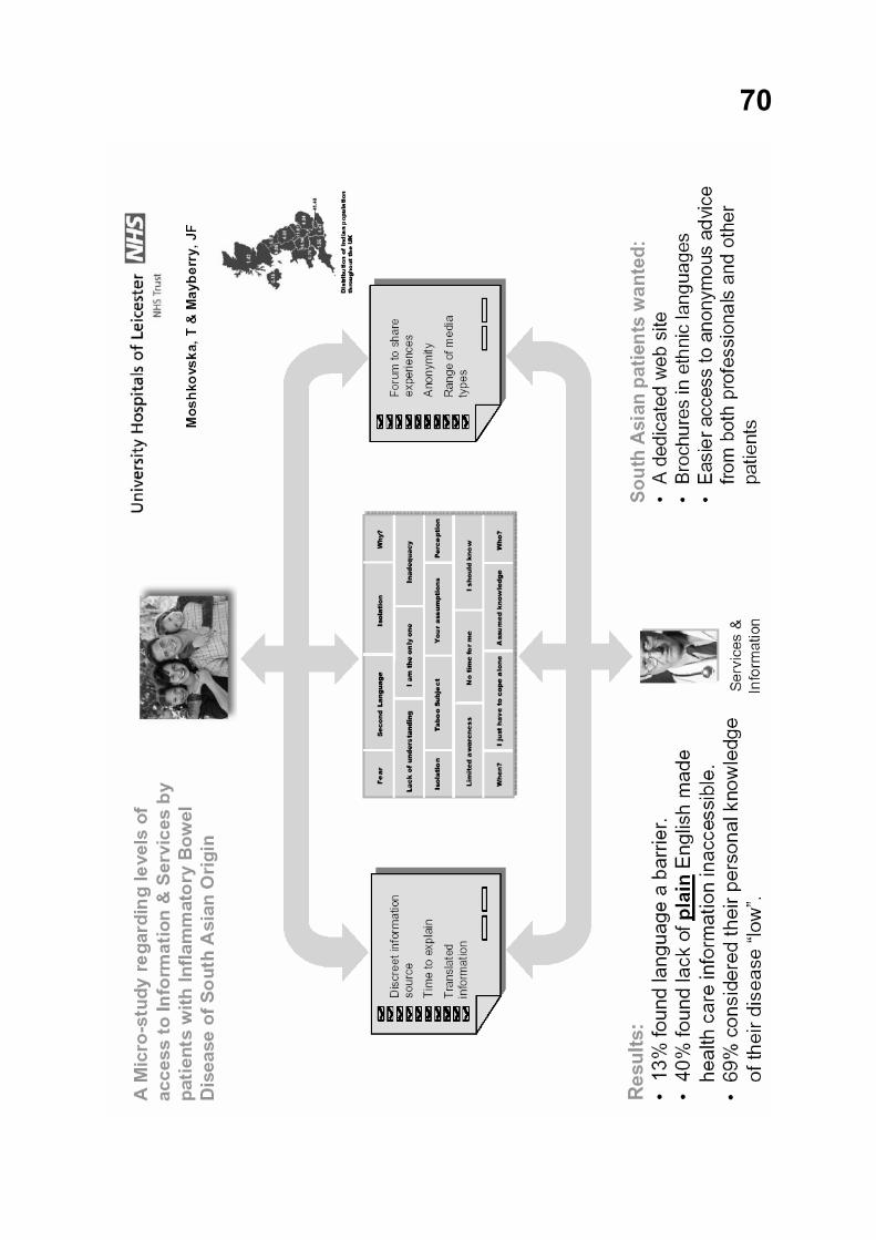
70

71
Interleukin 27 controls the development of Th-17 and iTreg cells via differential effects on the transcription factor STAT1 Clemens Neufert1, Christoph Becker1, Stefan Wirtz1, Massimo C. Fantini2, Benno Weigmann1, Markus F. Neurath1 1 Lab. of Immunology, I. Medical Clinic, University of Mainz, Mainz, Germany 2 University of Tor Vergata Rome, Rome, Italy Introduction: Immuno-suppressive inducible regulatory T-cells (iTregs) and highly pro-inflammatory Th-17 cells can develop from the same precursors depending on the cytokine milieu. Interleukin 27 (IL-27) is frequently present at sites of inflammation including intestinal lesions of patients suffering from Crohn´s disease. Remarkably, IL-27 can promote both anti- and pro-inflammatory immune reactions. Here, we have analyzed mechanisms how IL-27 may drive such divergent immune responses. Methods: CD4+CD25- naïve T-cells were polyclonally stimulated and grown with TGF-beta (for iTregs) or TGF-beta plus IL-6 (for Th-17 cells) in the presence or absence of IL-27. Cells were analyzed after 3–5 days with different methods such as flow cytometry for Foxp3 and IL-17A. Functional studies included CFSE based co-culture proliferation assays, and cells from animals deficient in proteins activated by IL-27 were used to address signaling pathways. Results: Our data demonstrate, that IL-27 can potently interfere with the development of anti-inflammatory iTregs, and that the reduced number of Foxp3+ cells correlates with a diminished suppressive capacity. Strikingly, IL-27 is also able to inhibit the induction of highly pro-inflammatory Th-17 cells. However, whereas the blockade of Th-17 development is dependent on the transcription factor STAT1, the suppression of iTregs is STAT1 independent. Discussion/Conclusion: By utilizing different signaling pathways for the blockade of Th-17 and iTreg cells, IL-27 could shape T cell driven immune responses and might play opposing roles in a way that goes beyond a counterregulation of the cell type predominantly present at a site of immune reaction. In summary, our data demonstrate that IL-27 controls the development of Th-17 and iTreg cells via differential effects on STAT1.

72
Traits of clinical presentation of new diagnosed IBD colitis and their changing over the years M. Shapiro, R. Simantov and E. Scapa Institute of Gastroenterology, Asaf Harofe Hospital, Rehovot, Israel Introduction: In spite of many diagnostic criteria of Crohn’s disease and ulcerative colitis were developed during the past decades, a clinical distinction between these two conditions is difficult and, in some cases, impossible. We conducted a study of clinical traits of new diagnosed IBD colitis in patients who referred to our hospital-based GI clinic during the years 1997–2005. Methods: All colonoscopic records of new colitis cases in ambulatory and hospitalized patients from 1997 to 2005 were reviewed. Demographic data, clinical course, complaints, extent of disease, and histological picture were retrieved from clinical, endoscopic, and pathological reports. Patients with infectious, ischemic, and diverticulaes-related colitis were excluded. Results: 281 patients were diagnosed with IBD colitis between years 1997–2005. 134 were female and 147 male, average age 43.3 years. A percentage of proctitis decreased from 32.1 in 1997–2000 to 20.6 (p < 0.05) in 2001–2005, and percentage of total involved colitis increased from 15.3 to 24, consequently. We found an additional peak of new colitis in ages 51–60 during years 2001–2005 (16.2% of all patients) in comparison with years 1997–2000 (8.2% of all patients). There was no statistical difference in other characteristics over the years 1997–2005. Discussion/Conclusion: We have found that some clinical and epidemiological traits of new diagnosed IBD colitis are changing over the years.

73
Vascular endothelial growth factor (VEGF) in patients with ulcerative colitis A. Stadnicki1,2, D. Frysz-Naglak2, E. Klimacka-Nawrot1, I. Stadnicka3 1Department of Basic Biomedical Sciences, Medical University of Silesia, Katowice, Poland 2Department of Gastroenterology, St. Barbara’s Hospital, Sosnowiec, Poland, 3Department of Neurology, District Hospital, Sosnowiec, Poland Vascular endothelial growth factor (VEGF) is an multifunctional cytokine, that plays a role in angiogenesis and inflammatory response. An increased serum level of VEGF in inflammatory bowel disease is though as mainly platelets origin, however VEGF plasma concentration may reflect its tissue expression. In the present study we evaluate VEGF in colonic tissue as well as VEGF levels in plasma and serum of patients with ulcerative colitis (UC). Methods: Serum and plasma samples were taken from 20 UC active patients, 14 UC inactive patients and 15 normal controls. VEGF concentration in serum and plasma was determined by ELISA. VEGF intestinal localization was estimated by immunohistochemistry. Results: Serum level of VEGF was significantly higher (p < 0.01) in UC active patients (114.4 ± 69.6 pg/ml), as compared with UC inactive patients (44.3 ± 27.4), and when compared with controls (39.7 ± 31.2). The plasma VEGF level was found to be significantly higher in UC active patients (45.3 ± 3.5 pg/ml) as compared with healthy controls (42.6 ± 1.8). However the plasma level of VEGF in inactive UC patients (43.3 ± 2.1) and in control subjects was similar. VEGF protein was visualized in enterocytes and endothelial cells in UC colonic tissue and in normal intestine, however the specific reaction for VEGF staining was more pronounced in UC inflammatory tissue. Conclusion: Increased VEGF levels in serum and plasma in active UC patients may reflect VEGF overexpression in intestinal inflammatory tissue.
74
Polyphenolic compounds of plant origin protect from murine colitis Sabine Westphal, Wolfram Domschke, Torsten Kucharzik Department of Medicine B, University of Münster, Germany Introduction: Inflammatory bowel disease (IBD) is comprised of Crohn’s disease and ulcerative colitis. Because of severe side effects of some of the current immunosuppressive therapies, there is need for developing new therapeutic strategies. The use of active components of plants is becoming an increasingly attractive approach for the treatment of various inflammatory disorders. Polyphenolics recently have been suggested to exert anti-inflammatory activities. In this study we determined EGCG (epigallocatechingallate) the major polyphenolic compound constituent found in green tea, piperine, an alkaloid derived from black pepper, used to enhance the bioavailability of EGCG and curcumin, a polyphenolic pigment from turmeric. The aim of this study was to evaluate different polyphenolics regarding their anti-inflammatory activity in murine colitis models and to determine the underlying mechanisms. Materials and methods: The effects of curcumin and EGCG/piperine have been evaluated in the transfer colitis and DSS colitis model. To induce T cell mediated colitis female ragl-/- mice were treated i.v. with 1 x 10⁶ CD4+CD25- T cells. Mice developed colitis 3–4 weeks posttransfer. To induce chemically mediated colitis, female C57BL/6 mice received 2% DSS in drinking water. Mice received either a combination of 150 µM EGCG plus 100 µM piperine 3 times a week or a diet containing 2% curcumin. Follow-up parameters included weight loss, histology, colon length and cytokine levels in colon mucosa, spleen and mesenteric lymph nodes determined by ELISA and real time PCR. Results: Both curcumin and the combination of EGCG plus piperine significantly attenuated colitis induced weight loss and shortening of the colon. Histological colitis scores were significantly improved during treatment with both agents. TNF-α levels in spleen and mesenteric lymph nodes were significantly decreased 4 weeks posttransfer in mice treated with curcumin or EGCG/piperine. In addition, we observed significantly decreased levels of IL-12 in the colon of mice treated with curcumin or EGCG/piperine. Similar results for these agents could be obtained in DSS colitis. Conclusions: In conclusion, polyphenolics such as EGCG/piperine as well as curcumin attenuate colitis in two different models of colitis. Our findings suggest that polyphenolic compounds may be of therapeutical value for the treatment of human IBD.

75
Reprimo as a potential biomarker for early detection of sporadic diffuse-type gastric carcinoma in plasma E. Aravena1, C. Bernal2, M. Vargas2, C. Villarroel2, F. Ossandon2, E. Santibañez2, I. Diaz2, C. Barrientos1, M. Palma1, R. Estela1, A. Corvalan2 1Chilean-Japanese Institute of Digestive Diseases, Hospital Clínico San Borja Arriarán; 2Department of Pathology, Catholic University of Chile, Santiago, Chile Introduction: Sporadic diffuse-type gastric carcinoma (SDGC) is the most aggressive form of gastric cancer and its precursor lesions are frequently not detected at early stage. Aberrant hypermethylation, a mechanism of inactivation of tumor suppressor genes (TSG), is an emerging approach to early diagnosis of SDGC. Methods: To identify potential biomarkers for early detection of SDGC we studied the aberrant hypermethylation profile of 24 genes covering major cellular pathways by Methylation Specific Polymerase Chain Reaction (MS-PCR) in 32 retrospective cases of SDGC. Most frequently methylated genes were further explored in 69 prospectively collected cases (14 patients with SDCG including tumor mucosa [T], normal mucosa [N] and plasma [P], 29 age-matched noncancer symptomatic controls [N y P] and 26 age-matched noncancer asymptomatic controls [P]). Unsupervised hierarchical clustering analysis was performed to identify subgroups according to the aberrant hypermethylation status in the retrospective cases and two relevant genes (Reprimo and APC) were evaluated in the prospective group. Results: Eleven genes were hypermethylated in at least 50% of the SDGC retrospective cases and 23 genes showed hypermethylation in at least one of the tested cases. Unsupervised hierarchical clustering analysis clustered the cases in two branches that appeared to differ in mucinous/signet ring cell histological type (p = 0.03 by Fisher’s exact test). Among the prospective SDGC cases group, Reprimo and APC were identified in 100% and 88% of the tumor mucosa, 96% and 88% of N and 70% and 82% of P respectively. In 29 age-matched noncancer symptomatic controls, Reprimo and APC were identified in 35% and 100% of N and 25% and 70% of P respectively. In 26 age-matched noncancer asymptomatic controls, Reprimo and APC were identified in 0% and 65% of P (p < 0.001). Conclusions: The aberrant hypermethylation profile of 24 TSG is associated with clinical-pathological characteristics of SDGC (mucinous/signet ring cell histology). The detection of aberrant hypermethylation of promoter region of Reprimo in plasma may be useful for early detection of SDGC.

76
Ursodeoxycholic acid mediated cell cycle arrest of adenocarcinoma cells likely operates through induction of NAG-1 F. Bresso*1, E. Edlundh-Rose2, M. D'Amato3, A. Are4, G. Greicius4, A. Liedén4, U. Sjöqvist1, J. Lundeberg2, V. Arulampalam4, R. Löfberg5, S. Pettersson4 1Gastrocentrum Medicin, Karolinska University Hospital, 2Dept. of Gene Technology, Royal Institute of Technology, 3Department of Biosciences and Nutrition, 4Strategic Research Center IRIS and Department of Microbiology, Tumor and Cell Biology, Karolinska Institutet, 5Department of Medicine, Karolinska University Hospital, Stockholm, Sweden Introduction: Colorectal cancer is a feared complication in inflammatory bowel diseases. In patients with longstanding, extensive colitis and primary sclerosing cholangitis (PSC), the cumulative risk reaches up to 50% after 25 years of disease. Ursodeoxycholic acid, UDCA, used in PSC treatment, was observed as having chemopreventive properties in colitis and adenoma associated colon cancer. We aimed to delineate the anti-cancer properties of UDCA in colonic epithelial adenocarcinoma cell line and understand features of its effects on cell cycle. Methods: UDCA’s effect on cell cycle was evaluated using FACS analysis. In a SW480-based in vitro system, microarray analyses were performed using KTH 29.8k human cDNA microarrays to identify target genes of UDCA and qRT-PCR was used for validation. UDCA target genes’ proteins were evaluated using western-blot. Results: UDCA has an antiproliferative effect in SW480 inducing G1 arrest. Colonic epithelial SW480 cells were treated with either 500 mM UDCA or ethanol for 2, 6, 10, 24 and 48 hours and gene expression analysis was performed. Four and 60 genes were significantly differentially expressed respectively at 6 and 10 hours. Twenty-four genes out of the 31 selected for validation were confirmed. NAG-1 was identified, and as such, may be linked to the UDCA induced G1 arrest we observed. Discussion/Conclusion: NAG-1 is a putative molecular effector of the chemopreventive activity of UDCA. Knowledge of other UDCA target genes may allow for a better understanding of how UDCA works, in order to improve its efficacy. Finally, these genes may be useful biomarkers to monitor the effectiveness of UDCA treatment.
77
Immunohistochemical evaluation of Ki-67, PCNA and MCM2 protein in patients with advanced gastric cancer Jolanta Czyzewska1, Katarzyna Guzinska-Ustymowicz2, Andrzej Kemona2, Anna Pryczynicz2, Halina Kemona1, Roman Bandurski3, Mariusz Gryko3, Bogusław Kędra3 Departament of Clinical Laboratory Diagnostics1, Departament of General Pathomorphology2, Department of General Surgery and Gastroenterology3, Medical University of Bialystok, Poland Background/Aim: Evaluation of proliferating activity may provide important information in different types of tumors. Methods: Tumors from 100 patients with advanced gastric cancer were assessed by immunohistochemistry. Tissue sections were fixed in 10% buffered formaldehyde solution, embedded in paraffin and stained immunohistochemically with anti-human Ki-67, PCNA and MCM2 monoclonal antibodies. Results: No correlation was found between Ki-67, PCNA and MCM2 expression in maim mass of tumor, lymph node metastasis and age, sex of the patients, tumor localization, Lauren’s classification, Bormann’s classification. However, we found a strong association between expression of this proteins in main mass of tumors and lymph node metastasis and presence of lymph node metastasis. Also the histological differentiation was correlated with positive expression of PCNA in main mass of tumor. A statistically significant association was observed between the expression of MCM2, PCNA protein in lymph node metastasis and MCM2 in main mass of tumor and depth of invasion. We have found correlation between survival time and expression of PCNA protein in main mass of tumor. There was no association between expression of Ki-67, MCM2 in main mass of tumor and overall survival time. Address for correspondence: Jolanta Czyzewska Department of Clinical Laboratory Diagnostics Medical University of Bialystok ul. Waszyngtona 15a PL-15-274 Bialystok Poland E-mail: [email protected]

78
A study concerning the incidence of gastrointestinal cancer in several regions of Romania O. Fira-Mladinescu1, Corneluta Fira-Mladinescu1,2, Sorina Doroftei2, Felicia Sas3, Laura S. Gotia1, Danina Muntean1, Brigitha Vlaicu1
1Research Centre in Preventive Medicine and 2Departament of Hygiene, “Victor Babes” University of Medicine and Pharmacy, Timisoara, Romania, 3Timis Public Health Agency, Timisoara, Romania Introduction: Cancer-related morbidity and mortality progressively augmented during the past years; therefore the assessment of cancer incidence is important both for prevention and treatment of the disease. Accordingly, the present study was aimed at identifying the Romanian regions with increased risk for gastrointestinal cancer. Methods: The study targeted Romanian population which is divided in 7 statistical regions. The incidence of digestive cancer was calculated from the cancer registry of each county. The investigation included 10 years (1994–2003), considering the mean value of this period. Comparison of the means values was performed by the ANOVA test, and a p value < 0.05 was considered statistically significant. Results: Within the 7 Romanian regions, cancer incidence (expressed as number of new cases/100,000 inhabitants) varied between 13.29 ± 1.60 and 19.89 ± 2.37 for gastric cancer; between 7.28 ± 1.97 and 12.13 ± 3.98 for the colonic cancer; between 8.29 ± 2.61 and 11.89 ± 3.01 for rectal and anal cancer; between 1.49 ± 0.23 and 2.23 ± 0.5 for esophageal cancer; between 0.25 ± 0.11 and 0.41 ± 0.19 for cancer of small intestine. Statistically significant differences were for all the above mentioned digestive cancer, but the small intestine location. Discussion/Conclusion: The high incidence of gastric, colon, rectal and anal cancer represents a major health problem, mainly in the North-West, West and Central regions of Romania, which requires the investigation of potential risk factors within these geographical areas.

79
Multipolar radiofrequency ablation in the treatment of hepatic tumors J. Genov, N. Grigorov, R. Mitova, B. Golemanov, L. Dinkov, M. Donov Clinic of Gastroenterology, University Hospital “Queen Joanna”, Sofia, Bulgaria Aim: To assess the effectiveness and safety of multipolar RFA in the treatment of hepatic tumors. Methods: 57 patients with 94 hepatic tumors (21 pts with 29 HCC, 36 pts with 65 metastases, age 46–76, tumor size 2.0–11.5 cm) were treated with percutaneous multipolar RFA using 3 bipolar internally cooled applicators. The achieved destruction was assessed with contrast-enhanced ultrasound, CT and cytological examination. Patients were followed-up for 1–18 months. A comparison was made to monopolar RFA with perfused electrodes in 196 patients with 354 lesions (63 pts 78 HCC, 133 pts 276 metastases), followed-up 1–46 months. Results: Complete destruction (CD) after multipolar RFA was achieved in all lesions < 3.0 cm, in 82.9% of tumors sized 3.0–5.0 cm (77.8% of HCC vs. 84.4% of metastases) and in 36.8% of tumors > 5.0 cm (42.9% vs. 33.3%). Major complications occurred in 7%. Local tumor progression (LTP) was observed in 15.8% of the lesions. After monopolar RFA CD was attained in 88.4% of lesions < 3.0 cm (89.3% of HCC vs. 88.3% of metastases), in 60.7% of tumors sized 3.0–5.0 cm (71.8% vs. 56%) and in 18.8% of tumors > 5.0 cm (27.3% vs. 13.3%). Major complications occurred in 7.1% and LTP – in 28.4%. Conclusions: The safety of multipolar RFA is similar to monopolar RFA. The effectiveness of the procedure depends on the size and origin of the tumor. Multipolar RFA is preferred in lesions > 3 cm, accomplishing CD in fewer sessions, with lower rate of LTP.

80
The risk of development of gastric cancer in patients with precancerous changes after Helicobacter pylori eradication Amelia Genunche-Dumitrescu, P. Mitrut, Daniela Badea, M. Badea University of Medicine and Pharmacy, Clinical Hospital of Emergency, Craiova, Romania Introduction: The aim of this study is the assessment the risk of development of gastric cancer (GC) at patients with precancerous changes (PC) at three years after Helicobacter pylori (HP) eradication. Methods: We included in this study 125 patients (51 females, 74 males) with preexistent precancerous changes. A multiannual and comparative study was performed on two groups of patients. A group patients (77 cases) have HP infection, who was eradicated three years ago (HP absence was monitoring in last three years). B group consist of 48 patients without HP infection. We use rapid urease test and serologic testing for determined HP infection. The history and duration of HP eradication was also quantified. Results: The incidence of the PC were: atrophic gastritis (66 cases), gastric ulcer (18 cases), gastrectomy (23 cases), gastric polypus (13 cases) and Menetrier gastritis (5 cases). A group contain all Menetrier gastritis cases, atrophic gastritis (41 cases), gastric ulcer (12 cases), gastrectomy (9 cases), gastric polypus (10 cases). GC was develop in 20 patients (25.97%) of the A group and in 5 cases (10.42%) of the B group. Majority of Menetrier gastritis cases (4 cases) developed GC. In A group, endoscopic forms of the early GC were: type I (polypoid) in 6 cases, type II (superficial) in 2 cases and type III (ulcerated) in 5 cases. In advanced GC we found type Borrmann I in 2 cases, type II in 4 cases and Borrmann IV only one case. Group B had advanced GC in Borrmann forms: II (2 cases), III (one case). The early GC we found in only one case. The risk of GC development, was significantly great (p < 0.01) in patients with long duration of HP eradication, but these parameters was not correlation. Discussion/Conclusion: The risk of development GC, in patients with PC, was significantly great after HP eradication comparativ with never infected patients. This risk is higher when HP eradication necessitate more therapy cure.

81
On the basis of its expression in primary hepatocellular carcinoma, sperm protein 17 is a potential immunotherapeutic target Fabio Grizzi1, Giorgia Ceva-Grimaldi1, Barbara Franceschini1, Sonia Di Biccari1, Eldo E. Frezza2, Everardo Cobos3, Nicola Dioguardi1, Maurizio Chiriva-Internati4 1Laboratories of Quantitative Medicine, Istituto Clinico Humanitas IRCCS, Rozzano, Milan, Italy; 2Department of Surgery, 3Hematology and Oncology, and 4Microbiology and Immunology, Texas Tech University Health Science Center, Lubbock, Texas, USA Introduction: Hepatocellular carcinomas (HCCs) remain a major public health problem throughout the world. Surgical resection is the primary treatment strategy, but many cases are unsuitable because of the presence of intrahepatic or distant metastases at the time of diagnosis. It has therefore become compelling to recognise the disease as early as possible, and to improve alternative strategies such as immunotherapy and gene therapy. Here we investigate whether the cancer-testis antigen, sperm protein 17 (Sp17), is expressed in a series of primary HCCs. Methods: We immunohistochemically studied the expression of Sp17 in formalin-fixed, paraffin-embedded HCC specimens taken from 23 patients (M:F = 19:4; mean age 71 ± 6 years; range: 61–82). The immunohistochemical reaction was categorised into one of three distinct patterns: a) spotted (Sp17 restricted to isolated and scattered neoplastic cells); b) clustered (Sp17 limited to groups of neoplastic cells heterogeneously distributed in the lesion); or c) diffuse (Sp17 distributed throughout the neoplastic cells). Results: Sp17 was found in 96% of tumoral tissues, but not in adjacent non-tumoral tissue. Of the 22 HCC tissue samples immunopositive for Sp17, four (18%) showed a spotted pattern; six (27%) a clustered pattern, and twelve (55%) a diffuse pattern. Conclusion: The high frequency of Sp17 expression in primary HCC suggests using the antigen as a potential immunotherapeutic target and a helpful tool in disease management. Our findings also suggest that further experimental models are needed for exploring the role of Sp17-immunopositive cells in the pathogenesis of HCC.
82
MCM-2 protein expression predict prognosis of G2pT3 colorectal cancer Katarzyna Guzinska-Ustymowicz, Anna Pryczynicz, Ewa Stepień, Jolanta Czyzewska, Andrzej Kemona Departament of General Pathomorphology, Medical University of Bialystok, Poland Aim & methods: Minichromosome maintenance protein 2 (Mcm-2) is a useful biomarker of cell-cycle state in human tissue samles. The aim of this study was to assess the expression of Mcm-2 in moderately differentiated (G2) and pT3 adenocarcinomas. Methods: Mcm-2 protein expression was assessed by immunohistochemistry in formalin-fixed, paraffin-embedded colorectal cancer tissue from 55 patients. Results: The nuclear Mcm-2 immunostaing was observed for all cases. No statistical significant correlation was observed between expression of Mcm-2 and age, sex of patient and localization of tumor. However, a strong association was found between presence of lymph metastases and expression of Mcm-2 in main mass of tumor. Conclusion: This study may suggest that Mcm-2 can be an indicator of lymph node metastases. Address for correspondence: Katarzyna Guzinska-Ustymowicz Department of General Pathomorphology Medical University of Bialystok Ul. Waszyngtona 13 PL-15-889 Bialystok Poland E-mail: [email protected]

83
The expression of connexins 26, 32 and 43 as well as adhesion proteins – E-cadherin and beta-catenin in colorectal adenomas and adenocarcinomas Luiza Kanczuga-Koda1, Mariola Sulkowska2, Mariusz Koda2, Stanisław Sulkowski2 1Department of Medical Pathology, 2Department of General Pathology, Medical University of Bialystok, Waszyngtona 13, PL-15-269 Bialystok, Poland; E-mail: [email protected] Introduction: One of the most important events associated with development and progression of the primary tumor as well as the metastasis is the alteration in gap junctional intercellular communication, which appears to play a role in carcinogenesis as a result of inhibition signalling pathways controlling many biological processes in the cell. The aim of the study was to evaluate the expression and localization of gap junction proteins – connexins (Cx26, Cx32, Cx43) and adhesion proteins (E-cadherin and beta-catenin) in colorectal adenomas and cancers as well as to estimate the correlation between assessed proteins and relationships with selected anatomoclinical features. Methods: Immunohistochemical staining using specific antibodies was performed in 151 colorectal cancer samples and 71 adenomas including a proportionate number of cases from low to high grades of dysplasia. The control group was 30 slides with the normal colorectal mucosa. Results: These studies revealed that during colorectal carcinogenesis altered expression and localization of connexins as well E-cadherin and beta-catenin is common phenomenon in neoplastic cells. These changes appeared even in adenomas as well as in colorectal cancers, but in malignant tumors there were the most distinct, what might suggest, that during tumor progression loss of the functional gap junctions and permanent adhesion complexes between cells is present. Furthermore, overexpression of connexins in cancerous cells seem to be the favorable prognostic factor. The statistically significant correlation between expression of connexins and adhesion proteins could be the point to close relations between these proteins. Discussion/Conclusion: Changes in the expression of adhesion proteins might be an additional factor responsible for incorrect localization of connexins in cancerous cells.

84
Relationships between the obesity hormone leptin, its receptor and hypoxia-inducible factor-1alpha in human colorectal cancer Mariusz Koda1, Mariola Sulkowska1, Andrzej Wincewicz1, Luiza Kanczuga-Koda1, Sandra Cascio2,3, Giuseppe Colucci4, Antonio Russo3, Eva Surmacz2 and Stanisław Sulkowski1. 1Department of Pathomorphology, Medical University of Bialystok, PL-15-269 Bialystok, Poland; 2Sbarro Institute for Cancer Research and Molecular Medicine, Temple University, Philadelphia, PA 19122, USA; 3Section of Medical Oncology, Department of Surgical and Oncology, Università di Palermo, I-90127 Palermo, Italy 4Division of Medical Oncology, National Institute of Oncology, I-70126 Bari, Italy; E-mail: [email protected] Introduction: Leptin (obesity protein, Ob) is a 16 kDa cytokine produced mainly by adipose tissue and it has been first described as a hormone regulating energy balance in the brain. Later studies documented that the hormone can also influence malignant progression. Numerous studies demonstrated that different tumor tissues and cell lines respond to leptin and express functional leptin receptors (ObR). Previously, we found that the expression of leptin and ObR can be stimulated by hypoxia-mimetic agents. The aim of this study was analysis of the abundance of the leptin system and hypoxia-inducible factor-1alpha (HIF-1alpha) in human colorectal cancer as well as assessment of correlations among these biomarkers. Methods: The study included specimens of primary colorectal cancers from 135 patients who underwent curative resection. We used immunohistochemistry for analysis of the expression of leptin, ObR and HIF-1alpha. Results: Immunoreactivity for leptin, ObR and HIF-1alpha protein was observed in 69/135 (51.1%), 129/135 (95.5%) and 88/135 (65.2%), of colorectal cancers, respectively. Statistically significant positive correlations were noted between leptin and HIF-1alpha (p = 0.005, r = 0.243), ObR and HIF-1alpha (p < 0.001, r = 0.325) as well as leptin and ObR (p < 0.001, r = 0.426) in the group of all patients as well as in various subgroups depending on clinicopathological features. Discussion/Conclusion: Our current observations suggest that HIF-1alpha may play a role in the progression of colorectal cancer via upregulation of the leptin system.
85
Hepatocellular carcinoma GR. I. and multifocal nodular steatosis V. Kupčová, E. Šeligová, M. Szántová, L. Turecký, M. Smutný Medical School of Comenius University, Bratislava, Slovakia We are presenting a case of a 57-year-old woman after mastectomy because of carcinoma mammae in 1988. In 1989 a solitary liver lesion was found on the ultrasonography and a liver resection (segmentectomia V. seg.) was performed. In March 1990 repeatedly an operation for a finding of a lesion in the right liver lobe (excisio cunoides lobi dx. hepatis, seg. VIII.) was done. Both of the histological findings (1989, 1990) showed an extremely well differentiated hepatocellular carcinoma grading I – borderline lesion (HCC-I). In 1992 ultrasound and computer tomography (CT) showed multiple hypodense diffuse nodules (20 mm) and metastatic process was suspected. The patient has refused further biopsy. Control CT in 1993 showed an increase of one of the nodules to 30 mm, others have decreased in size. Tumor markers were negative. Next CT in 1994 and March 1995 was identical with that in 1993 and diagnosis of nodular steatosis or nodular hyperplastic steatofibrosis was suspected. CT in Dec. 1995 showed regression of nodules, only one pathological lesion was found. MR imaging in Dec. 1995 could not exclude malignity of this lesion. In March 1996 repeated operation (resectio tangentialis seg. IV. et VII nonanatomica) was performed. Histological finding was without any signs of malignity, only steatosis was found. The 13-year follow up of the woman showed, that in this patient with repeated liver resection because of HCC-I (1989, 1990) nodular steatosis resp. nodular hyperplastic steatofibrosis was developed. This work was supported by VEGA grant 1/4277/07
86
Melatonin increases the phosphorylation of heat shock protein 27 in pancreatic carcinoma PANC-1 cells A. Leja-Szpak1, J. Jaworek1, J. Szklarczyk1, J. Bonior1, S.J. Konturek2, W.W. Pawlik2 1Department of Medical Physiology, Health Care Faculty, School of Medicine Jagiellonian University, Cracow, Poland 2Chair of Physiology, Medical Faculty, School of Medicine, Jagiellonian University, Cracow, Poland Background: Recent evidence suggests that heat shock protein 27 (Hsp 27) could be involved in the progression of tumor growth. It has been reported that melatonin protects pancreatic cells and various tissues against inflammatory damage and im proves the outcome of experimental pancreatitis. Aim: To investigate the effect of melatonin on Hsp 27 protein level in human pancreatic carcinoma cells (PANC-1). Methods: PANC-1 cells were incubated in the standard medium DMEM supplemented with 10% fetal bovine serum at 37˚C with 5% CO2 and humidified atmosphere under basal conditions or in the presence of various doses of melatonin (10-6, 10-8, 10-10 or 10-12 M). Control experiments were performed with the vehicle only (0.1% DMSO) without melatonin. After 24 h and 48 h the cells were harvested, the cytoplasmic and nuclear proteins were isolated for Western Blot and immunoblotting studies. Results: Incubation of the PANC-1 cells with melatonin to PANC-1 cells resulted in the stimulation of both cytoplasmic (by 270%, 240%, 260% or 180% respectively) and nuclear (by 200%, 195%, 180% or 130% respectvely nonphosphorylated Hsp 27 protein level after 24 of incubation. Above pools of nonphosphorylated chaperone protein levels were strongly diminished after subsequent 24 h. Administration of melatonin siginficantly stimulated production of nuclear phosphorylated Hsp 27 protein level (by 180%, 170%, 130% or 110%) in PANC-1 cells after 48 h incubation. Conclusion: Melatonin stimulates phosphorylation of Hsp 27 in human pancreatic carcinoma cells (PANC-1) and thus could protect these cells from that.

87
Decrease of intrahepatic innate lymphocytes (NK and NKT cells) in patients with liver metastasis I. Manolova1, M. Gulubova2, D. Kyurkchiev3, I. Altunkova3, G. Cirovski4, A. Julianov4, J. Jovchev4 1Laboratory of Clinical Immunology, 2Department of General and Clinical Pathology, 4Department of General Surgery, Medical Faculty, Trakia University, Armeiska str. 11, BG-6000 Stara Zagora, 3Laboratory of Clinical Immunology, University Hospital “St. Ivan Rilski”, Sofia, Bulgaria, E-mail: [email protected] Various lymphocyte populations with predominance of cytotoxic T lymphocytes and innate lymphocytes, such as natural killer (NK) and lymphocytes called NKT, were found in the human liver. Cytotoxic T cells, NK and NKT cells are effectors that realize tumor surveillance in the liver. Aim: We examined the main intrahepatic lymphocyte subpopulations, namely CD3+ lymphocytes, NKT lymphocytes expressing the CD3+CD56+ phenotype, CD56+ NK cells, CD4+, and CD8+ T cells in colorectal cancer (CRC) patients with and without liver metastasis. Methods: Surgically resected liver tissues were examined immunohistochemically using anti-CD4, CD8, and CD56 monoclonal antibodies and the proportion of each hepatic lymphocyte subset was evaluated by two-color flow cytometry in 32 CRC patients (16 with and 14 without liver metastases). Results: Immunohistochemically CD4+, CD8+, and CD56+ cells were gathered at the periphery of metastases. In the tumor tissue the three types of lymphocytes were presented, although CD56+ cells were less in number. Flow cytometry analysis showed a significant decrease in the proportion of CD3+CD56+ cells in metastatic livers, but not in normal livers (11.9 ± 10.3 vs. 24.2 ± 13.6%, p = 0.02). The percentage of intrahepatic CD3-CD56+ cells was also decreased in patients with metastasis compared to those without (10.5 ± 12.0 vs. 16.3 ± 8.9%, p = 0.058). Conclusion: NK and NKT cells are decreased in hepatic malignancy compared to histologically normal livers. Depletion of innate lymphocytes may underline susceptibility to metastatic liver diseases and could be a reason for the escape of metastatic cells from immunologic surveillance.

88
Management of early gastric cancer in a Latinoamerican hospital A. Menendez, E. Aravena, M. Gonzalez, C. Barrientos, A. Sandoval, M. Palma, H. Iturriaga Chilean Japanese Institute of Digestive Diseases, Hospital Clinico San Borja Arriaran, Santiago, Chile Introduction: Gastric cancer is the leading cause of death from cancer in our country. Our gastroenterology department has a a diagnostic center built with the support of the Japanese government. Therefore, endoscopic procedures, pathology and surgical management are based on guidelines of Japanese authors. The aims of our study are to describe the characteristics of early gastric cancer (EGC) and to analyze our experience with the treatment options: endoscopic mucosal resection (EMR) v/s surgery. Methods: Patients with EGC were identified from a database of 18,000 upper endoscopy reports for the period between January 1999 and August 2006. Clinical data, endoscopic and surgical biopsies with adenocarcinoma and treatment were analyzed. EMR was performed according to the Japanese guidelines. Results: Our gastroenterology department treated 27 patients with EGC (8.7% of gastric cancers). There where 69% men and 31% women with a mean age of 66.5 years ± SD 12.5 (34–85 years). The most frequent location was the upper third (50%), followed by the lower third (31%) and the middle third (19%) The most common macroscopic type was 0–IIc (26%). Histology showed a differentiated adenocarcinoma in 81% and an undifferentiated adenocarcinoma in 9% of the cases. Analyzing treatment options, 12 (44%) of the 27 patients were treated with EMR and 9 (33%) underwent gastrectomy. A complete resection after EMR was shown in biopsies of 8 patients. 4 patients showed positive borders after EMR and 2 of them underwent gastrectomy, but no cancer was found in the surgical specimen. Assessing treatment associated mortality, all the patients who were treated with EMR are still alive, however, 2 of the operated patients died because of early complications after surgery. Conclusions: The detection rate of EGC in our hospital is similar to those in Western countries and lower than in Japan. EMR is the initial approach in the treatment of EGC showing a succesful resection in 75% of these patients. EMR is an oncologically valid treatment and has no mortality and morbidity in our experience, in contrast to surgery that implies a risk of complications and death.

89
Experimetal studies in vivo regarding the effect of resveratrol extracted from vine on gastric neoplastic injuries P. Mitruţ*, F. Burada**, I. Rogoz**, Amelia Genunche-Dumitrescu* *Medical Clinic, **Departments of Genetic and Cells Biology, UMF, Craiova, Romania Grant CNCSIS 954/ 2007 Resveratrol is considered to be the first natural medicine product which offers firm data about its efficiency in certain status of cancer. Biological activities of resveratrol connected with preventing and curing cancer are connected with enzymatic biotransformation, preserving natural cell cycle regulation, inhibition of proliferation, apoptosis induce and inhibition of the proliferation tumors and angiogenesis. Aim of study: Testing the cytogenetic effect and the potential of inducing apoptosis of resveratrol on adenocarcinoma cell cultures Methods: There used standard cell lines KATO III. Testing the cytogenetic effect of the resveratrol preparation observed the frequency of poliploide cells in the cultures, the frequency of aneuploide cells in the cultures, the types of structural chromosomial changes and their frequency. The cytogenetic evaluation of the treated cell cultures and of the witnesses ones will be done at 4 or 5 passage and to each passage we added resveratrol. In the begining we utilized the use concentrations 0.1, 1, 10, 100 mmol/l. In the same time we studied the witness culture, which be undercultivated together with the tested culture. For chromosomial analysis we used standard cytogenetic techniques. For keeping cell cultures we used a medium MEM or RPMI supplemented with 10% fetal bovine serum, penicillin, streptomycin and L-glutamine. Studying the apoptotic effect of resveratrol was made on cell cultures, too. There observed the citoplasmatic changes, nuclear changes (tahicromy, picnosis) formation of apoptotic corpuscule and the process of eating away of the cell residue. The apoptotic index will be determined by quantitative morphometry. Results and discussion: Treatment with resveratrol arrested KATO-III cells in the G0 and G1 phase of the cell cycle and eventually induced apoptotic cell death, but had a minimal effect on cell lysis. The cytogenetic evaluation of the treated cell cultures was regarded high frequency of polyploide cells in the cultures and the frequency of aneuploide cells in the cultures. The apoptotic index determined by quantitative morphometry was high after resveratrol. Conclusions: Results indicate that resveratrol has potential as a chemopreventive agent against gastric cancer because it exerts an overall deactivating effect on human gastric adenocarcinoma cells. Evaluation of anticarcinogenic effect of resveratrol extracted from local natural sources on cell cultures of gastric cancer with possible preventive and even curative medical implications.

90
Anemia in the patients with digestive cancers – An alert signal or an expression of an advanced neoplasm of the digestive tube? R. Mihaila1, E.C. Rezi2, L. Tiurean2, M. Deac1, R. Mihaila3 1Lucian Blaga University Sibiu, Faculty of Medicine, Sibu, Romania 2Departmental Clinical Hospital Sibiu, Romania 3Public Health Authority Sibiu, Romania Introduction: Gastrointestinal bleeding is the major cause of iron-deficiency anemia in men and women over 50 years of age. An accurate evaluation of this anemia to detect a convincing explanation is therefore necessary. Methods: We studied a group formed by 143 patients which were hospitalized on the IInd Medical Department of the County Clinical Hospital from Sibiu, Romania, during 2002–2006. 38 of them were diagnosed with digestive cancers. 105 of them formed the control group. Results: The medium age of the control group was 58.4 ± 11.68 years of age, compared with the age of the patients with digestive cancer were the medium age was 69.84 ± 8.88 years. 42.1% from the patients with cancer were women. 57.89% of the patients with digestive cancers presented anemia, compared with only 20.95% of the patients from the control group. 37% of the patients were diagnosed with carcinoma of the ascending colon. The average level of hemoglobin was, in the control group of 12.78 ± 1.89 g/dl, compared with only 10.984 ± 2.96 g/dl at the patients with digestive cancers (p < 0.0001). Also, we could find the same results regarding the hematocrit, which was, in average, 37.52% at the control group, compared with only 32.79% at the patients with digestive cancers (p < 0.001). The number of platelets was higher at the patients with digestive cancers, compared with the control group (p < 0.0001). The cause is probably a reactive trombocytosis, as a response to anemia and cancer. From the total of 143 patients which were studied, 45 (31.46%) had the hemoglobin level under 12 g/dl. From these, 48.8% were diagnosed with a digestive cancer. The relative risk of developing a digestive cancer at the patients with the hemoglobin level under 12 g/dl was 2.993. We could not find any correlation between the age and the hemoglobin level (r = -0.149). Discussion/Conclusion: More then a half from the patients with digestive cancers present iron-deficit. Anemia is an important alert sign which can suggests a digestive cancer. Carcinoma of the ascending colon is a frequent cause of iron-deficiency anemia. The hospitalized patients presenting anemia have a third times higher risk of a digestive cancer than those without anemia. Evaluation of anemic patients in order to exclude a carcinoma of the colon has a high priority in these cases.
91
Tissue transglutaminase could participate in ECM remodeling in invasive front in colorectal cancer A. Portyanko, J. Gorgun, P. Kovalev Belarusian State Medical University, Belarusian Medical Academy of Postgraduate Education, City Pathological Bureau, Minsk, Belarus Introduction: Remodeling of extracellular matrix in invasive front is a key event in invasive growth of malignant tumors. Invasion is a significant step in metastasing cascade in malignant tumors. Tissue transglutaminase (tTG) is a Ca2+-dependent enzyme which cross-links proteins via epsilon(gamma-glutamyl)lysine bridges. There is increasing evidence that tTG is involved in wound repair and tissue stabilization, as well as in physiological mechanisms leading to cell death. Methods: We have studied 19 non-mucous well- and moderately differentiated colon cancers. TTG expression was assessed immunohistohemically in formalin-fixed, paraffin-embedded specimens from cancer surgery. Results: TTG expression was found in all cases either in epithelial cancer cells or in tumor stromal cells. Cancer epithelial cells in central areas showed tTG positivity in 8 cases versus 15 cases of positive staining of cancer cells in invasive front. Stromal cells surrounding cancer nests in invasive front expressed tTG in 18 cases versus 8 cases of tTG expression in stromal cells in central areas of tumor. In cases with positive tTG staining of central tumor areas the intensity of staining of cells in invasive front was higher comparing with central parts. Discussion/Conclusion: These results indicate that tTG expression is upregulated in the invasive front in colorectal cancer both in epithelial and stromal cells. The present study shows that tTG may play an important role in matrix processing during colorectal cancer growth.

92
Ileal mucosa changes after total colectomy in patients with juvenile polyposis and familial adenomatous polyposis Alexander S. Tertychnyy, Dmitry M. Konovalov, Alexander G. Talalaev Pediatric Faculty, Russian State Medical University, Moscow, Russia Introduction: We studied follow-up ileal mucosa biopsies from 11 patients with juvenile polyposis and 4 patients with familial adenomatous polyposis who had been previously underwent total colectomy with mucosal proctoectomy with the creation of straight ileoanal anastomosis. Patients’ age ranged from 9 to 36 years (mean 18.7 years). The follow-up ranged from 13 months to 15 years (mean 87.5 months). Methods: The biopsy specimens have been taken from the posterior wall of terminal ileum. Paraffin sections were stained with H&E, van Gieson, periodic acid Schiff (PAS) and Gomori’s aldehyde fushcin – Alcian-blue (GAF-AB) stains. Six biopsies were subjected to electron microscopy analysis. Results: On histological examination ileal mucosa revealed focal shortening of villi and lengthened crypts. The villi were lined by increased number of goblet cells. Mucin histochemical studies demonstrated partial conversion of the epithelium to a colonic sulfomucin mucin profile by positive GAF-AB staining in cytoplasm of goblet cells. Only 2 out of 15 patients had increased number of mononuclear cells in lamina propria. The electron microscopy examination revealed shortening and rarification of microvilli of absorbtive enterocytes. Discussion/Conclusion: Follow-up biopsies show incomplete and focal neocolonic transformation of ileal mucosa. Nevertheless, in most of the cases ileal mucosa preserved its architectural and histochemical characteristics. We failed to identify evidences of chronic inflammation that made us suggest that chronic terminal ileitis is not a common long-term event in patients with polyposis coli syndromes after total colectomy.
93
Molecular and immunohistochemical analysis in HNPCC diagnosis Doina Verdeş, Roxana Popescu, Smaranda Goţia1, Daniela Puşcaşiu Departaments of Cell and Molecular Biology, 1Department of Physiology, University of Medicine and Pharmacy “V. Babes”, Timisoara, Romania HNPCC syndrome caused by inactivation of the genes which are implicated in the repairing systems of the DNA. MMR Gene family involves in this specific order: MSH2, MLH1, MSH6, PMS2, PMS1, MSH3. Methods: 8 patients have been investigated. The immunohistochemistry study of the expression changes in the MMR proteins and amplified PCR molecular tests of the MLH1 and MSH2 genes has been made from biopsies. Two antibodies have been used: anti-MLH1 and anti-MSH2 Results: The study has been reveled the loss of the expression of one of these proteins in the tumor cells nuclei. The immunohistochemistry study allowed the diagnosis in 7 of 8 cases. In 90% of the cases, HNPCC reveals an unstable phenotype, characterized thru the appearance of new alleles. The molecular analysis has been made in 5 of the 7 cases; 2 cases reveled MSI-H phenotype characterized thru the instability of at least 2 markers. Discussion/Conclusion: Immunohistochemistry is a simple, fast and cheap method; it has a 92% sensitivity and 100% specificity. Detecting MSI phenotype using the molecular analysis is a reliable method, but is laborious and expensive. Immunohistochemical analysis of tumors may possibly substitute the PCR based MSI analysis in a considerable percentage of the HNPCC cases.

94
May the GST-pi expression and Ile105Val GSTP1 gene polymorphism predict the colorectal cancer outcome after adjuvant chemotherapy? T. Vlaykova1, M. Gulubova2, D. Vlaykova3, I. Manolova4, G. Cirovski5, P. Chilingirov6 1Dept. of Chemistry and Biochemistry, 2Dept. of General and Clinical Pathology, 3Under-graduate student, 5Dept of General Surgery, Medical Faculty, 4Laboratory of Clinical Immunology, University Hospital, Trakia University, 6Oncology Hospital, Stara Zagora, 11 Armeiska Str, BG-6000 Stara Zagora, Bulgaria, E-mail: [email protected] Introduction: Some studies revealed that glutathione S-transferase pi (GST-pi), one of the isoenzymes of xenobiotic-metabolizing enzymes glutathione-S-transferases (EC. 2.5.1.18) is involved in susceptibility to cancers and is overexpressed in colon carcinoma and in drug-resistant tumors. The GSTP1 gene is a polymorphic and an A→G transition in exon 5 of the gene resulting in a codon 105 Ile→Val amino acid substitution has been identified. The aim of the current study was to investigate the expression of GST-pi in primary colorectal cancers, to determine the Ile105Val GSTP1 genotypes of the colorectal patients and to elucidate their potential role as prognostic and predictive markers after adjuvant chemotherapy. Methods: The expression of GST-pi was evaluated by immunohistochemistry in 132 biopsies from patients suffering from colonic (n = 81) and rectal (n = 51) cancers. The PCR-RFLP-based method was applied for detection of GSTP1 genotype of 80 of the enrolled patients. Results: Patients with weak GST-pi expression in tumor tissue had significantly longer overall survival after the surgical therapy (median of 58 months) compared to those with strong GST-pi expression (median of 19 months, p = 0.0004, Logrank test). This association was even stronger when the analysis was stratified according to the tumor stage. The beneficial effect of the weak GST-pi expression was valid both for patients treated with adjuvant chemotherapy (n = 63, p = 0.008) and those that did not receive any postoperative therapy (n = 66, p = 0.019). The distribution and the frequencies of the Ile105Val GSTP1 genotypes among the CRC patients were as the following: 55 (0.69) Ile/Ile, 18 (0.22) Ile/Val, and 7 (0.09) Val/Val. The genotype distribution did not correlate with GST-pi expression and did not affect the overall survival of patients either with adjuvant therapy or without such. Discussion/Conclusion: Based on our observation, we suggest that the GST-pi expression, but not the Ile105Val GSTP1 polymorphism may be a valuable prognostic marker for overall survival of the patients with colorectal cancer, but its predictive impact on the response to adjuvant chemotherapy is uncertain. Key words: colorectal carcinoma, GST-pi, gene polymorphism, immunohisto-chemistry, PCR-RFLP, prognosis This work is done with the financial support of research project VU-L-05, funded by the Ministry of Education and Science, Bulgaria

95
Preoperative serum protein levels of EPO and their comparison to VEGF and VE-cadherin in human colorectal cancers Andrzej Wincewicz1, Anna Chabowska1, Mariola Sulkowska1, Adrian Chabowski2, Michal Mysliwiec3, Krystyna Pawlak3, Mariusz Koda1, Boguslaw Kedra4, Stanislaw Sulkowski1 1Departments of Medical and General Pathomorphology Waszyngtona St. 13, PL-15-269 Bialystok, Medical University of Bialystok, Poland 2Department of Physiology, Medical University of Bialystok, Poland 3Department of Nephrology and Transplantology, Medical University of Bialystok, Poland 4Department of General and Gastrointestinal Surgery, Medical University of Bialystok, Poland Introduction: EPO stimulates erythropoiesis and induces proliferation of endothelial cells. VEGF and VE-cadherin contribute to formation of vascular network during growth of cancer. VEGF and EPO follow the common way of HIF-1 dependent transcription and are synthesized by colorectal cancers. Therefore we compared these factors in sera of colorectal cancer patients. Methods: We applied commercially available ELISA kits to evaluate preoperative serum levels of EPO, VEGF and VE-cadherin in blood samples from 125 colorectal cancer patients and control group of 16 healthy volunteers. Results: Erythropoietin was significantly elevated in preoperative sera in all colorectal cancer patients (p = 0.013) compared with healthy volunteers. Among different anatomo-clinical features of the tumors the strongest statistical differences for EPO were observed in node-positive (p = 0.0031) and pT3 + pT4 (p = 0.0131) cancers in comparison to control group. VEGF positively correlated with EPO in preoperative sera in cases of node-negative colorectal cancer (p = 0.005, r = 0.345), in colon tumors (p = 0.038, r = 0.256) and set of individuals younger below sixties at their ages (p = 0.002, r = 0.465). No other correlation was noted among all of the examined groups. VE-cadherin serum levels were not correlated with serum EPO. Discussion/Conclusion: EPO levels might increase preoperatively as hallmark of greater cancer advancement. The relationships between VEGF and EPO could indicate co-ordination of their stimulating functions of angiogenesis in cancers with favorable clinical and pathological features. With deterioration of tumor characteristics, the productions of VEGF and EPO can be deregulated without expected dependence of their serum levels as consequence of their autonomic secretion by cancer.
96
Treatment of pyloric stenosis by intrapyloric injection of botulinum toxin – Pilot study C. Banciu, O. Chirileanu, Loredana Marian, Alina Pacurari, I. Romosan IV. Department of Internal Medicine, University of Medicine and Pharmacy Timisoara, Romania Introduction: intrasphincteric injection of botulinum toxin (Botx) represents one of the treatment modalities for pyloric stenosis (PS). Aims and objectives: the present study tries to compare the effect of two different doses of Botx and to observe the long-term response of the injection in pts with PS. Material and methods: 7pts (4 males, 3 females) with PS received doses of 100 U Botox in a single injection. 4 pts were randomized to be reinjected with another dose of 100 U Botx after 30 days. Clinical, radiological and endoscopic assessments were performed at baseline, at one and three months after initial injection of Botx and at the end of follow-up (mean: 6 months). Results: At one month after initial injection 88% of pts responded to Botx, clinical, radiological and endoscopic improvement could be observed. After three months, however, 3 pts who received only the initial 100 U Botx injections, experienced a relapse of clinical symptoms (regurgitation, vomiting), compared to the four pts who received a second 100 U Botx injection after 30 days. At 6 months follow-up, the 4 pts reinjected with a second dose of Botx were still in remission with good clinical status and endoscopic remission. Conclusions: Intrapyloric injection of Botx represents a simple and safe method of treatment in PS. Two intraspincteric injections of 100 U Botx 30 days apart seem to be more effective and have the best long-lasting effect in PS. Further studies on larger number of pts must undergo in order to support these conclusions, also comparative studies with other endoscopic procedures (balloon dilation) or surgery.

97
Detection of small polyps by cap fitted colonoscopy compared with standard colonoscopy C. Banciu, O. Chirileanu, Loredana Marian, Alina Pacurari, I. Romosan IV. Dept. of Internal Medicine, University of Medicine and Pharmacy Timisoara, Romania Introduction: Several new methods for colonoscopy are tested to improve detection of small polyps or early (in situ) carcinoma. Objectives: the present study wants to compare the grade of detection of small polyps by standard colonoscopy (scol) and cap fitted colonoscopy (cfcol), respectively. Material and methods: 55 pts with positive FOBT were randomized to undergo either scol (27 pts) or cfcol (28), using the same colonoscope, just adding a transparent hood at the tip of the colonoscope. Results: Polyp and early carcinoma detection rate was significantly higher in the cfcol group (47.2%) vs. the scol group (38.1%). Using cfcol polyps were more efficient and easier detected, especially under folds of the sigmoid and transverse colon compared with scol (85.3% vs. 63.5%, p = 0.003). Discussion/Conclusions: Among other more expensive endoscopic methods (magnification, NBI, autofluorecence etc) colonoscopy using a cap at the tip of the endoscope represents a cheap but efficient method to improve the view and to detect small polyps or early colon cancers by seeing under colonic folds. Adding chromoendoscopy to this technique will improve further more the detection rate of polyps.
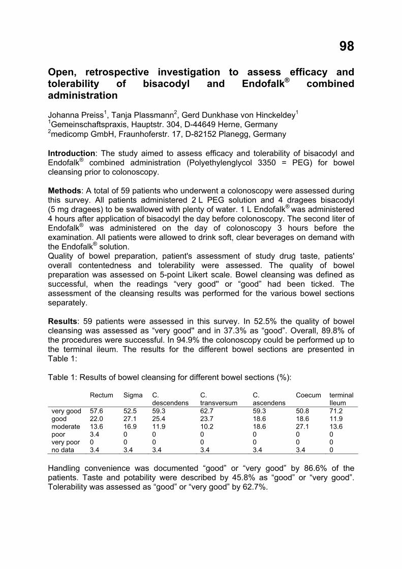
98
Open, retrospective investigation to assess efficacy and tolerability of bisacodyl and Endofalk® combined administration Johanna Preiss1, Tanja Plassmann2, Gerd Dunkhase von Hinckeldey1 1Gemeinschaftspraxis, Hauptstr. 304, D-44649 Herne, Germany 2medicomp GmbH, Fraunhoferstr. 17, D-82152 Planegg, Germany Introduction: The study aimed to assess efficacy and tolerability of bisacodyl and Endofalk® combined administration (Polyethylenglycol 3350 = PEG) for bowel cleansing prior to colonoscopy. Methods: A total of 59 patients who underwent a colonoscopy were assessed during this survey. All patients administered 2 L PEG solution and 4 dragees bisacodyl (5 mg dragees) to be swallowed with plenty of water. 1 L Endofalk® was administered 4 hours after application of bisacodyl the day before colonoscopy. The second liter of Endofalk® was administered on the day of colonoscopy 3 hours before the examination. All patients were allowed to drink soft, clear beverages on demand with the Endofalk® solution. Quality of bowel preparation, patient's assessment of study drug taste, patients' overall contentedness and tolerability were assessed. The quality of bowel preparation was assessed on 5-point Likert scale. Bowel cleansing was defined as successful, when the readings “very good'' or “good” had been ticked. The assessment of the cleansing results was performed for the various bowel sections separately. Results: 59 patients were assessed in this survey. In 52.5% the quality of bowel cleansing was assessed as “very good'' and in 37.3% as “good”. Overall, 89.8% of the procedures were successful. In 94.9% the colonoscopy could be performed up to the terminal ileum. The results for the different bowel sections are presented in Table 1: Table 1: Results of bowel cleansing for different bowel sections (%): Rectum Sigma C.
descendens C. transversum
C. ascendens
Coecum terminal Ileum
very good 57.6 52.5 59.3 62.7 59.3 50.8 71.2 good 22.0 27.1 25.4 23.7 18.6 18.6 11.9 moderate 13.6 16.9 11.9 10.2 18.6 27.1 13.6 poor 3.4 0 0 0 0 0 0 very poor 0 0 0 0 0 0 0 no data 3.4 3.4 3.4 3.4 3.4 3.4 0
Handling convenience was documented “good” or “very good” by 86.6% of the patients. Taste and potability were described by 45.8% as “good” or “very good”. Tolerability was assessed as “good” or “very good” by 62.7%.

Conclusion: Application of 2 L Endofalk® + 4 dragees bisacodyl result in successful bowel cleansing prior to colonoscopy. The addition of bisacodyl to the bowel preparation procedure with the ability to reduce total liquid solution intake compared to PEG intake alone seems to be an appropriate alternative to achieve successful bowel cleansing.

99
Risk factors for intra- and retroperitoneal perforations in patients after therapeutic ERCP M. Shapiro, L. Kopel, D. Abramovith, M. Yair, E. Broide, and E. Scapa Institute of Gastroenterology, Assaf Harofeh Hospital, Israel Introduction: Therapeutic ERCP is a procedure that may cause a number of serious complications. One of them is a retroperitoneal or/and intraperitoneal perforation. However, the real incidence and risk factors of asymptomatic perforations is scarcely lighted in medical literature. Moreover, we know very little about the "natural history" of these perforations. Methods: We prospectively performed a low-radiation abdominal CT scan in patients who underwent therapeutic ERCP. Demographic characteristics, type of procedure (papillotomy or pre-cut), type of perforation (intra- or retroperitoneal), treatment, and outcome were evaluated. Results: Ninety patients were recruited, signed informed consent and underwent abdominal CT. In twelve of them (13.3%) retroperitoneal perforation was diagnosed. In three patients perforation was retro- and intraperitoneal. All patients with isolated retroperitoneal perforation had an uneventful recovery. Two patients with intraperitonael perforation had surgical intervention. One recovered completely and the other (88-year-old female with severe comorbidity) died from multi-organ failure. We found that the group of perforation included more male patients than the no perforation group (58% versus 37%) and underwent more pre-cuts that patients with no perforations (83% versus 67%). Discussion/Conclusion: Asymptomatic retroperitoneal perforation after therapeutic ERCP is common. However, usually it has no clinical significance. Male gender and pre-cut are risk factors. Intraperitoneal perforation frequently required surgery and may cause death, particularly in elderly patients with comorbidity.

100
Bispectral monitoring during endoscopic retrograde cholangiopancreatography Dr. Sharan Shetty, Dr. Talal Valliani, Dr. Ivona Bernova, Dr. Jayshri Shah, Dr. Ankur Srivastava, and Dr. Robert Przemioslo Department of Gastroenterology, Frenchay Hospital, Bristol, UK Introduction: Bispectral (BIS) has been used in anaesthesia and intensive care to attain a target level of sedation. It uses specialized analysis and conversion of electroencephalogram (EEG) signals into numerical value of sedation depth. Its use during gastrointestinal has been reported in only few studies. We present our of BIS monitoring during endoscopic retrograde pancreatography (ERCP) Methods: BIS monitoring was performed in 36 consecutive patients undergoing ERCP during Nov 06 to Apr 07. During the procedure patient’s levels of sedation was assessed with Ramsay Sedation Scale (RSS) and were monitored for unplanned events, procedure duration, and use of reversal agents, recovery time and recall after the procedure. In addition these patients had routine endoscopy monitoring. Results: Among the 36 patients there were 10 males and 26 females. Mean age was 65.75 years (range 36–94 yrs).The dose of Midazolam ranged from 2–15 mg with mean of 6.63 mg and fentanyl l25 to 100 micrograms with mean of 66.80 mcg. Baseline BIS ranged from 91–100 with mean values of 96.11.The mean BIS values during the procedure was 82.18 (range 74.73 to 89.85) with corresponding mean RSS scale scores 3.5 (range 3–4.28). Three patients required reversal agents and 2 patients had hypoxic events during the procedure. The mean procedure and recovery times were 32.86 (range 10–65 min) and respectively 24.08 (7–45 min). In the post procedure survey of endoscopy experience with 18 patients 33% had some nausea with or without vomiting with overall mean satisfaction of 7.5 out of 10 during the procedure. There were no untoward incidents with the use of BIS during ERCP. Discussion: BIS score correlate with level of sedation. Even though patients had mean dose of midazolam more than 5 mg they had acceptable levels of sedation and BIS scores. One third had some nausea/vomiting post procedure but overall satisfaction was good. BIS monitoring was safe during ERCP. Conclusions: BIS augments routine monitoring during ERCP. The level of sedation can be easily titrated with BIS scores. It is easy and safe to have BIS monitoring during ERCP.

Author Index to Poster Abstracts (Name - Poster Number) Abramovith, D. 99 Aerts, R. 8 Altunkova, I.P. 87 Amzolini, A. 30 Ancusa, V.M. 7 Andreeva, D.I. 42 Aravena, E. 75, 88 Are, A. 76 Arulampalam, V. 76 Asaumi, H. 34 Assche, G. Van 8 Aydin, A. 9 Badea, D. 80 Badea, M. 80 Bagci, S. 9 Bakowska, A. 1 Banciu, C. 7, 96, 97 Bandurski, R. 77 Baraban, O.V. 22 Baretton, G. 57 Barmada, M. 38 Barrientos, C. 75, 88 Becheanu, C. 2, 50 Becheanu, G. 51 Becker, C. 58, 71 Beckmann, R. 57 Beglinger, C. 38 Bergel, D. 13 Bernaczyk, P.S. 52 Bernal, C. 75 Bernova, I. 100 Bethke, B. 55 Bezna, M. 30 Bintintan, V. 36 Blonska-Fajfrowska, B. 13, 43 Bodger, K. 67 Bonior, J. 35, 47, 86 Borkowska, A. 3 Bozina, N. 24, 59 Bresso, F. 76 Brinar, M. 24, 59, 64 Brodzicki, J. 1 Broide, E. 99 Brown, E. 67 Brzozowski, T. 5, 6 Buchel, O. 8, 11
Bula-Mucha, M. 43 Burada, F. 89 Burganova, G.R. 42 Calcic, C. 2 Camacho, S. 69 Cascio, S. 84 Celinski, K. 44, 46 Ceranowicz, P. 48, 49 Ceva-Grimaldi, G. 81 Chabowska, A. 95 Chabowski, A. 95 Chang, K.-H. 23 Charua Guindic, L. 69 Chilingirov, P. 94 Chirileanu, O. 96, 97 Chiriva-Internati, M. 81 Cieszkowski, J. 48, 49 Ciocirlan, M. 68, 68 Cioffi, M. 60 Cirovski, G. 87, 94 Ciurea, T. 30 Cobos, E. 81 Colucci, G. 84 Corvalan, A. 75 Cukovic-Cavka, S. 17, 24, 59, 64 Czaja, M. 3 Czechowska, G. 44, 46 Czerniak, T. 28 Czyzewska, J. 77, 82 Dagalp, K. 9 D’Amato, M. 76 Damian, D. 25, 26 Deac, A. 10 Deac, M. 10, 90 Decaestecker, J. 8 Delinska-Galinska, A. 3 Dembinski, A. 48, 49 Dembinski, M. 49 Di Biccari, S. 81 Diaz, I. 75 Diculescu, M. 51, 68 Dienes, H.P. 32 Dinkov, L. 79 Dioguardi, N. 81 Domschke, W. 74

Donov, M. 79 Doroftei, S. 78 Drebber, U. 32 Dries, V. 32 Drozdowicz, D. 5, 6 Dublina, E.S. 61 Dumbrava, M. 51 Dunkhase von Hinckeldey, G. 98 Edlundh-Rose, E. 76 Elefterescu, R. 10 Ercin, C.N. 9 Estela, R. 75 Fantini, M. 58, 71 Fevery, J. 8, 11 Fira-Mladinescu, C. 63, 78 Fira-Mladinescu, O. 78 Flehmig, B. 31 Franceschini, B. 81 Frezza, E.E. 81 Frysz-Naglak, D. 73 Funariu, G. 36 Galle, P.R. 58 Gazizov, I.M. 42 Genov, J. 79 Genunche-Dumitrescu, A. 4, 80, 89 Gheorghe, C. 51 Gheorghe, L. 68 Giaquinto, S. 60, 62 Goldis, E. 53 Golemanov, B. 79 González, M. 37, 88 Gorgun, J.V. 54, 91 Gorun, C. 7 Gotia, L.S. 63, 78 Gotia, S.R. 7, 63, 93 Greicius, G. 76 Greinwald, R. 57 Grigorescu, M. 25, 26 Grigorescu, M.D. 25 Grigorov, N. 79 Grizzi, F. 81 Groza, N. 36 Grubelic-Ravic, K. 24, 59, 64 Gryko, M. 77 Gulubova, M.V. 87, 94 Gumerova, A.A. 42 Guzinska-Ustymowicz, K. 77, 82
Hanck, C. 38 Harsch, S. 12, 16 Hoffmeister, A. 39 Hübner, S. 58 Hurduc, V. 2 Ianosi, G. 30 Ilieva, A. 27 Improta, A. 60 Iturriaga, H. 88 Jankowski, A. 43 Jaworek, J. 35, 40, 41, 47, 86 Jimenez Bobadilla, B. 69 Johnson, H.E. 65 Jonderko, K. 13, 43 Jovchev, J. 87 Julianov, A. 87 Kaligin, M.S. 42 Kalinova, K. 27 Kaminska, B. 1 Kaminska, M. 13, 43 Kanczuga-Koda, L. 83, 84 Karaeren, N. 9 Karimi, D. 57 Kasicka-Jonderko, A. 13, 43 Kassabian, S. 38 Kedra, B. 52, 77, 95 Kemona, A. 77, 82 Kemona, H. 77 Khoroshko, N. 18 Kiassov, A.P. 42 Kitsenko, E. 29 Klimacka-Nawrot, E. 73 Koda, M. 83, 84, 95 Konovalov, D.M. 92 Konturek, P. 6, 48, 49 Konturek, S.J. 5, 6, 35, 40, 41, 47, 86 Konze, E. 32 Kopel, L. 99 Korobowicz, E. 44, 46 Korolczuk, A. 44, 46 Korzon, M. 1, 3 Kosilko, S.A. 14 Kot, M. 35, 40, 41, 47 Kotlowska-Kmiec, A. 1 Kovalev, P. 91 Kownacki, P. 48, 49 Kozielska, E. 3 Kozlova, N.M. 14, 15, 19, 20 Krasnoperova, Y.Y. 66

Kratzsch, J. 39 Krznaric, Z. 24, 59, 64 Kuchar, E. 28 Kucharzik, T. 74 Kuhlisch, E. 55, 57 Kulinsky, V.I. 15 Kupcová, V. 85 Kurtz, U. 21 Kwiecien, S. 5 Kyurkchiev, D. 87 Ladic, A. 64 Lai, G.N. 11 Landolfi, C. 62 Landowski, P. 1 Larova, A.A. 66 Lazarev, A.M. 66 Lee, C.-C. 31 Lee, S.-H. 23 Leja-Szpak, A. 35, 40, 86 Leone, A. de 60, 62 Leonova, Z.A. 15 Lesanu, G. 2, 50 Lesanu, R. 2 Liberek, A. 1 Lichovetskaja, Z. 18 Liedén, A. 76 Lin, M.-H. 33 Lin, S.-M. 31 Löfberg, R. 76 Lubivy, E. 29 Luces, C. 67 Luczak, G. 3 Lukina, E. 18, 29 Lundeberg, J. 76 Lungu, V. 53 Lupu, A. 68 Lyons, M.A. 21 Macko, M. 41 Madisch, A. 55, 57 Madro, A. 44, 46 Manav, A. 32 Manolova, I.M. 87, 94 Marenich, N.S. 56 Marian, L. 96, 97 Martinez Bistrain, A. 69 Martínez Martínez, A. 69 Mayberry, J.F. 70 Mazurkiewicz, M. 6 Menendez, A. 37, 88
Miehlke, S. 55, 57 Mihaila, Rodica 10, 90 Mihaila, Romeo 10, 90 Mitis-Musiol, M. 40 Mitova, R. 79 Mitrut, P. 4, 80, 89 Mocanu, T. 25 Molinari, A.M. 60 Monteleone, G. 58 Morgner-Miehlke, A. 55, 57 Moschopulos, M. 51 Moshkovska, T. 70 Mössner, J. 21, 39 Mukhina, J.G. 56 Müller, R. 57 Muntean, D. 78 Mysliwiec, M. 95 Mysliwiec, P. 52 Navarrete Cruces, T. 69 Nawrot-Porabka, K. 40 Neagoe, D. 30 Nemova, I.S. 66 Nesterov, A.S. 66 Neufert, C. 58, 71 Neurath, M.F. 58, 71 Nevens, F. 8, 11 Normann, A. 31 Odenthal, M. 32 Opacic, M. 17 Oracz, G. 45 Oralewska, B. 45 Osorio Hernandez, R.M. 69 Ossandon, F. 75 Otsuki, M. 34 Pacurar, D. 2, 50 Pacurari, A. 96, 97 Pajdo, R. 5, 6 Palma, M. 75, 88 Parisi, M. 60 Pawlak, K. 95 Pawlik, M.W. 5, 6 Pawlik, W.W. 5, 6, 35, 40, 41, 47, 48, 49, 86 Petrascu, O. 10 Pettersson, S. 76 Pevnev, G.O. 42 Pirenne, J. 8 Plassmann, T. 98

Plata-Nazar, K. 3 Popescu, R. 93 Portyanko, A. 54, 91 Potapov, A.S. 61 Potaturkina-Nesterova, N.I. 66 Preiss, J. 98 Prozorow-Król, B. 44, 46 Pryczynicz, A. 77, 82 Przemioslo, R. 100 Ptak-Belowska, A. 5 Puscasiu, D. 93 Racanel, F. 30 Radic, D. 64 Radys, W. 1 Razina, I.V. 66 Renner, O. 12, 16 Reszec, J. 52 Rezi, C.E. 90 Riegler, G. 60, 62 Riegler, L. 62 Rogoz, I. 89 Rojnic Kuzman, M. 59 Romosan, I. 7, 96, 97 Rose-John, S. 58 Roßner, S. 39 Russo, A. 84 Russo, M.I. 60 Rustemovic, N. 17 Ryzko, J. 45 Salvatelli, M. 60 Sandoval, A. 88 Santibañez, E. 75 Sas, F. 78 Sayal, A. 9 Scapa, E. 72, 99 Schäffeler, E. 16 Scheffler, M. 32 Schegoleva, M.G. 66 Scheller, J. 58 Schimmel, S. 12, 16 Schirmacher, P. 32 Schleicher, S. 31 Schulze, F. 32 Seicean, R. 36 Seitz, G. 57 Seligová, E. 85 Selvaggi, F. 60 Semenov, S.V. 66 Sertic, J. 24, 59
Shah, J. 100 Shapiro, M. 72, 99 Shetty, S. 100 Simantov, R. 72 Sjöqvist, U. 76 Sliwowski, Z. 6 Slomka, M. 44, 46 Smith, T.R. 65 Smutny, M. 85 Socha, J. 45 Solovjova, T. 18 Srivastava, A. 100 Stadnicka, I. 73 Stadnicki, A. 73 Stahl, T. 39 Stange, E.-F. 12, 16 Steenbergen, W. van 11 Stepien, E. 82 Stolte, M. 55, 57 Strohmeyer, A. 12, 16 Sukhanova, G. 29 Sulkowska, M. 83, 84, 95 Sulkowski, S. 83, 84, 95 Surmacz, E. 84 Susan, L.M. 7, 63 Sysoeva, E.P. 18, 29 Szántová, M. 85 Szarszewski, A. 1, 3 Szczepanik, M. 41 Szenborn, L. 28 Szklarczyk, J. 41, 47, 86 Taguchi, M. 34 Talalaev, A.G. 56, 92 Terlikowski, S.J. 52 Tertychnyy, A. 56, 92 Teupser, D. 21 Tipaldi, C. 60 Tiurean, L. 90 Tomaszewska, R. 47, 48, 49 Tschen, S.-Y. 31, 33 Tsimbalova, E.G. 61 Tuennemann, J. 39 Turecky, L. 85 Turumin, J.L. 14, 15, 19, 20 Turumina, H.E. 20 Valliani, T. 100 Vargas, M. 75 Varnholt, H. 32 Vasiljev, S. 29
Verdes, D. 93 Vere, C. 30 Vernic, C. 53 Verslype, C. 11 Vietri, M.T. 60 Villarroel, C. 75 Vlaicu, B. 78 Vlaykova, D. 94 Vlaykova, T. 94 Vorobjev, A.I. 18 Voronkova, E. 29 Vucelic, B. 17, 24, 59, 64 Wang, A. 33 Wang, C.-H. 23, 31, 33 Warzecha, Z. 48, 49 Watanabe, S. 34 Weaver, S.A. 65 Wedemeyer, I. 32 Weigmann, B. 71 Westphal, S. 74 Whitcomb, D.C. 38 Wincewicz, A. 52, 84, 95 Wirtz, S. 71 Wittenburg, H. 21 Wonschik, S. 55, 57 Yair, M. 99 Yemelyanchik, S.V. 22 Yesilova, Z. 9 Zaharie, T. 25 Zajac, K. 41 Zapucioiu, C. 2 Zemelka, M. 41 Zimatkin, S.M. 22 Zimnoch, L. 52 Zubkova, E.A. 66

Where medicineand pharmaceuticals meet –
a tried and trusted link
Falk FoundationDr. Falk Pharma
We help
by providing innovative medicines for bowel and liver diseases.
We promote knowledge
from 1967 onwards, there have been more than 200 internationalFalk Symposia and Workshops with over 100,000 participants frommore than 100 countries
1 million doctors and patients have attended over 11,000Falk educational meetings during the last 30 years
our Literature and Patient Information Service –including on-line ordering through our website.
www.falkfoundation.de www.drfalkpharma.de
Leinenweberstr. 5 79108 Freiburg Germany Tel +49 (0) 761/1514-0 Fax +49 (0) 761/1514-321 E-Mail [email protected]