Embed Size (px)
Citation preview

P1: SFK/UKS P2: SFK
BLBS110-c03 BLBS110-Kubicek June 21, 2012 7:54 Trim: 244mm×172mm
Chapter 3
The Tools—Part 1: Enzymology ofCellulose Degradation
3.1 General Properties and Classification of Enzymes ThatHydrolyze Polysaccharides
Enzymes that are capable of cleaving the glycosidic bonds in oligo- or polysaccharides(including cellulose and hemicelluloses) are generally summarized under the term “glyco-side hydrolases (GHs).” The hydrolysis of the glycosidic linkage leads to the formation ofa sugar hemiacetal or hemiketal and the corresponding free aglycone. There are severalmeans for categorization of these enzymes: the IUB (International Union of Biochemistry)enzyme nomenclature classifies them—as all enzymes in general—according to their EC(Enzyme Commission) number. This is a numerical classification scheme for enzymes based onthe reactions they catalyze. Unfortunately, the EC classification does not distinguish betweengenetically and structurally different enzymes as long as they catalyze the same reaction, whichis particularly unsatisfying in the case of GHs as I will explain in more detail later.
In the case of GHs, there are several additional differences between enzymes belonging tothe same EC group, which are therefore traditionally used for further classification. One suchdifference is based on the topology of the action on the macromolecular substrate: exo-GHscleave their substrate on one of the end of the polymer (most frequently, but not always, atthe nonreducing end), whereas endo-GHs cleave within a chain (Figure 3.1a). In terms ofthe biological benefit for this, this principle aids to an efficient attack on any macromoleculebecause the endo-acting enzyme provides an increasing number of oligomers that can beattacked by the exo-enzyme. It is therefore not surprising that endo- and exo-GHs have beenshown to act in synergy (called “exo-endo-synergism”) and their simultaneous presence thusincreases the rate of hydrolysis (Wood and McCrae, 1972).
Another related principle is the grouping of GHs into glycanases and glycosidases. Theterm “glycanases” is used for enzymes that preferentially act on polymeric or high-molecularweight substrates (which is then replaced by specifically naming the polymer and the linkagespecificity; e.g., �-1,4-mannanase), whereas the latter term describes enzymes that are active
Fungi and Lignocellulosic Biomass, First Edition. Christian P. Kubicek.C© 2013 John Wiley & Sons, Inc. Published 2013 by John Wiley & Sons, Inc.
45
P1: SFK/UKS P2: SFK
BLBS110-c03 BLBS110-Kubicek June 21, 2012 7:54 Trim: 244mm×172mm
46 Fungi and Lignocellulosic Biomass
Figure 3.1. Basic mechanisms of oligo- and polysaccharide cleavage by glycosyl hydrolases: (a) endo-and exo-type hydrolysis, (b) retaining mechanism, and (c) inverting mechanism.
on oligomers. While basically simple, this distinction becomes difficult when distinguishingexoglycanases from glycosidases, however, because exoglycanases will act on the same solubleoligomers and from the same end as the glycosidases. Reese et al. (1968) has therefore proposeda method to distinguish exoglycanases from glycosidases: when acting on the same substrate(e.g., a cellooligodextrin), the affinity of an exoglycanase will increase with the polymerizationlength (i.e., the Km will decrease), whereas the opposite will be observed with �-glycosidases.Unfortunately, this principle has been ignored in many cases where GHs were described, whichled to some confusion about their identity in the literature.
Another important way to distinguish GHs is the reaction mechanism, that is, whether theposition of the hydroxylic group at the C-1 atom of the sugar moiety in the glycosidic bond isinverted and retained (Figures 3.1b and c). The case of an inverting hydrolysis generally occursvia only one step, that is, as single-displacement mechanism, which involves an oxocarbeniumion-like transition states (Figure 3.2a), and catalysis proceeds by the canonical acid/basemechanism using (mostly) an E or D, which are typically located 6–11 A
′apart (McCarter
and Withers, 1994), as amino acid participants.A hydrolysis in which retention of the configuration is maintained (also known as “Koshland
retaining mechanism”; Koshland, 1953) mostly involves a two-step, double-displacement re-action that proceeds via a covalent glycosyl-enzyme intermediate (Figure 3.2b). Also this

P1: SFK/UKS P2: SFK
BLBS110-c03 BLBS110-Kubicek June 21, 2012 7:54 Trim: 244mm×172mm
3 The Tools—Part 1: Enzymology of Cellulose Degradation 47
Figure 3.2. Reaction mechanisms of glycoside hydrolases. (a) Hydrolysis of a glycoside by a single-displacement mechanism; (b) hydrolysis by the Koshland retaining mechanism.
mechanism uses an oxocarbenium ion-like transition state and occurs with acid/base catal-ysis provided by an E or D residue. However, here these two amino acids are located muchcloser (5.5 A
′) than in the inverting mechanism. The two steps occur as follows: in the first,
one of the two amino acid residues performs a nucleophilic attack and displaces the aglyconeto form a glycosyl enzyme intermediate. In the second step, the covalent bond between theenzyme and the glycosyl chain is hydrolyzed, which is aided by the residue that acts as theLewis base and which deprotonates the water molecule (Williams, 2011).
Almost all of the GHs and particularly the cellulases and hemicellulases are distributedamong various gene families that are genetically and structurally strongly different. In orderto pay attention to this fact, Bernard Henrissat has nearly 20 years ago started to establish aclassification concept that is based on amino acid similarity (and thus, although not initiallypursued by him, phylogenetic relationship) in the respective enzymes. The first proof-of-principle was obtained by demonstrating the classification of cellulases into several distinct
P1: SFK/UKS P2: SFK
BLBS110-c03 BLBS110-Kubicek June 21, 2012 7:54 Trim: 244mm×172mm
48 Fungi and Lignocellulosic Biomass
Table 3.1. Enzymes and associated modules currently covered by CAZy.
CAZy Abbreviation FunctionCurrentNumbera
Glycoside hydrolases GH Hydrolysis and/or rearrangementof glycosidic bonds
125
Glycosyl transferases GT Formation of glycosidic bonds 94
Polysaccharide lyases PL Nonhydrolytic cleavage ofglycosidic bonds
22
Carbohydrate esterases CD Hydrolysis of carbohydrate esters 16
Carbohydrate binding modules CBM Adhesion to carbohydrates 64
Data modified from http://www.cazy.org/Home.html (Cantarel et al., 2009).aChecked last November 3, 2011.
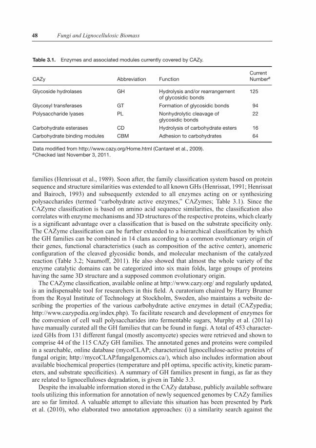
families (Henrissat et al., 1989). Soon after, the family classification system based on proteinsequence and structure similarities was extended to all known GHs (Henrissat, 1991; Henrissatand Bairoch, 1993) and subsequently extended to all enzymes acting on or synthesizingpolysaccharides (termed “carbohydrate active enzymes,” CAZymes; Table 3.1). Since theCAZyme classification is based on amino acid sequence similarities, the classification alsocorrelates with enzyme mechanisms and 3D structures of the respective proteins, which clearlyis a significant advantage over a classification that is based on the substrate specificity only.The CAZyme classification can be further extended to a hierarchical classification by whichthe GH families can be combined in 14 clans according to a common evolutionary origin oftheir genes, functional characteristics (such as composition of the active center), anomericconfiguration of the cleaved glycosidic bonds, and molecular mechanism of the catalyzedreaction (Table 3.2; Naumoff, 2011). He also showed that almost the whole variety of theenzyme catalytic domains can be categorized into six main folds, large groups of proteinshaving the same 3D structure and a supposed common evolutionary origin.
The CAZyme classification, available online at http://www.cazy.org/ and regularly updated,is an indispensable tool for researchers in this field. A curatorium chaired by Harry Brumerfrom the Royal Institute of Technology at Stockholm, Sweden, also maintains a website de-scribing the properties of the various carbohydrate active enzymes in detail (CAZypedia;http://www.cazypedia.org/index.php). To facilitate research and development of enzymes forthe conversion of cell wall polysaccharides into fermentable sugars, Murphy et al. (2011a)have manually curated all the GH families that can be found in fungi. A total of 453 character-ized GHs from 131 different fungal (mostly ascomycete) species were retrieved and shown tocomprise 44 of the 115 CAZy GH families. The annotated genes and proteins were compiledin a searchable, online database (mycoCLAP; characterized lignocellulose-active proteins offungal origin; http://mycoCLAP.fungalgenomics.ca/), which also includes information aboutavailable biochemical properties (temperature and pH optima, specific activity, kinetic param-eters, and substrate specificities). A summary of GH families present in fungi, as far as theyare related to lignocelluloses degradation, is given in Table 3.3.
Despite the invaluable information stored in the CAZy database, publicly available softwaretools utilizing this information for annotation of newly sequenced genomes by CAZy familiesare so far limited. A valuable attempt to alleviate this situation has been presented by Parket al. (2010), who elaborated two annotation approaches: (i) a similarity search against the

P1: SFK/UKS P2: SFK
BLBS110-c03 BLBS110-Kubicek June 21, 2012 7:54 Trim: 244mm×172mm
3 The Tools—Part 1: Enzymology of Cellulose Degradation 49
Table 3.2. Glycosyl hydrolase clans.
Clan GH Families Structure Main Enzyme Activities
GH-A 1, 2, 5, 10, 17, 26,30, 35, 39, 42, 50,51, 53, 59, 72, 79,86, 113
(�/�)8 Various
GH-B 7, 16 �-jelly roll Cellulases, endo-�-1,3/1,4-glucanases
GH-C 11, 12 �-jelly roll Xylanases, endo-�-1,3-glucanases
GH-D 27, 31, 36 (�/�)8 �-Galactosidases, �-xylosidases
GH-E 33, 34, 83, 93 Sixfold �-propeller Sialidase, neuraminidase,�-1,5-endoarabinase
GH-F 43, 62 Fivefold �-propeller �-L-arabinofuranosidases
GH-G 37, 63 (�/�)6 �-Glucosidase, trehalase
GH-H 13, 70, 77 (�/�)8 Amylases and related �-glucosidases
GH-I 24, 46, 80 �+� Lysozyme, chitosanase
GH-J 32, 68 Fivefold �-propeller Invertases, inulinases
GH-K 18, 20, 85 (�/�)8 Chitinases, N-acetyl-�-glucosaminidases
GH-L 15, 65, 125 (�/�)6 Glucoamylase, trehalose phosphorylase,�-1,6-mannosidase
GH-M 8, 48 (�/�)6 Bacterial endo-processive cellulases andchitinases
GH-N 28, 49 �-helix Pectinase, �-1,6-glucanase
entire nonredundant sequences of the CAZy database and (ii) an automatic annotation usinglinks or correspondences between the CAZy families and protein family domains (CAZymesAnalysis Toolkit; it is available at http://cricket.ornl.gov/cgi-bin/cat.cgi).
In the further course of this and the following chapters dealing with the lignocellulosesGHs, I will use a combined approach starting with a grouping according to the substrate thatis hydrolyzed (cf. Table 3.3) and using the CAZyme concept as a subsequent classificationcriterion for each of these groups. As far as it is known and relevant to the understanding,I will also provide information on the protein structure, substrate specificity, and reactionmechanism for these protein families.
3.2 Fungal Cellulolytic Enzymes
As explained in Chapter 2, the canonical view of hydrolysis of cellulose involves the ac-tion of two types of cellulases in an exo-/endo-synergy, followed by a �-glucosidase thathydrolyzes the soluble cellodextrin oligomers to glucose. However, it has been recognizedfor some time now that the differentiation of cellulases into endoglucanases and cellobio-hydrolases is an oversimplification: cellulases have evolved to a continuum of overlappingmodes of actions ranging from totally random endoglucanases through processive endoglu-canases to strictly exo-acting highly processive cellobiohydrolases (Teeri, 1997; Kurasin andValjamae, 2011). The exact roles of individual enzymes with different degrees of processivityand endo-activity in cellulose degradation are not known. The genome sequences of more than
P1: SFK/UKS P2: SFK
BLBS110-c03 BLBS110-Kubicek June 21, 2012 7:54 Trim: 244mm×172mm
50 Fungi and Lignocellulosic Biomass
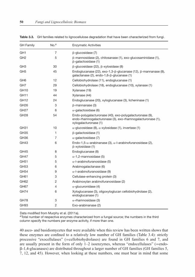
Table 3.3. GH families related to lignocellulose degradation that have been characterized from fungi.
GH Family No.a Enzymatic Activities
GH1 7 �-glucosidase (7)
GH2 5 �-mannosidase (2), chitosanase (1), exo-glucosaminidase (1),�-galactosidase (1)
GH3 30 �-glucosidase (22), �-xylosidase (8)
GH5 45 Endoglucanase (22), exo-1,3-�-glucanase (12), �-mannanase (8),galactanase (2), endo-1,6-�-glucanase (1)
GH6 12 Cellobiohydrolase (11), endoglucanase (1)
GH7 29 Cellobiohydrolase (18), endoglucanase (10), xylanase (1)
GH10 19 Xylanase (19)
GH11 44 Xylanase (44)
GH12 24 Endoglucanase (20), xyloglucanase (3), licheninase (1)
GH26 3 �-mannanase (3)
GH27 6 �-galactosidase (6)
GH28 54 Endo-polygalacturonase (40), exo-polygalacturonase (9),endo-rhamnogalacturonase (3), exo-rhamnogalacturonase (1),xylogalacturonase (1)
GH31 10 �-glucosidase (8), �-xylosidase (1), invertase (1)
GH35 1 �-galactosidase (1)
GH36 7 �-galactosidase (7)
GH43 6 Endo-1,5-�-arabinanase (3), �-l-arabinofuranosidase (2),�-xylosidase (1)
GH45 8 Endoglucanase (8)
GH47 5 �-1,2-mannosidase (5)
GH51 5 �-l-arabinofuranosidase (5)
GH53 6 Arabinogalactanase (6)
GH54 9 �-l-arabinofuranosidase (9)
GH61 3 Cellulase-enhancing protein (3)
GH62 2 Arabinoxylan arabinofuranosidase (2)
GH67 4 �-glucuronidase (4)
GH74 6 Xyloglucanase (3), oligoxyloglucan cellobiohydrolase (2),endoglucanase (1)
GH78 3 �-rhamnosidase (3)
GH93 2 Exo-arabinanase (2)
Data modified from Murphy et al. (2011a).aTotal number of respective enzymes characterized from a fungal source; the numbers in the thirdcolumn specify the numbers per enzyme activity, if more than one.
40 asco- and basidiomycetes that were available when this review has been written shows thatthese enzymes are confined to a relatively low number of GH families (Table 3.4): strictlyprocessive “exocellulases” (=cellobiohydrolases) are found in GH families 6 and 7, andare usually present in the form of only 1–2 isoenzymes, whereas “endocellulases” (=endo-�-1,4-glucanases) are distributed throughout a larger number of GH families (GH families 5,7, 12, and 45). However, when looking at these numbers, one must bear in mind that some

P1: SFK/UKS P2: SFK
BLBS110-c03 BLBS110-Kubicek June 21, 2012 7:54 Trim: 244mm×172mm
Tab
le3.
4.D
istr
ibut
ion
ofce
llula
seG
Hs
infu
ngi.
Cel
lula
ses
�-g
luco
sid
ases
GH
Fam
ilyP
MO
Phy
llum
Cla
ssS
pec
ies
56
712
451
361
Pez
izom
ycot
aE
urot
iom
ycet
esA
.nid
ulan
s15
23
11
320
9P
eziz
omyc
ota
Eur
otio
myc
etes
A.n
iger
102
24
03
177
Pez
izom
ycot
aE
urot
iom
ycet
esP
enic
illiu
mch
ryso
genu
m13
12
30
317
4P
eziz
omyc
ota
Leot
iom
ycet
esS
cler
otin
iasc
lero
tioru
m5
13
22
112
9P
eziz
omyc
ota
Pez
izom
ycet
esTu
ber
mel
anos
por
um6
00
11
26
4P
eziz
omyc
ota
Leot
iom
ycet
esB
lum
eria
gram
inis
var.
hord
ei0
00
00
00
2P
eziz
omyc
ota
Leot
iom
ycet
esB
otry
tisci
nere
a5
13
12
514
10P
eziz
omyc
ota
Sor
dar
iom
ycet
esFu
sariu
mgr
amin
earu
m15
12
41
322
15P
eziz
omyc
ota
Sor
dar
iom
ycet
esFu
sariu
mgr
amin
earu
m3
12
21
317
13P
eziz
omyc
ota
Sor
dar
iom
ycet
esN
ectr
iaha
emat
ococ
ca18
13
61
538
12P
eziz
omyc
ota
Sor
dar
iom
ycet
esN
euro
spor
acr
assa
73
51
11
914
Pez
izom
ycot
aS
ord
ario
myc
etes
Mag
nap
orth
egr
isea
133
63
12
1917
Pez
izom
ycot
aS
ord
ario
myc
etes
Pod
osp
ora
anse
rina
154
62
21
1133
Pez
izom
ycot
aS
ord
ario
myc
etes
T.at
rovi
ride
141
23
14
143
Pez
izom
ycot
aS
ord
ario
myc
etes
T.re
esei
111
22
12
133
Pez
izom
ycot
aS
ord
ario
myc
etes
T.vi
rens
161
24
22
173
Pez
izom
ycot
aD
othi
deo
myc
etae
Myc
osp
here
llagr
amin
earu
m0
01
11
NI
NI
2P
eziz
omyc
ota
Dot
hid
eom
ycet
aeS
tago
nosp
ora
nod
orum
34
54
3N
I16
30B
asid
iom
ycot
aU
stila
gino
myc
etes
Ust
ilago
may
dis
00
00
30
30
Bas
idio
myc
ota
Puc
cini
omyc
etes
Puc
cini
agr
amin
isva
r.tr
itici
90
72
00
23
Bas
idio
myc
ota
Aga
ricom
ycet
esS
chiz
ophy
llum
com
mun
e18
12
11
312
22B
asid
iom
ycot
aA
garic
omyc
etes
Lacc
aria
bic
olor
220
03
00
28
Bas
idio
myc
ota
Aga
ricom
ycet
esP
ostia
pla
cent
a36
00
40
49
4B
asid
iom
ycot
aA
garic
omyc
etes
P.ch
ryso
spor
ium
201
92
02
1115
Bas
idio
myc
ota
Aga
ricom
ycet
esS
erp
ula
lacr
yman
s20
10
1N
IN
I10
5B
asid
iom
ycot
aA
garic
omyc
etes
C.c
iner
ea26
57
1N
IN
I7
33
Dat
ata
ken
from
Kub
icek
etal
.(20
11),
Eas
twoo
det
al.(
2011
),an
dG
ood
win
etal
.(20
11).
NIm
eans
“no
info
rmat
ion”
was
avai
lab
le;P
MO
,pol
ysac
char
ide
mon
ooxy
gena
ses,
pre
viou
sly
bel
ieve
dto
be
clas
sG
H61
cellu
lase
-enh
anci
ngp
rote
ins.
51

P1: SFK/UKS P2: SFK
BLBS110-c03 BLBS110-Kubicek June 21, 2012 7:54 Trim: 244mm×172mm
52 Fungi and Lignocellulosic Biomass
of these GH families (particularly GH5) also contain a number of enzymes with other sub-strate specificities (such as endomannanases, �-1,6-galactanase, �-1,3-mannanase, xyloglu-canases), whereas other families comprise cellulases only. �-Glucosidases are predominantlyfound in the GH1 and GH3 families, which however also contain other glycosidases such as�-galactosidase, �-mannosidases, and others.
In addition, it became recently very clear that there are further proteins (called “cellulaseenhancing proteins”) that are now known to strongly and synergistically raise the activityof the cellulases but do not exhibit any enzymatic activity on cellulose themselves (i.e., thenonhydrolytic “endoglucanase” CEL61, and the expansin-like protein swollenin; Saloheimoet al., 2002; Harris et al., 2010). They may thus fulfill the role of the missing link in the earlytheory of cellulase action, the Cx-C1 model (see Chapter 2), and be the “swelling factor,”C1, a nonhydrolytic component that functions to make the substrate more accessible to Cx.Therefore, these proteins will also be described in this chapter.
Further, cellulases and related GHs very often display a modular structure that consistsof a catalytic domain and a polysaccharide-binding domain connected by a loop (“hinge”)region, which has been first discovered during the investigation of cellobiohydrolases I andII of Trichoderma reesei (Teeri et al., 1987). Because these polysaccharide-binding domainsoccur in most of the fungal cellulases, I decided to describe them first.
3.2.1 Cellulose-Binding Domains
Noncatalytic polysaccharide-binding modules of GHs were originally defined as cellulose-binding domains (CBDs) because the first examples investigated bound tightly to crystallinecellulose (reviewed by Boraston et al., 2004). Since thereafter, several polysaccharide-bindingmodules were found, which bind to polysaccharides other than cellulose; however, the moregeneral term carbohydrate-binding module is used to reflect this diversity in specificity(Boraston et al., 1999). In analogy to the GH classification, CBMs are also divided into familiesbased on amino acid sequence similarity, of which today 63 families have been defined andincluded in the CAZy database (http://www.cazy.org/Carbohydrate-Binding-Modules.html).They display a broad spectrum of ligand specificity, including very specific CBM-ligand in-teractions but some of them also bind to a broader range of different carbohydrates. Mostof them are found in GHs that act on insoluble polysaccharides, indicating the necessity oflocating the respective enzymes to such insoluble substrates. Interestingly, the largest numberof CBM families is found only in bacterial enzymes, whereas only about 25% of the currentlyknown CBMs are present in fungal enzymes. Table 3.5 lists those CBM families and thecarbohydrates they bind to, which have been found in fungi and that act on polysaccharidesfound in lignocellulose biomass.
Regarding the structure, the most important conformational element of most CBMs is the�-sheet. The folds and architecture displayed by these �-sheets have led to a classificationinto seven families, of which the �-sandwich is the most recurrent fold (Boraston et al., 2004;Hashimoto, 2006).
Unfortunately, the grouping of CBM families based on the conservation of the protein foldis not predictive of function, as specific amino acids or binding-site topographies are notconserved. Consequently, another classification of CBMs based on structural and functionalsimilarities has been proposed: “surface-binding” CBMs (type A), “glycan-chain-binding”CBMs (type B), and “small-sugar-binding” CBMs (type C) (Boraston et al., 2004; see alsoTable 3.5). Type A CBMs consist of the canonical �-sandwich structure, which is composed
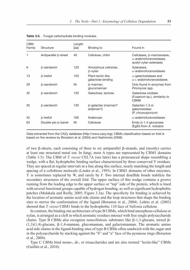
P1: SFK/UKS P2: SFK
BLBS110-c03 BLBS110-Kubicek June 21, 2012 7:54 Trim: 244mm×172mm
3 The Tools—Part 1: Enzymology of Cellulose Degradation 53
Table 3.5. Fungal carbohydrate binding modules.
CBMFamily Structure
Length[aa] Binding to Found in
1 Antiparallel �-sheet 40 Cellulose, chitin Cellulases, �-mannanases,�-arabinofuranosidases,acetyl xylan esterases
6 �-sandwich 120 Amorphous cellulose,�-xylan
Xylanases,�-arabinofuranosidases
13 �-trefoil 150 Plant-lectin-likegalactose binding
�-galactosidases and�-L-arabinofuranosidases
29 �-sandwich NI �-mannan,glucomannan
Only found in enzymes fromPiromyces spp.
32 �-sandwich 120 Galactose, lactose Galactose oxidase(Fusarium sp.), similarity toCBM6
35 �-sandwich 130 �-galactan (mannan?arabinan?)
Galactan-1,3-�-galactosidase(P. chrysosporium)
42 �-trefoil 160 Arabinose �-arabinofuranosidases
63 Double-psi-�-barrel NI Cellulose Endo-�-1,4-glucanase(EglD) from A. nidulans
Data extracted from the CAZy database (http://www.cazy.org); CBMs classification based on fold isbased on the reviews by Boraston et al. (2004) and Hashimoto (2006).
of two �-sheets, each consisting of three to six antiparallel �-strands, and (mostly) carriesat least one structural metal ion. In fungi, most A types are represented by CBM1 domains(Table 3.5). The CBM of T. reesei CEL7A (see later) has a pronounced shape resembling awedge, with a flat, hydrophobic binding surface characterized by three conserved Y residues.They are spaced at regular intervals in a line along this surface, nearly matching the length andspacing of a cellobiose molecule (Linder et al., 1995). In CBM1 domains of other enzymes,Y is sometimes replaced by W, and rarely by F. Two internal disulfide bonds stabilize thesecondary structures of the overall fold. The upper surface of this wedge contains a grooverunning from the leading edge to the upper surface or “top” side of the protein, which is linedwith several functional groups capable of hydrogen bonding, as well as significant hydrophobicpatches (Mulakala and Reilly, 2005; Figure 3.3a). The specificity for binding is achieved bythe location of aromatic amino acid side chains and the loop structures that shape the bindingsites to mirror the conformation of the ligand (Boraston et al., 2004). Lehtio et al. (2003)showed that T. reesei CBM1 binds to the hydrophobic 110 face of Vallonia cellulose.
In contrast, the binding site architecture of type B CBMs, which bind amorphous cellulose orxylan, is arranged as a cleft in which aromatic residues interact with free single polysaccharidechains. Type B CBMs also recognize noncellulosic substrates like �-1,3-glucans, mixed �-(1,3)(1,4)-glucans, �-1,4-mannan, glucomannan, and galactomannan. The aromatic aminoacid side chains in the ligand-binding sites of type B CBMs often sandwich with the sugar unitin the polysaccharide by stacking against the “b” and “a” face of the pyranose rings (Borastonet al., 2004).
Type C CBMs bind mono-, di-, or trisaccharides and are also termed “lectin-like” CBMs(Guillen et al., 2010).

P1: SFK/UKS P2: SFK
BLBS110-c03 BLBS110-Kubicek June 21, 2012 7:54 Trim: 244mm×172mm
54 Fungi and Lignocellulosic Biomass
(a)
(c)
(b)
Figure 3.3. Structure of cellobiohydrolase CEL7A from T. reesei: (a) the CBM1 cellulose-bindingdomain (accession number 1CBH), (b) structure of the catalytic domain of CEL7A (accession number1CEL), and (c) side view of the complex of CBH I docked onto the surface of the model cellulosemicrofibril. (I acknowledge the US Department of Energy Genomic Science program and the websitehttp://genomicscience.energy.gov for the kind gift of this figure.)
Calcium is known to play a significant role in the interaction of lectins with their targetligands, either by maintaining the binding site in the correct conformation or via directcoordination with the carbohydrate itself. Indeed, xylan recognition by a bacterial CBM35(from Cellvibrio spp., in Abf62A) is Ca2+-dependent, but it has not been investigated asyet whether this also accounts for the CBM35 modules in ascomycetous �-mannanases, �-galactosidases, and �-arabinofuranosidases.
CBMs are generally believed to have three roles, which aid to the function of their cognatecatalytic modules: (i) providing a proximity effect, (ii) a targeting function (i.e., bringingthe enzyme and particularly the catalytic module to the substrate), and (iii) a disruptive

P1: SFK/UKS P2: SFK
BLBS110-c03 BLBS110-Kubicek June 21, 2012 7:54 Trim: 244mm×172mm
3 The Tools—Part 1: Enzymology of Cellulose Degradation 55
function. With regards to the last point, binding of CBM1 domains from a T. pseudokoningiiand Penicillium janthinellum cellobiohydrolase 1 to cellulose was shown to cause structuralchanges and release of short fibers, and the authors concluded that the CBM1 protein acts bydisrupting hydrogen bonds between cellulose chains (Gao et al., 2001; Wang et al., 2008).Mulakala and Reilly (2005) suggested that the CBM1 of T. reesei CEL7A domain wedges itselfunder a free reducing chain end on the crystalline cellulose surface and feeds it to the activesite tunnel of the catalytic domain. However, Igarashi et al. (2009), using high-speed atomicforce microscopy to study real-time sliding of CEL7A molecules on crystalline cellulose,showed that the catalytic domain without the CBD moved with a velocity similar to that ofthe intact Cel7A enzyme with a speed of 3.5 nm/s, and this sliding was absent in proteinswith loss-of-catalysis mutations in the catalytic domain. Consequently, the CBM1 module isdispensable for lifting and feeding of the cellulose chain. They therefore concluded that therole of the cellulose-binding CBM1 is merely to increase the enzyme concentration on thecrystalline substrate.
For a deeper and more comprehensive review about all (not only fungal) CBMs, I refer thereader to the articles by Boraston et al. (2004), Shoseyov et al. (2006), Hashimoto (2006) andGuillen et al. (2010).
3.2.2 Cellobiohydrolases (EC 3.2.1.91)
GH7 Cellobiohydrolase Cel7A/CBH1
As mentioned earlier, fungal cellobiohydrolases are found in the glycosyl hydrolase familiesGH6 and GH7. Thereby, cellobiohydrolase I (CBH1, which according to its categorizationinto GH7 is now called CEL7A) is the archetypus of fungal cellulases, with which most of themechanistic and structural studies have been performed, and its characterization from differentfungi outnumbers that of all other cellulases (Table 3.6). CEL7A from T. reesei was the firstcellulase protein that was purified and characterized, whose gene was cloned, and whose 3D
Table 3.6. Characterized cellobiohydrolase I proteins from fungi.
Species Protein ID (Genbank) Reference
PezizomycotaA. aculeatus BAA25183 Takada et al., 1998A. niger AAF04491 Gielkens et al., 1999A. nidulans AAM54069 Lockington et al., 2002Penicillium chrysogenum AAV65115, AAX84833 Hou et al., 2007Penicillium janthinellum CAA41780 Koch et al., 1993Humicola grisea var. thermoidea BAA74517 Takashima et al., 1998Thermoascus aurantiacus AAL16941, AAL83303 Hong et al., 2003T. koningii CAA49596 Wey et al., 1994T. reesei CAH10320 Fagerstam and Pettersson, 1980Melanocarpus albomyces CAD56667 Haakana et al., 2004Cochliobolus carbonum AAC49089 Sposato et al., 1995Cryphonectria parasitica AAB00479 Wang and Nuss, 1995
BasidiomycotaIrpex lacteus BAA76363 Hamada et al., 1999P. chrysosporium AAB46373, CAA80253 Vanden Wymelenberg et al., 1993
Data retrived by searching MycoClap (Murphy et al., 2011) for “cellobiohydrolase 1.”

P1: SFK/UKS P2: SFK
BLBS110-c03 BLBS110-Kubicek June 21, 2012 7:54 Trim: 244mm×172mm
56 Fungi and Lignocellulosic Biomass
structure had been elucidated. This all is due to the fact that this enzyme makes up morethan 60% of the total protein secreted by T. reesei during growth under cellulase-inducingconditions (see Chapter 7), and it is also the major cellulase protein secreted by other fungi(with the exception of brown rot fungi, see Chapter 2). Orthologs of CEL7A have been foundin all ascomycete and white-rot basidiomycete genomes, and a phylogenetic analysis reflectsthe species phylogeny, thus implying that CEL7A is an essential component of fungi.
The CEL7A protein consists of an N-terminal catalytic domain, a C-terminal CBM1carbohydrate-binding domain, and an unstructured “hinge” domain that links these both(Figures 3.3b and c). The latter linker is rich in S and T residues, which are also highly O-glycosylated. Beckham et al. (2010) performed simulations that suggested that the linker is anintrinsically disordered protein of 16 A extension and thus considerably expands the operatingrange of Cel7A. The 3D structures of the catalytic domain of CEL7A from T. reesei (Divneet al., 1994), Phanerochaete chrysosporium (Munoz et al., 2001), from the thermophilic fungusTalaromyces emersonii (Grassick et al., 2004), and from Melanocarpus albomyces (Parkkinenet al., 2008) have been solved. They are built up around a �-jelly roll folded framework, inwhich two large antiparallel �-sheets pack face-to-face to form a highly curved �-sandwich.This �-sandwich is further extended along both edges by several of the loops that connect the�-strands, resulting in a long (about 50 A) substrate-binding surface that runs perpendicularto the �-strands of the inner, concave �-sheet. A few further short �-helical segments occurin some of the loops at the periphery of the structure (Figure 3.3b; Stahlberg, 2011).
The catalytic domain of CEL7A contains this approximately 50 A-long active-site tunnelthat is decorated with amino acid side chains that contains ten subsites (−7 to +3) for theglycosyl units of a cellulose chain (Divne et al., 1994). The cellulose chain enters from the−7 subsite and is thread through the tunnel in a manner that twists the cellulose chain almostupside down. Once in the tunnel, the cellulose chain is then processively cleaved, two glucosylresidues at a time. The tunnel contains a total of seven glucosyl-binding sites, four of whichare formed by W residues, which are major determinants for the formation of the −7, −4,−2, and +1 glucosyl-binding sites. The glucosyl rings seem to slide over the indole surface,and thus likely interact only rather weakly. This could be important for hydrolysis becausestrong interactions throughout the tunnel would hamper the advancement of the chain in thedirection of the active site (Divne et al., 1994). All glucosyl units in the eleven different bindingsites are in phase and aligned such that the glucosyl units at the active site, located at −1,are oriented for catalysis. The proposed nucleophile and acid/base catalysts, E212 and E217(Divne et al., 1994; Stahlberg et al., 1996), are positioned roughly on opposite sides of theglycosidic linkage that is to be cleaved, with their carboxylate groups about 6 A apart. The sitesfor product binding are located at +1/+2, indicating that hydrolysis of the glycosyl-enzymeintermediate may proceed without prior release of the cellobiose product and suggests a productejection mechanism during processive hydrolysis of cellulose (Ubhayasekera et al., 2005).
As also indicated by the spacing of E212 and E217 (see earlier), CEL7A is a retainingglycosidase that acts by a double-displacement mechanism (Figure 3.2b). These two E-residuesperfectly mirror the typical -E1-X-D-X-X-E2- consensus motif, in which E1 acts as the catalyticnucleophile and E2 as general acid/base. Interestingly, the CEL7A enzymes act from thereducing ends of the cellulose chains, which is in conflict with the general IUBMB definitionof cellobiohydrolases (EC 3.2.1.91), which implies that they would act from the nonreducingends of cellulose.
The progress of action of CEL7A has been summarized by Levine et al. (2010): the enzymefirst adsorbs to the crystalline regions of the cellulose surface via the CBM domain, thendiffuses over the surface. Eventually, it binds a nonreducing end of a cellulose chain, catalyzes
P1: SFK/UKS P2: SFK
BLBS110-c03 BLBS110-Kubicek June 21, 2012 7:54 Trim: 244mm×172mm
3 The Tools—Part 1: Enzymology of Cellulose Degradation 57
the hydrolysis event, generating cellobiose, and finally desorbs from the cellulose surface orstarts to bind to the next nonreducing end. Fox et al. (2011) recently reported that the rate ofCEL7A-catalyzed hydrolysis of crystalline cellulose is limited by the rate of binding of theenzyme to the cellulose chains, which in turn was equivalent to the rate of initial productionof hydrolysis products and dependent on the micromorphology of the cellulose surface. Thisrate was enhanced in the presence of endoglucanases (see later).
At the time of this writing, the interplay between the catalytic and the CBM1 domain is stilla matter of intense modeling and debate. Zhao et al. (2008) considered that the CBM1 modulewould prevent the catalytic domain from diffusing away from the surface of the substrate, andthus maintain it there in a high local concentration. Bu et al. (2009) showed that CBM1 alone
exhibits regions of stability on the hydrophobic face of cellulose at every 5 and 10 A′, which
would correspond to a glucose unit and a cellobiose unit, respectively. They observed that inthe presence of hydrolyzed cellulose chain ends, CBM1 exerted a thermodynamic driving forceto translate away from the free cellulose chain ends and concluded that it thus may be a drivingforce on the enzyme during processive hydrolysis of cellulose. Zhao et al. (2008) suggestedthat the linker segment might store energy, in the manner of a compressed spring, perhapsforcing the chain further into the active site after each bond scission and product escape ordriving the CBM1 to advance along the substrate chain, pulling the catalytic domain along afterit (Zhao et al., 2008). Indeed, the water bound by the heavy glycosylation of the linker chaincould create a gel-like zone between the catalytic and the cellulose-binding modules, whichwould inhibit their relative motions and push the smaller CBM1 forward along the chain, awayfrom the heavier catalytic domain, thus promoting processivity (Zhong et al., 2009).
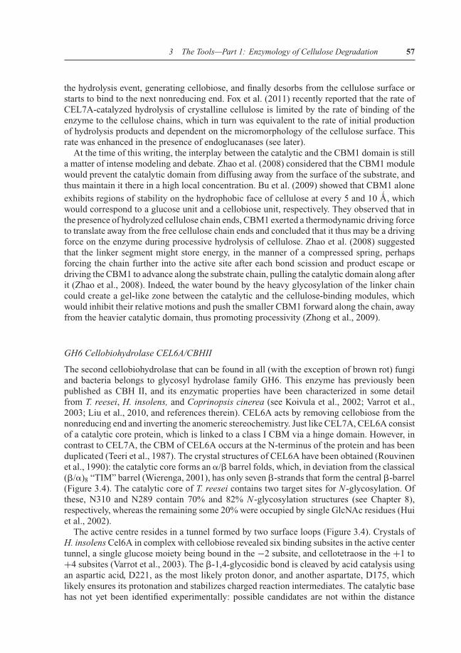
GH6 Cellobiohydrolase CEL6A/CBHII
The second cellobiohydrolase that can be found in all (with the exception of brown rot) fungiand bacteria belongs to glycosyl hydrolase family GH6. This enzyme has previously beenpublished as CBH II, and its enzymatic properties have been characterized in some detailfrom T. reesei, H. insolens, and Coprinopsis cinerea (see Koivula et al., 2002; Varrot et al.,2003; Liu et al., 2010, and references therein). CEL6A acts by removing cellobiose from thenonreducing end and inverting the anomeric stereochemistry. Just like CEL7A, CEL6A consistof a catalytic core protein, which is linked to a class I CBM via a hinge domain. However, incontrast to CEL7A, the CBM of CEL6A occurs at the N-terminus of the protein and has beenduplicated (Teeri et al., 1987). The crystal structures of CEL6A have been obtained (Rouvinenet al., 1990): the catalytic core forms an �/� barrel folds, which, in deviation from the classical(�/�)8 “TIM” barrel (Wierenga, 2001), has only seven �-strands that form the central �-barrel(Figure 3.4). The catalytic core of T. reesei contains two target sites for N-glycosylation. Ofthese, N310 and N289 contain 70% and 82% N-glycosylation structures (see Chapter 8),respectively, whereas the remaining some 20% were occupied by single GlcNAc residues (Huiet al., 2002).
The active centre resides in a tunnel formed by two surface loops (Figure 3.4). Crystals ofH. insolens Cel6A in complex with cellobiose revealed six binding subsites in the active centertunnel, a single glucose moiety being bound in the −2 subsite, and cellotetraose in the +1 to+4 subsites (Varrot et al., 2003). The �-1,4-glycosidic bond is cleaved by acid catalysis usingan aspartic acid, D221, as the most likely proton donor, and another aspartate, D175, whichlikely ensures its protonation and stabilizes charged reaction intermediates. The catalytic basehas not yet been identified experimentally: possible candidates are not within the distance

P1: SFK/UKS P2: SFK
BLBS110-c03 BLBS110-Kubicek June 21, 2012 7:54 Trim: 244mm×172mm
58 Fungi and Lignocellulosic Biomass
Figure 3.4. Three-dimensional structure of cellobiohydrolase II CEL6A from T. reesei (1CB2).
of a hydrogen bond to a water molecule that could act as the nucleophile in this invertingmechanism. Thus, the current interpretation proposes that the water is deprotonated througha “solvent wire” through to one of the conserved D residues near the active center (Piens andDavies, 2011).
The crystal structure of CBHII also shows a tyrosine residue, Y169, located close enoughto the hydrolytic target bond to be involved in catalysis. Exchange of this residue to a pheny-lalanine (Y169F) increased the association constants of the mutant enzyme for cellobioseand cellotriose threefold and simultaneously reduced the catalytic constants toward the samesubstrates fourfold (Koivula et al., 1996). The data suggest that Y169 interacts with a glu-cose ring in the substrate at the second subsite, thereby distorting the glucose ring into amore reactive conformation. In addition, Y169 may affect the protonation state of the activesite carboxylates, D175 and D221 (Koivula et al., 1996).
Interestingly, Varrot et al. (2003) showed that the catalytic core of H. insolens Cel6Aundergoes several conformational changes upon substrate binding, the most significant ofwhich is a closing of the two active site loops (residues 174–196 and 397–435) with mainchain movements of up to 4.5 A
′.
3.2.3 Endo-β-1,4-Glucanases (EC 3.2.1.4)
GH5 Endo-β-1,4-Glucanases
Glycosyl hydrolase family GH5 is one of the largest of all GH families and contains enzymesacting on a wide variety of substrates. Most of them are endo-�-1,4-glucanases and endo-�-1,4-mannanase, but other activities include endo-�-1,6-galactanase, endo-�-1,3-mannanase,endo-�-1,4-xylanase, as well as high specificity xyloglucanases. St. John et al. (2010) have
P1: SFK/UKS P2: SFK
BLBS110-c03 BLBS110-Kubicek June 21, 2012 7:54 Trim: 244mm×172mm
3 The Tools—Part 1: Enzymology of Cellulose Degradation 59
recently shown that many members of GH5 actually should be classified as glycosyl hydro-lase family 30, but the fungal endo-�-1,4-glucanases remained unaffected from this shift.Unfortunately, the 3D structure of these cellulases has only been solved for bacterial enzymes(e.g., Clostridium thermocellum endoglucanase CelC and Bacillus agaradhaerens Cel5A;Dominguez et al., 1995; Davies et al., 1998). They are members of Clan GH-A and thus have aclassical (�/�)8-TIM barrel fold, with two E residues forming the active center. GH5 enzymesare retaining enzymes and act by the Koshland double-displacement mechanism and the twocatalytic residues (catalytic nucleophile and general acid/base) are known to be E’s found atthe C-terminal ends of �-strands four (acid/base) and seven (nucleophile; Davies, 2011).
As for fungi, endo-�-1,4-glucanases from GH5 have been purified and characterized fromT. reesei (previously called EGIII), Aspergillus spp., and also anaerobic fungi (Saloheimo et al.,1988; Eberhard et al., 2000; Hara et al., 2003). However, none of these has been investigatedin more detail.
GH7 Endo-β-1,4-Glucanases
Endo-�-1,4-glucanases from glycosyl hydrolase family GH7 are produced by most fungi inmajor amounts and have therefore been characterized in some detail from several sources.Being a member of GH7, they are very similar in structure to CEL7A CBH1 (displaying a�-jelly roll-derived framework) and in the basic mechanism of action, that is, they attack atthe reducing end by a retaining mechanism. All these features have been explained in detail atcellobiohydrolase CEL7A earlier (Section 3.2.2) and will thus not be repeated here. However,the major difference between the cellobiohydrolase and endoglucanase enzymes of GH7 isthat the latter have an open substrate binding cleft instead of a tunnel, which enables theattack in the middle of the cellulose molecule and thus endo-action. Similar findings havealso been reported for GH6 enzymes, which also (although not in fungi) contain endo-�-1,4-glucanases. When the structure of the first GH6 endo-�-1,4-glucanase was solved, the activecenter was observed in a long open groove and provided the first hint that endo or exo activitycould be modulated through display of the active center either in an open grove (for endo)or loop-enclosed tunnel (for exo), respectively. Proof for this was obtained by showing thatthe exo activity was changed to endo when the extended loops of a cellobiohydrolase fromthe prokaryote Thermomonospora fusca were truncated (Meinke et al., 1995; Figure 3.5).Since the loops that form the active center of the cellobiohydrolases are flexible and showmultiple conformations, the enzyme may switch between exo and endo activity depending onthe conformational changes of that loop. There is thus an ongoing debate whether true exo-and endo-cellulases actually exist (cf. Stahlberg, 2011).
As with other cellulase, CEL7B occurs in several isoforms that exhibit different isoelectricpoints. In CEL7B, this has been studied in some detail: T. reesei CEL7B was shown to occur inat least 14 different glycoforms (Garcıa et al., 2001). The major isoform contained only a singleN-linked GlcNAc-, and a single probably O-linked mannose residue. A minor population ofthe enzyme contained Man5–7GlcNAc2 antennae, and all the others contained a negativelycharged phosphate ester on the N-glycans. Eriksson et al. (2004), using another preparation ofCEL7B, arrived at basically similar results although they varied in detail, indicating that thesemodifications are dependent on biochemical processes that vary with the culture conditions.Of the five potential sites found in the wild-type enzyme, they found only N56 and N182to be N-glycosylated. GlcNAc2Man5 was identified as the predominant N-glycan, althoughlesser amounts of GlcNAc2Man7 and glycans carrying a mannophosphodiester bond were

P1: SFK/UKS P2: SFK
BLBS110-c03 BLBS110-Kubicek June 21, 2012 7:54 Trim: 244mm×172mm
60 Fungi and Lignocellulosic Biomass
CBHII
C
E2
N
Figure 3.5. �-Carbon skeletons of the T. reesei cellobiohydrolase CEL6A and T. fusca endoglucanaseE2 catalytic domains. The views are chosen to illustrate differences in the accessibilities of the two activesites. C and N, respectively, indicate the carboxyl- and amino-proximal loops that cover the active site ofCBH II. ( C© 1995 The American Society for Biochemistry and Molecular Biology.)
also detected. In addition, they detected partial deamidation of N259 and a partially occupiedO-glycosylation sites.
GH12 Endo-β-1,4-Glucanases
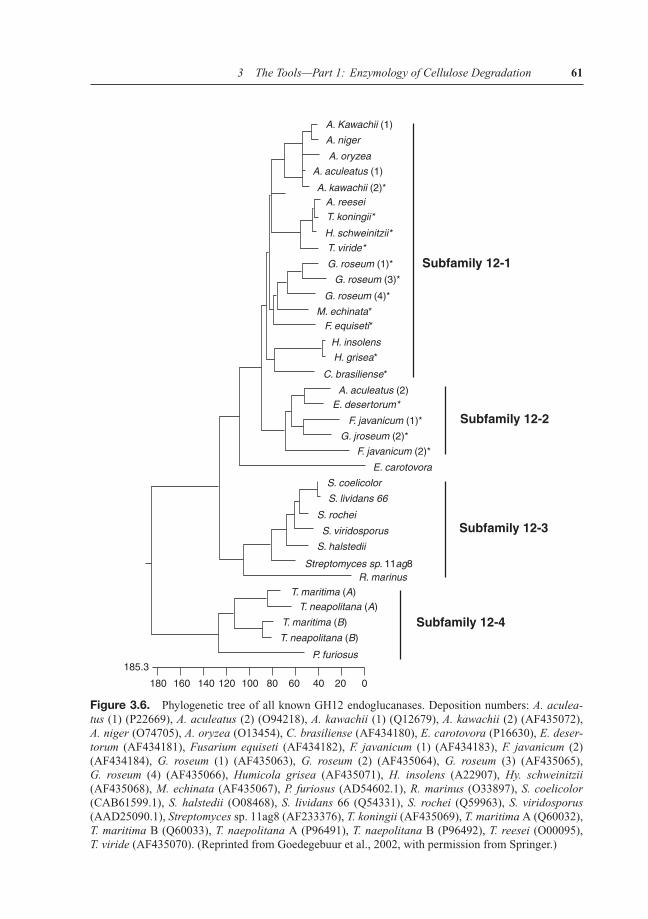
Members of GH family 12 (GH 12) are distributed throughout the bacterial and fungal king-doms and comprise small (20–25 kDa) proteins that—in contrast to most other cellulases—lacka CBD. They are therefore unable to bind to crystalline cellulose and hydrolyze only amorphouscellulose (Henriksson et al., 1999). They perform their hydrolysis via a double-displacementreaction and a glycosyl-enzyme intermediate that results in retention of the anomeric config-uration in the product (Schulein, 1997; Birsan et al., 1998). A phylogenetic analysis shows adivision into five subfamilies of which two exclusively comprise fungal enzymes: 12–1 (fungalgroup I), 12–2 (fungal group II), 12–3 (Streptomyces group including Rhodothermus marinus),12–4 (Thermophiles group), and 12–5 (Erwinia carotovora) (Figure 3.6; Goedegebuur et al.,2002). Some fungi (e.g., Gliocladium roseum) appeared to have duplicated their cel12 genesin subfamily 12–1.
The enzymes from subfamily 12–2 showed the presence of an additional domain withunknown function, and also their catalytic domain was different from the other members. Inaddition, while most of the GH12 glycosyl hydrolase proteins comprise mainly endo-�-1,4-glucanase activities, the enzyme from Aspergillus niger and Malbrachea cinnamomea thatare members of subfamily 12–2 were reported to exhibit xyloglucanase activity (Schuleinet al., 2002; Powlowski et al., 2009). Powlowski et al. (2009) detected a short deletion andinsertion following D112 and I129, respectively, in the A. niger protein AnXEG12A thatare conserved among the subfamily 12–2 sequences but not subfamily 12–1 sequences. A
P1: SFK/UKS P2: SFK
BLBS110-c03 BLBS110-Kubicek June 21, 2012 7:54 Trim: 244mm×172mm
3 The Tools—Part 1: Enzymology of Cellulose Degradation 61
A. Kawachii (1)
Subfamily 12-1
Subfamily 12-2
Subfamily 12-3
Subfamily 12-4
A. niger
A. aculeatus (1)
A. oryzea
A. kawachii (2)*A. reesei
T. koningii*
H. schweinitzii*
T. viride*
G. roseum (1)*
G. roseum (3)*
G. roseum (4)*
M. echinata*F. equiseti*
H. insolens
H. grisea*
C. brasiliense*
A. aculeatus (2)E. desertorum*
F. javanicum (1)*
G. jroseum (2)*
F. javanicum (2)*
E. carotovora
S. coelicolor
S. lividans 66
S. rochei
S. viridosporus
S. halstedii
Streptomyces sp. 11ag8R. marinus
T. maritima (A)
T. maritima (B)
T. neapolitana (B)
P. furiosus185.3
180 160 140 120 100 80 60 40 20 0
T. neapolitana (A)
Figure 3.6. Phylogenetic tree of all known GH12 endoglucanases. Deposition numbers: A. aculea-tus (1) (P22669), A. aculeatus (2) (O94218), A. kawachii (1) (Q12679), A. kawachii (2) (AF435072),A. niger (O74705), A. oryzea (O13454), C. brasiliense (AF434180), E. carotovora (P16630), E. deser-torum (AF434181), Fusarium equiseti (AF434182), F. javanicum (1) (AF434183), F. javanicum (2)(AF434184), G. roseum (1) (AF435063), G. roseum (2) (AF435064), G. roseum (3) (AF435065),G. roseum (4) (AF435066), Humicola grisea (AF435071), H. insolens (A22907), Hy. schweinitzii(AF435068), M. echinata (AF435067), P. furiosus (AD54602.1), R. marinus (O33897), S. coelicolor(CAB61599.1), S. halstedii (O08468), S. lividans 66 (Q54331), S. rochei (Q59963), S. viridosporus(AAD25090.1), Streptomyces sp. 11ag8 (AF233376), T. koningii (AF435069), T. maritima A (Q60032),T. maritima B (Q60033), T. naepolitana A (P96491), T. naepolitana B (P96492), T. reesei (O00095),T. viride (AF435070). (Reprinted from Goedegebuur et al., 2002, with permission from Springer.)

P1: SFK/UKS P2: SFK
BLBS110-c03 BLBS110-Kubicek June 21, 2012 7:54 Trim: 244mm×172mm
62 Fungi and Lignocellulosic Biomass
Figure 3.7. Structure of T. reesei endoglucanase CEL12A (accession number 1H8V).
comparison between enzymes from subfamily 1 and A. niger AnXEG12A (subfamily 12–2)showed that the D112 deletion would shorten a loop region that in Cel12A constricts thesubstrate-binding cleft. The insertion SST after I129 in AnXEG12A, on the other hand, isadjacent to the so-called “cord” region of Cel12A, which contributes amino acid residues tothe substrate-binding cleft that are likely involved in binding the reducing end of the substrate(Sandgren et al., 2001). Powlowski et al. (2009) therefore speculated that this insertion mayalter the substrate-binding properties of xyloglucanase relative to Cel12A.
GH12 endo-�-1,4-glucanases belong to clan GH-C and exhibit a compact �-sandwichstructure that is curved to create an extensive cellulose-binding site on the concave face ofthe �-sheet (Sulzenbacher et al., 1999; Figure 3.7). Structures from T. reesei, H. insolens,and T. citrinoviride are available and display a consistent framework for proteins of the GH12family (Sandgren et al., 2005). The protein comprises two �-sheets, of six and nine strands,packed on top of one another, and a single �-helix. The concave surface of the nine-stranded�-sheet forms a large substrate-binding groove in which the active-site residues are located.It comprises a carboxylic acid trio, similar to that of GH families 7, in which the strictlyconserved D99 also forms hydrogen bonds to the invariant E116. The binding crevice is linedwith both aromatic and polar amino acid side chains, which may play a role in substratebinding. The enzyme contains one disulfide bridge and is glycosylated at D164 by a singleN-acetyl glucosamine residue.
GH45 Endo-β-1,4-Glucanases
The GHs belonging to GH45 have so far only been described as endo-�-1,4-glucanases, andpreviously been considered to belong to cellulase family K. They are known from bacteria andfungi only.
The first GH45 endoglucanase described was endoglucanase V from H. insolens (Davieset al., 1993) and T. reesei EGV (Saloheimo et al., 1994). The protein is unusually small (242

P1: SFK/UKS P2: SFK
BLBS110-c03 BLBS110-Kubicek June 21, 2012 7:54 Trim: 244mm×172mm
3 The Tools—Part 1: Enzymology of Cellulose Degradation 63
Figure 3.8. Structure of T. reesei endoglucanase CEL45A (accession number 2ENG).
amino acids), yet—in contrast to the small GH12 cellulases—contains a CBD. The catalyticand the CBDs are separated by only 36 amino acid-long linker, the smallest one known forcellulases. EGV consists of a six-stranded �-barrel domain with long interconnecting loops(Figure 3.8). A 40 A
′groove exists along the surface of the enzyme, and this contains the
catalytic residues, D10 and D121, which sit to either side of the substrate-binding groove in anideal conformation for facilitating cleavage by inversion, their carboxyl groups being separatedby approximately 8.5 A
′(Davies et al., 1995). A disordered loop is located above the active
center that becomes ordered upon the binding of cellooligosaccharides (Davies et al., 1995).D121 (located in an HxD motif) acts as the general acid and D10 most likely as the generalbase (located in a YxD motif; Davies et al., 1995). Unlike other cellulases, their pH optimumlies around neutrality, and they have thus been strongly investigated for their application in thetextile/detergent industries (Schulein et al., 1998).
Seven subsites were detected, but no sugar seems to bind to the −1 subsite at the pointof cleavage, and the geometry of the cleavage site suggests that the enzyme would favor thebinding of the transition state (an elongated glycosidic bond) rather than the substrate. Uponsubstrate binding, the above described loop structure reorganizes (called “lid flipping”), whichcauses an increase in the hydrophobic environment of the catalytic proton donor, enclosing theactive site at the point of cleavage and bringing a third aspartate (D114) in close proximity tothe substrate (Davies et al., 1995).
The structure of GH45 endo-�-1,4-glucanases has also been found in so-called “Barwinendocellulases,” plant defense proteins of unknown function (Ludvigsen and Poulsen, 1992).This similarity is due to the fact that both display a general, yet distant similarity in their �-barrelstructure with the domain 1 of plant expansins (for review, read Sampedro and Cosgrove, 2005).Expansins are plant proteins that form a distinct protein family with high sequence identity(so-called �-expansins). They have been proposed to disrupt hydrogen bonding betweencellulose microfibrils or between cellulose and other cell wall polysaccharides without havinghydrolytic activity (McQueen-Mason and Cosgrove, 1994; Whitney et al., 2000). In thisway, they are thought to allow the sliding of cellulose fibers and enlargement of the cell

P1: SFK/UKS P2: SFK
BLBS110-c03 BLBS110-Kubicek June 21, 2012 7:54 Trim: 244mm×172mm
64 Fungi and Lignocellulosic Biomass
wall. Expansin domain 1 proteins also share a number of conserved cysteine residues withthe GH45 family proteins. It is interesting that several residues that make up the catalyticsite of GH45 endo-�-1,4-glucanases are also conserved in expansin, including the catalyticacid N, because until now neither �- nor �-expansin protein have been reported to displayhydrolytic activity.
3.2.4 β-1,4-Glucosidases
�-glucosidases (EC 3.2.1.21) are produced by all fungi, and they are found in GH families 1 and3. Both of them are large families that contain various �-glycosidases and that also show broadsubstrate specificities. �-glucosidases isolated so far exhibit high structural variability, partlyreflecting the intracellular/extracellular localization of the enzyme (Baldrian and Valaskova,2008). The detected molecular masses range from 35 to 640 kDa. While the small enzymes withmolecular masses up to 100 kDa are monomeric and usually extracellular, homo-oligomericenzymes have also been isolated, most frequently from basidiomycetes.
Most of the GH1 �-glucosidases are intracellular enzymes. They act by the retainingKoshland double-displacement mechanism and are able to cleave soluble �-linked oligosaccha-rides from chain lengths up to nine glucose residues, as well as aglycone-linked �-glucosides.Many of them exhibit both �-glucosidase and �-galactosidase activity in the same protein.They are competitively inhibited by the product glucose and by �-glucono- and cellobionolac-tone, which could arise as a product of the action of cellobiose dehydrogenase (see Chapters2 and 5). Enzyme catalytic details have been published for the enzyme from almond andAgrobacterium tumefaciens but not yet for any fungal enzyme (Withers, 2011a). Their 3Dstructure has only recently been reported (for the two GH1 �-glucosidases from P. chrysospo-rium and for the single enzyme from T. reesei; Nijikken et al., 2007; Jeng et al., 2011). Theybelong to clan GH-A and are organized in a classical �/�8-TIM barrel fold, of which eachcontains a slot-like active site cleft and a more variable outer opening, related to its functionin processing different lengths of �-1,4-linked glucose derivatives. While the two essentialE residues for hydrolysis are spatially conserved in the active site, the residues around theaglycone-binding site are not. One of the isoenzymes of P. chrysosporium (BGL1A) has aunique aglycone specificity compared to other structurally known GH1 enzymes (i.e., activitytoward aryl-�-D-glucopyranosides), which is lacking from the other GH1 �-glucosidases andwhich correlates with a unique subsite at +1 (Nijikken et al., 2007).
The second GH family that contains �-glucosidase enzymes is GH3. This is a very largefamily with most enzymes originating from microorganisms. The family 3 �-D-glucosidasesare broad specificity exo-hydrolases that remove single glucosyl residues from the nonreduc-ing ends of oligo- and polysaccharides using the retaining Koshland double-displacementmechanism. They are active on a wide range of substrates, including �-D-glucans, �-D-oligoglucosides and aryl-�-D-glucosides, �-1,3-D-glucans, �-1,4-D-glucans, �-1,3/1,4-D-glucans, �-1,6-D-glucans, and some �-D-oligoxyloglucosides (Hrmova et al., 1998). Althoughthe GH3 �-glucosidases comprise the main extracellular fungal �-glucosidases, none of themhas yet been structurally characterized. Such data are only available for the protein frombarley (Varghese et al., 1999), which shows a two-domain, globular protein. The first partcomprises—like the GH1 family �-glucosidases—a (�/�)8-TIM barrel domain, whereas thesecond one is arranged in a six-stranded �-sandwich, with three �-helices flanking the sheeton either side. The two domains are connected by a 16-amino acid helix-like linker. The broadsubstrate specificity is likely caused by the fact that the glucosyl residue occupying binding

P1: SFK/UKS P2: SFK
BLBS110-c03 BLBS110-Kubicek June 21, 2012 7:54 Trim: 244mm×172mm
3 The Tools—Part 1: Enzymology of Cellulose Degradation 65
subsite −1 is tightly locked into a relatively fixed position, whereas the position of the glu-cosyl residue at subsite +1 is rather flexible. The active site is therefore largely independentof substrate conformation and will consequently accommodate a range of substrates (Hrmovaet al., 2002).
While the fungal GH3 �-glucosidases are generally extracellular enzymes and secreted intothe medium, many of them (notoriously that of T. reesei; Kubicek, 1981) remain tightly boundto the cell wall and are therefore not found in the extracellular fluid. One could speculate thatthis location, close to the cell membrane, would provide an ecological advantage because theproducts of hydrolysis are formed close to the respective mono- and disaccharide transportersand do not diffuse in the medium. Rath et al. (1995) identified a cell wall heteroglycan,composed of mannose, galactose, glucose, and glucuronic acid, that is responsible for thebinding of �-glucosidase to the T. reesei cell walls.
3.3 Nonenzymatic Proteins Involved in Cellulose Hydrolysis
3.3.1 GH61 Proteins
The GH61 family comprises enzymes that were originally classified as endo-�-1,4-glucanasesbased on a very weak activity in one family member from T. reesei (Karlsson et al., 2001).However, more recent elucidation of the 3D structures and properties of GH61 proteins fromT. reesei, Thielavia terrestris, and Thermomyces aurantiacus (Karkehabadi et al., 2008; Harriset al., 2010) confirmed that they lack enzymatic activity, but—in contrast—are able to enhancethe degradation of cellulose by other cellulases in the presence of metal ions.
A genome-wide inventory of GH61 gene families shows that the respective members havebecome dramatically amplified in several asco- and basidiomycetes such as C. cinerea, Po-dospora anserina, Chaetomium globosum, and Stagonospora nodorum, in which more than25 GH61-encoding proteins are present. On the other hand, no GH61-encoding genes havebeen detected in the genome sequences of yeasts or in noncellulolytic filamentous fungi suchas Rhizopus oryzae, Ustilago maydis, or Coccidioides immitis. A phylogenetic analysis of theGH61 amino acid sequences from ascomycetes and basidiomycetes is discordant with thespecies tree and they are rather distributed throughout all branches (Figure 3.9), suggestingthat they have either become duplicated and diverged in an already very early evolutionaryperiod (the split between asco- and basidiomycetes is estimated to have happened 600 millionyears ago; Berney and Pawlowski, 2006) or are subject to frequent horizontal gene transferbetween asco- and basidiomycetes (Harris et al., 2010). The fact that this diversity has beenmaintained until today further implies the operation of a significant selective pressure, whichin turn lends to hypothesize an important ecological role of the GH61 family enzymes. Harriset al. (2010) also noted that the evolution of GH61 genes has gone in different direction indifferent species: C. cinerea, for example, has a much larger number of closely related paralogsthat must be the product of relatively recent duplication event. In contrast, the phylogeneticdistribution of GH61 proteins from Chaetomium globosum and Phaeosphaeria nodorum in thephylogenetic tree is much more dispersed and indicates a long-time ongoing diversification oftheir respective genes. Interestingly, gene loss seems to have occurred in some GH61 branchesin some fungi like Aspergillus and Trichoderma spp. (Harris et al., 2010).
The structure of the GH61 proteins consists of a compact single-domain �-sandwich struc-ture that is formed by two sheets in a variation of a fibronectin type III fold (Goll et al., 1998;Figure 3.10). Consistent with the absence of enzymatic activity, the protein is devoid of surface

P1: SFK/UKS P2: SFK
BLBS110-c03 BLBS110-Kubicek June 21, 2012 7:54 Trim: 244mm×172mm
66 Fungi and Lignocellulosic Biomass
Figure 3.9. Phylogeny of GH61 proteins available in the Uniprot and GeneSeqP databases. Theaccession number and identified species are shown for each entry. (Reprinted with permission from Harriset al., 2010. Stimulation of Lignocellulosic Biomass Hydrolysis by Proteins of Glycoside HydrolaseFamily 61: Structure and Function of a Large, Enigmatic Family. Biochemistry. Copyright 2012 AmericanChemical Society.)
crevices that could act as binding pockets for a substrate. Also, the conserved catalytic acidicD or E residues that are present in almost all known GHs are absent (Harris et al., 2010).
When added in concentrations between 5% and 20% of the total protein, GH61 proteinscause a dramatic increase in cellulose hydrolysis by cellulase mixtures such as the one fromT. reesei. The effect was dependent on the presence of a divalent metal ion (Harris et al., 2010).There are three highly conserved H residues at the surface near the N-terminus of GH61A,two of which bind a metal ion that by default appears to be zinc.
Most recently, Phillips et al. (2011) identified the GH61 proteins of N. crassa to actually becarbohydrate monooxygenases (in fact those monooxygenases that were required for cellulose

P1: SFK/UKS P2: SFK
BLBS110-c03 BLBS110-Kubicek June 21, 2012 7:54 Trim: 244mm×172mm
3 The Tools—Part 1: Enzymology of Cellulose Degradation 67
Figure 3.10. Structure of T. reesei cellulase-enhancing protein CEL61B (accession number 2VTC).
oxidation in the presence of cellobiose dehydrogenase), which have different regiospecifitieson the cellulose chain, resulting in oxidized products modified at either the reducing ornonreducing end of the glucan chain. In contrast to previous models where oxidative enzymeswere thought to produce reactive oxygen species that randomly attacked the substrate, theGH61 enzymes catalyze the direct oxidation of cellulose. The electron transfer necessary fortheir action was shown to come from cellobiose dehydrogenase (see also Chapter 2), thuslinking this enzyme to cellulose oxidation. This is intriguing, because not all fungi—notablyT. reesei—have such an enzyme, but their cellulases were nevertheless stimulated by theGH61 proteins (Harris et al., 2010). T. reesei has been shown to be able to oxidize cellulose(Szakmary et al., 1991) and thus must possess an alternative system for electron transfer to itsGH61 proteins.
Interestingly, there is also a structural similarity between T. reesei GH61B and the chitin-binding protein CBP21 from Serratia marcescens, a protein that belongs to the CBM33 familyand stimulates the chitin-degrading activity of chitinases while having no chitinase activityitself (Vaaje-Kolstad et al., 2005; Karkehabadi et al., 2008; Harris et al., 2010). This similarityis supported by the findings that the above mentioned H residues for metal ion binding areconserved in both proteins, and mutation of these residues significantly impacts the ability ofboth proteins to stimulate chitinase and cellulase activity by CBP21 and GH61B, respectively(Vaaje-Kolstad et al., 2003). A CBP21 homolog from Thermobifida fusca was able to alsostimulate the hydrolysis of filter paper by cellulases (Moser et al., 2008). A phylogeneticanalysis that combines both the GH61 and the CBM33 proteins is still lacking, but could testthe hypothesis whether they form an ancient superfamily of proteins destined to enhance thedegradation of polysaccharide substrates by monooxygenation of the carbohydrates.
3.3.2 Swollenin
Saloheimo et al. (2002) first described a gene and the encoded protein from T. reesei thathas an C-terminal expansin-like domain with homology to the group 1 grass pollen allergens

P1: SFK/UKS P2: SFK
BLBS110-c03 BLBS110-Kubicek June 21, 2012 7:54 Trim: 244mm×172mm
68 Fungi and Lignocellulosic Biomass
(pfam 01357) and an N-terminal CBD. This protein was able to loosen the structure of Valloniacell walls (that consist mainly of cellulose) while retaining its integrity and not releasing anysoluble sugars and was thus called swollenin (SWO1). This property is also reminiscent ofexpansins (vide supra).
The limited sequence similarity of swollenin to the expansins is in the same range of identityas the similarity between the GH family 45 endoglucanases and individual expansins (Brotmanet al., 2008). However, there is almost no sequence conservation detectable between EGL5 andSWOI, and it is thus weaker than conservation between CEL45A and expansins. The sequencemotif HFD, which forms a part of the active site of the family 45 hydrolases, is conserved inthe expansins and is replaced by HLD in SWOI. Interestingly, swollenin also exhibits somesequence similarity to the fibronectin III-type repeats of mammalian titin proteins. The latterform �-sandwich domains that have been suggested to be able to unfold and refold easily, thusenabling the protein to stretch. This property could be important for swollenin, if its functionis to allow slippage of cellulose microfibrils in plant cell walls as suggested for expansions(Saloheimo et al., 2002).
All the available data today suggest that the function of swollenin is to bind to cellulosiccompounds and loosen the hydrogen bonds. Levasseur et al. (2006) demonstrated that fusionof swollenin to feruloyl esterase A of A. niger results in a synergistic increase of ferulic acidrelease. They speculate that the CBD of SWOI may increase the local concentration of thefused enzyme close to the substrate and facilitate the lateral diffusion of the FAEA along thesurface of the cellulose microfibrils and consequently increase the final hydrolysis yields.
The genome of T. reesei also contains a gene encoding a second swollenin (SWO2), which isalso expressed during growth on cellulose (C.P. Kubicek, unpublished data), but which has notbeen investigated yet. Swollenin genes are also present in the genomes of other Trichodermaspp. such as T. asperellum, a well-known biocontrol agent and inducer of plant defenseresponses (Brotman et al., 2008), where it was shown to be expressed during colonizationof the plant rhizosphere. Root colonization rates were reduced in transformants silenced inswollenin gene expression. Swollenin was also capable of stimulating local defense responsesin cucumber roots and leaves and to afford local protection toward infection by plant pathogens,but mutation studies showed that this was due to the presence of the CBD in SWO1 that isapparently recognized by the plant as a microbe-associated molecular pattern. Interestingly,swollenins appear to be absent from other fungi except for A. fumigatus and its close relativeNeosartorya fischeri. The reason for this has so far remained unexplored.