Embed Size (px)
Citation preview

About OMICS GroupOMICS Group is an amalgamation of Open Access Publications and worldwide international science conferences and events. Established in the year 2007 with the sole aim of making the information on Sciences and technology ‘Open Access’, OMICS Group publishes 500 online open access scholarly journals in all aspects of Science, Engineering, Management and Technology journals. OMICS Group has been instrumental in taking the knowledge on Science & technology to the doorsteps of ordinary men and women. Research Scholars, Students, Libraries, Educational Institutions, Research centers and the industry are main stakeholders that benefitted greatly from this knowledge dissemination. OMICS Group also organizes 500 International conferences annually across the globe, where knowledge transfer takes place through debates, round table discussions, poster presentations, workshops, symposia and exhibitions.

OMICS International Conferences
OMICS International is a pioneer and leading science event organizer, which publishes around 500 open access journals and conducts over 500 Medical, Clinical, Engineering, Life Sciences, Pharma scientific conferences all over the globe annually with the support of more than 1000 scientific associations and 30,000 editorial board members and 3.5 million followers to its credit.
OMICS Group has organized 500 conferences, workshops and national symposiums across the major cities including San Francisco, Las Vegas, San Antonio, Omaha, Orlando, Raleigh, Santa Clara, Chicago, Philadelphia, Baltimore, United Kingdom, Valencia, Dubai, Beijing, Hyderabad, Bengaluru and Mumbai.

Fungal diseases of the scalp skin in the Trichologist practice.
Dr. Inga ZemiteVeselibas Centrs 4 Latvia

Definition of fungi The living world is divided into the five kingdoms of
Planta, Animalia, Fungi, Protista and Monera. Generally speaking fungi are:
eukaryotica, heterotrophic unicellular to filamentous, rigid cell walled, spore- bearing organisms that usually reproduce by both sexual and asexual
means. they are insensitive to antibacterial antibiotics.
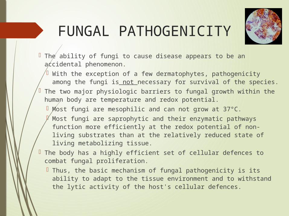
FUNGAL PATHOGENICITY The ability of fungi to cause disease appears to be an accidental
phenomenon. With the exception of a few dermatophytes, pathogenicity among
the fungi is not necessary for survival of the species. The two major physiologic barriers to fungal growth within the human
body are temperature and redox potential. Most fungi are mesophilic and can not grow at 37°C. Most fungi are saprophytic and their enzymatic pathways function
more efficiently at the redox potential of non-living substrates than at the relatively reduced state of living metabolizing tissue.
The body has a highly efficient set of cellular defences to combat fungal proliferation. Thus, the basic mechanism of fungal pathogenicity is its ability to
adapt to the tissue environment and to withstand the lytic activity of the host's cellular defences.

FUNGAL PATHOGENICITY The development of human mycoses is related
primarily to the immunological status of the host and amount of the environmental exposure, rather than to the infecting organism.
A few of fungi have the ability to cause infections in healthy humans by having a unique enzymatic capacity, exhibiting thermal dimorphism, by having an ability to block hosts cell-mediated
immune defenses. There are then many "opportunistic" fungi which
cause infections to patients whose normal defense mechanisms are impaired.
CLINICAL GROUPINGS FOR FUNGAL INFECTIONS
SKIN MYCOLOGY
Superficial MycosesCutaneous Mycoses
Subcutaneous Mycoses
INFECTIOUS DISEASE MYCOLOGY
Dimorphic Systemic Mycoses Opportunistic Systemic Mycoses

Dermatomycoses Superficial fungal infections (dermatomycoses) are very
common and occur throughout the world.
Most of these infections are caused by dermatophytic moulds (the terms tinea and ringworm are synonymous with dermatomycosis). Dermatophytic infections are contagious diseases caused by
either a human (anthropophilic) or animal (zoophilic) species of dermatophyte fungi.
A second group of superficial infections is caused by yeasts. Candida species cause infections of the mucous membranes,
skin and fingernails (candidiasis) and Malassezia furfur (Pityrosporum orbiculare) infects the skin,
usually the trunk (pityriasis versicolor). Both organisms are considered to be commensals of humans.

Dermatomycoses The organisms are transmitted by either
direct contact with infected host (human or animal) or by direct or indirect contact with infected exfoliated
skin or hair in combs, hair brushes, clothing, theatre seats, furniture, caps, towels, bed linens, hotel rugs, locker room floors etc.
Depending on the species the organism may be viable in the environment for up to 15 months,
There is an increased susceptibility to infection when there is a preexisting injury to the skin such as scratches, scares, burns, excessive temperature and humidity.

Dermatophytes
Dermatophytes are fungi that can cause infections of the skin, hair, and nails due to their ability to utilize keratin.
They require keratin for nutrition and must live on stratum corneum, hair, or nails to survive.
The organisms colonize the keratin tissues and inflammation is caused by host response to metabolic by-products.
These infections are known as ringworm or tinea, in association with the infected body part.

Dermatophytes
The dermatophytes consist of three genera: Epidermophyton produces only macroconidia, no
microconidia and consists of 2 species, one of which is a pathogen.
Microsporum - both microconidia and rough-walled macroconidia characterize Microsporum species. There are 19 described species but only 9 are involved in human or animal infections.
Trichophyton - the macroconidia of Trichophyton species are smooth-walled. There are 22 species, most causing infections in humans or animals.

Dermatophytes The dermatophytes are classified as
anthropophilic, zoophilic or geophilic according to their normal habitat: Anthropophilic are restricted to human hosts and
produce a mild, chronic inflammation. Zoophilic organisms are found primarily in animals
and cause marked inflammatory reactions in humans who have contact with infected cats, dogs, cattle, horses, birds, or other animals. This is followed by a rapid termination of the infection.
Geophilic species are usually recovered from the soil but occasionally infect humans and animals. They cause a marked inflammatory reaction, which limits the spread of the infection and may lead to a spontaneous cure but may also leave scars.

Geophilic , zoophilic dermatophytes
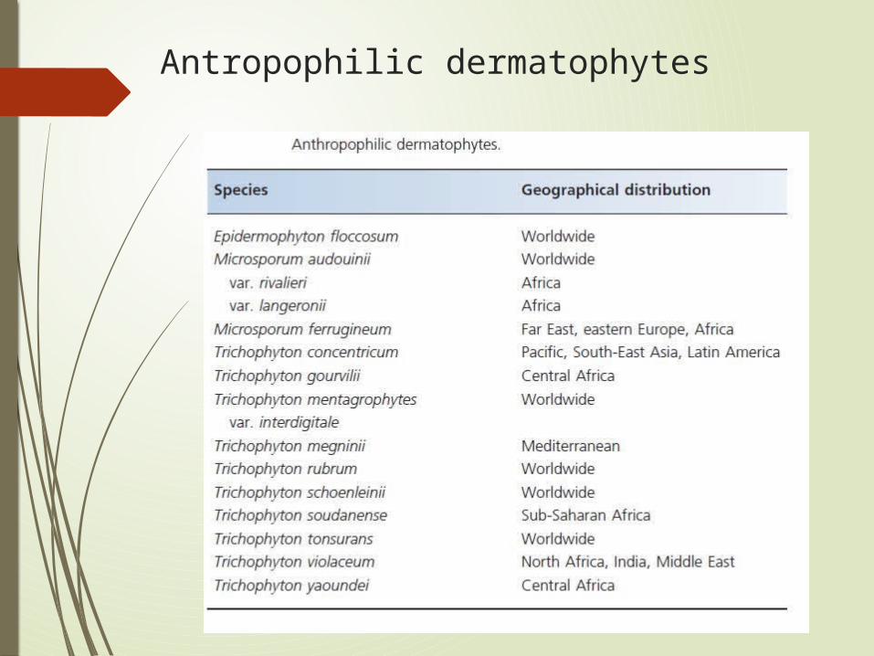
Antropophilic dermatophytes

Dermatophytes
Common dermatophytes include: Tinea barbae Tinea capitis Tinea corporis Tinea cruris Tinea pedis and dermatophytid reaction
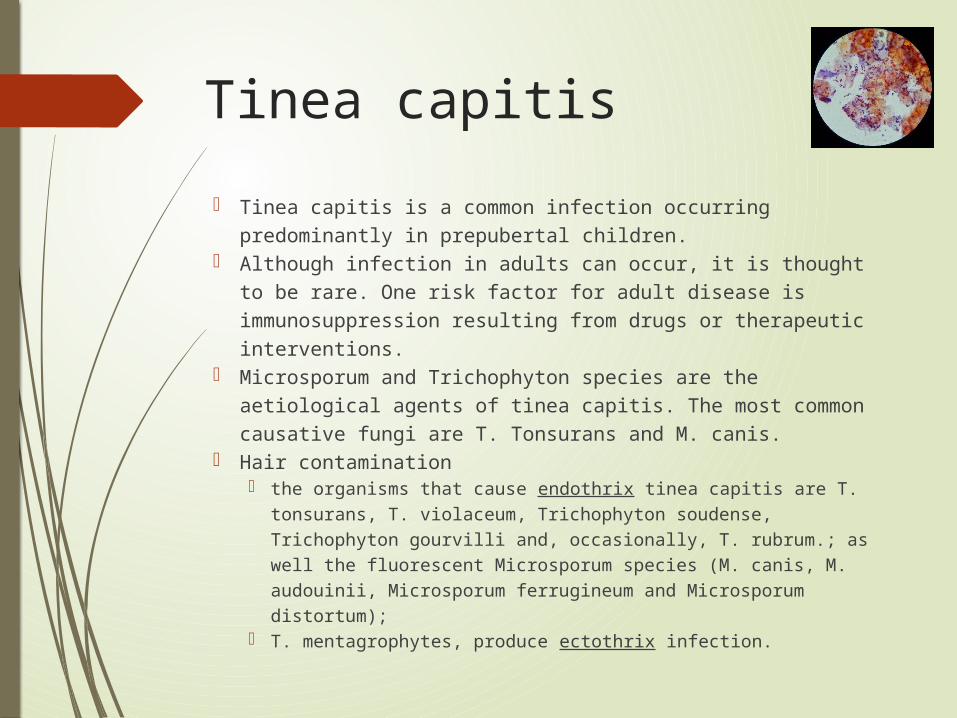
Tinea capitis Tinea capitis is a common infection occurring predominantly
in prepubertal children. Although infection in adults can occur, it is thought to be
rare. One risk factor for adult disease is immunosuppression resulting from drugs or therapeutic interventions.
Microsporum and Trichophyton species are the aetiological agents of tinea capitis. The most common causative fungi are T. Tonsurans and M. canis.
Hair contamination the organisms that cause endothrix tinea capitis are T.
tonsurans, T. violaceum, Trichophyton soudense, Trichophyton gourvilli and, occasionally, T. rubrum.; as well the fluorescent Microsporum species (M. canis, M. audouinii, Microsporum ferrugineum and Microsporum distortum);
T. mentagrophytes, produce ectothrix infection.

Ectothrix and Endothrix

Tinea capitis A variety of clinical presentations of tinea capitis are
recognized as being inflammatory or noninflammatory and are usually associated with patchy alopecia.
However, the infection may be widespread, and the clinical appearances can be subtle.
In urban areas, tinea capitis should be considered in the differential diagnosis of children older than 3 months with a scaly scalp until proven negative by mycological examination.
Infection may also be associated with painful regional lymphadenopathy, especially in the inflammatory variants.
Pertinent physical findings are limited to the skin of scalp, eyebrows, and eyelashes.

Microsporum

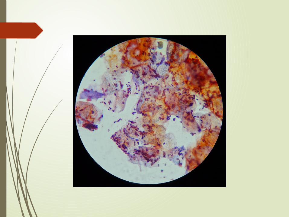
Fungal hyphae and yeast cells of Trichophyton rubrum seen on the stratum corneum of tinea capitis. Periodic acid-Schiff stain, magnification 250X. Medscape

Photomicrograph depicting an endoectothrix invasion of a hair shaft by Microsporum audouinii. Intrapilary hyphae and spores around the hair shaft are seen (hematoxylin and eosin stain with Periodic acid-Schiff counterstain, magnification X 250). Medscape

Tinea capitis Primary skin lesions of tinea capitis begin as red
papules with progression to grayish ring-formed patches containing perifollicular papules.
Pustules with inflamed crusts, exudate, matted infected hairs, and debris may be seen.
Black dot tinea capitis refers to an infection with fracture of the hair, leaving the infected dark stubs of broken hairs visible in the follicular orifices. Black dots may occur within a single patch or diffusely across the scalp.

Tinea capitis Alopecia is the most common presentation as a
discrete patch of alopecia, with or without scale that may mimic alopecia areata.
Patients with tinea capitis also develop posterior cervical adenopathy, which helps to distinguish tinea capitis from other cutaneous diseases that result in alopecia, such as alopecia areata.
The development of pustules and abscesses, known as a kerion, is another possible presentation. Such abscesses can be painful and several centimetres in diameter. A kerion is an advanced form of tinea capitis and is a hypersensitive reaction. It can occur on some parts of the scalp.

Favus (tinea favosa) Favus (also termed tinea favosa) is a severe form of
tinea capitis. Favus is a chronic infection caused most commonly
by T schoenleinii and, occasionally, by T violaceum or Microsporum gypsum.
Scalp lesions are characterized by the presence of yellow cup-shaped crusts termed scutula, which surround the infected hair follicles.
Favus is seen predominantly in Africa, the Mediterranean, and the Middle East and, rarely, in North America and South America, usually in descendants of immigrants from endemic areas.
Favus usually is acquired early in life and has a tendency to cluster in families.
In favus, infected hairs appear yellow.

Candidiasis A primary or secondary mycotic infection caused by members of
the genus Candida. The clinical manifestations may be acute, subacute or chronic to
episodic. Involvement may be localized to the mouth, throat, skin, scalp,
vagina, fingers, nails, bronchi, lungs, or the gastrointestinal tract, or become systemic as in septicaemia, endocarditis and meningitis.
In healthy individuals, Candida infections are usually due to impaired epithelial barrier functions and occur in all age groups, but are most common in the newborn and the elderly.
They usually remain superficial and respond readily to treatment.
Systemic candidiasis is usually seen in patients with cell-mediated immune deficiency, and those receiving aggressive cancer, immunosuppression, or transplantation therapy.
Several species of Candida may be etiological agents, most commonly, Candida albicans.

Malassezia spp.
Taxonomic Classification
Kingdom: FungiPhylum: BasidiomycotaClass: HymenomycetesOrder: TremellalesFamily: FilobasidiaceaeGenus: Malassezia
Malassezia furfur
Malassezia pachydermatis

The yeast genus Malassezia
The implication of the yeast genus Malassezia in skin diseases has been characterized by controversy, since the first description of the fungal nature of pityriasis versicolor in 1846 by Eichstedt.
This is underscored by the existence of Malassezia yeasts as commensal but also by their implication in diseases with distinct absence of inflammation despite the
heavy fungal load (pityriasis versicolor) or with
characteristic inflammation (eg, seborrheic dermatitis, atopic dermatitis, folliculitis, or psoriasis).

The yeast genus Malassezia
The description of 14 Malassezia species and epidemiologic studies did not reveal pathogenic species but rather disease-associated subtypes within species.
Emerging evidence demonstrates that the interaction of Malassezia yeasts with the skin is multifaceted and entails constituents of the fungal wall (melanin, lipid
cover), enzymes (lipases, phospholipases), and metabolic products (indoles), as well as the cellular components of the epidermis
(keratinocytes, dendritic cells, and melanocytes). Understanding the complexity of their interactions
will explain the picture of the clinical presentation of Malassezia-associated diseases and unravel the complexity of skin homeostatic mechanisms.

The yeast genus Malassezia
Although Malassezia yeasts are a part of the normal microflora, under certain conditions they can cause superficial skin infection, such as pityriasis versicolor and Malassezia folliculitis.
Moreover the yeasts of the genus Malassezia have been associated with: seborrheic dermatitis and dandruff, atopic dermatitis, psoriasis, and, less commonly, with confluent and reticulated papillomatosis, onychomycosis, and transient acantholytic dermatosis.
It is difficult to study the clinical role of Malassezia species due to the relative complexity in isolation, cultivation and identification.
It is important to consider the clinical, mycologic, and immunologic aspects of the various skin diseases associated with Malassezia.

Tinea versicolor Tinea versicolor (also known as dermatomycosis furfuracea,
pityriasis versicolor, and tinea flava) is a condition characterized by a skin eruption on the trunk and proximal extremities.
Recent research has shown that the majority of tinea versicolor is caused by the Malassezia globosa fungus, although Malassezia furfur is responsible for a small number of cases.
These yests are normally found on the human skin and only become troublesome under certain circumstances, such as a warm and humid environment, although the exact conditions that cause initiation of the disease process are poorly understood.

Seborrhoeic deramtitis Seborrhoeic dermatitis is a papulosquamous disorder
patterned on the sebum-rich areas of the scalp, face, and trunk.
In addition to sebum, this dermatitis is linked to Malassezia, immunologic abnormalities, and activation of complement.
Its severity ranges from mild dandruff to exfoliative erythroderma.

Malassezia-related Skin Diseases
The third form of Malassezia infections of the skin involves the hair follicle. This condition is typically localized to the back, the chest, and the extremities.
This form can be clinically difficult to differentiate from bacterial folliculitis. The presentation of Pityrosporum folliculitis is a perifollicular, erythematous papule or pustule.
Predisposing factors include diabetes, high humidity, steroid or antibiotic therapy, and immunosuppressant therapy.

Treatment Internationally approved guidelines for the diagnosis and
management of Malassezia-related skin diseases are lacking. There is guidelines for the diagnostic procedures and management
of pityriasis versicolor, seborrhoeic dermatitis and Malassezia folliculitis.
Main recommendations in most cases of pityriasis versicolor and seborrhoeic dermatitis include topical treatment which has been shown to be sufficient.
As first choice, treatment should be based on topical antifungal medication.
A short course of topical corticosteroid or topical calcineurin inhibitors has an anti-inflammatory effect in seborrhoeic dermatitis.
Systemic antifungal therapy may be indicated for widespread lesions or lesions refractory to topical treatment.
Maintenance therapy is often necessary to prevent relapses. In the treatment of Malassezia folliculitis systemic antifungal
treatment is probably more effective than topical treatment but a combination may be favourable.

Lab

Laboratory Specimen Processing In general, direct microscopy and culture should be
performed on all specimens received by the laboratory. Microscopy provides vital information, often an immediate
presumptive diagnosis is possible, which is of particular importance in the immunosuppressed patient. Microscopy usually consists of either (a) wet mounts in 10% KOH with Parker ink, or india ink, (b) smears for Gram, Giemsa and PAS staining, and (c) histopathology of tissue sections.
Routinely, cultures should be maintained for one month. Cultures should be examined regularly, fungal growths
identified and significant isolates reported as soon as possible.

Specimen Collection
Skin should be scraped from the margin of the lesion onto folded black paper or directly on microscope slide
Hair should be plucked, not cut, from the edge of the lesion
Choose hairs that fluoresce under a Wood's lamp or, if none fluoresce, choose broken or scaly ones

Direct Examination
A small sample of the specimen is selected for direct microscopic examination and investigated for the presence of fungal elements
The specimen is mounted in a small amount of potassium hydroxide The KOH slides are gently heated and allowed to clear for 30 to 60
minutes before examining on a light or phase contrast microscope
When present in the direct examination dermatophytes appear as non-pigmented, septated elements; hyphae rounding up into arthroconidia are also diagnostic of dermatophyte involvement.
When hair is involved the arthroconidia may be found on the periphery of the hair shaft (ectothrix) or within the shaft (endothrix)
Malassezia furfur infections (tinea versicolor) are diagnosed by the presence of spherical yeast cells with a single bud and a collar and short curved hyphal strands

Culture
Hair is cut into short segments Each specimen is divided between at least two
types of culture media The use of antibiotics will inhibit the overgrowth
of bacteria and incorporation of cycloheximide will prevent the overgrowth of the rapidly growing saprophytic fungi
The cultures are incubated at 30°C and examined frequently for 4 weeks


Research2013 Jan Feb Mar Apr May Jun Jul Aug Sep Oct Nov Dec All
Altogether 108 80 79 84 70 100 107 91 90 92 118 85 1104 Round spores 29 21 19 21 16 22 16 23 21 18 28 20 254Round and ovale spores 3 5 3 6 4 8 14 15 13 16 12 8 107Round, ovale and bacteria 2 2 0 1 1 0 0 0 0 0 0 0 6 367Ovale spores 24 20 23 21 21 39 41 25 21 20 34 14 303Ovale spores and bacteria 19 7 8 11 14 13 7 11 13 15 8 17 143 446 Bacteria 9 10 17 12 5 13 14 8 7 11 22 18 146 No microflora 22 15 9 12 9 5 15 9 15 12 14 8 145 Demodex 0 2 1 0 0 0 0 1 0 0 1 0 5

Research2013 Altogether
Altogether 1104 Round spores 254 Round and ovale spores 107 Round, ovale spores and bacteria 6 367 33,24%Ovale spores 303 Ovale spores and bacteria 143 446 40,39% Bacteria 146 13,22% No microflora 145 13,13% Demodex 5 0,36%

Research
Cultures altogether: 308Trichophyton violaceum 21Trichophyton mentagrophytes var. interdigitale 1Trichophyton tonsurans 11Trichophyton spp 1Candida 9Microsporum ferrugineum 2
Microsporum gypseum 1

Facts
Removal of fungal infection from infected scalp skin stops hair loss (already in the one month
time – while treatment is going on) allows hair to grow back more efficiently helps to gain volume back improves hair cosmetic condition – shine
and structure

Conclusions
Fungal infections on the skin is much more often then we suspect them
Patient with longstanding hair loss must be investigated for fungal infection
If dermatomycosis is found, appropriate treatment must be done
Further investigation to elucidate this subject is needed

Our friendly team – doctor Inga Zemite, doctor Ausma Eglite with her assistant Victoria and nurse Ita
Veselibas centrs 4, Riga, Latvija
Thank you for your attention!

Let us meet again..We welcome you all to our future conferences of OMICS International
5th International Conference and Expo on
Cosmetology, Trichology & Aesthetic Practices On
April 25-27, 2016 at Dubai, UAE
http://cosmetology-trichology.conferenceseries.com/