Embed Size (px)
Citation preview

P A R T I. THE BASIS OF SELECTIVE TOXICITY
FUNDAMENTAL ASPECTS OF SELECTIVE TOXICITY
Adrien Albert Department of Medical Chemistry. John Curtin School of Medical Research
Canberra, Australia
It is of the very natuIt of a drug to be toxic, and the majority of drugs in the Pharmacopoeia have won their place because they are able to exert a valuable repressive action on some natural metabolic process. Toxicity, then, is usually a drug's most characteristic and valued property; and yet for a drug to be useful, the toxicity must be selective.'
This introduction leads naturally to a definition. Selective toxicity has to d o with the injuring of one kind of living matter without harming some other kind, even though the two are in intimate contact. Either reversible or permanent injury may be preferred, depending on the problem in hand.
The living matter which is to be injured is known as the uneconomic form, and the matter which is to be protected is called the economic form. They may be related to one another as parasite and host, or they may be two tissues in the one organism. General anaesthetics admirably illustrate what is meant by selective toxicity. The more toxic the anaesthetic, the greater its value, but only if the toxicity is selective for the central nervous system alone and is completely reversible with time. Antiparasitic agents, on the other hand, are preferably irreversible toxicants.
In agriculture, many agents are known which injure fungi, insects, and even competitiw plant life without injury to the economic species. In animal husbandry, too, drugs enable man's economic animals to be safely freed from many kinds ot parasites, external and internal. The continuance, not to mention expansion, of agriculture and animal breeding necessitates the constant search for improved selectively toxic agents. Biological controls,* which are preferable to chemical methods, are so seldom discoverable that the search for new chemicals must necessarily form the main line of defense.
That branch of selective toxicity which is concerned with the removal of parasiltes (including bacteria) from man and his adopted animals is called chemotherapy, and many of its underlying principles are well understood. The name pharmacodynamics is usually reserved for that more complex situation where the economic and the uneconomic cells are both part of the same animal. Thus the uneconomic cells may be a malignant growth in an otherwise healthy host, or they may be some part of the nervous system which, by malfunction, has disrupted the normal harmony of bodily functions.
The drugs or other chemical agents used for selectively toxic purposes, will be referred to, briefly, as agents. An agent exerts its selectivity by one or other of two main principles. Either (a) it is equitoxic to both economic and uneconomic cells, but is accumulated mainly by the uneconomic cells, or ( b ) it reacts fairly specifically with (1) a cytological or (2 ) a biochemical feature which is absent from, or does not play so important a part in, the economic cells. Although these several divisions tend to merge into one another, they form the basis of a useful
* An example of biological control is the introduction of the parasitic wasp Tiphia vernalis from China to the Atlantic seaboard of the U.S.A. to reduce the population density of the Japanese beetle.
5

6 Annals New York Academy of Sciences
classification. For example many agents cause a strong and specific physiological response in spite of an unfavorable distribution, and hence the reason for their selectivity is to be sought under (b). Let us now review a few examples from each division.
Selectivity Through Distribution
The simplest cases of selectivilty achieved through differences in distribution or accumulation depend on gross morphology. Thus the large surface area (per unit weight) of an insect, compared to that of a mammal upon which it is resting, causes the inseot to retain relatively more of any agent applied as a spray.
A similar, but more subtle, example is seen in the spraying of cereal crops with a 20 per cent aqueous solution of sulphuric acid, which is practiced in many parts of the world.2 This treatment spares the monocotyledonous cereals and yet kills the weeds which are mainly dicotyledons. Such strong acid solutions, of course, are equally injurious to the cytoplasm of both wheat and weed, but they do not gain access to that of the wheat. This is because the exterior of the wheat plant is smooth, whereas the weeds are externally rough. Hence the sulphuric acid is shed by the wheat plant but accumulated by the weeds which consequently die. Further details of this example are as follows:
The leaves of cereal crops are waxy and upright; the growing tissues are basal and protected by leaf-sheaths. Thus the wax prevents penetration, the droplets run off the leaves, and the meristems do not come in contact with the spray. On the other hand, most dicotyledonous weeds have flat waxless leaves, which catch the spray; moreover the growing point is exposed and vulnerable, because it is the apex of the shoot.3
Phenothiazine, used as an oral anthelmintic in sheep, affords another example of selectivity achieved by favorable distribution. The cells of the host's intestinal tract take up very lilttle of this agent, whereas the parasite accumulates it readily?
Examples of selective partitioning between various 'tissues of a single organism will now be given.
As little as one microgram of cyanocobalamin (vitamin Blz) injected into the arm or leg muscle of a patient with pernicious anaemia, initiates the formation of new erythrocytes in the bone marrow. In spite of an initial dilution of IOlO-fold in the body fluids, this small dose is specifically accumulated by the marrow.
Iodine is similarly selectively accumulated by the thyroid gland, a process readily followed wilth radioactive iodine. Of an oral dose of only gram, 80 per cent can be demonstrated in this gland within a few hours.'
Inflamed lung tissue can selectively concentrate penethamate (the diethyl- aminoethyl ester of penicillin) if this drug is injeoted into the muscles of an arm or leg. The drug becomes hydrolysed in situ to penicillin, and hence it is a valued therapeutic agent for bacterial infections of the lung5
The bacterial toxin of Clostridium botulinum, the most poisonous substance known, is selectively accumulated by peripheral motor nerve endings where it prevents the normal secretion of acetylcholine. The minimal lethal dose is lo-'* g. per kg. (mouse). Thus 1 mg. can kill 1,200 tons of living matter. The toxin is usually consumed in contaminated food. Although the pathway from the gastro- intestinal tract to the nerve endings is long and provides many opportunities for the sequestering of this large protein molecule, very little is lost on the way.6
These examples of highly seleative distribution raise the question of what generally takes place when an agent is applied to an organism (or given by mouth, or injected). Obviously it must traverse one or several semipermeable membranes before the effective receptor is encountered. For example an antimalarial agent,

Albert: Selective Toxicity 7
given by mouth, must penetrate the gut/blood barrier, then the erythrocyte nieni- brane, and finally that of the malarial parasite.
On both sides of each membrane the concentration of a drug continually dccreases through storage, excretion, and inactivation (FIGURE I ).
Sforage may take place in lipoids ( for liposoluble substances, e.g. thiopentone), on nucleic acids ( for cationic substances, e.g. quinacrine), or on serum albumin ( for anionic substances, e.g. suramin and the coumarins). Storage is usually freely reversible. I t can be beneficial when it tends to keep the blood level of a drug constant in an infectious disease; hut it can be disadvantageous if it permanently keeps most of the drug out of circulation.
Excretion may be through the kidneys, the bile-duct (and hence into the gut) , or through the lungs. For some drugs, inactivation precedes excretion. Inactiva- tion involves the making or breaking of covalent bonds and is usually irreversible.
STORAGE \ + DRUG
DRUG I 4
kidney bile-duct lungs
’,,,ETioN
A 0
S STORAGE
R I p J T
- ++ FREE e--,
+ INACTIVATION I 0
FIGURE 1. Principal factors involved in the distribution of a drug. The words “absorption” represent semipermeable membranes.
When a n agent escapes from the circulation by penetrating the membranes of blood vessels, it is subject to further decreases in concentration by storage and inactivation. Fortunately, it can usually not be excreted from such a site, a limita- tion which helps to maintain concentration.
Finally, when the drug penetrates the last membrane surrounding the relevant receptor, combination can occur with the receptor if drug and receptor are sufficiently complementary: a t this stage the biological effect characteristic of the agent begins.
The frequency and size of the dose depend on all ,these factors. Sometimes there is an additional complication: the agent which acts on the receptor is not ad- ministered as such, but as a “pro-agent” which is later metabolized to the true agent.
In this overall picture of distribution, the easy reversibility of most of the steps should be noted. Even the combination between agent and receptor is usually loose, and subject to easily disturbed equilibria ( F I G U R E 2 ) .
Because of their complexity, distribution phenomena offer great scope for selectivity, even though this often rests on a graduated (rather than on an all-or- nothing) basis. The quantitative study of the distribution of agents in various organisms is beginning to attract greater attention, and is a most profitable topic for further study. The subject has been reviewed fairly r e ~ e n t l y . ~

8 Annals New York Academy of Sciences
DRUG & 8 Kd 1 KY
Membrane Membrane Receptor
K, Binding constant with transporting agent
K,,, Diffusion constant
K, Binding constant with receptor
FIGURE 2. Some quantitative aspects of distribution
Selectivity Through Comparative Cytology
An outstanding example of cytological difference is offered by the comparison of plants and animals.
Plants differ from animals in many ways: e.g. the presence of cell walls and of a photosynthetic mechanism, absence of a nervous system and muscles, and of an efficient circulatory system. At the intracellular level, the similarities outweigh the differences. However, let us examine some cytological differences, commencing with cell walls.
Cell walls constitute a type of structure which differs greatly among various classes or organisms. Cell walls lend rigidity to a cell, but do not govern permea- bility, which is a function of thin lipoprotein membranes. The cells of multi- cellular animals usually have no walls. As regards monocellular animals, many protozoa (e.g., Ameba) lack cell walls, whereas some others have walls of cel- lulose about 230 A thick (e.g. Chlamydomonas) or of protein (e.g. Eirneria), or silica (e.g. the Foraminifera). Multicellular plants have walls formed from micro- fibrils of cellulose (10-20 m p wide, and of variable length) embedded in an amorphous matrix of hemicellulosv and pectins.
The cell wall of fungi is also composed of carbohydrate: in mycelial forms this is usually chitin* (polymerized N-acetyl-D-glucosamine) , whereas yeast cell walls contain mannan, glucan and some protein, but no aminosugars. Unlike higher organisms, fungi are under high internal pressure, and they burst if the cell wall is removed (e.g. by applying chitinase) unless the osmotic pressure of the medium is simultaneously increased. Whoever can devise a chemical that specifically ruptures fungal cell walls, or prevents their biosynthesis while the organism is growing, will have produced a selective fungicide of great potential utility.
Bacteria, too, are under high internal turgor pressure, estimated to be about 20 atmospheres for Gram-positive, and five atmospheres for Gram-negative,
Bursting is prevented by a thick cell wall which often constitutes 25 per cent of the dry weight. When the protoplast (i.e. all of the cell inside the wall) grows, additional quantities of cell wall must be synthesized. Several biocides owe their place in therapeutics to their ability to prevent this synthesis, notably penicillin,1° oxamycin, bacitracin, novobiocin, vancomycin, and the cephalo- sporins.ll Other biocides can break preformed cell walls, notably the quaternary amines, the phenols, and the polypeptide antibiotics. In every case, the bacteria rupture under their own turgor pressure.
* Chitin also forms a large part of the exoskeleton of insects and crustacea.

Albert: Selective Toxicity 9
This bacterial cell wall, which is highly porous, acts as a restrainer to the very thin (about four molecules thick) lipoprotein membrane which is the true osmotic barrier (see below). Bacterial cell walls have unique chemical composition.12 The highly polymerized glycopeptide, which forms its main framework and sub- stance, consists of repeating units of acetylglucosamine linked to acetylmuramic acid, which is in turn linked to a short polypeptide. Penicillin causes the ultimate bursting of bacteria by blocking the incorporation of acetylmuramic acid into the new cell wall.13
The presence of muramic acid and also of a peptide in the cell wall are features which differentiate bacteria from all other organisms, especially as the peptide contains some amino-acids never found in proteins (e.g. D.alanine, diaminopime- lic acid). But the mere presence of a cell wall differentiates bacteria from the higher animals.
Acetylmuramic acid ( F I G U R E 3a) is acetylglucosamine etherified at 0 - 3’ by lactic acid, The lactic acid is, in turn, linked to the polypeptide by an amide group (e.g. FIGURE 3 b ) . Digestion of the cell wall with the enzyme lysozyme, which is found in tears and other secretions of the human body, hydrolyses the link between the two carbohydrates: acetylglucosamine and acetylmuramic acid, and once again the bacterium bursts under its own turgor pressure.
The aminoacid sequence in the polypeptide differs from species to species. Some bacteria also have teichoic acids linked to the polysaccharide framework of the cell wall. These acids are polymers of glycerol phosphate and ribitol phos- phate. The occurrence of teichoic acids in nature is apparently confined to bacteria.
Immediately inside this cell wall of unique composition, is the lipoprotein cell membrane 60-100 A thick. The lipoid part of this membrane is chemically un- usual in having no cholesterol, and the protein part has the unusual feature that much of it consists of enzymes, particularly those of the tricarboxylic acid cycle. Thus more than 90 per cent of the succinic, malic, lactic, and formic dehydroge- nases, and of cytochrome oxidase are present in the cytoplasmic membrane of
CH20H I
Acetylmuramic acid
A
(D-loctyl)-L-Ala-D-Glu-l-lys-D-Ala -D-Ala
B
FIGURE 3. Representative constituent parts of the polymer framework of bacterial cell wall.

10 Annals New York Academy of Sciences
Micrococcus lysodeikticus, and Staphylococcus uurew.14 Bacteria differ from most other organisms in having no mitochondria, and the above cytoplasmic membrane apparently performs the function of these bodies in phosphorylative oxidation of carbohydrates. The presence of so many vital enzymes in such an exposed position should make bacteria particularly vulnerable to selectively toxic agents. Moreover, bacteria are the only cells without a nucleus; instead, they have strands of nucleic acids lying in the cytoplasm, unprotected by the usual nuclear membrane.
Viruses are non-cellular forms of life structurally simpler than bacteria. The complete infective virus, such as exists extracellularly, consists of a core of either deoxyribosenucleic acid ( D N A ) , or ribosenucleic acid ( R N A ) but not both. This core is surrounded by a protective coat of protein and the two com- ponents are arranged in a highly ordered fashion. The D N A is double stranded in some species, but exists as a single strand in others. Some viruses have lipides, also, which seem to regulate permeability. Aliphatic diamines and magnesium are frequently present, and copper is often found in association with viruses of the pox group.
Thus a picture emerges of a highly ordered structure for viruses, and even at the chemical level they begin to appear less simple than had been thought. Struc- tural complexity is particularly evident in those viruses that infect bacteria. Thus the head of T 2 coliphage contains a single molecule of D N A (M.W. = lo*) which weighs 2 X pg and contains 2 X lo5 nucleotide pairs. This molecule is neatly folded (presumably by aliphatic diamines or magnesium ions, or both) into an approximately spherical mass, and is surrounded by neatly packed protein molecules. To this “head” is attached a “tail” built of a series of structures. First there is an outer sheath consisting of a myosin-like contractile protein that is associated with about 110 molecules of adenosine t r ipho~phate’~ (FIGURE 4) . Other structures in the tail include (a) a solid core, (b) a spiked plate on the tip, ( c ) a lysozyme-like enzyme, ( d ) a series of fibres that are wound around the tip. These structures play a coordinated part in wpturing the bacterial exterior, and injecting the infective D N A into the bacterial cytoplasm.16
The complexity of some virus structures has offered hope for the discovery of specific antiviral drugs, harmless to the host’s cells. Last year, two such drugs emerged from successful clinical trials, viz. Marboran ( 1 -methyl-isatin p-semi- thiocarbazone) and IUdR (5-iodo-deoxyuridine), active against smallpox17 and herpes,’* respectively.
Although gross cytological differences between phyla are easy to demonstrate, evidence is now being accumulated of more subtle differences within a single species. Thus the mitochondria of the rat retina does not swell in response to vitamin A, whereas those of rat liver d o so, and this suggests a structural dif- ference.
Selectivity Through Conipararive Biochemistry
Some of the greatest successes of selective toxicity may yet come from the study of comparative biochemistry. The literature of this subject, long neglected, has recently become more accessible through publication of a multivolume work.’$ Nevertheless, very much more remains to be discovered. In particular, little is known of the comparative biochemistry of the various tissues of the human body, although this knowledge is so necessary for pharmacodynamics. However i t is clear that striking biochemical differences between species are to be found more in anabolic (building up) than in catabolic (breaking down) processes.
Deoxyribosenucleic acid ( D N A ) contains most of the cell’s genetic informa-

Albert: Selective Toxicity 11
' Head Membrane
Tail Core
Plate
Tail Fibers
FIGURE 4. Diagram showing several of thc components of a bacteriophage (L. M. Kozloff, personal communication).
TABLE 1 DNA OF BACTERIA
c Guanine 4- Cytosine
Adenine + Thymine Species
Closrridium perfringens Staphylococcus aureus Pasteurella tularensis Proteus vulgaris Escherischia coli Shigella dysenteriae Salmonella typhosa Corynebacterium diphtheriae Azobacter species Brucella abortus Pseudomonas aerogenes Mycobacterium tuberculosis Actinomyces species
0.45 0.53 0.53 0.68 1.09 1.14 1.14 1.20 1.28 1.37 2.03 2.08 2.80

12 Annals New York Academy of Sciences
tion, which it uses t o make specific ribosenucleic acids ( R N A ) which in turn produce specific proteins, including enzymes. The comparative biochemistry of D N A is therefore of special interest.
The D N A of bacteria differs from that of higher organisms in being paired to a nonbasic protein.20 The ratio of certain purine and pyrimidine bases differs greatly from one bacterial species to another. In TABLE 1 , the last column gives the sum of the two amphoteric bases divided by that of the two monofunctional bases.z1 This figure ranges from 0.45 to 2.80, whereas in higher animals and plants it has so far been found to vary only between 0.6 and 0.9. Moreover, higher plants have one-quarter of the cytosine replaced by methylcystosine. Similarly, ratios have been determined also for protozoa, fungi, algae, and invertebrate animals. It is not a t once clear how to apply this knowledge to the discovery of selectively toxic agents; nevertheless it reveals differences at a very fundamental level, which must have considerable significance in the quest.
Small amounts of unusual purine and pyrimidine bases are found in the nucleic acids of bacteria, yeasts, and viruses, e.g. 5-hydroxymethylcytosine and 5-hy- droxymethyluracil. There must be a significant difference between bacteria and mammals a t the R N A level, because the tetracycline antibiotics inhibit the migra- tion of aminoacids from transfer-RNA to messenger-RNA in bacteria, but not in mammals.z2
The comparative biochemistry of proteins (including enzymes and the poly- peptide hormones) is somewhat better known. Strong species differences are shown by homologous proteins, i.e. those serving similar functions in related species. For example, the N-terminal aminoacids of fibrinogenz3 differ in various mammals as shown in FIGURE 5.
Homologous enzymes are often immunologically distinct, e.g. the hexokinase from yeast and that from mammalian brain. More marked chemical differences also exist. Crystalline aldolases from yeasts and bacteria require iron, whereas those from plants and mammals do not.z4 The chemotherapeutic success of anti- monials in the treatment of schistosomiasis depends on blocking phosphofructo- kinase in the worms, whereas the homologous enzyme in man is unaffected.25
Quantitative factors enter into the distribution of an enzyme in the different organs of a single organism. More glutamine synthetase is present in human brain
Man a n d Pig: Ala, Tyr
ox: Glu, Tyr
Dog a n d Horse: Thr, Tyr
Sheep: Gly, Ala, Tyr
Glombaeck 8 Yamashina,1958
FIGURE 5. N-terminal aminoacids of a typical protein, showing species differences.

Albert: Selective Toxicity 13
than in liver, and this enzyme is absent from all other human tissues. Of the char- acteristic enzymes present in muscle, some (such as aconitase and oxaloacetate transacetase) are much more abundant in heart than in skeletal muscle, whereas the reverse is true of aldolase.26
The composition of the protein of cytochrome C shows remarkable species dif- fe rence~. ’~ The two neighboring cysteine residues of this enzyme are linked to side-chains on the porphyrin ring by thioether bonds. The sequences of amino- acids in this region are shown in FIGURE 6 (this FIGURE is to be read vertically; aminoacids not printed are identical with those in the top row).
The polypeptide animal hormones have similar species differences,28 and two examples are shown in F I G U R E 7 (which is to be read as suggested above for FIGURE 6 ) .
Mammals and fish: - L y s - C y s - A l a - G l u - C y s - ~ i s - F e -
Chicken: -Ser-
Silkworm: -Arg-
Y e a r t : -Arg- -Glu-Leu-
FIGURE 6. Species differences in the aminoacid composition of cytochrome c.
Sheep: -Pro-Ala-Gly-Glu-Asp-Asp-Glu-Ala-Ser-Glu-
Eg: -ASP- -Ala-Glu- -Leu-Ala-
Schwattx. 1959
FIGURE 7. Species differences in the aminoacid composition of adrenocorticotropic hor- mone.
Interesting differences have been observed at the aminoacid level, and some examples were given when discussing bacterial cell wall (above). Aminoacids of the abnormal D-configuration d o not form a part of biologically active cell sub- stances a t any level of evolution higher than that of the earthworm (Lumbricus ferrestris) which uses a phosphagen containing D-serine (it is the O-phosphodies- ter of guanidoethanol and D-serine) .29
Among various pathways to common amino acids, it may be noted that bacte- rial lysine is made only by the decarboxylation of diaminopimelic acid (which is peculiar to bacteria and their closest relatives), whereas fungal and mammalian lysine are made only by direct synthesis.30
From this discussion of nucleic acid and proteins, we proceed naturally to the fundamentals of nitrogen catabolism. The end products of this catabolism display more species variety than d o those of fat or carbohydrate degradation. These end-products range in complexity from ammonia to the alkaloids. TABLE 2 shows the end-products of protein and purine metabolism among vertebrates; it is no less varied among simpler forms of life.
What is still more surprising, these end-products can vary within a single spe-

14 Annals New York Academy of Sciences TABLE 2
END-PRODUCTS OF NITROGEN METABOLISM
Vertebrates Protein metabolism Purine metabolism
Fish (teleost) ammonia Amphibia Reptiles (snake) uric acid uric acid Birds uric acid uric acid Man and higher apes urea uric acid Other mammals allantoin
Baldwin, 1948
cies, depending on the stage of development. It can be been in FIGURE 8, for example, that a chicken in the egg passes, in turn, through the stages of excreting ammonia (fish-like) , then urea ( frog-like) , and finally uric acid (typical of birds) all within a few days. This metabolic sequence brings to mind the well-known necessity for using different drugs to deal with parasites at different stages of their metabolic cycle.
We can now consider some aspects of comparative biosynthesis, especially of those purines and pyrimidines from which nucleic acids are built. In this field, species differences have provided a large number of very valuable chemothera- peutic drugs, some of them discovered accidentally and others by the logical application of biochemical principles.
0 Ammonia
0 Urea 1 0 Uric acid
FIGURE 8. Varying nature of nitrogen end-products in the chick embryo. (Adapted from Baldwin, 1948.)

Albert: Selective Toxicity 15
Pteridines in the form of folic acid (1 ) and its 5, 6 , 7, 8-hydrogenated deriva- tives, play a dominant role in catalyzing the biosynthesis of purines, pyrimidines, and methionine, and also in the interconversion of glycine and serine. Antifolic agents interrupt the synthesis of nucleic acids at concentrations which d o not affect protein synthesis.
Pteroylglutamic acid (folic acid) (1)
Very few pathogenic bacteria can absorb preformed folic acid,g1 and hence they are severely injured by any drug which can interfere with the biosynthesis or utilization of this growth-factor. On the other hand, mammals are not capable of synthesizing folic acid, but absorb i t easily from the diet. Man, and other mammals, are reasonably unaffected by antifolic drugs in moderate doses. Hence the antifolic field has proved a very fertile one for the discovery of antibacterial drugs, and it has been successfully extended to the antimalarial and antileukaemic fields.
The molecule of folic acid ( I ) , consists of three portions which, reading from left to right, comprise residues of glutamic acid, p-aminobenzoic acid, and a pteri- dine. Sulphonamide drugs, which resemble p-aminobenzoic acid (PAB) closely in both charge and dimensions ( F I G U R E 9), antagonize the incorporation of PAB into the folic acid molecule, a reaction which man does not carry out. To this biochemical difference, they owe their outstanding success as selectively toxic drugs.
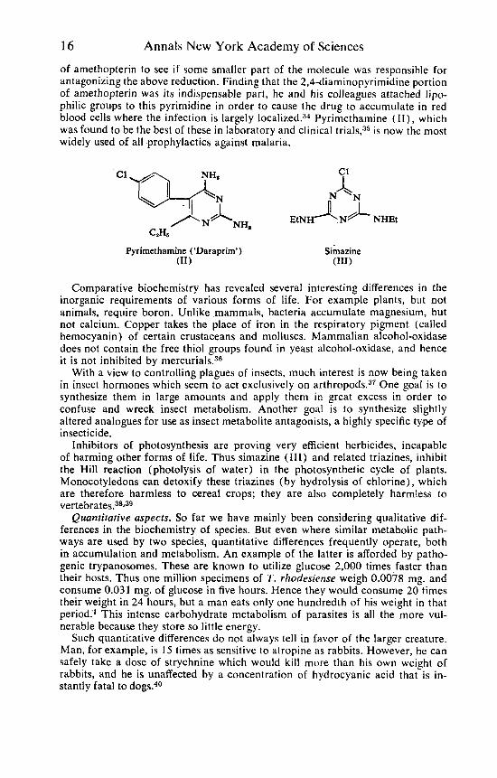
Analogues of folic acid, in which the 4-hydroxy-group is replaced by an amino- group, have proved useful in the management of leukemia in young people.32 Amethopterin (methotrexate), a typical example, is the 4-amino- I O-methyl- derivative of folic acid ( I ) . This, and analogous, 4-aminopteridines seem to exert their biological activity by inhibiting the bioreduction of 7,8-dihydrofolic acids to 5,6,7,8-tetrahydrofolic acids, usually a n essential step in the functioning of the natural pteridines.”j
When it was found that 2,4-diamino-6,7-diphenylpteridine was as active as quinine in suppressing malaria in chicks, Hitchings continued the simplification
-f - +\/”
I I -2.4 A+ I I
P A B S U L P H A N I L A M I D E
FIGURE 9. Molecular dimensions of p-aminobenzoic acid (PAB) and sulphanilamide.
16 Annals New York Academy of Sciences
of amethopterin to see i f some smaller part of the molecule was responsible for antagonizing the above reduction. Finding that the 2,4-diaminopyrimidine portion of amethopterin was its indispensable part, he and his colleagues attached lipo- philic groups to this pyrimidine in order to cause the drug to accumulate in red blood cells where the infection is largely localized.x4 Pyrimethamine ( I I ) , which was found to be the best of these in laboratory and clinical trials,:j5 is now the most widely used of all prophylactics against malaria.
Pyrirnetharnine (‘Daraprirn’) (11)
Comparative biochemistry has revealed several interesting differences in the inorganic requirements of various forms of life. For example plants, but not animals, require boron. Unlike mammals, bacteria accumulate magnesium, but not calcium. Copper takes the place of iron in the respiratory pigment (called hemocyanin) of certain crustaceans and molluscs. Mammalian alcohol-oxidase does not contain the free thiol groups found in yeast alcohol-oxidase, and hence it is not inhibited by mercurials.36
With a view to controlling plagues of insects, much interest is now being taken in insect hormones which seem to act exclusively on arthropod^.^^ One goal is to synthesize them in large amounts and apply them in great excess in order to confuse and wreck insect metabolism. Another goal is to synthesize slightly altered analogues for use as insect metabolite antagonists, a highly specific type of insecticide.
Inhibitors of photosynthesis are proving very efficient herbicides, incapable of harming other forms of life. Thus simaziae (111) and related triazines, inhibit the Hill reaction (photolysis of water) in the photosynthetic cycle of plants. Monocotyledons can detoxify these triazines (by hydrolysis of chlorine), which are therefore harmless to cereal crops; they are also completely harmless t o ~ertebrates.3~239
Quantitative aspects. So far we have mainly been considering qualitative dif- ferences in the biochemistry of species. But even where similar metabolic path- ways are used by two species, quantitative differences frequently operate, both in accumulation and metabolism. An example of the latter is afforded by patho- genic trypanosomes. These are known to utilize glucose 2,000 times faster than their hosts. Thus one million specimens of T . rhodesiense weigh 0.0078 mg. and consume 0.03 1 mg. of glucose in five hours. Hence they would consume 20 times their weight in 24 hours, but a man eats only one hundredth of his weight in that period:’ This intense carbohydrate metabolism of parasites is all the more vul- nerable because they store so little energy.
Such quantitative differences d o not always tell in favor of the larger creature. Man, for example, is 15 times as sensitive to atropine as rabbits. However, he can safely take a dose of strychnine which would kill more than his own weight of rabbits, and he is unaffected by a concentration of hydrocyanic acid that is in- stantly fatal to dogs.40

Albert: Selective Toxicity 17
Quantitative differences in the distribution of enzymes in the mammalian body were referred to above, when discussing the comparative biochemistry of enzymes.
Conclusion and Summary
In comparing various species, and also different tissues in a single species, many differences have been found in the ability to accumulate various chemicals; also many important cytological and biochemical differences have been revealed. These discoveries, which are continuing a,pace, provide a firm basis for the search for further drugs (and other toxic substances) that will be equal in selectivity to the best in use today.
References
1. ALBERT. A. 1964. Selective Toxicitv. 3rd Ed. John Wilev & Sons. New York. N. Y. 2.
3.
4.
5.
6. 7.
8.
9.
10. 11.
12.
13.
14.
15.
16.
17.
18.
19.
20.
21.
BLACKMAN, G. E. 1946. Weed conk01 in cereals by chemical methods. Agriculture (Lon- don). 53: 16-22.
BLACKMAN, G. E. 1948. Recent developments in the control of weeds. J. Roy. Hort. SOC. 73: 134-144.
LAZARUS, M. & W. P. RODGERS. 1951. Mode of action of phenothiazine as anthelmintic. Aust. J . Sci. Res. 4B: 163-179.
HEATHCOTE, A. G. S. & E. NASSAU. 1951. Concentration of penicillin in the lungs. Effect of two penicillin esters in chronic pulmonary infections. Lancet 1: 1255.
VAN HEYNINGEN, W. 1950. Bacterial Toxins. B. H. Blackwell, Ltd. Oxford, England. NELSON, E. 1961. Kinetics of drug absorption, distribution, metabolism, and excretion.
J. Pharm. Sci. 50: 181-192. MITCHELL, P. D. & J . MOYLE. 1956. Osmotic function and structure in bacteria. Symp.
SOC. Gen. Microbiol. (Cambridge University Press) 6: 150-180. MITCHELL, P. D. & J. MOVLE. 1957. Autolytic release and osmotic properties of “proto-
plasts” from Staphylococcus arrreus. J. Gen. Microbiol. 16: 184-194. LEDERBERG, J. 1957. Mechanism of action of penicillin. J. Bacteriol. 73: 144. ALBERT, A. 1963. Correlations between microbiological morphology and the chemistry of
biocides. Adv. Appl. Microbiol. 5: 1-50. WORK, E. 1961. The mucopeptides of bacterial cell walls. J. Gen. Microbiol. 25: 167-
189. ROGERS, H. J. & J . MANDELSTAM. 1962. Inhibition of cell wall mucopeptide formation
in Escherichia coli by benzylpenicillin. Biochem. J. 84: 299-303. MITCHELL, P. D. 1963. Molecule, group and electron translocation through natural mem-
branes. Biochem. SOC. Symp. (Cambridge, Eng.) 22: 142-169. KOZLOFF, L. & M. LUTE. 1959. A contractile protein in the tails of bacteriophage T2.
J. Biol. Chem. 234: 539-546. KOZLOFF, L., M. LUTE & K. HENDERSON. 1957. Viral invasion. Rupture of thiol ester
bonds in the bacteriophage tail. J. Biol. Chem. 228: 511-528. BAUER, D., C. ST. VINCENT, C. KEMPE & A. DOWNIE. 1963. Prophylactic treatment of
smallpox contacts with N-methylisatin p-thiosemicarbazone. Lancet 2: 494-496 KAUFMAN, H. E. 1962. Clinical cure of herpes simplex keratitis by 5-iodo-2’-deoxy-
uridine. Proc. SOC. Exptl. Biol. Med. 109: 251-252. FLORKIN, M. & H. MASON (Eds.). Comparative Biochemistry. Academic Press. New
York, N. Y. MASON, D. & D. POWELSON. 1956. Nuclear division as observed in live bacteria by a new
technique. J. Bacteriol. 71: 474-479. BELOZERSKY, A. & A. SPIRIN. 1958. A correlation between the compositions of deoxy-
ribonucleic and ribonucleic acids. Nature 182: 11 1-1 12. 22. RENDI, R. & S. OCHOA. 1961. Enzyme specificity in activation and transfer of amino-
23. BLOMBACK B. & 1. YAMASHINA. On the N-terminal aminoacids in fibrogen and
24. BARD, R. & L. GUNSALUS. 1950. Glucose metabolism of Clostridium perfringens: ex-
25. MANSOUR, T. & E. BUEDING. 1954. The action of antimonials on glycolytic enzymes of
26. DIXON, M. & E. WEBB.
acids to ribonucleoprotein particles. Science 133: 1367.
fibrin. Arkiv Kemi. 12: 299-319.
istence of a metallo aldolase. J. Bacteriol. 59: 387-400.
Schistosoma mansoni. Brit. J. Pharmacol. 9: 459-462.
1958.
1958. Enzymes. Longmans. London, England.
18 Annals New York Academy of Sciences 27. PALBUS, S. & H. TUPPY.
28. SCHWART-Z, D.
29. ENNOK, A. H., H. ROSENBERG, R. ROSSITER, 1. BEATTY & T. GAFFNEY.
1959. A hemopeptide from a tryptic hydrolysate of cyto- chrome c. Acta Chem. Scand. 13: 641-646.
Patterns in the aminoacid sequences of the corticotropins. Nature 183: 464-465.
1960. The isola- tion and characterization of D-serine ethanolamine diester from earthworms. Biochem. J. 75: 179-187.
1959. On biochemical evolution. A dichotomy in microbial lysine synthesis. Fed. Proc. 18: 345.
1961. The relationship of cellular per- meability to the degree of inhibition by amethopterin and pyrimethamine in several species of bacteria. Biochem. Pharmacol. 6: 113-124.
1959.
30. VOGEL, H.
3 1 . WOOD. R. C., R. FERONE & G. H. HITCHINGS.
32. FARBER, S. 1952. Folic acid antagonists in leukemia treatment. Blood 7: 97-190. 33. OSBORN, M., M. FREEMAN & F. HUENNEKENS. 1958. Inhibition of dihydrofolic reduc-
34. HIlCHINGS, G. Daraprim as an antagonist of folk and folinic acids. Trans. Roy.
35. FALCO, E., L. GOODWIN, G. HITCHINGS, 1. ROLLO & P. RUSSELL. 1951. 2,4-Diamino-
36. BARRON, E. S. G. Mechanisms of carbohydrate metabolism. An essay on com-
37. KARLSON, P. 1963. Chemistry and biochemistry in insect hormones. Angew. Chem.
38. EXER, B. 1958. Uber Pflanzenwachstumregulatoren. Experientia. 14: 136-137. 39. G Y S I N , H.
40. BALDWIN, E.
tase by aminopterin and amethopterin. Proc. SOC. Exptl. Biol. Med. 97: 429-431. 1952.
SOC. Trop. Med. Hyg. 46: 467-473.
pyrimidines-a new series of antimaliarials. Brit. J. Pharmacol. 6: 185-200.
parative biochemistry. Adv. Enzymol. 3: 149-189.
Intern. Ed. Eng. 2: 175-182.
1943.
1962. Triazine herbicides. Their chemistry, biological properties and mode
1948. An Introduction to Comparative Biochemistry. Cambridge Uni- of action. Chem. Ind. (London) : 1393-1400.
versity Press. London, England.