Embed Size (px)
Citation preview

Helsinki University Biomedical Dissertations No. 62
FUNCTIONS OF THE COREGULATOR PROTEIN SNURF (RNF4)
IN TRANSCRIPTION AND CELL GROWTH
MARIKA HÄKLI
INSTITUTE OF BIOMEDICINE / PHYSIOLOGY
UNIVERSITY OF HELSINKI
FINLAND
ACADEMIC DISSERTATION
To be publicly discussed with the permission of
the Medical Faculty of the University of Helsinki, in the lecture hall 2,
Biomedicum Helsinki, Haartmaninkatu 8, on March 19th, 2005,
at 12 o’clock
Helsinki 2005
2
Supervised by
Professor Olli A. Jänne
University of Helsinki
&
Professor Jorma J. Palvimo
University of Kuopio
Reviewed by
Docent Asta Pirskanen
University of Oulu
&
Docent Ilkka Julkunen
National Public Health Institute,
Helsinki
ISBN 952-10-2368-6 (paperback)
ISBN 952-10-2369-4 (PDF)
ISNN 1457-8433
http://ethesis.helsinki.fi
Yliopistopaino
Helsinki 2005

3
Äidille ja Isälle

4
CONTENTS
ABSTRACT............................................................................................................................................ 6
ORIGINAL PUBLICATIONS ............................................................................................................. 7
ABBREVIATIONS................................................................................................................................ 8
REVIEW OF THE LITERATURE ................................................................................................... 10
1. TRANSCRIPTIONAL REGULATION........................................................................................ 101.1 Regulatory elements of the gene .............................................................................................. 101.2 General transcription machinery ............................................................................................ 111.3 Sequence-specific transcription factors................................................................................... 12
1.3.1 General characteristics of the nuclear receptor family ..................................................... 131.3.1.1 DNA-binding domain ................................................................................................ 131.3.1.2 Ligand-binding domain ............................................................................................. 141.3.1.3 Activation function 1 ................................................................................................. 15
1.3.2 Androgen receptor (AR) ................................................................................................... 161.3.3 Androgen action................................................................................................................ 17
1.4 Transcriptional coregulators................................................................................................... 191.4.1 Covalent modifiers............................................................................................................ 21
1.4.1.1 Histone acetyltransferases, HATs.............................................................................. 211.4.1.2 Histone deacetylases, HDACs................................................................................... 221.4.1.3 Histone methyltransferases, HMTs ........................................................................... 23
1.4.2 ATP-dependent chromatin remodelling ........................................................................... 231.4.3 Mediators .......................................................................................................................... 24
2. RING FINGER PROTEINS.......................................................................................................... 252.1 RING finger domain structure ................................................................................................. 252.2 Function of the RING domain.................................................................................................. 30
3. THE UBIQUITINATION SYSTEM............................................................................................. 323.1 The ubiquitination pathway and proteasome .......................................................................... 32
Ubiquitin ................................................................................................................................ 353.2 Enzymes of the ubiquitination pathway ................................................................................... 36
3.2.1 Ubiquitin-activating E1 enzyme................................................................................... 373.2.2 Ubiquitin-conjugating E2 enzymes .............................................................................. 373.2.3 Ubiquitin E3 ligases...................................................................................................... 38
3.3 Substrate specificity and regulation of ubiquitination............................................................. 403.4 Alternative ubiquitin signals and their function ...................................................................... 413.5 Ubiquitin-binding domains...................................................................................................... 43
4. SUMO-1 CONJUGATION ........................................................................................................... 444.1 The SUMO pathway................................................................................................................. 454.2 Enzymes of the sumoylation pathway ...................................................................................... 47
4.2.1 SUMO-activating E1 enzyme....................................................................................... 474.2.2 SUMO-conjugating E2 enzymes .................................................................................. 474.2.3 SUMO E3 ligases ......................................................................................................... 484.2.4 SUMO proteases........................................................................................................... 48
4.3 Function of SUMO-1 ............................................................................................................... 495. NUCLEAR STRUCTURES.......................................................................................................... 52
5.1 Chromatin ................................................................................................................................ 535.2 Nuclear matrix ......................................................................................................................... 555.3 Sites of transcription................................................................................................................ 565.4 PML nuclear bodies................................................................................................................. 56

5
AIMS OF THE STUDY ...................................................................................................................... 58
MATERIALS AND METHODS........................................................................................................ 59
Methods of unpublished results ......................................................................................... 60
RESULTS AND DISCUSSION.......................................................................................................... 61
1. SNURF POSSESSES PROTEIN- AND DNA-BINDING ACTIVITY (I, II, III, IV AND V) ........................................................................................................................................... 61
Characteristics of SNURF ................................................................................................. 61SNURF and protein-protein interactions ........................................................................... 63SNURF and DNA/nucleosome binding............................................................................. 65
2. SNURF AS A TRANSCRIPTIONAL COREGULATOR (I, II, III, AND V)................................ 673. SNURF HAS UBIQUITIN E3 LIGASE ACTIVITY (IV) ........................................................... 694. COVALENT MODIFICATIONS OF SNURF (IV, V) ................................................................ 705. SUBCELLULAR LOCALIZATION OF SNURF (I AND V)........................................................ 72
CONCLUSIONS.................................................................................................................................. 74
ACKNOWLEDGEMENTS ................................................................................................................ 75
REFERENCES .................................................................................................................................... 76

6
ABSTRACT
Gene expression is a strictly regulated process, which involves RNA polymerase II, sequence-specific
transcription factors, general transcription factors and coregulatory proteins that cooperate with each
other to achieve transcription activation or repression from a given gene promoter at a given time
(development, growth, and homeostasis) and place (tissue/cell specificity). Androgen receptor (AR) is
a sequence-specific transcription factor that delivers the messages of male steroids (androgens) to
transcription machinery, and thus is responsible for normal sexual development and maintenance of
spermatogenesis. The AR-mediated gene transcription is initiated when androgen binds to the ligand-
binding domain (LBD) of AR, which then leads to nuclear translocation, homodimerization and
binding to the androgen response element (ARE) via the DNA-binding domain (DBD). Increasing
number of coregulatory proteins that bind androgen receptor have been identified. They enhance
(coactivators) or repress (corepressors) AR-mediated transcription via modifying chromatin structures
by histone acetylation or deacetylation and by ATPase-mediated chromatin remodelling. They can
also connect the enhancer binding proteins to basal transcription machinery or can recruit additional
regulatory proteins into the transcription machinery. The activities of transcription factors and
cofactors are regulated by covalent modifications such as phosphorylation, methylation, acetylation,
ubiquitination and sumoylation, which contribute to protein stability and structure, subcellular
localization, or protein-protein and protein-DNA interactions.
In the present study, we have characterized the functions of a novel RING finger protein,
SNURF (small nuclear RING finger), that was discovered as an AR-interacting protein. SNURF
enhances the transcriptional activity of both nuclear receptors and hormone-independent transcription
factors, such as promoter specificity protein 1 (Sp1), and thus acts as a transcriptional coactivator. In
addition to steroid-receptors, SNURF interacts with many different proteins of transcriptional control
such as steroidogenic factor 1 (SF-1) and TATA-binding protein (TBP). Moreover, it exhibits binding
activity towards nucleic acids and nucleosomes. SNURF binds various types of DNA molecules
without sequence-specificity and the DNA-binding activity of SNURF correlates with its coactivation
function in Sp1-regulated transcription. SNURF specifically regulates the expression of luteinizing
hormone (LH), which is involved in the synthesis of sex steroids in ovaries and testis, from the LHβ-
promoter by connecting two promoter elements, the distal and the proximal regulatory elements,
through interactions with SF-1 and Sp1. SNURF possesses RING finger-dependent ubiquitin E3
ligase activity and cooperates with various ubiquitin-conjugating E2 enzymes in ubiquitin-mediated
protein degradation pathway. The human counterpart of SNURF, the RNF4, has been shown to inhibit
cell growth also in a RING finger-dependent fashion. SNURF is covalently modified by self-
ubiquitination, sumoylation and phosphorylation, but the role of these modifications in SNURF
function remains elusive. SNURF associates with promyelocytic leukemia protein 3 (PML-3) through
a non-covalent interaction with small ubiquitin-like modifier 1 (SUMO-1). PML-3 is able to abolish
the coactivation function of SNURF in Sp1-regulated transcription, which parallels the ability of
PML-3 to recruit nucleoplasmic SNURF to PML nuclear bodies. Taken together, these results indicate
that SNURF is a multifunctional transcriptional coregulator and suggest an important role for SNURF
in cell growth control.

7
ORIGINAL PUBLICATIONS
The thesis is based on the following original articles, which are referred to in the text by their Roman
numerals.
I Moilanen A-M, Poukka H, Karvonen U, Häkli M, Jänne OA, and Palvimo JJ (1998)
Identification of a novel RING finger proteins as a coregulator in steroid receptor-mediated
gene transcription. Mol Cell Biol 18: 5128-5139
II Häkli M, Karvonen U, Jänne OA, and Palvimo JJ (2001) The RING finger protein SNURF is a
bifunctional protein possessing DNA binding activity. J Biol Chem 276: 23653-23660
III Curtin D, Ferris H, Häkli M, Gibson M, Jänne OA, Palvimo JJ, and Margaret A. Shupnik
(2004) Small nuclear RING finger protein (SNURF) stimulates the rat luteinizing hormone-β
promoter by interacting with Sp1 and steroidogenic factor-1 and protects from androgen
suppression. Mol Endocrinol 18: 1263-1276
IV Häkli M, Lorick K, Weissman AM, Jänne OA, and Palvimo JJ (2004) Transcriptional
coregulator SNURF (RNF4) possesses ubiquitin E3 ligase activity. FEBS Lett 560: 56-62
V Häkli M, Karvonen U, Jänne OA, and Palvimo JJ (2005) SUMO-1 promotes association of
SNURF (RNF4) with PML nuclear bodies. Exp Cell Res 304: 224-233
In addition, some unpublished data are presented.
Original publication I was also included in the thesis “Novel coregulators of androgen receptor action”
by Anu-Maarit Moilanen, and “SNURF and Ubc9 as coregulators of androgen receptor function” by
Hetti Poukka.
The original publications are reproduced with permission of the copyright holders.

8
ABBREVIATIONS
AF activation functionAR androgen receptorARE androgen response elementATP adenosine triphosphatebp base pairCBP CREB-binding proteincDNA complementary deoxyribonucleic acidCOS-1 simian virus 40-transformed monkey kidney cell lineCTD C-terminal domain of the largest subunit of RNA polymerase IICV-1 monkey kidney cell lineDBD DNA-binding domainDHT 5α-dihydrotestosteroneDRIP vitamin D receptor-interacting proteinEMSA electrophoretic mobility shift assayER estrogen receptorF9 murine embryonal carcinoma cell lineGFP green fluorescent proteinGnRH gonadotropin hormone-releasing hormoneGR glucocorticoid receptorGRIP1 glucocorticoid receptor-interacting protein 1GST glutathione S-transferaseGTF general transcription factorHAT histone acetyltransferaseHDAC histone deacetylaseHeLa human cervix carcinoma cell lineHMG high mobility group proteinHsp heat shock proteinkDa kilodaltonLβT2 secretory gonadotrope cell lineLBD ligand-binding domainLH luteinizing hormoneLUC luciferaseMR mineralocorticoid receptormRNA messenger ribonucleic acidNLS nuclear localization signalNR nuclear receptorPCAF p300/CBP-associated factorPIC preinitiation complexPML promyelocytic leukemia gene productPML NB PML nuclear bodyPol II RNA polymerase IIPPAR peroxisome proliferator-activated receptorPR progesterone receptorRAR retinoid acid receptorRNF4 RING finger 4 proteinRXR retiboid X receptorSF-1 steroidogenic factor 1Sf9 Spodoptera frugiperda insect cell line

9
SNURF small nuclear RING finger proteinSp1 promoter specificity protein 1SRC steroid receptor coactivatorSUMO small ubiquitin-like modifierSWI/SNF Switch/sucrose non-fermentingTAF TBP-associated factorTBP TATA-binding proteinTR thyroid hormone receptorTRAP TR-associated proteinUb ubiquitinUBA ubiquitin-associated domainUbc ubiquitin-conjugating enzymeUIM ubiquitin-interacting motifVDR vitamin D3 receptor

10
REVIEW OF THE LITERATURE
1. TRANSCRIPTIONAL REGULATION
1.1 Regulatory elements of the gene
Eukaryotes contain three RNA polymerases (RNA polymerases I, II, and III) of which RNA
polymerase II is responsible for the transcription of protein-coding genes (mRNA genes). Gene
expression is regulated by promoter regions, which are typically located upstream of transcription start
site (+1). The core promoter region, which is generally situated within ~ -35 to + 35 of the start site, is
recognized by the basal RNA polymerase II (Pol II) transcriptional machinery. The core promote is
the minimal DNA region required for the transcription initiation complex assembly and initiation of
RNA transcription in vitro. The core promoter may contain DNA elements such as TATA-box, TFIIB
recognition element (BRE), the initiator element (Inr), and the downstream core promoter (DPE).
Each of these DNA elements is found in only a subset of core promoters; certain promoters may
contain all, none or only some of these elements. The TATA-box is found approximately in one third
of human core promoters (Suzuki et al. 2001), located around −25 to −30 of the start site and is
recognized by TATA-binding protein (TBP). The Inr element contains transcription start site(s),
associates with TBP-associated proteins (TAFIIs) of the TFIID complex and RNA polymerase II, and
can function without TATA-box element. BRE element, which is recognized by the TFIIB, is located
upstream of the TATA-box (Lagrange et al. 1998, Evans et al. 2001). The DPE element mostly exist
in TATA-less promoters and is located downstream of transcription start site (at + 28 to +32). In the
DPE-dependent basal transcription, the Inr element is required for TFIID interaction (Burke and
Kadonaga 1997, Kutach and Kadonaga 2000). These different combinations of core promoter
elements contribute to the differential regulation of gene expression. Proximal promoters are ∼6-20-nt
long sequences (such as CCAAT box, Sp1 box) located in the near vicinity of core promoter region
(at -100 to -200), and typically contribute to the efficiency of transcription initiation. The distal
transcription regulatory regions (enhancers and silencers) are located several kilobases (up- or
downstream) from the transcription start site.
The general transcription factors (GTFs), such as Pol II, TBP, and TFIIB, bind to the core
promoter region. Sequence-specific transcription factors usually recognise proximal and distal
regulatory regions and stimulate or repress the recruitment of the GTFs and Pol II to the promoter.
11
The third major class of transcription regulators is the group of coregulators, which function is to
connect sequence-specific transcription factors to the GTFs or to modify chromatin structure.
1.2 General transcription machinery
The basal transcription by RNA polymerase II is a multi-step process, where preinitiation-stage is
followed by initiation, elongation, termination, and mRNA processing. The preinitiation complex
(PIC) is composed of Pol II and GTFs (TFIID, -B, -E, -F, -H, and -A), and it is assembled on the core
promoter (reviewed by Orphanides et al. 1996). The PICs can be composed of different set of factors
at distinct promoters (reviewed Müller and Tora 2003). TBP-type factors (TLFs) may play
complementing roles in transcriptional regulation from the TATA-less promoters (reviewed by
Dantonel et al. 1999 and Ohbayashi et al. 2003). Also TAFIIs, subunits of the TFIID complex, can
bind the Inr and BRE element. Also TBP-free TAFII-complexes, such as yeast SAGA, may
functionally replace the TFIID from certain promoters.
At the TATA-box containing promoters, TBP binds the core promoter and forms a binding
surface for other components of the transcription machinery by bending DNA, while some of TAFIIs
interact with Inr and DPE-element (Oelgeschläger et al. 1996, Burke et al. 1997, Chalkley et al. 1999).
The BRE element facilitates TFIIB binding to the TBP-DNA complex, and TFIIB and TFIIA stabilize
the TBP-DNA complex on the core promoter. Then TFIIB recruits TFIIF together with Pol II to the
promoter and transcription is initiated when TFIIE and TFIIH incorporate into the PIC. TFIIH
possesses helicase activity and catalyzes ATP-dependent melting of the promoter at transcription start
site (open complex formation) and is required for the promoter clearance during transcription. The
carboxy-terminal domain of the largest subunit of Pol II (CTD) is phosphorylated by TFIIH-
associated kinases during transcription initiation (Liu Y et al. 2004). The Pol II, TFIIB TFIIH and
TFIIF dissociate from the promoter and leave the remaining PIC complex (TFIID-TFIIA) at the
promoter, or alternatively, the mediator coregulatory complex may remain associated with the core
promoter together with TFIIA, -D, -E, and -H for waiting for the assembly of the second transcription
complex (reinitiation) (Yudkovsky et al. 2000). The entry of RNA polymerase II into progressive
elongation stage is followed by dephosphorylation on Ser5, and phosphorylation of Ser2 within the
CTD (Komarnitsky et al. 2000). mRNA synthesis by Pol II is boosted by TFIIF and elongation factors
such as the Elongings and ELL (Eissenberg et al. 2002, Garret et al. 1994). The mRNA processing
events (5´-end capping, intron splicing and 3´-end maturation) occur while the nascent mRNA is
being synthesized by Pol II. mRNA processing factors associate with the CTD of elongating Pol II
and perform their action at a certain time point. The transcription termination, where the transcription
12
complex dissociates, generally occurs 4 kbp beyond of the poly(A) signal. Gene transcription is
repetitive and thus is able to synthesize multiple copies of identical mRNA molecule from the same
template by contacting the remaining PIC complex at transcription start site (transcription
reinitiation). Reinitiation may be down-regulated by CTD dephosphorylation by specific
phosphatases, or Pol II transcription components can be ubiquitinated and degraded by the proteasome
(Reviewed by Lin et al. 2002 and Tansey 2001, Mitsui and Sharp 1999). Recent study suggests that
long yeast genes are able to form loops, where the promoter and termination regions are brought
together in the early stages of transcriptional activation, and thus explain the use of the same factors
such as TFIIB and TFIID in transcription and termination processes (O´Sullivan et al. 2004).
1.3 Sequence-specific transcription factors
In eukaryotes, there are thousands of protein-coding genes, whose transcription by Pol II is
predominantly mediated by a network of numerous sequence-specific DNA-binding transcription
factors. These factors bind to the proximal promoter and distal transcriptional regulatory regions,
enhancers or silencers, located upstream of the promoter and induce or repress gene expression via
association with Pol II, GTFs, and cofactors (Reviewed by Tjian and Maniatis 1994). A typical
sequence-specific transcription factor has a DNA-binding domain (DBD) that mediates the binding of
the protein to specific DNA sequences. There are many different DBDs, such as basic domains helix-
loop-helix (bHLH) and leucine zipper (bZip) found in myogenic transcription factor D (MyoD) and
activator protein-1 (AP-1), respectively (Ma et al. 1994, Glover and Harrison 1995). The zinc finger
(Cys2His2) is found in promoter specificity protein-1 (Sp1) and nuclear receptors (Kadonaga et al.
1998, Schwabe et al. 1991). The helix-loop-helix domain (homeo- and ETS-domains) is found in
Hox8, and a domain with the β-scaffold is found in the nuclear factor kappa B (NF-κB) and p53
(reviewed by Grimm and Baeuerle 1993, Cho et al. 1994). Also, the regions outside the DBD and
protein-protein interactions may act to increase the specificity of DNA-binding (reviewed by
Marmorstein and Fitzgerald 2003, Janknecht et al. 1994). The bHLH-, the bZIP- and zinc finger
domains allow their dimerization when binding to DNA. The DNA-binding module can be joined to
other functional modules, such as activation domain (AD) or repression domain (ID), di- and
multimerization domain, regulatory domain and nuclear localization signal (NLS). Transcription
factors are translocated to nucleus via NLS or nuclear translocation can be delivered along with other
proteins. The transactivation and repression domains are highly variable regions and they act as a
platform for other transcriptional regulators, such as coactivators and corepressors. For instance, Sp1
contains two glutamine-rich transactivation domains, which bind other transcription proteins such as

13
TAFII130 (Courey and Tjian 1988, Gill et al. 1994). Some transcription factors are controlled via the
regulatory region, such as nuclear receptors via ligand-binding domain (LBD).
1.3.1 General characteristics of the nuclear receptor family
Nuclear receptors (NRs) comprise the largest family of transcription factors and are involved in the
regulation of a wide variety of cellular processes from development to homeostasis. They are a family
of ligand-inducible transcription factors that regulate gene expression in response to small lipophilic
molecules such as steroid hormones, T3/T4 thyroid hormone, vitamin D, retinoids and eicosanoids.
The human genome contains 48 genes encoding for NRs, and the total amount of different NRs is
even higher due to alternative splicing and/or alternative promoter usage (Robinson-Rechavi et al.
2001, Kastner et al. 1990). All members of the NR family share a modular structure consisting of
distinct domains (Fig. 1).
Fig. 1. The structural and functional domains of a nuclear receptor. AF-1, the activation function 1; DBD, theDNA binding domain; CTE, the carboxyl-terminal extension; The hinge region; LBD, the ligand-bindingdomain; AF-2, the activation function 2.
1.3.1.1 DNA-binding domain
The DNA-binding domain assembles with the NRs in a defined configuration into the response-
element (RE) of the ligand-responsive gene (reviewed by Glass 1994). The DBD consisting of 66-68
amino acids is the most conserved region among NRs. Three dimensional structures of many DBDs
have been determined in a complex with the DNA response element and have revealed that DBDs are
structurally conserved as well. The DBD contains two zinc-binding modules, where four conserved
cysteine residues (Cys4) coordinate one zinc ion in each binding module (Freedman et al. 1988,
reviewed by Khorasanizadeh and Rastinejad 2001). The DBD involves two α-helices; the α-helix1
resides after the first zinc finger, and it is responsible for specific binding of the factor to the major
groove of the half-site (six to eight nucleotides) within the RE; the α-helix2 is located after the second

14
zinc finger, and it is not in contact with DNA, but is important for the overall folding of the DBD
(Schwabe et al. 1993, Alroy and Freedman 1992, Luisi et al. 1991, reviewed by Freedman 1992).
Many NRs bind DNA as homo- or heterodimers, where the N-terminal residues of the second zinc
finger in both receptors mediate dimerization (Forman et al. 1992, Kurokawa et al. 1993, Dahlman-
Wright et al. 1991, reviewed by Glass 1994). Receptor dimerization is dependent on the DNA, and the
response element must contain two half-sites in a specific orientation (inverted or direct repeat, non-
repeat) and spacing (1-5 nucleotides) (Khorasaizadeh et al. 2001). Some NRs such as thyroid hormone
receptor (T3R, Quack et al. 2001), constitutive androstane receptor (CAR, Frank et al. 2003) and SF-1
(Wilson et al. 1993), can bind RE as a monomer, where the carboxyl-terminal extension (CTE) of the
DBD associates with the 5´ flanking sequence of the DNA half-site (Harding and Lazar 1995, Meinke
and Sigler 1999). The CTE is a part of the hinge region and a variable region among NRs. The CTD
of TR and VDR forms a third α-helix, which is needed for higher affinity DNA binding and correct
spacing with RXR heterodimer by making extensive contacts along the phosphate backbone of DNA
(Rastinejad et al. 1995, Shaffer and Gewirth 2002). In addition to DNA-binding and dimerization,
DBD binds various regulatory proteins such as non-histone proteins HMGB1- and -2, which facilitate
NR binding to DNA and the transcription activity of NR (Melvin et al. 2002). The border region
between the DBD and hinge region in steroid receptors typically contains a NLS for nuclear transport
(Poukka et al. 2000).
1.3.1.2 Ligand-binding domain
Numerous three-dimensional structures of the ligand-binding domains (LBDs) bound to their
appropriate ligands have been solved (reviewed by Renaud and Moras 2000). The C-terminal LBD is
positioned as three-layer sandwich to form a hydrophobic pocket for the ligand (Wurtz et al. 1996,
Bourguet et al. 2000a). Helix 12 sticks out from the LBD core in the absence of cognate ligand, but it
is rearranged in response to ligand binding by folding against the core of the LBD and creates a lid
over the ligand-binding pocket (Bourguet et al. 1995, Renaud et al. 1995, Kallenberger et al. 2003).
Helix 12 contains the activation function 2, the AF-2 domain, which is important for the binding of
coregulators (Danielian et al. 1992). In the agonist-induced conformation, the LBD displaces co-
repressors and reveals a binding surface for coactivators with a leucine-rich consensus sequence, the
LXXLL-motif (L=leucine, X= any amino acid, also called NR box) (Chen H et al. 1997, Ogryzko et
al. 1996). In the antagonist-bound ER, helix 12 is displaced from the position required for coactivator
binding and blocks the binding regions of coactivators in helix 3 and 4 (Brzozowski et al. 1997, Shiau
et al. 1998). Some antagonists, so-called inverse-agonists, are able to facilitate corepressor binding via
a conformational change in LBD (Xu et al. 2002). In the absence of a ligand, corepressors bind to

15
LBD and partially stabilize the ligand-free conformation (Pissios et al. 2000, Pratt and Toft 1997).
Some NRs, such as liver receptor homolog 1 (LHR-1) and estrogen-related receptor 3 (ERR3), are in
an active conformation with an empty ligand pocket (Sablin et al. 2003, Greschik et al. 2002) or the
ligand-binding pocket can be occupied with the side chains of the NR (Kallen et al. 2004).
Interestingly, nur-related factor 1 (Nurr1) lacks the ligand-binding pocket and cofactor binding sites,
but it is continuously active because of its fold that mimics the agonist-bound LBD (Wang Z et al.
2003). NRs, whose physiological ligands are not yet known, are called orphan receptors, such as
steroidogenic factor 1 (SF-1) (Wilson et al. 1993), Nurr1 and testis receptors (TR2 and TR4). In
addition to binding to ligands and coregulators, LBD is able to bind heat shock proteins and form
dimers in the absence of DNA (Lee et al. 1996, Glass 1994, Bourguet et al. 2000b, Depoix et al. 2001,
reviewed by Pratt and Toft 1997).
1.3.1.3 Activation function 1
The activation function 1 (AF-1) region is located in the N-terminal region of NR, which is the most
variable region among NRs. Activities of different AF-1 domains vary considerably. The activity of
AF-1 is not dependent on the ligand and has been defined to be continuously active (Tora et al. 1989).
AF-1 is important in mediating cell type- and promoter-specific responses (Meyer et al. 1992,
McInerney and Katzenellenbogen 1996, Ikonen et al. 1997). AF-1 can function independently, but it
communicates with AF-2 to gain the full response in transcription (Kraus et al. 1995, Ikonen et al.
1997, Tetel et al. 1999, Ali et al. 1993). In vitro binding studies with purified AF-1 and AF-2
fragments of progesterone receptor (PR) showed that these two domains bind each other directly
(Tetel et al. 1999). Under physiological conditions, ligand binding to PR induces a conformation
change and thus facilitates the interaction between AF-1 and AF-2, and coactivators have been shown
to stabilize this structure (Tetel et al. 1999). In addition to coactivator binding, AF-1 is able to
associate with GTFs (Hentschke and Borgmeyer 2003, Deblois and Giguere 2003, reviewed by Beato
and Sanchez-Pacheco 1996).
NRs have been divided into six subfamilies on the basis of the evolution of the conserved
DNA- and ligand-binding domains, and differences in the mechanism of action (DNA binding and
dimerization) (Laudet 1997, NRNC, nuclear receptor nomenclature committee 1999, and an update of
the nomenclature is available in a web site, http://www.ens-lyon.fr/LBMC/laudet/nomenc.html). The
subfamily I is the largest subfamily containing thyroid receptor (TR), peroxisome proliferator
activated receptors (PPAR), vitamin D receptor (VDR), and all-trans retinoic acid receptors (RAR).
The subfamily II contains 9-cis retinoid acid receptor (RXR), and testis receptors (TR2 and TR4).

16
Steroid receptors, including androgen receptor, estrogen receptors (ERα and -β), progesterone
receptor (PR), mineralocorticoid receptor (MR), glucocorticoid receptor (GR), and estrogen receptor-
regulated receptors (ERRs), which bind DNA as a homodimer upon ligand binding belong to the
group of the subfamily III. The subfamily IV includes the nur-related factor 1 (Nurr1), and the
subfamily V contains steroidogenic factor 1 (SF-1) that binds DNA as a monomer. The subfamily VI
contains RTR that forms homodimers. Finally, there is also an additional subfamily called subfamily 0
that contains receptors that lack one of the conserved domains (DBD or LBD), such as DAX1.
Members of subfamilies I and IV are able to form heterodimers with RXR while binding to DNA.
1.3.2 Androgen receptor (AR)
Androgen receptor (AR) plays a key role in the proper development and function of male reproductive
organs in response to the androgens (testosterone and 5α-dihydrotestossterone, DHT). AR is
expressed in a variety of genital tissues (testis, prostate, and ovaries) and also in non-genital tissues,
such as the brain (reviewed by Quigley et al. 1995). The human AR gene is located on the long arm on
X chromosome (q11-12) and encodes a protein with 919 amino acid residues containing N-terminal
AF-1 domain (142-485 aa), DBD (529-618 aa), LBD (662-919 aa) and the hinge region with a
bipartite nuclear localization signal (608-625 aa). The AR gene is autoregulated by androgens, and up-
and down-regulation have been reported in different cell lines. Transcription factors that upregulate
AR are promoter specificity protein 1 (Sp1), cAMP-response element binding protein (CREB) and c-
myc, while NF-κB and NF-1 have been shown to down-regulate the AR gene (Chen S et al. 1997,
Mizokami et al. 1994, Grad et al. 1999, Supakar et al. 1995, Song et al. 1999). Examples of androgen-
induced genes are prostate-specific antigen (PSA), probasin, and antiapoptotic factor p21, while
androgen-repressed genes are the tumor suppressing genes serpin and maspin (Nelson et al. 2002,
Eder et al. 2003, Jiang and Wang 2003, Umar 2003a and 2003b).
The N-terminal region of AR contains a number of amino acid repeat sequences, including
poly-glutamine (Q), poly-glycine (G) and poly-proline (P) repeats. The N-terminus is structurally
flexible and adopts a more stable helical conformation upon specific protein-protein interactions with
e.g. TFIIF or p160, and consequently it is more potent for additional protein binding (reviewed Reid et
al. 2002a, 2002b, and 2003). The N-terminal region of AR is highly active compared to other NRs. The
primary transcription activation domain of AR is located in the N-terminal region, whereas the LBD
harbors a comparatively weak activation function (MacLean et al. 1997, Simental et al. 1991).
Interaction between the N- and C-terminal LBD regions, where helix 12 of LBD and the FXXLF
sequence of N-terminus forms the intramolecular interaction, is important for AR-dependent gene

17
activation (Steketee et al. 2002, Ikonen et al. 1997, reviewed by He and Wilson 2002). AR binds DNA
as a homodimer, recognizes the sequence of DNA often as a direct repeat sequence and adopts the
“head-to-head” binding configuration.
Since AR is located on X chromosome and therefore present only as a single copy in males,
mutations within this gene will result in a direct phenotypic manifestation. The shorter length of the
N-terminal poly-Q stretch has an increased risk of prostate cancer and patterned baldness, while
shorter poly-Q repeats correlates with infertility (Correa-Cerro et al. 1999, Edwards et al. 1999, Ellis
et al. 2001, reviewed by McEwan 2001b). Expansion of the poly-Q repeat to more than 40 residues
results into misfolding and aggregation of AR and causes a spial bulbar muscular atrophy (Kennedy´s
disease), a neurodegenerative condition associated with selective neuronal cell death in brainstem and
spinal cord (reviewed by McEwan 2001b). Natural AR mutations that cause partial or complete
androgen insensitivity syndrome (AIS) have proven the significance of AR in normal male sex
differentiation. In complete AIS, males are phenotypically females with female external genitalia. In
partial AIS, the male phenotype varies from near-normal male to near-normal female (reviewed by
Quigley et al. 1995 and Avila et al. 2001).
1.3.3 Androgen action
Testis is responsible for the production of sperm and the synthesis of testosterone in the adult male,
but also the adrenal glands produce other less potent androgens, such as androstenedione (Konety et
al. 2001). Both testicular functions are regulated by the central nervous system (CNS) via follicle-
stimulating hormone (FSH) and luteinizing hormone (LH). LH regulates testosterone (T) synthesis in
Leydig cells, and FSH controls the spermatogenesis in Sertoli cells. Mutations in LH leads to the
absence of Leydig cells, azoospermic and lack of spontaneous puberty (Weiss et al. 1992). Secretion
of LH and FSH from the anterior pituitary is regulated by gonadotropin hormone-releasing hormone
(GnRH), which is synthesized in hypothalamus and secreted as pulses. GnRH recognizes and binds to
its receptors on pituitary gonatropes, which then leads to the release of LH and FSH (Clarke and
Cummins 1982). Testicular hormones (testosterone, estradiol (E2) and inhibin) decrease FSH and LH
secretion by decreasing the sensitivity of the pituitary to GnRH stimulation and GnRH production
(Matsumoto and Bremner 1984, Sheckter et al. 1989). In testis, LH stimulates directly the synthesis of
a steroidogenic acute regulatory (StAR) protein, which has an essential role in the transfer of
cholesterol from the outer to the inner mitochondrial membrane where cholesterol is converted to
pregnenolone. Thereafter, steroid hormone biosynthesis takes place in the smooth endoplasmic
reticulum. Testosterone, the most abundant androgen, is released from Leydic cells into circulation
and diffuses into various cells. As a lipophilic ligand, testosterone enters the cytoplasm by diffusing

18
through the plasma membrane (Fig. 2). In certain types of cells, testosterone is converted by the 5α-
reductase to 5α-dihydrotestosterone (DHT), which is the more active androgen. Testosterone regulates
the feedback of gonadotrophin synthesis and secretion, spermatogenesis and sexual differentation of
wolffian ducts, whereas DHT regulates the differentation and development of the prostate, the
external genetalia and several secondary male characteristics during puberty.
Fig. 2. Simplified model of the androgen action in cell. The lipid hormone, testosterone, enters to cytoplasmthrough plasma membrane by diffusion. Testosterone can be converted to the more active 5α-dihydrotestosterone (DHT) by the 5α-reductase. When androgen binds to AR, AR-bound inhibitory proteins(hsp, heat scock protein) dissoate and AR is transported into the nucleus. Alternatively, AR may bind hormonewithin the nucleus. Further, AR dimerizes, and binds to appropriate response elements (ARE) and regulatesgene transcription. Coregulator proteins (CoAc) contact the AR dimer and PIC complex (Pol II, TBP, TFIIAand B components are indicated) and modulate hormone-dependent transcription.
The newly synthesized AR is bound by a number of molecular chaperones such as the heat
shock protein (Hsp) -90, -70, -54, -56, p23 and by certain immunophilins (Davies et al. 2002, Pratt
and Toft 1997) and stays in an inactive form (Fig. 2). These molecular chaperones appear to maintain
the receptor in a conformation capable of binding its ligand (reviewed by Pratt and Toft 1997 and
McEwan et al. 2001a). Upon testosterone or DHT binding to LBD, AR undergoes a series of
conformational changes, which leads to the release of Hsps and results in the translocation of activated
19
AR into the nucleus. It has also shown that these molecular chaperons may accompany the receptor
into the nucleus (Kaul et al. 2002). In the nucleus, AR homodimerizes and DBD recognizes the
androgen response elements (AREs) of the androgen-responsive genes.
The PSA gene is the most well-studied AR target gene. In addition to Sp1 and AP-1 binding
sites, the regulatory regions of PSA contains at least three AREs, two of which locate in the proximal
promoter region and the third one in the enhancer region (Cleutjens et al. 1997). Chromatin
immunoprecipitation (ChIP) with antibodies against acetylated histones showed that upon androgen
stimulation, the regions around the three AREs contain acetylated histones, indicating the
rearrangement of chromatin structure by ligand-bound AR (Shang et al. 2002). In the same study,
coactivators such as CBP and GRIP1 were also shown to be recruited to the AREs upon ligand-
binding (Shang et al. 2002), but in the case of antagonist (bicalutamide) binding to AR, the proximal
promoter AREs were associated by NCoR and SMRT, which indicates that antagonist-bound AR was
recruiting histone deacetylase activity (Shang et al. 2002). AR may communicate with the general
transcription machinery by direct protein-protein interactions or indirectly through coregulators
(reviewed by Heinlein and Chang 2002). AR may also regulate transcription by enhancing the
assembly of the PIC complex, clearance of the promoter region and elongation. (McEwan and
Gustafsson 1997, Lee et al. 2000, Lee et al. 2001, Kang et al. 2002).
1.4 Transcriptional coregulators
Coregulators are proteins that can interact with sequence-specific transcription factors and play an
important role in mediating or facilitating the effects of these factors to the basal transcription
machinery either via direct interactions with components of the basal transcription machinery or
through modification of chromatin structure (Fig. 3). These factors can either enhance (coactivators)
or repress (corepressors) gene transcription (reviewed by McKenna and O´Malley 2002, reviewed by
Näär et al. 2001, Robyr et al. 2000). Coregulators can be divided into four groups. The first group
consists of histone covalent modifiers, histone acetyltransferases (HATs), histone methyltransferases
(HMTs), and histone deacetylases (HDACs), that can add acetyl- and methyl-groups or remove
acetyl-groups from chromatin, respectively, and thus regulate the chromatin access for other
transcription regulators (reviewed by Schreiber and Bernstein 2002 and Cheung 2000 and Jenuwein
and Allis 2001). The second group contains ATP-dependent chromatin-modelling complexes, such as
the switch/sucrose non-fermentable (SWI/SNF) family of proteins, which disrupt the condensed
structure of chromatin and increase the accessibility of transcription regulators in a non-covalent

20
manner. The third group includes mediators, such as TRAP/DRIP, which act as bridging factors
between general transcription machinery and transcription factors through protein-protein interactions.
Fig. 3. The action of transcriptional coregulators. Coactivators; the chromatin remodelling complex (e.g.SWI/SNF complex) modifies the chromatin structure; the histone acetylase complex (HATs, e.g. PCAFcomplex) modifies the chromatin structure via covalent histone acetylation; the mediator complex (e.g. TRAPand TRIP complexes) connects sequence-specific transcription factors to basal transcription machinery viaprotein-protein-interactions. Corepressors; the histone deacetylase complex (HDACs, e.g. Sin3-HDAC)modifies chromatin structure by removing acetyl-groups of histone tails. NR; nuclear receptor, TBP; TATA-binding protein, TAFs; TBP-associating proteins, A-, B-, E-, F-, H-; TFIIA, TFIIB, TFIIE, TFIIF, TFIIH,respectively, Pol II; RNA polymerase II. ; protein-protein-interactions; ; chromatin modifyingactivity.
And the fourth group is characterised by coregulators of diverse or unknown function (reviewed by
Näär et al. 2001, Roeder 1998, McKenna et al. 1999). The breast cancer susceptibility gene 1
(BRCA1) is one member of the fourth group. BRCA1 possesses ubiquitin ligase activity in
combination with BARD (BRCA1-associated RING domain protein 1), it interacts with various
transcription proteins, and acts as a coactivator for p53 and AR, and corepressor for unliganded NRs
(Brzovic et al. 2001, Hashizume et al. 2001, Scully et al. 1997, Bochar et al. 2000, Yarden and Brody
1999, Zhang et al. 1998, Park et al. 2000). The ubiquitin-conjugating enzyme UbcH7 is a coactivator
for steroid receptors (Verma et al. 2004). PIAS (protein inhibitor of activated signal transducer and

21
activator of transcription) proteins are SUMO ligases that have been shown to modulate the activities
of NRs (Kotaja et al. 2002, Tan et al. 2002). The non-histone proteins HMG-1 and -2 are known to
enhance the transcription of steroid receptors and p53 via increasing DNA-binding activity of
receptors (Boonyaratanakornkit et al. 1998, Melvin and Edwards 1999). Nuclear receptor corepressors
N-CoR (NR corepressor) and SMRT (silencing mediator for RAR and TR) bind unliganded NRs and
recruit histone deacetylases for silencing gene expression (Chen and Evans 1995, Hörlein et al. 1995,
Li et al. 2000, Wen et al. 2000).
It has now become clear that the transcriptional coactivation or corepression involves a
dynamic interplay of multiple distinct coregulator complexes, and rapid promoter association and
dissociation of various coregulators occurs temporally and in a cyclic manner (Burakov et al. 2002,
Shang et al. 2000, Acevedo et al. 2003, Liu XF et al. 2004). In addition, it seems that there is no
specific order for the function of coregulators; rather, each promoter possesses its own characteristic
order in recruiting and displacing transcription factors and coregulators.
1.4.1 Covalent modifiers
Histone covalent modifiers alter the chromatin structure by modifying the N-terminal tails of core
histones. Acetylation of lysine residues by histone acetyltransferases is thought to neutralize the basic
charge of histone tails and thus decrease their affinity towards negatively charged DNA and loosen the
chromatin structure. Furthermore, histone acetylation attracts other transcription coregulators such as
the chromatin modelling complexes (Lee et al. 1993, Anderson et al. 2001, Sewack et al. 2001).
Histone deacetylases catalyze removal of acetyl groups from histones resulting in gene silencing.
Methylation of histones by histone methyltransferases is associated with gene silencing and activation
depending on the specific lysine residue and the level of the modification (mono, di or tri).
1.4.1.1 Histone acetyltransferases, HATs
Four families of histone acetyltransferases have been identified in the nucleus; PCAF/GCN5, MYST,
p300/CBP and the p160 protein family. They all contain an acetyl-CoA binding site and they have
been found as a part of large complexes, such as SAGA, ADA and NuA4. Each of these complexes
contains a specific composition of subunits, and thus has distinct histone substrates and target
complexes to distinct gene promoters via interaction with different transcription factors (reviewed by

22
Roth 2001). CREB-binding protein (CBP) and p300 share several conserved functional domains and
are rather general and diversified transcriptional coactivators (Arany et al. 1994, and reviewed by
Janknecht and Hunter 1996). Ito T et al. (2000) showed that acetylation of histones by p300/CBP
favors H2A/H2B dimers to escape from nucleosomes and enhance transcription. Many nuclear
receptors and other transcription factors, such as p53 and general transcription factors TBP, TFIIB and
TFIIF, interact with p300/CBP, indicating that p300/CBP also acts as a connector between sequence-
specific and general transcription factors (reviewed by Chan 2001). p300/CBP has also been shown to
form multicomponent coactivator complexes with other HATs, like pCAF, SRC-1 and SCR-3
(Ogryzko et al. 1996, Yao et al. 1996, Chen H et al. 1997). Steroid receptor coactivator 1, SRC-1
(NcoA-1), a member of the p160 family, is a common coactivator for nuclear receptors and harbors
the histone acetyltransferase activity in its C-terminal region. The central region of SRC-1 contains a
nuclear receptor-interacting region within three LXXLL-motifs (NR-box) (Ornate et al. 1995). SRC-1
knock out mice revealed that SRC-1 is needed for efficient steroid hormone action especially for
estrogen and progesterone action in the uterus and mammary gland and for androgen action in the
prostate and testis (Xu et al. 1998). Later on, it was reported that SRC-1 and AR expression levels
were elevated in recurrent prostate cancer (Gregory et al. 2001). SRC-2 (TIF2/GRIP1/NcoA-2) and
SRC-3 are coactivators for NRs, but they are able to coactivate other transcription factors. Finally,
several HATs like p300/CBP, TIP60 and PCAF can acetylate non-histone proteins such as p53,
HMG-1, and AR (Sakaguchi et al. 1998, Barlev et al. 2001, Munshi et al. 1998, Fu et al. 2000,
Gaughan et al. 2002). Acetylation of HMG-1 results in the disassembly of enhancesomes and
silencing of transcription (Munshi et al. 1998). Acetylation of AR controls co-regulation recruitment
and has been shown to promote growth of prostate cancer (Fu et al. 2000, Fu et al. 2003).
1.4.1.2 Histone deacetylases, HDACs
Gene silencing is often associated with deacetylation of histones. Histone deacetylase complexes,
HDACs, catalyze removal of acetyl groups from lysine residues not only from histones, but also from
non-histone proteins. Histone acetylases HDAC1 and HDAC2 are subunits of switch-independent 3
protein (Sin3) complex and ATPase-dependent chromatin remodelling NuRD complex (reviewed by
Khochbin 2001). In lymphocytes, the DNA-binding protein Ikaros can recruit NuRD complex to the
regions of heterochromatin, and it has been suggested to maintain the inactive state of chromatin or
remodel active chromatin structure to inaccessible structure (Kim J et al. 1999). Also a transcription
repressor, KRAB-zinc finger protein (KAP-1), recruits NuRD to specific promoters to repress
transcription (Schultz et al. 2001). Corepressors of nuclear receptors, SMRT and NcoR, interact
directly with HDAC3 and stimulate its deacetylase activity (Fischle et al. 2002, Li et al. 2000, Zhang
et al. 2002, Wen et al. 2000). Interestingly, antiestrogen- and promoter-bound ER is able to

23
sequentially recruit N-CoR-HDAC3-complex and NuRD-HDAC1 to the promoter region, which
results in H3 and H4 deacetylation and release of Poll II from the promoter. The latter event might
occur through remodelling chromatin structure by NuRD (Liu XF et al. 2004).
1.4.1.3 Histone methyltransferases, HMTs
Histones H3 and H4 can be methylated on lysine or arginine residues by lysine methyltransferases,
such as Suv39HI and arginine methyltransferases like CARM1/PRMT4 and PRMT1 (Rea et al. 2000,
Strahl et al. 2001, Ma et al. 2001). CARM 1 is a coactivator for nuclear receptors, but this activity
takes place only in the presence of p300/CBP and p160 (Chen et al. 1999, Koh et al. 2001).
Interestingly, CARM1 was found to be integrated with SWI/SNF remodelling components to form
nuclear methylation activator complex, NUMAC (Xu et al. 2004). NUMAC coactivated ER-mediated
transcription, and CARM1 stimulated the chromatin remodelling activity of NUMAC. HMT activity
can be recruited to chromatin by the methyl-CpG-binding protein MeCP2, which has shown to
enhance methylation of lysine 9 in H3 and lead to gene silencing (Fuks et al. 2003). On the contrary,
the H3-Lys9 methylation is required for DNA methylation (Tamaru and Selker 2001). PRMT1 and
CARM1 can also catalyze methylation of non-histone proteins like STAT1 and CBP, respectively,
and in both systems methylation inhibits recruitment of coregulators (Mowen et al. 2001, Xu et al.
2001).
1.4.2 ATP-dependent chromatin remodelling
ATP-dependent chromatin remodelling increases the accessibility for regulatory proteins to recognize
and interact with their specific target elements on DNA through various mechanisms. They can, for
instance, remove histones from the promoter region, change nucleosome positions or replace histones
with histone variants (Reinke and Horz 2003, Fazzio et al. 2003, Krogan et al. 2003). Chromatin
remodelling complexes all have an ATP-hydrolyzing core that exhibits homology to the helicase
family proteins, which, in turn, catalyze the progressive separation of duplex DNA into single-
stranded DNA (reviewed by Flaus and Owen-Hughes 2001). Other subunits of these complexes are
thought to modulate the remodelling activity of the ATPase subunit, or they participate in targeting
the remodelling complexes to specific promoters (Ito et al. 1999, reviewed by Längst and Becker
2001, Xu et al. 2004). There are four major chromatin remodelling family: ISWI, SWI2, CHD and
Ino80. ISWI-family members, such as human chromatin accessibility complex hCHRAC and human
nucleosome remodelling factor, hNURF, contain C-terminal SAINT-like domain, which binds both
DNA and proteins, within ATPase subunit (Grüne et al. 2003). The complex remodels nucleosomes
without disruption or displacement of the histone octamer, but catalyze nucleosome sliding and this

24
activity is dependent of histone H4 tails (Hamiche et al. 1999, Längst et al. 1999, Clapier et al. 2001,
Schwanbeck et al. 2004). CHD-family members like human NuRD contains the chromodomain that
mediates specific interaction with proteins (Kelley et al. 1999). The NuRD complex contains also
histone deacetylation subunits and has been suggested to play a repressive role in nuclear receptor-
mediated transcription (Feng and Zhang 2003, Underhill et al. 2000, Fujita et al. 2003, Fujita et al.
2004, Liu XF et al. 2004). The SWI/SNF-family members, such as human NUMAC and BAF, and
yeast SWI/SNF and RSC, are characterized by ATPases with a bromodomain that is involved in
binding with acetylated peptide (Martens et al. 2003). The SWI/SNF complexes that contain BRG-1
as an ATPase subunit, have been proposed to be essential in steroid receptor-dependent chromatin
remodelling and gene regulation (Direnzo et al. 2000, Fryer and Archer 1998, Huang et al. 2003, Xu
et al. 2004). In the Ino80 family, the ATPase domain is split into two segments by an insert, and so
far, only yeast members, Ino80.com and SWR.com, of this family have been identified. The
remodelling activity of CWR complex can exchange canonical H2A for H2A variants (Krogan et al.
2003, Mizuguchi et al. 2004).
1.4.3 Mediators
The Mediator complex was initially identified in yeast cells, while searching proteins that interact
with the unphosphorylated C-terminal domain (CTD) of the largest subunit of Pol II (Flanagan et al.
1991, Thompson et al. 1993, Hengartner et al. 1995, Kim et al. 1994). The yeast and metazoan
Mediators are multisubunit complexes containing from 7 to 25 different proteins. The human mediator
complexes isolated by different laboratories include the TRAP, DRIP, ARC, SMCC, NAT, and
PC2/CRSP complexes (Fondell et al. 1996, Fondell et al. 1999, Boyer et al. 1999, Ito et al. 2002, Näär
et al. 1999, Sun et al. 1998, Rachez et al. 1998, Rachez et al. 1999, reviewed by Malik and Roeder
2000, Ryu et al. 1999). These complexes share similar subunit composition with each other, and part
of their subunits are related to yeast Mediator components. Thyroid-hormone-receptor-associated
protein (TRAP) and vitamin D-interacting proteins (DRIP) were copurified with ligand-bound TR and
VDR, respectively, and were found to act as coactivators (Fondell et al. 1996, Rachez et al. 1999).
The 220-kDa subunit of these complexes, referred to as TRAP220 and DRIP230, contains the
LXXXL-motif for direct NR binding (Yan et al. 2000). TRAP220 knock-out mice fail to develop
beyond 10.5 days postconception, and fibroblast derived from TRAP220 -/- embryos do not maintain
efficient TR-mediated activation (Ito M et al. 2000). The negative regulator of activated transcription
(NAT) and SRB/mediator coactivation complex (SMCC) are also able to repress both induced and
basal transcription (Akoulitchev et al. 2000, Gu et al. 1999, Sun et al. 1998). Mediators can interact
with Pol II and general transcription factors TBP, TFIIB, TFIIE and TFIIF (reviewed by Malik and

25
Roeder 2000, Park et al. 2001). Structural studies of yeast Mediator, CRSP (cofactor required for Sp1
activation) and TRAP complexes by electron microscopy have revealed that mediators can bind Pol II
through multiple domains (Asturias et al. 1999, Taatjes et al. 2002, Dotson et al. 2000, Näär 2002) and
that the conformation of mediator is significantly altered upon binding to Pol II or activator protein.
Interestingly, Mediator and p300/CBP-SRC function synergistically during ER-mediated
transcription, and ER-bound mediator promotes the formation of the PIC complex for subsequent
rounds of transcription reinitiation (Acevedo et al. 2003).
2. RING FINGER PROTEINS
The RING finger motif was originally named after a protein that was encoded by the Really
Interesting New Gene (Freemont 2000, Lovering et al.1993). To date, hundreds of members of the
RING finger protein family have been identified. RING finger motifs are found in many regulatory
proteins throughout the plant, animal, fungal, viral and protozoan kingdom. RING finger proteins are
localized both in the cell nucleus and cytoplasm. These proteins function in a many cellular processes,
including oncogenesis, apoptosis, development, viral replication and protein degradation. Several
RING proteins are implicated in human diseases. For example, PML is responsible for acute
promyelocytic leukemia when it forms a fusion protein with retinoid acid receptor α (RARα) (Jensen
et al. 2001) and RING finger motif of BRCA1 (Breast Cancer gene 1) is a site for numerous mutations
found in families genetically predisposed to breast and ovarian cancer (Ruffner et al. 2001). Parkin
protein is disrupted in autosomal recessive familial juveline parkinsonism (AP-JP). Many viral RING
finger proteins are also critical for virus replication (Saurin et al. 1996).
2.1 RING finger domain structure
Zinc-binding domains are common, relatively small protein motifs that fold around one or more zinc
ions. These domains have been divided into a number of classes, based primarily on the number and
the arrangement of the zinc-chelating histidine and cysteine residues (Schwabe and Klug 1994). For
instance, the classical zinc finger motif (ZnF), exemplified by the ZnFs of the transcription factor
TFIIIA (Miller et al. 1985), is characterised by two conserved cysteines and histidines (C2H2), which
bind tetrahedrally to a zinc atom. Another class of zinc finger proteins has been described in the
nuclear receptor family of proteins, where the motif binds two zinc atoms to form a single folded
domain with four cysteine ligands for each zinc (reviewed by Schwabe and Rhodes 1991). The zinc
finger found in GAL4 DNA binding domain (Marmorstein et al. 1992, Kraulis et al. 1992, Baleja et
al. 1992), binds two zinc atoms through six cysteines with the metals sharing two of the ligands.
26
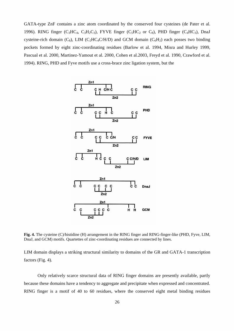
GATA-type ZnF contains a zinc atom coordinated by the conserved four cysteines (de Pater et al.
1996). RING finger (C3HC4, C3H2C3), FYVE finger (C5HC2 or C8), PHD finger (C4HC3), DnaJ
cysteine-rich domain (C8), LIM (C2HC4C/H/D) and GCM domain (C6H2) each posses two binding
pockets formed by eight zinc-coordinating residues (Barlow et al. 1994, Misra and Hurley 1999,
Pascual et al. 2000, Martinez-Yamout et al. 2000, Cohen et al.2003, Freyd et al. 1990, Crawford et al.
1994). RING, PHD and Fyve motifs use a cross-brace zinc ligation system, but the
Fig. 4. The cysteine (C)/histidine (H) arrangement in the RING finger and RING-finger-like (PHD, Fyve, LIM,DnaJ, and GCM) motifs. Quartettes of zinc-coordinating residues are connected by lines.
LIM domain displays a striking structural similarity to domains of the GR and GATA-1 transcription
factors (Fig. 4).
Only relatively scarce structural data of RING finger domains are presently available, partly
because these domains have a tendency to aggregate and precipitate when expressed and concentrated.
RING finger is a motif of 40 to 60 residues, where the conserved eight metal binding residues

27
(cysteines and histidines) bind two divalent zinc ions. There are two different variants, the C3HC4
(RING-CH) and a C3H2C3 -type (RING-H2), which are clearly related despite the presence of cysteine
(C) or histidine (H) in the fifth position. The spacing of cysteines and histidine in the RING-finger
motif is C-x2-C-x(9-39)-C-x(1-3)-H-x(2-3)-C/H-x2-C-x(4-48)-C-x2-C, where x is any amino acid. Four pairs
of metal-binding residues sequester two zinc atoms at distinct tetrahedral sites (Zheng et al. 2000).
The first and third pairs (C4-pair) ligate the zinc ion in position 1, while the zinc in position 2 is
ligated by the second and the fourth pair (C3H) of the RING finger motif. The structure of the zinc
ligation is unique and is referred to as the "cross-brace" motif (Fig. 5), which is also found in PHD
and Fyve domains. Within the RING domain the sulphydryl group of cysteine and the imidazolyl
nitrogen of histidine are ligating the metal ions (Barlow et al. 1994, Everett et al. 1993). Conserved
metal-binding residues can be substituted for other metal-binding amino acids (Asp and Thr).
Exceptionally in the RAG-1 (recombination-activating gene 1) and Rbx-1 (Ring box protein-1, also
known as ROC1 and HRT1) the first zinc-binding site of RING finger structure is a part of an unique
binuclear cluster with the cysteine bridging two zinc atoms, leading to co-ordination of three zinc
ions.
Fig. 5. Simple model of the “cross-brace” structure of the RING finger domain showing conserved cysteines(C) and histidines (H) and secondary structure elements (Dodd et al. 2004). Zn; a zinc ion.
Metal binding has been shown to stabilize the RING structure of PML (promyelocytic
leukemia protein) and MAT-1 (Menage a trois) (Borden 1995). In addition to zinc(II) binding, RING
finger can ligate cobolt(II) and cadmium(II) but with a lower affinity (Lovering et al. 1993, von Arnim
and Deng 1993, Upton et al.1994). Interestingly, the C4-site (site one) in the RING finger domain of
BRCA1 or HDM2 (human double minute 2) possesses a higher affinity towards zinc atom, whereas
the C3H -site (site 2) possesses a lower zinc-binding activity (Roehm and Berg 1997, Lai et al. 1998).

28
The metal binding is anticooperative, since the zinc-binding affinity of the C3H -site decreases ~ 20-
fold, when a metal ion binds to the C4-site.
The atomic resolution structures have been solved for RING domains of RAG-1, PML, MAT-
1, IEEVH and EL-5. Also the structures of BRCA1-BARD1 (BRCA1-associated ring domain protein
1) heterodimeric RING-RING complex, c-Cbl-Ubc7 complex and Rbx-1 containing SCF (Skp1-
Cullin-F-box protein) ubiquitin ligase complex have been solved. The RING C3HC4 secondary
structure adopts a ββαβ fold in Herpes simplex virus type 1 immediate-early protein Vmw110
(IEEVH), RAG-1, BRCA1 and MAT-1 (Fig. 6), but PML has a βββ-loop-α-β-fold (Everett et al.
1993). Conserved residues within the α-helix, β-sheets and loops contribute to a compact hydrophobic
180°
Fig. 6. The three-dimensional structure (NMR) of the RING finger domain of IEEHV presented as a ribbondiagram. The right site shows the molecule after a 180°C rotation around the vertical axis compared the leftside (Gervais et al. 2001). Black sphere presents zinc atom. Zinc ligating residues are shown in black.
core in IEEHV (immediate early protein from Equine herpes virus type 1) and MAT1 (Barlow et al.
1994, Gervais et al. 2001). The variable spacing in the consensus sequence leads to differences in
three-dimensional fold around the zinc-binding site 2 in PML, IEEHV and RAG1. The distance
between the two zinc-binding sites is likely to be the same in all known RING fingers, given the
absolute conservation of the spacing of the two residues between histidine ligand of the C3H-site and
the first cysteine ligand of the C4-site. For instance, the inter-zinc distance in PML, RAG1 and IEEHV

29
RING finger structure is the same (14Å) (Barlow et al. 1994, Borden et al. 1995, Bellon et al. 1997).
Also the charge distribution at the surface of RING finger domains could be an essential factor that
modulates RING finger activity, since, for example, the mutations of charged amino acids within the
RING domain affect the function of PML and IEEVH (Boddy et al. 1997). Generally, there is very
little sequence homology between RING proteins outside the consensus RING sequence, but these
regions regulate the specificity of protein-protein interactions and the structural diversity within this
family of proteins (Zheng et al. 2000, Brzovic et al. 2001). It appears that the RING finger motif
adopts significantly varying three-dimensional structures, while maintaining some structural
conservation including the overall topology of the central β-strands, the cross-braced Zn2+ binding
system and the packing of conserved residues which form the hydrophobic core of the molecule. This
suggests that the RING finger motif forms a convenient scaffold, which can be structurally varied to
reflect the diversity in its molecular function. The RING finger domain cannot be substituted even
between closely related RINGs without a change in function. For instance, even though the RING
fingers of BRCA1 and Rpt1 have more than 90% sequence identity, a BRCA1 form containing the
RINGRpt1-substitution does not maintain the BAP1 interaction anymore (Jensen et al. 1998).
RING fingers are often associated with distinct domains. For instance, the RING finger
domains of TRAF 2-5 are followed by five zinc fingers, a coiled-coil and a TRAF domain (Schwabe
and Klug 1994). The DNA repair proteins RAD5 and RAD16 have a RING finger that is interleaved
with ATPase domains. The inhibitor of apoptosis (IAP3) contains three BIR (baculovirus IAP repeat)
domains in front of the RING finger (Laren et al. 2003). Many RING finger proteins, such as Parkin
and the human homologue of Drosophila Ariadne (HHARI), that are involved in protein
ubiquitination and degradation are characterized by the presence of two RING finger domains
separated by the cysteine-rich IBR (the in between RING fingers) domain or DRIL (double RING
finger linked) domain (Moynihan et al. 1999, Zhang et al. 2000). This tripartite domain is called as a
TRIAD (two RING fingers and a DRIL). Mutations in the RING-IBR-RING of Parkin cause the AR-
JP, and RING2 mutations are found in a rare form of parkinsonism (Morett and Bork 1999, Shimura
et al. 2001). Interestingly, a NMR structural study of the RING2 in TRIAD of HHARI revealed that
RING2 has a totally different three-dimensional fold when compared to the classical RING finger, and
it binds only one zinc atom. In addition, the RING2 of HHARI possesses ubiquitin E3 ligase activity
(Capili et al. 2004). Most frequently, RING domains are associated with cysteine-rich zinc-binding
domains, B-boxes, and α-helical coiled-coil domain and referred to as RBCC-domains. This domain
is found in PML, estrogen-responsive RING finger (Efp), TIFα1, neuregulin receptor degradation
pathway protein 1 (Nrdp1) and KAP-1 (Fagioli et al. 1998, Inoue et al. 1991, Qiu and Goldberg 2002,

30
reviewed by Saurin et al. 1996). The RBCC domain appears to be an integral structural unit requiring
every one of its subdomains for proper function of the protein in which it is found.
2.2 Function of the RING domain
A small zinc-ligating domain can facilitate multiple intermolecular interactions between nucleic acids
and proteins. GATA-type ZnF can bind both DNA and proteins, and the proteins with multiple
GATA-type motifs can play a complex role in regulating transcription through an interplay of these
different binding selectivities and affinities. Other ZnFs have more specific functions, such as DNA-
binding ZnFs in the nuclear hormone receptor proteins and small-molecule-binding ZnFs in protein
kinase C. When the RING finger was initially identified, it was thought that it acted as a specific
DNA-binding domain (Freemont et al. 1991, van Lohuizen et al. 1991, Haupt et al. 1991), but RING
finger together with PHD, Fyve and LIM domains appear to act exclusively in protein-protein
interactions. Several intriguing characteristics evident from the studies of RING domains have made it
difficult to establish a single biochemical function for RINGs.
RING finger proteins are known to be involved in the assembly of large protein complexes.
RING finger is able to interact with other RING finger domains and non-RING containing sequences.
Further, RING finger can use both zinc-binding regions independently for binding different proteins
(Roehm and Berg 1997, reviewed by Kentsis and Borden 2000). The arenaviral protein Z is the
smallest known RING finger protein (90 aa) constituted almost entirely by its RING domain
(reviewed by Riviere et al. 1987). The Z protein self-assembles into ordered spherical structures via its
RING finger domain in vitro and these structures resemble the nuclear domains formed by Z protein
during virus infection (Kentsis et al. 2002a). Isolated RING domains of BRCA1, KAP-1, mel-18 and
PML are able to self-assembly in vitro (Kentsis et al. 2002b). Mutating the first zinc-binding site of
RING finger abolishes the capacity to self-assemble in vitro and also in vivo, but in contrast, the
second zinc-binding site mutation does not destroy the RING finger domain structure or eliminate the
self-assembly activity (Kentsis et al. 2002a,b, Peng et al. 2000, Borden et al. 1995, Campbell et al.
2000). Interestingly, the BRCA1 cancer predisposing RING mutant (C64G) protein fails to self-
assemble in vitro, form nuclear domains or suppress tumor growth in vivo (Kentsis et al. 2002b, Jin et
al. 1997). In addition to homodimerization, BRCA1 can form heterodimers with another RING finger
protein BARD1 through RINGBRCA1-RINGBARD1 interaction. Also BARD1 homodimerizes (Brzovic
et al. 1998, Meza et al. 1999). The RING finger domain of the PML is important for the formation of
PML multiprotein complexes that are referred to as PML nuclear bodies (PML NBs). Disruption of
RING domain destroys the PML NBs and further correlates with a loss of growth suppression and

31
apoptotic activities (Borden et al. 1997, Mu et al. 1994). Also the polycomb group protein Bmi1 exists
in a large 2-5 MDa protein complexes, and mutations in the conserved residues within RING finger of
Bmi1 disperses the Polycomp complexes and leads to anterior-posterior transformation of the axial
skeleton (Alkema et al. 1997). The sequence determinants for RING finger binding of RING-less
proteins are not well characterized. However, one potential proline-rich consensus sequence has been
defined as PxBxPJxP, where B is Leu/Val, J is Ala/ser and X is any amino acid, and is called as
FRODO (Funky RING oligomerization domain) (Kentsis and Borden 2000). The first zinc-binding
site of the RING finger has been shown to maintain the interaction with the FRODO.
There is a lot of evidence that the RING domain mediates ubiquitin E3 ligase activity
(reviewed by Jackson et al. 2000 and Joazeiro et al. 2000, Lorick et al. 1999). Generally, the RING
finger of E3 binds the ubiquitin-conjugation enzyme (E2) (Lorick et al. 1999, Zheng et al. 2000). The
crystal structure study of ubiquitin E3 ligase, c-Cbl, complexed with E2 enzyme, Ubc7, revealed that
the hydrophobic groove formed by the helix and the two zinc-chelating loops of the c-Cbl RING
finger forms the major contact region with loops 1 and 2 of Ubc7, but also the region preceding the
RING finger participates in E2 binding (Zheng et al. 2000). The multisubunit E3s, such as APC and
SCF, always contain a RING finger subunit such as Apc11 and Rbx1, respectively. The organization
and the function of these complex E3s are critically dependent on the RING finger (Seol et al 1999).
Mutations in the first zinc-binding site reduces ubiquitin ligase activity of SCF, but the second zinc-
binding site is irreparable for ubiquitin ligation (Ohta et al. 1999), indicating that the protein-
interactions by the zinc-binding site 2 are critical for normal function of the SCF complex. The RING
finger of Rbx1 is the primary binding region for E2 and serves as a secondary binding region for Cul1
in SCF complex (Zheng et al. 2002). The BRCA1-BARD1 complex functions as a RING ubiquitin E3
ligase (Hashizume et al. 2001, Kentsis et al. 2002b). In vitro ubiquitination reactions examined by
electron microscopy, showed that RINGBRCA1:BARD1 bodies efficiently scaffold multiple UbcH5Cs on
their surface with several chains of polyubiquitins (Kentsis et al. 2002b). In addition, a RING-like
domain (SP-RING;Siz/PIAS-RING) of PIAS proteins, which has been suggested to have a similar
three-dimensional structure as RING finger, binds directly to SUMO-conjugating enzyme (E2) Ubc9
and is required for PIAS SUMO E3 ligase activity (Kahyo et al. 2001, Kotaja et al. 2002).
RING finger proteins involve in many cellular processes. Many of the biological functions of
the PML, such as growth and transformation suppressive action, are mediated through PML NBs and
require an intact RING finger domain (Melnick and Licht 1999, Borden at al. 1997, Mu et al. 1994).
RING-mediated oligomerization of KAP-1 is required for its association with the DNA-dependent
transcriptional repression domain (KRAB) of KOX-1, thereby it mediates transcriptional repression
32
(Peng et al. 2000). Also the RING finger LIM domain-binding protein, RLIM, is a corepressor that
recruits histone deacetylases HDAC2 and Sin3 (Bach et al. 1999). Interestingly, also the RING-
mediated oligomerization stimulates catalytic activity of MAPKKK/MEKK1 and its
autophosphorylation, which is needed for the JNK activation (Baud et al. 1999). RING ubiquitin E3
ligases MDM2, IPC0, BRCA1-complex, pirh2, COP1 and Topors are involved in ubiquitination and
degradation of the tumor suppressor protein p53 in addition to other protein targets (Kubbutat et al.
1997, Haupt et al. 1997, Boutell and Everett 2003, Leng et al. 2003, Dong et al. 2003, Dornan et al.
2004, Rajendra et al. 2004). c-Cbl attenuates signaling by the growth factor receptors EGFR and
PGDFR via inducing their ubiquitination and degradation. Interestingly, oncogenic variants of c-Cbl
have been shown to contain mutated forms of the RING (Levkowitz et al. 1998, Miyake et al. 1998,
Joazeiro et al. 1999, Blake et al. 1991, Langdon et al. 1989).
3. THE UBIQUITINATION SYSTEM
Protein proteolysis is increasingly understood to be an important general mechanism by which cells
regulate protein levels and consequently their function at specific times. Constant protein turnover
serves many critical regulatory roles, including quality control by confirming degradation of
misfolded proteins due to mutations or damage in the protein. This function is especially important in
non-dividing cells, such as neurons, where the accumulation of malfunctioning proteins would be
highly deleterious (reviewed by Kopito 2000 and Sherman and Goldberg 2001 and Petrucelli and
Dawson 2004). In eukaryotic cells, the main mechanism for such control involves specific covalent
modification by ubiquitin/polyubiquitin, which labels target proteins for proteolysis and subsequent
degradation by the 26S proteasome or, as in the case of cell surface receptors, the ubiquitin
conjugation causes their down-regulation by the endosomal-lysosomal pathway (reviewed by
Schwartz and Ciechanover 1999). Ubiquitination can also directly alter the function and intracellular
localization of proteins. The ubiquitination pathway is an ATP-dependent and a multistep process,
involving at least three types of enzymes, known as E1, E2 and E3. Numerous studies have
demonstrated that ubiquitination plays an important regulatory role in many cellular events, such as
cell cycle, apoptosis, inflammation, DNA repair, and stress responses.
3.1 The ubiquitination pathway and proteasome
Ubiquitin is transcribed as an inactive precursor molecule with a C-terminal extension of several
amino acids, which is processed by a specific protease, ubiquitin carboxy-terminal hydrolase, to make
the carboxy-terminal double glycine (Gly76-Gly77) motif available for conjugation. Ubiquitination is

33
initiated by the activation of ubiquitin at the C-terminal Gly76 by ubiquitin-activating enzyme (E1) in
an ATP-dependent manner (Fig. 7). Synthesized C-terminal adenylate intermediate serves as the
Fig. 7. The ubiquitination pathway. Free ubiquitin (Ub) is cleaved from the ubiquitin precursor by ubiquitincarboxy-terminal hydrolase (UCH). Ubiquitin is activated in an ATP-dependent manner with the formation of athiol-ester linkage between E1 and the carboxy terminus of ubiquitin. Ubiquitin is transferred to E2. E2associates with E3, which may have the substrate already bound. For HECT domain E3s, ubiquitin istransferred to the active site cysteine of the HECT domain followed by ubiquitin transfer to the substrate (S).For RING E3, current evidence indicates that ubiquitin is transferred directly from E2 to the substrate(reviewed by Weissman 2001).
donor of ubiquitin to a cysteine (Cys) in the E1 active site and the thiol ester bond is formed between
ubiquitin and E1. Then the ubiquitin-conjugating enzyme (E2) accepts the activated ubiquitin from E1
with its own active Cys residue and, again, the thiol ester bond is formed. Finally, E2 ubiquitinates the
substrate in cooperation with ubiquitin E3 ligase. The HECT-type E3 accepts ubiquitin from E2, and
forms thiol linkage with ubiquitin and then shifts it to protein substrate. The RING-type ubiquitin E3
ligases do not form a thiol linkage with ubiquitin, rather they facilitate direct ubiquitin transfer from
E2 to the substrate. E3s dictate the specificity of ubiquitination by recognizing the target protein. In
34
most cases, the ubiquitination of proteins is mediated by lysine dependent-ubiquitination pathway,
where ubiquitin is conjugated into ε-NH2 group of an internal Lys residue. For instance, Lys21 and
Lys22 of IκBα have been shown to be the targets of ubiquitin conjugation, but in cyclin B any single
lysine residue can serve as an ubiquitin acceptor (King et al. 1996).
Proteasomes are present both in the cytoplasm and the nucleus. In the cytoplasm, proteasomes
associate with the cytoskeletal networks (reviewed by Wojcik and DeMartino 2003) and the outer
surface of the endoplasmic reticulum (ER) (reviewed by Brodsky et al. 1999). In the nucleus,
proteasomes are spread throughout the nucleoplasm, but are absent in the nucleoli, and sometimes
associate with discrete subnuclear domains called the PML nuclear bodies (PML NBs). Proteasomes
exist in multiple forms such as a free 20S proteasome, a 26S proteasome, a 20S proteasome associated
with PA28 and a hybrid proteasome (reviewed by DeMartino and Slaughter 1999 and Glickman and
Ciechanover 2002). Polyubiquitinated proteins are recognized and degraded by the 26S proteasome
(Chu-Ping et al. 1994). The S26 proteasome is a ∼ 2.4 MDa complex composed of two subcomplexes;
a 20S core particle, which carries the protease complex, and one or two 19S regulatory particles,
which regulate the function of the former and select the substrates. The polyubiquitinated proteins are
recognized and bound by the proteasome (Thrower et al. 2000) and the ubiquitin chain is cleaved from
the protein substrate by deubiquitination enzymes (DUBs). It has been suggested that specific
ubiquitin chain-recognition adaptors serve to distinguish between these ubiquitin chains and they are
likely play a role in the substrate delivery to the proteasome. For instance, HHR23 (human homologue
of yeast RAD23) translocates ubiquitinated target proteins to the proteasome via its UDP (ubiquitin-
domain protein) and UBA (ubiquitin-associated) domains, which bind catalytically active proteasome
and ubiquitin chains of the target protein, respectively (Chen and Madura 2002, Elsasser et al. 2004)
The protein is then unfolded and translocated into the 20S core proteasome in an ATP-dependent
manner and subsequently the protein is hydrolyzed into small peptides. Ubiquitin is spared from
degradation through its release from the substrate by ubiquitin hydrolases, and it is recycled back into
the ubiquitin pathway (Swaminathan et al. 1999, reviewed by Hershko and Ciechanover 1998).
Interestingly, not all ubiquitinated proteins are fully degraded, since some proteins, such as the
transcription factor p105 (a precursor of p50), and Spt23, are processed into a truncated form by
proteasome and thus the proteasome can regulate the activity of the protein (Palombella et al. 1994,
Hoppe et al. 2000). Proteasomes have also a capacity to degrade certain non-ubiquitinated substrates,
which can either be directly recognized by the proteasome or be presented to the proteasome by
cofactors, like antizyme in the case of ornithine decarboxylase (Murakami et al. 1992). Ubiquitination
is a reversible process due to deubiquitination enzymes (DUBs). DUBs are cysteine proteases that
specifically hydrolyse the amide bond immediately adjacent to the carboxy-terminal residue and these
35
proteases are divided into two categories; UCHs (ubiquitin-COOH hydrolases) and UBPs (ubiquitin-
specific proteases). UCHs remove short and flexible peptide chains from the COOH terminus of
ubiquitin, whereas UBPs cleave the isopeptide bond between ubiquitins (Ub-Ub) or between the Ub
and the protein. DUBs are involved in the generation of free ubiquitin, recycling of ubiquitin, editing
polyubiquitin chains (Kovalenko et al. 2003, Trompouki et al. 2003), and aiding proteasome-
dependent degradation (Reviewed by D’Andrea and Pellman 1998). Inhibitors of the ubiquitin-
proteasome pathway directly target and inhibit the 20S proteasome, rather than the upstream
ubiquitination. Lactacystin and its derivative clasto-lactacystin β-lactone, metabolites of Streptomyces
lactacystinaeus, irreversibly inhibit the 20S proteasome, as it forms a covalent bond with the catalytic
N-terminal residue of the proteasomal β-subunit (Fenteany et al. 1995, Ostrowska et al. 1997, Dick et
al. 1996). Another natural inhibitor, epoxomicin (EXM) is a peptide (originally isolated from a species
of Actinomycetes) that has a relatively high specificity for the 20S proteasome, and it covalently binds
to the catalytic subunits of the proteasome (Meng et al. 1999). Synthetic peptide aldehydes, MG-132
(benzyloxycarbonyl-leucinyl-leucinyl-leucinal), MG-115 (carbobenzoxy-L-leucyl-L-leucyl-L-nor-
valinal) and PSI (carbo-benzoxy-L-isoleucyl-gamma-t-butyl-L-glutamyl-L-alanyl-L-leucinal) arrest
reversibly the proteasome activity by inhibiting the chymotrypsin-like activity (Rock et al. 1994, Wilk
and Figueiredo-Pereira 1993). Since the ubiquitin-proteasome pathway is critical for the proliferation
and survival of cells, including cancer cells, proteasome inhibitors are of great interest as potential
therapeutics for cancer. In general, proteasome inhibition tends to induce apoptosis in proliferating
cells (Drexler 1997). The first proteasome inhibitor has been approved for use in patients with
relapsed and refractory multiple myeloma. Bortezomib (PS-341) is a modified dipeptidyl boronic acid
that binds reversibly to the chymotryptic site in the proteasome and induces apoptosis in different
cancer cell lines (Adams et al. 1999).
Ubiquitin
Ubiquitin (Ub) was first discovered as a lymphocyte differentiation-promoting factor in 1975
(Goldstein 1975). Ubiquitin is a small 8.5-kDa polypeptide (76 amino acid residues) that is
ubiquitously expressed in all eukaryotic cells, and it is found throughout the cell (ubique, latin for
everywhere). Ubiquitin is highly conserved during evolution, with only three amino acid changes
from yeast to human. Ubiquitin consisting of both alpha-helices and beta-sheets (ββαββαβ-fold), that
form a highly stable, globular structure containing a hydrophobic core with C-terminal glycine
residues (Gly76-Gly77) protruding from the main body of the protein (Fig. 8) (Vijay-Kumar 1987).
Figure 8 also shows the 3D structure of SUMO-1 (small ubiquitin-like modifier 1), which is
conjugated to proteins in a similar fashion as ubiquitin, but with different enzyme components and
different effect on the function of the target protein (see section 4. SUMO-1 conjugation).
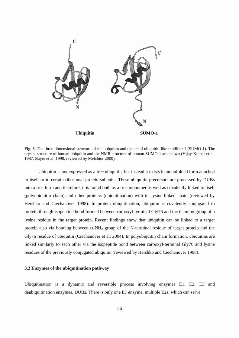
36
Ubiquitin SUMO-1
Fig. 8. The three-dimensional structure of the ubiquitin and the small ubiquitin-like modifier 1 (SUMO-1). Thecrystal structure of human ubiquitin and the NMR structure of human SUMO-1 are shown (Vijay-Kumar et al.1987, Bayer et al. 1998, reviewed by Melchior 2000).
Ubiquitin is not expressed as a free ubiquitin, but instead it exists in an unfolded form attached
to itself or to certain ribosomal protein subunits. These ubiquitin precursors are processed by DUBs
into a free form and therefore, it is found both as a free monomer as well as covalently linked to itself
(polyubiquitin chain) and other proteins (ubiquitination) with its lysine-linked chain (reviewed by
Hershko and Ciechanover 1998). In protein ubiquitination, ubiquitin is covalently conjugated to
protein through isopeptide bond formed between carboxyl-terminal Gly76 and the ε-amino group of a
lysine residue in the target protein. Recent findings show that ubiquitin can be linked to a target
protein also via bonding between α-NH2 group of the N-terminal residue of target protein and the
Gly76 residue of ubiquitin (Ciechanover et al. 2004). In polyubiquitin chain formation, ubiquitins are
linked similarly to each other via the isopeptide bond between carboxyl-terminal Gly76 and lysine
residues of the previously conjugated ubiquitin (reviewed by Hershko and Ciechanover 1998).
3.2 Enzymes of the ubiquitination pathway
Ubiquitination is a dynamic and reversible process involving enzymes E1, E2, E3 and
deubiquitination enzymes, DUBs. There is only one E1 enzyme, multiple E2s, which can serve
37
several E3 enzymes, and an even a greater number of E3s. This multi-step reaction involving many
different enzyme components enables protein ubiquitination to achieve a high degree of specificity
and diversity. Another large group of proteins involved in this process is the deubiquitinating
enzymes, a family of ubiquitin-specific proteases that cleave ubiquitin from ubiquitin-conjugated
proteins and are thought to act at different steps in the ubiquitin pathway.
3.2.1 Ubiquitin-activating E1 enzyme
A single E1 enzyme (UBA) in human and yeast is required to activate ubiquitin for all conjugating
reactions and to transfer it to all known E2s. E1 is essential for cell viability, since the deletion of
UBA1 in yeast is lethal (McGrath 1991). E1 is found in the nucleus and cytosol, and the localization
of E1 is cell cycle-regulated with a predominant nuclear localization in the G2 phase (Grenfell et
al.1994, Trausch, et al. 1993). E1 is also phosphorylated in mammalian cells by cdc2 kinase (Nagai et
al. 1995). Mutation of Cys residue of the active site abolishes the E1 activity (Hatfield and Vierstra
1992).
3.2.2 Ubiquitin-conjugating E2 enzymes
There are 13 isoforms of E2s in yeast and more than 20 in mammals (reviewed by Jentsch 1992 and
Hochstrasser 1996). E2s all share an active site ubiquitin binding Cys residue within the core
structure, Ubc domain, required for binding E3s. E2s are subdivided into four classes. Examples of
class I are Ubc4, Ubc5, Ubc7 and Ubc9-13, which contain UBC domain and require E3s for substrate
recognition and the ubiquitin transfer to target proteins. Several E2 enzymes are more complex than
the class I members and have unique N-terminal (class II) or C-terminal (class III) extensions of
various lengths or both of them (class IV). Class II contains Ubc1, Ubc2 (RAD6), Ubc3(Cdc34), Ubc6
and Ubc8. The C-terminal extension of Ubc2 appears to mediate interactions with histones and is also
needed for histone ubiquitination (Sung et al. 1988). Also the C-terminal extension is needed for
anchoring Ubc6 to the cytosolic side of endoplasmic reticulum (ER) (Sommer and Jentsch 1993).
Ubc2/RAD6 acts along with Ubr1/E3α to target N-end rule substrates (Xie and Varshavsky 1999).
Ubc4 and ubc5 E2s participate in the turnover of short-lived and misfolded proteins. Ubc13 is
involved in error-free postreplicative repair pathway in yeast (Brusky et al. 2000). It also forms a
complex with Mms2p, an ubiquitin-conjugating enzyme variant (UEV) without a catalytic cysteine
within UBC domain and causes the assembly of polyubiquitin chains linked through Lys-63
(Hofmann and Pickart 2001). Ubc6 associates with Ubc7 and they have been implicated in

38
endoplasmic ER-associated degradation (ERAD) pathway (Chen et al.1993, Kim BW et al. 2003).
Two UBC domain-containing proteins, Ubc9 and Ubc12, do not conjugate ubiquitin, but conjugate
ubiquitin-like molecules, SUMO (small ubiquitin-like modifier) and RUB-1/Nedd8 (related to
ubiquitin 1/Neural precursor cell-Expressed Developmentally down-regulated), respectively (Johnson
and Blodel 1997, Gong and Yeh 1999). The E2 terminology is not standardized across different
species, since similar names given to yeast and mammalian enzymes do not reflect functional or
structural homology. For instance, human UBCH1 is not the human homolog of yeast Ubc1, but
rather the homolog of yeast Ubc2/RAD6. Human UbcH5 is one of the most active E2s and it is
involved in ubiquitination of p53 and IκBα (Scheffner et al.1994, Gonen, et al.1999). Human UBCH6
and UBCh7 are involved in targeting of soluble proteins in the cytosol (Nuber et al.1996).
3.2.3 Ubiquitin E3 ligases
Ubiquitin E3 ligases play a critical roles in the ubiquitination cascade by recruiting E2s with ubiquitin
and recognizing the target protein and thereby facilitating ubiquitin transfer from E2 to the target
protein. The E3s can be divided into two groups: the homologous to E6-AP carboxy terminus (HECT)
domain-containing E3s and the RING domain-containing E3s. RING-type E3s are further divided into
single subunit and multisubunit E3s. Also RING-like ubiquitin ligase domains, plant homeodomain
(PHD) domain, the U-box and HUL-1, have been identified (van Sant et al. 2001, Hagglund and
Roizman 2003).
3.2.3.1 HECT-type ubiquitin E3 ligases
The first enzyme described in this family was E6-AP (E6-associated protein), which targets p53 for
degradation in the presence of human papillomavirus (HPV) oncoprotein E6 (Scheffner et al. 1993).
E6-AP was found to promote ubiquitination of p53 and SRC family kinase Blk in the absence of E6
(Scheffner et al. 1993, Oda et al. 1999), suggesting its role as an ubiquitin E3 ligase. Mutations in E6-
AP have been implicated in the pathogenesis of Angelman syndrome, a severe form of inherited
mental and motor retardation (Kishino et al. 1997). Later on, the family of proteins harboring HECT,
which shares a region of ~350 amino acid residues similar to the C-terminal domain of the E6-AP,
was revealed. This domain contains conserved Cys-residue that forms a covalent thiol-ester
intermediate with ubiquitin. In addition to the chemistry of ubiquitination, the C-terminal HECT
domain mediates the binding of distinct subclass of E2s, such as human UbcH5, UbcH7 and UbcH8
(Scheffner et al. 1994, Nuber et al. 1996, Kumar et al. 1997). HECT domain proteins contain an N-
terminal protein kinase C-related C2 domain and WW (two conserved tryptophan residues) domains

39
responsible for cellular localization and substrate recognition. The target proteins of HECT E3 ligases
predominately possess proline-rich motif, PpxY, for WW domain recognition. Yeast has only one
HECT E3 ligase, Rsp5p, which regulates a number of important cellular processes including
mitochondrial inheritance, internalization of cell surface receptors, and transcription. The disruption of
the rsp5 gene is also lethal (Fisk and Yaffe 1999, Huibregtse et al. 1997, Hein et al. 1995, reviewed by
Rotin 2000). Higher eukaryotes have several related Nedd4 proteins, such as Nedd4-1 and -2, Smurf1
and -2, Bul1 and Itch. Nedd4 ubiquitinates subunits of the epithelial sodium channel leading to down-
regulation of the number of active channels (Kamynina et al. 2001). The Smad ubiquitin regulatory
factor 1 and 2 (Smurf 1 and 2) participate in the control of cell growth, differentiation and apoptosis
via playing distinct functions in the regulation of signaling pathways triggered by the TGF-β
superfamily (Arora and Warrior 2001).
3.2.3.2 RING-type ubiquitin E3 ligases
In contrast to HECT-type E3s, RING-type E3s do not form a thioester bond with ubiquitin, but rather
bring the ubiquitin-E2 complex and substrate into close proximity and promote ubiquitin transfer
directly from E2 to the substrate (reviewed by Hershko and Ciechanover 1998). The RING finger
protein E3 family is composed of two distinct groups, single and multisubunit proteins. In single
protein RING-type E3s, such as MDM2, Cbl and Parkin, the RING finger domain and the substrate
recognition site are in the same molecule. For instance, Cbl E3 ligase is a 120-kDa protein, harboring
RING finger domain for recruiting E2-ubiquitin complex and TKB domain for binding the target
protein, tyrosine phosphorylated receptor tyrosine kinases. The crystal structure of Cbl-RING and
Ubc7 shows that the interaction surface for Ubc7 in RING domain is similar to that of HECT domain,
and also Ubc7 uses the same structural elements for the interaction with both domains, although there
is no sequence similarity between RING finger and HECT domain. MDM2 has E3 ligase activity
towards itself (autoubiquitination) and as well as to tumor suppressor protein p53. Autoubiquitination
of MDM2 does not only render proteins for degradation by the proteasome, but it also reduces its
ubiquitination activity towards p53. Autoubiquitination of IAP E3 ligase induces its degradation and
promotes apoptosis.
Many RING finger proteins are members of multisubunit ubiquitin E3 ligase complex, such as
APC (anaphase promoting complex), SCF (Skp1-Cullin1-F-box) and VCB-CUL2 (von-Hippel-
Lindau-ElongingC/B-Cul2). The small Rbx1/Hrt1/Roc1 RING finger protein is involved in E2-
ubiquitin recruitment together with Cullin in SCF or VCB-CUL2 E3s and assembly of other
components of the complex. The F-protein such as Skip2 in SCF complex recognizes the substrate.
The most complex multisubunit E3 is APC, which contains at least 13 subunits (reviewed by Hershko
40
and Ciechanover 1998, Vodermaier 2004). The APC and SCF complexes are the key players in the
control of cell cycle, where APC/C seems to inactivate many cell cycle-advancing protein activities
from the previous cell cycle stage, whereas SCF removes blocking agents of the cell cycle progression
(reviewed by Page and Hieter 1999). Interestingly, degradation of the SCF component Skp2 is
mediated by APC in the phase G1 of the cell-cycle, and therefore APC influences also the SCF-
dependent event (Wei et al. 2004).
3.3 Substrate specificity and regulation of ubiquitination
Targeting of a protein via the ubiquitin system must require specific recognition signals for
appropriate E3 ligases, but thus far our knowledge of these signals is limited. Only a few motifs or
structures that identify proteins as proteolytic substrates have been defined precisely. Certain amino
acid sequences called destruction signals or degrees appear to be the signals for degradation. Many
short-lived proteins contain a PEST sequence, a short stretch of approximately eight amino acids
enriched with proline (P), glutamic acid (E), serine (S) and threonine (T) (Rogers et al. 1986, reviewed
by Rechsteiner and Rogers 1996). PEST sequences often contain minimum consensus
phosphorylation sites (S/TP) for certain protein kinases (Yaglom et al. 1995). Phosphorylation within
the PEST region seems to be required for ubiquitination and degradation of NF-κB1 p105, the
inhibitor of the NF-κB, and transcription activator Gcn4 (Kornitzer et al. 1994, Lang et al. 2003). The
N-end rule pathway targets proteins carrying destabilizing N-terminal residues called N-degron for
degradation. The N-degron is recognized by ubiquitin E3 ligase, which then ubiquitinates the lysine
residue of the target protein. The relationship between the nature of the N-terminal amino acid
residues of the protein and its half-life is called the N-end rule (Bachmair et al. 1986, Varshavsky
1997). The E3α/Ubr1 has N-degron recognition site for substrates with basic NH2-termini and
hydrophobic termini, but also for non-N-end rule substrates (Kwon et al. 1998, Reiss, et al. 1988). For
example, DIAP1 (Drosophila inhibitor-of-apoptosis protein 1) degradation by the N-end rule pathway
is needed for its regulation activity in apoptosis (Ditzel et al. 2004). New degrons continue to be
identified at a regular rate. The Cdc20-APC/cyclosome (APC/C) ubiquitin E3 ligase complex
recognizes and binds the destruction box (D-box; Arg-xx-Leu-xxxx-Asn) of cyclin B and securin and
directs them for proteolysis during metaphase/anaphase (Yamano et al. 1998, Zur and Brandeis 2001).
Also APC/C-Cdh1 binds KEN-box (Lys-Glu-Asn) of hTK1 (human thymidine kinase 1) (Ke and
Chang 2004) and directs its to the proteasome. Signals may also be buried in the hydrophobic core.
Misfolded proteins probably expose similar hydrophobic pockets, which are buried in the correctly
folded structure, and these signals may be seen by the Ub-machinery yielding the protein to become
ubiquitinated and proteolytically processed (Sadis et al. 1995, Gilon et al. 1998.

41
Interaction of E3 and degron can be modulated by posttranslational modifications occurring at
different stages of cellular events. Phosphorylation triggers degradation of CDK regulators at
appropriate points in the cell cycle (reviewed by Peters 2002 and Deshaies and Ferrell 2001) or
stabilizes the protein such as Pds1, an inhibitor of anaphase initiation, in response to DNA damage
(Agarwal et al. 2003). Hydroxylation on proline residue and acetylation of HIF-1α (hypoxia inducible
factor-1α) are critical for recognition by the pVHL ubiquitination complex (Ivan et al. 2001, Jaakkola
et al. 2001). Also RNA polymerase II needs phosphorylation and proline hydroxylation for
recognition by the pVHL complex and thus its ubiquitination (Kuznetsova et al. 2003). p53 protein is
acetylated and ubiquitinated on the same Lys residues, and it seems that acetylation blocks also
ubiquitination of unacetylated Lys residues and thus stabilizes p53 (Li M et al. 2002). In addition to
acetylation, also methylation and sumoylation can be modifying same lysine residue as ubiquitin, and
it is possible that these modifications regulate each other by competing for the same lysine residues.
Sumoylation of lysine residue in IκBα and Smad4 has been demonstrated to block ubiquitination at an
identical site, protecting these proteins from degradation (Desterro et al. 1998, Lin et al. 2003). Also
protein association and dissociation of substrate can regulate its ubiquitination. Heterodimerization of
the yeast mating factors MATa1 and MAT2, or homodimerization of nuclear factor NF-IL6 may mask
an element that otherwise serves as the target for ubiquitination and proteasome degradation (Johnson
et al. 1998, Hattori et al. 2003). It is also possible that dimerization activates subsequent proteasome
processing such as in the case of dimerization of transcription factors ATF2 (activating transcription
factor 2) and c-Jun (Fuchs and Ronai 1999).
3.4 Alternative ubiquitin signals and their function
A single protein can be ubiquitinated on one or several lysines with a single ubiquitin
(monoubiquitination) moiety, with lysine-linked chains of ubiquitin (polyubiquitination) or
combination of these two. Polyubiquitin chains can be built to the lysine residue of the target protein
through repetitive conjugation of ubiquitin via distinct Lys residues (Lys6, Lys11, Lys27, Lys29,
Lys33, Lys48, and Lys63) of preceding ubiquitin (Peng et al. 2003). The fate of a target protein
depends on the number of ubiquitin molecules conjugated as well as the lysine linkage in the ubiquitin
chain (Fig. 9). In some cases, polyubiquitination of protein requires additional ubiquitin chain
elongating factor, named E4 (Koegl et al. 1999). In yeast, E4 binds to the ubiquitin moieties of short
conjugates and catalyzes ubiquitin elongation in conjunction with E1, E2, and E3. Interestingly,
transcription coactivator p300 appears to possess the E4 activity, and polyubiquitinates p53, but p53
must be first monoubiquitinated by MDM2 (Grossman et al. 2003). Proteins modified with ubiquitin
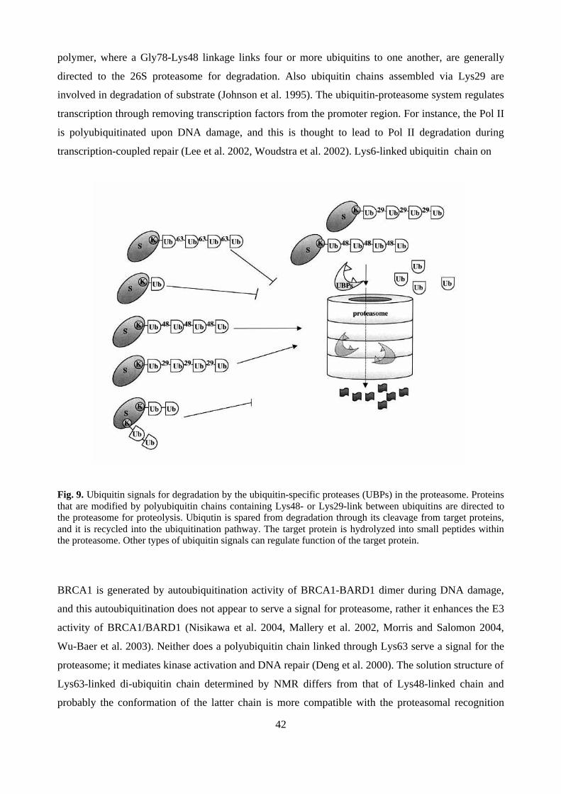
42
polymer, where a Gly78-Lys48 linkage links four or more ubiquitins to one another, are generally
directed to the 26S proteasome for degradation. Also ubiquitin chains assembled via Lys29 are
involved in degradation of substrate (Johnson et al. 1995). The ubiquitin-proteasome system regulates
transcription through removing transcription factors from the promoter region. For instance, the Pol II
is polyubiquitinated upon DNA damage, and this is thought to lead to Pol II degradation during
transcription-coupled repair (Lee et al. 2002, Woudstra et al. 2002). Lys6-linked ubiquitin chain on
Fig. 9. Ubiquitin signals for degradation by the ubiquitin-specific proteases (UBPs) in the proteasome. Proteinsthat are modified by polyubiquitin chains containing Lys48- or Lys29-link between ubiquitins are directed tothe proteasome for proteolysis. Ubiqutin is spared from degradation through its cleavage from target proteins,and it is recycled into the ubiquitination pathway. The target protein is hydrolyzed into small peptides withinthe proteasome. Other types of ubiquitin signals can regulate function of the target protein.
BRCA1 is generated by autoubiquitination activity of BRCA1-BARD1 dimer during DNA damage,
and this autoubiquitination does not appear to serve a signal for proteasome, rather it enhances the E3
activity of BRCA1/BARD1 (Nisikawa et al. 2004, Mallery et al. 2002, Morris and Salomon 2004,
Wu-Baer et al. 2003). Neither does a polyubiquitin chain linked through Lys63 serve a signal for the
proteasome; it mediates kinase activation and DNA repair (Deng et al. 2000). The solution structure of
Lys63-linked di-ubiquitin chain determined by NMR differs from that of Lys48-linked chain and
probably the conformation of the latter chain is more compatible with the proteasomal recognition

43
signal (Varadan et al. 2004). In addition, Lys11, -27 and -33 ubiquitin chains has been observed, but
the functions of these chains are unknown (Peng, et al. 2003). Lys-11 polyubiquitins on BAG-1
protein does not lead to degradation, but stimulation of the degradation-independent association of
BAG-1 with the proteasome (Alberti, et al. 2002). Monoubiquitination does not normally target a
protein for degradation, but appears to act as a signal for trafficking, DNA repair, gene silencing and
vesicle sorting or as a substrate for polyubiquitin conjugation (reviewed by Johnson 2002 and Pickart
2000). Sumoylated lysine of PCNA (proliferating cell nuclear antigen) becomes monoubiquitinated
during DNA damage by UV light (Stelter and Ulrich 2003). The same lysine can be further Lys63-
polyubiquitinated, which is important in error-free repair of the damaged DNA (Hoege et al. 2002).
Monoubiquitination is the principal signal responsible for the movement of receptor tyrosine kinases
(RTKs) from the plasma membrane to the lysosome (Haglund et al. 2003, Jeong et al. 2002).
Moreover, the monoubiquitination of p53 by MDM2 E3 ligase is critical for its nuclear export (Li M
et al. 2003). Monoubiquitination of transcription regulators has been shown to be involved in
transcription activation. Monoubiquitination of coactivator protein CIITA, which regulates the
expression of major histocompatibility complex (MHC) II class gene, is suggested to enhance the
assembly of CTIIA into a transcriptional complex at the promoter and thus increase gene expression
(Greer et al. 2003).
Since ubiquitination plays an essential role in a broad array of cellular functions, the
malfunction of this pathway will cause numerous diseases. In neurodegenerative diseases, such as the
Angelman sydrome and Parkinson the ubiquitin E3 ligase, E6-AP or Parkin, respectively, or the
ubiquitin carboxy-terminal hydrolase-1 (UCH-L1) have been shown to be mutated (Kishino et al.
1997, Shimura et al. 2000, Saigoh et al. 1999). The ubiquitin protease UCH BAP1 has been shown to
enhance the growth inhibitory activity of BRCA1, and mutations in BAP1 gene have been observed in
some lung carcinoma cell lines (Jensen et al. 1998). Also mutations that interfere with ubiquitin E3
ligase activity of BRCA1 are found in breast cancer (Ruffner et al. 2001). MDM2 is overexpressed in
a significant number of human tumors, which is thought to suppress normal p53 levels and suffocate
the p53 response to cell stress (Reifenberger et al. 1993, reviewed by Michael and Oren 2003).
Additionally, alterations of genes of the APC subunits have been found in colon cancer cell lines
(Wang Q et al. 2003).
3.5 Ubiquitin-binding domains
The information of ubiquitin signals can be transmitted within the cell by proteins that bind
noncovalently to ubiquitin and ubiquitinated proteins. Ubiquitin and poly-ubiquitin chains can be

44
recognized by different classes of ubiquitin-binding domains; the UIM (ubiquitin-interacting motif),
the UBA (ubiquitin-associated), the CUE (coupling of ubiquitin conjugation to ER degradation), the
UEV (ubiquitin E2 variant/UBC-like), the NZF (Npl4 zinc finger), the GAT (GGA/Tom1) and the
PAZ (polyubiquitin associated zinc finger). The UBA domain consists of three helices and exposes
hydrophobic interface, which contacts ubiquitin (Mueller and Feigon 2002). The UBA domain of
RAD23 and that of Dsk2p have shown binding preference to Lys48-linked ubiquitin chains
(Funakoshi et al. 2002, Raasi and Pickart 2003) and to be essential for their function in proteolysis
(Rao and Sastry 2002). CUE and UBA are structurally similar and bind both mono-, polyubiquitin and
ubiquitinated proteins (Bertolaet et al. 2001). The UIM motif forms an amphipathic α-helix and two
or three UIMs can be present in a protein (Swanson et al. 2003). UIM and CUE domains promote
monoubiquitination of proteins containing these sequences (Shih et al. 2003, Polo et al. 2002). The
UIM domains of both Vps27 and Hse1 are needed for sorting of ubiquitinated proteins for degradation
at the endosome (Bilodeau et al. 2002). The UEV domain is related to the catalytic domain of E2
enzymes, but lacks the catalytic cysteine that is essential for ubiquitin conjugation. In the Ubc13-
Mms2 heterodimer the Mms2 protein binds ubiquitin through its UEV domain, which is required for
linking ubiquitin molecules to one to another via Lys-63 isopeptide bond (Mckenna et al. 2001,
Mckenna et al. 2003a, Mckenna et al. 2003b). Tsg101/Vsp23 UEV domain binds ubiquitin with a
different interface than the UEV domain of Msm2. Ubiquitin binding of Tsg101/Vsp23 does not
involve linking ubiquitin chains, but is required for sorting ubiquitinated proteins into the internal
vesicles of multivesicular bodies (Katzmann et al. 2001, Bilodeau et al. 2003, Teo et al. 2004). The
ubiquitin binding activity of the GAT domain in GGA protein is needed for ubiquitination within its
GAT domain and ubiquitin-dependent transport to endosomes (Shiba et al. 2004, Scott et al. 2004).
The PAZ domain of HDAC6 (histone deacetylase 6) interacts only with polyubiquitin and similar zinc
finger domains are present in several DUBs and in BRCA1-associated proteins 1 and 2 (Hook et al.
2002). Ubiquitin binding of the Vsp36 NZF domain is required for efficient vacuolar sorting of
ubiquitinated proteins in yeast (Alam et al. 2004). Although the NZF domain is structurally distinct
from UIM, GAT, UBA and CUE, all of these domains bind the same hydrophobic surface (the "Ile-
44" hydrophobic surface) of ubiquitin (Alam et al. 2004, Mueller and Feigon 2002, Kang et al. 2003,
Swanson et al. 2003, Scott et al. 2004).
4. SUMO-1 CONJUGATION
Over the past 15 years, a number of proteins related to ubiquitin, UBLs (ubiquitin-like proteins), have
been discovered. Most of these proteins resemble ubiquitin in their primary and higher order structure,
but they have unique properties. These UBLs can be divided into two subclasses: Type-1 UBLs

45
SUMO, RUB/Nedd8 (Parry and Estelle 2004), FAT10 (Raasi et al. 2001), ISG15 (reviewed Ritchie
and Zhang 2004), Ufm1 (Komatsu et al. 2004), Hub1/UBL5 (Luders et al. 2003), Atg12 and Atg8
(reviewed by Ohsumi and Mizushima 2004) are conjugated to target proteins in a manner similar, but
not identical, to the ubiquitination pathway, whereas type-2 UBLs (also called UDP, ubiquitin-domain
proteins), such as Parkin, RAD23, and DSK2 contain an ubiquitin-like domain that is not covalently
conjugated to other proteins, but has been found to direct proteins to the proteasome (Chen and
Madura 2002, Leggett et al. 2002). SUMO (small ubiquitin-like modifier) is probably the most
investigated of the UBLs over the past few years.
4.1 The SUMO pathway
SUMOs (small ubiquitin-related modifiers) belong to a highly conserved protein family found in all
eukaryotes. SUMO is structurally related to ubiquitin containing the ββαββαβ ubiquitin-fold, although
SUMO-1 and ubiquitin share only ~18 % primary sequence identity (Bayer et al. 1998, Jin et al. 2001)
(Fig. 8). However, the surface charge distribution of SUMO is very different from that of ubiquitin.
SUMOs have also unstructured N-terminal extension that is not present in ubiquitin and that may
provide an additional surface for protein interactions (Bayer et al. 1998). Saccharomyces cerevisiae
contain a single SUMO protein encoded by the SMT3 gene, while mammals contain three different
SUMOs; SUMO-1 (also called PIC1, GMP1, Ubl1 and Smt3c), SUMO-2 (sentrin-3, Smt3a), and
SUMO-3 (sentrin-2, Smt3b) (reviewed by Melchior 2000, Müller et al. 2001, Seeler and Dejean,
2003). SUMO-1 shares 48 % identity with SUMO-2, and SUMO-2 and SUMO-3 are 95% identical to
each other. The crystal structure of the SUMO-2 is similar to that of SUMO-1 (Huang et al. 2004).
Localization studies of SUMO variants showed that SUMO-1, -2 and -3 share a similar distribution all
over the nucleoplasm, including PML NBs. However, SUMO-1 is also localized to nucleoli, the
nuclear envelope and cytoplasmic foci. The distribution of different SUMOs change rapidly during
cell cycle. During mitosis, SUMO-1 is more avidly localized to the mitotic spindle, and during late
anaphase, it is recruited to re-formed nuclear envelopes and further accumulates in the nucleus.
SUMO-2 and SUMO-3 share similar cell cycle-dependent localization, which is distinct from that of
SUMO-1. For instance, they are not concentrated in spindle microtubules and they are found in
chromatin regions earlier than SUMO-1 (Ayaydin and Dasso 2004). Recently, a human SUMO-4 gene
was discovered, and its protein product has 87% homology with SUMO-2. Interestingly, a
polymorphism in the SUMO-4 gene appears to be associated with type 1 diabetes (Bohren et al. 2004,
Guo et al. 2004). Plants (Aradopsis), in turn, might contain up to eight SUMO genes (Lois et al. 2003,
Kurepa et al. 2003).

46
Like ubiquitin, the precursor of SUMO requires processing to remove four amino acids from
its C terminus by cysteine proteases (SUMO proteases) to reveal diglycine motif (Gly-Gly) for
conjugation. Covalent attachment of SUMO on target protein is catalyzed by three enzymatic steps,
which are analogous to the ubiquitination pathway, consisting of the SUMO-activating (E1) enzyme
(heterodimer of SAE1/Aos1/Sua1 and SAE2/Uba2), the SUMO-conjugating (E2) enzyme (Ubc9), and
several SUMO E3 ligases (PIAS/Siz, RanBP2, and Pc2). First, SUMO is activated by E1 in the
presence of ATP and then it is transmitted to the active cysteine of E2 from where it is directed to the
ε-amino group of lysine in the target protein. In contrast to ubiquitination pathway where ubiquitin
ligases are responsible for target recognition, Ubc9 efficiently transfers SUMO to selected targets in
vitro. However, the specific SUMO E3 ligase appears to be required for efficient modification in vivo
(Fig. 10).
Fig. 10. The SUMO conjugation pathway. After SUMO is proteolytically processed by C-terminal hydrolases,it serves as the substrate in the ATP-dependent formation of an isopeptide bond between SUMO and the lysinein the target protein. This reaction is mediated by SAE1/SAE2 (E1), Ubc9 (E2) and SUMO ligases (E3).Sumoylation is a reversible reaction, since the isopeptide bond can be cleaved by isopeptidases (Adapted fromMelchior 2000).
In most cases, the lysine residue is embedded in a consensus sequence, ψKxE/D, where ψ is a large
hydrophobic amino acid (Ile, Leu, or Val), K is lysine, X is any amino acid residue, and E and D are
glutamic and aspartic acid, respectively. Both Ubc9 and E3s contribute to substrate specificity.
Sumoylation of the target protein can be regulated by other posttranslational modification.
Phosphorylation of c-Jun, p53, IκBα and PML reduces SUMO-1 modification, whereas the heat shock
factor 1 (HSF1) must be phosphorylated in order to be sumoylated (Müller et al. 1998, Hietakangas et
al. 2003, Desterro et al. 1998, Everett et al. 1999). Also other lysine-dependent modifications,
acetylation, methylation and ubiquitination may regulate sumoylation by modifying the same lysine
residue (Sapetschnig et al. 2002, Braun et al. 2001, Desterro et al. 1998, Ross et al. 2003).
Very little is know about the functions of SUMO-2 and SUMO-3, but at least they have been
implicated to play a role in the response to environmental stress (Saitoh and Hinchey 2000). As

47
SUMO-2 and SUMO-3 contain the ψKXE/D consensus motif in their N-terminal region, they can
form SUMO chains (Tatham et al. 2001). Although SUMO-1 lacks this sequence, it may be able to
form SUMO chains via different lysine acceptors (Pichler et al. 2002). SUMO chains may have
important functions in vivo , since, for instance, they may affect amyloid protein processing and thus
could be involved in the pathogenesis of Alzheimer´s disease (Li Y et al. 2003). Like ubiquitination,
sumoylation is a reversible and dynamic process. Removal of SUMO from modified proteins is
executed by SUMO-specific isopeptidases of the SUSP/SENP family.
4.2 Enzymes of the sumoylation pathway
4.2.1 SUMO-activating E1 enzyme
Most organisms have only a single SUMO-activating enzyme, E1, which is required for conjugation
of all SUMO variants to their target proteins. The SUMO E1 is a heterodimer (SAE1/SAE2), which is
structurally and functionally related to ubiquitin-activating E1 enzyme, UBA1. The SAE1 (SUMO
activating enzyme 1, also called Aos1 and Sua1) subunit resembles the N-terminal half of UBA1,
whereas the SAE2 (also called Uba2) subunit corresponds to the C-terminal part of UBA1 and
contains the active site cysteine (Desterro et al. 1999, Gong et al. 1999, Okuma et al. 1999). However,
Aradopsis has two SAE1 genes, whose products probably form a heterodimer with SAE2 (Kurepa et
al. 2003). In the SUMO activation step, SAE1/SAE2 consumes ATP to adenylate the C-terminal
glycine of SUMO-1 before it forms the high-energy thiolester bond between the C-terminal glycine of
SUMO and the active site cysteine in SAE2.
4.2.2 SUMO-conjugating E2 enzymes
The SUMO moiety is transferred from SAE1/SAE2 to the active site cysteine (Cys 93) of the SUMO-
conjugating enzyme (E2), forming a SUMO-E2 thioester complex. SUMO pathway involves only a
single SUMO E2, Ubc9, which shares substantial sequence and structural similarity with ubiquitin
E2s (Tong et al. 1997). The positively charged N-terminal region of Ubc9 binds non-covalently to the
negatively charged surface of all SUMOs, but not with the positively charged surface of ubiquitin
(Tatham et al. 2003, Liu et al. 1999). This may have a role in modifier discrimination since Ubc9 is
specific to SUMO and does not function with ubiquitin (Desterro et al. 1997, Gong et al. 1997). The
SUMO binding region of Ubc9 overlaps with that of E1, and E1 binding of Ubc9 is needed for the
thiolester bond formation (Bencsath et al. 2002). Non-covalent SUMO binding of Ubc9 is important
for the transfer of SUMO from E1 to E2, although it is distant from the C-terminal active site cysteine
of Ubc9. This C-terminal region of Ubc9 also binds the ψKXE/D consensus motif in the substrate
48
(Tatham et al. 2003, Tong et al. 1997, Bernier-Villamor et al. 2002). Most of the SUMO targeted
proteins are able to bind Ubc9.
4.2.3 SUMO E3 ligases
Even though Ubc9 is capable of recognizing and binding the SUMO modification site in a substrate, a
specific SUMO E3 ligase may be required for efficient and properly targeted modification in vivo.
SUMO E3 ligases have the capacity to associate with Ubc9 and increase the rate of target protein
modification. To this date, only three kinds of SUMO E3 ligases have been discovered. The Siz/PIAS
(Sap and Miz/protein inhibitors of activated STAT) protein family shares some similarity with RING-
finger ubiquitin E3 ligases, particularly an essential RING-like motif (Johnson and Gupta 2001,
Kahyo et al. 2001, Sachdev et al. 2001). In S. cerevisiae, Siz1p is required for sumoylation of septins
in vivo, and it enhances in vitro sumoylation (Johnson and Gupta 2001, Takahashi et al. 2001).
Mammalian PIAS proteins (PIAS1, -3, -x and -y, and splicing derivatives) bind Ubc9 with their RING
finger-like domain and are able to bind the target protein (Kotaja et al. 2002, Liang et al. 2004) and
SUMO. SUMO ligation activity is clearly dependent on the RING finger-like motif. PIAS proteins act
as SUMO E3s for LEF1, p53, AR and c-Jun (Kahyo et al. 2001, Sachdev et al. 2001, Schmidt and
Müller 2002, Kotaja et al. 2002). Recently, RanBP2 (Ran-binding protein 2)/Nup358 and Pc2
(Polycomb protein) were shown to have SUMO E3 ligase activity (Pichler et al. 2002, Kagey et al.
2003). RanBP2 has SUMO E3 activity toward MDM2, Sp100 and HDAC4, heterogeneous nuclear
ribonucleoproteins, hnRNP C and M proteins (Kirsh et al. 2002, Pichler et al. 2002, Miyauchi et al.
2002, Vassileva and Matunis 2004). Pc2 is able to sumoylate the corepressor protein CtBP (carboxyl-
terminal binding protein) (Kagey et al. 2003). Also SUMO E3 ligases are themselves sumoylated
(Kotaja et al. 2002). Interestingly, all three ligase types localize to specific subcellular complexes
suggesting that these structures may serve as modification sites. PIAS proteins are found in the close
proximity of PML or other nuclear bodies, Pc2 in Polycomb group (PcG) bodies and RanBP2 in
nuclear pore complex (NPC). The role of E3 ligases may be to stabilize the interaction between Ubc9
and the target protein.
4.2.4 SUMO proteases
SUMO proteases can act both in the maturation of the pre-SUMO (carboxy-terminal hydrolases) and
in the cleavage of SUMO from modified proteins (isopeptidases). These cysteine proteases are distinct
from ubiquitin-specific proteases, and they all contain a C-terminal ULP-domain (200 aa), which
harbors the catalytically active region (Mossessova and Lima 2000). SUMO proteases have variable

49
N-terminal domains, which localize the enzymes to different regions in the cells and thus appear to be
responsible for desumoylation of different proteins. Saccharomyces cerevisiae contains two SUMO
proteases, Upl1 and Ulp2/Smt4 (Li and Hochstrasser 1999 and 2000). Ulp2p is predominantly located
in the nucleus, whereas Ulp1p colocalizes with nuclear pore complex proteins. Ulp1 is capable of
processing both SUMO maturation and SUMO substrate cleavage, and it is required for the G2/M
transition of the cell cycle (Li and Hochstrasser 1999). Ulp2 is localized in the nucleus, but it is not
essential for cell viability (Li and Hochstrasser 2000). In mammals, SENP1 is primarily a nuclear
protease that is found in PML nuclear bodies. It has been shown to cleave SUMO from PML and
histone deacetylase 1 (HDAC1) (Gong et al. 2000, Bailey and O´Hare 2004, Cheng et al. 2004).
SENP2 in turn is a nuclear envelope-associated protease (Hang and Dasso 2002). SuPr-1, a splice
variant of mouse SENP2, is able to alter the distribution of Daxx, CBP and Sp3 in PML NBs (Best et
al. 2002, Ross et al. 2002). SENP3/SMT3IP1 is localized in the nucleolus (Nishida et al. 2000)
whrereas SENP6/SUSP1 is found in the cytosol (Kim et al. 2000).
4.3 Function of SUMO-1
A number of transcription factors, and chromatin-associated and DNA repair proteins have shown to
be posttranslationally modified by SUMO-1. SUMO-1 modification regulates protein-protein
interactions, protein stability, activity and conformation, and subcellular localization. For instance,
sumoylated form of the RanGAP, a GTPase-activating protein that plays a role in nuclear import,
binds more actively to the nuclear pore complex (NCP)-anchored RanBP2. This may enable RanGAP
to be recruited from cytoplasm to the NCP (Matunis et al. 1996, Mahajan et al. 1997). In addition to
SUMO-1 conjugation, other regions of RanGAP are needed for proper interaction with RanBP2,
suggesting that the binding is mediated through regions of SUMO-1 and RanBP2 or the sumoylation
induces structural changes, which then reveals the RanBP2-binding element of RanGAP (Matunis et
al. 1998 499). Moreover, the SUMO-1-modified RanGAP is found in the spindle microtubules during
mitosis (Joseph et al. 2002). Translocation of the MAP kinase MEK1 and the NF-κB essential
regulator protein (NEMO) between the nucleus and the cytosol is regulated by sumoylation (Galy et
al. 2002, Huang et al. 2003). Subnuclear localization of PML is regulated by SUMO-1 conjugation,
while SUMO-1-modified PML exists in nuclear PML NBs (Kamitani et al. 1998, Duprez et al. 1999).
SUMO-1-modified PML recruits many transcription factors and chromatin modifiers, such as Sp100,
CBP, Daxx, to PML NBs (Kamitani et al. 1998, Müller et al. 1998, Duprez et al. 1999, Ishov et al.
1999). Many of these proteins are also SUMO-1 modified (reviewed by Zhong et al. 2000), but it
50
seems that sumoylation of PML rather than sumoylation of other proteins is the controlling factor in
PML NB formation (Hofmann et al. 2000, Fogal et al. 2000), e.g. sumoylation deficient Sp100 is
recruited PML NBs (Sternsdorf et al. 1997). However, SUMO-1 conjugation of Sp100 enhances its
interaction with heterochromatin protein 1 (HP1) in vitro (Seeler et al. 2001), suggesting that this
PML-associated protein may recruit additional proteins via its SUMO-1 modification. Interestingly,
different SUMO variants may direct certain SUMO target proteins to different subcellullar
compartments (Saitoh and Hinchey 2000). Transcription factor SATB2 localizes to nuclear dots when
conjugated to SUMO-1, but to the nuclear periphery when conjugated to SUMO-3 (Dobreva et al.
2003).
Many transcription factors and coregulators are sumoylated, suggesting that sumoylation plays
a role in the regulation of gene expression. In most cases, sumoylation appears to repress transcription.
For example, when SUMO-1 is fused to the DNA-binding domain of Gal4, this fusion protein
represses transcription. This result may indicate that SUMO-1 is able to recruit transcriptional
repression proteins, such as HDACs, to the promoter (Ross et al. 2002, Yang et al. 2003).
Interestingly, the coactivator protein p300 interacts with the HDAC6 only when the repressor domain
of p300 is sumoylated (Girdwood et al. 2003). Transcription is also repressed when sumoylated
transcription factor Elk-1 binds to HDAC2 (Yang and Sharrocks 2004). In addition, sumoylation of
histone H4 represses transcription, which is likely to occur through interaction with corepressor
proteins HDAC1 and HP1 (Shiio and Eisenman 2003). An interesting example of sumoylation is that
of glucocorticoid receptor (GR). GR contains so called synergy control motifs, which are found in
several transcription factors, like in AR, and attenuate the activity of promoters having multible GR-
binding elements (GREs), but not that of promoters with single GRE-sites (Iniguez-Lluhi and Pearce
2000). These synergy control motifs harbor the classical SUMO-1 attachment sites, and they are
indeed sumoylated, which is suggested to prevent the synergistic action of multible GRs bound to the
same promoter (Iniguez-Lluhi and Pearce 2000, Tian et al. 2002, Subramanian et al. 2003, Poukka et
al. 2000b). Although sumoylation of transcription factors is mainly correlated with transcriptional
repression, SUMO-1 has also been shown to have positive effects on transcription. For example,
sumoylation of the coactivator GRIP1 (SRC-2) facilitates AR-interaction and thus enhances AR-
mediated transcription (Kotaja et al. 2002b). In addition, it has been suggested that sumoylation of the
transcription factor Tcf-4 is needed for its activation function (Yamamoto et al. 2003).
SUMO-1 modification has been shown to protect some proteins from proteasome-mediated
degradation. Sumoylation stabilizes IκBα and Smad4 likely by competing with the same lysine
residue involved in ubiquitination (Desterro et al. 1998, Lin et al. 2003). Interestingly, degradation of

51
CREB by ubiquitin-proteasome system leads to induction of proinflamatory genes right after hypoxic
stress, but it is stabilized via sumoylation one or two days after the stress (Comerford et al. 2003).
Yeast proliferating cell nuclear antigen (PCNA) can be modified on the same lysine by SUMO-1 and
ubiquitin. However, the SUMO-1 modification regulates the function rather than the stability of
PCNA (Stelter and Ulrich 2003, Haracska et al. 2004). Sumoylation of yeast PCNA may promote
replication by increasing the activity of translesion DNA polymerase and reducing the recombinant-
dependent bypass mechanism in the S phase, while ubiquitination of PCNA may promote DNA repair
processes (Stelter and Ulrich 2003, Haracska et al. 2004, Hoege et al. 2002).
SUMO-1 is able to alter the activity of thymine DNA glycosylase (TDG) that is involved in
base excision repair during DNA damage (Hardeland et al. 2002). The unmodified TDG binds and
excises a mutant base, becomes sumoylated and then dissociates from the DNA product. The affinity
of TDG towards its DNA substrate is reduced possibly due to a conformational change of TDG, where
an intramolecular interaction occurs between the SUMO-1-binding motif of TDG and the conjugated
SUMO-1. Further, SUMO proteases might restore the enzymatic activity of TDG. In the case of heat
shock factors 1 and 2 (HSF1 and HSF2), sumoylation enhances their binding activity to DNA in vitro
(Hong et al. 2001, Goodson et al. 2001). In vivo, heat shock treatment induces sumoylation of HSF1,
which is linked to the activation of stress-induced gene expression.
Several proteins have been shown to bind non-covalently to SUMO-1 and some SUMO-1-
binding motifs have been identified (Minty et al. 2000, Engelhardt et al. 2003, Rosendorff et al. 2004 ,
Song et al. 2004). According to Minty et al. (2000), protein motifs containing Ser/Thr- and Glu/Asp-
rich sequences preceded by hydrophobic residues are able to mediate the interaction with free SUMO-
1. Rosendorff et al. (2004) found that acidic amino acid-containing regions followed by hydrophobic
residues [consensus: (D/E)3V/TIEV] provide binding platform for SUMO-1. A recent study by Song
et al. (2004), in turn, showed that hydrophobic regions possessing V/IxV/IV/I motif (SMB) mediates
SUMO-1 interaction rather than the consensus sequence proposed by Minty et al. (2000). Indeed,
SUMO-1-binding proteins like PML, PIASx and SAE-2 contain the latter SMB-motif. In addition,
SMB is also capable of recognizing all three SUMO variants (SUMO-1, -2, and -3) (Song et al. 2004).
Many proteins that bind noncovalently to SUMO-1, such as Daxx, IE2 and Dnmt3b, PIAS and PML,
are also SUMO-1 modified (Ryu et al. 2000, Kotaja et al. 2002, Kang et al. 2001, Ahn et al. 2001). It
has been suggested that both covalent and non-covalent SUMO-1 binding enhances the complex
formation between various SUMO-1 modified proteins. For instance, PML NB-associated proteins,
such as Sp100, Daxx, CBP and HIPK2, contain the latter SMB-motif. Also HDAC6 and HDAC2 that

52
are recruited by sumoylated p300 and Elk-1, respectively, contain the SMB (Bernier-Villamout et al.
2002, Yang and Sharrocks 2004).
Sumoylation has been reported to play a role in some diseases, such as in neurodegenerative
disorders and infections. In Huntington´s disease (HD), SUMO modification of Huntingtin protein
appears to contribute to HD pathology (Steffan et el. 2004). Several viral and bacterial pathogens may
benefit from the host cell sumoylation system. For instance, Yersinia pestis contains a SUMO protease
homology protein YopJ, whose activity contributes to downregulation of host immune response
(reviewed by Wilson and Rangasamy 2001). An interesting study on Gam1 protein, which is essential
for the replication of the avian adenovirus CELO, demonstrates that Gam1 is able to interfere with the
SUMO-1 pathway and thus may alter gene expression of the host cell. Gam1 binds E1 heterodimer
complex, which results in the inhibition of E1 activity and whole sumoylation pathway in vitro and in
vivo. In addition, the protein levels of SAE-1 and -2, and Ubc9 are down-regulated upon CELO
infection or Gam1 overexpression (Boggio et al. 2004). Moreover, many tumor suppressors and
oncogenes are modified and regulated by SUMO and, therefore, they may contribute to the
development of cancer. Interestingly, the acute promyelocytic leukaemia (APL) is successfully treated
with arsenic trioxide, which restores sumoylation of the PML-RARα-fusion protein (Müller et al.
1998).
5. NUCLEAR STRUCTURES
The nucleus is the place for many vital and primary biochemical events. Recent advances in imaging
technologies have confirmed that the nucleus contains compartments and domains with specialized
functions in a fashion similar to the cytoplasm. Some nuclear domains have a dynamic structure and
rapid exchange occurs between many domains and the nucleoplasm (reviewed by Misteli 2001). The
nucleus is surrounded by a nuclear envelope, which is a double membrane structure that harbors
nuclear pores through which the material between the nucleus and cytoplasm is transported (Stofler et
al. 1999). Nuclear DNA is organized into distinct chromosomal territories, where different
chromosomes display different gene densities, transcriptionally silent heterochromatin and
transcriptionally active euchromatin (Boyle et al. 2001, Tanabe et al. 2002). Also various protein-rich
regulatory compartments are represented with distinct domains, such as the PML nuclear bodies, Cajal
bodies and nucleolus. Nucleoli are sites of ribosomal RNA synthesis and processing and assembly of
ribosomal subunits (Spector 1993). Cajal bodies (or coiled bodies) contain pre-mRNA splicing factors
and they are implicated in snRNP assembly, snRNA metabolism and posttranscriptional modification
of newly assembled of splicesomal snRNAs (Matera 1999, Sleeman et al. 1999, Stanik et al. 2003).
53
PML bodies have been suggested to play a role in transcriptional regulation and to be targets of viral
infections. The nuclear matrix is maintaining the spatial arrangement of the genome and other nuclear
components. Below, the chromatin structures, PML NBs, transcriptionally active regions and nuclear
matrix is presented in more detail in light of transcription control.
5.1 Chromatin
Mammalian DNA has to be firmly packaged in order to fit inside the nucleus. Chromatin is composed
of nucleosomes in which 146-bp of DNA is wrapped around a histone octamer that consists of two
molecules each of core histones H2A, H2B, H3 and H4. Each nucleosome is separated from the next
one by a region of linker DNA to which the linker histone H1 and high mobility group proteins
(HMGs) bind (reviewed by Hill 2001). This string of nucleosomes can be further condensed to a 30-
nm fibre and further wound into higher order structures, larger solenoid structure and finally into
chromosomes (Horn and Peterson 2002). The majority of DNA is not expressed and thus forms
condensed heterochromatin structures. Current study of chromatin fiber structure of the human
genome by Gilbert et al. (2004) showed that heterochromatin is surprisingly heterogeneous structure
and there is not a simple structural division between heterochromatin and transcriptionally active
euchromatin. Gilbert and coworkers also presented that open chromatin fibers (active region) correlate
with regions of the highest gene density, but not necessarily with gene expression in view of the fact
that inactive genes can be found in domains of open chromatin, and conversely active genes within
regions of low gene density can be found in condensed chromatin fibers. Thus, the ability of a gene to
be activated does not simply depend on the condensation state of chromatin density. It was thought
that heterochromatin contains a network of stable protein-protein interactions that block access of
transcription factors to the silent genes.
Recent studies, which demonstrate the mobility of the heterochromatic proteins in living cells
by FRAP-technique (fluorescence recovery after photobleaching), have revealed that heterochromatin-
interacting proteins are not stably-bound but are rather in a constant flux (Cheutin et al. 2003,
Festenstein et al. 2003). Non-histone HP1 is recruited by methylation of histone H3 tail (N terminus)
and is believed to promote the formation of these dense chromatin structures and to act as a
corepressor of genes within transcriptionally active chromatin (Grewal and Elgin 2002, reviewed by
Li Y et al. 2002). The mobility of HP1 increases during cell activation, and the phosphoacetylation of
H3 tails release the HP1 from chromatin (Mateescu et al. 2004).

54
Modulation of chromatin structure plays a central role in the regulation of cellular processes,
such as DNA replication, repair, recombination and transcription (reviewed Kornberg and Lorch
1999). Two most important enzymatic activities that regulate the chromatin access during
transcription are chromatin remodelling and covalent chromatin modification activities. The histone
acetyltransferase complexes and ATP-dependent chromatin remodelling complexes cooperate with
sequence-specific transcription factors to help the transcriptional machinery gain access to the
promoter of certain genes (Näär et al. 2001).
Covalent modifications of chromatin that include acetylation, methylation, phosphorylation,
ADP-ribosylation, sumoylation and ubiquitination usually occur on the N-terminal tails of core
histone that protrude from the nucleosomes, with the exception of ubiquitination that occurs at the C-
terminal tails of H2A and H2B (reviewed by Strahl and Allis 2000 and Berger 2001). Acetylation on
lysine residues of histone tails by histone acetyltransferases correlates with the gene activity. In
transcription activation, the HAT component acetylates nucleosomes in the vicinity of the promoter
and recruits chromatin remodelling components, such as SWI/SNF (Vignali et al. 2000, Syntichaki et
al. 2000). Then these remodelers mobilize nucleosomes by remodelling nucleosomes (the bulging
mechanism) or removing them from the promoter (Boeger et al. 2003, Reinke and Hortz 2003).
Acetylation of the promoter region can also stabilize the interactions of transcription machinery with
the active chromatin region. The changes in chromatin followed from histone acetylation are reversed
by deacetylation by histone deacetylases (reviewed by Kurdistani and Grunstein 2003, Robyr et al.
2002).
Also DNA methylation on CpG nucleotides silences gene expression, possibly through
recruitment of HDACs and histone methyltransferases by methyl CpG-binding proteins or induction
of the formation of chromatin structures that decrease the efficiency of Pol II elongation (reviewed by
Li 1999 and Bird and Wolffe 1999, El-Osta et al. 2000, Lorincz et al. 2004, Fuks et al. 2003).
Phosphorylation of all five histones has been reported (van Holde 1989). Phosphorylation of linker
histone H1 stabilizes higher order chromatin structure and hinders the access of transcriptional
coactivators to the promoter DNA (Horn et al. 2002, Lee and Archer 1998, Cheung et al. 2002, Hill
and Imbalzano 2000). Phosphorylation of H3 on Ser10 is crucial for chromosome condensation and
cell cycle progression during mitosis and meiosis (Gurley et al. 1978), but it is also important for gene
expression, probably via acting as a binding site for transcription machinery (Barratt et al. 1994).
Methylation occurs both on arginine and lysine residues of H3 and H4 by histone
methyltransferases (Bannister et al. 2002, reviewed by Fischle et al. 2003 and Kouzarides 2002).

55
Histone methylation have multiple effects on chromatin structure, since dimethylation of Lys9 and
trimethylation of Lys27 in H3 are associated with gene silencing, while methylation of Lys4, Lys36
and Lys79 is associated with gene activation (reviewed by Kouzarides 2002). Also ubiquitination of
H2B correlates with gene expression (Nickel et al. 1989). Yeast H2B ubiquitination has been
suggested to have an overlapping function with acetylation of H3, through the parallel recruitment of
chromatin remodeling factors (Johnson 2002). However, ubiquitin-conjugation to H2B can also lead
to H3 methylation and gene silencing in yeast (Sun and Allis 2002, Dover et al. 2002). Interestingly,
sumoylation of H4 leads to association with HDAC and HP1, and at least within an artificial reporter
model; this correlates with gene silencing (Shiio and Eisenman 2003).
5.2 Nuclear matrix
The nuclear matrix or nucleoskeleton is a structural framework within nucleus that has been
implicated in a number of cellular events and arrangement of chromatin (reviewed by Nickerson
2001). The nuclear matrix consists of the nuclear lamina that lies at the interface between chromatin
and inner nuclear membrane, and the internal fibrogranular ribonucleoprotein framework, which
contains over 200 nuclear matrix proteins, such as lamins, heterogeneous ribonucleoprotein (hnRNP)
and nuclear mitotic apparatus protein (NuMA) (Nickerson et al. 1997, Monneron and Bernhardt 1969,
Fey et al. 1986, Mattern et al. 1996, reviewed by Mancini et al. 1996, Goldman et al. 2002). The
protein composition of nuclear matrix varies in a cell type- and developmental stage-dependent
manner (Fey and Penman 1988, Dworetzky et al. 1990). Various nuclear proteins interact with the
nuclear matrix, which supports their assembly into functional multisubunit complexes involved in
transcription, RNA splicing and DNA replication (Nickerson 2001). Nuclear matrix proteins, such as
SAF-A (scaffold attachment factor A) and ARBP (attachment region binding protein), that
specifically bind AT-rich DNA sequences called S/MARS (scaffold/matrix-associated regions)
generate an active and silenced chromatin state by recruiting HATs and HDACs, respectively
(Martens et al. 2002). The SABT1 (special AT-rich sequence binding 1) targets chromatin
remodelling factors to specific chromatin domains and it interacts with RNA (Yasui et al. 2002,
Durrin and Krontiris 2002). Nuclear matrix proteins can also bind chromatin via association with HP1
(Ye and Worman 1996) and transcription factors, such as retinoblastoma protein (Rb), GR, AR and
active phosphorylated Pol II (Mancini et al. 1994, Markiewicz et al. 2002, van Steensel et al. 1995,
Tang and DeFranco 1996, Patturajan et al. 1998, Nayler et al. 1998). Interestingly, disruption of
normal nuclear lamin assembly significantly inhibits Poll II activity, but not that of Pol I or Pol III
(Spann et al. 2002, Kumaran et al. 2002). Taken together, these results suggest that nuclear matrix
plays an important role in gene expression.

56
5.3 Sites of transcription
To identify the sites of gene expression in the nucleus it is important to elucidate the relationship of
nuclear architecture and genomic function. The sites of RNA synthesis have been examined by
labelling permeabilized cells with bromouridine triphosphate or bromouridine and light and electron
microscopy. The majority of labelled transcripts is concentrated at 500-1000 transcription foci (40-80
nm) in proliferating mammalian cell (Jackson et al. 1998, Pombo et al. 1999, Elbi et al. 2002, Grande
et al. 1997), but Pol II is also localized to numerous domains outside these “transcription factories”
(Jackson et al. 1998). A recent study by Osborne et al. (2004) shows that many transcriptionally active
genes colocalize into the same transcription factory at a high frequency and inactive genes are
localized apart from these sites. Thus, these results suggest that activated genes are recruited to
preassembled Pol II compartments rather than that each gene assembles its own transcription complex.
5.4 PML nuclear bodies
The promyelocytic leukaemia (PML) bodies (also known as PML nuclear body, PML NB; PML
oncogenic domain, POD; nuclear domain 10, ND10; Kremer body, Kr-body) are nuclear domains that
are disrupted in human acute promyelocytic leukaemia (APL) cells (Dyck et al. 1994, Koken et al.
1994). A typical cell nucleus contains about 10-30 PML NBs with the appearance of doughnut-shape
and size variation from 0.2 to 1.0 µm in diameter. The number of PML NBs varies between cell types
and the stage of cell cycle (Zhong et al. 2000b, Everett et al. 1999). Several models have been
proposed for the function of the PML NBs. They have been thought to serve as storage pools for
proteins waiting for degradation or to modulate nuclear protein concentrations by preserving them at
PML bodies until needed (reviewed by Maul et al. 2000). In addition, PML NBs may serve as a
location of post-translational modification of its protein components (Hofmann et al. 2002, D´Orazi et
al. 2002, Bernassola et al. 2004). The major structural component of PML NBs is the PML protein.
PML knock-out mice are viable, but sensitive to tumor-promoting agents and display chromosome
instability, suggesting that PML is a tumor suppressor protein (Wang et al. 1998a and 1998b).
Chromosomal translocation of the PML gene on chromosome 15 into the proximity of the RARα gene
on chromosome 17 in APL cells results in PML-RARα-fusion protein that mislocalizes PML and thus
destroys PML NBs into smaller domains (Melnick and Licht 1999, Koken et al. 1994, Dyck et al.
1994, Daniel 1993). Treatment of leukemic cells with retinoic acid or arsenic trioxide (As2O3) results
in reformation of PML NBs (Huang et al. 1988, Zhu et al. 1997 and 2002). More than forty proteins,

57
including SUMO-1, nuclear antigen Sp100, p53 and CBP are listed to localize either transiently or
constitutively with these domains (Zhong et al. 2000a, Hodges et al. 1998, Dellaire et al. 2003, Zhong
et al. 2000b).
PML NBs are thought to represent sites of specific nuclear processes, such as transcription,
DNA replication or repair, and appear to be the targets of viral infections (reviewed by Maul 1998 and
Everett 2001). PML NBs are regularly found in juxtaposition to distinct nuclear structures, such as
splicing speckle domains and Cajal bodies, and are tightly associated with nuclear matrix (Dyck et al.
1994). Recent studies have proven that PML NBs are mobile (Muratani et al. 2002, Görisch et al.
2004) and that the dynamics and accessibility of chromatin may target nuclear bodies to specific
nuclear subcompartments where they carry out their biological function (Görisch et al. 2004).
Interestingly, PML NBs are stably surrounded by chromatin, but during inhibition of transcription and
chromatin condensation or cleavage, the PML NBs breakdown into microstructures, “PML
microbodies”, suggesting that chromatin integrity and condensation stage can influence also PML NB
structure and stability (Eskiw et al. 2004). Similar to transcription inhibition, PML NBs are disrupted
by heat-shock or heavy metal stress, but the released subunits of PML NBs are distinct from “PML
microbodies”, since they lack Sp100 and SUMO-1 (Eskiw et al. 2003). PML NBs may also regulate
chromatin structure, since HP1 and chromatin-modifying proteins HATs and HDACs accumulate in
PML NBs (Seeler et al. 1998, Wu et al. 2001, Doucas et al. 1999, Boisvert et al. 2001). Many
transcription regulators, such as HIPK2, p53, BRCA1 and MDM2, concentrate into PML NBs, and
the transcriptional activity of these nuclear bodies is further supported by demonstration of nascent
RNA on the surface of the PML NBs and association specific gene loci with PML NBs (Kim YH et
al. 1999, Louria-Hayon et al. 2003, Xu et al. 2003, Boisvert et al. 2000, Shiels et al. 2001, Sun et al.
2003). PML may regulate p53- and the Sp1-mediated transcription by recruiting these transcription
factors into PML NBs (Vallian et al. 1998, Fogal et al. 2000). On the other hand, RNA polymerase II,
TFIIF or DNA has not been found in these nuclear domains (Boisvert et al. 2001), arguing against
PML NBs to be the sites of active transcription.
58
AIMS OF THE STUDY
We were interested in identifying androgen receptor (AR)-interacting proteins that interact with the
DNA-binding domain (DBD) and the hinge region of AR. The aim of this study was to characterize
the biological function of one of these newly discovered AR-binding proteins, termed small nuclear
RING finger protein (SNURF), in more detail. The specific aims of this study were:
• To examine the function of SNURF in the AR-dependent and -independent transcriptional
regulation
• To investigate the functional regions of SNURF by searching for interaction partners
• To study the potential role of SNURF as a RING ubiquitin E3 ligase
• To study SNURF as a target of covalent modifications
• To examine subcellular localization of SNURF
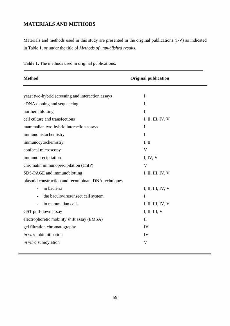
59
MATERIALS AND METHODS
Materials and methods used in this study are presented in the original publications (I-V) as indicated
in Table 1, or under the title of Methods of unpublished results.
Table 1. The methods used in original publications.
Method Original publication
yeast two-hybrid screening and interaction assays I
cDNA cloning and sequencing I
northern blotting I
cell culture and transfections I, II, III, IV, V
mammalian two-hybrid interaction assays I
immunohistochemistry I
immunocytochemistry I, II
confocal microscopy V
immunoprecipitation I, IV, V
chromatin immunoprecipitation (ChIP) V
SDS-PAGE and immunoblotting I, II, III, IV, V
plasmid construction and recombinant DNA techniques
- in bacteria I, II, III, IV, V
- the baculovirus/insect cell system I
- in mammalian cells I, II, III, IV, V
GST pull-down assay I, II, III, V
electrophoretic mobility shift assay (EMSA) II
gel filtration chromatography IV
in vitro ubiquitination IV
in vitro sumoylation V
60
Methods of unpublished results
RNA- EMSA
A central region of SNURF gene (71-280 bp) was inserted between PstI and HincII of pGem3Z vector
(Promega). In vitro transcribed RNA (232 ribonucleotides) was produced as described in in vitro RNA
synthesis protocol of Promega. Shortly, pGem3Z-SNURF(71-280 bp) was linearized with XbaI and
the template DNA was purified by phenol/chloroform extraction and ethanol precipitation. The DNA
template was incubated with Rnase inhibitor, ATP, UTP, GTP, SP6 RNA polymerase and [γ32P]CTP
and incubated for 20 minutes at 37 °C. Samples were treated with DnaseRQ1 for 15 minutes at 37 °C
and the transcribed RNA was purified with gel filtration. GST-SNURF (60 ng) was incubated with32P-RNA-probe and 4 units of Rnase inhibitor in a buffer containing 20 mM Hepes (pH 7.9), 50 mM
KCl, 1 mM MgCl2, 10% (v/v), 2 mM DTT, 0.35 mM EDTA, 0.025% (v/v) Nonidet P-40, 0.25 mM
PMSF, 2 µg/ml pepstatin A, 2 µg/ml leupeptin, and 2 µg/ml aprotinin in a reaction vol of 20 µl for 30
minutes at room temperature. Competitors, yeast tRNA (1 µg) (Sigma) and pGem3z-vector (200 ng)
were included before addition of the 32P-RNA probe. Protein-RNA complexes were resolved on 4%
polyacrylamide gel in 0.25 x Tris-borate-EDTA at °4C. Complexes were detected by autoradiography.
In vitro phosphorylation
GST-SNURF protein was produced in bacteria and purified as described (Häkli et al. 2000). Casein
kinase II (CKII) was obtained from Promega. GST-SNURF (1 µg) was incubated with 10 ng of CKII,
100 µM ATP, 1 µCi [γ32P]ATP in a buffer containing 25 mM Tris-HCl (pH 7.4), 200 mM NaCl, 1
mM DTT, 10 mM MgCl2, in a volume of 40 µl. Samples were incubated at 37 °C for 1 h and reactions
were terminated by adding concentrated SDS-PAGE buffer and heat denaturation. Samples were
fractioned by 15% SDS-PAGE and detected by autoradiography. Alternatively, GST-SNURFs (1 µg)
resolved on SDS-PAGE gels were stained with Coomassie staining.

61
RESULTS AND DISCUSSION
1. SNURF POSSESSES PROTEIN- AND DNA-BINDING ACTIVITY (I, II, III, IV and V)
Characteristics of SNURF
In addition to mediating dimerization and binding to response element in DNA, the DNA-binding
domain of nuclear receptor can act as a binding platform for regulatory proteins. We were interested
to find out proteins that interact with the DBD and the hinge region of androgen receptor. Yeast two-
hybrid screening was used to screen a mouse embryonic day 10.5 cDNA domain library, in which the
amino acid residues 554–644 comprising the DBD and N-terminal part of the hinge region of human
AR fused to LexA DNA-binding domain was used as a bait. A novel protein termed small nuclear
RING finger (SNURF) was one of the discovered proteins. SNURF mRNAs of 3-kb and 1.6-kb in
size were identified when SNURF cDNA was used as a probe in Northern blotting of various rat and
human tissues. The 3-kb SNURF mRNA was found in all tissues, but the smaller, 1.6-kb mRNA was
expressed only in the rat testis. The human and mouse SNURF homolog, which is called RNF4, was
identified by Chiariotti and coworkers in the same year as SNURF (Chiariotti et al. 1998). RNF4
shows expression pattern similar to that of SNURF, and its gene was mapped to chromosome 4p16.3,
which is associated with different neoplastic diseases (Chiariotti et al. 1998, Galili et al. 2000,
Shivapurkar 1999, Caron et al. 1996).
Full-length SNURF cDNA was isolated from rat testis cDNA library by conventional
hybridization methods, and an open reading frame (ORF) with 194 amino acid residues was revealed.
The mouse and human RNF4 exhibit 97% and 91% sequence identity with SNURF, respectively (Fig.
11A), but no other homologs are found. Based on the primary SNURF sequence, a bipartite NLS in
the N-terminal part, a SV-40-like NLS in the central region of SNURF, and a RING finger (C3HC4)
structure in the C terminus (amino acids 136-180) (Fig. 11B), which is fully conserved in RNF4 (Fig.
11A), were identified in rat SNURF.

62
A
20SNURF 1 MSTRNPQRKRRGGAVNSRQTQKRTRETTSTPEISLEAEPIELVETVGDEIVDLTCESLEPVVVDL 65mRNF4 1 MSTRNPQRKRRGGTVNSRQTQKRTRETTSTPEVSLETEPIELVETVGDEIVDLTCESLEPVVVDL 65hRNF4 1 MST----RKRRGGAINSRQAQKRTREATSTPEISLEAEPIELVETAGDEIVDLTCESLEPVVVDL 61 + + + + + - - - - - - - -
121SNURF 66 THNDSVVIVEERRRPRRNGRRLRQDHADSCVVSSDDEELSKDKDVYVTTHTPRSTKDEGTTGLRP 130mRNF4 66 THNDSVVIVEERRRPRRNGRRLRQDHADSCVVSSDDEELSRDKDVYVTTHTPRSTKDDGATGPRP 130hRNF4 62 THNDSVVIVDERRRPRRNARRLPQDHADSCVVSSDDEELSRDRDVYVTTHTPRNARDEGATGLRP 126 + + + + - - -
136 180 SNURF 131 SGTVSCPICMDGYSEIVQNGRLIVSTECGHVFCSQCLRDSLKNANTCPTCRKKINHKRYHPIYI 194mRNF4 131 SGTVSCPICMDGYSEIVQNGRLIVSTECGHVFCSQCLRDSLKNANTCPTCRKKINHKRYHPIYI 194hRNF4 127 SGTVSCPICMDGYSEIVQNGRLIVSTECGHVFCSQCLRDSLKNANTCPTCRKKINHKRYHPIYI 190 + + +
B
L K
T
G
Y
E
D
I
VS
C
P
I
C
M
S
V Q N GR
L
I
V
S
T
E
C
GV
FC
C
Q
S
L
R
DS N
T
C
P
T
C
R
K
K
I
Zn Zn
NA
136
139 163
166 160 177
158 180
H
Fig. 11. A, Sequence alignment of SNURF and its mouse (mRNF4) and human (hRNF4) counterparts. RINGfinger (C3HC4) motif is indicated by dark grey shading and the cysteine and histidine residues mediatingputative zinc binding within the RING finger motif are bolded. Bolded italics present amino acid residues thatdiffer between the three homologous sequences, and dash lines indicate absent amino acid residues. Underlinedsequences present NLSs, and the grey shading region between amino acids 20-121 is AR-interacting region.Basic and acidic aa regions are indicated + and –, respectively. Protein database sequence identificationnumbers: SNURF (AAC35248), mRNF4 (AAF00620) and hRNF4 (AAC52022). B, Schematic “cross-braced”structure of SNURF/RNF4 RING finger motif. Zinc (Zn)-coordinating amino acid residues are numbered andcircled with light grey spheres.
SNURF is a hydrophilic protein, and its charge distribution is asymmetrical with alternating basic and
acidic amino acid clusters. Such charged regions have been involved in DNA binding and
transcriptional activation (van Hoy et al.1993, Niessing et al. 2003). This electrical asymmetry of
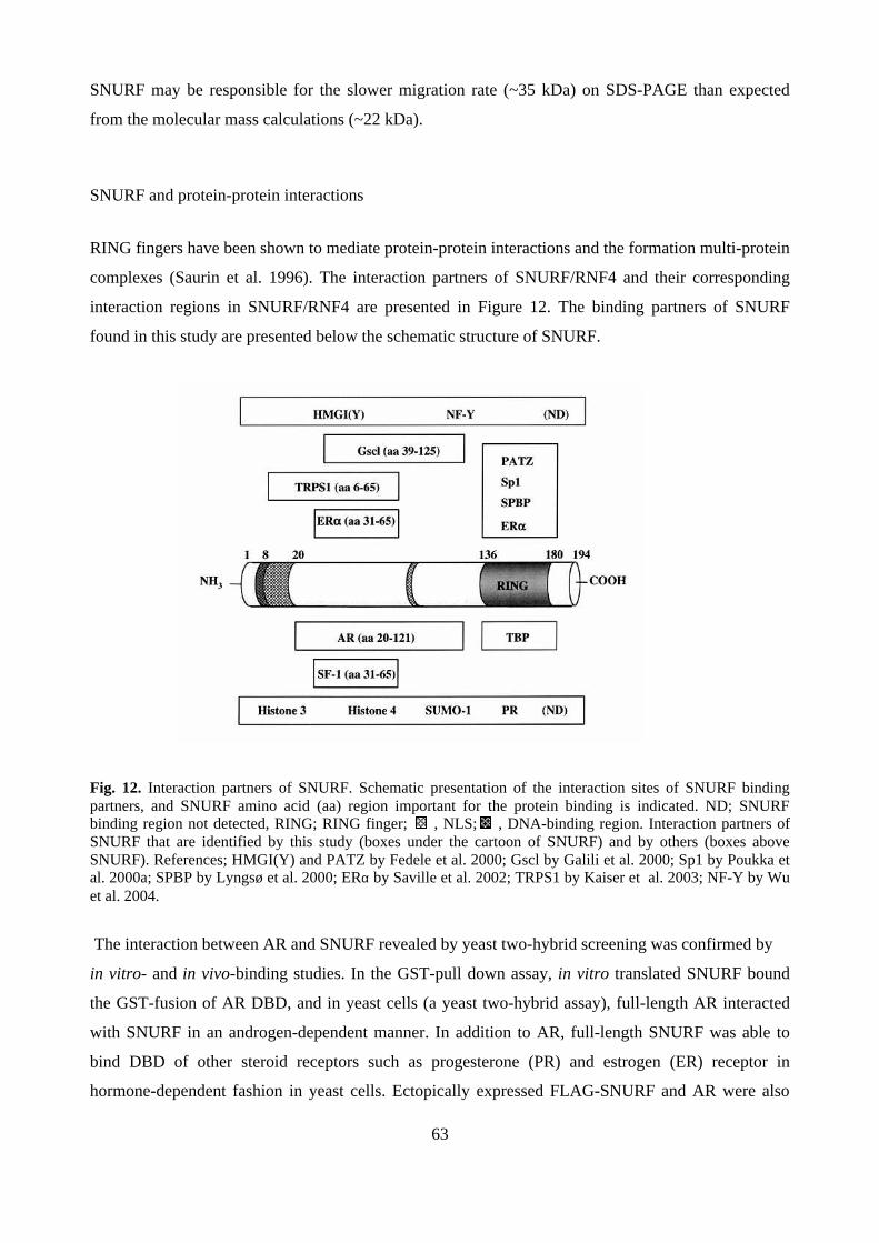
63
SNURF may be responsible for the slower migration rate (~35 kDa) on SDS-PAGE than expected
from the molecular mass calculations (~22 kDa).
SNURF and protein-protein interactions
RING fingers have been shown to mediate protein-protein interactions and the formation multi-protein
complexes (Saurin et al. 1996). The interaction partners of SNURF/RNF4 and their corresponding
interaction regions in SNURF/RNF4 are presented in Figure 12. The binding partners of SNURF
found in this study are presented below the schematic structure of SNURF.
Fig. 12. Interaction partners of SNURF. Schematic presentation of the interaction sites of SNURF bindingpartners, and SNURF amino acid (aa) region important for the protein binding is indicated. ND; SNURFbinding region not detected, RING; RING finger; , NLS; , DNA-binding region. Interaction partners ofSNURF that are identified by this study (boxes under the cartoon of SNURF) and by others (boxes aboveSNURF). References; HMGI(Y) and PATZ by Fedele et al. 2000; Gscl by Galili et al. 2000; Sp1 by Poukka etal. 2000a; SPBP by Lyngsø et al. 2000; ERα by Saville et al. 2002; TRPS1 by Kaiser et al. 2003; NF-Y by Wuet al. 2004.
The interaction between AR and SNURF revealed by yeast two-hybrid screening was confirmed by
in vitro- and in vivo-binding studies. In the GST-pull down assay, in vitro translated SNURF bound
the GST-fusion of AR DBD, and in yeast cells (a yeast two-hybrid assay), full-length AR interacted
with SNURF in an androgen-dependent manner. In addition to AR, full-length SNURF was able to
bind DBD of other steroid receptors such as progesterone (PR) and estrogen (ER) receptor in
hormone-dependent fashion in yeast cells. Ectopically expressed FLAG-SNURF and AR were also

64
observed to form a complex in mammalian cells (COS-1), when co-immunoprecipitated with anti-
FLAG antibody and furthermore, the N-terminal part of SNURF RING finger was shown to be
required for this interaction. SNURF interaction region in AR localizes to the region flanking the AR
DBD and hinge region as revealed by yeast two-hybrid screening. Later on, Poukka et al. 2000a
performed more detailed examination of this binding region, and found that the bipartite NLS in the
vicinity of the DBD/hinge-borderline was critical for the interaction. In addition, AR DBD point
mutations that are observed in patients with partial androgen insensitivity syndrome and male breast
cancer weakened the interaction between AR-DBD and SNURF (Poukka et al. 2000a). In the
interactions with ERα, both the RING and the central acidic region (aa 31-65) of SNURF were needed
for the interaction (Saville et al. 2002). The same acidic region of SNURF was also needed for the
interaction with the orphan nuclear receptor steroidogenic factor 1 (SF-1 or NR5A1). In addition to
sequence-specific transcription factors, SNURF was able to bind to the basal transcription machinery
protein TBP with its RING finger region that was also needed for binding to other transcription
factors, such as POZ AT-hook zinc finger protein (PATZ), promoter specificity protein 1 (Sp1), and
the activator of stromelysin 1 gene transcription (SPBP) (Lyngsø et al. 2000, Fedele et al. 2000,
Poukka et al. 2000a). SNURF interacts also with the repressor of GATA-mediated transcription, the
TRPS1 (trichorhino-phalangeal syndrome) protein with its amino-terminal region (aa 6-65) (Kaiser et
al. 2003).
Interestingly, when SNURF region of amino acids 21-186 was used as a bait in the yeast two-
hybrid screening SUMO-1 and Ubc9 were identified as SNURF-interacting proteins (Poukka,
unpublished results), which suggests that SNURF might be involved in SUMO modifications. We
studied these interactions by using the GST-pull down assay and found that SNURF bound efficiently
to GST-SUMO-1, but not to GST-Ubc9, suggesting that additional protein(s) mediate the SNURF-
Ubc9 interaction in yeast cells. Several proteins have been shown to bind noncovalently to SUMO,
but little is known about the effects of this function. However, noncovalent SUMO binding has been
shown to influence protein localization and activity (Minty et al. 2000, Kotaja et al. 2002, Engelhardt
et al. 2003). A recent study by Song et al. (2004) revealed that hydrophobic consensus sequence
V/IxV/IV/I is required for binding to all three SUMO variants (SUMO-1, -2,and -3). SNURF contains
one perfect SUMO-1 binding motif, 71VVIV74, in the central region of SNURF. However, the SNURF
mutant (∆66-98) lacking this region was still able to bind SUMO-1 in GST-pull down assays, albeit
less efficiency than the wild-type protein, indicating that there is an additional SUMO-1 binding site
in SNURF (unpublished results). This secondary SUMO-1-binding region in SNURF could be the61VVVD64 sequence (Song et al. 2004). We examined the possible interaction between SNURF and
another RING finger- and well-known SUMO-binding- protein PML3 (promyelocytic leukemia
65
protein 3) by using immunoprecitation techniques. Immunoprecipitation with anti-SNURF antibody
revealed that PML3 was complexed with SNURF, but only when SUMO-1 was coexpressed. Some of
the SNURF-interacting protein partners, such as SF-1 an AR, have been found to be sumoylated
(Poukka et al. 2000b, Chen et al. 2004), and thus SUMO-1 may be involved in mediating these
interactions. Finally, SNURF was found to reside in large (≥500-kDa) multiprotein complexes, as
assessed by size fractionation of protein lysates of F9 cells and mouse testis by gel filtration
chromatography. In addition, E. coli-produced purified SNURF was eluted in fractions of ≥100-kDa,
suggesting that SNURF is able to self-assemble. Taken together, SNURF is able to interact with many
diverse transcription regulators via multiple regions and in vivo it appears to be a component in a
multiprotein complex.
SNURF and DNA/nucleosome binding
It has been speculated that RING finger structure possesses DNA-binding activity similar to other
zinc-binding molecules such as Gal4, but up until now none of the DNA-binding RING finger
proteins have been shown to contact DNA via their RING fingers. Nucleotide-binding ability of
SNURF was examined by electrophoretic mobility shift assay (EMSA) in vitro. Recombinant SNURF
was found to bind DNA in a non-sequence specific manner with different types of DNA (single- and
double-stranded DNA, supercoiled and linear plasmid DNA, and four-way junction DNA), but there
was no clear binding preference between linear or branched DNA. The capacity to bind normal and
abnormal DNA without sequence specificity is a common property of architectural proteins, such as
HP1 (reviewed by Zlatanova and van Holde 1998). GST-SNURF interacted with DNA in a
cooperative fashion that was dependent on the length of the DNA fragment. Also human RNF4
showed a similar DNA-binding behavior (Wu et al. 2004). A comparable DNA-binding capacity has
been characterized for HP1 and BRCA1 (Zhao et al. 2000, Paull et al. 2001). BRCA1 interacts with
DNA as a multimer and is able to generate DNA loops (Paull et al. 2001). Since SNURF is also able
to self-associate (Lyngsø et al. 2000), it is possible that SNURF can bind DNA as a multimer. The
RING finger structure of SNURF was not needed for its DNA-binding activity, but the deletion of the
positively charged N-terminal region (aa 1-20) abolished the DNA-binding activity of SNURF.
Further examination revealed that basic amino acids 8RKRR11 play a critical role in DNA binding.
Many DNA-binding proteins, such as Bicoid, mel-8 and HMGY(I), associate with DNA through their
basic amino acid regions (Niessing et al. 2000). Since Arg-rich clusters are also involved in RNA
recognition (Muchardt et al. 2002, Burd et al. 1994), we examined whether SNURF is able to bind
RNA as well. GST-SNURF bound the RNA-probe (232 nucleotides) efficiently also in the presence of
a cold competitor transfer RNA (tRNA) (90-fold excess) (Fig. 13, lanes 2 and 3). However, a lower

66
(0.3-fold) molar ratio of linearized plasmid DNA was able to compete with RNA, since smaller
SNURF-RNA complexes started to appear (Fig. 13, lane 4).
32P-RNA
GST-SNURF – + + +
tRNA DNA
1 2 3 4
Fig. 13. SNURF is a RNA-binding protein. In EMSA, GST-SNURF was incubated with in vitro-transcribed32P-RNA (lanes 2-4). In lanes 3 and 4, RNA-binding of SNURF is competed with tRNA (90-fold excess) andlinearized plasmid DNA (0.3-fold excess), respectively.
Since SNURF was able to interact with proteins and various DNA molecules, we were
interested in studying whether SNURF is able to bind nucleosomes. We reconstituted DNA on
mononucleosomes in vitro and performed EMSA assays with different amounts of GST-SNURF.
SNURF was able to bind efficiently to nucleosomes, albeit with a lower activity compared to naked
DNA. The DNA binding-deficient SNURF mutant (∆1-20) was also practically unable to bind
nucleosomes. GST-SNURF pull-down experiments showed that SNURF is able to recruit histones H3
and H4, suggesting that both DNA- and protein recognitions are involved in nucleosome binding.
Interestingly, HP1α that is also a small (191 amino acids) protein with DNA- and RNA-binding
activities binds to nucleosomes and histones H3 and H4 (Polioudaki et al. 2001, Zhao et al. 2000,
Meehan et al. 2003, Murchardt et al. 2002). This suggests that SNURF and HP1 share similar
functional properties that are involved in the regulation of chromatin function. In addition, SNURF
binds non-histone chromatin modeling protein HMGI(Y), which is involved in transcription and cell

67
growth regulation (Fedele et al. 2000). Since SNURF is able to bind a variety of transcription
regulators, DNA and nucleosomes, it may promote assembly of nucleoprotein structures involved in
transcription control.
2. SNURF AS A TRANSCRIPTIONAL COREGULATOR (I, II, III, and V)
Since SNURF was able to bind AR, we studied whether SNURF was influencing AR-dependent
transcription. We performed reporter gene assays, in which SNURF was coexpressed with AR in
cultured CV-1 and COS-1 cells. These assays showed that SNURF was able to enhance transcriptional
activity of AR from different AR-regulated promoters, such as minimal ARE2-TATA-LUC and
natural and more complex rat probasin-LUC, in an androgen-dependent fashion. This activity of
SNURF was dependent on the AR-binding region of SNURF. Interestingly, SNURF was also able to
enhance basal transcription from probasin-promoter and minimal TATA-LUC reporters, which was
dependent on the RING finger of SNURF. Later on, Poukka and coworkers showed that the activity of
SNURF in AR-mediated transcription depends on the cell type. Namely, in COS-1 cells, the
coactivator function of the SNURF RING finger mutant (SNURF-C177/180S), in which the second
zinc-binding site was mutated, was practically inactive, but this mutant was active in CV-1 cells
(Poukka et al. 2000a). Interestingly, the PATZ protein is capable of switching SNURF coactivation
function to corepression function in AR-mediated transcription by interacting with SNURF (Pero et
al. 2002).
SNURF is not specific for AR, since it was able to enhance the activity of other steroid
receptors, such as the progesterone receptor (PR) and glucocorticoid receptor (GR) in the presence of
appropriate hormone. In addition, SNURF enhanced transcription from promoters containing binding
sites for Sp1 and activator protein 1 (AP-1). The latter coactivator function of SNURF was dependent
on the intact RING finger, which supports the Sp1 interaction. However, also the N-terminal region
was required for both Sp1- and AR-regulated transcription (Poukka et al. 2000a). Since SNURF uses
its N-terminal region for DNA binding, we examined whether DNA binding is required for SNURF
action in transcription regulation. We cotransfected COS-1 cells with Sp12-TATA-LUC or ARE2-
TATA-LUC together with SNURF or its DNA-binding mutants. We found that the DNA-binding
activity of SNURF clearly correlates with its coactivation function in both cases (AR; unpublished
results). Therefore, the DNA-binding activity of SNURF may also be needed in ERα-regulated
transcription, since the N-terminal deletion of SNURF abolishes the coactivation of ERα, although
SNURF was still maintaining ER-interaction and nuclear localization (Saville et al. 2000). We also
examined whether SNURF enhanced Sp1 binding to a GC-rich promoter sequence in EMSA, but
68
there was no clear cooperativity between Sp1 and SNURF. Neither was AR binding to its
corresponding promoter element influenced by SNURF (AR; unpublished result). Interestingly, RNF4
has been shown to promote both DNA binding and transcriptional activity of SPBP (Lyngø et al.
2000).
Since SNURF mRNA is also expressed in mouse brain, we were interested in studying
whether SNURF is able to influence the regulation of luteinizing hormone β -gene expression in
pituitary tissues, where GnRH plays a stimulative role, and androgens and AR play a repressive role
(negative feedback). We were able to detect endogenous SNURF protein in mouse pituitary tissues
and in pituitary gonadotrope LβT2 cell line by anti-SNURF antibody. Furthermore, endogenous
SNURF physically associated with the native LHβ promoter in LβT2 cells as, revealed by anti-
SNURF antibodies and chromatin immunoprecipitation (ChIP) assay. In transient transfection assays,
ectopically expressed SNURF enhanced the basal and GnRH-regulated transcription from the LHβ-
promoter. Enhancer regions of the LHβ gene, where the distal enhancer harbors binding sites for Sp1
and the proximal enhancer contains binding sites for SF-1 and Egr-1, were both needed for full
SNURF activity. The stimulatory effect of SNURF on the LHβ-promoter was abolished, when the
Sp1- or the SF-1-binding region of SNURF was deleted. Interestingly, SNURF was able to prevent the
androgen-mediated suppression of GnRH-stimulation of LHβ. AR has previously been shown to
interfere with the transcription factors that stimulate the LHβ promoter (Curtin et al. 2001, Jorgensen
et al. 2001); AR may form a complex with SF-1, which leads to repression of LHβ transcription
(Jorgensen et al. 2001). Our results suggest that SNURF is also able to block the entry of AR to the
LHβ promoter via binding to SF-1 and Sp1. cAMP signaling system has been shown to regulate the
LHβ promoter via Sp1, Egr-1 and SF-1 in LβT2 cells, but the precise mechanism of its action on the
LHβ promoter is not known. SF-1 is not mandatory for the cAMP-induced activation of the LHβ
promoter, but it amplifies the cAMP-response via an unknown mechanism (Horton and Halvorson
2004). Interestingly, SNURF expression is enhanced by forskolin and PMA (phorbol myristate,
another LH agonist) in cultured granulosa cells of follicles (Hirvonen-Santti et al. 2003). Therefore, it
would be interesting to study whether SNURF is participating in the cAMP-induced activation of the
LHβ promoter in pituitary tissue. In view of this, it is interesting that the transcription factor NF-Y has
been shown to regulate the bovine LHβ promoter and contribute to cAMP signalling (Keri et al. 2000,
Zhong et al. 2000). Moreover, a recent study showed that RNF4 is a coactivator for NF-Y and it can
enhance transcription of GTP cyclohydrolase I (GCH), which is an essential gene in the synthesis of
neurotransmitters dopamine and serotonin (Wu et al. 2004).

69
SNURF fulfills only some of the characteristics of a “classical” coregulator. It interacts with
many transcription factors, whose transcription activity SNURF either enhances (e.g. AR and Sp1) or
represses (TRPS1) (Kaiser et al. 2003). Since SNURF can bind to DNA, a large number of enhancer-
binding proteins and to proteins of general transcription machinery, SNURF may bridge the sequence-
specific transcription factors to the general transcription machinery, and thus enhance or repress
transcription. In agreement with the latter observation, SNURF has been shown to cooperate with
TBP in coactivation of ERα-mediated transactivation (Saville et al. 2002).
3. SNURF HAS UBIQUITIN E3 LIGASE ACTIVITY (IV)
Since many RING finger-containing proteins mediate ubiquitin E3 ligase activity, we have studied the
ability of SNURF to cooperate with E2 enzyme by using in vitro ubiquitination assays, in which
immobilized GST-SNURF was incubated with recombinant labelled ubiquitin, ubiquitin-activating
enzyme (E1) and various E2s. Our result showed that SNURF is able to mediate the ubiquitin E3
ligase activity via cooperation with different E2s, such as Ubch5A/B, HHR6B (RAD6), E2-25K,
MmUbc7 and UbcH13. This multiple usage of E2 is a rare quality among the known ubiquitin E3s.
UbcH5 has previously been shown to cooperate with many E3s, such as MDM2, BRCA1-BRAD1,
SCF and APC, and to ubiquitinate p53 (Ostendorff et al. 2002, Brzovic et al. 2003). HHR6 (RAD6)
has been implicated in the regulation of chromatin structure via histone ubiquitination (Robzyk et al.
2000, Haas et al. 1990). As in the case of many other E3s, such as BRCA1 and MDM2, also SNURF
was self-ubiquitinated in a RING-finger-dependent manner (Fang et al. 2000, Chen et al. 2002). We
used several SNURF-interacting partners, such as AR, Sp1, SF-1, and PML-3, to screen for
substrate(s) for the SNURF ubiquitin E3 ligase activity. SNURF was, however, not able to enhance
the ubiquitination level of these proteins, thus the specific substrate(s) other than SNURF itself was
not discovered. Since the endogenous SNURF was found in large multiprotein complexes in F9 cells,
SNURF may act as an essential RING finger subunit in an E3 multiprotein complex. Further work
should be performed to identify potential endogenous interaction partners of SNURF. It should be
possible to purify SNURF-multiprotein complex by using SNURF-affinity chromatography and
identify SNURF-bound proteins with mass spectrometric analyses. Unfortunately, our current
antibodies were not suitable for such an approach. Mammalian two-hybrid screening with full-length
SNURF as a bait could provide another tool to study the components of SNURF-containing
complexes. Results from these experiments may also lead us to identify specific ubiquitination targets
of SNURF. Interestingly, MDM2 ubiquitin E3 ligase has recently been shown to repress transcription
activity of p53 via Nedd8 (ubiquitin-like molecule) conjugation (Xirodimas et al. 2004). Therefore, it
70
would be interesting to study whether SNURF is able to mediate conjugation of ubiquitin-like
molecules, such a Nedd8, ISG15 and FAT10.
SNURF has been suggested to play a role in spermatid maturation (Yan et al. 2002). SNURF
is down-regulated in germ cell tumors and RAS-transformed cells (Pero et al. 2001, Zuber et al. 2000,
Hirvonen-Santti et al. 2003), suggesting a role for SNURF in the pathogenesis of testicular germ cell
cancer. Interestingly, overexpression of SNURF inhibits cell proliferation (Pero et al. 2001). The
RING finger SNURF mutants that are inactivate as ubiquitin E3 ligases show attenuated coregulatory
function as well as inhibitory function in cell growth. Similarly, RING finger mutated BRCA1 fails to
inhibit the cell growth and also the ubiquitin E3 ligase activity of the BRCA1/BARD heterodimer is
lost as a consequence of the mutation within the RING of BRCA1. Therefore, it has been suggested
that the E3 ubiquitin ligase activity of BRCA1-BARD1 would promote tumor suppression (Chen et al.
2002, Jin et al. 1997, reviewed by Baer and Ludwig 2002). Accordingly, it would be important to
study the ubiquitin E3 ligase activity of SNURF in the context of cell growth regulation in more
detail.
4. COVALENT MODIFICATIONS OF SNURF (IV, V)
SNURF is mainly mono- and di-ubiquitinated in vitro and in vivo, but the addition of MG-132, a
proteasome inhibitor, to cultured cells increases total SNURF ubiquitination level and especially,
polyubiquitinated SNURF forms become more evident. SNURF contains nine lysine residues.
Although we have used a versatile deletion series of SNURF (Poukka et al. 2000a), we were not able
to pinpoint the target lysines, suggesting that SNURF can simultaneously be ubiquitinated on several
lysine residues or a deletion of one ubiquitination site may lead to ubiquitination of another lysine.
Some proteins, such as c-Jun, exhibit seemingly random ubiquitination patterns, while others, such as
IκBα, are ubiquitinated with high specificity (Weissman 2001). Mutations in the RING finger motif
did not stabilize the SNURF protein, suggesting that SNURF autoubiquitination is not simply
targeting SNURF for degradation, but rather modifies SNURF function, possibly its subcellular
localization, DNA binding or protein-protein interactions. MDM2, for instance, regulates its
degradation by auto-ubiquitination, since the destruction of its RING finger structure stabilizes
MDM2 in cells (Fang et al. 2000, Geyer et al. 2000), whereas in the case of BRCA1,
autoubiquitination does not lead to degradation, rather to a modulation of its function (Nishikawa et
al. 2004).
71
Since SNURF was able to bind non-covalently to SUMO-1 in vitro, we studied whether
SNURF is also covalently conjugated to SUMO-1. SNURF does not contain precise sumoylation
consensus sequence, ψKXE/D. However, many proteins appear to be sumoylated through lysine
residues, which surrounding sequences do not fulfill the SUMO consensus sequence (Hoege et al.
2002, Lee et al. 2003). We performed in vitro sumoylation assays and observed that SNURF can be
conjugated to two SUMO-1 molecules in the presence of SUMO-activating enzyme (E1), Ubc9 and
SUMO-1. Studies in COS-1 cells suggested that at least three SUMO-1 molecules can be conjugated
to SNURF. SNURF has one potential site Lys121 within the TKDE sequence, but the deletion of this
SNURF region (deletion ∆121-143) did not alter the sumoylation pattern of SNURF. Interestingly, the
RING finger SNURF mutant was more avidly sumoylated than the wild-type SNURF in COS-1 cells,
suggesting that sumoylation and autoubiquitination compete for the same target lysine(s) in SNURF
or that the destruction of RING finger can reveal hidden sumoylation sites. Interestingly, SUMO-1
conjugation of MDM2 shifts its E3 activity towards p53 ubiquitination and diminishes its
autoubiquitination and thus its degradation (Buschmann et al. 2001). SUMO conjugation of SNURF
may regulate the stability, protein-protein interactions, DNA-binding or subnuclear localization of
SNURF. However, SUMO-1-mediated SNURF-PML3 complex formation was independent on the
covalent SUMO-1 conjugation.
SNURF sequence contains many consensus target sites for various kinases, such as protein
kinase A (PKA), protein kinase C (PKC) and casein kinase II (CKII). There are seven potential
phosphorylation sites for CKII, which lie within the AR-binding region of SNURF. We performed in
vitro phosphorylation reactions, where various bacterially produced and purified GST–SNURF
proteins were incubated with CKII and [γ32P]ATP. A shown in Fig. 14, wild-type SNURF was
efficiently phosphorylated by CKII (lanes 2 and 9), but the SNURF mutants 1-94 and ∆99-118 were
not phosphorylated (lanes 3 and 6). Next, we mutated two serine residues (Ser98 and Ser99)
simultaneously (lane 8 and 10) or separately (lanes 11 and 12) within this region, and found that both
serines were targets of CKII phosphorylation. Lack of CKII phosphorylation resulted in a slight
increase in SNURF coactivation function in both Sp1- and AR-regulated transcription (data not
shown), suggesting that CKII-mediated phosphorylation of SNURF does not play a major role in
transcription. Phosphorylation may, in turn, regulate sumoylation and/or ubiquitination of SNURF,
since, in the case of IκBα, phosphorylation inhibits SUMO modification and enhances ubiquitination
(Desterro et al. 1998).

72
WT
1-94
95-1
94
∆66-
98
∆99-
118
∆121
-143
S98-
99A
S98A
S99D
WT
GST
S98-
99A
1 2 3 4 5 6 7 8
9 10 11 12
Fig. 14. SNURF is phosphorylated by casein kinase II in vitro. GST (lane 1), GST-SNURF wild-type (lanes 2and 9) and its mutants (lanes 3-8 and 10-12) were incubated with CKII and [γ32P]ATP. Samples werefractionated by 15% SDS-PAGE gel and detected by autoradiography.
5. SUBCELLULAR LOCALIZATION OF SNURF (I and V)
SNURF mRNA was found to be expressed in various mouse and human tissues as revealed by
Northern blotting. Immunohistochemistry of rat prostate epithelial cells by anti-SNURF antibodies
revealed that SNURF is localized into the nucleus. Furthermore, immunocytochemical staining of
cultured CV-1 cells revealed that the endogenous SNURF was localized into small nuclear speckles in
CV-1 cells. Kaiser et al. (2003) have obtained similar results with primary human fibroblast.
Interestingly, in the cells of the nervous system of developing mouse embryo and adult mouse, the
localization of RNF4 was primarily cytoplasmic (Galili et al. 2000), suggesting that the localization of
SNURF is regulated in a tissue-specific manner and that SNURF may participate in shuttling
information between the cytoplasm and the nucleus. Poukka et al. (2000c) showed that SNURF is
capable of modulating nuclear targeting of AR by facilitating nuclear import during ligand induction
and retarding export of AR after hormone withdrawal in CV-1 cells.
73
Since SUMO-1 is an essential factor in the formation of the PML nuclear bodies (PML NBs)
and many RING finger proteins, such as PML, BRCA1 and MDM2, are found to colocalize to these
structures, we investigated potential colocalization of SNURF, SUMO-1 and PML in HeLa cells by
using immunostaining and confocal microscopy. Interestingly, a significant portion of the endogenous
SNURF colocalized with PML and SUMO-1 into the same nuclear structures. In addition, ectopic
expression of SNURF and PML isoforms, PML3 (PML IV) and PML-L (PML III), showed that
SNURF is specifically recruited to PML NBs by PML3. Likewise, the EGFP-SUMO-1 was localized
with ectopically expressed SNURF in the nucleus and the localization was independent on the RING
finger structure of SNURF. Furthermore, triple staining of ectopically expressed SUMO-1, PML3 and
SNURF revealed perfect localization into PML NBs. Interestingly, overexpression of PML3, but not
PML-L, was able to repress SNURF coactivition function in Sp1-regulated transcription, suggesting
that PML3 was able to regulate SNURF via recruiting SNURF into PML NBs and store SNURF in a
transcriptionally inactive form. Also Daxx and p53 are specifically recruited to PML NBs and their
transcription activity is repressed by PML3 (Fogal et al. 2000, Kim et al. 2003). PML can be
considered as a tumor suppressor protein (Wang et al. 1998a and 1998b). Its expression is down-
regulated in many cancers, such as in germ cell tumors, and absent in progressive tumors of prostate
and central nervous system (Gurrieri et al. 2004). SNURF is down-regulated in germ cell tumors and
overexpression of SNURF inhibits cell proliferation (Yan et al. 2002, Galili et al. 2000), therefore, we
suggest that SNURF and PML may communicate in the regulation of transcription and cell growth.

74
CONCLUSIONS
In the present work, we have examined the functional characteristics of rat small nuclear RING finger
protein SNURF. Highly conserved SNURF homologs in human and mouse are called RNF4, but no
other close homologs or related proteins exist. SNURF is a hydrophilic protein that contains zinc-
binding RING-finger motif, which is known to maintain protein-protein interactions and ubiquitin E3
ligase activity. The results of this study are summarized below:
• SNURF is a transcriptional coregulator protein. SNURF interacts with steroid receptors as well as
other transcription regulators, such as, SF-1 and TBP, and enhances transactivation function of AR
and Sp1. SNURF is able to stimulate LHβ gene expression via interaction with Sp1 and SF-1 that
are bound to distal and proximal regulatory elements within the LHβ promoter. SNURF may act
as a bridging factor between sequence-specific transcription factors and the basal transcription
machinery.
• SNURF is able to bind DNA and RNA. SNURF binds to various types of DNA without apparent
sequence specificity. Furthermore, SNURF is able to bind to nucleosomes. The DNA-binding
activity of SNURF correlates with its transcriptional coactivation function. SNURF may thus
promote the assembly of nucleoprotein structures involved in transcriptional control.
• SNURF is a ubiquitin E3 ligase capable of cooperating with various ubiquitin-conjugating E2
enzymes. The E3 function is dependent on SNURF RING finger structure. SNURF may serve as
an essential RING component in a multiprotein E3 complex.
• SNURF is able to ubiquitinate itself (autoubiquitination), and it is covalently modified by the
SUMO-1 conjugation and phosphorylation.
• SNURF is a nuclear protein that is recruited to PML nuclear bodies via interaction with PML3 and
SUMO-1. Localization of SNURF to PML NBs represses its activity in transcription regulation.

75
ACKNOWLEDGEMENTS
Tämä väitöskirjatyö laadittiin professori Olli Jänteen ja professori Jorma Palvimon tutkimusryhmässäHelsingin yliopiston Biolääketieteen laitoksen fysiologian osastolla. Kiitän Olli Jännettä ohjauksestaja loistokkaasti varustetusta työympäristöstä Biomedicum Helsinki-tutkimuskeskuksessa. Olen syvästikiitollinen Jorma Palvimolle asiantuntevasta ja innostavasta ohjauksesta sekä itsenäisyydestä.
Kiitokset Asta Pirskaselle ja Ilkka Julkuselle rakentavista huomautuksista ja ehdotuksistatarkastaessanne väitöskirjaani.
Kiitokset Helsinki Biomedical graduate school -tutkijakoululle saamastani rahoituksesta jakoulutuksesta. Lämmin kiitos Emil Aaltosen Säätiölle, Otto A. Malmin lahjoitusrahastolle, Jalmari jaRauha Ahokkaan Säätiölle ja Farmoksen Tutkimus- ja Tiedesäätiölle saamastani tuesta jakannustuksesta
Marikki Laiholle ja Ilkka Julkuselle lämmin kiitos toimiessanne tutkimusprojektini seurantaryhmässäavustavana ja kannustavana jäsenenä.
I am grateful to Denis Curtin, Hearther Ferris, Matthew Gibson, Ulla Karvonen, Kevin Lorick, Anu-Maarit Moilanen, Hetti Poukka, Margaret Shupnik and Allan Weissmann for productive collaboration.
Leenalle, Seijalle, Pirjolle, Saijalle, Johannalle, Katjalle, Riitalle, Katille, Heikille ja Birgitilleerityislämminkiitos saamastani avusta ja mukavasta työskentelyseurasta. Annelle, Pirjolle, Mallalle jaNiinalle kiitokset käytännönasioiden hoidosta. Kiitokset Marc Baumannille avusta proteiinikemianosa-alueella.
Lämmin kiitos kaikille OAJ- ja ”Orphan group”-tutkimusryhmämme jäsenille mukavastatyöympäristöstä, sekä yhteistyöstä että avunannosta. Leenalle, Seijalle ja Marikalle sydämellinenkiitos kannustuksesta ja välittämisestä. Sirpalle, Karoliinalle ja Lauralle kiitos virkistävästäyhteiselosta Penkereen alakerran tutkijakammiossa ja Sirpalle vielä kiitos SNURF-hetkistä. FörstiUllalle erityiskiitos värikkäistä huumorinkukkasista. Ne olivat tarpeen! Kiitokset Shalle, Nooralle,Andriille, Tanyalle, Henrikille, Fu-Pingille, Zhigangille ja Jamekselle hauskoista hetkistä. Minnaa jaAnnaa kiitän suuresti avuliaasta ja piristävästä yhteistyöstä.
Annelle, Jaanalle, Marjalle, Nathalielle, Päiville, Sirpalle, Tarjalle ja Terhille kiitos ystävyydestä.
Olen kiitollinen pikkuauringoista, Rikistä ja muista ”karvanapeista”, jotka ovat vaeltaneet rinnallanitämän pitkän työrupeaman aikana.
Äidille, Isälle, Tiitukselle, Marjaanalle, Mikalle ja Stefanille kiitokset saamastani tuesta ja paljostamuusta.
Helsinki, maaliskuu 2005

76
REFERENCES
Acevedo ML, Kraus WL (2003) Mediator and p300/CBP-steroid receptor coactivator complexes have distinctroles, but function synergistically, during estrogen receptor alpha-dependent transcription with chromatintemplates. Mol Cell Biol 23:335-348
Adams J, Palombella VJ, Sausville EA, Johnson J, Destree A, Lazarus DD, Maas J, Pien CS, Prakash S, ElliottPJ (1999) Proteasome inhibitors: a novel class of potent and effective antitumor agents. Cancer Res 59:2615-2622
Agarwal R, Tang Z, Yu H, Cohen-Fix O (2003) Two distinct pathways for inhibiting pds1 ubiquitination inresponse to DNA damage. J Biol Chem 278:45027-45033
Ahn JH, Xu Y, Jang WJ, Matunis MJ, Hayward GS (2001) Evaluation of interactions of humancytomegalovirus immediate-early IE2 regulatory protein with small ubiquitin-like modifiers and theirconjugation enzyme Ubc9. J Virol 75:3859-3872
Akoulitchev S, Chuikov S, Reinberg D (2000) TFIIH is negatively regulated by cdk8-containing mediatorcomplexes. Nature 407:102-106
Alam SL, Sun J, Payne M, Welch BD, Blake BK, Davis DR, Meyer HH, Emr SD, Sundquist WI (2004)Ubiquitin interactions of NZF zinc fingers. EMBO J 23:1411-1421
Alberti S, Demand J, Esser C, Emmerich N, Schild H, Hohfeld J (2002) Ubiquitylation of BAG-1 suggests anovel regulatory mechanism during the sorting of chaperone substrates to the proteasome. J Biol Chem277:45920-45927
Ali S, Metzger D, Bornert JM, Chambon P (1993) Modulation of transcriptional activation by ligand-dependentphosphorylation of the human oestrogen receptor A/B region. EMBO 12:1153-1160
Alkema MJ, Bronk M, Verhoeven E, Otte A, van 't Veer LJ, Berns A, van Lohuizen M (1997) Identification ofBmi1-interacting proteins as constituents of a multimeric mammalian polycomb complex. Genes Dev 11:226-240
Alroy I, Freedman LP (1992) DNA binding analysis of glucocorticoid receptor specificity mutants. NucleicAcids Res 20:1045-1052
Anderson JD, Lowary PT, Widom J (2001) Effects of histone acetylation on the equilibrium accessibility ofnucleosomal DNA target sites. J Mol Biol 307:977-985
Arany Z, Sellers WR, Livingston DM, Eckner R (1994) E1A-associated p300 and CREB-associated CBPbelong to a conserved family of coactivators. Cell 77:799-800
Arora K, Warrior R (2001) A new Smurf in the village. Dev Cell 1:441-442
Asturias FJ, Jiang YW, Myers LC, Gustafsson CM, Kornberg RD (1999) Conserved structures of mediator andRNA polymerase II holoenzyme. Science 283:985-987
Avila DM, Zoppi S, McPhaul MJ (2001) The androgen receptor (AR) in syndromes of androgen insensitivityand in prostate cancer. J Steroid Biochem Mol Biol 76:135-142
Ayaydin F, Dasso M (2004) Distinct In Vivo Dynamics of Vertebrate SUMO Paralogues. Mol Biol Cell15:5208-5218

77
Bach I, Rodriguez-Esteban C, Carriere C, Bhushan A, Krones A, Rose DW, Glass CK, Andersen B, IzpisúaBelmonte JC, Rosenfeld MG (1999) RLIM inhibits functional activity of LIM homeodomain transcriptionfactors via recruitment of the histone deacetylase complex. Nat Genet 22:394-399
Bachmair A, Finley D, Varshavsky A (1986) In vivo half-life of a protein is a function of its amino-terminalresidue. Science 234:179-186
Baer R, Ludwig T (2002) The BRCA1/BARD1 heterodimer, a tumor suppressor complex with ubiquitin E3 ligase activity.Curr Opin Genet Dev 12:86-91
Bailey D, O'Hare P (2002) Herpes simplex virus 1 ICP0 co-localizes with a SUMO-specific protease. J GenVirol 83:2951-2964
Bailey D, O'Hare P (2004) Characterization of the localization and proteolytic activity of the SUMO-specificprotease, SENP1. J Biol Chem 279:692-703
Baleja JD, Marmorstein R, Harrison SC, Wagner G (1992) Solution structure of the DNA-binding domain ofCd2-GAL4 from S. cerevisiae. Nature 356:450-453
Bannister AJ, Schneider R, Kouzarides T (2002) Histone methylation: dynamic or static? Cell 109:801-806
Barlev NA, Liu L, Chehab NH, Mansfield K, Harris KG, Halazonetis TD, Berger SL (2001) Acetylation of p53activates transcription through recruitment of coactivators/histone acetyltransferases. Mol Cell 8:1243-1254
Barlow PN, Luisi B, Milner A, Elliott M, Everett R (1994) Structure of the C3HC4 domain by 1H-nuclearmagnetic resonance spectroscopy. A new structural class of zinc-finger. J Mol Biol 237:201-211
Barratt MJ, Hazzalin CA, Cano E, Mahadevan LC (1994) Mitogen-stimulated phosphorylation of histone H3 istargeted to a small hyperacetylation-sensitive fraction. Proc Natl Acad Sci U S A 91:4781-4785
Batonnet S, Leibovitch MP, Tintignac L, Leibovitch SA (2004) Critical role for lysine 133 in the nuclearubiquitin-mediated degradation of MyoD. J Biol Chem 279:5413-5420
Baud V, Liu ZG, Bennett B, Suzuki N, Xia Y, Karin M (1999) Signaling by proinflammatory cytokines:oligomerization of TRAF2 and TRAF6 is sufficient for JNK and IKK activation and target gene induction viaan amino-terminal effector domain. Genes Dev 13:1297-1308
Bayer P, Arndt A, Metzger S, Mahajan R, Melchior F, Jaenicke R, Becker J (1998) Structure determination ofthe small ubiquitin-related modifier SUMO-1. J Mol Biol 280:275-286
Beato M, Sanchez-Pacheco A (1996) Interaction of steroid hormone receptors with the transcription initiationcomplex. Endocr Rev 17:587-609
Bellon SF, Rodgers KK, Schatz DG, Coleman JE, Steitz TA (1997) Crystal structure of the RAG1 dimerizationdomain reveals multiple zinc-binding motifs including a novel zinc binuclear cluster. Nat Struct Biol 4:586-591
Bencsath KP, Podgorski MS, Pagala VR, Slaughter CA, Schulman BA (2002) Identification of amultifunctional binding site on Ubc9p required for Smt3p conjugation. J Biol Chem 277:47938-47945
Berger SL (2001) An embarrassment of niches: the many covalent modifications of histones in transcriptionalregulation. Oncogene 20:3007-3013
Bernassola F, Salomoni P, Oberst A, Di Como CJ, Pagano M, Melino G, Pandolfi PP (2004) Ubiquitin-dependent degradation of p73 is inhibited by PML. J Exp Med 199:1545-1557
Bernier-Villamor V, Sampson DA, Matunis MJ, Lima CD (2002) Structural basis for E2-mediated SUMOconjugation revealed by a complex between ubiquitin-conjugating enzyme Ubc9 and RanGAP1. Cell 108:345-356
78
Bertolaet BL, Clarke DJ, Wolff M, Watson MH, Henze M, Divita G, Reed SI (2001) UBA domains of DNAdamage-inducible proteins interact with ubiquitin. Nat Struct Biol 8:417-422
Best JL, Ganiatsas S, Agarwal S, Changou A, Salomoni P, Shirihai O, Meluh PB, Pandolfi PP, Zon LI (2002)SUMO-1 protease-1 regulates gene transcription through PML. Mol Cell 10:843-855
Bilodeau PS, Urbanowski JL, Winistorfer SC, Piper RC (2002) The Vps27p Hse1p complex binds ubiquitinand mediates endosomal protein sorting. Nat Cell Biol 4:534-539
Bird AP, Wolffe AP (1999) Methylation-induced repression--belts, braces, and chromatin. Cell 99:451-454
Blake TJ, Shapiro M, Morse HC 3rd, Langdon WY (1991) The sequences of the human and mouse c-cbl proto-oncogenes show v-cbl was generated by a large truncation encompassing a proline-rich domain and a leucinezipper-like motif. Oncogene 6:653-657
Bloom J, Amador V, Bartolini F, DeMartino G, Pagano M (2003) Proteasome-mediated degradation of p21 viaN-terminal ubiquitinylation. Cell 115:71-82
Bochar DA, Wang L, Beniya H, Kinev A, Xue Y, Lane WS, Wang W, Kashanchi F, Shiekhattar R (2000)BRCA1 is associated with a human SWI/SNF-related complex: linking chromatin remodeling to breast cancer.Cell 102:257-265
Boddy MN, Duprez E, Borden KL, Freemont PS (1997) Surface residue mutations of the PML RING fingerdomain alter the formation of nuclear matrix-associated PML bodies. J Cell Sci 110:2197-2205
Boeger H, Griesenbeck J, Strattan JS, Kornberg RD (2003) Nucleosomes unfold completely at atranscriptionally active promoter. Mol Cell 11:1587-1598
Boggio R, Colombo R, Hay RT, Draetta GF, Chiocca S (2004) A Mechanism for Inhibiting the SUMOPathway. Mol Cell 16:549-561
Bohren KM, Nadkarni V, Song JH, Gabbay KH, Owerbach D (2004) A M55V polymorphism in a novelSUMO gene (SUMO-4) differentially activates heat shock transcription factors and is associated withsusceptibility to type I diabetes mellitus. J Biol Chem 279:27233-27238
Boisvert FM, Hendzel MJ, Bazett-Jones DP (2000) Promyelocytic leukemia (PML) nuclear bodies are proteinstructures that do not accumulate RNA. J Cell Biol 148:283-292
Boisvert FM, Kruhlak MJ, Box AK, Hendzel MJ, Bazett-Jones DP (2001) The transcription coactivator CBP isa dynamic component of the promyelocytic leukemia nuclear body. J Cell Biol 152:1099-1106
Boonyaratanakornkit V, Melvin V, Prendergast P, Altmann M, Ronfani L, Bianchi ME, Taraseviciene L,Nordeen SK, Allegretto EA, Edwards DP (1998) High-mobility group chromatin proteins 1 and 2 functionallyinteract with steroid hormone receptors to enhance their DNA binding in vitro and transcriptional activity inmammalian cells. Mol Cell Biol 18:4471-4487
Borden KL (2000) RING domains: master builders of molecular scaffolds? J Mol Biol 295:1103-1112
Borden KL, Boddy MN, Lally J, O'Reilly NJ, Martin S, Howe K, Solomon E, Freemont PS (1995) The solutionstructure of the RING finger domain from the acute promyelocytic leukaemia proto-oncoprotein PML. EMBOJ 14:1532-1541
Borden KL, CampbellDwyer EJ, Salvato MS (1997) The promyelocytic leukemia protein PML has a pro-apoptotic activity mediated through its RING domain. FEBS Lett 418:30-34
Bourguet W, Germain P, Gronemeyer H (2000a) Nuclear receptor ligand-binding domains: three-dimensionalstructures, molecular interactions and pharmacological implications. Trends Pharmacol Sci 21:381-388
79
Bourguet W, Ruff M, Chambon P, Gronemeyer H, Moras D (1995) Crystal structure of the ligand-bindingdomain of the human nuclear receptor RXR-alpha. Nature 375:377-382
Bourguet W, Vivat V, Wurtz JM, Chambon P, Gronemeyer H, Moras D (2000b) Crystal structure of aheterodimeric complex of RAR and RXR ligand-binding domains. Mol Cell 5:289-298
Boutell C, Everett RD (2003) The herpes simplex virus type 1 (HSV-1) regulatory protein ICP0 interacts withand Ubiquitinates p53. J Biol Chem 278:36596-36602
Boyer TG, Martin ME, Lees E, Ricciardi RP, Berk AJ (1999) Mammalian Srb/Mediator complex is targeted byadenovirus E1A protein. Nature 399:276-279
Boyle S, Gilchrist S, Bridger JM, Mahy NL, Ellis JA, Bickmore WA (2001) The spatial organization of humanchromosomes within the nuclei of normal and emerin-mutant cells. Hum Mol Genet 10:211-219
Braun H, Koop R, Ertmer A, Nacht S, Suske G (2001) Transcription factor Sp3 is regulated by acetylation.Nucleic Acids Res 29:4994-5000
Breitschopf K, Bengal E, Ziv T, Admon A, Ciechanover A (1998) A novel site for ubiquitination: the N-terminal residue, and not internal lysines of MyoD, is essential for conjugation and degradation of the protein.EMBO J 17:5964-5973
Brodsky JL, McCracken AA (1999) ER protein quality control and proteasome-mediated protein degradation.Semin Cell Dev Biol 10:507-513
Brusky J, Zhu Y, Xiao W (2000) UBC13, a DNA-damage-inducible gene, is a member of the error-freepostreplication repair pathway in Saccharomyces cerevisiae. Curr Genet 37:168-174
Brzovic PS, Keeffe JR, Nishikawa H, Miyamoto K, Fox D 3rd, Fukuda M, Ohta T, Klevit R (2003) Bindingand recognition in the assembly of an active BRCA1/BARD1 ubiquitin-ligase complex. Proc Natl Acad Sci US A 100:5646-5651
Brzovic PS, Meza J, King MC, Klevit RE (1998) The cancer-predisposing mutation C61G disrupts homodimerformation in the NH2-terminal BRCA1 RING finger domain. J Biol Chem 273:7795-7799
Brzovic PS, Rajagopal P, Hoyt DW, King MC, Klevit RE (2001) Structure of a BRCA1-BARD1 heterodimericRING-RING complex. Nat Struct Biol 8:833-837
Brzozowski AM, Pike AC, Dauter Z, Hubbard RE, Bonn T, Engstrom O, Ohman L, Greene GL, GustafssonJA, Carlquist M (1997) Molecular basis of agonism and antagonism in the oestrogen receptor. Nature 389:753-758
Burakov D, Crofts LA, Chang CP, Freedman LP (2002) Reciprocal recruitment of DRIP/mediator and p160coactivator complexes in vivo by estrogen receptor. J Biol Chem 277:14359-14362
Burd CG, Dreyfuss G (1994) Conserved structures and diversity of functions of RNA-binding proteins. Science265:615-621
Burke TW, Kadonaga JT (1997) The downstream core promoter element, DPE, is conserved from Drosophilato humans and is recognized by TAFII60 Drosophila. Genes Dev 11:3020-31
Buschmann T, Lerner D, Lee CG, Ronai Z (2001) The Mdm-2 amino terminus is required for Mdm2 bindingand SUMO-1 conjugation by the E2 SUMO-1 conjugating enzyme Ubc9. J Biol Chem 276:40389-40395
Campbell Dwyer EJ, Lai H, MacDonald RC, Salvato MS, Borden KL (2000) The lymphocyticchoriomeningitis virus RING protein Z associates with eukaryotic initiation factor 4E and selectively repressestranslation in a RING-dependent manner. J Virol 74:3293-3300
80
Capili AD, Edghill EL, Wu K, Borden KL (2004) Structure of the C-terminal RING finger from a RING-IBR-RING/TRIAD motif reveals a novel zinc-binding domain distinct from a RING. J Mol Biol 340:1117-1129
Caron H, van Sluis P, Buschman R, Pereira do Tanque R, Maes P, Beks L, de Kraker J, Voute PA, VergnaudG, Westerveld A, Slater R, Versteeg R (1996) Allelic loss of the short arm of chromosome 4 in neuroblastomasuggests a novel tumour suppressor gene locus. Hum Genet 97:834-837
Chalkley GE, Verrijzer CP (1999) DNA binding site selection by RNA polymerase II TAFs: a TAF(II)250-TAF(II)150 complex recognizes the initiator. EMBO J 18:4835-4845
Chan HM, La Thangue NB (2001) p300/CBP proteins: HATs for transcriptional bridges and scaffolds. J CellSci 114:2363-2373
Chen A, Kleiman FE, Manley JL, Ouchi T, Pan ZQ (2002) Autoubiquitination of the BRCA1*BARD1 RINGubiquitin ligase. J Biol Chem 277:22085-22092
Chen D, Ma H, Hong H, Koh SS, Huang SM, Schurter BT, Aswad DW, Stallcup MR (1999) Regulation oftranscription by a protein methyltransferase. Science 284:2174-2177
Chen H, Lin RJ, Schiltz RL, Chakravarti D, Nash A, Nagy L, Privalsky ML, Nakatani Y, Evans RM (1997)Nuclear receptor coactivator ACTR is a novel histone acetyltransferase and forms a multimeric activationcomplex with P/CAF and CBP/p300. Cell 90:569-580
Chen JD, Evans RM (1995) A transcriptional co-repressor that interacts with nuclear hormone receptors.Nature 377:454-457
Chen L, Madura K (2002) Rad23 promotes the targeting of proteolytic substrates to the proteasome. Mol CellBiol 22:4902-4913
Chen P, Johnson P, Sommer T, Jentsch S, Hochstrasser M (1993) Multiple ubiquitin-conjugating enzymesparticipate in the in vivo degradation of the yeast MAT alpha 2 repressor. Cell 74:357-369
Chen S, Supakar PC, Vellanoweth RL, Song CS, Chatterjee B, Roy AK (1997) Functional role of aconformationally flexible homopurine/homopyrimidine domain of the androgen receptor gene promoterinteracting with Sp1 and a pyrimidine single strand DNA-binding protein. Mol Endocrinol 11:3-15
Chen WY, Lee WC, Hsu NC, Huang F, Chung BC (2004) SUMO modification of repression domainsmodulates function of nuclear receptor 5A1 (steroidogenic factor-1). J Biol Chem 279:38730-3875
Chen Z, Pickart CM (1990) A 25-kilodalton ubiquitin carrier protein (E2) catalyzes multi-ubiquitin chainsynthesis via lysine 48 of ubiquitin. J Biol Chem 265:21835-21842
Cheng J, Wang D, Wang Z, Yeh ET (2004) SENP1 enhances androgen receptor-dependent transcriptionthrough desumoylation of histone deacetylase 1. Mol Cell Biol 24:6021-6028
Cheung E, Zarifyan AS, Kraus WL (2002) Histone H1 represses estrogen receptor alpha transcriptional activityby selectively inhibiting receptor-mediated transcription initiation. Mol Cell Biol 22:2463-2471
Cheung P, Allis CD, Sassone-Corsi P (2000) Signaling to chromatin through histone modifications. Cell103:263-271
Cheutin T, McNairn AJ, Jenuwein T, Gilbert DM, Singh PB, Misteli T (2003) Maintenance of stableheterochromatin domains by dynamic HP1 binding. Science 299:721-725
Chiariotti L, Benvenuto G, Fedele M, Santoro M, Simeone A, Fusco A, Bruni CB (1998) Identification andcharacterization of a novel RING-finger gene (RNF4) mapping at 4p16.3. Genomics 47:258-265

81
Cho Y, Gorina S, Jeffrey PD, Pavletich NP (1994) Crystal structure of a p53 tumor suppressor-DNA complex:understanding tumorigenic mutations. Science 265:346-355
Chu-Ping M, Vu JH, Proske RJ, Slaughter CA, DeMartino GN (1994) Identification, purification, andcharacterization of a high molecular weight, ATP-dependent activator (PA700) of the 20 S proteasome. J BiolChem 269:3539-3547
Ciechanover A, Ben-Saadon R (2004) N-terminal ubiquitination: more protein substrates join in. Trends CellBiol 14:103-106
Clapier CR, Langst G, Corona DF, Becker PB, Nightingale KP (2001) Critical role for the histone H4 Nterminus in nucleosome remodeling by ISWI. Mol Cell Biol 21:875-883
Clarke IJ, Cummins JT (1982) The temporal relationship between gonadotropin releasing hormone (GnRH) andluteinizing hormone (LH) secretion in ovariectomized ewes. Endocrinology 111:1737-1739
Cleutjens KB, van der Korput HA, van Eekelen CC, van Rooij HC, Faber PW, Trapman J (1997) An androgenresponse element in a far upstream enhancer region is essential for high, androgen-regulated activity of theprostate-specific antigen promoter. Mol Endocrinol 11:148-161
Cohen SX, Moulin M, Hashemolhosseini S, Kilian K, Wegner M, Muller CW (2003) Structure of the GCMdomain-DNA complex: a DNA-binding domain with a novel fold and mode of target site recognition. EMBO J22:1835-1845
Comerford KM, Leonard MO, Karhausen J, Carey R, Colgan SP, Taylor CT (2003) Small ubiquitin-relatedmodifier-1 modification mediates resolution of CREB-dependent responses to hypoxia. Proc Natl Acad Sci U SA 100:986-991
Correa-Cerro L, Wohr G, Haussler J, Berthon P, Drelon E, Mangin P, Fournier G, Cussenot O, Kraus P, JustW, Paiss T, Cantu JM, Vogel W (1999) (CAG)nCAA and GGN repeats in the human androgen receptor geneare not associated with prostate cancer in a French-German population. Eur J Hum Genet 7:357-362
Courey AJ, Tjian R (1988) Analysis of Sp1 in vivo reveals multiple transcriptional domains, including a novelglutamine-rich activation motif. Cell 55:887-898
Crawford AW, Pino JD, Beckerle MC (1994) Biochemical and molecular characterization of the chickencysteine-rich protein, a developmentally regulated LIM-domain protein that is associated with the actincytoskeleton. J Cell Biol 124:117-127
Curtin D, Jenkins S, Farmer N, Anderson AC, Haisenleder DJ, Rissman E, Wilson EM, Shupnik MA (2001)Androgen suppression of GnRH-stimulated rat LHbeta gene transcription occurs through Sp1 sites in the distalGnRH-responsive promoter region. Mol Endocrinol 15:1906-1917
Dahlman-Wright K, Wright A, Gustafsson JA, Carlstedt-Duke J (1991) Interaction of the glucocorticoidreceptor DNA-binding domain with DNA as a dimer is mediated by a short segment of five amino acids. J BiolChem 266:3107-3112
D'Andrea A, Pellman D (1998) Deubiquitinating enzymes: a new class of biological regulators. Crit RevBiochem Mol Biol 33:337-352
Daniel MT, Koken M, Romagne O, Barbey S, Bazarbachi A, Stadler M, Guillemin MC, Degos L, ChomienneC, de The H (1993) PML protein expression in hematopoietic and acute promyelocytic leukemia cells. Blood82:1858-1867
Danielian PS, White R, Lees JA, Parker MG (1992) Identification of a conserved region required for hormonedependent transcriptional activation by steroid hormone receptors EMBO J 11:1025-1033

82
Dantonel JC, Wurtz JM, Poch O, Moras D, Tora L (1999) The TBP-like factor: an alternative transcriptionfactor in metazoa? Trends Biochem Sci 24:335-339
Davies TH, Ning YM, Sanchez ER (2002) A new first step in activation of steroid receptors: hormone-inducedswitching of FKBP51 and FKBP52 immunophilins. J Biol Chem 277:4597-4600
de Pater S, Greco V, Pham K, Memelink J, Kijne J (1996) Characterization of a zinc-dependent transcriptionalactivator from Arabidopsis. Nucleic Acids Res 24:4624-4631
Deblois G, Giguere V (2003) Ligand-independent coactivation of ERalpha AF-1 by steroid receptor RNAactivator (SRA) via MAPK activation. J Steroid Biochem Mol Biol 85:123-131
Dellaire G, Farrall R, Bickmore WA (2003) The Nuclear Protein Database (NPD): sub-nuclear localisation andfunctional annotation of the nuclear proteome. Nucleic Acids Res 31:328-330
DeMartino GN, Slaughter CA (1999) The proteasome, a novel protease regulated by multiple mechanisms. JBiol Chem 274:22123-22126
Deng L, Wang C, Spencer E, Yang L, Braun A, You J, Slaughter C, Pickart C, Chen ZJ (2000) Activation ofthe IkappaB kinase complex by TRAF6 requires a dimeric ubiquitin-conjugating enzyme complex and a uniquepolyubiquitin chain. Cell 103:351-361
Depoix C, Delmotte MH, Formstecher P, Lefebvre P (2001) Control of retinoic acid receptorheterodimerization by ligand-induced structural transitions. A novel mechanism of action for retinoidantagonists. J Biol Chem 276:9452-9459
Deshaies RJ, Ferrell JE Jr (2001) Multisite phosphorylation and the countdown to S phase. Cell 107:819-822
Desterro JM, Rodriguez MS, Hay RT (1998) SUMO-1 modification of IkappaBalpha inhibits NF-kappaBactivation. Mol Cell 2:233-239
Desterro JM, Rodriguez MS, Kemp GD, Hay RT (1999) Identification of the enzyme required for activation ofthe small ubiquitin-like protein SUMO-1. J Biol Chem 274:10618-10624
Desterro JM, Thomson J, Hay RT (1997) Ubch9 conjugates SUMO but not ubiquitin. FEBS Lett 417:297-300
Dick LR, Cruikshank AA, Grenier L, Melandri FD, Nunes SL, Stein RL (1996) Mechanistic studies on theinactivation of the proteasome by lactacystin: a central role for clasto-lactacystin beta-lactone. J Biol Chem271:7273-7276
DiRenzo J, Shang Y, Phelan M, Sif S, Myers M, Kingston R, Brown M (2000) BRG-1 is recruited to estrogen-responsive promoters and cooperates with factors involved in histone acetylation. Mol Cell Biol 20:7541-7549
Ditzel M, Wilson R, Tenev T, Zachariou A, Paul A, Deas E, Meier P (2003) Degradation of DIAP1 by the N-end rule pathway is essential for regulating apoptosis. Nat Cell Biol 5:467-473
Dobreva G, Dambacher J, Grosschedl R (2003) SUMO modification of a novel MAR-binding protein, SATB2,modulates immunoglobulin mu gene expression. Genes Dev 17:3048-3061
Dodd RB, Allen MD, Brown SE, Sanderson CM, Duncan LM, Lehner PJ, Bycroft M, Read RJ (2004) Solutionstructure of the kaposi's sarcoma-associated herpesvirus K3 N-terminal domain reveals a novel E2-bindingC4HC3-type RING domain. J Biol Chem 279:53840-53847
Dong Y, Hakimi MA, Chen X, Kumaraswamy E, Cooch NS, Godwin AK, Shiekhattar R (2003) Regulation ofBRCC, a holoenzyme complex containing BRCA1 and BRCA2, by a signalosome-like subunit and its role inDNA repair. Mol Cell 12:1087-1099

83
D'Orazi G, Cecchinelli B, Bruno T, Manni I, Higashimoto Y, Saito S, Gostissa M, Coen S, Marchetti A, DelSal G, Piaggio G, Fanciulli M, Appella E, Soddu S (2002) Homeodomain-interacting protein kinase-2phosphorylates p53 at Ser 46 and mediates apoptosis. Nat Cell Biol 4:11-19
Dornan D, Wertz I, Shimizu H, Arnott D, Frantz GD, Dowd P, O'Rourke K, Koeppen H, Dixit VM (2004) Theubiquitin ligase COP1 is a critical negative regulator of p53. Nature 429:86-92
Dotson MR, Yuan CX, Roeder RG, Myers LC, Gustafsson CM, Jiang YW, Li Y, Kornberg RD, Asturias FJ(2000) Structural organization of yeast and mammalian mediator complexes. Proc Natl Acad Sci U S A97:14307-14310
Doucas V, Tini M, Egan DA, Evans RM (1999) Modulation of CREB binding protein function by thepromyelocytic (PML) oncoprotein suggests a role for nuclear bodies in hormone signaling. Proc Natl Acad SciU S A 96:2627-2632
Dover J, Schneider J, Tawiah-Boateng MA, Wood A, Dean K, Johnston M, Shilatifard A (2002) Methylation ofhistone H3 by COMPASS requires ubiquitination of histone H2B by Rad6. J Biol Chem 277:28368-28371
Drexler HC (1997) Activation of the cell death program by inhibition of proteasome function. Proc Natl AcadSci U S A 94:855-860
Duprez E, Saurin AJ, Desterro JM, Lallemand-Breitenbach V, Howe K, Boddy MN, Solomon E, de The H,Hay RT, Freemont PS (1999) SUMO-1 modification of the acute promyelocytic leukaemia protein PML:implications for nuclear localisation. J Cell Sci 112:381-393
Durrin LK, Krontiris TG (2002) The thymocyte-specific MAR binding protein, SATB1, interacts in vitro with anovel variant of DNA-directed RNA polymerase II, subunit 11. Genomics 79:809-817
Dworetzky SI, Fey EG, Penman S, Lian JB, Stein JL, Stein GS (1990) Progressive changes in the proteincomposition of the nuclear matrix during rat osteoblast differentiation. Proc Natl Acad Sci U S A 87:4605-4609
Dyck JA, Maul GG, Miller WH Jr, Chen JD, Kakizuka A, Evans RM (1994) A novel macromolecular structureis a target of the promyelocyte-retinoic acid receptor oncoprotein. Cell 76:333-343
Eder IE, Haag P, Basik M, Mousses S, Bektic J, Bartsch G, Klocker H (2003) Gene expression changesfollowing androgen receptor elimination in LNCaP prostate cancer cells. Mol Carcinog 37:181-191
Edwards SM, Badzioch MD, Minter R, Hamoudi R, Collins N, Ardern-Jones A, Dowe A, Osborne S, Kelly J,Shearer R, Easton DF, Saunders GF, Dearnaley DP, Eeles RA (1999) Androgen receptor polymorphisms:association with prostate cancer risk, relapse and overall survival. Int J Cancer 84:458-465
Eissenberg JC, Ma J, Gerber MA, Christensen A, Kennison JA, Shilatifard A (2002) dELL is an essential RNApolymerase II elongation factor with a general role in development Proc Natl Acad Sci U S A 99: 9894-9899
Elbi C, Misteli T, Hager GL (2002) Recruitment of dioxin receptor to active transcription sites. Mol Biol Cell13:2001-2015
Ellis JA, Stebbing M, Harrap SB (2001) Polymorphism of the androgen receptor gene is associated with malepattern baldness. J Invest Dermatol 116:452-255
El-Osta A, Wolffe AP (2000) DNA methylation and histone deacetylation in the control of gene expression:basic biochemistry to human development. Gene Expr 9:63-75
Elsasser S, Chandler-Militello D, Muller B, Hanna J, Finley D (2004) Rad23 and Rpn10 serve as alternativeubiquitin receptors for the proteasome. J Biol Chem 279:26817-26822
84
Engelhardt OG, Boutell C, Orr A, Ullrich E, Haller O, Everett RD (2003) The homeodomain-interacting kinasePKM (HIPK-2) modifies ND10 through both its kinase domain and a SUMO-1 interaction motif and alters theposttranslational modification of PML. Exp Cell Res 283:36-50
Eskiw CH, Dellaire G, Bazett-Jones DP (2004) Chromatin contributes to structural integrity of promyelocyticleukemia bodies through a SUMO-1-independent mechanism. J Biol Chem 279:9577-9585
Eskiw CH, Dellaire G, Mymryk JS, Bazett-Jones DP (2003) Size, position and dynamic behavior of PMLnuclear bodies following cell stress as a paradigm for supramolecular trafficking and assembly. J Cell Sci116:4455-4466
Everett RD (2001) DNA viruses and viral proteins that interact with PML nuclear bodies. Oncogene 20:7266-7273
Everett RD, Barlow P, Milner A, Luisi B, Orr A, Hope G, Lyon D (1993) A novel arrangement of zinc-bindingresidues and secondary structure in the C3HC4 motif of an alpha herpes virus protein family. J Mol Biol234:1038-1047
Everett RD, Lomonte P, Sternsdorf T, van Driel R, Orr A (1999) Cell cycle regulation of PML modificationand ND10 composition. J Cell Sci 112:4581-4588
Fagioli M, Alcalay M, Tomassoni L, Ferrucci PF, Mencarelli A, Riganelli D, Grignani F, Pozzan T, Nicoletti I,Grignani F, Pelicci PG (1998) Cooperation between the RING + B1-B2 and coiled-coil domains of PML isnecessary for its effects on cell survival. Oncogene 16:2905-2913
Fang S, Jensen JP, Ludwig RL, Vousden KH, Weissman AM (2000) Mdm2 is a RING finger-dependentubiquitin protein ligase for itself and p53. J Biol Chem 275:8945-8951
Fazzio TG, Tsukiyama T (2003) Chromatin remodeling in vivo: evidence for a nucleosome sliding mechanism.Mol Cell 12:1333-1340
Fedele M, Benvenuto G, Pero R, Majello B, Battista S, Lembo F, Vollono E, Day PM, Santoro M, Lania L,Bruni CB, Fusco A, Chiariotti L (2000) A novel member of the BTB/POZ family, PATZ, associates with theRNF4 RING finger protein and acts as a transcriptional repressor. J Biol Chem 275:7894-7901
Feng Q, Zhang Y (2003) The NuRD complex: linking histone modification to nucleosome remodeling. CurrTop Microbiol Immunol 274:269-290
Fenteany G, Standaert RF, Lane WS, Choi S, Corey EJ, Schreiber SL (1995) Inhibition of proteasome activitiesand subunit-specific amino-terminal threonine modification by lactacystin. Science 268:726-731
Festenstein R, Pagakis SN, Hiragami K, Lyon D, Verreault A, Sekkali B, Kioussis D (2003) Modulation ofheterochromatin protein 1 dynamics in primary Mammalian cells. Science 299:719-721
Fey EG, Krochmalnic G, Penman S (1986) The nonchromatin substructures of the nucleus: theribonucleoprotein (RNP)-containing and RNP-depleted matrices analyzed by sequential fractionation andresinless section electron microscopy. J Cell Biol 102:1654-1665
Fey EG, Penman S (1988) Nuclear matrix proteins reflect cell type of origin in cultured human cells. Proc NatlAcad Sci U S A 85:121-125
Fischle W, Dequiedt F, Hendzel MJ, Guenther MG, Lazar MA, Voelter W, Verdin E (2002) Enzymatic activityassociated with class II HDACs is dependent on a multiprotein complex containing HDAC3 and SMRT/N-CoR. Mol Cell 9:45-57
Fischle W, Wang Y, Allis CD (2003) Histone and chromatin cross-talk. Curr Opin Cell Biol 15:172-183
85
Fisk HA, Yaffe MP (1999) A role for ubiquitination in mitochondrial inheritance in Saccharomyces cerevisiae.J Cell Biol 145:1199-1208
Flanagan PM, Kelleher RJ 3rd, Sayre MH, Tschochner H, Kornberg RD (1991) A mediator required foractivation of RNA polymerase II transcription in vitro. Nature 350:436-438
Flaus A, Owen-Hughes T (2001) Mechanisms for ATP-dependent chromatin remodelling. Curr Opin GenetDev 11:148-154
Fogal V, Gostissa M, Sandy P, Zacchi P, Sternsdorf T, Jensen K, Pandolfi PP, Will H, Schneider C, Del Sal G(2000) Regulation of p53 activity in nuclear bodies by a specific PML isoform. EMBO J 19:6185-6195
Fondell JD, Ge H, Roeder RG (1996) Ligand induction of a transcriptionally active thyroid hormone receptorcoactivator complex. Proc Natl Acad Sci U S A 93:8329-8333
Fondell JD, Guermah M, Malik S, Roeder RG (1999) Thyroid hormone receptor-associated proteins andgeneral positive cofactors mediate thyroid hormone receptor function in the absence of the TATA box-bindingprotein-associated factors of TFIID. Proc Natl Acad Sci U S A 96:1959-1964
Forman BM, Casanova J, Raaka BM, Ghysdael J, Samuels HH (1992) Half-site spacing and orientationdetermines whether thyroid hormone and retinoic acid receptors and related factors bind to DNA responseelements as monomers, homodimers, or heterodimers. Mol Endocrinol 6:429-442
Frank C, Gonzalez MM, Oinonen C, Dunlop TW, Carlberg C (2003) Characterization of DNA complexesformed by the nuclear receptor constitutive androstane receptor. J Biol Chem 278:43299-43310
Freedman LP (1992) Anatomy of the steroid receptor zinc finger region. Endocr Rev 13:129-145
Freedman LP, Luisi BF, Korszun ZR, Basavappa R, Sigler PB, Yamamoto KR (1988) The function andstructure of the metal coordination sites within the glucocorticoid receptor DNA binding domain. Nature334:543-546
Freemont PS (2000) RING for destruction? Curr Biol 10:R84-R87
Freemont PS, Hanson IM, Trowsdale J (1991) A novel cysteine-rich sequence motif. Cell 64:483-484
Freyd G, Kim SK, Horvitz HR (1990) Novel cysteine-rich motif and homeodomain in the product of theCaenorhabditis elegans cell lineage gene lin-11. Nature 344:876-879
Fryer CJ, Archer TK (1998) Chromatin remodelling by the glucocorticoid receptor requires the BRG1 complex.Nature 393:88-91
Fu M, Rao M, Wang C, Sakamaki T, Wang J, Di Vizio D, Zhang X, Albanese C, Balk S, Chang C, Fan S,Rosen E, Palvimo JJ, Jänne OA, Muratoglu S, Avantaggiati ML, Pestell RG (2003) Acetylation of androgenreceptor enhances coactivator binding and promotes prostate cancer cell growth. Mol Cell Biol 23:8563-8575
Fu M, Wang C, Reutens AT, Wang J, Angeletti RH, Siconolfi-Baez L, Ogryzko V, Avantaggiati ML, PestellRG (2000) p300 and p300/cAMP-response element-binding protein-associated factor acetylate the androgenreceptor at sites governing hormone-dependent transactivation. J Biol Chem 275:20853-20860
Fuchs SY, Ronai Z (1999) Ubiquitination and degradation of ATF2 are dimerization dependent. Mol Cell Biol19:3289-3298
Fujita N, Jaye DL, Kajita M, Geigerman C, Moreno CS, Wade PA (2003) MTA3, a Mi-2/NuRD complexsubunit, regulates an invasive growth pathway in breast cancer. Cell 113:207-219
Fujita N, Kajita M, Taysavang P, Wade PA (2004) Hormonal regulation of metastasis-associated protein 3transcription in breast cancer cells. Mol Endocrinol 18:2937-2949

86
Fuks F, Hurd PJ, Wolf D, Nan X, Bird AP, Kouzarides T (2003) The methyl-CpG-binding protein MeCP2 linksDNA methylation to histone methylation. J Biol Chem 278:4035-4040
Funakoshi M, Sasaki T, Nishimoto T, Kobayashi H (2002) Budding yeast Dsk2p is a polyubiquitin-bindingprotein that can interact with the proteasome. Proc Natl Acad Sci U S A 99:745-750
Galili N, Nayak S, Epstein JA, Buck CA (2000) Rnf4, a RING protein expressed in the developing nervous andreproductive systems, interacts with Gscl, a gene within the DiGeorge critical Dev Dyn 218:102-111
Galy A, Neron B, Planque N, Saule S, Eychene A (2002) Activated MAPK/ERK kinase (MEK-1) inducestransdifferentiation of pigmented epithelium into neural retina. Dev Biol 248:251-264
Gaughan L, Logan IR, Cook S, Neal DE, Robson CN (2002) Tip60 and histone deacetylase 1 regulateandrogen receptor activity through changes to the acetylation status of the receptor. J Biol Chem 277:25904-25913
Gervais V, Busso D, Wasielewski E, Poterszman A, Egly JM, Thierry JC, Kieffer B (2001) Solution structureof the N-terminal domain of the human TFIIH MAT1 subunit: new insights into the RING finger. J Biol Chem276:7457-7464
Geyer RK, Yu ZK, Maki CG (2000) The MDM2 RING-finger domain is required to promote p53 nuclearexport. Nat Cell Biol 2:569-573
Gilbert N, Boyle S, Fiegler H, Woodfine K, Carter NP, Bickmore WA (2004) Chromatin architecture of thehuman genome: gene-rich domains are enriched in open chromatin fibers. Cell 118:555-566
Gill G, Pascal E, Tseng ZH, Tjian R (1994) A glutamine-rich hydrophobic patch in transcription factor Sp1contacts the component of the Drosophila TFIID complex and mediates transcriptional activation. Proc NatlAcad Sci U S A 91:192-196
Gilon T, Chomsky O, Kulka RG (1998) Degradation signals for ubiquitin system proteolysis in Saccharomycescerevisiae. EMBO J 17:2759-2766
Girdwood D, Bumpass D, Vaughan OA, Thain A, Anderson LA, Snowden AW, Garcia-Wilson E, Perkins ND,Hay RT (2003) P300 transcriptional repression is mediated by SUMO modification. Mol Cell 11:1043-1054
Glass CK (1994) Differential recognition of target genes by nuclear receptor monomers, dimers, andheterodimers. Endocr Rev 15:391-407
Glickman MH, Ciechanover A (2002) The ubiquitin-proteasome proteolytic pathway: destruction for the sakeof construction. Physiol Rev 82:373-428
Glover JN, Harrison SC (1995) Crystal structure of the heterodimeric bZIP transcription factor c-Fos-c-Junbound to DNA. Nature 373:257-261
Goldman RD, Gruenbaum Y, Moir RD, Shumaker DK, Spann TP (2002) Nuclear lamins: building blocks ofnuclear architecture. Genes Dev 16:533-547
Goldstein G, Scheid M, Hammerling U, Schlesinger DH, Niall HD, Boyse EA (1975) Isolation of a polypeptidethat has lymphocyte-differentiating properties and is probably represented universally in living cells. Proc NatlAcad Sci U S A 72:11-15
Gonen H, Bercovich B, Orian A, Carrano A, Takizawa C, Yamanaka K, Pagano M, Iwai K, Ciechanover A(1999) Identification of the ubiquitin carrier proteins, E2s, involved in signal-induced conjugation andsubsequent degradation of IkappaBalpha. J Biol Chem 274:14823-14830
Gong L, Kamitani T, Fujise K, Caskey LS, Yeh ET (1997) Preferential interaction of sentrin with a ubiquitin-conjugating enzyme, Ubc9. J Biol Chem 272:28198-28201

87
Gong L, Li B, Millas S, Yeh ET (1999) Molecular cloning and characterization of human AOS1 and UBA2,components of the sentrin-activating enzyme complex. FEBS Lett 448:185-189
Gong L, Millas S, Maul GG, Yeh ET (2000) Differential regulation of sentrinized proteins by a novel sentrin-specific protease. J Biol Chem 275:3355-3359
Gong L, Yeh ET (1999) Identification of the activating and conjugating enzymes of the NEDD8 conjugationpathway. J Biol Chem 274:12036-12042
Goodson ML, Hong Y, Rogers R, Matunis MJ, Park-Sarge OK, Sarge KD (2001) Sumo-1 modificationregulates the DNA binding activity of heat shock transcription factor 2, a promyelocytic leukemia nuclear bodyassociated transcription factor. J Biol Chem 276:18513-18518
Görisch SM, Wachsmuth M, Ittrich C, Bacher CP, Rippe K, Lichter P (2004) Nuclear body movement isdetermined by chromatin accessibility and dynamics. Proc Natl Acad Sci U S A 101:13221-13226
Grad JM, Dai JL, Wu S, Burnstein KL (1999) Multiple androgen response elements and a Myc consensus sitein the androgen receptor (AR) coding region are involved in androgen-mediated up-regulation of ARmessenger RNA. Mol Endocrinol 13:1896-1911
Grande MA, van der Kraan I, de Jong L, van Driel R (1997) Nuclear distribution of transcription factors inrelation to sites of transcription and RNA polymerase II. J Cell Sci 110:1781-1791
Greer SF, Zika E, Conti B, Zhu XS, Ting JP (2003) Enhancement of CIITA transcriptional function byubiquitin. Nat Immunol 4:1074-1082
Gregory CW, He B, Johnson RT, Ford OH, Mohler JL, French FS, Wilson EM (2001) A mechanism forandrogen receptor-mediated prostate cancer recurrence after androgen deprivation therapy. Cancer Res61:4315-4319
Grenfell SJ, Trausch-Azar JS, Handley-Gearhart PM, Ciechanover A, Schwartz AL (1994) Nuclear localizationof the ubiquitin-activating enzyme, E1, is cell-cycle-dependent. Biochem J 300:701-708
Greschik H, Wurtz JM, Sanglier S, Bourguet W, van Dorsselaer A, Moras D, Renaud JP (2002) Structural andfunctional evidence for ligand-independent transcriptional activation by the estrogen-related receptor 3. MolCell 9:303-313
Grewal SI, Elgin SC (2002) Heterochromatin: new possibilities for the inheritance of structure. Curr OpinGenet Dev 12:178-187
Grimm S, Baeuerle PA (1993) The inducible transcription factor NF-kappa B: structure-function relationship ofits protein subunits. Biochem J 290:297-308
Grossman SR, Deato ME, Brignone C, Chan HM, Kung AL, Tagami H, Nakatani Y, Livingston DM (2003)Polyubiquitination of p53 by a ubiquitin ligase activity of p300. Science 300:342-344
Grüne T, Brzeski J, Eberharter A, Clapier CR, Corona DF, Becker PB, Müller CW (2003) Crystal structure andfunctional analysis of a nucleosome recognition module of the remodeling factor ISWI. Mol Cell 12:449-460
Gu W, Malik S, Ito M, Yuan CX, Fondell JD, Zhang X, Martinez E, Qin J, Roeder RG (1999) A novel humanSRB/MED-containing cofactor complex, SMCC, involved in transcription regulation. Mol Cell 3:97-108
Guo D, Li M, Zhang Y, Yang P, Eckenrode S, Hopkins D, Zheng W, Purohit S, Podolsky RH, Muir A, Wang J,Dong Z, Brusko T, Atkinson M, Pozzilli P, Zeidler A, Raffel LJ, Jacob CO, Park Y, Serrano-Rios M, LarradMT, Zhang Z, Garchon HJ, Bach JF, Rotter JI, She JX, Wang CY (2004) A functional variant of SUMO4, anew I kappa B alpha modifier, is associated with type 1 diabetes. Nat Genet 36:837-841

88
Gurley LR, D'Anna JA, Barham SS, Deaven LL, Tobey RA (1978) Histone phosphorylation and chromatinstructure during mitosis in Chinese hamster cells. Eur J Biochem 84:1-15
Gurrieri C, Capodieci P, Bernardi R, Scaglioni PP, Nafa K, Rush LJ, Verbel DA, Cordon-Cardo C, Pandolfi PP(2004) Loss of the tumor suppressor PML in human cancers of multiple histologic origins. J Natl Cancer Inst96:269-279
Haas A, Reback PM, Pratt G, Rechsteiner M (1990) Ubiquitin-mediated degradation of histone H3 does notrequire the substrate-binding ubiquitin protein ligase, E3, or attachment of polyubiquitin chains. J Biol Chem265:21664-21669
Hagglund R, Roizman B (2003) Herpes simplex virus 1 mutant in which the ICP0 HUL-1 E3 ubiquitin ligasesite is disrupted stabilizes cdc34 but degrades D-type cyclins and exhibits diminished neurotoxicity. J Virol77:13194-13202
Haglund K, Sigismund S, Polo S, Szymkiewicz I, Di Fiore PP, Dikic I (2003) Multiple monoubiquitination ofRTKs is sufficient for their endocytosis and degradation. Nat Cell Biol 5:461-466
Halvorson LM, Ito M, Jameson JL, Chin WW (1998) Steroidogenic factor-1 and early growth response protein1 act through two composite DNA binding sites to regulate luteinizing hormone beta-subunit gene expression. JBiol Chem 273:14712-14720
Hamiche A, Sandaltzopoulos R, Gdula DA, Wu C (1999) ATP-dependent histone octamer sliding mediated bythe chromatin remodeling complex NURF. Cell 97:833-842
Hang J, Dasso M (2002) Association of the human SUMO-1 protease SENP2 with the nuclear pore. J BiolChem 277:19961-19966
Haracska L, Torres-Ramos CA, Johnson RE, Prakash S, Prakash L (2004) Opposing effects of ubiquitinconjugation and SUMO modification of PCNA on replicational bypass of DNA lesions in Saccharomycescerevisiae. Mol Cell Biol 24:4267-4274
Hardeland U, Steinacher R, Jiricny J, Schar P (2002) Modification of the human thymine-DNA glycosylase byubiquitin-like proteins facilitates enzymatic turnover. EMBO J 21:1456-1464
Harding HP, Lazar MA (1995) The monomer-binding orphan receptor Rev-Erb represses transcription as adimer on a novel direct repeat. Mol Cell Biol 15:4791-4802
Hashizume R, Fukuda M, Maeda I, Nishikawa H, Oyake D, Yabuki Y, Ogata H, Ohta T (2001) The RINGheterodimer BRCA1-BARD1 is a ubiquitin ligase inactivated by a breast cancer-derived mutation. J Biol Chem276:14537-14540
Hatfield PM, Vierstra RD (1992) Multiple forms of ubiquitin-activating enzyme E1 from wheat. Identificationof an essential cysteine by in vitro mutagenesis. J Biol Chem267:14799-14803
Hattori T, Ohoka N, Inoue Y, Hayashi H, Onozaki K (2003) C/EBP family transcription factors are degradedby the proteasome but stabilized by forming dimer. Oncogene 22:1273-1280
Haupt Y, Alexander WS, Barri G, Klinken SP, Adams JM (1991) Novel zinc finger gene implicated as myccollaborator by retrovirally accelerated lymphomagenesis in E mu-myc transgenic mice. Cell 65:753-763
Haupt Y, Maya R, Kazaz A, Oren M (1997) Mdm2 promotes the rapid degradation of p53. Nature 387:296-299
Hauser HP, Bardroff M, Pyrowolakis G, Jentsch S (1998) A giant ubiquitin-conjugating enzyme related to IAPapoptosis inhibitors. J Cell Biol 141:1415-1422
He B, Wilson EM (2002) The NH(2)-terminal and carboxyl-terminal interaction in the human androgenreceptor. Mol Genet Metab 75:293-298

89
Hein C, Springael JY, Volland C, Haguenauer-Tsapis R, Andre B (1995) NPl1, an essential yeast geneinvolved in induced degradation of Gap1 and Fur4 permeases, encodes the Rsp5 ubiquitin-protein ligase. MolMicrobiol 18:77-87
Heinlein CA, Chang C (2002) Androgen receptor (AR) coregulators: an overview. Endocr Rev 23:175-200
Hengartner CJ, Thompson CM, Zhang J, Chao DM, Liao SM, Koleske AJ, Okamura S, Young RA (1995)Association of an activator with an RNA polymerase II holoenzyme. Genes Dev 9:897-910
Hentschke M, Borgmeyer U (2003) Identification of PNRC2 and TLE1 as activation function-1 cofactors of theorphan nuclear receptor ERRgamma. Biochem Biophys Res Commun 312:975-982
Hershko A, Ciechanover A (1998) The ubiquitin system. Annu Rev Biochem 67:425-479
Hietakangas V, Ahlskog JK, Jakobsson AM, Hellesuo M, Sahlberg NM, Holmberg CI, Mikhailov A, PalvimoJJ, Pirkkala L, Sistonen L (2003) Phosphorylation of serine 303 is a prerequisite for the stress-inducible SUMOmodification of heat shock factor 1. Mol Cell Biol 23:2953-2968
Hill DA (2001) Influence of linker histone H1 on chromatin remodeling. Biochem Cell Biol 79:317-324
Hill DA, Imbalzano AN (2000) Human SWI/SNF nucleosome remodeling activity is partially inhibited bylinker histone H1. Biochemistry 39:11649-11656
Hirvonen-Santti SJ, Rannikko A, Santti H, Savolainen S, Nyberg M, Jänne OA, Palvimo JJ (2003) Down-regulation of estrogen receptor beta and transcriptional coregulator SNURF/RNF4 in testicular germ cellcancer. Eur Urol 44:742-747
Hirvonen-Santti SJ, Sriraman V, Anttonen M, Savolainen S, Palvimo JJ, Heikinheimo M, Richards JS, JänneOA (2004) Small nuclear RING finger protein expression during gonad development: regulation bygonadotropins and estrogen in the postnatal ovary. Endocrinology 145:2433-2444
Hochstrasser M (1996) Ubiquitin-dependent protein degradation. Annu Rev Genet 30:405-439
Hodges M, Tissot C, Howe K, Grimwade D, Freemont PS (1998) Structure, organization, and dynamics ofpromyelocytic leukemia protein nuclear bodies. Am J Hum Genet 63:297-304
Hoege C, Pfander B, Moldovan GL, Pyrowolakis G, Jentsch S (2002) RAD6-dependent DNA repair is linkedto modification of PCNA by ubiquitin and SUMO. Nature 419:135-141
Hofmann H, Floss S, Stamminger T (2000) Covalent modification of the transactivator protein IE2-p86 ofhuman cytomegalovirus by conjugation to the ubiquitin-homologous proteins SUMO-1 and hSMT3b. J Virol74:2510-2524
Hofmann RM, Pickart CM (2001) In vitro assembly and recognition of Lys-63 polyubiquitin chains. J BiolChem 276:27936-27943
Hofmann TG, Moller A, Sirma H, Zentgraf H, Taya Y, Droge W, Will H, Schmitz ML (2002) Regulation ofp53 activity by its interaction with homeodomain-interacting protein kinase-2. Nat Cell Biol 4:1-10
Hong Y, Rogers R, Matunis MJ, Mayhew CN, Goodson ML, Park-Sarge OK, Sarge KD, Goodson M (2001)Regulation of heat shock transcription factor 1 by stress-induced SUMO-1 modification. J Biol Chem276:40263-40267
Hook SS, Orian A, Cowley SM, Eisenman RN (2002) Histone deacetylase 6 binds polyubiquitin through itszinc finger (PAZ domain) and copurifies with deubiquitinating enzymes. Proc Natl Acad Sci U S A 99:13425-13430

90
Hoppe T, Matuschewski K, Rape M, Schlenker S, Ulrich HD, Jentsch S (2000) Activation of a membrane-bound transcription factor by regulated ubiquitin/proteasome-dependent processing. Cell 102:577-586
Hörlein AJ, Näär AM, Heinzel T, Torchia J, Gloss B, Kurokawa R, Ryan A, Kamei Y, Soderstrom M, GlassCK (1995) Ligand-independent repression by the thyroid hormone receptor mediated by a nuclear receptor co-repressor. Nature 377:397-404
Horn PJ, Carruthers LM, Logie C, Hill DA, Solomon MJ, Wade PA, Imbalzano AN, Hansen JC, Peterson CL(2002) Phosphorylation of linker histones regulates ATP-dependent chromatin remodeling enzymes. Nat StructBiol 9:263-267
Horn PJ, Peterson CL (2002) Molecular biology. Chromatin higher order folding--wrapping up transcription.Science 297:1824-1827
Horton CD, Halvorson LM (2004) The cAMP signaling system regulates LHbeta gene expression: roles ofearly growth response protein-1, SP1 and steroidogenic factor-1. J Mol Endocrinol 32:291-306
Huang ME, Ye YC, Chen SR, Chai JR, Lu JX, Zhoa L, Gu LJ, Wang ZY (1988) Use of all-trans retinoic acidin the treatment of acute promyelocytic leukemia. Blood 72:567-572
Huang TT, Wuerzberger-Davis SM, Wu ZH, Miyamoto S (2003) Sequential modification of NEMO/IKKgamma by SUMO-1 and ubiquitin mediates NF-kappaB activation by genotoxic stress. Cell 115:565-576
Huang ZQ, Li J, Sachs LM, Cole PA, Wong J (2003) A role for cofactor-cofactor and cofactor-histoneinteractions in targeting p300, SWI/SNF and Mediator for transcription. EMBO J 22:2146-2155
Huibregtse JM, Yang JC, Beaudenon SL (1997) The large subunit of RNA polymerase II is a substrate of theRsp5 ubiquitin-protein ligase. Proc Natl Acad Sci U S A 94:3656-3661
Ikonen T, Palvimo JJ, Jänne OA (1997) Interaction between the amino- and carboxyl-terminal regions of the ratandrogen receptor modulates transcriptional activity and is influenced by nuclear receptor coactivators. J BiolChem 272:29821-29828
Iniguez-Lluhi JA, Pearce D (2000) A common motif within the negative regulatory regions of multiple factorsinhibits their transcriptional synergy. Mol Cell Biol 20:6040-6050
Inoue S, Kondo S, Hashimoto M, Kondo T, Muramatsu M (1991) Isolation of estrogen receptor-binding sites inhuman genomic DNA. Nucleic Acids Res 19:4091-4096
Ishov AM, Sotnikov AG, Negorev D, Vladimirova OV, Neff N, Kamitani T, Yeh ET, Strauss JF 3rd, Maul GG(1999) PML is critical for ND10 formation and recruits the PML-interacting protein daxx to this nuclearstructure when modified by SUMO-1. J Cell Biol 147:221-234
Ito M, Okano HJ, Darnell RB, Roeder RG (2002) The TRAP100 component of the TRAP/Mediator complex isessential in broad transcriptional events and development. EMBO J 21:3464-3475
Ito M, Yuan CX, Okano HJ, Darnell RB, Roeder RG (2000) Involvement of the TRAP220 component of theTRAP/SMCC coactivator complex in embryonic development and thyroid hormone action. Mol Cell 5:683-693
Ito T, Ikehara T, Nakagawa T, Kraus WL, Muramatsu M (2000) p300-mediated acetylation facilitates thetransfer of histone H2A-H2B dimers from nucleosomes to a histone chaperone. Genes Dev 14:1899-1907
Ito T, Levenstein ME, Fyodorov DV, Kutach AK, Kobayashi R, Kadonaga JT (1999) ACF consists of twosubunits, Acf1 and ISWI, that function cooperatively in the ATP-dependent catalysis of chromatin assembly.Genes Dev 13:1529-1539

91
Ivan M, Kondo K, Yang H, Kim W, Valiando J, Ohh M, Salic A, Asara JM, Lane WS, Kaelin WG Jr (2001)HIFalpha targeted for VHL-mediated destruction by proline hydroxylation: implications for O2 sensing.Science 292:464-468
Jaakkola P, Mole DR, Tian YM, Wilson MI, Gielbert J, Gaskell SJ, Kriegsheim Av, Hebestreit HF, MukherjiM, Schofield CJ, Maxwell PH, Pugh CW, Ratcliffe PJ (2001) Targeting of HIF-alpha to the von Hippel-Lindauubiquitylation complex by O2-regulated prolyl hydroxylation. Science 292:468-472
Jackson DA, Iborra FJ, Manders EM, Cook PR (1998) Numbers and organization of RNA polymerases,nascent transcripts, and transcription units in HeLa nuclei. Mol Biol Cell 9:1523-1536
Jackson PK, Eldridge AG, Freed E, Furstenthal L, Hsu JY, Kaiser BK, Reimann JD (2000) The lore of theRINGs: substrate recognition and catalysis by ubiquitin ligases. Trends Cell Biol 10:429-439
Janknecht R, Hunter T (1996) Transcription. A growing coactivator network. Nature 383:22-23
Janknecht R, Zinck R, Ernst WH, Nordheim A (1994) Functional dissection of the transcription factor Elk-1.Oncogene 9:1273-1278
Jensen DE, Proctor M, Marquis ST, Gardner HP, Ha SI, Chodosh LA, Ishov AM, Tommerup N, Vissing H,Sekido Y, Minna J, Borodovsky A, Schultz DC, Wilkinson KD, Maul GG, Barlev N, Berger SL, PrendergastGC, Rauscher FJ 3rd (1998) BAP1: a novel ubiquitin hydrolase which binds to the BRCA1 RING finger andenhances BRCA1-mediated cell growth suppression. Oncogene 16:1097-1112
Jensen K, Shiels C, Freemont PS (2001) PML protein isoforms and the RBCC/TRIM motif. Oncogene20:7223-7233
Jentsch S (1992) The ubiquitin-conjugation system. Annu Rev Genet 26:179-207
Jenuwein T, Allis CD (2001) Translating the histone code. Science 293:1074-1080
Jeong JW, Bae MK, Ahn MY, Kim SH, Sohn TK, Bae MH, Yoo MA, Song EJ, Lee KJ, Kim KW (2002)Regulation and destabilization of HIF-1alpha by ARD1-mediated acetylation. Cell 111:709-720
Jiang F, Wang Z (2003) Identification of androgen-responsive genes in the rat ventral prostate bycomplementary deoxyribonucleic acid subtraction and microarray. Endocrinology 144:1257-1265
Jin C, Shiyanova T, Shen Z, Liao X (2001) Heteronuclear nuclear magnetic resonance assignments, structureand dynamics of SUMO-1, a human ubiquitin-like protein. Int J Biol Macromol 28:227-234
Jin Y, Xu XL, Yang MC, Wei F, Ayi TC, Bowcock AM, Baer R (1997) Cell cycle-dependent colocalization ofBARD1 and BRCA1 proteins in discrete nuclear domains. Proc Natl Acad Sci U S A 94:12075-12080
Joazeiro CA, Weissman AM (2000) RING finger proteins: mediators of ubiquitin ligase activity. Cell 102:549-552
Joazeiro CA, Wing SS, Huang H, Leverson JD, Hunter T, Liu YC (1999) The tyrosine kinase negativeregulator c-Cbl as a RING-type, E2-dependent ubiquitin-protein ligase. Science 286:309-312
Johnson ES (2002) Ubiquitin branches out. Nat Cell Biol 4:E295-E298
Johnson ES, Blobel G (1997) Ubc9p is the conjugating enzyme for the ubiquitin-like protein Smt3p. J BiolChem 272:26799-26802
Johnson ES, Gupta AA (2001) An E3-like factor that promotes SUMO conjugation to the yeast septins. Cell106:735-744

92
Johnson ES, Ma PC, Ota IM, Varshavsky A (1995) A proteolytic pathway that recognizes ubiquitin as adegradation signal. J Biol Chem 270:17442-17456
Johnson PR, Swanson R, Rakhilina L, Hochstrasser M (1998) Degradation signal masking byheterodimerization of MATalpha2 and MATa1 blocks their mutual destruction by the ubiquitin-proteasomepathway. Cell 94:217-227
Jorgensen JS, Nilson JH (2001) AR suppresses transcription of the LHbeta subunit by interacting withsteroidogenic factor-1. Mol Endocrinol 15:1505-1516
Kadonaga JT, Carner KR, Masiarz FR, Tjian R (1987) Isolation of cDNA encoding transcription factor Sp1 andfunctional analysis of the DNA binding domain. Cell 51:1079-1090
Kagey MH, Melhuish TA, Wotton D (2003) The polycomb protein Pc2 is a SUMO E3. Cell 113:127-137
Kahyo T, Nishida T, Yasuda H (2001) Involvement of PIAS1 in the sumoylation of tumor suppressor p53. MolCell 8:713-718
Kaiser FJ, Möröy T, Chang GT, Horsthemke B, Ludecke HJ (2003) The RING finger protein RNF4, a co-regulator of transcription, interacts with the TRPS1 transcription factor. J Biol Chem 278:38780-38785
Kaiser UB, Halvorson LM, Chen MT (2000) Sp1, steroidogenic factor 1 (SF-1), and early growth responseprotein 1 (egr-1) binding sites form a tripartite gonadotropin-releasing hormone response element in the ratluteinizing hormone-beta gene promoter: an integral role for SF-1. Mol Endocrinol 14:1235-1245
Kallen J, Schlaeppi JM, Bitsch F, Filipuzzi I, Schilb A, Riou V, Graham A, Strauss A, Geiser M, Fournier B(2004) Evidence for ligand-Independent transcriptional activation of the human estrogen-related receptor alpha:Crystal structure of ERRalpha LBD in complex with PGC-1alpha. J Biol Chem 279:49330-49337
Kallenberger BC, Love JD, Chatterjee VK, Schwabe JW (2003) A dynamic mechanism of nuclear receptoractivation and its perturbation in a human disease. Nat Struct Biol 10:136-140
Kamitani T, Kito K, Nguyen HP, Wada H, Fukuda-Kamitani T, Yeh ET (1998) Identification of three majorsentrinization sites in PML. J Biol Chem 273:26675-26682
Kamynina E, Debonneville C, Bens M, Vandewalle A, Staub O (2001) A novel mouse Nedd4 proteinsuppresses the activity of the epithelial Na+ channel. FASEB J 15:204-214
Kang ES, Park CW, Chung JH (2001) Dnmt3b, de novo DNA methyltransferase, interacts with SUMO-1 andUbc9 through its N-terminal region and is subject to modification by SUMO-1. Biochem Biophys ResCommun 289:862-868
Kang RS, Daniels CM, Francis SA, Shih SC, Salerno WJ, Hicke L, Radhakrishnan I (2003) Solution structureof a CUE-ubiquitin complex reveals a conserved mode of ubiquitin binding. Cell 113:621-630
Kang Z, Pirskanen A, Jänne OA, Palvimo JJ (2002) Involvement of proteasome in the dynamic assembly of theandrogen receptor transcription complex. J Biol Chem 277:48366-48371
Kastner P, Krust A, Turcotte B, Stropp U, Tora L, Gronemeyer H, Chambon P (1990) Two distinct estrogen-regulated promoters generate transcripts encoding the two functionally different human progesterone receptorforms A and B. EMBO J 9:1603-1614
Katzmann DJ, Babst M, Emr SD (2001) Ubiquitin-dependent sorting into the multivesicular body pathwayrequires the function of a conserved endosomal protein sorting complex, ESCRT-I. Cell 106:145-155
Kaul S, Murphy PJ, Chen J, Brown L, Pratt WB, Simons SS Jr (2002) Mutations at positions 547-553 of ratglucocorticoid receptors reveal that hsp90 binding requires the presence, but not defined composition, of aseven-amino acid sequence at the amino terminus of the ligand binding domain. J Biol Chem 277:36223-36232

93
Ke PY, Chang ZF (2004) Mitotic degradation of human thymidine kinase 1 is dependent on the anaphase-promoting complex/cyclosome-CDH1-mediated pathway. Mol Cell Biol 24:514-526
Kentsis A, Borden KL (2000) Construction of macromolecular assemblages in eukaryotic processes and theirrole in human disease: linking RINGs together. Curr Protein Pept Sci 1:49-73
Kentsis A, Gordon RE, Borden KL (2002a) Self-assembly properties of a model RING domain. Proc Natl AcadSci U S A 99:667-672
Kentsis A, Gordon RE, Borden KL (2002b) Control of biochemical reactions through supramolecular RINGdomain self-assembly. Proc Natl Acad Sci U S A 99:15404-15409
Keri RA, Bachmann DJ, Behrooz A, Herr BD, Ameduri RK, Quirk CC, Nilson JH (2000) An NF-Y bindingsite is important for basal, but not gonadotropin-releasing hormone-stimulated, expression of the luteinizinghormone beta subunit gene. J Biol Chem 275:13082-13088
Khochbin S, Verdel A, Lemercier C, Seigneurin-Berny D (2001) Functional significance of histone deacetylasediversity. Curr Opin Genet Dev 11:162-166
Khorasanizadeh S, Rastinejad F (2001) Nuclear-receptor interactions on DNA-response elements. TrendsBiochem Sci 26:384-390
Kim BW, Zavacki AM, Curcio-Morelli C, Dentice M, Harney JW, Larsen PR, Bianco AC (2003) Endoplasmicreticulum-associated degradation of the human type 2 iodothyronine deiodinase (D2) is mediated via anassociation between mammalian UBC7 and the carboxyl region of D2. Mol Endocrinol 17:2603-2612
Kim EJ, Park JS, Um SJ (2003) Identification of Daxx interacting with p73, one of the p53 family, and itsregulation of p53 activity by competitive interaction with PML. Nucleic Acids Res 31:5356-5367
Kim J, Sif S, Jones B, Jackson A, Koipally J, Heller E, Winandy S, Viel A, Sawyer A, Ikeda T, Kingston R,Georgopoulos K (1999) Ikaros DNA-binding proteins direct formation of chromatin remodeling complexes inlymphocytes. Immunity 10:345-355
Kim KI, Baek SH, Jeon YJ, Nishimori S, Suzuki T, Uchida S, Shimbara N, Saitoh H, Tanaka K, Chung CH(2000) A new SUMO-1-specific protease, SUSP1, that is highly expressed in reproductive organs. J Biol Chem275:14102-14106
Kim YH, Choi CY, Kim Y (1999) Covalent modification of the homeodomain-interacting protein kinase 2(HIPK2) by the ubiquitin-like protein SUMO-1. Proc Natl Acad Sci U S A 96:12350-12355
Kim YJ, Bjorklund S, Li Y, Sayre MH, Kornberg RD (1994) A multiprotein mediator of transcriptionalactivation and its interaction with the C-terminal repeat domain of RNA polymerase II. Cell 77:599-608
King RW, Glotzer M, Kirschner MW (1996) Mutagenic analysis of the destruction signal of mitotic cyclins andstructural characterization of ubiquitinated intermediates. Mol Biol Cell 7:1343-1357
Kirsh O, Seeler JS, Pichler A, Gast A, Muller S, Miska E, Mathieu M, Harel-Bellan A, Kouzarides T, MelchiorF, Dejean A (2002) The SUMO E3 ligase RanBP2 promotes modification of the HDAC4 deacetylase. EMBO J21:2682-2691
Kishino T, Lalande M, Wagstaff J (1997) UBE3A/E6-AP mutations cause Angelman syndrome. Nat Genet15:70-73
Koegl M, Hoppe T, Schlenker S, Ulrich HD, Mayer TU, Jentsch S (1999) A novel ubiquitination factor, E4, isinvolved in multiubiquitin chain assembly. Cell 96:635-644
Koh SS, Chen D, Lee YH, Stallcup MR (2001) Synergistic enhancement of nuclear receptor function by p160coactivators and two coactivators with protein methyltransferase activities. J Biol Chem 276:1089-1098

94
Koken MH, Puvion-Dutilleul F, Guillemin MC, Viron A, Linares-Cruz G, Stuurman N, de Jong L, Szostecki C,Calvo F, Chomienne C (1994) The t(15;17) translocation alters a nuclear body in a retinoic acid-reversiblefashion. EMBO J 13:1073-1083
Komarnitsky P, Cho EJ, Buratowski S (2000) Different phosphorylated forms of RNA polymerase II andassociated mRNA processing factors during transcription. Genes Dev 14:2452-2460
Komatsu M, Chiba T, Tatsumi K, Iemura S, Tanida I, Okazaki N, Ueno T, Kominami E, Natsume T, Tanaka K(2004) A novel protein-conjugating system for Ufm1, a ubiquitin-fold modifier. EMBO J 23:1977-1986
Kopito RR (2000) Aggresomes, inclusion bodies and protein aggregation. Trends Cell Biol 10:524-530
Kornberg RD, Lorch Y (1999) Twenty-five years of the nucleosome, fundamental particle of the eukaryotechromosome. Cell 98:285-294
Kornitzer D, Raboy B, Kulka RG, Fink GR (1994) Regulated degradation of the transcription factor Gcn4.EMBO J 13:6021-6030
Kotaja N, Karvonen U, Jänne OA, Palvimo JJ (2002a) PIAS proteins modulate transcription factors byfunctioning as SUMO-1 ligases. Mol Cell Biol 22:5222-5234
Kotaja N, Karvonen U, Jänne OA, Palvimo JJ (2002b) The nuclear receptor interaction domain of GRIP1 ismodulated by covalent attachment of SUMO-1. J Biol Chem 277:30283-30288
Kouzarides T (2002) Histone methylation in transcriptional control. Curr Opin Genet Dev 12:198-209
Kovalenko A, Chable-Bessia C, Cantarella G, Israel A, Wallach D, Courtois G (2003) The tumour suppressorCYLD negatively regulates NF-kappaB signalling by deubiquitination. Nature 424:801-805
Kraulis PJ, Raine AR, Gadhavi PL, Laue ED (1992) Structure of the DNA-binding domain of zinc GAL4.Nature 356:448-450
Kraus WL, McInerney EM, Katzenellenbogen BS (1995) Ligand-dependent, transcriptionally productiveassociation of the amino- and carboxyl-terminal regions of a steroid hormone nuclear receptor. Proc Natl AcadSci U S A 92:12314-12318
Krogan NJ, Keogh MC, Datta N, Sawa C, Ryan OW, Ding H, Haw RA, Pootoolal J, Tong A, Canadien V,Richards DP, Wu X, Emili A, Hughes TR, Buratowski S, Greenblatt JF (2003) A Snf2 family ATPase complexrequired for recruitment of the histone H2A variant Htz1. Mol Cell 12:1565-1576
Kubbutat MH, Jones SN, Vousden KH (1997) Regulation of p53 stability by Mdm2. Nature 387:299-303
Kumar S, Kao WH, Howley PM (1997) Physical interaction between specific E2 and Hect E3 enzymesdetermines functional cooperativity. J Biol Chem 272:13548-13554
Kumaran RI, Muralikrishna B, Parnaik VK (2002) Lamin A/C speckles mediate spatial organization of splicingfactor compartments and RNA polymerase II transcription. J Cell Biol 159:783-793
Kurdistani SK, Grunstein M (2003) Histone acetylation and deacetylation in yeast. Nat Rev Mol Cell Biol4:276-284
Kurepa J, Walker JM, Smalle J, Gosink MM, Davis SJ, Durham TL, Sung DY, Vierstra RD (2003) The smallubiquitin-like modifier (SUMO) protein modification system in Arabidopsis. Accumulation of SUMO1 and –2conjugates is increased by stress. J Biol Chem 278:6862-6872
Kurokawa R, Yu VC, Näär A, Kyakumoto S, Han Z, Silverman S, Rosenfeld MG, Glass CK (1993)Differential orientations of the DNA-binding domain and carboxy-terminal dimerization interface regulatebinding site selection by nuclear receptor heterodimers. Genes Dev 7:1423-1435

95
Kutach AK, Kadonaga JT (2000) The downstream promoter element DPE appears to be as widely used as theTATA box in Drosophila core promoters. Mol Cell Biol 20:4754-4764
Kuznetsova AV, Meller J, Schnell PO, Nash JA, Ignacak ML, Sanchez Y, Conaway JW, Conaway RC,Czyzyk-Krzeska MF (2003) von Hippel-Lindau protein binds hyperphosphorylated large subunit of RNApolymerase II through a proline hydroxylation motif and targets it for ubiquitination. Proc Natl Acad Sci U S A100:2706-2711
Kwon YT, Reiss Y, Fried VA, Hershko A, Yoon JK, Gonda DK, Sangan P, Copeland NG, Jenkins NA,Varshavsky A (1998) The mouse and human genes encoding the recognition component of the N-end rulepathway. Proc Natl Acad Sci U S A 95:7898-7903
Lagrange T, Kapanidis AN, Tang H, Reinberg D, Ebright RH (1998) New core promoter element in RNApolymerase II-dependent transcription: sequence-specific DNA binding bytranscription factor IIB. Genes Dev12:34-44
Lai Z, Freedman DA, Levine AJ, McLendon GL (1998) Metal and RNA binding properties of the hdm2 RINGfinger domain. Biochemistry 37:17005-17015
Lang V, Janzen J, Fischer GZ, Soneji Y, Beinke S, Salmeron A, Allen H, Hay RT, Ben-Neriah Y, Ley SC(2003) betaTrCP-mediated proteolysis of NF-kappaB1 p105 requires phosphorylation of p105 serines 927 and932. Mol Cell Biol 23:402-413
Langdon WY, Hartley JW, Klinken SP, Ruscetti SK, Morse HC 3rd (1989) v-cbl, an oncogene from a dual-recombinant murine retrovirus that induces early B-lineage lymphomas. Proc Natl Acad Sci U S A 86:1168-1172
Längst G, Becker PB (2001) Nucleosome mobilization and positioning by ISWI-containing chromatin-remodeling factors. J Cell Sci 114:2561-2568
Längst G, Bonte EJ, Corona DF, Becker PB (1999) Nucleosome movement by CHRAC and ISWI withoutdisruption or trans-displacement of the histone octamer. Cell 97:843-852
Lareu RR, Lacher MD, Bradley CK, Sridaran R, Friis RR, Dharmarajan AM (2003) Regulated expression ofinhibitor of apoptosis protein 3 in the rat corpus luteum. Biol Reprod 68:2232-2240
Laudet V (1997) Evolution of the nuclear receptor superfamily: early diversification from an ancestral orphanreceptor. J Mol Endocrinol 19:207-226
Lee DK, Duan HO, Chang C (2000) From androgen receptor to the general transcription factor TFIIH.Identification of cdk activating kinase (CAK) as an androgen receptor NH(2)-terminal associated coactivator. JBiol Chem 275:9308-9313
Lee DK, Duan HO, Chang C (2001) Androgen receptor interacts with the positive elongation factor P-TEFband enhances the efficiency of transcriptional elongation. J Biol Chem 276:9978-9984
Lee DY, Hayes JJ, Pruss D, Wolffe AP (1993) A positive role for histone acetylation in transcription factoraccess to nucleosomal DNA. Cell 72:73-84
Lee HL, Archer TK (1998) Prolonged glucocorticoid exposure dephosphorylates histone H1 and inactivates theMMTV promoter. EMBO J 17:1454-1466
Lee HS, Aumais J, White JH (1996) Hormone-dependent transactivation by estrogen receptor chimeras that donot interact with hsp90. Evidence transcriptional repressors. J Biol Chem 271:25727-25730
Lee KB, Wang D, Lippard SJ, Sharp PA (2002) Transcription-coupled and DNA damage-dependentubiquitination of RNA polymerase II in vitro. Proc Natl Acad Sci U S A 99:4239-4244
96
Lee PS, Chang C, Liu D, Derynck R (2003) Sumoylation of Smad4, the common Smad mediator oftransforming growth factor-beta family signaling. J Biol Chem 278:27853-27863
Leggett DS, Hanna J, Borodovsky A, Crosas B, Schmidt M, Baker RT, Walz T, Ploegh H, Finley D (2002)Multiple associated proteins regulate proteasome structure and function. Mol Cell 10:495-507
Leng RP, Lin Y, Ma W, Wu H, Lemmers B, Chung S, Parant JM, Lozano G, Hakem R, Benchimol S (2003)Pirh2, a p53-induced ubiquitin-protein ligase, promotes p53 degradation. Cell 112:779-791
Levkowitz G, Waterman H, Zamir E, Kam Z, Oved S, Langdon WY, Beguinot L, Geiger B, Yarden Y (1998)c-Cbl/Sli-1 regulates endocytic sorting and ubiquitination of the epidermal growth factor receptor. Genes Dev12:3663-36674
Li E (1999) The mojo of methylation. Nat Genet 23:5-6
Li J, Wang J, Wang J, Nawaz Z, Liu JM, Qin J, Wong J (2000) Both corepressor proteins SMRT and N-CoRexist in large protein complexes containing HDAC3. EMBO J 19:4342-4350
Li M, Brooks CL, Wu-Baer F, Chen D, Baer R, Gu W (2003) Mono- versus polyubiquitination: differentialcontrol of p53 fate by Mdm2. Science 302:1972-1975
Li M, Luo J, Brooks CL, Gu W (2002) Acetylation of p53 inhibits its ubiquitination by Mdm2. J Biol Chem277:50607-50611
Li SJ, Hochstrasser M (1999) A new protease required for cell-cycle progression in yeast. Nature 398:246-251
Li SJ, Hochstrasser M (2000) The yeast ULP2 (SMT4) gene encodes a novel protease specific for the ubiquitin-like Smt3 protein. Mol Cell Biol 20:2367-2377
Li Y, Kirschmann DA, Wallrath LL (2002) Does heterochromatin protein 1 always follow code? Proc NatlAcad Sci U S A 99:16462-16469
Li Y, Wang H, Wang S, Quon D, Liu YW, Cordell B (2003) Positive and negative regulation of APPamyloidogenesis by sumoylation. Proc Natl Acad Sci U S A 100:259-264
Liang M, Melchior F, Feng XH, Lin X (2004) Regulation of Smad4 sumoylation and transforming growthfactor-beta signaling by protein inhibitor of activated STAT1. J Biol Chem 279:22857-22865
Lin PS, Marshall NF, Dahmus ME (2002) CTD phosphatase: role in RNA polymerase II cycling and theregulation of transcript elongation. Prog Nucleic Acid Res Mol Biol 72:333-365
Lin X, Liang M, Liang YY, Brunicardi FC, Feng XH (2003) SUMO-1/Ubc9 promotes nuclear accumulationand metabolic stability of tumor suppressor Smad4. J Biol Chem 278:31043-31048
Littlepage LE, Ruderman JV (2002) Identification of a new APC/C recognition domain, the A box, which isrequired for the Cdh1-dependent destruction of the kinase Aurora-A during mitotic exit. Genes Dev 16:2274-2285
Liu Q, Jin C, Liao X, Shen Z, Chen DJ, Chen Y (1999) The binding interface between an E2 (UBC9) and aubiquitin homologue (UBL1). J Biol Chem 274:16979-16987
Liu XF, Bagchi MK (2004) Recruitment of distinct chromatin-modifying complexes by tamoxifen-complexedestrogen receptor at natural target gene promoters in vivo. J Biol Chem 279:15050-15058
Liu Y, Kung C, Fishburn J, Ansari AZ, Shokat KM, Hahn S (2004) Two cyclin-dependent kinases promoteRNA polymerase II transcription and formation of the scaffold Mol Cell Biol 24:1721-1735

97
Lois LM, Lima CD, Chua NH (2003) Small ubiquitin-like modifier modulates abscisic acid signaling inArabidopsis. Plant Cell 15:1347-1359
Lorick KL, Jensen JP, Fang S, Ong AM, Hatakeyama S, Weissman AM (1999) RING fingers mediateubiquitin-conjugating enzyme (E2)-dependent ubiquitination. Proc Natl Acad Sci U S A 96:11364-11369
Lorincz MC, Dickerson DR, Schmitt M, Groudine M (2004) Intragenic DNA methylation alters chromatinstructure and elongation efficiency in mammalian cells. Nat Struct Mol Biol 11:1068-1075
Louria-Hayon I, Grossman T, Sionov RV, Alsheich O, Pandolfi PP, Haupt Y (2003) The promyelocyticleukemia protein protects p53 from Mdm2-mediated inhibition and degradation. J Biol Chem 278:33134-33141
Lovering R, Hanson IM, Borden KL, Martin S, O'Reilly NJ, Evan GI, Rahman D, Pappin DJ, Trowsdale J,Freemont PS (1993) dentification and preliminary characterization of a protein motif related to the zinc finger.Proc Natl Acad Sci U S A 90:2112-2116
Luders J, Pyrowolakis G, Jentsch S (2003) The ubiquitin-like protein HUB1 forms SDS-resistant complexeswith cellular proteins in the absence of ATP. EMBO Rep 4:1169-1174
Luisi BF, Xu WX, Otwinowski Z, Freedman LP, Yamamoto KR, Sigler PB (1991) Crystallographic analysis ofthe interaction of the glucocorticoid receptor with DNA. Nature 352:497-505
Lyngsø C, Bouteiller G, Damgaard CK, Ryom D, Sanchez-Munoz S, Norby PL, Bonven BJ, Jorgensen P(2000) Interaction between the transcription factor SPBP and the positive cofactor RNF4. An interplay betweenprotein binding zinc fingers. J Biol Chem 275:26144-26149
Ma H, Baumann CT, Li H, Strahl BD, Rice R, Jelinek MA, Aswad DW, Allis CD, Hager GL, Stallcup MR(2001) Hormone-dependent, CARM1-directed, arginine-specific methylation of histone H3 on a steroid-regulated promoter. Curr Biol 11:1981-1985
Ma PC, Rould MA, Weintraub H, Pabo CO (1994) Crystal structure of MyoD bHLH domain-DNA complex:perspectives on DNA recognition and implications transcriptional activation. Cell 77:451-459
Malik S, Roeder RG (2000) Transcriptional regulation through Mediator-like coactivators in yeast andmetazoan cells. Trends Biochem Sci 25:277-283
Mallery DL, Vandenberg CJ, Hiom K (2002) Activation of the E3 ligase function of the BRCA1/BARD1complex by polyubiquitin chains. EMBO J 21:6755-6762
Mancini MA, He D, Ouspenski II, Brinkley BR (1996) Dynamic continuity of nuclear and mitotic matrixproteins in the cell cycle. J Cell Biochem 62:158-164
Mancini MA, Shan B, Nickerson JA, Penman S, Lee WH (1994) The retinoblastoma gene product is a cellcycle-dependent, nuclear matrix-associated protein. Proc Natl Acad Sci U S A 91:418-422
Markiewicz E, Dechat T, Foisner R, Quinlan RA, Hutchison CJ (2002) Lamin A/C binding protein LAP2alphais required for nuclear anchorage of retinoblastoma protein. Mol Biol Cell 13:4401-4413
Marmorstein R, Carey M, Ptashne M, Harrison SC (1992) DNA recognition by GAL4: structure of a protein-DNA complex. Nature 356:408-414
Marmorstein R, Fitzgerald MX (2003) Modulation of DNA-binding domains for sequence-specific DNArecognition. Gene 304:1-12
Martens JH, Verlaan M, Kalkhoven E, Dorsman JC, Zantema A (2002) Scaffold/matrix attachment regionelements interact with a p300-scaffold attachment factor A complex and are bound by acetylated nucleosomes.Mol Cell Biol 22:2598-2606

98
Martinez-Yamout M, Legge GB, Zhang O, Wright PE, Dyson HJ (2000) Solution structure of the cysteine-richdomain of the Escherichia coli chaperone protein DnaJ. J Mol Biol 300:805-818
Mateescu B, England P, Halgand F, Yaniv M, Muchardt C (2004) Tethering of HP1 proteins to chromatin isrelieved by phosphoacetylation of histone H3. EMBO Rep 5:490-496
Matera AG (1999) Nuclear bodies: multifaceted subdomains of the interchromatin space. Trends Cell Biol9:302-309
Matsumoto AM, Bremner WJ (1984) Modulation of pulsatile gonadotropin secretion by testosterone in man. JClin Endocrinol Metab 58:609-614
Mattern KA, Humbel BM, Muijsers AO, de Jong L, van Driel R (1996) hnRNP proteins and B23 are the majorproteins of the internal nuclear matrix of HeLa S3 cells. J Cell Biochem 62:275-289
Maul GG (1998) Nuclear domain 10, the site of DNA virus transcription and replication. Bioessays 20:660-667
Maul GG, Negorev D, Bell P, Ishov AM (2000) Properties and assembly mechanisms of ND10, PML bodies,or PODs. J Struct Biol 129:278-287
McEwan IJ (2001a) Bakers yeast rises to the challenge: reconstitution of mammalian steroid receptor signallingin S. cerevisiae. Trends Genet 17:239-243
McEwan IJ (2001b) Structural and functional alterations in the androgen receptor in spinal bulbar muscularatrophy. Biochem Soc Trans 29:222-227
McEwan IJ, Gustafsson J (1997) Interaction of the human androgen receptor transactivation function with thegeneral transcription factor TFIIF. Proc Natl Acad Sci U S A 94:8485-8490
McGrath JP, Jentsch S, Varshavsky A (1991) UBA 1: an essential yeast gene encoding ubiquitin-activatingenzyme. EMBO J 10:227-236
McInerney EM, Katzenellenbogen BS (1996) Different regions in activation function-1 of the human estrogenreceptor required for antiestrogen- and estradiol-dependent transcription activation. J Biol Chem 271:24172-24178
McKenna NJ, Lanz RB, O'Malley BW (1999) Nuclear receptor coregulators: cellular and molecular biology.Endocr Rev 20:321-344
McKenna NJ, O'Malley BW (2002) Minireview: nuclear receptor coactivators--an update. Endocrinology143:2461-2465
McKenna S, Hu J, Moraes T, Xiao W, Ellison MJ, Spyracopoulos L (2003a) Energetics and specificity ofinteractions within Ub.Uev.Ubc13 human ubiquitin conjugation complexes. Biochemistry 42:7922-7930
McKenna S, Moraes T, Pastushok L, Ptak C, Xiao W, Spyracopoulos L, Ellison MJ (2003b) An NMR-basedmodel of the ubiquitin-bound human ubiquitin conjugation complex Mms2.Ubc13. The structural basis forlysine 63 chain catalysis. J Biol Chem 278:13151-13158
McKenna S, Spyracopoulos L, Moraes T, Pastushok L, Ptak C, Xiao W, Ellison MJ (2001) Noncovalentinteraction between ubiquitin and the human DNA repair protein Mms2 is required for Ubc13-mediatedpolyubiquitination. J Biol Chem 276:40120-40126
Meehan RR, Kao CF, Pennings S (2003) HP1 binding to native chromatin in vitro is determined by the hingeregion and not by the chromodomain. EMBO J 22:3164-3174
Meinke G, Sigler PB (1999) DNA-binding mechanism of the monomeric orphan nuclear receptor NGFI-B. NatStruct Biol 6:471-477

99
Melchior F (2000) SUMO--nonclassical ubiquitin. Annu Rev Cell Dev Biol 16:591-626
Melnick A, Licht JD (1999) Deconstructing a disease: RARalpha, its fusion partners, and their roles in thepathogenesis of acute promyelocytic leukemia. Blood 93:3167-3215
Melvin VS, Edwards DP (1999) Coregulatory proteins in steroid hormone receptor action: the role of chromatinhigh mobility group proteins HMG-1 and –2. Steroids 64:576-586
Melvin VS, Roemer SC, Churchill ME, Edwards DP (2002) The C-terminal extension (CTE) of the nuclearhormone receptor DNA binding domain determines interactions functional response to the HMGB-1/-2 co-regulatory proteins. J Biol Chem 277:25115-25124
Meng L, Mohan R, Kwok BH, Elofsson M, Sin N, Crews CM (1999) Epoxomicin, a potent and selectiveproteasome inhibitor, exhibits in vivo antiinflammatory activity. Proc Natl Acad Sci U S A 96:10403-10408
Meyer ME, Quirin-Stricker C, Lerouge T, Bocquel MT, Gronemeyer H (1992) A limiting factor mediates thedifferential activation of promoters by the human progesterone receptor isoforms. J Biol Chem 267:10882-10887
Meza JE, Brzovic PS, King MC, Klevit RE (1999) Mapping the functional domains of BRCA1. Interaction ofthe ring finger domains of BRCA1 and BARD1. J Biol Chem 274:5659-5665
Michael D, Oren M (2003) The p53-Mdm2 module and the ubiquitin system. Semin Cancer Biol 13:49-58
Miller J, McLachlan AD, Klug A (1985) Repetitive zinc-binding domains in the protein transcription factorIIIA from Xenopus oocytes. EMBO J 4:1609-1614
Minty A, Dumont X, Kaghad M, Caput D (2000) Covalent modification of p73alpha by SUMO-1. Two-hybridscreening with p73 identifies novel SUMO-1-interacting proteins and a SUMO-1 interaction motif. J BiolChem 275:36316-36323
Misra S, Hurley JH (1999) Crystal structure of a phosphatidylinositol 3-phosphate-specific membrane-targetingmotif, the FYVE domain of Vps27p. Cell 97:657-666
Misteli T (2001) Protein dynamics: implications for nuclear architecture and gene expression. Science 291:843-847
Mitsui A, Sharp PA (1999) Ubiquitination of RNA polymerase II large subunit signaled by phosphorylation ofcarboxyl-terminal Proc Natl Acad Sci U S A 96:6054-6059
Miyake S, Lupher ML Jr, Druker B, Band H (1998) The tyrosine kinase regulator Cbl enhances theubiquitination and degradation of the platelet-derived growth factor receptor alpha. Proc Natl Acad Sci U S A95:7927-7932
Miyauchi Y, Yogosawa S, Honda R, Nishida T, Yasuda H (2002) Sumoylation of Mdm2 by protein inhibitor ofactivated STAT (PIAS) and RanBP2 enzymes. J Biol Chem 277:50131-50136
Mizokami A, Yeh SY, Chang C (1994) Identification of 3',5'-cyclic adenosine monophosphate responseelement and other cis-acting elements in the human androgen receptor gene promoter. Mol Endocrinol 8:77-88
Mizuguchi G, Shen X, Landry J, Wu WH, Sen S, Wu C (2004) ATP-driven exchange of histone H2AZ variantcatalyzed by SWR1 chromatin remodeling complex. Science 303:343-348
Mizushima T, Hirao T, Yoshida Y, Lee SJ, Chiba T, Iwai K, Yamaguchi Y, Kato K, Tsukihara T, Tanaka K(2004) Structural basis of sugar-recognizing ubiquitin ligase. Nat Struct Mol Biol 11:365-370
Monneron A, Bernhard W (1969) Fine structural organization of the interphase nucleus in some mammaliancells. J Ultrastruct Res 27:266-288

100
Morett E, Bork P (1999) A novel transactivation domain in parkin. Trends Biochem Sci 24:229-231
Morris JR, Solomon E (2004) BRCA1 : BARD1 induces the formation of conjugated ubiquitin structures,dependent on K6 of ubiquitin, in cells during DNA replication and repair. Hum Mol Genet 13:807-817
Mossessova E, Lima CD (2000) Ulp1-SUMO crystal structure and genetic analysis reveal conservedinteractions and a regulatory element essential for cell growth in yeast. Mol Cell 5:865-876
Mowen KA, Tang J, Zhu W, Schurter BT, Shuai K, Herschman HR, David (2001) Arginine methylation ofSTAT1 modulates IFNalpha/beta-induced transcription. Cell 104:731-741
Moynihan TP, Ardley HC, Nuber U, Rose SA, Jones PF, Markham AF, Scheffner M, Robinson PA (1999) Theubiquitin-conjugating enzymes UbcH7 and UbcH8 interact with RING finger/IBR motif-containing domains ofHHARI and H7-AP1. J Biol Chem 274:30963-30968
Mu ZM, Chin KV, Liu JH, Lozano G, Chang KS (1994) PML, a growth suppressor disrupted in acutepromyelocytic leukemia. Mol Cell Biol 14:6858-6867
Muchardt C, Guilleme M, Seeler JS, Trouche D, Dejean A, Yaniv M (2002) Coordinated methyl and RNAbinding is required for heterochromatin localization of mammalian HP1alpha. EMBO Rep 3:975-981
Mueller TD, Feigon J (2002) Solution structures of UBA domains reveal a conserved hydrophobic surface forprotein-protein. J Mol Biol 2002 319:1243-1255
Müller F, Tora L (2003) The multicoloured world of promoter recognition complexes. EMBO J 23:2-8
Müller S, Hoege C, Pyrowolakis G, Jentsch S (2001) SUMO, ubiquitin's mysterious cousin. Nat Rev Mol CellBiol 2:202-210
Muller S, Matunis MJ, Dejean A (1998) Conjugation with the ubiquitin-related modifier SUMO-1 regulates thepartitioning of PML within the nucleus. EMBO J 17:61-70
Munshi N, Merika M, Yie J, Senger K, Chen G, Thanos D (1998) Acetylation of HMG I(Y) by CBP turns offIFN beta expression by disrupting the enhanceosome. Mol Cell 2:457-467
Murakami Y, Matsufuji S, Kameji T, Hayashi S, Igarashi K, Tamura T, Tanaka K, Ichihara A (1992) Ornithinedecarboxylase is degraded by the 26S proteasome without ubiquitination. Nature 360:597-599
Muratani M, Gerlich D, Janicki SM, Gebhard M, Eils R, Spector DL (2002) Metabolic-energy-dependentmovement of PML bodies within the mammalian cell nucleus. Nat Cell Biol 4:106-110
Näär AM, Beaurang PA, Zhou S, Abraham S, Solomon W, Tjian R (1999) Composite co-activator ARCmediates chromatin-directed transcriptional activation. Nature 398:828-832
Näär AM, Lemon BD, Tjian R (2001) Transcriptional coactivator complexes. Annu Rev Biochem 70:475-501
Näär AM, Taatjes DJ, Zhai W, Nogales E, Tjian R (2002) Human CRSP interacts with RNA polymerase IICTD and adopts a specific CTD-bound conformation. Genes Dev 16:1339-1344
Nayler O, Stratling W, Bourquin JP, Stagljar I, Lindemann L, Jasper H, Hartmann AM, Fackelmayer FO,Ullrich A, Stamm S (1998) SAF-B protein couples transcription and pre-mRNA splicing to SAR/MARelements. Nucleic Acids Res 26:3542-3549
Nelson PS, Clegg N, Arnold H, Ferguson C, Bonham M, White J, Hood L, Lin B (2002) The program ofandrogen-responsive genes in neoplastic prostate epithelium. Proc Natl Acad Sci U S A 99:11890-11895
Nickel BE, Allis CD, Davie JR (1989) Ubiquitinated histone H2B is preferentially located in transcriptionallyactive chromatin. Biochemistry 28:958-963

101
Nickerson J (2001) Experimental observations of a nuclear matrix. J Cell Sci 114:463-474
Nickerson JA, Krockmalnic G, Wan KM, Penman S (1997) The nuclear matrix revealed by eluting chromatinfrom a cross-linked nucleus. Proc Natl Acad Sci U S A 94:4446-4450
Niessing D, Driever W, Sprenger F, Taubert H, Jackle H, Rivera-Pomar R (2000) Homeodomain position 54specifies transcriptional versus translational control by Bicoid. Mol Cell 5:395-401
Nishida T, Tanaka H, Yasuda H (2000) A novel mammalian Smt3-specific isopeptidase 1 (SMT3IP1) localizedin the nucleolus at interphase. Eur J Biochem 267:6423-6427
Nishikawa H, Ooka S, Sato K, Arima K, Okamoto J, Klevit RE, Fukuda M, Ohta T (2004) Mass spectrometricand mutational analyses reveal Lys-6-linked polyubiquitin chains catalyzed by BRCA1-BARD1 ubiquitinligase. J Biol Chem 279:3916-3924
Nuber U, Schwarz S, Kaiser P, Schneider R, Scheffner M (1996) Cloning of human ubiquitin-conjugatingenzymes UbcH6 and UbcH7 (E2-F1) and characterization of their interaction with E6-AP and RSP5. J BiolChem 271:2795-2800
Oda H, Kumar S, Howley PM (1999) Regulation of the Src family tyrosine kinase Blk through E6AP-mediatedubiquitination. Proc Natl Acad Sci U S A 96:9557-9562
Oelgeschläger T, Chiang CM, Roeder RG (1996) Topology and reorganization of a human TFIID-promotercomplex. Nature 382:735-738
Ogryzko VV, Schiltz RL, Russanova V, Howard BH, Nakatani Y (1996) The transcriptional coactivators p300and CBP are histone acetyltransferases. Cell 87:953-959
Ohbayashi T, Shimada M, Nakadai T, Wada T, Handa H, Tamura T (2003) Vertebrate TBP-like protein(TLP/TRF2/TLF) stimulates TATA-less terminal deoxynucleotidyl transferase promoters in a transient reporterassay, and TFIIA-binding capacity of TLP is required for this function. Nucleic Acids Res 31:2127-2133
Ohsumi Y, Mizushima N (2004) Two ubiquitin-like conjugation systems essential for autophagy. Semin CellDev Biol 15:231-236
Ohta T, Michel JJ, Schottelius AJ, Xiong Y (1999) ROC1, a homolog of APC11, represents a family of cullinpartners with an associated ubiquitin ligase activity. Mol Cell 3:535-541
Okuma T, Honda R, Ichikawa G, Tsumagari N, Yasuda H (1999) In vitro SUMO-1 modification requires twoenzymatic steps, E1 and E2. Biochem Biophys Res Commun 254:693-698
Onate SA, Tsai SY, Tsai MJ, O'Malley BW (1995) Sequence and characterization of a coactivator for thesteroid hormone receptor superfamily. Science 270:1354-1357
Orphanides G, Lagrange T, Reinberg D (1996) The general transcription factors of RNA polymerase II. GenesDev 10:2657-2683
Osborne CS, Chakalova L, Brown KE, Carter D, Horton A, Debrand E, Goyenechea B, Mitchell JA, Lopes S,Reik W, Fraser P (2004) Active genes dynamically colocalize to shared sites of ongoing transcription. NatGenet 36:1065-1071
Ostendorff HP, Peirano RI, Peters MA, Schluter A, Bossenz M, Scheffner M, Bach I (2002) Ubiquitination-dependent cofactor exchange on LIM homeodomain transcription factors. Nature 416:99-103
Ostrowska H, Wojcik C, Omura S, Worowski K (1997) Lactacystin, a specific inhibitor of the proteasome,inhibits human platelet lysosomal cathepsin A-like enzyme. Biochem Biophys Res Commun 234:729-732
102
O'Sullivan JM, Tan-Wong SM, Morillon A, Lee B, Coles J, Mellor J, Proudfoot NJ (2004) Gene loopsjuxtapose promoters and terminators in yeast. Nat Genet 36:1014-1018
Page AM, Hieter P (1999) The anaphase-promoting complex: new subunits and regulators. Annu Rev Biochem68:583-609
Palombella VJ, Rando OJ, Goldberg AL, Maniatis T (1994) The ubiquitin-proteasome pathway is required forprocessing the NF-kappa B1 precursor protein and the activation of NF-kappa B. Cell 78:773-785
Park JJ, Irvine RA, Buchanan G, Koh SS, Park JM, Tilley WD, Stallcup MR, Press MF, Coetzee GA (2000)Breast cancer susceptibility gene 1 (BRCAI) is a coactivator of the androgen receptor. Cancer Res 60:5946-5949
Park JM, Gim BS, Kim JM, Yoon JH, Kim HS, Kang JG, Kim YJ (2001) Drosophila Mediator complex isbroadly utilized by diverse gene-specific transcription factors at different types of core promoters. Mol CellBiol 21:2312-2323
Parry G, Estelle M (2004) Regulation of cullin-based ubiquitin ligases by the Nedd8/RUB ubiquitin-likeproteins. Semin Cell Dev Biol 15:221-229
Pascual J, Martinez-Yamout M, Dyson HJ, Wright PE (2000) Structure of the PHD zinc finger from humanWilliams-Beuren syndrome transcription factor. J Mol Biol 304:723-729
Patturajan M, Wei X, Berezney R, Corden JL (1998) A nuclear matrix protein interacts with the phosphorylatedC-terminal domain of RNA polymerase II. Mol Cell Biol 18:2406-2415
Paull TT, Cortez D, Bowers B, Elledge SJ, Gellert M (2001) Direct DNA binding by Brca1. Proc Natl Acad SciU S A 98:6086-6091
Peng H, Begg GE, Schultz DC, Friedman JR, Jensen DE, Speicher DW, Rauscher FJ 3rd (2000) Reconstitutionof the KRAB-KAP-1 repressor complex: a model system for defining the molecular anatomy of RING-B box-coiled-coil domain-mediated protein-protein interactions. J Mol Biol 295:1139-1162
Pero R, Lembo F, Di Vizio D, Boccia A, Chieffi P, Fedele M, Pierantoni GM, Rossi P, Iuliano R, Santoro M,Viglietto G, Bruni CB, Fusco A, Chiariotti L (2001) RNF4 is a growth inhibitor expressed in germ cells but notin human testicular tumors. Am J Pathol 159:1225-1230
Pero R, Lembo F, Palmieri EA, Vitiello C, Fedele M, Fusco A, Bruni CB, Chiariotti L (2002) PATZ attenuatesthe RNF4-mediated enhancement of androgen receptor-dependent transcription. J Biol Chem 277:3280-3285
Peters JM (2002) The anaphase-promoting complex: proteolysis in mitosis and beyond. Mol Cell 9:931-943
Petrucelli L, Dawson TM (2004) Mechanism of neurodegenerative disease: role of the ubiquitin proteasomesystem. Ann Med 36:315-320
Pichler A, Gast A, Seeler JS, Dejean A, Melchior F (2002) The nucleoporin RanBP2 has SUMO1 E3 ligaseactivity. Cell 108:109-120
Pickart CM (2000) Ubiquitin in chains. Trends Biochem Sci 25:544-548
Pissios P, Tzameli I, Kushner P, Moore DD (2000) Dynamic stabilization of nuclear receptor ligand bindingdomains by hormone or corepressor binding. Mol Cell 6:245-253
Polioudaki H, Kourmouli N, Drosou V, Bakou A, Theodoropoulos PA, Singh PB, Giannakouros T, GeorgatosSD (2001) Histones H3/H4 form a tight complex with the inner nuclear membrane protein LBR andheterochromatin protein 1. EMBO Rep 2:920-925

103
Polo S, Sigismund S, Faretta M, Guidi M, Capua MR, Bossi G, Chen H, De Camilli P, Di Fiore PP (2002) Asingle motif responsible for ubiquitin recognition and monoubiquitination in endocytic proteins. Nature416:451-455
Pombo A, Jackson DA, Hollinshead M, Wang Z, Roeder RG, Cook PR (1999) Regional specialization inhuman nuclei: visualization of discrete sites of transcription by RNA polymerase III. EMBO J 18:2241-53
Poukka H, Aarnisalo P, Santti H, Jänne OA, Palvimo JJ (2000a) Coregulator small nuclear RING finger protein(SNURF) enhances Sp1- and steroid receptor-mediated transcription by different mechanisms. J Biol Chem275:571-579
Poukka H, Karvonen U, Jänne OA, Palvimo JJ (2000b) Covalent modification of the androgen receptor bysmall ubiquitin-like modifier 1 (SUMO-1). Proc Natl Acad Sci U S A 97:14145-14150
Poukka H, Karvonen U, Yoshikawa N, Tanaka H, Palvimo JJ, Jänne OA (2000c) The RING finger proteinSNURF modulates nuclear trafficking of the androgen receptor. J Cell Sci 113:2991-3001
Pratt WB, Toft DO (1997) Steroid receptor interactions with heat shock protein and immunophilin chaperones.Endocr Rev 18:306-360
Qiu XB, Goldberg AL (2002) Nrdp1/FLRF is a ubiquitin ligase promoting ubiquitination and degradation ofthe epidermal growth factor receptor family member, ErbB3. Proc Natl Acad Sci U S A 99:14843-14848
Quigley CA, De Bellis A, Marschke KB, el-Awady MK, Wilson EM, French FS (1995) Androgen receptordefects: historical, clinical, and molecular perspectives. Endocr Rev 16:271-321
Raasi S, Pickart CM (2003) Rad23 ubiquitin-associated domains (UBA) inhibit 26 S proteasome-catalyzedproteolysis by sequestering lysine 48-linked polyubiquitin chains. J Biol Chem 278:8951-8959
Raasi S, Schmidtke G, Groettrup M (2001) The ubiquitin-like protein FAT10 forms covalent conjugates andinduces apoptosis. J Biol Chem 276:35334-35343
Rachez C, Lemon BD, Suldan Z, Bromleigh V, Gamble M, Näär AM, Erdjument-Bromage H, Tempst P,Freedman LP (1999) Ligand-dependent transcription activation by nuclear receptors requires the DRIPcomplex. Nature 398:824-828
Rachez C, Suldan Z, Ward J, Chang CP, Burakov D, Erdjument-Bromage H, Tempst P, Freedman LP (1998) Anovel protein complex that interacts with the vitamin D3 receptor in a ligand-dependent manner and enhancesVDR transactivation in a cell-free system. Genes Dev 12:1787-1800
Rajendra R, Malegaonkar D, Pungaliya P, Marshall H, Rasheed Z, Brownell J, Liu LF, Lutzker S, Saleem A,Rubin EH (2004) Topors functions as an E3 ubiquitin ligase with specific E2 enzymes and ubiquitinates p53. JBiol Chem 279:36440-36444
Rao H, Sastry A (2002) Recognition of specific ubiquitin conjugates is important for the proteolytic functionsof the ubiquitin-associated proteins Dsk2 and Rad23. J Biol Chem 277:11691-11695
Rastinejad F, Perlmann T, Evans RM, Sigler PB (1995) Structural determinants of nuclear receptor assemblyon DNA direct repeats. Nature 375:203-211
Rea S, Eisenhaber F, O'Carroll D, Strahl BD, Sun ZW, Schmid M, Opravil S, Mechtler K, Ponting CP, AllisCD, Jenuwein T (2000) Regulation of chromatin structure by site-specific histone H3 methyltransferases.Nature 406:593-599
Rechsteiner M, Rogers SW (1996) PEST sequences and regulation by proteolysis. Trends Biochem Sci 21:267-271
104
Reid J, Betney R, Watt K, McEwan IJ (2003) The androgen receptor transactivation domain: the interplaybetween protein conformation and protein-protein interactions. Biochem Soc Trans 31:1042-1046
Reid J, Kelly SM, Watt K, Price NC, McEwan IJ (2002a) Conformational analysis of the androgen receptoramino-terminal domain involved in transactivation. Influence of structure-stabilizing solutes and protein-protein interactions. J Biol Chem 277:20079-20086
Reid J, Murray I, Watt K, Betney R, McEwan IJ (2002b) The androgen receptor interacts with multiple regionsof the large subunit of general transcription factor TFIIF. J Biol Chem 277:41247-41253
Reifenberger G, Liu L, Ichimura K, Schmidt EE, Collins VP (1993) Amplification and overexpression of theMDM2 gene in a subset of human malignant gliomas without p53 mutations. Cancer Res 53:2736-2739
Reinke H, Horz W (2003) Histones are first hyperacetylated and then lose contact with the activated PHO5promoter. Mol Cell 11:1599-1607
Reiss Y, Kaim D, Hershko A (1988) Specificity of binding of NH2-terminal residue of proteins to ubiquitin-protein ligase. Use of amino acid derivatives to characterize specific binding sites. J Biol Chem 263:2693-2698
Renaud JP, Moras D (2000) Structural studies on nuclear receptors. Cell Mol Life Sci 57:1748-1769
Renaud JP, Rochel N, Ruff M, Vivat V, Chambon P, Gronemeyer H, Moras D (1995) Crystal structure of theRAR-gamma ligand-binding domain bound to all-trans retinoic. Nature 378:681-689
Ritchie KJ, Zhang DE (2004) ISG15: the immunological kin of ubiquitin. Semin Cell Dev Biol 15:237-246
Riviere Y (1987) Mapping arenavirus genes causing virulence. Curr Top Microbiol Immunol 133:59-65
Robinson-Rechavi M, Carpentier AS, Duffraisse M, Laudet V (2001) How many nuclear hormone receptorsare there in the human genome? Trends Genet 17:554-556
Robyr D, Suka Y, Xenarios I, Kurdistani SK, Wang A, Suka N, Grunstein M (2002) Microarray deacetylationmaps determine genome-wide functions for yeast histone deacetylases. Cell 109:437-446
Robyr D, Wolffe AP, Wahli W (2000) Nuclear hormone receptor coregulators in action: diversity for sharedtasks. Mol Endocrinol 14:329-347
Robzyk K, Recht J, Osley MA (2000) Rad6-dependent ubiquitination of histone H2B in yeast. Science287:501-504
Rock KL, Gramm C, Rothstein L, Clark K, Stein R, Dick L, Hwang D, Goldberg AL (1994) Inhibitors of theproteasome block the degradation of most cell proteins and the generation of peptides presented on MHC classI molecules. Cell 78:761-771
Roeder RG (1998) Role of general and gene-specific cofactors in the regulation of eukaryotic transcription.Cold Spring Harb Symp Quant Biol 63:201-218
Roehm PC, Berg JM (1997) Sequential metal binding by the RING finger domain of BRCA1. Biochemistry36:10240-10245
Rogers S, Wells R, Rechsteiner M (1986) Amino acid sequences common to rapidly degraded proteins: thePEST hypothesis. Science 234:364-368
Rosendorff A, Illanes D, David G, Lin J, Kieff E, Johannsen E (2004) EBNA3C coactivation with EBNA2requires a SUMO homology domain. J Virol 78:367-377
Ross S, Best JL, Zon LI, Gill G (2002) SUMO-1 modification represses Sp3 transcriptional activation andmodulates its subnuclear localization. Mol Cell 10:831-842

105
Roth SY, Denu JM, Allis CD (2001) Histone acetyltransferases. Annu Rev Biochem 70:81-120
Rotin D, Staub O, Haguenauer-Tsapis R (2000) Ubiquitination and endocytosis of plasma membrane proteins:role of Nedd4/Rsp5p family of ubiquitin-protein ligases. J Membr Biol 176:1-17
Ruffner H, Joazeiro CA, Hemmati D, Hunter T, Verma IM (2001) Cancer-predisposing mutations within theRING domain of BRCA1: loss of ubiquitin protein ligase activity and protection from radiationhypersensitivity. Proc Natl Acad Sci U S A 98:5134-5139
Ryu S, Zhou S, Ladurner AG, Tjian R (1999) The transcriptional cofactor complex CRSP is required foractivity of the enhancer-binding protein Sp1. Nature 397:446-450
Ryu SW, Chae SK, Kim E (2000) Interaction of Daxx, a Fas binding protein, with sentrin and Ubc9. BiochemBiophys Res Commun 279:6-10
Sablin EP, Krylova IN, Fletterick RJ, Ingraham HA (2003) Structural basis for ligand-independent activation ofthe orphan nuclear receptor LRH-1. Mol Cell 11:1575-1585
Sachdev S, Bruhn L, Sieber H, Pichler A, Melchior F, Grosschedl R (2001) PIASy, a nuclear matrix-associatedSUMO E3 ligase, represses LEF1 activity by sequestration into nuclear bodies. Genes Dev 15:3088-3103
Sadis S, Atienza C Jr, Finley D (1998) Synthetic signals for ubiquitin-dependent proteolysis. Mol Cell Biol15:4086-4094
Saitoh H, Hinchey J (2000) Functional heterogeneity of small ubiquitin-related protein modifiers SUMO-1versus SUMO-2/3. J Biol Chem 275:6252-6258
Sakaguchi K, Herrera JE, Saito S, Miki T, Bustin M, Vassilev A, Anderson CW, Appella E (1998) DNAdamage activates p53 through a phosphorylation-acetylation cascade. Genes Dev 12:2831-2841
Sapetschnig A, Rischitor G, Braun H, Doll A, Schergaut M, Melchior F, Suske G (2002) Transcription factorSp3 is silenced through SUMO modification by PIAS1. EMBO J 21:5206-5215
Saurin AJ, Borden KL, Boddy MN, Freemont PS (1996) Does this have a familiar RING? Trends Biochem Sci21:208-214
Saville B, Poukka H, Wormke M, Jänne OA, Palvimo JJ, Stoner M, Samudio I, Safe S (2002) Cooperativecoactivation of estrogen receptor alpha in ZR-75 human breast cancer cells by SNURF and TATA-bindingprotein. J Biol Chem 277:2485-2497
Scheffner M, Huibregtse JM, Howley PM (1994) Identification of a human ubiquitin-conjugating enzyme thatmediates the E6-AP-dependent ubiquitination of p53. Proc Natl Acad Sci U S A 91:8797-8801
Scheffner M, Huibregtse JM, Vierstra RD, Howley PM (1993) The HPV-16 E6 and E6-AP complex functionsas a ubiquitin-protein ligase in the ubiquitination of p53. Cell 75:495-505
Schmidt D, Müller S (2002) Members of the PIAS family act as SUMO ligases for c-Jun and p53 and repressp53 activity. Proc Natl Acad Sci U S A 99:2872-2877
Schreiber SL, Bernstein BE (2002) Signaling network model of chromatin. Cell 111:771-778
Schultz DC, Friedman JR, Rauscher FJ 3rd (2001) Targeting histone deacetylase complexes via KRAB-zincfinger proteins: the PHD and bromodomains of KAP-1 form a cooperative unit that recruits a novel isoform ofthe Mi-2alpha subunit of NuRD. Genes Dev 15:428-443
Schwabe JW, and Klug A (1994) Zinc mining for protein domains. Nat Struct Biol 1:345-349

106
Schwabe JW, Fairall L, Chapman L, Finch JT, Dutnall RN, Rhodes D (1993) The cocrystal structures of twozinc-stabilized DNA-binding domains illustrate different ways of sequence-specific DNA recognition. ColdSpring Harb Symp Quant Biol 58:141-147
Schwabe JW, Klug A (1994) Zinc mining for protein domains. Nat Struct Biol 1:345-349
Schwabe JW, Rhodes D (1991) Beyond zinc fingers: steroid hormone receptors have a novel structural motiffor DNA recognition. Trends Biochem Sci 16:291-296
Schwanbeck R, Xiao H, Wu C (2004) Spatial contacts and nucleosome step movements induced by the NURFchromatin remodeling complex. J Biol Chem 279:39933-39941
Schwartz AL, Ciechanover A (1999) The ubiquitin-proteasome pathway and pathogenesis of human diseases.Annu Rev Med 50:57-74
Scott PM, Bilodeau PS, Zhdankina O, Winistorfer SC, Hauglund MJ, Allaman MM, Kearney WR, RobertsonAD, Boman AL, Piper RC (2004) GGA proteins bind ubiquitin to facilitate sorting at the trans-Golgi network.Nat Cell Biol 6:252-259
Scully R, Anderson SF, Chao DM, Wei W, Ye L, Young RA, Livingston DM, Parvin JD (1997) BRCA1 is acomponent of the RNA polymerase II holoenzyme. Proc Natl Acad Sci U S A 94:5605-5610
Seeler JS, Dejean A (2003) Nuclear and unclear functions of SUMO. Nat Rev Mol Cell Biol 4:690-699
Seeler JS, Marchio A, Losson R, Desterro JM, Hay RT, Chambon P, Dejean A (2001) Common properties ofnuclear body protein SP100 and TIF1alpha chromatin factor: role of SUMO modification. Mol Cell Biol21:3314-3324
Seeler JS, Marchio A, Sitterlin D, Transy C, Dejean A (1998) Interaction of SP100 with HP1 proteins: a linkbetween the promyelocytic leukemia-associated nuclear bodies and the chromatin compartment. Proc NatlAcad Sci U S A 95:7316-7321
Seol JH, Feldman RM, Zachariae W, Shevchenko A, Correll CC, Lyapina S, Chi Y, Galova M, Claypool J,Sandmeyer S, Nasmyth K, Deshaies RJ, Shevchenko A, Deshaies RJ (1999) Cdc53/cullin and the essentialHrt1 RING-H2 subunit of SCF define a ubiquitin ligase module that activates the E2 enzyme Cdc34. GenesDev 13:1614-1626
Sewack GF, Ellis TW, Hansen U (2001) Binding of TATA binding protein to a naturally positionednucleosome is facilitated by histone acetylation. Mol Cell Biol 21:1404-1415
Shaffer PL, Gewirth DT (2002) Structural basis of VDR-DNA interactions on direct repeat response elements.EMBO J 21:2242-2252
Shang Y, Hu X, DiRenzo J, Lazar MA, Brown M (2000) Cofactor dynamics and sufficiency in estrogenreceptor-regulated transcription. Cell 103:843-852
Shang Y, Myers M, Brown M (2002) Formation of the androgen receptor transcription complex. Mol Cell9:601-610
Sheckter CB, Matsumoto AM, Bremner WJ (1989) Testosterone administration inhibits gonadotropin secretionby an effect directly on the human pituitary. J Clin Endocrinol Metab 68:397-401
Sherman MY, Goldberg AL (2001) Cellular defenses against unfolded proteins: a cell biologist thinks aboutneurodegenerative diseases. Neuron 29:15-32
Shiau AK, Barstad D, Loria PM, Cheng L, Kushner PJ, Agard DA, Greene GL (1998) The structural basis ofestrogen receptor/coactivator recognition and the antagonism of this interaction by tamoxifen. Cell 95:927-937

107
Shiba Y, Katoh Y, Shiba T, Yoshino K, Takatsu H, Kobayashi H, Shin HW, Wakatsuki S, Nakayama K (2004)GAT (GGA and Tom1) domain responsible for ubiquitin binding and ubiquitination. J Biol Chem 279:7105-7111
Shiels C, Islam SA, Vatcheva R, Sasieni P, Sternberg MJ, Freemont PS, Sheer D (2001) PML bodies associatespecifically with the MHC gene cluster in interphase nuclei. J Cell Sci 114:3705-3716
Shih SC, Prag G, Francis SA, Sutanto MA, Hurley JH, Hicke L (2003) A ubiquitin-binding motif required forintramolecular monoubiquitylation, the CUE domain. EMBO J 22:1273-1281
Shiio Y, Eisenman RN (2003) Histone sumoylation is associated with transcriptional repression. Proc NatlAcad Sci U S A 100:13225-13230
Shimura H, Schlossmacher MG, Hattori N, Frosch MP, Trockenbacher A, Schneider R, Mizuno Y, Kosik KS,Selkoe DJ (2001) Ubiquitination of a new form of alpha-synuclein by parkin from human brain: implicationsfor Parkinson's disease. Science 293:263-269
Shivapurkar N, Sood S, Wistuba II, Virmani AK, Maitra A, Milchgrub S, Minna JD, Gazdar AF (1999)Multiple regions of chromosome 4 demonstrating allelic losses in breast carcinomas. Cancer Res 59:3576-3580
Simental JA, Sar M, Lane MV, French FS, Wilson EM (1991) Transcriptional activation and nuclear targetingsignals of the human androgen receptor. J Biol Chem 266:510-518
Sleeman JE, Lamond AI (1999) Newly assembled snRNPs associate with coiled bodies before speckles,suggesting a nuclear snRNP maturation pathway. Curr Biol 9:1065-1074
Sommer T, Jentsch S (1993) A protein translocation defect linked to ubiquitin conjugation at the endoplasmicreticulum. Nature 365:176-179
Song CS, Jung MH, Supakar PC, Chatterjee B, Roy AK (1999) Negative regulation of the androgen receptorgene promoter by NFI and an adjacently located multiprotein-binding site. Mol Endocrinol 13:1487-1496
Song J, Durrin LK, Wilkinson TA, Krontiris TG, Chen Y (2004) Identification of a SUMO-binding motif thatrecognizes SUMO-modified proteins. Proc Natl Acad Sci U S A 101:14373-14378
Song S, Kim SY, Hong YM, Jo DG, Lee JY, Shim SM, Chung CW, Seo SJ, Yoo YJ, Koh JY, Lee MC, YatesAJ, Ichijo H, Jung YK (2003) Essential role of E2-25K/Hip-2 in mediating amyloid-beta neurotoxicity. MolCell 12:553-563
Spann TP, Goldman AE, Wang C, Huang S, Goldman RD (2002) Alteration of nuclear lamin organizationinhibits RNA polymerase II-dependent transcription. J Cell Biol 156:603-608
Spector DL (1993) Macromolecular domains within the cell nucleus. Annu Rev Cell Biol 9:265-315
Steffan JS, Agrawal N, Pallos J, Rockabrand E, Trotman LC, Slepko N, Illes K, Lukacsovich T, Zhu YZ,Cattaneo E, Pandolfi PP, Thompson LM, Marsh JL (2004) SUMO modification of Huntingtin and Huntington'sdisease pathology. Science 304:100-104
Steketee K, Berrevoets CA, Dubbink HJ, Doesburg P, Hersmus R, Brinkmann AO, Trapman J (2002) Aminoacids 3-13 and amino acids in and flanking the 23FxxLF27 motif modulate the interaction between the N-terminal and ligand-binding domain of the androgen receptor. Eur J Biochem 269:5780-5791
Stelter P, Ulrich HD (2003) Control of spontaneous and damage-induced mutagenesis by SUMO and ubiquitinconjugation. Nature 425:188-191
Sternsdorf T, Jensen K, Will (1997) Evidence for covalent modification of the nuclear dot-associated proteinsPML and Sp100 by PIC1/SUMO-1. J Cell Biol 139:1621-1634
108
Strahl BD, Allis CD (2000) The language of covalent histone modifications. Nature 403:41-45
Strahl BD, Briggs SD, Brame CJ, Caldwell JA, Koh SS, Ma H, Cook RG, Shabanowitz J, Hunt DF, StallcupMR, Allis CD (2001) Methylation of histone H4 at arginine 3 occurs in vivo and is mediated by the nuclearreceptor coactivator PRMT1. Curr Biol 11:996-1000
Subramanian L, Benson MD, Iniguez-Lluhi JA (2003) A synergy control motif within the attenuator domain ofCCAAT/enhancer-binding protein alpha inhibits transcriptional synergy through its PIASy-enhancedmodification by SUMO-1 or SUMO-3. J Biol Chem 278:9134-9141
Sun X, Zhang Y, Cho H, Rickert P, Lees E, Lane W, Reinberg D (1998) NAT, a human complex containingSrb polypeptides that functions as a negative regulator of activated transcription. Mol Cell 2:213-222
Sun Y, Durrin LK, Krontiris TG (2003) Specific interaction of PML bodies with the TP53 locus in Jurkatinterphase nuclei. Genomics 2003 82:250-252
Sun ZW, Allis CD (2002) Ubiquitination of histone H2B regulates H3 methylation and gene silencing in yeast.Nature 418:104-108
Sung P, Prakash S, Prakash L (1988) The RAD6 protein of Saccharomyces cerevisiae polyubiquitinateshistones, and its acidic domain mediates this activity. Genes Dev 2:1476-1485
Supakar PC, Jung MH, Song CS, Chatterjee B, Roy AK (1995) Nuclear factor kappa B functions as a negativeregulator for the rat androgen receptor gene and NF-kappa B activity increases during the age-dependentdesensitization of the liver. J Biol Chem 270:837-842
Swaminathan S, Amerik AY, Hochstrasser M (1999) The Doa4 deubiquitinating enzyme is required forubiquitin homeostasis in yeast. Mol Biol Cell 10:2583-2594
Swanson KA, Kang RS, Stamenova SD, Hicke L, Radhakrishnan I (2003) Solution structure of Vps27 UIM-ubiquitin complex important for endosomal sorting and receptor downregulation. EMBO J 22:4597-4606
Syntichaki P, Topalidou I, Thireos G (2000) The Gcn5 bromodomain co-ordinates nucleosome remodelling.Nature 404:414-417
Taatjes DJ, Naar AM, Andel F 3rd, Nogales E, Tjian R (2002) Structure, function, and activator-inducedconformations of the CRSP coactivator. Science 295:1058-1062
Takahashi Y, Toh-e A, Kikuchi Y (2001) A novel factor required for the SUMO1/Smt3 conjugation of yeastseptins. Gene 275:223-231
Tamaru H, Selker EU (2001) A histone H3 methyltransferase controls DNA methylation in Neurospora crassa.Nature 414:277-283
Tan JA, Hall SH, Hamil KG, Grossman G, Petrusz P, French FS (2002) Protein inhibitors of activated STATresemble scaffold attachment factors and function as interacting nuclear receptor coregulators. J Biol Chem277:16993-7001
Tanabe H, Muller S, Neusser M, von Hase J, Calcagno E, Cremer M, Solovei I, Cremer C, Cremer T (2002)Evolutionary conservation of chromosome territory arrangements in cell nuclei from higher primates. Proc NatlAcad Sci U S A 99:4424-4429
Tang Y, DeFranco DB (1996) ATP-dependent release of glucocorticoid receptors from the nuclear matrix. MolCell Biol 16:1989-2001
Tansey WP (2001) Transcriptional activation: risky business. Genes Dev 2001 15:1045-1050

109
Tatham MH, Jaffray E, Vaughan OA, Desterro JM, Botting CH, Naismith JH, Hay RT (2001) Polymeric chainsof SUMO-2 and SUMO-3 are conjugated to protein substrates by SAE1/SAE2 and Ubc9. J Biol Chem276:35368-35374
Tatham MH, Kim S, Yu B, Jaffray E, Song J, Zheng J, Rodriguez MS, Hay RT, Chen Y (2003) Role of an N-terminal site of Ubc9 in SUMO-1, -2, and -3 binding and conjugation. Biochemistry 42:9959-9969
Teo H, Veprintsev DB, Williams RL (2004) Structural insights into endosomal sorting complex required fortransport (ESCRT-I) recognition of ubiquitinated proteins. J Biol Chem 279:28689-28696
Tetel MJ, Giangrande PH, Leonhardt SA, McDonnell DP, Edwards DP (1999) Hormone-dependent interactionbetween the amino- and carboxyl-terminal domains of progesterone receptor in vitro and in vivo. MolEndocrinol 13:910-924
Thompson CM, Koleske AJ, Chao DM, Young RA (1993) A multisubunit complex associated with the RNApolymerase II CTD and TATA-binding protein in yeast. Cell 73:1361-1375
Thrower JS, Hoffman L, Rechsteiner M, Pickart CM (2000) Recognition of the polyubiquitin proteolytic signal.EMBO J 19:94-102
Tian S, Poukka H, Palvimo JJ, Jänne OA (2002) Small ubiquitin-related modifier-1 (SUMO-1) modification ofthe glucocorticoid receptor. Biochem J 367:907-911
Tjian R, Maniatis T (1994) Transcriptional activation: a complex puzzle with few easy Cell 77:5-8
Tong H, Hateboer G, Perrakis A, Bernards R, Sixma TK (1997) Crystal structure of murine/human Ubc9provides insight into the variability of the ubiquitin-conjugating system. J Biol Chem 272:21381-21387
Tora L, White J, Brou C, Tasset D, Webster N, Scheer E, Chambon P (1989) The human estrogen receptor hastwo independent nonacidic transcriptional activation functions. Cell 59:477-487
Trausch JS, Grenfell SJ, Handley-Gearhart PM, Ciechanover A, Schwartz AL (1993) Immunofluorescentlocalization of the ubiquitin-activating enzyme, E1, to the nucleus and cytoskeleton. Am J Physiol 264:C93-C102
Trompouki E, Hatzivassiliou E, Tsichritzis T, Farmer H, Ashworth A, Mosialos G (2003) CYLD is adeubiquitinating enzyme that negatively regulates NF-kappaB activation by TNFR family members. Nature424:793-796
Umar A, Luider TM, Berrevoets CA, Grootegoed JA, Brinkmann AO (2003) Proteomic analysis of androgen-regulated protein expression in a mouse fetal vas deferens cell line. Endocrinology 144:1147-1154
Umar A, Ooms MP, Luider TM, Grootegoed JA, Brinkmann AO (2003) Proteomic profiling of epididymis andvas deferens: identification of proteins regulated during rat genital tract development. Endocrinology 144:4637-4647
Underhill C, Qutob MS, Yee SP, Torchia J (2000) A novel nuclear receptor corepressor complex, N-CoR,contains components of the mammalian SWI/SNF complex and the corepressor KAP-1. J Biol Chem275:40463-40470
Upton C, Schiff L, Rice SA, Dowdeswell T, Yang X, McFadden G (1994) A poxvirus protein with a RINGfinger motif binds zinc and localizes in virus factories. J Virol 68:4186-4195
Vallian S, Chin KV, Chang KS (1998) The promyelocytic leukemia protein interacts with Sp1 and inhibits itstransactivation of the epidermal growth factor receptor promoter. Mol Cell Biol 18:7147-7156
van Holde KE (1989) Chromatin, Springer-Verlag: New York

110
van Hoy M, Leuther KK, Kodadek T, Johnston SA (1993) The acidic activation domains of the GCN4 andGAL4 proteins are not alpha helical but form beta sheets. Cell 72:587-594
van Lohuizen M, Verbeek S, Scheijen B, Wientjens E, van der Gulden H, Berns A (1991) Identification ofcooperating oncogenes in E mu-myc transgenic mice by provirus tagging. Cell 65:737-752
van Sant C, Hagglund R, Lopez P, Roizman B (2001) The infected cell protein 0 of herpes simplex virus 1dynamically interacts with proteasomes, binds and activates the cdc34 E2 ubiquitin-conjugating enzyme, andpossesses in vitro E3 ubiquitin ligase activity. Proc Natl Acad Sci U S A 98:8815-8820
van Steensel B, Jenster G, Damm K, Brinkmann AO, van Driel R (1995) Domains of the human androgenreceptor and glucocorticoid receptor involved in binding to the nuclear matrix. J Cell Biochem 57:465-478
Varadan R, Assfalg M, Haririnia A, Raasi S, Pickart C, Fushman D (2004) Solution conformation of Lys63-linked di-ubiquitin chain provides clues to functional diversity of polyubiquitin signaling. J Biol Chem279:7055-7063
Varshavsky A (1997) The N-end rule pathway of protein degradation. Genes Cells 2:13-28
Vassileva MT, Matunis MJ (2004) SUMO modification of heterogeneous nuclear ribonucleoproteins. Mol CellBiol 24:3623-3632
Verma S, Ismail A, Gao X, Fu G, Li X, O'Malley BW, Nawaz Z (2004) The ubiquitin-conjugating enzymeUBCH7 acts as a coactivator for steroid hormone receptors. Mol Cell Biol 24:8716-8726
Vignali M, Steger DJ, Neely KE, Workman JL (2000) Distribution of acetylated histones resulting from Gal4-VP16 recruitment of SAGA and NuA4 complexes. EMBO J 19:2629-2640
Vijay-Kumar S, Bugg CE, Cook WJ (1987) Structure of ubiquitin refined at 1.8 Å resolution. J Mol Biol194:531-544
Vodermaier HC (2004) APC/C and SCF: controlling each other and the cell cycle. Curr Biol 14:R787-R796
von Arnim AG, Deng XW (1993) Ring finger motif of Arabidopsis thaliana COP1 defines a new class of zinc-binding domain. J Biol Chem 268:19626-19631
Wang Q, Moyret-Lalle C, Couzon F, Surbiguet-Clippe C, Saurin JC, Lorca T, Navarro C, Puisieux A (2003)Alterations of anaphase-promoting complex genes in human colon cancer cells. Oncogene 22:1486-1490
Wang Z, Benoit G, Liu J, Prasad S, Aarnisalo P, Liu X, Xu H, Walker NP, Perlmann T (2003) Structure andfunction of Nurr1 identifies a class of ligand-independent nuclear receptors. Nature 423:555-560
Wang ZG, Rivi R, Delva L, Konig A, Scheinberg DA, Gambacorti-Passerini C, Gabrilove JL, Warrell RP Jr,Pandolfi PP (1998a) Arsenic trioxide and melarsoprol induce programmed cell death in myeloid leukemia celllines and function in a PML and PML-RARalpha independent manner. Blood 92:1497-1504
Wang ZG, Ruggero D, Ronchetti S, Zhong S, Gaboli M, Rivi R, Pandolfi PP (1998b) PML is essential formultiple apoptotic pathways. Nat Genet 20:266-272
Wei W, Ayad NG, Wan Y, Zhang GJ, Kirschner MW, Kaelin WG Jr (2004) Degradation of the SCFcomponent Skp2 in cell-cycle phase G1 by the anaphase-promoting 428:194-198
Weiss J, Axelrod L, Whitcomb RW, Harris PE, Crowley WF, Jameson JL (1992) Hypogonadism caused by asingle amino acid substitution in the beta subunit of luteinizing hormone. N Engl J Med 326:179-183
Weissman AM (2001) Themes and variations on ubiquitylation. Nat Rev Mol Cell Biol 2:169-178

111
Wen YD, Perissi V, Staszewski LM, Yang WM, Krones A, Glass CK, Rosenfeld MG, Seto E (2000) Thehistone deacetylase-3 complex contains nuclear receptor corepressors. Proc Natl Acad Sci U S A 97:7202-7207
Wilk S, Figueiredo-Pereira ME (1993) Synthetic inhibitors of the multicatalytic proteinase complex(proteasome). Enzyme Protein 47:306-313
Wilson TE, Fahrner TJ, Milbrandt J (1993) The orphan receptors NGFI-B and steroidogenic factor 1 establishmonomer binding as a third paradigm of receptor-DNA interaction. Mol Cell Biol 13:5794-5804
Wilson VG, Rangasamy D (2001) Viral interaction with the host cell sumoylation system. Virus Res 81:17-27
Wojcik C, DeMartino GN (2003) Intracellular localization of proteasomes. Int J Biochem Cell Biol 35:579-589
Woudstra EC, Gilbert C, Fellows J, Jansen L, Brouwer J, Erdjument-Bromage H, Tempst P, Svejstrup JQ(2002) A Rad26-Def1 complex coordinates repair and RNA pol II proteolysis in response to DNA damage.Nature 415:929-933
Wu SM, Kuo WC, Hwu WL, Hwa KY, Mantovani R, Lee YM (2004) RNF4 is a coactivator for nuclear factorY on GTP cyclohydrolase I proximal promoter. Mol Pharmacol 66:1317-1324
Wu WS, Vallian S, Seto E, Yang WM, Edmondson D, Roth S, Chang KS (2001) The growth suppressor PMLrepresses transcription by functionally and physically interacting with histone deacetylases. Mol Cell Biol21:2259-2268
Wu-Baer F, Lagrazon K, Yuan W, Baer R (2003) The BRCA1/BARD1 heterodimer assembles polyubiquitinchains through an unconventional linkage involving lysine residue K6 of ubiquitin. J Biol Chem 278:34743-34746
Wurtz JM, Bourguet W, Renaud JP, Vivat V, Chambon P, Moras D, Gronemeyer H (1996) A canonicalstructure for the ligand-binding domain of nuclear receptors. Nat Struct Biol 3:87-94
Xie Y, Varshavsky A(1999) The E2-E3 interaction in the N-end rule pathway: the RING-H2 finger of E3 isrequired for the synthesis of multiubiquitin chain. EMBO J 18:6832-6844
Xirodimas DP, Saville MK, Bourdon JC, Hay RT, Lane DP (2004) dm2-mediated NEDD8 conjugation of p53inhibits its transcriptional activity. Cell 118:83-97
Xu HE, Stanley TB, Montana VG, Lambert MH, Shearer BG, Cobb JE, McKee DD, Galardi CM, Plunket KD,Nolte RT, Parks DJ, Moore JT, Kliewer SA, Willson TM, Stimmel JB (2002) Structural basis for antagonist-mediated recruitment of nuclear co-repressors by PPARalpha. Nature 415:813-817
Xu J, Qiu Y, DeMayo FJ, Tsai SY, Tsai MJ, O'Malley BW (1998) Partial hormone resistance in mice withdisruption of the steroid receptor coactivator-1 (SRC-1) gene. Science 279:1922-1925
Xu W, Chen H, Du K, Asahara H, Tini M, Emerson BM, Montminy M, Evans RM (2001) A transcriptionalswitch mediated by cofactor methylation. Science 294:2507-2511
Xu W, Cho H, Kadam S, Banayo EM, Anderson S, Yates JR 3rd, Emerson BM, Evans RM (2004) Amethylation-mediator complex in hormone signaling. Genes Dev 18:144-156
Xu ZX, Timanova-Atanasova A, Zhao RX, Chang KS (2003) PML colocalizes with and stabilizes the DNAdamage response protein TopBP1. Mol Cell Biol 23:4247-4256
Yaglom J, Linskens MH, Sadis S, Rubin DM, Futcher B, Finley D (1995) p34Cdc28-mediated control of Cln3cyclin degradation. Mol Cell Biol 15:731-741
Yamamoto H, Ihara M, Matsuura Y, Kikuchi A (2003) Sumoylation is involved in beta-catenin-dependentactivation of Tcf-4. EMBO J 22:2047-2059

112
Yamanaka K, Ishikawa H, Megumi Y, Tokunaga F, Kanie M, Rouault TA, Morishima I, Minato N, Ishimori K,Iwai K (2003) Identification of the ubiquitin-protein ligase that recognizes oxidized IRP2. Nat Cell Biol 5:336-340
Yamano H, Tsurumi C, Gannon J, Hunt T (1998) The role of the destruction box and its neighbouring lysineresidues in cyclin B for anaphase ubiquitin-dependent proteolysis in fission yeast: defining the D-box receptor.EMBO J 17:5670-5678
Yan W, Hirvonen-Santti SJ, Palvimo JJ, Toppari J, Jänne OA (2002) Expression of the nuclear RING fingerprotein SNURF/RNF4 during rat testis development suggests a role in spermatid maturation. Mech Dev118:247-253
Yao TP, Ku G, Zhou N, Scully R, Livingston DM (1996) The nuclear hormone receptor coactivator SRC-1 is aspecific target of p300. Proc Natl Acad Sci U S A 93:10626-10631
Yarden RI, Brody LC (1999) BRCA1 interacts with components of the histone deacetylase complex. Proc NatlAcad Sci U S A 96:4983-4988
Yasui D, Miyano M, Cai S, Varga-Weisz P, Kohwi-Shigematsu T (2002) ATB1 targets chromatin remodellingto regulate genes over long distances. Nature 419:641-645
Ye Q, Worman HJ (1996) Interaction between an integral protein of the nuclear envelope inner membrane andhuman chromodomain proteins homologous to Drosophila HP1. J Biol Chem 271:14653-14656
Yoshida Y, Chiba T, Tokunaga F, Kawasaki H, Iwai K, Suzuki T, Ito Y, Matsuoka K, Yoshida M, Tanaka K,Tai T (2002) E3 ubiquitin ligase that recognizes sugar chains. Nature 418:438-442
Yoshida Y, Tokunaga F, Chiba T, Iwai K, Tanaka K, Tai T (2003) Fbs2 is a new member of the E3 ubiquitinligase family that recognizes sugar chains. J Biol Chem 278:43877-43884
Yudkovsky N, Ranish JA, Hahn S (2000) A transcription reinitiation intermediate that is stabilized by activator.Nature 408 225-229
Zhang H, Somasundaram K, Peng Y, Tian H, Zhang H, Bi D, Weber BL, El-Deiry WS (1998) BRCA1physically associates with p53 and stimulates its transcriptional activity. Oncogene 16:1713-1721
Zhang J, Kalkum M, Chait BT, Roeder RG (2002) The N-CoR-HDAC3 nuclear receptor corepressor complexinhibits the JNK pathway through the integral subunit GPS2. Mol Cell 9:611-623
Zhang Y, Gao J, Chung KK, Huang H, Dawson VL, Dawson TM (2000) Parkin functions as an E2-dependentubiquitin- protein ligase and promotes the degradation of the synaptic vesicle-associated protein, CDCrel-1.Proc Natl Acad Sci U S A 97:13354-13359
Zhao T, Heyduk T, Allis CD, Eissenberg JC (2000) Heterochromatin protein 1 binds to nucleosomes and DNAin vitro. J Biol Chem 275:28332-28338
Zheng N, Schulman BA, Song L, Miller JJ, Jeffrey PD, Wang P, Chu C, Koepp DM, Elledge SJ, Pagano M,Conaway RC, Conaway JW, Harper JW, Pavletich NP (2002) Structure of the Cul1-Rbx1-Skp1-F boxSkp2SCF ubiquitin ligase complex. Nature 416:703-709
Zheng N, Wang P, Jeffrey PD, Pavletich NP (2000) Structure of a c-Cbl-UbcH7 complex: RING domainfunction in ubiquitin-protein ligases. Cell 102:533-539
Zhong S, Muller S, Ronchetti S, Freemont PS, Dejean A, Pandolfi PP (2000a) Role of SUMO-1-modified PMLin nuclear body formation. Blood 95:2748-2752
Zhong S, Salomoni P, Pandolfi PP (2000b) The transcriptional role of PML and the nuclear body. Nat Cell Biol2:E85-E90
113
Zhong ZD, Hammani K, Bae WS, DeClerck YA (2000) NF-Y and Sp1 cooperate for the transcriptionalactivation and cAMP response of human tissue inhibitor of metalloproteinases-2. J Biol Chem 275:18602-18610
Zhu J, Chen Z, Lallemand-Breitenbach V, de The H (2002) How acute promyelocytic leukaemia revivedarsenic. Nat Rev Cancer 2:705-713
Zhu J, Koken MH, Quignon F, Chelbi-Alix MK, Degos L, Wang ZY, Chen Z, de The H (1997) Arsenic-induced PML targeting onto nuclear bodies: implications for the treatment of acute promyelocytic leukemia.Proc Natl Acad Sci U S A 94:3978-3983
Zlatanova J, van Holde K (1998) Binding to four-way junction DNA: a common property of architecturalproteins? FASEB J 12:421-431
Zuber J, Tchernitsa OI, Hinzmann B, Schmitz AC, Grips M, Hellriegel M, Sers C, Rosenthal A, Schafer R(2000) A genome-wide survey of RAS transformation targets. Nat Genet 24:144-152
Zur A, Brandeis M (2001) Securin degradation is mediated by fzy and fzr, and is required for completechromatid separation but not for cytokinesis. EMBO J 20:792-801