Embed Size (px)
Citation preview
Functionalized Nanoscale Micelles Improve Drug Delivery for CancerTherapy in Vitro and in VivoTuo Wei,† Juan Liu,† Huili Ma,† Qiang Cheng,§ Yuanyu Huang,§ Jing Zhao,† Shuaidong Huo,†
Xiangdong Xue,† Zicai Liang,§ and Xing-Jie Liang*,†
†Chinese Academy of Sciences (CAS) Key Laboratory for Biological Effects of Nanomaterials and Nanosafety, National Center forNanoscience and Technology, No. 11, First North Road, Zhongguancun, Beijing, China 100190§Laboratory of Nucleic Acid Technology, Institute of Molecular Medicine, Peking University, Beijing, China 100871
*S Supporting Information
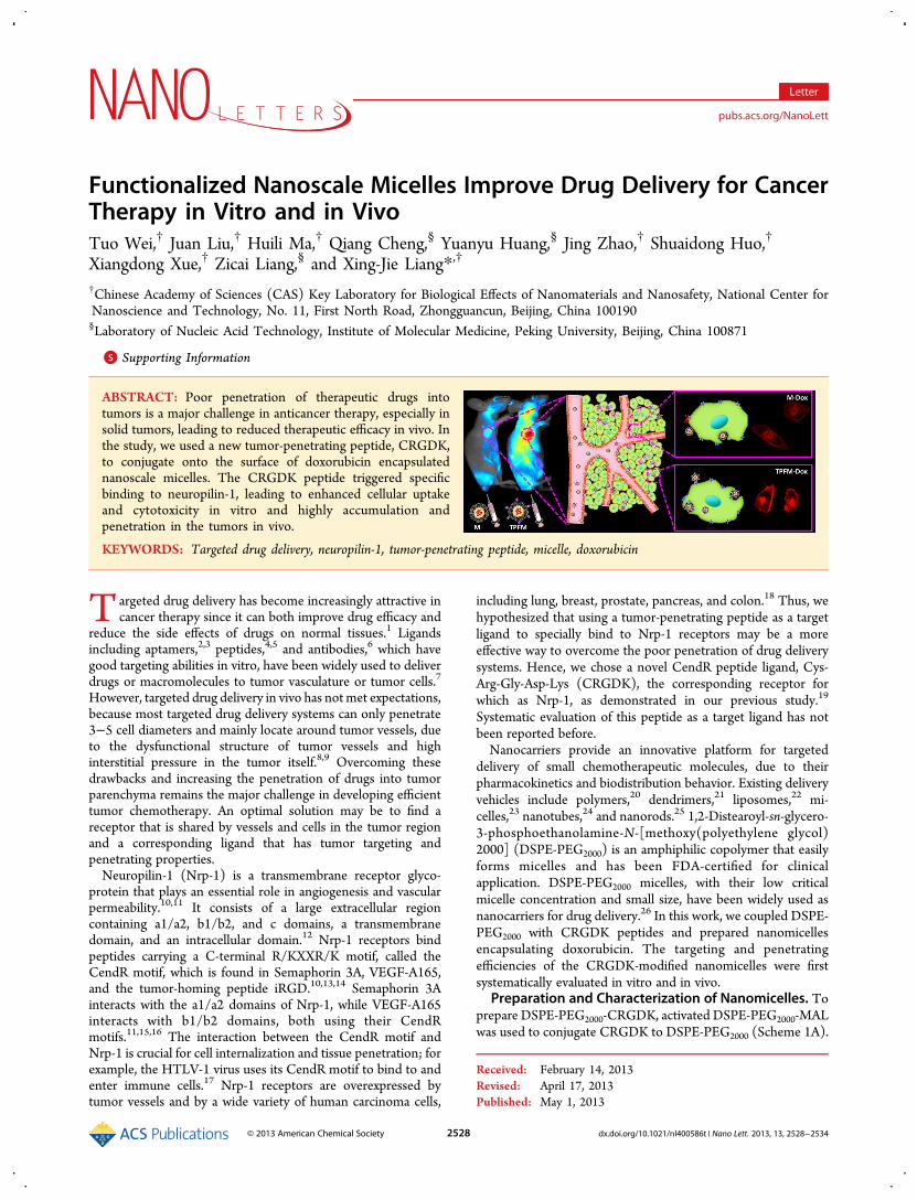
ABSTRACT: Poor penetration of therapeutic drugs intotumors is a major challenge in anticancer therapy, especially insolid tumors, leading to reduced therapeutic efficacy in vivo. Inthe study, we used a new tumor-penetrating peptide, CRGDK,to conjugate onto the surface of doxorubicin encapsulatednanoscale micelles. The CRGDK peptide triggered specificbinding to neuropilin-1, leading to enhanced cellular uptakeand cytotoxicity in vitro and highly accumulation andpenetration in the tumors in vivo.
KEYWORDS: Targeted drug delivery, neuropilin-1, tumor-penetrating peptide, micelle, doxorubicin
Targeted drug delivery has become increasingly attractive incancer therapy since it can both improve drug efficacy and
reduce the side effects of drugs on normal tissues.1 Ligandsincluding aptamers,2,3 peptides,4,5 and antibodies,6 which havegood targeting abilities in vitro, have been widely used to deliverdrugs or macromolecules to tumor vasculature or tumor cells.7
However, targeted drug delivery in vivo has not met expectations,because most targeted drug delivery systems can only penetrate3−5 cell diameters and mainly locate around tumor vessels, dueto the dysfunctional structure of tumor vessels and highinterstitial pressure in the tumor itself.8,9 Overcoming thesedrawbacks and increasing the penetration of drugs into tumorparenchyma remains the major challenge in developing efficienttumor chemotherapy. An optimal solution may be to find areceptor that is shared by vessels and cells in the tumor regionand a corresponding ligand that has tumor targeting andpenetrating properties.Neuropilin-1 (Nrp-1) is a transmembrane receptor glyco-
protein that plays an essential role in angiogenesis and vascularpermeability.10,11 It consists of a large extracellular regioncontaining a1/a2, b1/b2, and c domains, a transmembranedomain, and an intracellular domain.12 Nrp-1 receptors bindpeptides carrying a C-terminal R/KXXR/K motif, called theCendR motif, which is found in Semaphorin 3A, VEGF-A165,and the tumor-homing peptide iRGD.10,13,14 Semaphorin 3Ainteracts with the a1/a2 domains of Nrp-1, while VEGF-A165interacts with b1/b2 domains, both using their CendRmotifs.11,15,16 The interaction between the CendR motif andNrp-1 is crucial for cell internalization and tissue penetration; forexample, the HTLV-1 virus uses its CendR motif to bind to andenter immune cells.17 Nrp-1 receptors are overexpressed bytumor vessels and by a wide variety of human carcinoma cells,
including lung, breast, prostate, pancreas, and colon.18 Thus, wehypothesized that using a tumor-penetrating peptide as a targetligand to specially bind to Nrp-1 receptors may be a moreeffective way to overcome the poor penetration of drug deliverysystems. Hence, we chose a novel CendR peptide ligand, Cys-Arg-Gly-Asp-Lys (CRGDK), the corresponding receptor forwhich as Nrp-1, as demonstrated in our previous study.19
Systematic evaluation of this peptide as a target ligand has notbeen reported before.Nanocarriers provide an innovative platform for targeted
delivery of small chemotherapeutic molecules, due to theirpharmacokinetics and biodistribution behavior. Existing deliveryvehicles include polymers,20 dendrimers,21 liposomes,22 mi-celles,23 nanotubes,24 and nanorods.25 1,2-Distearoyl-sn-glycero-3-phosphoethanolamine-N-[methoxy(polyethylene glycol)2000] (DSPE-PEG2000) is an amphiphilic copolymer that easilyforms micelles and has been FDA-certified for clinicalapplication. DSPE-PEG2000 micelles, with their low criticalmicelle concentration and small size, have been widely used asnanocarriers for drug delivery.26 In this work, we coupled DSPE-PEG2000 with CRGDK peptides and prepared nanomicellesencapsulating doxorubicin. The targeting and penetratingefficiencies of the CRGDK-modified nanomicelles were firstsystematically evaluated in vitro and in vivo.
Preparation and Characterization of Nanomicelles. Toprepare DSPE-PEG2000-CRGDK, activated DSPE-PEG2000-MALwas used to conjugate CRGDK to DSPE-PEG2000 (Scheme 1A).
Received: February 14, 2013Revised: April 17, 2013Published: May 1, 2013
Letter
pubs.acs.org/NanoLett
© 2013 American Chemical Society 2528 dx.doi.org/10.1021/nl400586t | Nano Lett. 2013, 13, 2528−2534

CRGDK and DSPE-PEG2000-MAL (1:5, w/w) were dissolved in50 mM HEPES buffer (pH 6.5), stirring continuously at roomtemperature for 48 h. In this reaction, the thiol group (−SH) ofCRGDK is coupled to the maleimide group of DSPE-PEG2000-MAL.7,27,28 MALDI-TOF-MS analysis showed that the peak wasright-shifted after conjugation, indicating that CRGDK peptideshad been successfully conjugated to DSPE-PEG2000-MAL(Figure S1, Supporting Information).Doxorubicin-encapsulated DSPE-PEG2000 micelles (M-Dox)
and tumor-penetrating peptide-functionalized micelles (TPFM-Dox) were then prepared by a film dispersion method (Scheme1B). To prepare TPFM-Dox, DSPE-PEG2000 lipid was replacedby a mixture of DSPE-PEG2000-CRGDK andDSPE-PEG2000 (1:4w/w). The morphologies of M-Dox and TPFM-Dox were shownby TEM; bothM-Dox and TPFM-Dox micelles were spherical inshape, with a size of around 10 nm, and had good dispersion(Figure 1A). The average hydrodynamic diameter and surfacecharge were characterized by measuring the size and zetapotential with dynamic light scattering (DLS), as shown in TableS1, Supporting Information. The diameter of M-Dox was 11.5 ±0.3 nm, and the zeta potential was 0.2 ± 0.1 mV, while thediameter of TPFM-Dox was 13.1± 1.5 nm and the zeta potentialwas −6.9 ± 1.4 mV. Both M-Dox and TPFM-Dox had highencapsulation efficiencies (97.5% and 99.4%, respectively) andhigh drug loading capacities (16.3% and 15.3%, respectively).In Vitro Drug Release. Controlled and sustained drug
release is very important for drug delivery systems. The pH oftumor tissue is much lower than normal, because of lactic acidproduced due to hypoxia and acidic intracellular organelles.29,30
Therefore, the doxorubicin release profile from M-Dox orTPFM-Dox was evaluated by a dialysis method at pH 5.0 and 7.4(Figure 1B). Drug release fromM-Dox or TPFM-Dox was muchlower at pH 7.4 (30% and 34%, respectively) than at pH 5.0 (79%and 77%, respectively). The release profiles of M-Dox and
TPFM-Dox were similar at each pH. The result was consistentwith the previous report.31 The pH-sensitive drug release fromthe nanomicelles was related to the isoelectric points (PI) ofdoxorubicin andDSPE-PEG2000. When the pHwas reduced from7.4 to 5.0, dissociation of doxorubicin (PI = 9.06) was enhanced,and the electrostatic repulsion between drug moleculesincreased. Furthermore, the negatively charged DSPE-PEG2000molecules (PI = 5.93) became positively charged and lost theirelectrostatic attraction to doxorubicin.
Cytotoxicity Studies of M-Dox and TPFM-Dox. Toevaluate the cytotoxicity of CRGDK modified nanomicelles, wechose two human breast cancer cell lines, MDA-MB-231 andMCF-7. We confirmed that Nrp-1 receptors were expressed athigh levels on the surface of MDA-MB-231 cells, but at muchlower levels on MCF-7 cells using immunofluorescence (Figure2A). The expression of Nrp-1 receptors was also verified inmRNA level (Figure 2B) and protein level (Figure 2C) by RT-PCR and WB. These results are consistent with reports thatMDA-MB-231 cells overexpress Nrp-1 on their cell membrane.32
Next, we used these two cell lines to assess the efficacy of theNrp-1 target ligand CRGDK. We incubated free doxorubicin (F-Dox), M-Dox, or TPFM-Dox at doxorubicin concentrations of0.01−100 μg/mL for 24 h and evaluated the cell viability byMTT assay. Compared to F-Dox, M-Dox and TPFM-Dox weremore toxic to both MDA-MB-231 andMCF-7 cells (Figure 2D).ForMDA-MB-231 cells, M-Dox and TPFM-Dox showed a lowerIC50 (2.08 μg/mL and 0.38 μg/mL, respectively) than F-Dox
Scheme 1. Schematic Illustrations of the Preparation of theDoxorubicin-Encapsulating Micelles M-Dox and TPFM-Dox.(A) DSPE-PEG2000-CRGDK Was Synthesized by Couplingthe Thiol Group of CRGDK with the Maleimide Group ofDSPE-PEG2000-MAL. (B) Schematic Representations of theDoxorubicin-Encapsulating Micelles M-Dox and TPFM-Dox
Figure 1.Characterization of the doxorubicin-encapsulating micellesM-Dox and TPFM-Dox. (A) Transmission electron microscopy (TEM)images of M-Dox and TPFM-Dox after staining with 1% uranyl acetate.Scale bar = 50 nm. (B) Time course of doxorubicin release frommicellesat 37 °C at pH 5.0 or pH 7.4. Released doxorubicin was separated fromM-Dox or TPFM-Dox by dialysis and quantified by a spectropho-tometer.
Nano Letters Letter
dx.doi.org/10.1021/nl400586t | Nano Lett. 2013, 13, 2528−25342529

(4.89 μg/mL). Furthermore, the IC50 of TPFM-Dox was muchlower than that of M-Dox, which may be attributed to theimproved targeting efficacy of the CRGDK-modified micelles.However, there was little difference in the IC50 whenMCF-7 cellswere treated with M-Dox or TPFM-Dox (1.52 μg/mL and 1.94μg/mL, respectively), suggesting that the CRGDK ligand playeda key role in enhanced cytotoxicity in vitro.In addition, we evaluated the cytotoxicity of empty DSPE-
PEG2000 micelles to avoid the cytotoxicity caused by DSPE-PEG2000 micelles themselves. The result indicated that emptyDSPE-PEG2000 micelles had little toxicity to both MCF-7 cellsand MDA-MB-231 cells at the concentration from 0.05 to 500μg/mL, corresponding to the concentration of doxorubicin(Figure S2, Supporting Information). Hence, lower IC50 of M-DOX was not due to the toxic effect of DSPE-PEG2000 micellesthemselves, mainly by enhanced cellular internalization ofdoxorubicin when encapsulated into micelles.Competition Assay. To address the specificity of TPFM-
Dox for neuropilin-1 receptors, we performed a competitionassay on MDA-MB-231 cells. Cells were pretreated with excessanti-Nrp-1 monoclonal antibody (dilution 1:50) for 15 min andthen incubated with 10 μg/mL TPFM-Dox for 15 min.33,34 Theinternalization of TPFM-Dox into antibody-treated cells wasgreatly inhibited compared to untreated cells (Figure 3). Thisresult suggests that CRGDK-modified nanomicelles could bindspecifically to neuropilin-1 receptors.
Internalization and Distribution of Micelle-Encapsu-lated Doxorubicin in Vitro. We investigated the cellularuptake of CRGDK-modified micelles by MDA-MB-231 cellsusing laser confocal scanning microscopy. Cells were incubatedwith F-Dox, M-Dox, and TPFM-Dox at 37 °C for 0.5 or 1 h with
Figure 2. Expression of neuropilin-1 on cell membranes and cytotoxicity of various doxorubicin formulations. (A) Confocal images of Nrp-1 expression,detected with FITC-labeled anti-Nrp-1 antibody (green), on the membranes ofMDA-MB-231 andMCF-7 cells. The controls represent the cells are notpretreated with primary monoclonal Nrp-1 antibody, only treated by FITC-conjugated secondary antibody. Nuclei are stained with DAPI (blue). (B)RT-PCR and (C)Western blotting analysis of Nrp-1 expression inMDA-MB-231 andMCF-7 cells. (D) In vitro cytotoxicity, assessed byMTT assay, ofF-Dox (free doxorubicin), M-Dox, and TPFM-Dox against MDA-MB-231 and MCF-7 cells.
Figure 3. In vitro competition assay to detect targeting of neuropilin-1by TPFM-Dox inMDA-MB-231 cells. Cells were preincubated with (+)or without (−) anti Nrp-1 primary antibody for 15 min and thenincubated with TPFM-Dox for 15 min. Intracellular doxorubicin (red)was observed by confocal microscopy.
Nano Letters Letter
dx.doi.org/10.1021/nl400586t | Nano Lett. 2013, 13, 2528−25342530

the doxorubicin concentration at 10 μg/mL. The CRGDKmodification enhanced the cellular uptake of TPFM-Doxcompared with F-Dox and M-Dox (Figure 4A). This resultalso indicated that micelles entered cells more quickly than freedoxorubicin (F-Dox), which might be due to differentendocytosis pathways. In addition, cellular uptake increased asthe incubation time increased from 0.5 to 1 h.We next made quantitative measurements of the cellular
uptake of various Dox formulations using flow cytometry (Figure4B). MDA-MB-231 cells treated with TPFM-Dox showed aprominent right shift upon cytometric analysis, suggestinggreater cellular uptake of the CRGDK-modified micelles. Almostall cells were able to internalize Dox formulations, since nearly100% of cells had increased fluorescence except the controlgroup (Figure 4C). However, there was variation of the meanfluorescence intensity (MFI) among different Dox formulations(Figure 4D). The intracellular fluorescence intensity increasedwhen the treatment time was extended from 0.5 to 1 h. Themeanintensity of TPFM-Dox was 2.6-fold higher than F-Dox (p <0.01) and 1.6-fold higher than M-Dox (p < 0.01) after treatmentfor 0.5 h, indicating that CRGDK improves the target efficacy.
The difference of cellular uptake can be explained by theirdistinct uptake mechanisms. As shown in Figure S3, SupportingInformation, cellular uptake of M-Dox and TPFM-Doxdecreased markedly after treated with different endocytosisinhibitors, indicating that micelles entered cells by endocytosis.The cellular uptake inhibition to F-Dox was much lower thanthat to M-Dox and TPFM-Dox, indicating that F-Dox, a smallmolecule, mainly entered into cells by passive diffusionmechanism.35,36
The subcellular localization of F-Dox, M-Dox, and TPFM-Doxafter 4 h treatment was examined by confocal laser scanningmicroscopy. Lysotracker Green was used to identify endosomes/lysosomes. In cells treated with M-Dox and TPFM-Dox,doxorubicin fluorescence was localized in lysosomes and incytoplasm. Less F-Dox was localized in lysosomes (Figure S4,Supporting Information). The doxorubicin encapsulated nano-micelles entered into endosomes (pH 5.0−6.0) or lysosomes(pH 4.0−5.0) of cancer cells, and doxorubicin could easily escapefrom micelles and enter the cytoplasm,37,38 because of the pHsensitivity property. This can explain why the doxorubicinlocated both in endosomes/lysosomes and in cytoplasm.
Figure 4.Cellular uptake of F-Dox,M-Dox, and TPFM-Dox byMDA-MB-231 cells. (A) Confocal images of cells treated with free doxorubicin (F-Dox),M-Dox, or TPFM-Dox at 10 μg/mL for 0.5 and 1 h. (B) Quantitative analysis of micelle uptake by flow cytometry. (C) Percentages of cells withincreased fluorescence. (D) Mean fluorescence intensity of cells after 0.5 or 1 h. Control cells were untreated. The asterisk (**) represents data pointsthat have a highly significant difference (p < 0.01; two-tailed Student’s t tests).
Nano Letters Letter
dx.doi.org/10.1021/nl400586t | Nano Lett. 2013, 13, 2528−25342531

Enhanced Tumor Accumulation and Penetration inVivo. Before evaluating the tumor targeting and penetratingefficacy in mice, we used hemolysis analysis and blood smear textto evaluate the blood compatibility of empty DSPE-PEG2000
micelles. For hemolysis analysis, if erythrocytes are lysed,hemoglobin will be released and the supernatant will appearred and the hemoglobin in the supernatant can be measured bymeasuring the absorbance at 577 nmwith a reference wavelengthof 655 nm.39,40 As shown in Figure S5, Supporting Information,no visible hemolytic effects were seen even at the highest micelleconcentration of 320 μg/mL in PBS, indicating that the emptyDSPE-PEG2000 micelles had good hemocompatibility (<2%
hemolysis).41 For blood smears, mouse blood was mixed withempty nanomicelles at the same concentration used in the in vivoexperiments (1 mg/mL, in whole blood). The cells in the treatedblood showed no obvious aggregation or morphological changes(Figure S6, Supporting Information). These results demon-strated that the nanomicelles had good blood compatibility andcan be used for in vivo experiments.The tumor-targeting efficacy of CRGDK-modified micelles
was then evaluated in mice bearing armpit tumors derived fromhuman breast cancer MDA-MB-231 cells. Micelles loaded withthe fluorescent dye DiR were injected intravenously at a dosecorresponding to 200 ng/mL of DiR. Figure 5A showed the real-
Figure 5. Evaluation of tumor targeting and penetrating efficiencies of nanomicelles in vivo. Mice with armpit tumors derived fromMDA-MB-231 cellswere given tail vein injections of micelles loaded with the fluorescent dye DiR (A−D) or doxorubicin (10 mg/kg; E,F). Physiological saline wasadministered as a control. (A) In vivo images of mice 1, 3, 8, 12, and 24 h after treatment with DiR-loaded micelles. (B) Average fluorescence signals oftumors 1, 3, 8, 12, and 24 h after DiR treatment. The asterisk (**) represents data points that have highly significant difference (p < 0.01; two-tailedStudent’s t tests). (C, D) Ex vivo images of tumors and other tissues 24 h after DiR treatment (H, heart; Li, liver; S, spleen; Lu, lung; K, kidney; B, brain; I,intestine). (E, F) Frozen sections of tumors removed 24 h after treatment with Dox-loaded micelles were stained with DAPI to label nuclei (E) or CD31antibody to label tumor vessels (F). Red signal, doxorubicin; blue signal, DAPI; green signal, FITC-tagged CD31.
Nano Letters Letter
dx.doi.org/10.1021/nl400586t | Nano Lett. 2013, 13, 2528−25342532

time distribution and tumor accumulation of physiological saline(control), DiR-labeled micelles (M-DiR), and targeted micelles(TPFM-DiR) at 1, 3, 8, 12, and 24 h postinjection. After 1 h,strong DiR fluorescence was observed in the whole body, and by3 h, fluorescence accumulated in the tumors of micelle-treatedanimals (M-DiR or TPFM-DiR). As time elapsed, tumorfluorescence in the TPFM-DiR treated mice was notably higherthan in theM-DiR treated group (Figure 5A). Between 12 and 24h, the average M-DiR signal decreased significantly (p < 0.01),while the average TPFM-DiR signal continued to increase(Figure 5B). The average TPFM-DiR signal was significantlyhigher than that of M-DiR at all time-points examined.Compared to M-DiR, TPFM-DiR has excellent targetingefficiency and accumulates continuously in tumors. The ex vivofluorescent images of excised tumors further confirmed thehigher accumulation of TPFM-DiR compared to M-DiR (Figure5C). No obvious fluorescence uptake was observed in the heart,spleen, lung, kidney, intestine, and brain, but the fluorescencelevels in livers from M-DiR or TPFM-DiR treated animals werevery high, which may be attributed to the high macrophageuptake nature of livers (Figure 5D).42
To further test the accumulation and penetration of micelles,nude mice bearing tumors from MDA-MB-231 cells wereinjected with physiological saline, M-Dox and TPFM-Dox at adoxorubicin concentration of 10 mg/kg. After 24 h, tumors wereexcised, and tissue sections were examined. TPFM-Doxaccumulated at higher levels than M-Dox in the tumor tissue(Figure 5E), which is consistent with the in vivo imaging data. Toevaluate the penetration efficiency, we labeled tumor vessels withfluorescent-tagged antibody against the endothelial markerCD31. As shown in Figure 5F, TPFM-Dox spread more widelythan M-Dox around vessels, indicating better penetration. Wefurther used a method based on the simulated scatter diagrams toconfirm the better penetration of TPFM-Dox (Figure S7,Supporting Information). The distance that the drugs traveledafter leaving the vasculature was evaluated roughly. As a result,the penetration distance of M-Dox was 0.44 ± 0.38 μm, and thepenetration distance of TPFM-Dox was 7.16 ± 5.08 μm.Therefore, the TPFM-Dox enhanced penetration distance bymore than 15-fold compared to M-Dox. These results confirmedthat targeted micelles showed higher tumor targeting andpenetrating efficiency, leading to higher accumulation of drug inthe tumors.In summary, this work first evaluated the tumor targeting and
penetrating properties of CRGDK peptides systematically. Fromthe results above, we demonstrate that CRGDK peptides couldenhance tumor targeting and penetration of drug encapsulatednanocarriers both in vitro and in vivo and is a promisingcandidate for developing targeted drug delivery systems. Thenanocarriers and drugs forming TPFM-Dox are certified by Foodand Drug Administration (FDA), so TPFM-Dox may haveimportant clinical applications in cancer therapy. In addition,neuropilin-1 receptors are overexpressed in a wide variety ofhuman carcinoma cell lines and are implicated in the survival,migration, and invasion of tumor cells. Overexpression of Nrp-1receptors promotes tumor angiogenesis and tumor growth andalso correlates to metastatic spread.32,43,44 In addition to theaggressive human metastatic breast cancer cell line MDA-MB-231, the aggressive human metastatic prostate cancer cell linePC3 also overexpresses Nrp-1 receptors (data not shown).Therefore, CRGDK-modified nanocarriers may have greatpotential for treating metastatic cancers, which occur duringthe advanced stages of disease. Further work may be needed to
evaluate the anticancer therapeutic potential of CRGDK-modified nanocarriers, including their abilities to inhibit tumorinvasion and metastasis.
■ ASSOCIATED CONTENT*S Supporting InformationMaterials and methods to characterize CRGDK modifiednanomicelles and unmodified nanomicelles, including TEMimages, in vitro drug release, cell viability, competition assay,cellular uptake, blood compatibility, and in vivo experiments.Additional figures and tables. This material is available free ofcharge via the Internet at http://pubs.acs.org.
■ AUTHOR INFORMATIONCorresponding Author*E-mail: [email protected] ContributionsT.W. and J.L. contributed equally to this work.NotesThe authors declare no competing financial interest.
■ ACKNOWLEDGMENTSThis work was supported by the Chinese Natural ScienceFoundation project (no. 30970784, no. 81171455), a NationalDistinguished Young Scholars grant (31225009) from theNational Natural Science Foundation of China, the NationalKey Basic Research Program of China (2009CB930200), theChinese Academy of Sciences (CAS) “Hundred TalentsProgram” (07165111ZX), the CAS Knowledge InnovationProgram, and the State High-Tech Development Plan(2012AA020804).
■ ABBREVIATIONSNrp-1, neuropilin-1 receptors; F-Dox, free doxorubicin; M-Dox,DSPE-PEG2000 micelles encapsulating doxorubicin; TPFM-Dox,tumor-penetrating peptide-functionalized DSPE-PEG2000 mi-celles encapsulating doxorubicin
■ REFERENCES(1) Ruoslahti, E.; Bhatia, S. N.; Sailor, M. J. J. Cell Biol. 2010, 188 (6),759−768.(2) Guo, J. W.; Gao, X. L.; Su, L. N.; Xia, H. M.; Gu, G. Z.; Pang, Z. Q.;Jiang, X. G.; Yao, L.; Chen, J.; Chen, H. Z. Biomaterials 2011, 32 (31),8010−8020.(3) Kim, D.; Jeong, Y. Y.; Jon, S. ACS Nano 2010, 4 (7), 3689−3696.(4) Messerschmidt, S. K. E.; Musyanovych, A.; Altvater, M.; Scheurich,P.; Pfizenmaier, K.; Landfester, K.; Kontermann, R. E. J. ControlledRelease 2009, 137 (1), 69−77.(5) Nasongkla, N.; Shuai, X.; Ai, H.; Weinberg, B. D.; Pink, J.;Boothman, D. A.; Gao, J. Angew. Chem. 2004, 116 (46), 6483−6487.(6) Agemy, L.; Sugahara, K. N.; Kotamraju, V. R.; Gujraty, K.; Girard,O. M.; Kono, Y.; Mattrey, R. F.; Park, J. H.; Sailor, M. J.; Jimenez, A. I.Blood 2010, 116 (15), 2847−2856.(7) Lu, J.; Jeon, E.; Lee, B. S.; Onyuksel, H.; Wang, Z. J. J. ControlledRelease 2006, 110 (3), 505−513.(8) Heldin, C. H.; Rubin, K.; Pietras, K.; Ostman, A. Nat. Rev. Cancer2004, 4 (10), 806−813.(9) Jain, R. K. Annu. Rev. Biomed. Eng. 1999, 1 (1), 241−263.(10) Haspel, N.; Zanuy, D.; Nussinov, R.; Teesalu, T.; Ruoslahti, E.;Aleman, C. Biochemistry 2011, 50 (10), 1755−1762.(11) Jubb, A. M.; Strickland, L. A.; Liu, S. D.; Mak, J.; Schmidt, M.;Koeppen, H. J. Pathol. 2012, 226 (1), 50−60.(12) Jia, H.; Cheng, L.; Tickner, M.; Bagherzadeh, A.; Selwood, D.;Zachary, I. Br. J. Cancer 2010, 102 (3), 541−552.
Nano Letters Letter
dx.doi.org/10.1021/nl400586t | Nano Lett. 2013, 13, 2528−25342533
(13) Teesalu, T.; Sugahara, K. N.; Kotamraju, V. R.; Ruoslahti, E. Proc.Natl. Acad. Sci. U.S.A. 2009, 106 (38), 16157−16162.(14) Sugahara, K. N.; Teesalu, T.; Karmali, P. P.; Kotamraju, V. R.;Agemy, L.; Greenwald, D. R.; Ruoslahti, E. Science 2010, 328 (5981),1031−1035.(15) Hong, T.M.; Chen, Y. L.;Wu, Y. Y.; Yuan, A.; Chao, Y. C.; Chung,Y. C.; Wu, M. H.; Yang, S. C.; Pan, S. H.; Shih, J. Y. Clin. Cancer Res.2007, 13 (16), 4759−4768.(16) Jarvis, A.; Allerston, C. K.; Jia, H.; Herzog, B.; Garza-Garcia, A.;Winfield, N.; Ellard, K.; Aqil, R.; Lynch, R.; Chapman, C. J. Med. Chem.2010, 53 (5), 2215−2226.(17) Lambert, S.; Bouttier, M.; Vassy, R.; Seigneuret, M.; Petrow-Sadowski, C.; Janvier, S.; Heveker, N.; Ruscetti, F. W.; Perret, G.; Jones,K. S. Blood 2009, 113 (21), 5176−5185.(18) Bielenberg, D. R.; Pettaway, C. A.; Takashima, S.; Klagsbrun, M.Exp. Cell Res. 2006, 312 (5), 584−593.(19) Kumar, A.; Ma, H.; Zhang, X.; Huang, K.; Jin, S.; Liu, J.; Wei, T.;Cao, W.; Zou, G.; Liang, X.-J. Biomaterials 2011, 33 (4), 1180−1189.(20) Graf, N.; Bielenberg, D. R.; Kolishetti, N.; Muus, C.; Banyard, J.;Farokhzad, O. C.; Lippard, S. J. ACS Nano 2012, 6 (5), 4530−4539.(21) Yu, T.; Liu, X.; Bolcato-Bellemin, A. L.; Wang, Y.; Liu, C.;Erbacher, P.; Qu, F.; Rocchi, P.; Behr, J. P.; Peng, L. Angew. Chem. 2012,124 (34), 8606−8612.(22) Liu, J.; Ma, H.;Wei, T.; Liang, X. J.Chem. Commun. 2012, 48 (40),4869−4871.(23) Peters, D.; Kastantin, M.; Kotamraju, V. R.; Karmali, P. P.;Gujraty, K.; Tirrell, M.; Ruoslahti, E. Proc. Natl. Acad. Sci. U.S.A. 2009,106 (24), 9815−9819.(24) Bhirde, A. A.; Patel, V.; Gavard, J.; Zhang, G.; Sousa, A. A.;Masedunskas, A.; Leapman, R. D.; Weigert, R.; Gutkind, J. S.; Rusling, J.F. ACS Nano 2009, 3 (2), 307−316.(25) Wang, L.; Liu, Y.; Li, W.; Jiang, X.; Ji, Y.; Wu, X.; Xu, L.; Qiu, Y.;Zhao, K.; Wei, T. Nano Lett. 2011, 11 (2), 772−780.(26) Kastantin, M.; Ananthanarayanan, B.; Karmali, P.; Ruoslahti, E.;Tirrell, M. Langmuir 2009, 25 (13), 7279−7286.(27) Liu, X.; Ruan, L.; Mao, W.; Wang, J. Q.; Shen, Y.; Sui, M. Int. J.Med. Sci. 2010, 7 (4), 197−208.(28) Yang, T.; Choi, M. K.; Cui, F. D.; Kim, J. S.; Chung, S. J.; Shim, C.K.; Kim, D. D. J. Controlled Release 2007, 120 (3), 169−177.(29) Aryal, S.; Hu, C.M. J.; Zhang, L.ACSNano 2009, 4 (1), 251−258.(30) Yuan, Q.; Yeudall, W. A.; Yang, H. Biomacromolecules 2010, 11(8), 1940−1947.(31) Tang, N.; Du, G.; Wang, N.; Liu, C.; Hang, H.; Liang, W. J. Natl.Cancer Inst. 2007, 99 (13), 1004−1015.(32) Ellis, L. M. Mol. Cancer Ther. 2006, 5 (5), 1099−1107.(33) Danhier, F.; Vroman, B.; Lecouturier, N.; Crokart, N.; Pourcelle,V.; Freichels, H.; Jerome, C.; Marchand-Brynaert, J.; Feron, O.; Preat, V.J. Controlled Release 2009, 140 (2), 166−173.(34) Murphy, E. A.; Majeti, B. K.; Barnes, L. A.; Makale, M.; Weis, S.M.; Lutu-Fuga, K.; Wrasidlo, W.; Cheresh, D. A. P Natl Acad Sci USA2008, 105 (27), 9343−9348.(35) Lee, Y.; Park, S. Y.; Mok, H.; Park, T. G. Bioconjugate Chem. 2007,19 (2), 525−531.(36) Yi, Y.; Kim, J. H.; Kang, H. W.; Oh, H. S.; Kim, S. W.; Seo, M. H.Pharm. Res. 2005, 22 (2), 200−208.(37) Ganta, S.; Devalapally, H.; Shahiwala, A.; Amiji, M. J. ControlledRelease 2008, 126 (3), 187−204.(38) Shenoy, D.; Little, S.; Langer, R.; Amiji, M. Mol. Pharmaceutics2005, 2 (5), 357−366.(39) Chen, Y.; Chen, H.; Guo, L.; He, Q.; Chen, F.; Zhou, J.; Feng, J.;Shi, J. ACS Nano 2009, 4 (1), 529−539.(40) Yu, T.; Malugin, A.; Ghandehari, H. ACS Nano 2011, 5 (7),5717−5728.(41) Zhao, Y.; Sun, X.; Zhang, G.; Trewyn, B. G.; Slowing, I. I.; Lin, V.S. Y. ACS Nano 2011, 5 (2), 1366−1375.(42) Xiang, Y.; Liang, L.; Wang, X.; Wang, J.; Zhang, X.; Zhang, Q. J.Controlled Release 2011, 152 (3), 402−410.
(43) Frankel, P.; Pellet-Many, C.; Lehtolainen, P.; D’Abaco, G. M.;Tickner, M. L.; Cheng, L.; Zachary, I. C. EMBO Rep. 2008, 9 (10), 983−989.(44) Guttmann-Raviv, N.; Kessler, O.; Shraga-Heled, N.; Lange, T.;Herzog, Y.; Neufeld, G. Cancer Lett. 2006, 231 (1), 1−11.
Nano Letters Letter
dx.doi.org/10.1021/nl400586t | Nano Lett. 2013, 13, 2528−25342534