Embed Size (px)
Citation preview

Microorganisms 2021, 9, 1434. https://doi.org/10.3390/microorganisms9071434 www.mdpi.com/journal/microorganisms
Article
Functional Redundancy and Specialization of the Conserved
Cold Shock Proteins in Bacillus subtilis
Patrick Faßhauer 1, Tobias Busche 2, Jörn Kalinowski 2, Ulrike Mäder 3, Anja Poehlein 4, Rolf Daniel 4
and Jörg Stülke 1,*
1 Department of General Microbiology, GZMB, Georg‐August‐University Göttingen, 37077 Göttingen,
Germany; patrick.fasshauer@uni‐goettingen.de 2 Center for Biotechnology (CeBiTec), Bielefeld University, 33615 Bielefeld, Germany;
[email protected]‐bielefeld.de (T.B.); [email protected]‐bielefeld.de (J.K.) 3 Interfaculty Institute for Genetics and Functional Genomics, University Medicine Greifswald,
17487 Greifswald, Germany; ulrike.maeder@uni‐greifswald.de 4 Department of Genomic and Applied Microbiology, GZMB, Georg‐August‐University Göttingen,
37077 Göttingen, Germany; [email protected] (A.P.); [email protected] (R.D.)
* Correspondence: [email protected]; Tel.: +0049‐551‐3933781
Abstract: Many bacteria encode so‐called cold shock proteins. These proteins are characterized by
a conserved protein domain. Often, the bacteria have multiple cold shock proteins that are
expressed either constitutively or at low temperatures. In the Gram‐positive model bacterium
Bacillus subtilis, two of three cold shock proteins, CspB and CspD, belong to the most abundant
proteins suggesting a very important function. To get insights into the role of these highly abundant
proteins, we analyzed the phenotypes of single and double mutants, tested the expression of the
csp genes and the impact of CspB and CspD on global gene expression in B. subtilis. We
demonstrate that the simultaneous loss of both CspB and CspD results in a severe growth defect,
in the loss of genetic competence, and the appearance of suppressor mutations. Overexpression
of the third cold shock protein CspC could compensate for the loss of CspB and CspD. The
transcriptome analysis revealed that the lack of CspB and CspD affects the expression of about
20% of all genes. In several cases, the lack of the cold shock proteins results in an increased read‐
through at transcription terminators suggesting that CspB and CspD might be involved in the
control of transcription termination.
Keywords: Bacillus subtilis; cold shock proteins; quasi‐essential
1. Introduction
Bacillus subtilis is the model organism for a large group of Gram‐positive bacteria,
among them serious pathogens and biotechnologically relevant bacteria. Due to this
relevance, B. subtilis is one of the best‐studied organisms. However, for about 25% of all
proteins, the function is completely unknown, or the proteins are only poorly
characterized [1]. Similarly, in the artificially created genome of the minimal organism
Mycoplasma mycoides JCVI‐syn3.0, about one‐third of all encoded proteins are of
unknown function [2]. These numbers demonstrate how far we are from a complete
understanding of even seemingly simple model organisms.
We are interested in a comprehensive understanding of B. subtilis. Obviously, the
large fraction of unknown proteins is a major challenge. Among the unknown and
poorly characterized proteins, the large majority is not or only weakly expressed under
standard growth conditions and probably only important under very specific conditions
[3]. However, among the 100 most abundant proteins, there are six proteins for which
no clear function has been identified [4]. These highly abundant unknown proteins are
Citation: Faßhauer, P.; Busche, T.;
Kalinowski, J.; Mäder, U.; Poehlein, A.;
Daniel, R.; Stülke, J. Functional
Redundancy and Specialization of the
Conserved Cold Shock Proteins in
Bacillus subtilis. Microorganisms 2021, 9,
1434. https://doi.org/10.3390/
microorganisms9071434
Academic Editor: Imrich Barák
Received: 03 June 2021
Accepted: 30 June 2021
Published: 2 July 2021
Publisher’s Note: MDPI stays neutral
with regard to jurisdictional claims in
published maps and institutional
affiliations.
Copyright: © 2021 by the authors.
Licensee MDPI, Basel, Switzerland.
This article is an open access article
distributed under the terms and
conditions of the Creative Commons
Attribution (CC BY) license
(http://creativecommons.org/licenses/b
y/4.0/).

Microorganisms 2021, 9, 1434 2 of 15
likely to be of major relevance for the cell, and their functional analysis should be given a
high priority. Among these proteins are the cold shock proteins CspB and CspD, which
are thought to act as RNA chaperones in B. subtilis [5]. The cold shock proteins are a large
protein family that are all characterized by a conserved cold shock domain (see [6] for a
phylogenetic tree). Among the bacteria, the cold shock proteins are nearly ubiquitous, and
most bacteria encode multiple cold shock proteins. B. subtilis has three cold shock proteins,
CspB, CspC, and CspD. The genes encoding the B. subtilis cold shock proteins are highly
expressed under a wide variety of growth conditions, and with 31,200 and 21,500 protein
molecules per cell, CspB and CspD, respectively, even belong to the 15 most abundant
proteins in B. subtilis [4,7].
The presence of multiple closely related proteins in an organism always raises the
question of whether they participate in the same function or if their specific activity is
slightly different. There are a few examples of families of closely related proteins in B.
subtilis. In the case of ABC transporters or classic amino acid and ion transporters, both
functional overlaps, as well as specific functions, have been observed [8–10]. In some
cases, as described for the three diadenylate cyclases, all proteins have the same enzymatic
activity, but their expression and activities are differentially controlled [11,12]. Similarly,
the four DEAD‐box RNA helicases have distinct domains in addition to the active protein
core, and these proteins have independent functions [13]. Finally, in some cases,
regulatory systems consisting of multiple parts, such as two‐component regulatory
systems, ECF sigma factors, PTS‐controlled RNA‐binding antitermination proteins, or
stress signaling proteins, specificity is achieved by co‐evolving interacting partners of
these systems [14–17].
For the B. subtilis cold shock proteins, it has been observed that the bacteria are not
viable in the absence of all three proteins [5]. This, on the one hand, supports the idea that
these highly abundant proteins play a major role in the B. subtilis cell and, on the other
hand, suggests that there is at least some functional overlap between them. In
Staphylococcus aureus, the CspA protein is unique among the three cold shock proteins
since it is the only one that is strongly constitutively expressed and important for the
expression of the virulence factor staphyloxanthin [18,19]. It was shown that a single
amino acid in CspA, a conserved proline residue, is responsible for this regulatory effect
[18].
To get more insights into the function(s) of the cold shock proteins of B. subtilis, we
studied their expression as well as phenotypes of mutants. The deletion of cspB and cspD
was found to result in the rapid acquisition of suppressor mutations that often result in
increased expression of cspC, suggesting that CspC might at least partially take over the
function of CspB and CspD if it is overexpressed. We also studied the global RNA profile
of a strain lacking CspB and CspD and observed that these proteins are important for
transcription elongation and termination and that they can control the read‐through at
transcription terminators.
2. Materials and Methods
2.1. Bacterial Strains, Growth Conditions, and Phenotypic Characterization
All B. subtilis strains used in this study are listed in Table 1. All strains are derived
from the laboratory strain 168 (trpC2). B. subtilis was grown in Lysogeny Broth (LB
medium) (21). LB plates were prepared by addition of 17 g Bacto agar/l (Difco) [20,21].
Quantitative studies of lacZ expression in B. subtilis were performed as described
previously [21]. One unit of ‐galactosidase is defined as the amount of enzyme which
produces 1 nmol of o‐nitrophenol per min at 28 °C.
Microorganisms 2021, 9, 1434 3 of 15
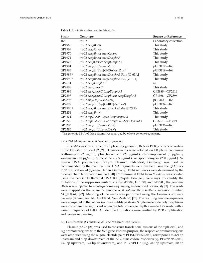
Table 1. B. subtilis strains used in this study.
Strain Genotype Source or Reference
168 trpC2 Laboratory collection
GP1968 trpC2 ΔcspB::cat This study
GP1969 trpC2 ΔcspC::spec This study
GP1970 trpC2 ΔcspB::cat ΔcspC::spec This study
GP1971 trpC2 ΔcspB::cat ΔcspD::aphA3 This study
GP1972 trpC2 ΔcspC::spec ΔcspD::aphA3 This study
GP1984 trpC2 amyE::(PcspC‐lacZ cat) pGP3117→168
GP1986 trpC2 amyE::(PcspC[G‐65A]‐lacZ cat) pGP3119→168
GP1989 1 trpC2 ΔcspB::cat ΔcspD::aphA3 PcspC‐[G‐65A] This study
GP1990 1 trpC2 ΔcspB::cat ΔcspD::aphA3 Pveg‐[G‐10T] This study
GP2614 trpC2 ΔcspD::aphA3 42
GP2888 trpC2 Δveg::ermC This study
GP2896 trpC2 Δveg::ermC ΔcspD::aphA3 GP2888→GP2614
GP2897 trpC2 Δveg::ermC ΔcspB::cat ΔcspD::aphA3 GP1968→GP2896
GP2898 trpC2 amyE::(Pveg‐lacZ cat) pGP3133→168
GP2899 trpC2 amyE::(Pveg‐[G‐10T]‐lacZ cat) pGP3134→168
GP2900 1 trpC2 ΔcspB::cat ΔcspD::aphA3 degS[P245S] This study
GP3251 trpC2 ΔcspB::tet This study
GP3274 trpC2 cspC‐A58P‐spec ΔcspD::aphA3 This study
GP3275 trpC2 cspC‐A58P‐spec ΔcspB::tet ΔcspD::aphA3 GP3251→GP3274
GP3283 trpC2 amyE::(PcspB‐lacZ cat) pGP3136→168
GP3286 trpC2 amyE::(PcspD‐lacZ‐cat) This study 1 The genomic DNA of these strains was analyzed by whole‐genome sequencing.
2.2. DNA Manipulation and Genome Sequencing
B. subtilis was transformed with plasmids, genomic DNA, or PCR products according
to the two‐step protocol [20,21]. Transformants were selected on LB plates containing
erythromycin (2 μg/mL) plus lincomycin (25 μg/mL), chloramphenicol (5 μg/mL),
kanamycin (10 μg/mL), tetracycline (12.5 μg/mL), or spectinomycin (250 μg/mL). S7
Fusion DNA polymerase (Biozym, Hessisch Oldendorf, Germany) was used as
recommended by the manufacturer. DNA fragments were purified using the QIAquick
PCR purification kit (Qiagen, Hilden, Germany). DNA sequences were determined by the
dideoxy chain termination method [20]. Chromosomal DNA from B. subtilis was isolated
using the peqGOLD Bacterial DNA Kit (Peqlab, Erlangen, Germany). To identify the
mutations in the suppressor mutant strains GP1989, GP1990, and GP2900, the genomic
DNA was subjected to whole‐genome sequencing as described previously [3]. The reads
were mapped on the reference genome of B. subtilis 168 (GenBank accession number:
NC_000964) [22]. Mapping of the reads was performed using the Geneious software
package (Biomatters Ltd., Auckland, New Zealand) [23]. The resulting genome sequences
were compared to that of our in‐house wild‐type strain. Single nucleotide polymorphisms
were considered as significant when the total coverage depth exceeded 25 reads with a
variant frequency of ≥90%. All identified mutations were verified by PCR amplification
and Sanger sequencing.
2.3. Construction of Translational LacZ Reporter Gene Fusions
Plasmid pAC5 [24] was used to construct translational fusions of the cspB, cspC, and
veg promoter regions with the lacZ gene. For this purpose, the respective promoter regions
were amplified using the oligonucleotide pairs PF151/PF152 (cspB, corresponds to 379 bp
upstream and 5 bp downstream of the ATG start codon, respectively), PF97/PF98 (cspC,
237 bp upstream, 125 bp downstream), and PF127/PF118 (veg, 280 bp upstream, 50 bp

Microorganisms 2021, 9, 1434 4 of 15
downstream), and chromosomal DNA of B. subtilis 168 as the template. The PCR products
were digested with EcoRI and BamHI and cloned into pAC5 linearized with the same
enzymes. The resulting plasmids were pGP3136, pGP3117, and pGP3133 for the cspB,
cspC, and veg promoters, respectively. Plasmids pGP3119 and pGP3134 for the cspC and
veg mutant upstream regions were constructed accordingly using chromosomal DNA of
the suppressor mutants GP1986 and GP1990 as the template, respectively. The promoter
region of cspD (194 bp upstream, 30 bp downstream) was fused to lacZ using a PCR prod‐
uct constructed with oligonucleotides PF246/PF247 and PF250/PF251 using pAC5 as a
template and PF248/PF249 with chromosomal DNA from B. subtilis 168 as a template. The
fragments were joined and amplified in a PCR as described previously for the deletion of
genes [25,26]. The fusion constructs were integrated into the amyE site of the B. subtilis
chromosome by the transformation of B. subtilis 168 with linearized plasmids or the PCR
product. The resulting strains were GP3283 (cspB wild type promoter), GP1984 (cspC wild
type promoter), GP1986 (cspC mutant promoter), GP3286 (cspD wild type promoter),
GP2898 (veg wild type promoter), and GP2899 (veg mutant promoter).
2.4. Construction of Mutants
Deletion of the csp genes was achieved by transformation with PCR products con‐
structed using oligonucleotides (see Table S1) to amplify DNA fragments flanking the tar‐
get genes and intervening antibiotic resistance cassettes as described previously [25,26].
The strain GP3274 harboring the CspC‐A58P variant was created using the multiple‐mu‐
tation reaction method, as described previously [27], to generate the cspC fragment fol‐
lowed by the combination of the fragment with the downstream flanking region and a
spectinomycin resistance cassette as described above.
2.5. RNA‐Sequencing
B. subtilis 168 wild type and the cspB cspD double mutant GP1971 were grown in LB
medium to an OD600 of 0.2 and were harvested by mixing them 5:3 with frozen killing
buffer (20 mM Tris‐HCl pH 7.5, 6 mM MgCl2, 20 mM NaN3) followed by snap‐freezing of
the pellets in liquid nitrogen. RNA isolation and quality assessment were performed as
described previously [7] with an additional DNase I treatment using TURBO DNase (Am‐
bion). The RNA quality was checked by Trinean Xpose (Trinean, Gentbrugge, Belgium)
and the Agilent RNA Nano 6000 kit using an Agilent 2100 Bioanalyzer (Agilent Technol‐
ogies, Böblingen, Germany). The Ribo‐Zero rRNA Removal Kit (Bacteria) from Illumina
(San Diego, CA, USA) was used to remove the rRNA. The TruSeq Stranded mRNA Li‐
brary Prep Kit from Illumina was applied to prepare the cDNA libraries. Final libraries
were sequenced paired‐end on an Illumina MiSeq system (San Diego, CA, USA) using
75 bp read length.
Trimmed reads were mapped to the B. subtilis 168 genome sequences (NCBI GenBank
accession number AL009126.3) using Bowtie2 [28]. The annotation AL009126.3 from the
NCBI RefSeq database was augmented with the RNA features previously annotated [7]).
In order to perform differential gene expression analysis, DEseq2 [29] was used as a part
of the software ReadXplorer v2.2 [30]. Statistically significant expression changes (ad‐
justed p‐value ≤ 0.01) with log2 fold change >1.0 or <−1.0 were used. RNA‐seq data have
been deposited in the ArrayExpress database at EMBL‐EBI (www.ebi.ac.uk/arrayexpress
(accessed on 02 July 2021)) under accession number E‐MTAB‐10658.
2.6. Qualitative PCR and Real‐Time Quantitative Reverse Transcription PCR
For RNA isolation, the cells were grown in LB medium and harvested at an OD600 of
1.2 for qualitative PCR and an OD600 of 0.5–0.8 for quantitative RT‐PCR. Preparation of
total RNA was carried out as described previously [31]. cDNAs for qualitative PCR were
synthesized using the RevertAid First Strand cDNA Synthesis Kit from ThermoFisher ac‐
cording to the manufacturer’s instructions. cDNAs for qRT‐PCR were synthesized using

Microorganisms 2021, 9, 1434 5 of 15
the One‐Step RT‐PCR kit (BioRad, Feldkirchen, Germany) as described [32]. qRT‐PCR was
carried out on the iCycler instrument (BioRad), following the manufacturer’s recom‐
mended protocol by using the primers indicated in Table S1. The rpsE and rpsJ genes en‐
coding constitutively expressed ribosomal proteins were used as internal controls. Data
analysis and the calculation of expression ratios as fold changes were performed as de‐
scribed [32]. qRT‐PCR experiments were performed in triplicate.
2.7. Microscopy
For microscopy, cells were grown at 37 °C in liquid LB medium overnight. The over‐
night culture was directly used for microscopy or for inoculation of LB medium, which
was incubated at 37 °C to an OD600 of 0.3–0.5. Images were acquired using an Axioskop 40
FL fluorescence microscope, equipped with digital camera AxioCam MRm and Axio‐
Vision Rel (version 4.8, Carl Zeiss, Oberkochen, Germany) software for image processing
(Carl Zeiss, Göttingen, Germany) and Neofluar series objective at ×100 primary magnifi‐
cation.
3. Results
3.1. Relative Contribution of the Cold Shock Proteins to the Growth of B. Subtilis
It has been reported that B. subtilis is not viable in the absence of the three cold shock
proteins CspB, CspC, and CspD [5]. To get better insights into the role(s) of the individual
cold shock proteins, we constructed a set of single and double mutants. In agreement with
the published data, deletion of all three csp genes was not possible. First, we compared
the growth of the strains on plates at 37 °C and 15 °C. As shown, in Figure 1A, at 37 °C,
all single mutants, as well as the cspB cspC and cspC cspD double mutants, grew indistin‐
guishable from the wild‐type strain B. subtilis 168. In contrast, the cspB cspD double mutant
GP1971 barely formed colonies; instead, this mutant acquired suppressor mutations that
restored growth (Figure 1A, see below). At 15 °C, growth of the cspB cspC double mutant
was also strongly impaired comparable to the cspB cspD double mutant (Figure 1B),
whereas the cspC cspD double mutant showed a slight growth defect.
Figure 1. Growth of csp knockout mutants. The wild‐type strain B. subtilis 168 and the csp mutants
GP1968 (∆cspB), GP1969 (∆cspC), GP2614 (∆cspD), GP1970 (∆cspB ∆cspC), GP1971 (∆cspB ∆cspD),
and GP1972 (∆cspC ∆cspD) were cultivated on LB‐agar (A) at 37 °C for one day and (B) at 15 °C for
11 days. The results are representative of three biological replicates.
Microscopic analysis of the cell morphologies of the different csp mutant strains in‐
dicated that the cold shock proteins did not affect cell morphology during logarithmic
growth (see Figure 2A). Similarly, loss of cspC or cspD did not affect the morphology in
the stationary phase. In contrast, the deletion of cspB in GP1968 resulted in the formation
of elongated cells in the stationary phase suggesting a defect in cell wall biosynthesis
and/or cell division or in the entry to stationary phase (Figure 2B). The cspB cspD double
mutant showed a highly aberrant cell morphology with curly cells in the stationary phase

Microorganisms 2021, 9, 1434 6 of 15
(see Figure 2B). The strong phenotype of the cspB cspD double mutant is in good agree‐
ment with the poor growth of this mutant.
Figure 2. Phenotypic analysis of B. subtilis csp mutants. The wild‐type strain B. subtilis 168 and the
csp mutants GP1968 (∆cspB), GP1969 (∆cspC), GP2614 (∆cspD), GP1970 (∆cspB ∆cspC), GP1971
(∆cspB ∆cspD), and GP1972 (∆cspC ∆cspD) were analyzed for morphology by phase‐contrast mi‐
croscopy. Scale bars, 5 μM. (A) Cells were cultivated in liquid LB medium at 37 °C to an OD600 of
0.3–0.5. (B) Cells were cultivated in liquid LB medium at 37 °C overnight to the stationary growth
phase.
In our attempts to combine the different mutations by genetic transformation, we
observed that the cspB cspD double mutant GP1971 had completely lost genetic compe‐
tence, whereas all single mutants, as well as the other double mutants, were not affected.
Taken together, these data indicate that none of the individual cold shock proteins is
essential for the viability of B. subtilis. Moreover, strains expressing either CspB or CspD
exhibited few phenotypic effects. In contrast, the simultaneous loss of the latter two pro‐
teins had drastic consequences for growth, cell morphology, and genetic competence. This
suggests that the two proteins have similar and overlapping activities and that their func‐
tion cannot be taken over by CspC.
In several cases, such as for diadenylate cyclases and undecaprenyl pyrophosphate
phosphatases of B. subtilis, the proteins have very similar functions but are unable to re‐
place each other because they are expressed under different conditions [12,33,34]. In con‐
trast, all cold shock genes are highly expressed under all conditions [1,7], and CspB and
CspD do even belong to the most abundant proteins in B. subtilis [4]. To get more insights
into the expression of the three csp genes in B. subtilis, we constructed and analyzed fu‐
sions of their promoter regions to a promoterless lacZ reporter gene encoding β‐galacto‐
sidase. As shown in Table 2, all three genes were highly expressed at both 15 °C and 37
°C. However, the expression of the cspB and cspD genes did not respond to the tempera‐
ture, whereas cspC expression was about five times higher at 15 °C as compared to the
expression at 37 °C. Thus, based on the expression response to temperature, only cspC can

Microorganisms 2021, 9, 1434 7 of 15
be called a cold shock protein. The high constitutive expression of cspB and cspD is in
excellent agreement with the central function of the encoded proteins in B. subtilis.
Table 2. Promoter activities of the B. subtilis csp genes.
Strain Promoter Enzyme Activity in Units/Mg of Protein a
15 °C 37 °C
GP3283 cspB 14,900 ± 1680 11,750 ± 560
GP1984 cspC 30,950 ± 2900 6320 ± 280
GP3286 cspD 11,300 ± 1600 19,600 ± 450 a All measurements were performed in triplicate. The standard deviations are indicated.
3.2. Suppressor Analysis of the CspB CspD Double Mutant
As mentioned above, the cspB cspD double mutant GP1971 was impaired in growth
and formed suppressor mutants after two days of incubation on plates. In order to get
more insights into the function of these cold shock proteins, we isolated and characterized
a set of suppressor mutants that was able to grow in the absence of CspB and CspD. Three
of these mutants were subjected to whole‐genome sequencing. Each of the strains carried
a single point mutation. In the case of strain GP1989, we identified a point mutation up‐
stream of the cspC gene. Strain GP1990 had a mutation upstream of the constitutively ex‐
pressed veg gene, and GP2900 carried a mutation affecting the sensor kinase DegS that
resulted in a substitution of Pro‐245 by serine. We then analyzed three additional sup‐
pressor mutants for mutations in the cspC and veg upstream regions as well as in the degS
coding sequence. They all carried mutations upstream of cspC, highlighting the im‐
portance of this mutation, whereas no mutations potentially affecting veg and degS were
found. Since the csp genes are not part of the regulon controlled by the DegS‐DegU two‐
component system, we focused our further analyses on the mutations in the upstream
regions of cspC and veg.
In four suppressor mutants, we found single point mutations in the 5’ untranslated
region of the cspC gene (RNA feature S179 [7]). The 5’ UTRs of cspB and cspC contained
two conserved regions that were dubbed cold shock boxes [5]. The identified mutations
in the cspC upstream regions were located in one of the cold shock boxes or very close to
it (see Figure 3). We assumed that these mutations might affect the expression of cspC. To
test this idea, we fused the G‐65A control region of the mutant strain GP1989 to the lacZ
reporter gene lacking its own transcription and translation signals and compared the ex‐
pression of the lacZ gene to that driven by the wild type control region. For this purpose,
the strains GP1984 and GP1986 carrying the lacZ gene under the control of the wild type
and mutant cspC upstream regions, respectively, were cultivated in LB medium at 37 °C,
and the resulting β‐galactosidase activities were determined. For the wild type, we ob‐
served 4360 (±210) units of β‐galactosidase activity per mg of protein, whereas the activity
was increased to 9450 (±710) units for the mutant cspC upstream region. This indicates that
the mutation resulted in higher cspC expression, which in turn is likely to be the reason
for the suppression of the cspB cspD double mutant.
Figure 3. Genetic organization of the cspC promoter region. Sequence of the promoter region showing the −35 and −10
regions. The 5’UTR (RNA feature S179) is highlighted in grey. CSBoxes, cold shock boxes; RBS, ribosomal binding site;
mutations found in ∆cspB ∆cspD suppressor mutants are highlighted in red.

Microorganisms 2021, 9, 1434 8 of 15
The mutation in the 5’UTR of veg affected the ribosomal binding site (GGUGGA UGUGGA), suggesting reduced expression of the veg gene in the suppressor mutant
GP1990. Again, we fused the wild type and mutant control regions of veg to the lacZ re‐
porter gene and compared the β‐galactosidase activities. For B. subtilis GP2898 (wild type),
we observed 34 (±1) units of β‐galactosidase activity per mg of protein. In contrast, the
activity was reduced to 2 (±1) units of β‐galactosidase for the lacZ gene under the control
of the mutant veg 5’ UTR in GP2899. This indicates that a reduced expression of the veg
gene might help to overcome the deleterious effect of the simultaneous loss of the two
major cold shock proteins. To test this idea, we combined the veg gene deletion with the
cspB cspD deletion and observed the growth of the strains on complex medium. While the
cspB cspD double mutant GP1971 grew only poorly, growth was restored both if the ex‐
pression of the veg gene was reduced or if the veg gene was deleted (Figure 4). These ob‐
servations indicate that the Veg protein may play a role in RNA metabolism in B. subtilis,
and that it becomes toxic in the absence of the major cold shock proteins.
Figure 4. Growth of veg knockdown and knockout mutants in the ΔcspB ΔcspD background. The
wild‐type strain B. subtilis 168, GP2888 (∆veg), GP1971 (∆cspB ∆cspD), GP1990 (∆cspB ∆cspD suppres‐
sor with knockdown mutation of veg), and GP2897 (∆cspB ∆cspD ∆veg) were cultivated on LB‐agar at
37 °C for 36 h. The results are representative of three biological replicates.
3.3. A Modified CspC Protein Can Take over the Functions of CspB and CspD
As mentioned above, the three cold shock proteins of B. subtilis are highly similar to
each other. A recent study on the cold shock proteins of S. aureus identified the proline
residue at position 58 in CspA as functionally important [18]. This residue is located on
the protein’s surface close to the RNA‐binding site [18]. The corresponding residue con‐
served in B. subtilis CspB and CspD, but not in CspC. We, therefore, decided to replace
the alanine present at this position in CspC with a proline residue and to assay whether
the modified protein could compensate for the lack of CspB and CspD. For this purpose,
we combined the cspC (A58P) allele with the deletions of cspB and cspD. The resulting
strain was GP3275. We then compared GP3275 and the corresponding cspB cspD double
mutant GP1971 with respect to growth and genetic competence (see Figure 5). As shown,
CspC‐A58P could compensate for the loss of both CspB and CspD for growth, and partial
compensation was observed for genetic competence. This indicates that native expression
of the CspC‐A58P variant is sufficient for the suppression of the cspB cspD double mutant,
similar to increased expression of the wild type CspC protein.

Microorganisms 2021, 9, 1434 9 of 15
Figure 5. The cspC (A58P) variant restores growth and genetic competence in the ΔcspB ΔcspD background. The left panel
shows the strains B. subtilis wild type 168, GP1971 (∆cspB ∆cspD), and GP3275 (ΔcspB ΔcspD cspC (A58P) that were culti‐
vated on LB‐agar at 37 °C for 36 h. The right panel shows the transformants obtained after transformation an equal number
of competent cells with 200 ng of chrom. DNA. Data are representative of three biological replicates.
3.4. RNA‐Seq Analysis Suggests a Role for Cold Shock Proteins in Transcription Termination
and Elongation
While it is clear from the data presented above that the cold shock proteins play a
pivotal role in the physiology of B. subtilis, even at 37 °C, nothing is known about the
specific functions of these proteins. A study on the E. coli cold shock proteins suggested
that they act as transcriptional antiterminators [35]. To test whether the cold shock pro‐
teins of B. subtilis are involved in transcription as well, we performed an RNA‐seq analysis
with the wild‐type strain and the cspB cspD double mutant GP1971. This analysis allowed
us to not only get insights into the regulation of the B. subtilis genes by the cold shock
proteins but also to study their impact on the expression of intergenic regions. In total, 542
and 305 genes exhibited an increased and decreased expression in the cspB cspD double
mutant with more than twofold changes of expression. The most strongly affected tran‐
scription units (more than 20‐fold change in expression) are listed in Table 3 (see Table S2
for the complete dataset). Among the most strongly upregulated genes were many in‐
volved in the utilization of different carbon sources. The most strongly downregulated
genes were dominated by genes involved in aerobic and anaerobic regulation. The genes
belong to the Rex and Fnr regulons (see Table 3).
Table 3. Effect of the deletion of cspB and cspD on the expression of B. subtilis genes and operons.
Transcription Unit Function 1 Remarks Fold Regulation
Upon cspB cspD
Deletion
mRNAs with increased amounts upon deletion of cspB and cspD
liaIH Resistance against cell wall
antibiotics Activated by LiaR 150
rbsACB Ribose utilization Repressed by CcpA 110
maeN Malate uptake Activated by MalR 62
tlpA Chemotaxis receptor SigD regulon 47
ywsB General stress protein SigB regulon 45
manPA‐yjdF Mannose utilization Activated by ManR 36
yodTSR Spore metabolism SigE regulon 32
yorR Unknown, SPβ prophage 30
ybdN Unknown Repressed by AbrB 23
uxaC Hexuronate utilization Repressed by ExuR and
CcpA 21
mRNAs with decreased amounts upon deletion of cspB and cspD
cydABCD Respiration, cytochrome bd
oxidase
Repressed by CcpA
and Rex −640

Microorganisms 2021, 9, 1434 10 of 15
ldh‐lctP Overflow metabolism Repressed by Rex −530
narGHJI Nitrate respiration Activated by Fnr −210
ywcJ Putative nitrate channel Repressed by Rex −163
arfM Regulation of anaerobic
genes Activated by Fnr −90
mhqNOP Resistance against oxidative
stress Repressed by MhqR −64
cotJC Spore coat protein SigE regulon −35 1 Functional information is based on the SubtiWiki database [1].
As the E. coli Csp proteins act as transcriptional antiterminators, we also studied the
expression of intergenic regions of the most strongly CspB/CspD responsive genes. Strik‐
ingly, in several cases, we observed an increased read‐through at transcriptional termina‐
tors in the double mutant, e.g., between the manR and manP genes and the liaH and liaG
genes (see Figure 6). This finding suggests that CspB and CspD support termination in‐
stead of acting as antiterminators in these intergenic regions. In contrast, the lack of CspB
and CspD resulted in reduced read‐through at the transcription termination structures
between the pyrR and pyrP and pyrP and pyrB genes. The regulation of transcription be‐
tween these genes involves the binding of the PyrR protein to the intergenic regions in the
presence of UMP or UTP. PyrR binding results in the termination of transcription between
pyrR and pyrP and pyrP and pyrB [36]. The reduced read‐through in the absence of CspB
and CspD allows two conclusions: the cold shock protein may either act as antiterminator
proteins at the pyr operon, or they may interfere with the binding of PyrR to its target
regions thus allowing a basal read‐through in this operon.
Figure 6. Differences in transcription in 168 and the ∆cspB ∆cspD mutant. The intergenic regions
between manR‐manP, liaH‐liaG, pyrR‐pyrP‐pyrB in B. subtilis wild type 168 (upper panel) and the
∆cspB ∆cspD mutant GP1971 (lower panel) aligned with the paired reads generated by RNA‐sequenc‐
ing are shown. The data are representative of three biological replicates. Pictures of aligned reads
were created using the Geneious Software version 2020.0.4 (Biomatters Ltd., New Zealand).
3.5. Analysis of Csp‐Dependent Transcription Read‐Through
To verify the effect of CspB and CspD on transcription read‐through, we performed
PCR analyses for the intergenic regions of the man, the lia, and the pyr operons. For this
purpose, total mRNA was converted to cDNA and used as a template for PCR assays. To
make sure that the analysis was specific for the intergenic regions, we amplified regions
covering 400 nucleotides upstream and downstream of the transcription terminators. As

Microorganisms 2021, 9, 1434 11 of 15
positive and negative controls, we used assays with chromosomal DNA as the template
and assays with mRNA that had not been subjected to reverse transcription, respectively.
As shown in Figure 7A, clear products corresponding to the manR‐manR, liaH‐liaG, pyrR‐
pyrP, and pyrP‐pyrB intergenic regions could be observed if the PCR analysis was per‐
formed using the cDNA as a template. In the case of the man and lia operons, the intensity
of these products was strongly increased for the cspB cspD double mutant GP1971 as com‐
pared to the wild‐type strain 168. In contrast, little effect of the inactivation of the cspB and
cspD genes was detected for the intergenic regions of the pyr operon.
Figure 7. Transcriptional read‐through in 168 and the ∆cspB ∆cspD mutant. (A) Qualitative PCR on
read‐through transcripts. Total RNA was extracted from 168 and GP1971. cDNA was synthesized
and served as a template in a PCR with primers annealing up‐ and downstream of the terminators
between manR‐manP, liaH‐liaG, pyrR‐pyrP‐pyrB. ‘+ control’: standard PCR using chromosomal DNA
of B. subtilis 168 as the template, ‘‐RT’: control sample without reverse transcriptase, ‘+RT’: sample
with reverse transcriptase. (B) Fold changes in the expression of read‐through transcripts in a ∆cspB
∆cspD (GP1971) mutant relative to levels in the wild‐type strain (168) are shown. RNA was purified
from each strain, and quantitative RT‐PCR was performed using primer sets amplifying up‐ and
downstream of the terminators in the indicated intergenic regions. Shown values represent the
mean of three biological replicates. Errors bars indicate the standard deviations.
In order to get further evidence for the effect of CspB and CspD on read‐through, we
performed quantitative RT‐PCR analyses for the intergenic regions (see Figure 7B). In
agreement with the RNA‐seq data and the qualitative PCR analysis, we observed in‐
creased read‐through in the cspB cspD double mutant for the manR‐manP and liaH‐liaG
intergenic regions. For the intergenic regions of the pyr operon, we found reduced read‐
through, again in agreement with the RNA‐seq data.
Thus, our results suggest that CspB and CspD are involved in the control of tran‐
scription elongation at terminator structures in B. subtilis; however, the increased expres‐
sion level of the gene upstream of the terminator may also play a role in reduced termi‐
nation in the cspB cspD double mutant.
4. Discussion
Cold shock proteins are widespread among bacteria, and many species encode sev‐
eral of these proteins. This is exemplified by the actinobacterium Lentzea guizhouensis
DHSC013 with as many as 15 distinct csp genes [37]. On the other side, some bacteria do
not express any cold shock proteins. In the Bacilli, the proteins are ubiquitous, indicating
that they are very important for the biology of these bacteria. Our results indicate that the
cold shock proteins CspB and CspD performed a quasi‐essential function in the cell and

Microorganisms 2021, 9, 1434 12 of 15
suggest that at least one of these proteins should be present in a minimal genome based on
B. subtilis [38,39].
B. subtilis encodes the three cold shock proteins CspB, CspC, and CspD. CspB and
CspD are highly similar to each other (81% identical residues). CspC is also similar to both
proteins, but the identical residues account only for 71% (with CspD) and 69% (with
CspB). The evaluation of the expression as well as the analysis of mutants demonstrated
that CspB and CspD are the key cold shock proteins. Actually, the term cold shock pro‐
teins is misleading since they are strongly constitutively expressed under a wide range of
conditions [7]. Strains lacking one of the cold shock proteins had no or only slightly
changed phenotype alterations as compared to the wild type. However, the concomitant
loss of the highly similar and constitutively expressed proteins CspB and CspD had severe
consequences: growth was strongly impaired, the cells had an aberrant morphology, and
lost genetic competence. These observations suggest that CspB and CspD are functionally
redundant: at least one of them is required, and a double mutant rapidly acquires sup‐
pressor mutations indicating that CspB and CspD together are quasi‐essential for B. sub‐
tilis. In contrast, CspC seemed to play a major role at low temperatures: this was the only
cold shock gene that was induced at low temperatures (see Table 2), and double mutants
lacking CspC exhibited impaired growth at 15 °C. In the case of the cspB cspC double mu‐
tant, the growth defect was as severe as observed for the cspB cspD mutant, suggesting
that CspB is more important than CspD.
Even though CspC seems to be specialized distinctly as compared to CspB and CspD,
we observed suppression of the cspB cspD double mutant by overexpression of CspC, in‐
dicating that CspC can take over the function of the other proteins if it is present at high
amounts. Similarly, we have observed suppression of a topA mutant lacking the quasi‐
essential DNA topoisomerase I by overexpression of topoisomerase IV even though the
two enzymes are specialized in distinct functions [40]. Interestingly, one amino acid ex‐
change in CspC was sufficient to allow suppression of the cspB cspD mutant even at a wild
type expression rate: This mutation affects a conserved proline residue that was not pre‐
sent in CspC. Upon introduction of a proline in position 58 of CspC, the protein can func‐
tionally replace CspB and CspD, even with respect to genetic competence.
The suppressor analysis also identified the inactivation of the veg gene as sufficient
to allow the growth of a strain lacking CspB and CspD. The precise function of the consti‐
tutively expressed Veg protein has not been identified, but a veg mutant was impaired in
the expression of biofilm genes and biofilm formation [41]. Interestingly, the Veg protein
was recently identified as an mRNA‐associated protein in Clostridium difficile [42]. Sup‐
pression of the cspB cspD double mutant by inactivation of Veg suggests that the proteins
might act as antagonists.
Two lines of evidence suggest a function related to transcription for CspB and CspD:
First, the corresponding E. coli proteins play a role in transcription initiation and antiter‐
mination [35,43]. Second, a B. subtilis strain lacking the quasi‐essential endoribonuclease
RNase Y readily acquires mutations that affect the RNA polymerase and different tran‐
scription factors, such as the transcription elongation factor GreA and the RNA polymer‐
ase recycling factor RpoE. In addition to these mutations, inactivation of CspD was found
to compensate for the lack of RNase Y [44]. Since all other suppressor mutants affected
transcription in B. subtilis, it is likely that this was also the case for CspD. The analysis of
the transcriptome of the B. subtilis cspB cspD double mutant confirmed the involvement of
these proteins in the expression of about 20% of all genes. The high resolution of the RNA‐
seq analysis allowed us to interrogate whether the cold shock proteins might have a func‐
tion at transcription terminators. Indeed, we observed altered read‐through at several
transcription terminators in the cspB cspD mutant. In contrast to the findings for E. coli,
both our RNA‐seq data as well as the PCR‐based validation indicated that CspB and CspD
favored transcription termination rather than antitermination.
Further studies will have to address the molecular interactions of the cold shock pro‐
teins and the specific role of CspC at optimal and low temperatures.

Microorganisms 2021, 9, 1434 13 of 15
Supplementary Materials: The following are available online at www.mdpi.com/arti‐
cle/10.3390/microorganisms9071434/s1, Table S1: Oligonucleotides used in this study. Table S2. Fold
changes of the RNA‐seq analysis of the cspB cspD double mutant GP1971 as compared to the wild
type strain B. subtilis 168
Author Contributions: Conceptualization, P.F. and J.S.; methodology, P.F.; validation, P.F., A. P.;
investigation, P.F., T.B., J.K., U.M., and R.D.; resources, J.S.; writing—original draft preparation, P.F.
and J.S.; writing—review and editing, P.F., U.M., and J.S.; supervision, J.S.; funding acquisition, J.S.
All authors have read and agreed to the published version of the manuscript.
Funding: This research was funded by the EU Horizons 2020 program (Grant Rafts4Biotech,
720776).
Institutional Review Board Statement: Not applicable.
Informed Consent Statement: Not applicable.
Data Availability Statement: RNA‐seq data have been deposited in the ArrayExpress database at
EMBL‐EBI (www.ebi.ac.uk/arrayexpress) under accession number E‐MTAB‐10658.
Acknowledgments: We thank Marc Schaffer for excellent technical assistance.
Conflicts of Interest: The authors declare no conflict of interest.
References
1. Michna, R.H.; Zhu, B.; Mäder, U.; Stülke, J. SubtiWiki 2.0—An integrated database for the model organism Bacillus subtilis.
Nucleic Acids Res. 2016, 44, D654–D662.
2. Hutchison, C.A.; Chuang, R.Y.; Noskov, V.N.; Assad‐Garcia, N.; Deerinck, T.J.; Ellisman, M.H.; Gill, J.; Kannan, K.; Karas, B.J.;
Ma, L.; et al. Design and synthesis of a minimal bacterial genome. Science 2016, 351, aad6253.
3. Reuß, D.R.; Altenbuchner, J.; Mäder, U.; Rath, H.; Ischebeck, T.; Sappa, P.K.; Thürmer, A.; Guérin, C.; Nicolas, P.; Steil, L.; et al.
Large‐scale reduction of the Bacillus subtilis genome: Consequences for the transcriptional network, resource allocation, and
metabolism. Genome Res. 2017, 27, 289–299.
4. Eymann, C.; Dreisbach, A.; Albrecht, D.; Bernhardt, J.; Becher, D.; Gentner, S.; Tam, L.T.; Büttner, K.; Buurman, G.; Scharf, C.;
et al. A comprehensive proteome map of growing Bacillus subtilis cells. Proteomics 2004, 4, 2849–2876.
5. Graumann, P.; Wendrich, T.M.; Weber, M.H.W.; Schröder, K.; Marahiel, M.A. A family of cold shock proteins in Bacillus subtilis
is essential for cellular growth and for efficient protein synthesis at optimal and low temperatures. Mol. Microbiol. 1997, 25, 741–
756.
6. Feliciano, J.R.; Seixas, A.M.M.; Pita, T.; Leitao, J.H. Comparative genomics and evolutionary analysis of RNA‐binding proteins
of Burkholderia cenocepacia J2315 and other members of the B. cepacia complex. Genes 2020, 11, 231.
7. Nicolas, P.; Mäder, U.; Dervyn, E.; Rochat, T.; Leduc, A.; Pigeonneau, N.; Bidnenko, E.; Marchadier, E.; Hoebeke, M.; Aymerich,
S.; et al. Condition‐dependent transcriptome reveals high‐level regulatory architecture in Bacillus subtilis. Science 2012, 335, 1103–
1106.
8. Gebhard, S. ABC transporters of antimicrobial peptides in Firmicutes bacteria—Phylogeny, function and regulation. Mol. Mi‐
crobiol. 2012, 86, 1295–1317.
9. Krüger, L.; Herzberg, C.; Rath, H.; Pedreira, T.; Ischebeck, T.; Poehlein, A.; Gundlach, J.; Daniel, R.; Völker, U.; Mäder, U.; et al.
Essentiality of c‐di‐AMP in Bacillus subtilis: Bypassing mutations converge in potassium and glutamate homeostasis. PLoS Genet.
2021, 17, e1009092.
10. Klewing, A.; Koo, B.M.; Krüger, L.; Poehlein, A.; Reuß, D.; Daniel, R.; Gross, C.A.; Stülke, J. Resistance to serine in Bacillus
subtilis: Identification of the serine transporter YbeC and of a metabolic network that links serine and threonine metabolism.
Environ. Microbiol. 2020, 22, 3937–3949.
11. Commichau, F.M.; Heidemann, J.L.; Ficner, R.; Stülke, J. Making and breaking of an essential poison: The cyclases and phos‐
phodiesterases that produce and degrade the essential second messenger cyclic di‐AMP in bacteria. J. Bacteriol. 2019, 201,
e00462‐18.
12. Mehne, F.M.P.; Schröder‐Tittmann, K.; Eijlander, R.T.; Herzberg, C.; Hewitt, L.; Kaever, V.; Lewis, R.J.; Kuipers, O.P.; Tittmann,
K.; Stülke, J. Control of the diadenylate cyclase CdaS in Bacillus subtilis: An autoinhibitory domain limits c‐di‐AMP production.
J. Biol. Chem. 2014, 289, 21098–21107.
13. Lehnik‐Habrink, M.; Rempeters, L.; Kovács, Á.; Wrede, C.; Baierlein, C.; Krebber, H.; Kuipers, O.P.; Stülke, J. The DEAD‐box
RNA helicases in Bacillus subtilis have multiple functions and act independent from each other. J. Bacteriol. 2013, 195, 534–544.
14. Procaccini, A.; Lunt, B.; Szurmant, H.; Hwa, T.; Weight, M. Dissecting the specificity of protein‐protein interaction in bacterial
two‐component signaling: Orphans and crosstalks. PLoS ONE 2011, 6, e19729.
15. Helmann, J.D. Bacillus subtilis extracytoplasmic function (ECF) sigma factors and defense of the cell envelope. Curr. Opin. Mi‐
crobiol. 2016, 30, 122–132.

Microorganisms 2021, 9, 1434 14 of 15
16. Schilling, O.; Herzberg, C.; Hertrich, T.; Vörsmann, H.; Jessen, D.; Hübner, S.; Titgemeyer, F.; Stülke, J. Keeping signals straight
in transcription regulation: Specificity determinants for the interaction of a family of conserved bacterial RNA‐protein couples.
Nucleic Acids Res. 2006, 34, 6102–6115.
17. Pané‐Farré, J.; Lewis, R.J.; Stülke, J. The RsbRST stress module in bacteria: A signalling system that may interact with different
output modules. J. Mol. Microbiol. Biotechnol. 2005, 9, 65–76.
18. Catalan‐Moreno, A.; Caballero, C.J.; Irurzun, N.; Cuesta, S.; López‐Sagaseta, J.; Toledo‐Arana, A. One evolutionary selected
amino acid variation is sufficient to provide functional specificity in the cold shock protein paralogs of Staphylococcus aureus.
Mol. Microbiol. 2020, 113, 826–840.
19. Catalan‐Moreno, A.; Cela, M.; Mendenez‐Gil, P.; Irurzun, N.; Caballero, C.J.; Caldelari, I.; Toledo‐Arana, A. RNA ther‐
moswitches modulate Staphylococcus aureus adaptation to ambient temperatures. Nucleic Acids Res. 2021, 49, 3409–3426.
20. Sambrook, J.; Fritsch, E.F.; Maniatis, T. Molecular Cloning: A Laboratory Manual, 2nd ed.; Cold Spring Harbor Laboratory: Cold
Spring Harbor, NY, USA, 1989.
21. Kunst, F.; Rapoport, G. Salt stress is an environmental signal affecting degradative enzyme synthesis in Bacillus subtilis. J. Bac‐
teriol. 1995, 177, 2403–2407.
22. Barbe, V.; Cruveiller, S.; Kunst, F.; Lenoble, P.; Meurice, G.; Sekowska, A.; Vallenet, D.; Wang, T.; Moszer, I.; Médigue, C.; et al.
From a consortium sequence to a unified sequence: The Bacillus subtilis 168 reference genome a decade later. Microbiology 2009,
155, 1758–1775.
23. Kearse, M.; Moir, R.; Wilson, A.; Stones‐Havas, S.; Cheung, M.; Sturrock, S.; Buxton, S.; Cooper, A.; Markowitz, S.; Duran, C.; et
al. Geneious basic: An integrated and extendable desktop software platform for the organization and analysis of sequence data.
Bioinformatics 2012, 28, 1647–1649.
24. Martin‐Verstraete, I.; Débarbouillé, M.; Klier, A.; Rapoport, G. Mutagenesis of the Bacillus subtilis “‐12, ‐24” promoter of the
levanase operon and evidence for the existence of an upstream activating sequence. J. Mol. Biol. 1992, 226, 85–99.
25. Lehnik‐Habrink, M.; Pförtner, H.; Rempeters, L.; Pietack, N.; Herzberg, C.; Stülke, J. The RNA degradosome in Bacillus subtilis:
Identification of CshA as the major helicase in the multiprotein complex. Mol. Microbiol. 2010, 77, 958–971.
26. Guérout‐Fleury, A.M.; Shazand, K.; Frandsen, N.; Stragier, P. Antibiotic‐resistance cassettes for Bacillus subtilis. Gene 1995, 167,
335–336.
27. Hames, C.; Halbedel, S.; Schilling, O.; Stülke, J. MMR: A method for the simultaneous introduction of multiple mutations into
the glpK gene of Mycoplasma pneumoniae. Appl. Environ. Microbiol. 2005, 71, 4097–4100.
28. Langmead, B.; Trapnell, C.; Pop, M.; Salzberg, S.L. Ultrafast and memory‐efficient alignment of short DNA sequences to the
human genome. Genome Biol. 2009, 10, R25.
29. Love, M.I.; Huber, W.; Anders, S. Moderated estimation of fold change and dispersion for RNA‐seq data with DESeq2. Genome
Biol. 2014, 15, 550.
30. Hilker, R.; Stadermann, K.B.; Schwengers, O.; Anisiforov, E.; Jaenicke, S.; Weisshaar, B.; Zimmermann, T.; Goesmann, A. ReadX‐
plorer 2—Detailed read mapping analysis and visualization from one single source. Bioinformatics 2016, 32, 3702–3708.
31. Schilling, O.; Frick, O.; Herzberg, C.; Ehrenreich, A.; Heinzle, E.; Wittmann, C.; Stülke, J. Transcriptional and metabolic re‐
sponses of Bacillus subtilis to the availability of organic acids: Transcription regulation is important but not sufficient to account
for metabolic adaptation. Appl. Environ. Microbiol. 2007, 73, 499–507.
32. Diethmaier, C.; Pietack, N.; Gunka, K.; Wrede, C.; Lehnik‐Habrink, M.; Herzberg, C.; Hübner, S.; Stülke, J. A novel factor con‐
trolling bistability in Bacillus subtilis: The YmdB protein affects flagellin expression and biofilm formation. J. Bacteriol. 2011, 193,
5997–6007.
33. Mehne, F.M.P.; Gunka, K.; Eilers, H.; Herzberg, C.; Kaever, V.; Stülke, J. Cyclic‐di‐AMP homeostasis in Bacillus subtilis: Both
lack and high‐level accumulation of the nucleotide are detrimental for cell growth. J. Biol. Chem. 2013, 288, 2004–2017.
34. Zhao, H.; Sun, Y.; Peters, J.M.; Gross, C.A.; Garner, E.C.; Helmann, J.D. Depletion of undecaprenyl phosphatases disrupts cell
envelope biogenesis in Bacillus subtilis. J. Bacteriol. 2016, 198, 2925–2935.
35. Bae, W.; Xia, B.; Inouye, M.; Severinov, K. Escherichia coli CspA‐family RNA chaperones are transcription antiterminators. Proc.
Natl. Acad. Sci. USA 2000, 97, 7784–7789.
36. Switzer, R.L.; Turner, R.J.; Lu, Y. Regulation of the Bacillus subtilis pyrimidine biosynthetic operon by transcriptional attenua‐
tion: Control of gene expression by an mRNA‐binding protein. Prog. Nucleic Acid Res. Mol. Biol. 1999, 62, 329–367.
37. Galperin, M.Y.; Wolf, Y.I.; Makarova, K.S.; Vera Alvarez, R.; Landsman, D.; Koonin, E.V. COG database update: Focus on mi‐
crobial diversity, model organisms, and widespread pathogens. Nucleic Acids Res. 2021, 49, D274–D281.
38. Commichau, F.M.; Pietack, N.; Stülke, J. Essential genes in Bacillus subtilis: A re‐evaluation after ten years. Mol. BioSyst. 2013, 9,
1068–1075.
39. Reuß, D.R.; Commichau, F.M.; Gundlach, J.; Zhu, B.; Stülke, J. The blueprint of a minimal cell: MiniBacillus. Microbiol. Mol. Biol.
Rev. 2016, 80, 955–987.
40. Reuß, D.R.; Faßhauer, P.; Mroch, P.J.; Ul‐Haq, I.; Koo, B.M.; Poehlein, A.; Gross, C.A.; Daniel, R.; Brantl, S.; Stülke, J. Topoiso‐
merase IV can functionally replace all type 1A topoisomerases in Bacillus subtilis. Nucleic Acids Res. 2019, 47, 5231–5242.
41. Lei, Y.; Oshima, T.; Ogasawara, N.; Ishikawa, S. Functional analysis of the protein Veg, which stimulates biofilm formation in
Bacillus subtilis. J. Bacteriol. 2013, 195, 1697–1705.
42. Lamm‐Schmidt, V.; Fuchs, M.; Sulzer, J.; Gerovac, M.; Hör, J.; Dersch, P.; Vogel, J.; Faber, F. Grad‐seq identifies KhpB as a global
RNA‐binding protein in Clostrioides difficile that regulates toxin production. microLife 2021, 2, uqab004.

Microorganisms 2021, 9, 1434 15 of 15
43. Giangrossi, M.; Brandi, A.; Gioliodori, A.M.; Gualerzi, C.O.; Pon, C.L. Cold‐shock‐induced de novo transcription and translation
of infA and role of IF1 during cold adaptation. Mol. Microbiol. 2007, 64, 807–821.
44. Benda, M.; Woelfel, S.; Fasshauer, P.; Gunka, K.; Klumpp, S.; Poehlein, A.; Kálalová, D.; Šanderová, H.; Daniel, R.; Krásný, L.;
et al. Quasi‐essentiality of RNase Y in Bacillus subtilis is caused by its critical role in the control of mRNA homeostasis. Nucleic
Acids Res. 2021, doi:10.1093/nar/gkab528.