Embed Size (px)
Citation preview

Functional morphology of the metapleural gland in workers of the antCrematogaster inflata (Hymenoptera, Formicidae)
Johan Billen,1,a Rosli Hashim,2 and Fuminori Ito3
1 Zoological Institute, Katholieke Universiteit Leuven, B-3000 Leuven, Belgium2 Institute of Biological Science, University of Malaya, 50603 Kuala Lumpur, Malaysia
3 Faculty of Agriculture, Kagawa University, Ikenobe, Miki 761-0795, Japan
Abstract. Workers of Crematogaster inflata possess the largest metapleural glands (relative tobody size) known among ants, with reservoirs extending anteriorly up to the junction betweenthe pro- and the mesothorax, and with over 1400 secretory cells on both sides together. Thislarge secretory capacity is related to the gland’s defensive function, which, in members of thisspecies, is directed against larger arthropod and vertebrate enemies, and apparently not againstmicroorganisms, in contrast to other ants, where the gland produces antibiotics. The gland isnot equipped with any direct musculature. Secretion release is probably caused by contractionof the oblique longitudinal thorax muscles or by passive expulsion caused by external pressure.
Additional key words: exocrine glands, social insects, histology
Ants live in colonies that represent potentiallyvery valuable resources for predators. The colonyas a whole, however, is generally well protected bythe combined defensive power of its individual mem-bers. The common defense mechanisms of ant work-ers include both mechanical and chemical elements.They can rely on their mandibles as powerful instru-ments for biting, spray formic acid if they are for-micine ants, or use their sting, which acts as a syringeto inject the contents of the venom gland. Besidesthese defense strategies that involve the head and theabdomen, the thorax can also play an important rolethrough the secretory products of the metapleuralgland. This gland is known for the elaboration ofantibiotics that suppress the development of micro-organisms (Maschwitz et al. 1970; Schildknecht &Koob 1970, 1971; Beattie et al. 1986; Bot et al. 2001,2002; Poulsen et al. 2002a,b).
In workers of the southeast Asian ant Cremato-gaster inflata SMITH 1857, the posterior part of thethorax is bright yellow, in contrast to the otherwiseblack appearance of these ants. The yellow region cor-responds with the position of an enlarged metapleuralgland, which, in this species, functions in the elabora-tion of a sticky defensive secretion (Maschwitz 1974;Jones et al. 2005). The obvious black-and-yellow color
pattern was also found to be a warning signal for pre-dators, as exemplified by cases of Batesian mimicry bya similarly colored member of the genus Camponotusand of beetles in the genus Drusilla (Maruyama et al.2003; Ito et al. 2004). We examined the morphology ofthis gland in workers ofC. inflata, and linked its struc-tural characteristics with the defensive function it per-forms. In his 1974 paper, Maschwitz also included thecongeneric species Crematogaster difformis SMITH
1857, workers of which also have enlarged metapleu-ral glands, but with the production of antibiotics astheir main function. Crematogaster difformis is prob-ably a mis-identification of Crematogaster sewardiFOREL 1901 (Hosoishi & Ogata 2009). As we had ac-cess to embedded material of C. sewardi, we also ex-amined the metapleural gland of members of thisspecies for comparison.
Methods
Foraging workers of Crematogaster inflata werecollected at the Ulu Gombak Field Station, peninsu-lar Malaysia. Posterior thorax halves were fixed incold 2% glutaraldehyde, buffered at pH 7.3 with50molL�1 Na-cacodylate and 150molL�1 saccha-rose. Postfixation was performed in 2% osmium tetr-oxide in the same buffer and was followed bydehydration in a graded acetone series. Tissues wereembedded in Araldite and sectioned with a Reichert
Invertebrate Biology 130(3): 277–281.r 2011, The American Microscopical Society, Inc.DOI: 10.1111/j.1744-7410.2011.00230.x
aAuthor for correspondence.
E-mail: [email protected]
Ultracut E microtome (Reichert, Austria). Semithinsections (1mm thick) were stained with methyleneblue and thionin and viewed with an Olympus BX-51 microscope (Olympus, Hamburg, Germany). Athin layer of Pattexs-glue was applied onto the sideof the tissue block first touching the knife, which al-lowed us to make aligned ribbons of serial sectionsthat were used for 3D-reconstruction using theAmira software program (Visage Imaging, Berlin,Germany). Estimation of the number of secretorycells was carried out by counting the cumulativenumber of cells occurring in serial sections throughthe entire thorax at 30-mm intervals (which is the av-erage cell diameter). Workers of Crematogaster sew-ardi (which, likeC. inflata, belongs to the Physocremagroup: Hosoishi & Ogata 2009), collected in theBogor Botanical Gardens, Indonesia, were preparedthe same way for comparison of the number ofsecretory cells in their metapleural glands.
Thin sections (70nm) of the metapleural glandsof members of C. inflata were double stained withlead citrate and uranyl acetate and examined in a ZeissEM900 electron microscope (Zeiss, Oberkochen,Germany). For scanning microscopy, air-driedmaterial was mounted on stubs, coated with gold,and examined using a JEOL JSM-6360 scanningmicroscope (JEOL, Tokyo, Japan).
Results
The paired metapleural gland of each worker ofCrematogaster inflata occupies a large portion of theposterior part of the thorax. At each side, the glandopens to the outside through a large round openingwith a diameter of B80mm just below the metatho-racic spiracle (Fig. 1A–C). The opening is not associ-ated with any muscular elements, and is surrounded bya smooth thick cuticle, which results in a permanentlyopen contact with the exterior (Fig. 1C). At each side,this opening leads to a very large reservoir sac thatstretches anteriorly to the border between the pro- andthe mesothorax (Fig. 1D,E), which roughly corre-sponds with the part that has a yellow appearanceexternally. The reservoir wall is formed by a thin cuticlewith a thickness of 1mm and an underlying squamousepithelium. Its fragility often causes a partially col-lapsed appearance on histological sections. When fullyextended, each reservoir reaches a length of r700mm,a height of 400mm, and a width of 350mm.
The rounded glandular cells occur in the anterior ofthe reservoir, toward the body midline (Fig. 1F). Thenumber of cells is estimated at 720745 per side (n55ants) or41400 for the entire gland. A cell count in twoworkers of the congenericCrematogaster sewardi yielded
a total number of 747 and 774 cells, approximately halfthe number found in workers ofC. inflata. The cells havean average diameter of 30mm, each cell being connectedto the reservoir through an accompanying duct cell(according to the classification of class-3 exocrine glandsof Noirot & Quennedey (1974)). The cells are oftengrouped in clusters, as can be seen by the occurrenceof bundles of ten to 15 duct cells (Fig. 1G). Nuclei arespherical with a diameter of B10mm, and are centrallylocalized. At the ultrastructural level, the secretory cellsare characterized by the presence of an end apparatus,comprising a central cuticular duct surrounded by mi-crovilli, and numerous mitochondria (Fig. 1H). Free ri-bosomes are scattered over the cytoplasm, although noclear endoplasmatic reticulum could be found.
Muscle fibers that insert directly onto themetapleuralgland were not found either surrounding the reservoir,or in the region of the lateral openings where secretion isdischarged to the outside. Indirect muscular elementsthat attach to body parts other than the metapleuralgland, however, are represented by the paired obliquelongitudinal thoracic muscles. These run from thepronotum to the coxal region (Fig. 1D,E), and assuch form contractile cord-like bundles touching themidventral side of the reservoirs (Fig. 1D–F).
Discussion
The metapleural gland of workers of Cremato-gaster inflata is much larger than that of other ants,both in the proportional extent of its reservoirs and inthe absolute number of its secretory cells. With a totalnumber of over 1400 secretory cells, metapleuralglands of C. inflata have a secretory capacity that isconsiderably larger than that of the metapleural glandof any other ant species. Cell counts for the metapleu-ral gland in members of the congeners Crematogasterclariventris MAYR 1895 and Crematogaster depressaLATREILLE 1802 yield a total of 350 and 400 cells, re-spectively, which is among the highest known numberamong myrmicine ants (Fanfani & Giovannotti 1994).Members of the congeneric Crematogaster sewardi,which also belongs to the Physocrema group, alsohave enlarged metapleural glands (Maschwitz 1974;Hosoishi & Ogata 2009), but with total cell numbersof 747 and 774 in the two workers checked, their size isonly half that of the glands of C. inflata. Maschwitz(1974) reported that the metapleural glands of C.sewardi (misidentified as Crematogaster difformis)show the usual antibiotic function. Leaf-cutting antsare also noted for their large metapleural glands, with400 cells per side in members of the genus Atta(Schoeters & Billen 1993) and 200–600 cells per sidein workers of Acromyrmex octospinosus (REICHERT
278 Billen, Hashim, & Ito
Invertebrate Biologyvol. 130, no. 3, September 2011

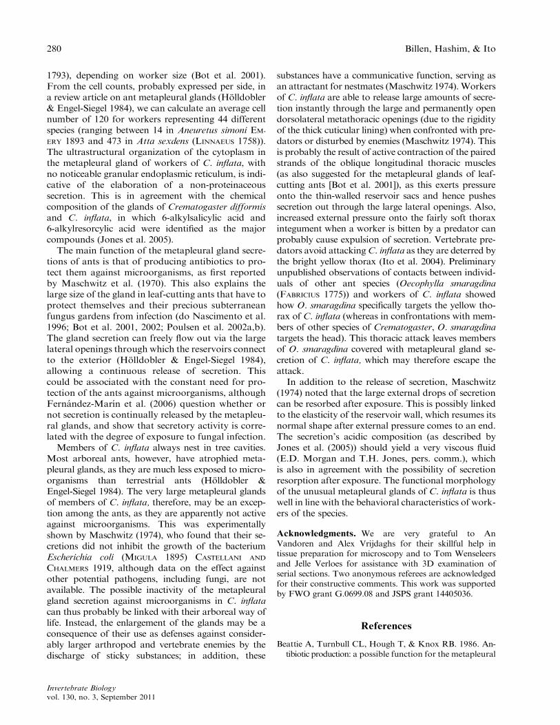
Fig. 1. Metapleural glands of workers of Crematogaster inflata. A. Scanning electron micrograph of the thorax andabdomen of a worker of C. inflata, showing the position of the large opening of the metapleural gland (MO). The dashed
line indicates the internal extent of the metapleural gland. B. Detail of a metapleural gland opening. C. Semithin crosssection through the posterior part of a metapleural gland showing the large lateral opening surrounded by a thickenedcuticle. D. Scanning electron micrograph of KOH-treated half thorax (for removal of soft tissues), showing a large
metapleural gland reservoir (R) and thoracic muscles (TM). E. Semithin longitudinal section near the body midline,showing clustering of secretory cells at the anterior side of the reservoir. F. Semithin cross section through the mesothoraxin the anterior region of the metapleural gland, with massive clusters of secretory cells near the side of reservoirs closest to
the body midline. G.Detail of secretory cells and reservoir; arrows indicate clustered duct cells.H. Transmission electronmicrograph showing detail of the secretory cell cytoplasm, with numerous mitochondria (M) and an end apparatusformed by a cuticular ductule (ct) surrounded by microvilli (mv). fc, foreleg coxa; hc, hindleg coxa; mc, midleg coxa; MO,metapleural gland opening; MS, metathoracic spiracle; N, nucleus; oe, oesophagus; R, reservoir; SC, secretory cells; SG,
salivary gland; TG, thoracic ganglion; TM, thoracic muscles.
Metapleural glands of Crematogaster inflata 279
Invertebrate Biologyvol. 130, no. 3, September 2011
1793), depending on worker size (Bot et al. 2001).From the cell counts, probably expressed per side, ina review article on ant metapleural glands (Holldobler& Engel-Siegel 1984), we can calculate an average cellnumber of 120 for workers representing 44 differentspecies (ranging between 14 in Aneuretus simoni EM-
ERY 1893 and 473 in Atta sexdens (LINNAEUS 1758)).The ultrastructural organization of the cytoplasm inthe metapleural gland of workers of C. inflata, withno noticeable granular endoplasmic reticulum, is indi-cative of the elaboration of a non-proteinaceoussecretion. This is in agreement with the chemicalcomposition of the glands of Crematogaster difformisand C. inflata, in which 6-alkylsalicylic acid and6-alkylresorcylic acid were identified as the majorcompounds (Jones et al. 2005).
The main function of the metapleural gland secre-tions of ants is that of producing antibiotics to pro-tect them against microorganisms, as first reportedby Maschwitz et al. (1970). This also explains thelarge size of the gland in leaf-cutting ants that have toprotect themselves and their precious subterraneanfungus gardens from infection (do Nascimento et al.1996; Bot et al. 2001, 2002; Poulsen et al. 2002a,b).The gland secretion can freely flow out via the largelateral openings through which the reservoirs connectto the exterior (Holldobler & Engel-Siegel 1984),allowing a continuous release of secretion. Thiscould be associated with the constant need for pro-tection of the ants against microorganisms, althoughFernandez-Marın et al. (2006) question whether ornot secretion is continually released by the metapleu-ral glands, and show that secretory activity is corre-lated with the degree of exposure to fungal infection.
Members of C. inflata always nest in tree cavities.Most arboreal ants, however, have atrophied meta-pleural glands, as they are much less exposed to micro-organisms than terrestrial ants (Holldobler &Engel-Siegel 1984). The very large metapleural glandsof members of C. inflata, therefore, may be an excep-tion among the ants, as they are apparently not activeagainst microorganisms. This was experimentallyshown by Maschwitz (1974), who found that their se-cretions did not inhibit the growth of the bacteriumEscherichia coli (MIGULA 1895) CASTELLANI AND
CHALMERS 1919, although data on the effect againstother potential pathogens, including fungi, are notavailable. The possible inactivity of the metapleuralgland secretion against microorganisms in C. inflatacan thus probably be linked with their arboreal way oflife. Instead, the enlargement of the glands may be aconsequence of their use as defenses against consider-ably larger arthropod and vertebrate enemies by thedischarge of sticky substances; in addition, these
substances have a communicative function, serving asan attractant for nestmates (Maschwitz 1974).Workersof C. inflata are able to release large amounts of secre-tion instantly through the large and permanently opendorsolateral metathoracic openings (due to the rigidityof the thick cuticular lining) when confronted with pre-dators or disturbed by enemies (Maschwitz 1974). Thisis probably the result of active contraction of the pairedstrands of the oblique longitudinal thoracic muscles(as also suggested for the metapleural glands of leaf-cutting ants [Bot et al. 2001]), as this exerts pressureonto the thin-walled reservoir sacs and hence pushessecretion out through the large lateral openings. Also,increased external pressure onto the fairly soft thoraxintegument when a worker is bitten by a predator canprobably cause expulsion of secretion. Vertebrate pre-dators avoid attackingC. inflata as they are deterred bythe bright yellow thorax (Ito et al. 2004). Preliminaryunpublished observations of contacts between individ-uals of other ant species (Oecophylla smaragdina(FABRICIUS 1775)) and workers of C. inflata showedhow O. smaragdina specifically targets the yellow tho-rax of C. inflata (whereas in confrontations with mem-bers of other species of Crematogaster, O. smaragdinatargets the head). This thoracic attack leaves membersof O. smaragdina covered with metapleural gland se-cretion of C. inflata, which may therefore escape theattack.
In addition to the release of secretion, Maschwitz(1974) noted that the large external drops of secretioncan be resorbed after exposure. This is possibly linkedto the elasticity of the reservoir wall, which resumes itsnormal shape after external pressure comes to an end.The secretion’s acidic composition (as described byJones et al. (2005)) should yield a very viscous fluid(E.D. Morgan and T.H. Jones, pers. comm.), whichis also in agreement with the possibility of secretionresorption after exposure. The functional morphologyof the unusual metapleural glands of C. inflata is thuswell in line with the behavioral characteristics of work-ers of the species.
Acknowledgments. We are very grateful to AnVandoren and Alex Vrijdaghs for their skillful help intissue preparation for microscopy and to Tom Wenseleersand Jelle Verloes for assistance with 3D examination ofserial sections. Two anonymous referees are acknowledgedfor their constructive comments. This work was supportedby FWO grant G.0699.08 and JSPS grant 14405036.
References
Beattie A, Turnbull CL, Hough T, & Knox RB. 1986. An-tibiotic production: a possible function for the metapleural
280 Billen, Hashim, & Ito
Invertebrate Biologyvol. 130, no. 3, September 2011

glands of ants (Hymenoptera: Formicidae). Ann. En-tomol. Soc. Am. 79: 448–450.
Bot ANM, Obermayer ML, Holldobler B, & Boomsma JJ.
2001. Functional morphology of the metapleural glandin the leaf-cutting ant Acromyrmex octospinosus. Insect.Soc. 48: 63–66.
Bot ANM, Ortius-Lechner D, Finster K, Maile R, &
Boomsma JJ. 2002. Variable sensitivity of fungi and bac-teria to compounds produced by the metapleural glandsof leaf-cutting ants. Insect. Soc. 49: 363–370.
Fanfani A & Giovannotti M. 1994. Metapleural glands inCrematogaster clariventris Mayr and C. depressa Latr.(Formicidae, Myrmicinae). Accad. Naz. Lincei. 267:
259–265.Fernandez-Marın H, Zimmerman JK, Rehner SA, & Wci-
slo WT. 2006. Active use of the metapleural glands by
ants in controlling fungal infection. Proc. R. Soc. B 273:1689–1695.
Holldobler B & Engel-Siegel H. 1984. On the metapleuralgland of ants. Psyche 91: 201–224.
Hosoishi S & Ogata K. 2009. A taxonomic revision of theAsian endemic subgenus Physocrema of the genusCrematogaster (Hymenoptera Formicidae). Zootaxa
2062: 15–36.Ito F, Hashim R., Sze Huei Y, Kaufmann E, Akino T, &
Billen J. 2004. Spectacular Batesian mimicry in ants.
Naturwissenschaften 91: 481–484.Jones TH, Brunner SR, Edwards AA, Davidson DW, &
Snelling RR. 2005. 6-Alkylsalicylic acids and 6-alkylre-sorcylic acids from ants in the genus Crematogaster from
Brunei. J. Chem. Ecol. 31: 407–417.Maruyama M, Yek SH, Hashim B, & Ito F. 2003. A new
myrmecophilous species of Drusilla (Coleoptera,
Staphylinidae, Aleocharinae) from Peninsular Malaysia,as possible Batesian mimic associated with Cremato-gaster inflata (Hymenoptera, Formicidae, Myrmicinae).
Jpn. J. Syst. Entomol. 9: 267–275.Maschwitz U. 1974. Vergleichende Untersuchungen zur
Funktion der Ameisenmetathorakaldruse. Oecologie16: 303–310.
Maschwitz U, Koob K, & Schildknecht H. 1970. EinBeitrag zur Funktion der Metathorakaldruse der Amei-sen. J. Insect Physiol. 16: 387–404.
do Nascimento RR, Schoeters E, Morgan ED, Billen J, &Stradling DJ. 1996. Chemistry of metapleural gland se-cretions of three attine ants, Atta sexdens rubropilosa,
Atta cephalotes, and Acromyrmex octospinosus (Hymen-optera: Formicidae). J. Chem. Ecol. 22: 987–1000.
Noirot C & Quennedey A. 1974. Fine structure of insect
epidermal glands. Annu. Rev. Entomol. 19: 61–80.Poulsen M, Bot ANM, Currie CR, & Boomsma JJ. 2002a.
Mutualistic bacteria and a possible trade-off betweenalternative defence mechanisms in Acromyrmex leaf-
cutting ants. Insect. Soc. 49: 15–19.Poulsen M, Bot ANM, Nielsen MG, & Boomsma JJ.
2002b. Experimental evidence for the cost and hygienic
significance of the antibiotic metapleural gland secretionin leaf-cutting ants. Behav. Ecol. Sociobiol. 52: 151–157.
Schildknecht H & Koob K. 1970. Pflanzliche Bioregula-
toren als Inhaltsstoffe der Metathorakaldrusen derKnotenameisen (Myrmicinae). Angew. Chem. 82: 181.
FFF 1971. Myrmicacin: das erste Insekten-Herbicid.Angew. Chem. 83: 110.
Schoeters E & Billen J. 1993. Anatomy and fine structure ofthe metapleural gland in Atta (Hymenoptera, Formic-idae). Belg. J. Zool. 123: 19–27.
Metapleural glands of Crematogaster inflata 281
Invertebrate Biologyvol. 130, no. 3, September 2011