Embed Size (px)
Citation preview

Available online at www.sciencedirect.com
www.elsevier.com/locate/brainres
b r a i n r e s e a r c h 1 5 3 0 ( 2 0 1 3 ) 3 2 – 4 3
0006-8993/$ - see frohttp://dx.doi.org/10.
nCorresponding autE-mail addresses:
Research Report
Functional brain organization of preparatoryattentional control in visual search
Patrick Bourkea,n, Steven Brownb, Elton Nganc, Mario Liottib
aDepartment of Psychology, University of Lincoln, Brayford Pool, Lincoln LN6 7TS, United KingdombDepartment of Psychology, Simon Fraser University, Burnaby, CanadacDepartment of Psychology, University of British Columbia, Vancouver, Canada
a r t i c l e i n f o
Article history:
Accepted 19 July 2013
Looking for an object that may be present in a cluttered visual display requires an
advanced specification of that object to be created and then matched against the incoming
Available online 26 July 2013
Keywords:
Visual-search
Template
Preparatory
Representation
fMRI
Sensory-motor
nt matter & 2013 Elsevie1016/j.brainres.2013.07.03
hor. Fax: +44 1522 [email protected],
a b s t r a c t
visual input. Here, fast event-related fMRI was used to identify the brain networks that are
active when preparing to search for a visual target. By isolating the preparation phase of
the task it has been possible to show that for an identical stimulus, different patterns of
cortical activation occur depending on whether participants anticipate a ‘feature’ or a
‘conjunction’ search task. When anticipating a conjunction search task, there was more
robust activation in ventral occipital areas, new activity in the transverse occipital sulci
and right posterior intraparietal sulcus. In addition, preparing for either type of search
activated ventral striatum and lateral cerebellum. These results suggest that when
participants anticipate a demanding search task, they develop a different advanced
representation of a visually identical target stimulus compared to when they anticipate
a nondemanding search.
& 2013 Elsevier B.V. All rights reserved.
r B.V. All rights reserved.2
[email protected] (P. Bourke).
1. Introduction
In a complex visual scene the object to which we attend is notalways the most intrinsically salient e.g. the brightest orlargest. Rather the things to which we attend are more oftenthose that are relevant to our current goals and interests. Forexample, we can find our keys on the desk amongst theclutter or our car amongst many others in a large car park.These illustrate the general case of having something ofcurrent importance ‘in mind’ and seeking for that precisevisual information in a cluttered visual world. This ability hasbeen empirically studied with ‘Visual search’, an experimen-tal paradigm that simulates these conditions (e.g. Treisman
and Gelade, 1980). In this, participants are asked to decidewhether a ‘target’ such as a specific colored letter is presentor not among a display of many similar items. Despiteaspects of visual search being studied for over 30 years, thecognitive neuroscience of the formation of the advancedrepresentation of the target is poorly understood.
The global network of areas involved in visual search taskshas been well documented in functional imaging studies (e.g.Anderson et al., 2010; Donner et al., 2000; Kim et al., 2012;Leonard et al., 2000; Nobre et al., 2003). The most consistentlyactivated areas include superior parietal cortex, intraparietalsulcus, and occipital cortex along with various parts of frontalcortex (Anderson et al., 2007). However, understanding what

b r a i n r e s e a r c h 1 5 3 0 ( 2 0 1 3 ) 3 2 – 4 3 33
aspect of visual search is performed by which part of thisnetwork of regions remains undetermined. Specifically, thedesign of these earlier studies have not allowed the ‘prepareto search’ and the ‘search’ element of the task to be sepa-rated. In the first, people have to develop and maintain someadequate representation of the item to be found and in thesecond they have to match incoming stimuli against this.
From other lines of work, reasonable expectations can beformed as to the brain areas that may be involved in preparingto search for a target. A short term description of currentlyrelevant visual information is often thought to be implemen-ted by biasing feature maps in extra-striate regions of occipitalcortex (Desimone and Duncan, 1995). Consistent with this,sustained activation over the posterior scalp has been shownas people hold representations of targets for which they areabout to search (Carlisle et al., 2011). In the human brain,feature maps for shapes and colors seem to exist in ventraloccipital cortex (Beauchamp et al., 1999; Corbetta et al., 1990,1991; Shulman et al., 1999, 2003). Furthermore, it has beendemonstrated that such maps are activated when people arepreparing to process visual input to find a target (Chawla et al.,1999; Giesbrecht et al., 2003). For example, Giesbrecht et al.(2003) showed increased bilateral activation in the fusiformregion when people are waiting to make an orientationdecision about a soon to be displayed colored, rectangle. It isclear that in visual search some similar advanced specificationof the target must be formed and ‘held in mind’ prior to anysearch. The task must proceed by comparing multiple items ina visual display against this representation. It seems likelytherefore that in visual search, the advanced specification ofthe target will also be found to be implemented here.
Importantly, as human cognition is highly flexible it seemslikely that the advance specification of target identity will varywith the current task demands. A target could be identical in twosearch tasks, but its advanced representation is predicted to besimpler when the upcoming task is expected to be undemandingcompared to when it is expected to be demanding. For examplethe pre-biasing that might occur when preparing to find a red Xis likely to be different when the task is expected to be a ‘feature’search i.e. all distracters will be green Os, relative to when thetask is expected to be a ‘conjunction’ search i.e. distracters willbe green Xs and red Os. In the first, a simple representation willsuffice to perform the task. This could be implemented neuro-logically by the detection of any activation in feature mapscoding other than green or O or by activation in red or diagonalfeature maps. In contrast when the distracters will be green Xsand red Os, a more elaborated representation of the targetincluding its relationship with the distracters must be formed(Duncan and Humphreys, 1989). If so, it is likely that for theidentical stimulus, when preparing for such an undemandingfeature search there will be less neural activation than whenpreparing for a demanding conjunction search. This may bedetected as a smaller fMRI signal.
In addition to the variable activity in feature maps that mightbe expected to be seen in occipital cortex, the ‘preparing tosearch’ phase of a visual search task is likely to include otherregions that are involved in modulating this sensory activity. Afrontal–parietal control system is often proposed (e.g. Desimoneand Duncan, 1995; Woldorff et al., 2004) that sends bias signals tofeature maps in ventral occipital cortex. Supporting evidence has
come from studies where a representation of a target locationhas to be developed. In this approach a symbolic cue is giventhat indicates the likely location of an upcoming target. Partici-pants use this advanced representation to facilitate targetdetection when it occurs (Hopfinger et al., 2000; Woldorff et al.,2004) or simply attend to that location (Kastner et al., 1999). Forexample Kastner et al. (1999) asked participants to attend to onelocation and count the occurrence of one of four complexcolorful images presented there. In such studies, during this‘attended’ interval, activation is seen in both occipital cortex,consistent with the biasing of visual spatial maps, and frontaland parietal areas, possibly involved in biasing suchmaps. Whilesomewhat variable across studies the frontal activation typicallyincludes the frontal eye fields. In visual search studies frontaland parietal activation is also often reported, however it is neverclear whether this reflects the source of the bias signal or theattentional movements that are part of later search and matchoperations. In contrast to the frontal areas that are active invisual search which vary across studies, the parietal activation ishighly consistent. An area near the posterior portion of theintraparietal sulcus is active (Donner et al., 2000; Leonards et al.,2000; Nobre et al., 2003). In addition, Shulman et al. (1999)reported increased activation here when participants weremaintaining information during an interval, regarding move-ment direction. Similarly, Giesbrecht et al. (2003) identified aregion that includes a similar parietal area as responsible for therepresentation of task relevant information concerning coloredshapes and location. This area has also been reported to beinvolved in other visual short term memory tasks (McNab andKlingberg, 2007; Todd and Marois, 2004). It seems likely that thisarea may be involved in maintaining the advanced specificationof the target during ‘prepare to search’ as part of a frontal–parietal control system (Desimone and Duncan, 1995).
No study has explicitly isolated the network of areas thatsupport the development and maintenance of an advancedrepresentation of the target in visual search from the othercomponents of the task. Therefore, no study has been able toexplore whether neural activation when preparing to searchfor a target, differs as a function of the anticipated demand ofthe task. The present study aims to address these limitationsby separating the brain activation during the preparation tosearch for a target, from the later components of a visualsearch task. By isolating this time period, the changes inneural activity that might underlie the flexible creation ofadvanced specifications is investigated. This is done bypresenting participants with identical targets but in contextsthat indicate that their search will be undemanding ordemanding, i.e. a feature search or a classic conjunctionsearch. To minimize the interpretative processes that sym-bolic indication of the current target and distracter informa-tion would have produced, spatial, shape and colorinformation is given in a very concrete way, see Fig. 1.
2. Results
2.1. Reaction time results
Reaction time results are shown in Fig. 2. There was a maineffect of the ‘type of search’ factor with feature search being

Fig. 2 – Reaction time of correct responses when the target ispresent and when it is absent in feature search andconjunction search conditions. Vertical lines show thestandard error of means.
Fig. 1 – Sequence of screens in a trial (top–bottom) with percent of different trial types. A conjunction search trial is illustrated.In feature search the target shared no features with the distracters.
b r a i n r e s e a r c h 1 5 3 0 ( 2 0 1 3 ) 3 2 – 4 334
faster than conjunction search, F(1, 15)¼110.29, po.001.There was a main effect of the ‘presence of the target’ factorwith ‘target present’ being faster than ‘target absent’,F(1, 15)¼22.47, po.001. There was a significant interactionbetween the two factors, F(1, 15)¼22.80, po.001. As shown inFig. 2, there was little lengthening of reaction time when thetarget was absent in feature search but a substantial increase
when it was absent in conjunction search. Error rates werelow in feature search (1.37%) and higher in conjunctionsearch (13.82%). Any trial with an incorrect response wasexcluded from the subsequent fMRI analysis.
2.2. Results – fMRI (preparation)
2.2.1. Effects of preparation for a feature search – “AttendPrepare” (feature) versus “Watch” (feature)Full Talairach coordinates are given in Tables 1 and 2. Preparingto perform a ‘feature’ search resulted in four clusters of sig-nificant BOLD activity in extrastriate visual cortex, ventrally inright inferior occipital gyrus and fusiform gyrus BA19, moredorsally in right middle occipital gyrus BA18, and more poster-iorly and medially in the left lingual gyrus and right cuneus (BA17/18) in the occipital pole [see Fig. 3, top row].
Furthermore, there were significant clusters of BOLDactivity in left ventral Striatum and adjacent anterior insula,left posterior thalamus, right anterior lateral cerebellum, andin the pons.
2.2.2. Effects of preparation for a conjunction search – “AttendPrepare” (conjunction) versus “Watch” (conjunction)Results are shown in Tables 1 and 2, Fig. 3 middle row.Preparing to perform a conjunction search yielded significant

b r a i n r e s e a r c h 1 5 3 0 ( 2 0 1 3 ) 3 2 – 4 3 35
BOLD activity in extrastriate visual cortex, including bilateralinferior occipital gyri and fusiform gyri BA19 (ventrally), andmore dorsally, bilateral middle occipital gyri BA18/19, in thevicinity of the transverse occipital sulcus (TOS). Additionally,there was significant BOLD activity in right superior parietallobe (BA7), in the proximity of the intraparietal sulcus, [seeFig. 3, middle row]. Finally, there were clusters in right poster-ior thalamus, bilateral ventral striatum, bilateral anterior lat-eral cerebellum, midline cerebellum and pons (Table 2).
2.2.3. Selective effects of preparing for a feature searchcompared to preparing for a conjunction search – “AttendPrepare” (conjunction) versus “Attend Prepare” (feature)Regions where BOLD activity was greater when preparing for aconjunction relative to a feature search were bilateral ventraloccipital cortex (inferior occipital/fusiform gyri BA19), bilateraldorsal occipital cortex (middle occipital gyri BA18/19, in theproximity of TOS), and right superior parietal lobule BA7, inthe vicinity of IPS [see Fig. 3, bottom row]. Please note thatBOLD effects around the right intraparietal sulcus and indorsal occipital cortex coincided across the two contrasts of“Attend Prepare” (conjunction) versus “Watch” (conjunction)and “Attend Prepare” (conjunction) versus “Attend Prepare”(feature) (Fig. 3, bottom and middle rows and Table 1).
3. Discussion
Fast event related fMRI was used to isolate the brain networksthat are active during preparing to search for a visual targetfrom the later components of a visual search task. The resultsshow a distinctive network activated during the preparationphase of the task. Of particular interest was the way in whichthe representation of a given target would vary in anticipationof a ‘feature’ relative to a ‘conjunction’ search task. In prepar-ing for either type of search, ventral occipital areas wereactivated, notably to a greater extent in advance of theconjunction search. In addition, when preparing for a conjunc-tion search, unique activity was seen in bilateral dorsaloccipital cortex and in the vicinity of the right intraparietalsulcus (Fig. 3). Finally, preparing for either type of searchactivated ventral striatum, cerebellum, thalamus and pons.
3.1. Varying representations with anticipated taskdemand
Activations seen in the ventral occipital region are consistentwith the idea that an advance specification of the target isimplemented by biasing feature maps in extra-striate regionsof occipital cortex (Chawla et al., 1999; Desimone andDuncan, 1995; Giesbrecht et al., 2003; Stokes et al., 2009). Thisportion of extrastriate visual cortex is similar to that which,in previous studies, has been argued to encode both shapeand color information (e.g. Beauchamp et al., 1999; Shulmanet al., 2003, 1999). Significantly, preparing to search for theidentical target, but in the context of expecting it to beamongst very similar distracters rather than very differentdistracters produced different results. When expecting aconjunction search task, the same ventral occipital areawas activated as when expecting a feature search task, but
more robustly. This would be consistent with an interpreta-tion that activity in this area reflects the formation of anadvanced specification of the target and that a more exten-sive representation is formed when participants anticipate amore demanding visual search. For example, when an easyfeature search is expected, color or shape maps may beactivated, when a demanding conjunction search is expectedcolor and shape maps or more complex representations maybe activated.
Target and distracter relationships may be establishedoutside of the dorsal occipital region followed by signals sentback to bias early processing. However, at least part of thedevelopment of the advanced representation may arisedirectly out of local comparisons of target and distracteritems during the ‘preparation’ display. During this phase ofthe task the ‘target’ needs to be compared to the distractersso as to prepare for the upcoming search task. In the secondpart of the task similar comparisons are made in the ‘search’display. This repeated local comparison of items is similar tothe local comparison of display items that is intrinsic in theinter-trial priming procedure (Müller et al., 1995; Found andMüller, 1996; Pollmann et al., 2000). Processing on one trialalters the state of the perceptual system for the next searchtrial. For example, if a search is performed in the colordimension, that dimension is altered so that it is processedfaster on the next trial. This type of activity is thought toproduce biasing or weighting of perceptual dimensions e.g.color (Found and Müller, 1996). Similar speeding effects arefound when conjunction searches are repeated (Weidneret al., 2002), driven mainly by repeating the distractors insuccessive trials (Kristjánsson et al., 2002; Geyer et al., 2006).We might therefore suspect that similar bottom-up biasingdevelops and stays active during the ‘preparation’ stage ofthe current task thus forming part of the preparatory set.
While, something akin to searching the display must haveoccurred i.e. comparison of target and distracters, thisappears to have been done without overt eye movements.The fMRI data show no evidence of frontal eye field activationduring this part of the task. As increased activity in thisregion is reported to be a consistent finding in humanneuroimaging studies (McDowell et al., 2008), the lack of itis indicative of the task being done without eye-movement.While sub-threshold activations cannot be excluded, giventhe clear instructions, that participants were practice ondoing the task without eye movement outside the scannerand reported being able to do so, it seems probable thatparticipants were largely successfully in following theinstructions to fixate the central box
The idea of biased feature maps is consistent with a broadrange of previous work it does not however fit well with theresults of McMains et al., 2007. They demonstrated that therewas a general increase in neural activity when preparing for atarget event. However, this was non-specific e.g. in brain areasconsidered specialized for color processing, preparing for acolor stimulus produced equal activation as preparing for amovement stimulus. This discrepancy could be understood ina number of ways. A key feature of the current study is that thetarget changed on every trial. This contrasts with the blockdesign used in McMains et al. (2007) in which the same targetidentity is used over 18 s blocks of stimuli. It may be that the

Table 1 – Talairach coordinates of occipital and parietal areas activated during ‘preparation to search’.
Conjunction prepare versus conjunction watch Feature prepare versus feature watch Conjunction prepare versus feature prepare
BA x y z cl-s T x y z cl-s T x y z cl-s T
Ventral occipitalR IOG/GF19 31 �71 �7 126 11.6 35 �79 �9 116 14.9 25 �80 0 197 9.7
25 �78 0 9.7 41 �68 �7 12.2 – – –
33 �84 1 9.7 33 �71 �7 9.5 – – –
L IOG/GF19 �29 �62 �14 146 10.7 – – – �26 �80 �9 150 6.0�42 �74 �9 10.5 – – – – – –
Dorsal occipitalL GOm 18 (TOS) �32 �85 16 40 10.5 – – – – – �30 �86 13 303 6.7
�28 �79 19 6.2R GOm 19 (TOS) 31 �82 23 15 9.8 – – – – – 33 �84 23 82 7.6
Occipital poleR GOm 18 – – – 35 �92 11 30 10.8 – – –
L Ling 18 – – – �22 �89 �5 14 10.1 – – –
R Cu 17/18 – – – 12 �97 1 45 10.1 – – –
– – – 16 �100 8 9.1 – – –
ParietalR LPs7 (IPS) 29 �62 40 74 11.1 – – – 27 �56 45 73 6.1
25 �64 47 10.2
Average coordinates of 15 participants. Values are peak coordinates (mm in Talairach space). Preparation for a conjunction search and preparation for a feature search are contrasted to their passiveviewing controls (left and center) and to each other (right). Areas in which peak activations occur are labeled by lobe/gyrus/sulcus and followed by a number indicating the corresponding Brodmannarea. Abbreviations: R, right; L, left; IOG/GF19, inferior occipital gyrus/fusiform gyrus; GOm 18 (TOS), middle occipital gyrus (transverse occipital sulcus); Ling18, lingual gyrus; Cu17/18, Cuneus; LPs7(IPS), superior parietal lobule (intraparietal sulcus); cl-s, cluster size.
brain
research
1530
(2013)32–43
36
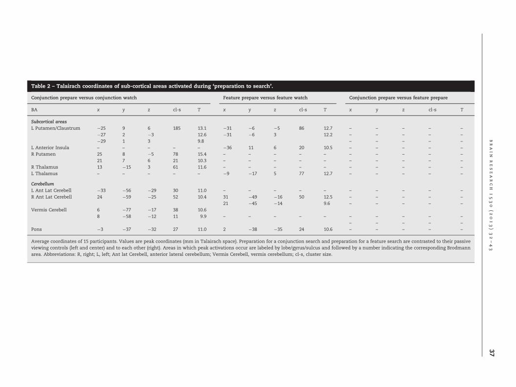
Table 2 – Talairach coordinates of sub-cortical areas activated during ‘preparation to search’.
Conjunction prepare versus conjunction watch Feature prepare versus feature watch Conjunction prepare versus feature prepare
BA x y z cl-s T x y z cl-s T x y z cl-s T
Subcortical areasL Putamen/Claustrum �25 9 6 185 13.1 �31 �6 �5 86 12.7 – – – – –
�27 2 �3 12.6 �31 �6 3 12.2 – – – – –
�29 1 3 9.8 – – – – –
L Anterior Insula – – – – – �36 11 6 20 10.5 – – – – –
R Putamen 25 8 �5 78 15.4 – – – – – – – – – –
21 7 6 21 10.3 – – – – – – – – – –
R Thalamus 13 �15 3 61 11.6 – – – – – – – – – –
L Thalamus – – – – – �9 �17 5 77 12.7 – – – – –
CerebellumL Ant Lat Cerebell �33 �56 �29 30 11.0 – – – – – – – – – –
R Ant Lat Cerebell 24 �59 �25 52 10.4 31 �49 �16 50 12.5 – – – – –
21 �45 �14 9.6 – – – – –
Vermis Cerebell 6 �77 �17 38 10.68 �58 �12 11 9.9 – – – – – – – – – –
– – – – –
Pons �3 �37 �32 27 11.0 2 �38 �35 24 10.6 – – – – –
Average coordinates of 15 participants. Values are peak coordinates (mm in Talairach space). Preparation for a conjunction search and preparation for a feature search are contrasted to their passiveviewing controls (left and center) and to each other (right). Areas in which peak activations occur are labeled by lobe/gyrus/sulcus and followed by a number indicating the corresponding Brodmannarea. Abbreviations: R, right; L, left; Ant lat Cerebell, anterior lateral cerebellum; Vermis Cerebell, vermis cerebellum; cl-s, cluster size.
brain
research
1530
(2013)32–43
37

Fig. 3 – Group activation map (15 subjects), superimposed on one subject's rendered brain during ‘preparing to search’.Superior view, left lateral, right lateral and posterior views are shown. Top row, activations produced by preparing to searchfor a feature (Att_Prep_Feat) relative to watching the control display (Watch_Feat). Middle row, activations produced bypreparing to search for a conjunction (Att_Prep_Conj) relative to watching the control display (Watch_Conj). Bottom row,preparing to search for a conjunction compared to preparing to search for a feature. Abbreviations: R, right; L, left; GL, lingualgyrus; Fus, fusiform; IPS, intraparietal sulcus; TOS, transverse occipital sulcus. Note: Top and middle row used FWE correctedcontrasts. Bottom row uses FDR corrected contrasts and a different scale.
b r a i n r e s e a r c h 1 5 3 0 ( 2 0 1 3 ) 3 2 – 4 338
constant need to establish a new target representation createsa level of activation not seen when a single representation isformed and held. Alternatively, the results of Giesbrecht et al.,2003 indicate that somewhat different results are to beexpected when stimuli are presented centrally (as in thecurrent study) or more peripherally (as in McMains et al.(2007)). They found that target specific preparatory activity
(color or location) was seen with central presentations but thisis less clearly the case with more peripheral presentations.
In addition to the ventral occipital areas, activity wasobserved in the vicinity of the transverse occipital sulci (TOS)when the upcoming visual search task was expected to bedemanding. TOS may hence have a similar role as the ventraloccipital regions in target representation. Alternatively, the

b r a i n r e s e a r c h 1 5 3 0 ( 2 0 1 3 ) 3 2 – 4 3 39
transverse occipital sulci may contribute to enhanced targetrepresentation by the suppression of distracters (Wokciulikand Kanwisher, 1999). Such an explanation seems particularlylikely given the current design. Here for reasons of experi-mental control of visual saliency, during the preparation phasethe target is always shown surrounded by distracters andwhen the upcoming task will be a conjunction search thosedistracters are always very similar to the target. Furthermore,the suppression of distracters has been shown to be a highlyefficient way of biasing the perceptual system so that targets‘pop-out’. This is true for both feature and conjunctionsearches. One way in which a color-form conjunction searchcould be done would be to by inhibiting one color and thensearching within the target color for a unique shape (e.g.Treisman and Sato, 1990). For example when the target is ared O among red Xs and blue Os, the color (blue) could besupressed and the search done on the shape dimension. Inthis case the unique rounded shape will pop-out from thestraight oriented lines of the X distractors. Importantly, whenthis is used in one trial, it carries over to the next trial (Geyeret al., 2010), i.e. the inhibition remains active. In response tothe current ‘prepare screen’ it seems likely that inhibition isdeveloped in separating the target from distractors and main-tained beyond the end of the prepare phase where it activelybiases search during the latter part of the task. If so part of theactivation we see, perhaps in the transverse occipital sulcimay be due to active inhibition of one of the colors or shapemaps. Similarly, relevant dimension weighting would beestablished during the preparation phase of ‘feature’ search.The features used were highly efficient ‘redundantly definedtargets’ dissimilar from the surrounding distractors in bothcolor and shape. Search for such targets could be speeded bythe active suppression of the non-target features,(Krummenacher et al., 2001, 2002). Neural activity reflectingthe development and maintenance of such suppression wouldbe active during the ‘preparation’ stage.
It is worth noting that while distracter displays areidentical in the Attend Prepare” versus “Watch” analysesthey are not in the comparison of the two preparationconditions. This may contribute to the more robust activa-tions seen in the latter analysis.
3.2. The posterior brain system
In addition to the occipital activations, a single right later-alized intraparietal activation was seen when preparing toperform a ‘conjunction’ search task, consistent with a largebody of literature reporting activations in foci along thelength of the intraparietal sulcus in similar tasks (e.g.Donner et al., 2000; Leonard et al., 2000). The current resultsrestrict the IPS activation during preparation to a single focus.This focus (Fig. 3 and Table 1), corresponds closely to thatdescribed by Donner et al. (2000) as AIP (anterior intraparietal)and by Leonards et al. (2000) as MIPS (medial intraparietal). Itis also close to the location identified by Nobre et al. (2003) asinvolved in the overall demand of a search task.
Its role in preparing to search needs to be considered inthe context of the lack of evidence of frontal activation duringthis part of the task. Until now it was never clear whether thefrontal activations that were seen in earlier visual search
studies reflected the representation of the target for whichpeople were searching or some other aspect of the task.The current results provide an answer to this – at least for theconcrete stimuli used here (i.e. ‘target to be searched for’ wasindicated by a visually identical stimulus to the ‘targetpresented’). Only posterior cortex and sub-cortical brain areasare found to be active when representing the target andpreparing to respond to it in an up-coming visual searchdisplay. No evidence of frontal activation was seen. While anegative result, and therefore difficult to interpret, this wouldbe consistent with the view that the biasing of perceptualmaps arises through local comparison of items in the preparedisplay and not through top-down control. It is possible thatfrontal cortex may have been engaged while learning therequirements of the task (during the instructions or thepractice phase). However, for performance in the scannerthe current results indicate that all aspects of the preparationto search are accomplished without frontal cortex involve-ment. This is consistent with a growing body of work thatshows no evidence of frontal involvement in the building andmaintenance of specific short-term representations of visualtargets. For example Shulman et al. (1999) found no frontalactivation while participants prepared to detect a specificdirection of motion in a visual display. The results alsodovetail with recent Event Related Potential (ERP) studiesshowing sustained activation over posterior scalp whenpeople are maintaining a template of an item for which theyare about to search (Carlisle et al., 2011). More broadly, it hasbeen argued from recent neuroimaging work on visual work-ing memory (see Postle, 2006) that frontal areas only becomeinvolved when transformation rather than memory per se, isrequired. While true for visual features such as motion, shapeand color, an exception seems to be the advanced represen-tation of visual stimuli at specific spatial locations. This hasbeen robustly shown to activate frontal areas (e.g., Kastneret al., 1999; Woldorff et al., 2004) and may relate to the closeconnection between visual spatial attention and motor plan-ning (Deubel and Schneider, 1996). The current results wouldbolster the position that apart from preparing to detecttargets at a specific location, the advanced representation oftarget features is achieved outside of frontal cortex.
Theoretically this result is important because it is oftenassumed that in visual search a signal is being sent fromcortical regions outside of the visual areas to bias featuremaps in an appropriate and flexible way (Desimone andDuncan, 1995). However, for the concrete visual stimuli usedhere, the traditional assumption that frontal–parietal net-works are necessarily involved in forming and maintaining arepresentation of a target is not supported. Given this, analternative explanation for the role of the intra-parietalsulcus activation in preparing to search is required. While itis possible that the intra-parietal area by itself is involved inmaintaining the advanced specification of the target during‘prepare to search’ and sending bias signals to early visualcortex, this seems increasingly unlikely. Recent variants ofthe Todd and Marois (2004) visual short-term memory para-digm suggest that IPS activity is more related to the variousattentional demands of tasks rather than any specific coding(Magen et al., 2009; Mitchell and Cusack, 2008). Magen et al.(2009) argue that attentional demands increase once the

b r a i n r e s e a r c h 1 5 3 0 ( 2 0 1 3 ) 3 2 – 4 340
delay interval between the target memory display and theprobe is lengthened so leading to an increase in activation inthe Intra-parietal sulcus. This increased activation is non-specific, being found both in memory for visual information(colors) and spatial information. In the current study atten-tional demands were increased by changing the task frompreparing to search for a feature to preparing to search for aconjunction of two features. It is possible that rather thansending content specific bias signals, the intra-parietal areamay support the ongoing activation of occipital neurons thatare already encoding target and distractor information. Thisattentional allocation might increase when a more complexrepresentation needs to be maintained as in the currentstudy or sustained for a longer period as in Magen et al.(2009). An alternative is that the intra-parietal area may beprimarily receiving the output from the spatially precise andcolor and form specialized occipital neurons, perhaps as partof a process of transforming the visual input into motor spaceas suggested by Ellison et al. (2003). When participantsanticipate a demanding search task a more detailed repre-sentation of the target and distracters may be implementedin these occipital regions. Their output may be what isreflected in increased activation in the intra-parietal sulcus.
3.3. Stimulus response reassignment as targetrepresentations change
Concurrent activity in posterior cortex and striatum stronglysuggest that the advanced representation of any target invisual search may be best considered as a visual-motor ratherthan a solely visual representation. In the present study, theidentity of the target to which people should prepare torespond varied from trial to trial, thus the stimulus-responserepresentation also changed on every trial. In addition, mixedamongst the ‘prepare to search’ trials were ‘watch’ trials whichindicated that no response would be needed, in which case aswitch from the previous stimulus-response representationwould also have to be generated. This may be the processingthat is being reflected as activity in the striatum. Such aninterpretation would be consistent with earlier work e.g. Cooleset al. (2004). They explicitly examined the substrate of visualstimulus-response rule switching in the striatum and otherareas. Participants were cued as to whether to respond to thesame object as in the previous trial or to another object.Significant activation in the ventral striatum was found asparticipants switched between which of the two concrete (i.e.visually identical) objects to respond to. While in Cooles et al.(2004) the analysis was restricted to areas of interest wholebrain analysis of a similar task has supported participation ofthe striatum while also demonstrating cerebellar involvement(Bischoff-Grethe et al., 2002). They explicitly contrasted tem-plate switching and response reassignment. They report rightanterior-lateral cerebellum (lobule VI) activation duringresponse reassignment, similar to that seen in the currentresults.
These studies suggests that in the current visual searchtask with its concrete visual targets, a likely function ofventral striatum and right anterior-lateral cerebellum isresponse reassignment to a visual stimulus, which is com-pleted during the ‘prepare to search’ phase of the task and is
independent of task demand. The involvement of the stria-tum in maintaining a visual-motor template is plausiblegiven that in non-human primates at least, there are sub-stantial input–output connections from higher-order visualareas to the region around the caudate nucleus/putamen andit has been linked to both perception and memory (Levy et al.,1997; Saint-Cyr et al., 1990; Zink et al., 2003).
An alternative role for the putamen is suggested by theresults of McNab and Klingberg (2007). They showed thatincreased activity in the left putamen was seen whenparticipants had to actively ignore yellow colored discs ratherthan treating them as potential targets in a short term visualmemory task. The current task also required ignoring dis-tracters during the preparations stage and a similar functionmay be accomplished by the putamen here.
4. Conclusion
The current study identified a network of brain regionsactivated when preparing to search for a visual targetembedded in a display of distracters. This was done byisolating it from the BOLD signal changes produced by thelater components of the task. Target identity varied from trialto trial, requiring participants to form a new representation ofthe target on each trial. In addition, participants knew inadvance how demanding the search was likely to be on agiven trial. It was hypothesized that for an identical visualtarget, a simpler representation would be formed when theexpected demand of the upcoming search task was low.It was expected that this would lead to a correspondingchange in neural activity. The results show a network ofneural areas activated in the posterior brain and in sub-cortical areas when ‘preparing to search’. Importantly for thehypothesis when preparing to perform a demanding visualsearch task, identical targets produce new and additionalneural activation in occipital and parietal areas. Future workwill need to identify which attentional processes are involvedin producing this pattern of result e.g. inhibition of distractorsor activation of target representations, the relative involve-ment of the identified areas in different attentional processesand the extent of their involvement when the ‘prepare dis-play’ is present relative to activity in the interval before thetarget display. Furthermore, to achieve a full understanding itwill also be necessary to establish the directionality of effectsand the timing of their activation during visual search. Forthe latter, fMRI effective connectivity analysis and methodol-ogies with high spatiotemporal resolution (such as MEG) willbe needed. These limitations notwithstanding, this is the firstfast event-related fMRI study to identify neural correlates ofthe preparatory phase of visual search and their modulationby the anticipated demand of the visual search.
5. Experimental procedures
5.1. Participants
Seventeen participants took part in the study (8 female, oneleft-handed, mean age 28.277.89 years) with normal or

b r a i n r e s e a r c h 1 5 3 0 ( 2 0 1 3 ) 3 2 – 4 3 41
corrected-to-normal vision. None admitted to current or pasthistory of neurological or psychiatric conditions, learningdisabilities, alcohol/substance abuse or current use of pre-scription medications (as ascertained through a medicalhistory checklist). One subject was discarded for not achiev-ing sufficient proficiency in the visual search task during thetraining session (see below), and a second was eliminateddue to technical problems during the MRI session, yielding afinal sample size of 15 subjects. The study was performed inagreement with the regulations of the University of BritishColumbia Behavioural Ethical Board. Participants took part ina behavioral session outside the scanner (45 min), where theyhad a chance to practice the visual search task until theyexceeded a desired level of performance (475% accuracy).This session took place within 2 days prior the fMRI session.
5.2. Task
The task was designed to avoid the order invariant problemand so enable the ‘prepare to search’ phase of a trial to beisolated from the later elements of the visual search task(Ollinger et al., 2001). It involved having to decide whether apre-defined target (a colored letter) was present or notamongst distracters (other colored letters). Visual stimuliwere viewed through a periscopic mirror positioned about10 cm above the eyes of the participants. Throughout all trialsa central outline box was present in the middle of the displayand participants were asked to keep their eyes fixed on thisduring a trial. The full sequence is shown in Fig. 1.
Trials began with the ‘Condition Display’ in which theoutline of the centrally positioned box turned blue or yellow.The color instructed participants to either “Attend” to (outlineof the box turning blue) or simply “Watch” (outline of the boxturning yellow) the upcoming display. This was used it toinform the participants as to whether they could simply watchthe display on the upcoming trial or should prepare them-selves to perform a search task. Activity during the “Watch”condition was later subtracted from activity in the “Attend”conditions in order to control for brain activation caused bysimply viewing rather than actively attending to the displays.
This initial ‘Condition Display’ was followed after 200 msby the onset of a ‘Prepare Display’, which in the “Attend”conditions (“Attend Prepare-Only” trials and “Attend Prepare+Target” trials, see Fig. 1) informed the subject as to thetarget and type of search to prepare for on that trial. The‘Prepare Display’ was comprised of the target for whichparticipants would shortly have to search, shown inside thecentral box, surrounded by the 31 or 32 distracters that couldbe present in the subsequent ‘Search Display’ (see Fig. 1,columns 2 and 3). The equivalent display in the “Watch” trialswas constructed in the same way except that the centralsquare was filled with a ‘#’ symbol. The distracter sets werematched across conditions.
The ‘Prepare Display’ was presented for 800 ms, and wasfollowed by the white central fixation box remaining on thescreen for a further 1000 ms. After this in the “Watch” trialsand “Attend Prepare-Only” trials (Fig. 1, left and centralcolumns), no further stimuli were presented. The centralwhite outline box remained on the screen and trials endedfollowing a variable interval (mean of 1850 ms, pseudo
randomly jittered with a range of 800–2900 ms). In “AttendPrepare+Target” trials however, a ‘Search Display’ followedthe 1000 ms fixation and participants had to decide as quicklyand accurately as possible if the target was present or absentin the display of 32 letters by pressing one of two keys on afiber optic keypad with the index fingers of either hand. In the‘Search Display’ the central box was empty and the desig-nated target could be either present, replacing one of thedistracters that was in the ‘Prepare Display’ (target present50%) or it could be omitted (target absent, 50%). The ‘SearchDisplay’ was shown for 1000 ms and then replaced by ascreen with just the white central box.
The target and distracter stimuli used to make the ‘Pre-pare Displays’ and the ‘Search Displays’, varied from trial totrial. The relationship between the target and the distractersdetermined whether a given visual search trial would be afeature or a conjunction search. In feature search trials, targetand distracters had no feature in common (e.g. a yellow Mamongst blue Ss). In conjunction search trials, as illustratedin Fig. 1, the target and distracters always shared one feature[e.g. a yellow M, amongst yellow Ss (same color) and blue Ms(same shape)]. Equal numbers of feature and conjunctionvisual search trials were included. The same ‘Prepare Dis-plays’ used in “Attend Prepare+Target” trials were used in the“Attend Prepare-Only” trials and in “Watch” control trials (butwith the central target replaced by a ‘#’). To enable theisolation of the BOLD signal produced during the preparephase from that produced by the target search phase, onethird of trials were “Watch” trials, one third were “AttendPrepare-Only” and one third were “Attend Prepare+Target”(see, Ollinger et al., 2001).
There were 3 runs of 196 trials. After every run, feedbackwas given in the form of mean reaction time and the numberof their errors shown in the center of the screen for 30 s.
5.3. Image acquisition
Echo-planar images were collected on a Philips GyroscanIntera 3.0-T scanner, equipped with a 6-channel SENSE coil.Conventional spin-echo T1-weighted sagittal localizers wereused to view head position and to graphically prescribe thefunctional image volumes. Functional image volumes sensi-tive to the blood oxygen-level dependent (BOLD) contrastsignal were collected with a gradient echo sequence (TR/TE2000/30 ms, 901 flip angle, field of view 210�143�240 mm3
(anteroposterior, feet–head, right–left), 3 mm slice thickness,slice gap 1 mm, 36 axial slices).
5.4. Image processing
PAR/REC format data from the 3T Philips system were con-verted to Analyze format using MRIcro (Rorden C: MRIcro.http://www.mricro.com). The converted images were thenanalyzed using SPM5 (Wellcome Institute of Cognitive Neurology, http://www.fil.ion.ucl.ac.uk/spm/) for image reorientation, realignment, normalization into Montreal NeurologicalInstitute space, and smoothing with a Gaussian kernel (8 mmfull width at half maximum) to compensate for inter-subjectanatomical differences and optimize the signal to noise ratio.

b r a i n r e s e a r c h 1 5 3 0 ( 2 0 1 3 ) 3 2 – 4 342
5.5. fMRI: within subjects
Event-related BOLD responses were modeled for the follow-ing trial types: “Watch” (feature), “Watch” (conjunction),“Attend Prepare” (feature) “Attend Prepare” (conjunction)“Attend Target” (feature) and “Attend Target” (conjunction)by the convolution of stimulus-onset vectors for each trialtype with the synthetic hemodynamic response functionprovided in SPM2. The stimulus onset vectors coincided with‘Condition Display’ onset for “Watch” and “Attend Prepare”trials and with ‘Search Display’ onset in “Attend Target”trials. Eight nuisance regressors (six sets of realignmentparameters, and the mean signal from white matter andcerebro-spinal fluid voxels respectively) were included in themodel. The magnitude of the BOLD responses for each trialtype were calculated using the GLM implemented in SPM2.
To evaluate the selective effects of preparing to search fora target relative to passively looking at a display, the follow-ing contrast images were specified: Prepare for a featuresearch – “Attend Prepare” (feature) versus “Watch” (feature),and prepare for a conjunction search – “Attend Prepare”(conjunction) versus “Watch” (conjunction). To evaluate theselective effects of preparing for a feature search compared topreparing for a conjunction search, the contrast “AttendPrepare” (feature) versus “Attend Prepare” (conjunction) wasspecified.
5.6. fMRI: between subjects
Contrast images for each subject were entered into two ran-dom effects analyses. Pair sample t-tests were set up to test thenull hypotheses of no difference between trial types in themean amplitude of the fitted hemodynamic response for anyof these event types. We first applied the more conservativeFWE method for correction of multiple comparisons, t (14)48.71, po.05, cluster-size410. This approach yielded severalsignificant clusters for contrasts involving the lower controlstate (“Watch” trials). However, for the higher level contrast of“Attend Prepare” (feature) versus “Attend Prepare” (conjunc-tion) no clusters reached significance at the .05 level. We thenopted for selecting the more liberal FDR method for multiplecomparison correction, with the statistical threshold set at t(14)44.6, po.05, cluster size420. This approach was indeedsuccessful in yielding significant activation clusters for thiscontrast. Fig. 3 illustrates the main results of these contrasts,highlighting the common regions activated in the contrastsinvolving “Attend Prepare” (conjunction) (middle and bottomrows). All reported coordinates are in Talairach space, follow-ing conversion from Montreal Neurological Institute (MNI)space, using the program mni2tal (Brett et al., 2001).
Acknowledgments
We thank Alan Woodford and Brian Luus for their assistancewith programming and analysis, anonymous reviewers andTim Hodgson for valuable feedback on earlier versions of themanuscript and Vince di Lollo for early discussions. Thiswork was supported by the Royal Society London, Interna-tional Short-term Overseas Visit Grant (2004/R6) and the
University of Lincoln to (PB) and by the Canada Foundationfor Innovation and the National Science and EngineeringResearch Council of Canada to (ML).
r e f e r e n c e s
Anderson, E.J., Mannan, S.K., Husain, M., Rees, G., Sumner, P.,Mort, D.J., McRobbie, D., Kennard, C., 2007. Involvementof prefrontal cortex in visual search. Exp. Brain Res. 180,289–302.
Anderson, E.J., Mannan, S.K., Rees, G., Sumner, P., Kennard, C.,2010. Overlapping functional anatomy for working memoryand visual search. Exp. Brain Res. 200, 91–107.
Beauchamp, M.S., Haxby, J.V., Jennings, J.E., DeYoe, E.A., 1999. AnfMRI version of the Frnsworth–Munsell 100-Hue test revealsmultiple color-selective areas in human ventraloccipitotemporal cortex. Cereb. Cortex 9, 257–263.
Bischoff-Grethe, A., Ivry, R., Grafton, S., 2002. Cerebellarinvolvement in response reassignment rather than attention.J. Neurosci. 22, 546–553.
Brett, M., Leff, A.P., Rorden, C., Ashburner, J., 2001. Spatialnormalization of brain images with focal lesions using costfunction masking. NeuroImage. 14, 486–500.
Carlisle, N., Arita, J.T., Pardo, D., Woodman, G., 2011. Attentionaltemplates in visual working memory. J. Neurosci. 31,9315–9322.
Chawla, D., Rees, G., Friston, J.K., 1999. The physiological basis ofattentional modulation in extrastriate visual areas. Nat.Neurosci., 671–676.
Cooles, R., Clark, L., Robbins, T.W., 2004. Differential responses inhuman striatum and prefrontal cortex to changes in objectand rule relevance. J. Neurosci. 24, 1129–1135.
Corbetta, M., Miezin, F.M., Dobmeyer, S., Shulman, G.L., Petersen, S.E.,1990. Attentional modulation of neural processingof shape, color, and velocity in humans. Science 248, 1556–1559.
Corbetta, M., Miezin, F.M., Dobmeyer, S., Shulman, G.L., Petersen, S.E.,1991. Shape, color and speed: functional anatomy by positronemission tomography. J. Neurosci. 11, 2383–2402.
Desimone, R., Duncan, J., 1995. Neural mechanisms of selectivevisual attention. Annu. Rev. Neurosci. 18, 193–222.
Donner, T.H., Kettermann, A., Diesch, E., Ostendorf, F., Villringer, A.,Brandt, S.A., 2000. Involvement of the frontal eye field andmultiple parietal areas in covert visual selection duringconjunction search. Eur. J. Neurosci. 12, 3407–3414.
Deubel, H., Schneider, W., 1996. Saccade target selection andobject recognition: evidence for a common attentionalmechanism. Vision Res. 36, 1827–1839.
Duncan, J., Humphreys, G.W., 1989. Visual search and stimulussimilarity. Psychol. Rev. 96, 433–458.
Ellison, A., Rushworth, M., Walsh, V., 2003. The parietal cortex invisual search: a visuomotor hypothesis. Suppl. Clin.Neurophysiol. 56, 321–333.
Found, A., Müller, H., 1996. Searching for unknown feature targetson more than one dimension: Investigating a “dimension-weighting” account. Percept. Psychophys. 58, 88–101.
Geyer, T., Müller, H., Krummenacher, J., 2006. Cross-trial primingin visual search for singleton conjunction targets: Role ofrepeated target and distractor features. Percept. Psychophys.68, 736–749.
Geyer, T., Shi, Z., Müller, H., 2010. Contextual cueing inmulticonjunction visual search is dependent on color- andconfiguration-based intertrial contingencies. J. Exp. Psychol.:Hum. Percept. Perform. 36, 515–532.
Giesbrecht, B., Woldorff, M.G., Song, A.W., Mangun, G.R., 2003.Neural mechanisms of top-down control during spatial andfeature attention. NeuroImage 19, 496–512.

b r a i n r e s e a r c h 1 5 3 0 ( 2 0 1 3 ) 3 2 – 4 3 43
Hopfinger, J.B., Buonocore, M., Mangun, G.R., 2000. The neuralmechanisms of top-down attentional control. Nat. Neurosci. 3,284–291.
Kastner, S., Pinsk, M., De Weerd, P., Desimone, R., Ungerleider, L.G., 1999. Increased activity in human visual cortex duringdirected attention in the absence of visual stimulation.Neuron. 22, 751–761.
Kim, K.K., Eliassen, J.C., Lee, S.K., Kang, E., 2012. Functionalneuroanatomy of visual search with differential attentionaldemands: an fMRI study. Brain Res. 1475, 49–61.
Kristjánsson, A., Wang, D., Nakayama, K., 2002. The role ofpriming in conjunctive visual search. Cognition 85, 37–52.
Krummenacher, J., Müller, H., Heller, D., 2001. Visual search fordimensionally redundant pop-out targets: evidence forparallel-coactive processing of dimensions. Pecept.Psychophys. 63, 901–917.
Krummenacher, J., Müller, H., Heller, D., 2002. Visual search fordimensionally redundant pop-out targets: parallel-coactiveprocessing of dimensions is location specific. J. Exp. Psychol.:Hum. Percept. Perform. 28, 1303–1332.
Leonard, U., Sunaert, S., van Hecke, P., Orban, G., 2000. Attentionmechanisms in visual search—an fMRI study. J. Cogn.Neurosci. 12 (Suppl. 2), 61–75.
Levy, R., Friedman, H.R., Davachi, L., Goldman-Rakic, P.S., 1997.Differential activation of the caudate nucleus in primatesperforming spatial and nonspatial working memory tasks.J. Neurosci. 17, 3870–3882.
Magen, H., Emmanouil, T.A., McMains, S.A., Kastner, S., Treisman, A.,2009. Attentional demands predict short-term memory loadresponse in posterior parietal cortex. Neuropsychologia 47,1790–1798.
McDowell, J.E., Dyckman, K.A., Austin, B.P., Clementz, B.A., 2008.Neurophysiology and neuroanatomy of reflexive andvolitional saccades: evidence from studies of humans. BrainCogn. 68, 255–271.
McMains, S.A., Fehd, H.M., Emmanouil, T., Kastner, S., 2007.Mechanisms of feature and space-based attention: responsemodulation and baseline increases. J. Neurophysiol. 98,2110–2121.
McNab, F., Klingberg, T., 2007. Prefrontal cortex and basal gangliacontrol access to working memory. Nat. Neurosci. 11, 103–107.
Mitchell, D., Cusack, R., 2008. Flexible, capacity-limited activity ofposterior parietal cortex in perceptual as well as visual short-term memory tasks. Cereb. Cortex 18, 1788–1798.
Müller, H., Heller, D., Ziegler, J., 1995. Visual search for singletonfeature targets within and across feature dimensions. Percept.Psychophys. 57, 1–17.
Nobre, A., Coull, J., Walsh, V., Frith, C., 2003. Brain activationsduring visual search: contributions of search efficiency versusfeature binding. NeuroImage 18, 91–103.
Ollinger, J.M., Shulman, G.L., Corbetta, M., 2001. Separatingprocesses within a trial in event-related functional MRI. I. Themethod. NeuroImage 13, 210–217.
Ollinger, J.M., Corbetta, M., Shulman, G.L., 2001. Separatingprocesses within a trial in event-related functional MRI. II.Analysis. NeuroImage 13, 218–229.
Pollmann, S., Weidner, R., Müller, H., von Cramon, D., 2000.A fronto-posterior network involved in visual dimensionchanges. J. Cogn. Neurosci. 12, 480–494.
Postle, B.R., 2006. Working memory as an emergent property ofthe mind and brain. Neuroscience 139, 23–38.
Saint-Cyr, J.A., Ungerleider, L.G., Desimone, R., 1990. Organizationof visual cortical inputs to the striatum and subsequentoutputs to the pallido-nigral complex in the monkey. J. Comp.Neurol. 298, 129–156.
Shulman, G.L., Ollinger, J.M., Akbudak, E., Conturo, T.E., Snyder, A.Z.,Petersen, S.F., Corbetta, M., 1999. Areas involved in encoding andapplying directional expectations to moving objects. J. Neurosci.19, 9480–9496.
Shulman, G.L., McAvoy, M.P., Cowan, M.C., Astafiev, S.V., Tansy, A.P.,Avossa, G., Corbetta, M., 2003. Quantitative analysis of attentionand detection signals during visual search. J. Neurophysiol. 90,3384–3397.
Stokes, M., Thompson, R., Nobre, A.C., Duncan, J., 2009. Shape-specific preparatory activity mediates attention to targets inhuman visual cortex. Proc. R. Soc. 106, 19569–19574.
Treisman, A., Gelade, G., 1980. A feature-integration theory ofattention. Cogn. Psychol. 12, 97–136.
Treisman, A., Sato, S., 1990. Conjunction search revisited. J. Exp.Psychol.: Hum. Percept. Perform. 16, 459–478.
Todd, J., Marois, R., 2004. Capacity limit of visual short-termmemory in human posterior parietal cortex. Nature 428,751–754.
Weidner, R., Pollmann, S., Müller, H., von Cramon, D., 2002. Top-down controlled visual dimension weighting: an event-relatedfMRI study. Cereb. Cortex 12, 318–328.
Woldorff, M., Hazlett, C., Fichtenholtz, H., Weissman, D., Dale, A.,Song, A., 2004. Functional parcellation of attentional controlregions of the human brain. J. Cogn. Neurosci. 16, 149–165.
Wojciulik, E., Kanwisher, N., 1999. The generality of parietalinvolvement in visual attention. Neuron 23, 747–764.
Zink, C., Pagnoni, G., Martin, M., Dhamala, M., Berns, G., 2003.Human striatal response to salient non-rewarding stimuli. J.Neurosci. 23, 8092–8097.