Embed Size (px)
Citation preview
Functional anatomy of the central nervous
system. Reticular and limbic systems.
1. Structure and functions of the nervous system
2. External structure of the spinal cord
3. Internal structure of the spinal cord
4. Development of the spinal cord
5. Anomalies of the spinal cord
6. General structure of the brain
7. Structure and functions of the brain component parts
8. Functional systems of the brain:
- reticular formation
- limbic system
Lecturer: PhD, professor Tamara Hacina

• It is the most complex system of the
body
• It determines responses of the body
to changes in internal and external
environments
• It also acts as a messenger and
coordination system for the body
• Functions:- The perception of stimuli
- Generation of nerve impulses
- Conduction of excitation
- Signal analysis
- Formation of the response
Generalities about the Nervous System

The main elements
of nervous tissue
* The nervous tissue consists of two cell types: neurocytes (neurons) and glial cells (collectively neuroglya).
* Neurocytes provide the basic functions:- generate excitement in response to stimulation,- spread of excitation,- transfer of excitation to the next link
* Gliocytes are cells that support and protect neurons, their functions:- mechanical- insulation- trophic- protection

Contents of the nervous
tissue :
I. The nervous cells = neurons - generate
& transmit nerve impulses;
II. Cells of the neuroglia :
*Oligodendrocytes - neuroglial cells of
ectodermal origin that myelinate axons
in the central nervous system and form
the white matter of the central nervous
system (unmyelinated axons are grey
matter)
*Astrocytes - star-shaped nutritive and
supportive glia cells of the central
nervous system
*Microglia - small neuroglial cells of
mesodermal origin, some of which are
phagocytic

Morphological classification of the neurons
in many ways
In this regard, the classification of neurons use several principles:• take into account the size and shape of the cell body;• the number and nature of branching processes;• the length of the neuron and the availability of specialized membranes.
The shape of cells, neurons can be spherical, granular, stellate, pyramidal, pyriform, fusiform (spindle-shaped), irregular, etc.
The size of the cell body varies from 5 microns to small granule cells up to 120-150 microns from the giant pyramidal neurons. The length of the neuron in humans ranges from 150 mm to 120 cm.
By the number of processes produce the following morphological types of neurons
• unipolar (single process) neurocytes present, for example, in the sensory trigeminal nucleus in the midbrain;• pseudo-cells are grouped near the spinal cord in the spinal ganglia;• bipolar neurons (have one axon and one dendrite), located in specialized sensory organs - retina, olfactory epithelium and bulb, auditory and vestibular ganglia;• multipolar neurons (have one axon and multiple dendrites), prevailing in the CNS.

Structure of the neuronI. Cell body
II. Processes:
Only one axon
One & more dendrites
Cell bodies form the gray matter
Processes form the white matter

Shapes
of the
nervous
tissue
cells
In the
nervous
system,
a synapse is a
structure that
permits a
neuron (or
nerve cell) to
pass an
electrical or
chemical
signal to
another cell
(neural or
otherwise).

Location of the
neurons1- Pseudounipolar,
found in the
dorsal root ganglia in the PNS,
have a single short process
that functions as an axon and
branches as a T-shape, of
which one process leads to
the spinal cord and the other
extends to a peripheral tissue.
2- Unipolar- found in the dorsal
horn of the spinal cord.
3- Bipolar neurons, found in
the olfactory epithelium and
retina, have only two
processes – one axon and
one dendrite.
4- Multipolar neurons, common
in the CNS and ANS, have one
axon and dozens of dentrites.
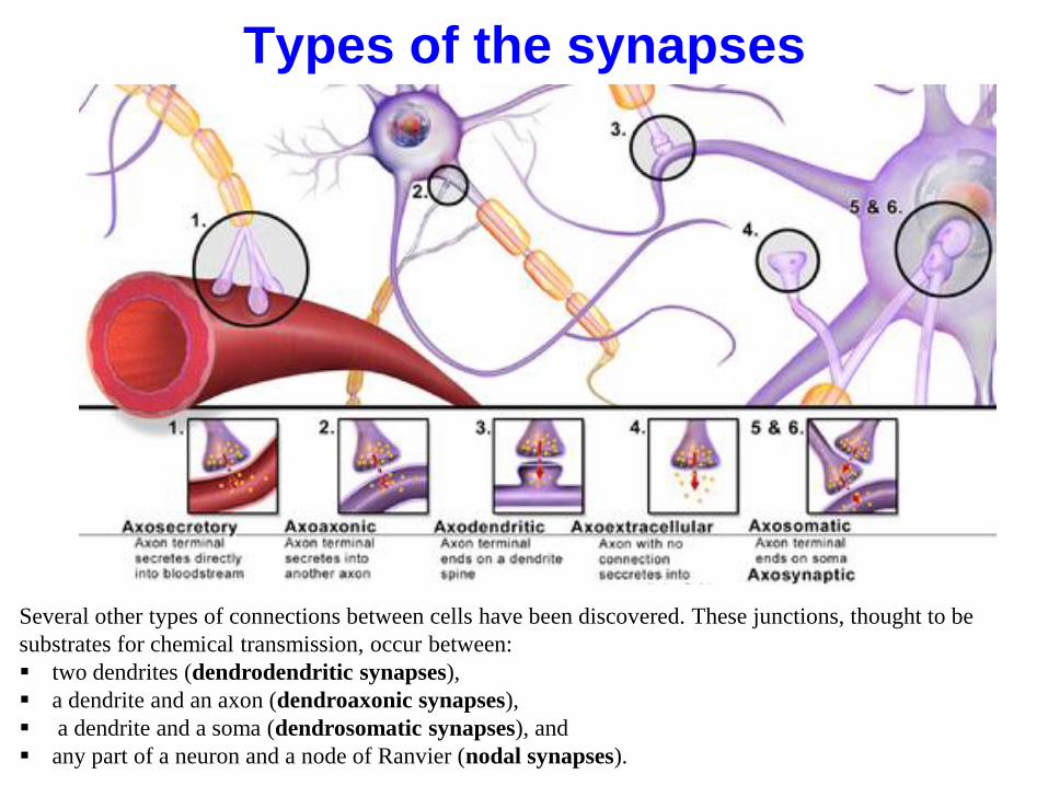
Types of the synapses
Several other types of connections between cells have been discovered. These junctions, thought to be
substrates for chemical transmission, occur between:
two dendrites (dendrodendritic synapses),
a dendrite and an axon (dendroaxonic synapses),
a dendrite and a soma (dendrosomatic synapses), and
any part of a neuron and a node of Ranvier (nodal synapses).
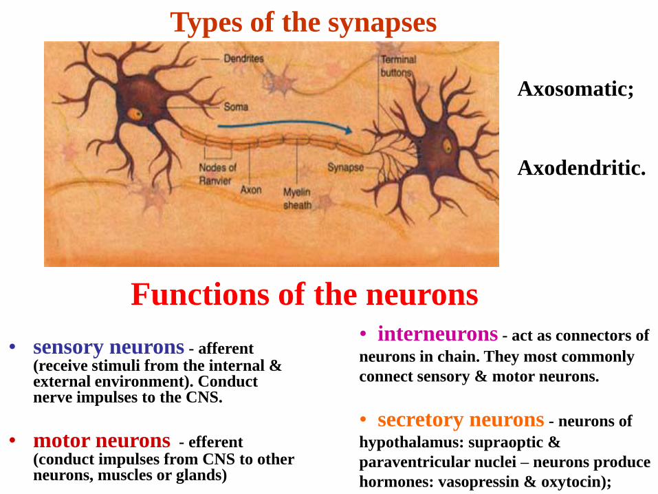
Types of the synapses
Axosomatic;
Axodendritic.
• sensory neurons - afferent (receive stimuli from the internal & external environment). Conduct nerve impulses to the CNS.
• motor neurons - efferent (conduct impulses from CNS to other neurons, muscles or glands)
• interneurons - act as connectors of
neurons in chain. They most commonly
connect sensory & motor neurons.
• secretory neurons - neurons of
hypothalamus: supraoptic &
paraventricular nuclei – neurons produce
hormones: vasopressin & oxytocin);
Functions of the neurons


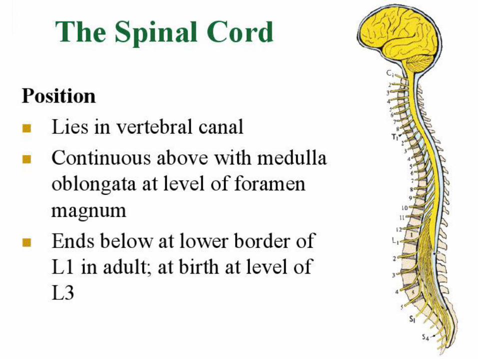

EXTERNAL
ANATOMY OF
THE SPINAL
CORD
The spinal cord is roughly
cylindrical and flattened in
anterior-posterior dimension.
Differential growth – early in
development the spinal cord
fills the entire vertebral canal.
By the time of birth, the tip of
the cord reaches only to level
L3-4.
At age 4-5 it had reached its
adult length and ceases to grow.
Differential growth, continuing
until adult statute is reached,
is responsible for the disparity
in length between the vertebral
canal and the spinal cord of the
adult.
Adult length –in the adult, the
spinal cord extends from the
foramen magnum where it is
continuous with the medulla of
the brain, to vertebral level L2.


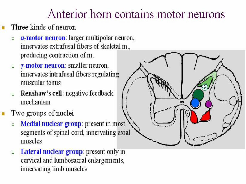


• Renshaw cells are inhibitory interneurons found in the gray matter of the spinal cord, and are associated in two ways with an alpha motor neuron.
• They receive an excitatory collateral from the alpha neuron's axon as they emerge from the motor root, and are thus "kept informed" of how vigorously that neuron is firing.
• They send an inhibitory axon to synapse with the cell body of the initial alpha neuron and/or an alpha motor neuron of the same motor pool.
• In this way, Renshaw cell inhibition represents a negative feedback mechanism. A Renshaw cell may be supplied by more than one alpha motor neuron collateral and it may synapse on multiple motor neurons.

Rexed laminae• They comprise a system of ten layers of grey matter (I-X), identified in the early
1950s by Bror Rexed to label portions of the spinal cord.
• They are defined by their cellular structure rather than by their location, but the location still remains reasonably consistent.
Laminae:
• Posterior/dorsal horn: I-VI
– Lamina I: marginal nucleus of spinal cord
or posteromarginal nucleus
– Lamina II: substantia gelatinosa of Rolando
– Laminae III/IV: nucleus proprius
– Lamina V: neck of the dorsal horn
– Lamina VI: base of the dorsal horn,
• Intermediate zone: VII and X
– Lamina VII: intermediomedial nucleus, intermediolateral nucleus, nucleus dorsalis in the thoracic and upper lumbar region
– Lamina X: central gray matter i.e. neurons bordering Central canal
• Anterior/ventral horn: VIII-IX
– Lamina VIII: motor interneurons
– Lamina IX: lateral (in limb regions) and medial motor neurons, also phrenic and spinal accessory nuclei at cervical levels, and Onuf's nucleus in the sacral region


Reflex and reflex arch
Reflex is a motor response that occurs following a
sensory stimulus.
The anatomical basis of a reflex is the reflex arc
formed by a receptor, afferent path, nerve center,
efferent path and effector.


Reflex
- the response of the organism to external or internal irritation, which is caused by the
action of the nervous system.
The reflex arch
- chain of neurons, which ensures the implementation of the reflex:
Bineuronal (tendinous )
sensory neuron+ motor neuron
Threeneuronal
sensory neuron+ interneuron+ motor neuron

SPINAL REFLEX• The knee jerk reflex is a
well known example of stretch reflex
• Tapping the knee cap (patella) pulls on the
tendon of the quadriceps femoris, which is an extensor muscle that extends the lower leg.
• When the muscle stretches in response to the pull of the tendon, information regarding this change in the muscle is conveyed by the afferent sensory neurons to the spinal cord and the central nervous system.

TYPES OF REFLEX ACTION
2 types of reflex action:
• a) CRANIAL REFLEX- brought about
by nerve impulses travelling through the
medulla oblongata.
• b) SPINAL REFLEX- brought
impulses travelling through the spinal
cord.

A dermatome is an area of skin
that is mainly supplied by a
single spinal nerve.
There are eight cervical nerves,
twelve thoracic nerves, five
lumbar nerves and five sacral
nerves.
Each of these nerves relays
sensation (including pain) from
a particular region of skin to the
brain.
Along the thorax and abdomen
the dermatomes are like a stack
of discs forming a human, each
supplied by a different spinal
nerve.
Along the arms and the legs, the
pattern is different: the
dermatomes run longitudinally
along the limbs. Although the
general pattern is similar in all
people, the precise areas of
innervation are as unique to an
individual as fingerprints.

Spinal cord lesions


Structural divisions of the brain
1. Myelencephalon = Madulla oblongata
2. Metencephalon = Cerebellum + Pons (Varoli‟s bridje)
3. Mesencephalon = Midbrain
4. Diancephalon = Between brain
5. Telencephalon = Endbrain
• brain stem

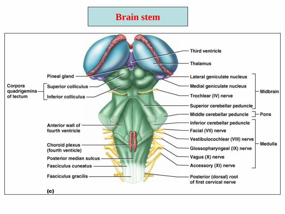
Brain stem


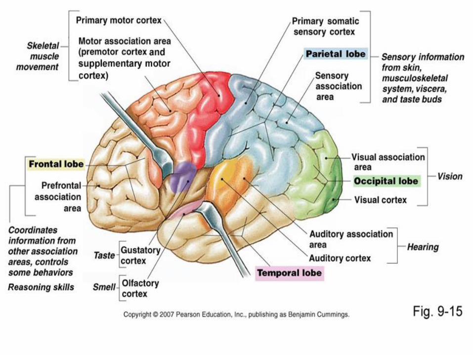
Frontal Lobe:
*is the largest lobe of the cerebrum
*is responsible for controlling speech,
thought, consciousness, and voluntary body
movements.
Parietal Lobe:
*is involved in controlling sensations such
as touch, pressure, and pain, as well as
texture and position.
Occipital Lobe:
*is located at the back of the head and is
involved in vision and reading.
Temporal Lobe:
•is involved in hearing, speech memory,
sight memory, and music memory.
•Each ear is connected to both sides of the
temporal lobes.
Insula:
*is located deep within the Sylvian fissure,
beneath the temporal and frontal lobes.
*it controls smell.
Cerebral lobes & their
functions:

White matter includes 3 types of
fibers:Associative – connect gyri of the same
hemisphere;
Commissural - connect gyri of both
hemispherae /right and left/
Projectional – connect the cerebral cortex
with underlying structures of the brain and
spinal cord
Gray matter forms the cortex and
basal nuclei:
1)the striated body:
caudate nucleus + lentiform nucleus
head, medial pallid nucleus
body lateral pallid nucleus,
tail putamen
2)the claustrum
3)the amygdaloid body

White
matter


Projection of motor
functions
on the brain

The study was carried out on the people who had
damage Insula and found that they were able
to give up smoking.
Scientists anticipate that research on Insula could
possible open the new doors for treatment
to drug addiction, anxiety, and eating disorders.
Scientists say that Insula is the „wellspring
of social emotions, things like lust and disgust,
pride and humiliation, guilt and atonement.‟
What does the Insula do? For example, the Insula “lights up” in brain scans when
people crave drugs, feel pain, anticipate pain, empathize
with others, listen to jokes, see disgust on someone‟s face,
are shunned in a social settings, listen to music, decide not to
buy an item, see someone cheat and decide to punish them,
and determine degrees of preference while eating chocolate.
Damage to the insula can lead to apathy, loss of libido and an
inability to distinguish fresh food from rotten.
Scientists will have to be very cautious since people
could possible not only loose desire for food and drink but
they could also loose interest in sex and work.

Functions of the cerebellum

Brain Stem – diencephalon, midbrain, pons,
medulla
*Organization is similar to spinal
cord (gray matter surrounded by
white fiber tracts) but contains
nuclei embedded in the white
matter
*Controls automatic behaviors
necessary for survival
*Provides the pathway for tracts
between higher and lower brain
centers
*Brain stem nuclei are associated
with 10 of the 12 pairs of cranial
nerves

The diencephalon consists of the:
thalamic region, hypothalamus,
subthalamus, and the third ventricle.
Thalamic region in turn includes:
1) optic thalamus,
2) epithalamus,
3) metathalamus
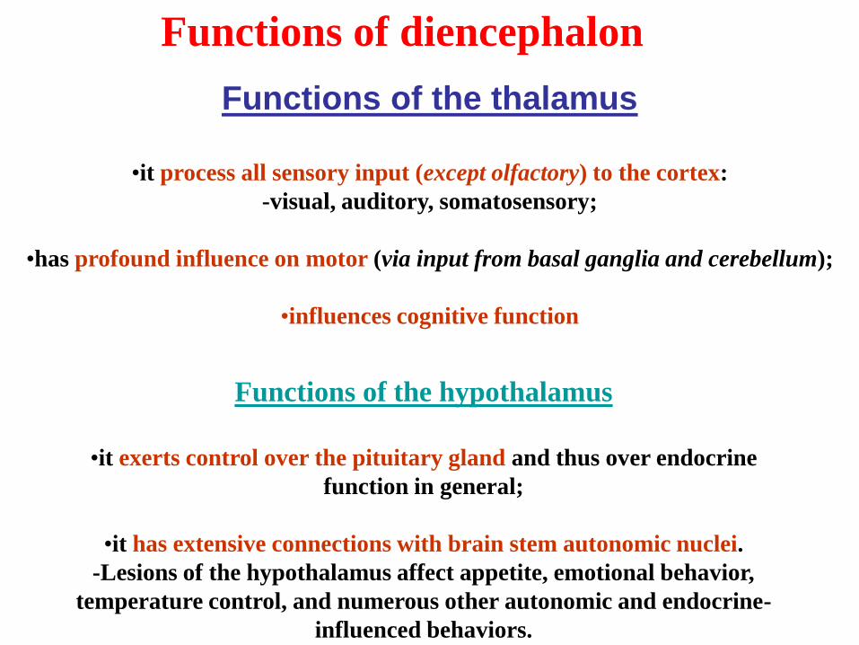
Functions of the thalamus
•it process all sensory input (except olfactory) to the cortex:
-visual, auditory, somatosensory;
•has profound influence on motor (via input from basal ganglia and cerebellum);
•influences cognitive function
Functions of the hypothalamus
•it exerts control over the pituitary gland and thus over endocrine
function in general;
•it has extensive connections with brain stem autonomic nuclei.
-Lesions of the hypothalamus affect appetite, emotional behavior,
temperature control, and numerous other autonomic and endocrine-
influenced behaviors.
Functions of diencephalon

Midbrain – between
diencephalon and pons
Midbrain structures include:
*Cerebral peduncles –
two bulging structures on
the ventral aspect that
contain descending
pyramidal motor tracts
*Cerebral aqueduct –
hollow tube that connects
the third and fourth
ventricles
*Subcortical visual and
hearing centers
*Various nuclei

Midbrain Nuclei
Nuclei that control cranial nerves III
(oculomotor) and IV (trochlear)
*Corpora quadrigemina – four domelike
protrusions of the dorsal midbrain
-Superior colliculi – visual reflex centers
-Inferior colliculi – auditory relay
centers
*Substantia nigra – functionally linked
to basal nuclei, contains melanin
pigment (precursor of dopamine)
*Red nucleus – largest nucleus (rich
blood supply) of the reticular formation;
relay nuclei for some descending motor
pathways

Pons – between midbrain and medulla oblongata
*Dorsally, it forms part
of the anterior wall of
the fourth ventricle
*Deep projection fibers
connect higher brain
centers and the spinal
cord
*The more superficial
or ventral fibers act as
relay between the motor
cortex and the
cerebellum
*Origin of cranial
nerves V (trigeminal),
VI (abducens), VII
(facial) and VIII
(vestibulocochlear)
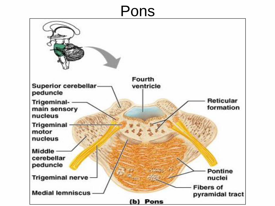
Pons
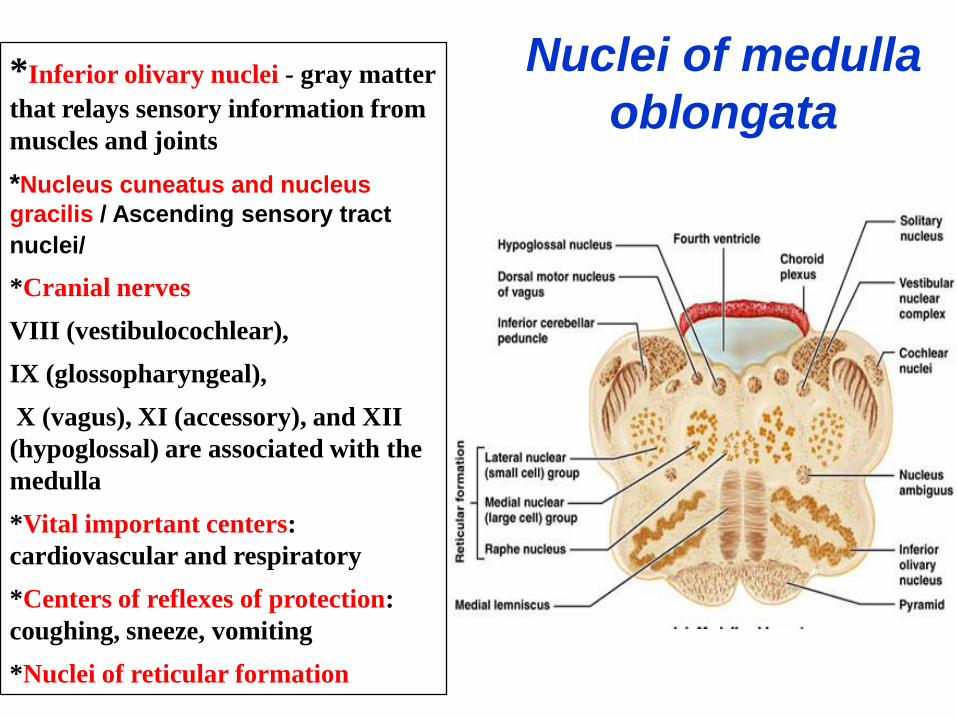
Nuclei of medulla
oblongata*Inferior olivary nuclei - gray matter
that relays sensory information from
muscles and joints
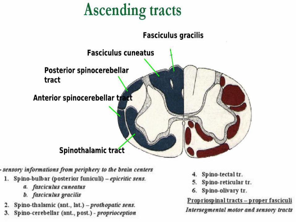
*Nucleus cuneatus and nucleus
gracilis / Ascending sensory tract
nuclei/
*Cranial nerves
VIII (vestibulocochlear),
IX (glossopharyngeal),
X (vagus), XI (accessory), and XII
(hypoglossal) are associated with the
medulla
*Vital important centers:
cardiovascular and respiratory
*Centers of reflexes of protection:
coughing, sneeze, vomiting
*Nuclei of reticular formation
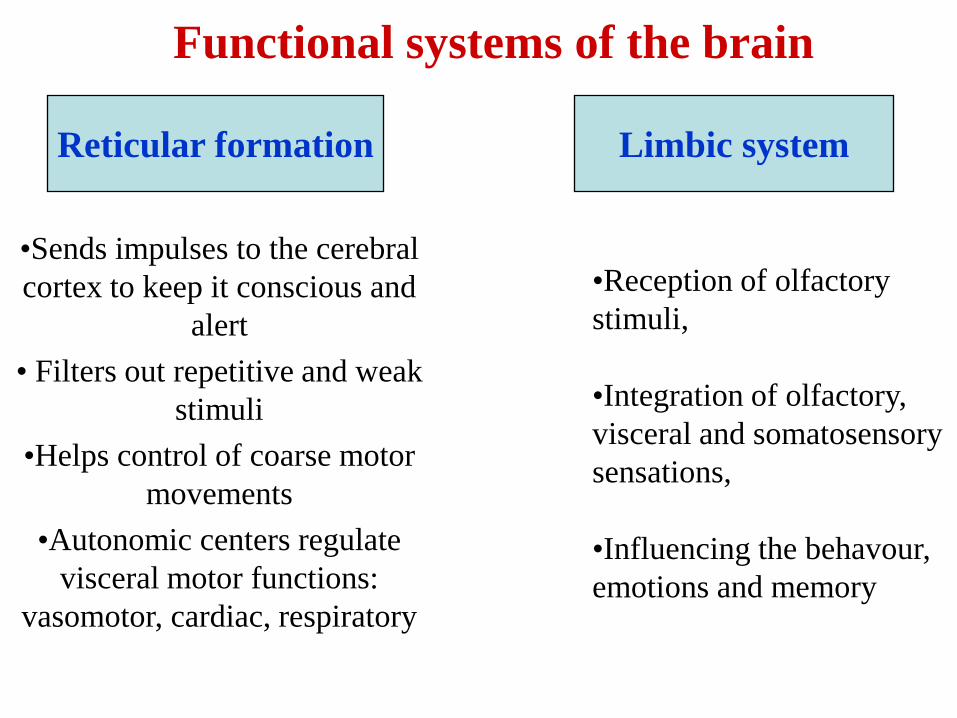
Functional systems of the brain
Reticular formation Limbic system
•Reception of olfactory
stimuli,
•Integration of olfactory,
visceral and somatosensory
sensations,
•Influencing the behavour,
emotions and memory
•Sends impulses to the cerebral
cortex to keep it conscious and
alert
• Filters out repetitive and weak
stimuli
•Helps control of coarse motor
movements
•Autonomic centers regulate
visceral motor functions:
vasomotor, cardiac, respiratory

• The shape and measures of the cells
are variable;
• Some of the cells are solitary, other
– form nuclei;
• Processes of the RS form rich network
in the brain stem;
• Dendrits have few branches, axons
have many relations /30.000/
• Their axons ascend and descend
along the long axis of the brainstem
• Ascending axons are carried in
the central tegmental tract
• Descending axons are carried in
the reticulospinal tract
The Reticular Neurons
Cells in the Reticular formation characterized by their relative size:
The largest cells are referred to as giant cells or giantocellular
The small cells are referred to as parvocellular.
Medium to large size cells are referred to as magnocellular


Specific features of the reticular system
1) the shape and measures of the cells are variable;
2) some of the cells are solitary, other – form nuclei;
3) processes with different structure and functions;
4) processes of the reticular system form rich
network in the brain stem;
5) dendrites have few branches, axons have
many relations /30.000/
Relations of the reticular system
3 groups of relations:
1- reticular formation with different parts of
the central nervous system,
2- different parts of the central nervous system
with the reticular formation
3- among different parts of reticular formation
Functions of the reticular system
1) regulates to the state of sleepness and wakeupness;
2) controls many of the stereotyped body movements:
postural motions of the limbs, turning and
bending motions of the head;
3) increases the tone of the muscle;
4) controls the brain‟s overall level of activity
5) selects the most important signals and sends
them to the cortex

Nuclei of RF
The reticular formation is an apparently (but not actually) diffusely organised
area that forms the central core of the brain stem.
•The reason it appears to be diffusely organised is twofold:
•Its pattern of connectivity is characterised by a great deal of convergence and
divergence, so that a single cell may respond to several different sensory modalities
or to stimuli applied practically anywhere on the body;
•Although it is involved in several quite separate functions, the areas involved in
these functions overlap considerably.
•At most levels of the brainstem, the reticular formation can be divided into 4
longitudinal zones arranged in a medial to lateral sequence, in addition a 5-th zone
is defined in the medulla:
The raphe nuclei
The paramedian zone;
The medial zone;
The lateral zone
The intermediate zone.

The longitudinal arrangement
of zones of the brain stem RF
The most lateral strips on both
sides are the lateral zones.
Along the midline is the
median or raphe zone.
In between is the paramedian
or the medial zone.

Functions of the Reticular
Formation
The reticular formation is involved in 4
general types of function:
• Motor control;
• Sensory control;
• Visceral control;
• And control of consciousness.

The reticular formation has two
components
• The ascending reticular formation is also called the reticular activating system. It is responsible for the sleep-wake cycle, thus mediating various levels of alertness. This part of the reticular system projects to the mid-line group of the thalamus, which also plays a role in wakefulness. From there, information is sent to the cortex.
• The descending reticular formation is involved in posture and equilibrium as well as autonomic nervous system activity. It receives information from the hypothalamus. The descending reticular formation also plays a role in motor movement.

Limbic System – “emotional or affective” brain
*Structures located
on the medial
aspects of cerebral
hemispheres and
diencephalon –
encircle the upper
part of the brain
stem (includes the
rhinencephalon,
amygdala,
hypothalamus, and
anterior nucleus of
the thalamus).
2 parts:
-peripheral
-central
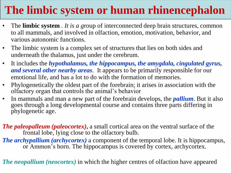
• The limbic system . It is a group of interconnected deep brain structures, common to all mammals, and involved in olfaction, emotion, motivation, behavior, and various autonomic functions.
• The limbic system is a complex set of structures that lies on both sides and underneath the thalamus, just under the cerebrum.
• It includes the hypothalamus, the hippocampus, the amygdala, cingulated gyrus, and several other nearby areas. It appears to be primarily responsible for our emotional life, and has a lot to do with the formation of memories.
• Phylogenetically the oldest part of the forebrain; it arises in association with the olfactory organ that controls the animal’s behavior
• In mammals and man a new part of the forebrain develops, the pallium. But it also goes through a long developmental course and contains three parts differing in phylogenetic age.
The paleopalleum (paleocortex), a small cortical area on the ventral surface of the frontal lobe, lying close to the olfactory bulb.
The archypallium (archycortex) a component of the temporal lobe. It is hippocampus, or Ammon’s horn. The hippocampus is covered by cortex, archycortex.
The neopallium (neocortex) in which the higher centres of olfaction have appeared
The limbic system or human rhinencephalon
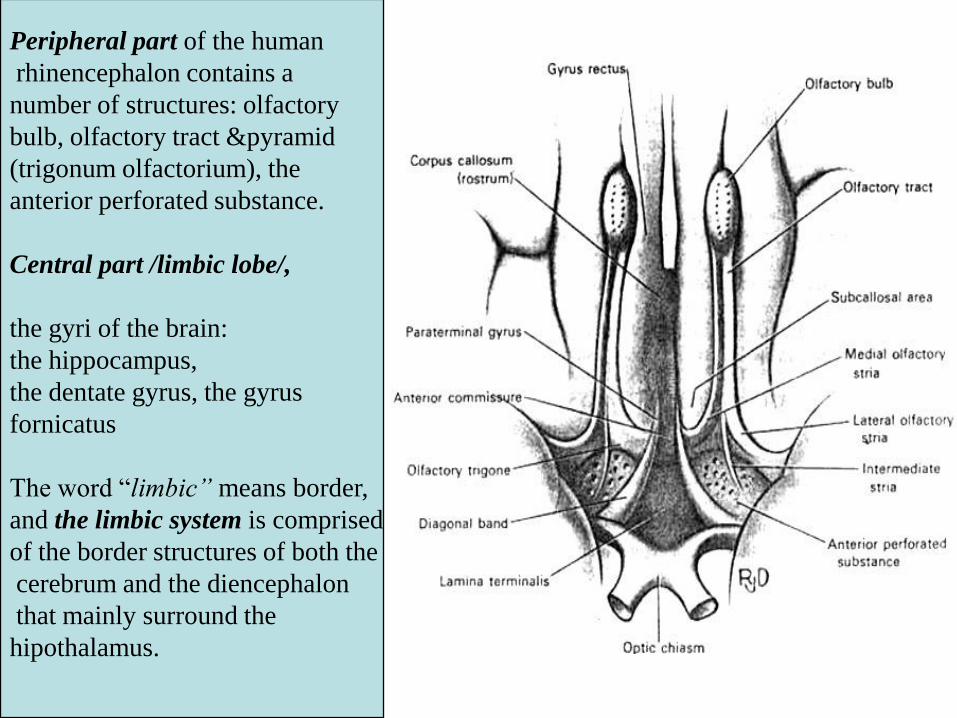
Peripheral part of the human
rhinencephalon contains a
number of structures: olfactory
bulb, olfactory tract &pyramid
(trigonum olfactorium), the
anterior perforated substance.
Central part /limbic lobe/,
the gyri of the brain:
the hippocampus,
the dentate gyrus, the gyrus
fornicatus
The word “limbic” means border,
and the limbic system is comprised
of the border structures of both the
cerebrum and the diencephalon
that mainly surround the
hipothalamus.

Limbic SystemThe limbic system interacts with the prefrontal lobes,
forming a connection between feelings (emotional
brain) and thoughts (cognitive brain)

Components of the limbic systemThe amygdala (also called the amygdaloid
body) is a small nuclear structure located deep inside each anterior temporal lobe /immediately anterior to the hippocampus/ and considered by anatomists to one of the basal ganglia.
The amygdalas are two almond-shaped masses of neurons on either side of the thalamus at the lower end of the hippocampus.
When it is stimulated electrically, animals respond with aggression. And if the amygdala is removed, animals get very tame and no longer respond to things that would have caused rage before. But there is more to it than just anger: When removed, animals also become indifferent to stimuli that would have otherwise have caused fear and even sexual responses.
Bilateral lesions of the temporal lobes and amygdala may result in the syndrome, which is characterized by patients who develop an aberrant tendency to examine almost anything orally, visually and tactually.
In addition, patients (males in particular) develop hypersexual tendencies toward any gender.

• The hippocampus consists of two “horns” that curve back from the amygdala. It appears to be very important in converting things that are “in your mind” at the moment (in short-term memory) into things that you will remember for the long run (long-term memory).
• If the hippocampus is damaged, a person cannot build new memories, and lives instead in a strange world where everything they experience just fades away, even while older memories from the time before the damage are untouched!
• Without the hippocampus, a person’s ability to store memories becomes very deficient.
• While a unilateral ablation of the hippocampus does not result in deficits, a bilateral ablation results in a loss of short term memory and an ability to store new information.


• The cingulate gyrus, the insula, and the parahippocampal gyrus all together form a ring of cerebral cortex in each cerebral hemisphere around the deeper structures of the limbic system.
• This ring of cortex is believed to allow association between conscious cerebral behavioral functions and subconscious behavioral functions of the deeper limbic system.
•The cingulate gyrus is the part of the
cerebrum that lies closest to the limbic
system, just above the corpus collosum.
It provides a pathway from the thalamus
to the hippocampus, seems to be
responsible for focusing attention
on emotionally significant events,
and for associating memories to smells
and to pain.

The limbic system includes many different cortical and subcortical brain
structures
• amygdale: involved in aggression and fear;
• cingulated gyrus: autonomic functions regulating heart rate and blood pressure as well as cognitive and attentional processing;
• fornicate gyrus: region encompassing the cingulated, hippocampus, and parahippocampal gyrus;
• hippocampus: required for the information of long term memories;
• hypothalamus: regulates the autonomic nervous system via hormone production and release. Affects and regulates blood pressure, heart rate, hunger, thirst, sexual arousal, and the sleep/wake cycle;
• mammilary body: importamt for the formation memory;
• nucleus accumbens: involved in reward, pleasure, and addiction;
• orbitofrontal cortex: required for decision making;
• parahippocampal gyrus: plays a role in the formation of spatial memory.

The mammillary bodies lie
immediately behind the
hypothalamus and function
in close association with the
thalamus, hypothalamus,
and brain stem to help
control many behavioral
functions such as the
person’s degree of
wakefulness and perhaps
also his feeling of well
being.

• The hypothalamus is a small part of the brain located just below the thalamus on both sides of the third ventricle. It sits just inside the two tracts of the optic nerve, and just above (and intimately connected with) the pituitary gland.
• The hypothalamus is one of the busiest parts of the brain, and is mainly concerned with homeostasis. Homeostasis is the process of returning something to some “set point.” It works like a thermostat: When your room gets too cold, the thermostat conveys that information to the furnace and turns it on. As your room warms up and the temperature gets beyond a certain point, it sends a signal that tells the furnace to turn off.
• The hypothalamus is responsible for regulating your hunger, thirst, response to pain, levels of pleasure, sexual satisfaction, anger and aggressive behavior, and more. It also regulates the functioning of the parasympathetic and sympathetic nervous systems, which in turn means it regulates things like pulse, blood pressure, breathing, and arousal in response to emotional circumstances.

• The hypothalamus sends instructions to the rest of the body in two ways. The first is to the autonomic nervous system. This allows the hypothalamus to have ultimate control of things like blood pressure, heartrate, breathing, digestion, sweating, and all the sympathetic and parasympathetic functions.
• The other way the hypothalamus controls things is via the pituitary gland. It is neurally and chemically connected to the pituitary, which in turn pumps hormones called releasing factors into the bloodstream. As you know, the pituitary is the so-called “master gland,” and these hormones are vitally important in regulating growth and metabolism.

The septum pellucidum• It lies anterior to the thalamus, superior to
the hypothalamus, and between the basal ganglia in the median plane of the cerebrum.
• The septal nuclei are located in the infero-medial portion of the frontal lobes.
• They receive afferents from:
- the hypothalamus and midbrai through the medial forebrain bundle,
- the amygdala via the diagonal band of Broca,
- the hippocampus via the fornix.
• Efferents from the septal nuclei project to the:
- hippicampus,
- hypothalamus,
- midbrain,
- habenular nuclei.
• The function of the septal nuclei is to provide a site of interaction between limbic and diencephalic structures.
• Physiological studies implicate the septal nuclei in modulating arousal, learning, emotion and sexual behavior. The septum, which lies in front of the thalamus, has areas that seem to be centers for orgasm.
• Stimulation of different parts of this septum can cause many different behavioral effects, including the phenomenon of rage.

Related areas• Besides the hypothalamus, hippocampus, and amygdala, there are other
areas in the structures near to the limbic system that are intimately connected to it:
• The ventral tegmental area of the brain stem (just below the thalamus) consists of dopamine pathways that seem to be responsible for pleasure. People with damage here tend to have difficulty getting pleasure in life, and often turn to alcohol, drugs, sweets, and gambling.
• The basal ganglia (including the caudate nucleus, the putamen, the globus pallidus, and the substantia nigra) lie over and to the sides of the limbic system, and are tightly connected with the cortex above them. They are responsible for repetitive behaviors, reward experiences, and focusing attention.
• The prefrontal cortex, which is the part of the frontal lobe which lies in front of the motor area, is also closely linked to the limbic system. Besides apparently being involved in thinking about the future, making plans, and taking action, it also appears to be involved in the same dopamine pathways as the ventral tegmental area, and plays a part in pleasure and addiction.

Functions of the limbic system
• Controls Emotions
• Emotional Responses
• Hormonal Secretions
• Mood
• Motivation
• Pain and Pleasure Sensations

• The reticulal formation has been reffered to many times in previous units as the “arousal” center for the central nervous system and is found centrally located in the brainstem.
• The limbic system is located just superior to the corpus callosum and has primary responsibilities for olfaction and emotions.
• These two areas work closely together in what may be reffered to as the “reticulolimbic system.”

Formation of neural tubeAt the beginning of the third week of development, the ectodermal germ layer
has the shape of a disc that is broader in the cephalic than the caudal region .
Development of the spinal meninges: a) the dura mater develops from the mesoderm of the sclerotomes which form the vertebral
column.
b) the arachnoid and pia mater develop from the neural crest.

• Spina bifida is a more common and usually less severe form of failure of fusion in which
there is failure of the neural arches of the vertebrae to meet, fuse, and enclose the spinal cord.
• Spinal bifida may be minimal, involving only failure of fusion of the vertebrae but the spinal
cord and meninges are normal and without external manifestation: this is called spina bifida
occulta.
• When the meninges are also involved and form a fluid filled cystic structure bulging
posteriorly then there is spina bifida with meningocele.
• When the spinal cord is carried outward with the meningocele then the condition is spina
bifida with myelomeningocele.
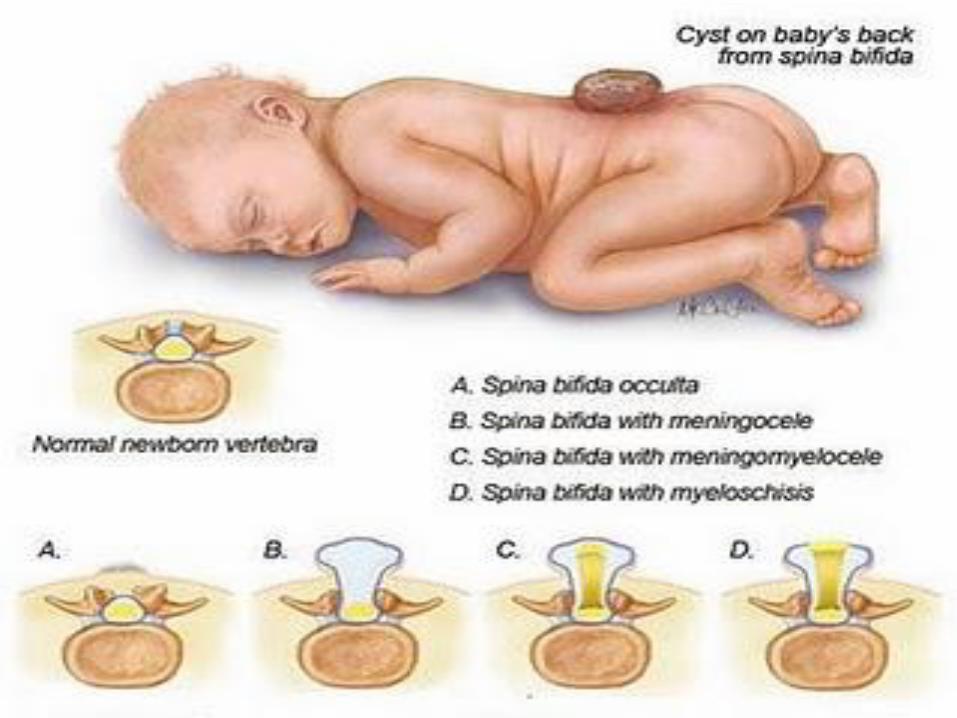

In the extreme example show, the spinal cord is at the surface in the form of the wide-open neural tube. This is spina bifida with myeloschisis.

Brain development
The human brain begins forming very
early in prenatal life (just three weeks
after conception), but in many ways,
brain development is a lifelong project.
Neural development refers to the
processes that generate, shape, and
reshape the nervous system, from the
earliest stages of embryogenesis to the
final years of life.
• The brain develops from the
cranial end of the neural tube as
follows:
• 1) The cranial end of the neural
tube expands to form the brain
swelling.
• 2) Two constrictions appear in the
brain swelling, dividing into 3
parts called primary brain vesicles:
• a) forebrain or prosencephalon,
• b) midbrain or mesencephalon,
• c) hindbrain or rhombencephalon.

3)The brain vesicles are
differentiated in 5
secondary brain vesicles:
a) the forebrain gives 2
optic vesicles (the future
eyes) and then divides
into:
- the median part called
diencephalons,
- two lateral diverticula
called telencephalic
vesicles (the future
cerebral hemispheres);
b) the midbrain remains
undivided;
c) the hindbrain gives rise
to the following
derivatives:
- the metencephalon
which forms the pons and
cerebellum;
- the myelencephalon
which forms the medulla
oblongata.

Anomalies of the brain
• Anencephaly is the most severe of neural tube defects. Babies with anencephaly
have underdeveloped brains and incomplete skulls and most of them do not survive
more than a few hours after birth. A baby born with this disorder is usually blind,
deaf, unconscious and unable to feel pain.
• Encephalocele is where there is an opening in the skull, from which the brain
protrudes. There can be a large sac like deformity which holds cerebrospianl fluid,
these can be even larger than the baby’s head.
• Microgyria: abnormal smallness of the convolutions of the brain.

END