Embed Size (px)
Citation preview
CASE REPORT Open Access
Fully digital workflow, integrating dentalscan, smile design and CAD-CAM: casereportMiguel Stanley1, Ana Gomes Paz1,3* , Inês Miguel1 and Christian Coachman2
Abstract
Background: This report is a presentation of a clinical case that follows a full digital workflow.
Case presentation: A 47-year old man presented with pain in the TMJ (temporomandibular joint) and whoseaesthetic concern was having a chipped maxillary central incisor veneer. The concern was solved following afully digital workflow: it was applied the digital smile design protocol, as well as CAD-CAM monolithic lithiumdisilicate ceramic veneers and crowns (following a minimal invasive preparation approach). The aim of this rehabilitationwas to solve a loss of vertical dimension, subsequent aesthetics and temporomandibular joint disorders.
Conclusion: Thanks to the evolution of technology in dentistry, it is possible to do a full digital case and solve problemssuch as loss of vertical dimension successfully. Nevertheless, more clinical studies are needed to obtain consistent resultsabout the digital work flow compared to the conventional technique in loss of vertical dimension cases.
Keywords: Digital workflow, Digital planning, Digital smile design, Intraoral scanner, 3D printer, CAD-CAM, Minimalpreparation, Monolithic lithium disilicate ceramic
BackgroundDigital work flow in dentistry has increased in recentyears due to the headway made in technologies such asintraoral scanners and software programs, which havecontributed to improve communication between theclinician and the dental technician. The Digital SmileDesign (DSD) is a digital tool which provides, from afacial perspective, rehabilitative aesthetic planning, bettercommunication between specialists and an improvementin the expected outcome of the treatments [1]. A dy-namic documentation of the smile is an important stepin the 2D/3D digital smile design process that can beperformed in a entire digital flow and will help in the re-habilitative procedures. The advantages of using videodocumentation are that it facilitates and simplifies thedocumentation process, improves smile design, facial
analysis, treatment planning, team communication andpatient education [2]. The DSD could be converted intoa conventional or virtual diagnostic model to facilitatesubsequent clinical treatments, i.e. CAD-CAM restora-tions [3–7]. The preparations for minimally invasivetreatments have become easily achievable in restorativedentistry because of the combination of the adhesivetechnique with restorative materials featuring translu-cent properties. Materials like lithium disilicate ceramic[8–11] have properties like those existing in naturalteeth so they have presented positive outcomes [12, 13].Another important tool that integrates the digital
workflow are the intraoral scanners. These are powerfuldevices that allow an immediate determination of thequality of the impression and have the capacity to easilysend the models to the laboratory using e-mail, thus redu-cing expense and time [14]. Nevertheless, there is limitedliterature about intraoral scanner potential capturing highquality impressions [15–24].Computer-aided design (CAD) software is an essen-
tial tool since it is responsible for guiding robotic de-vices which create objects and assemblies in a virtualenvironment [25].
* Correspondence: [email protected] Practice at White Clinic, Rua Dr. António Loureiro Borges. Edif. 5, 1°Andar Arquiparque; 1495-131 Algés-, Lisbon, Portugal3Endodontic Department at FMDUL, Private Practice at White Clinic, Rua Dr.António Loureiro Borges. Edif. 5, 1° Andar Arquiparque, 1495-131 Algés,PortugalFull list of author information is available at the end of the article
© The Author(s). 2018 Open Access This article is distributed under the terms of the Creative Commons Attribution 4.0International License (http://creativecommons.org/licenses/by/4.0/), which permits unrestricted use, distribution, andreproduction in any medium, provided you give appropriate credit to the original author(s) and the source, provide a link tothe Creative Commons license, and indicate if changes were made. The Creative Commons Public Domain Dedication waiver(http://creativecommons.org/publicdomain/zero/1.0/) applies to the data made available in this article, unless otherwise stated.
Stanley et al. BMC Oral Health (2018) 18:134 https://doi.org/10.1186/s12903-018-0597-0
This report is a presentation of a clinical case thatfollows a full digital workflow. After a minimal inva-sive preparation approach, the digital smile protocol,CAD-CAM monolithic lithium disilicate ceramic ve-neers and crowns was used to solve a loss of verticaldimension, subsequent aesthetics and temporoman-dibular joint disorders.
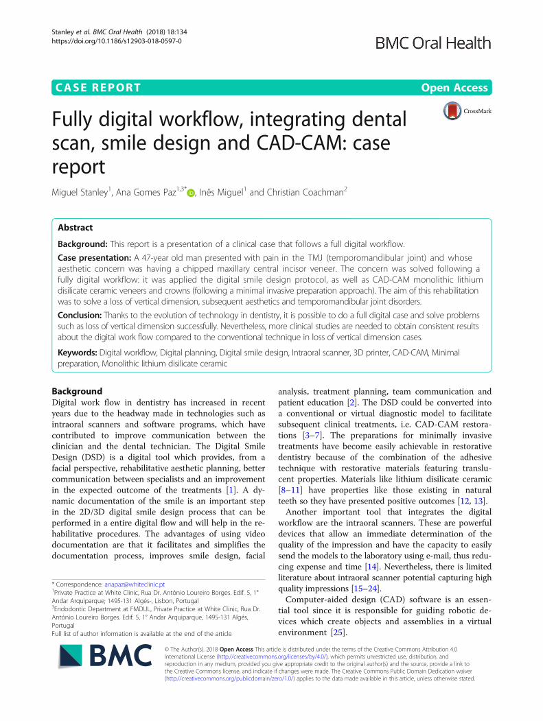
Case presentationIn 2015, a 47-year old man presented with pain in theTMJ (temporomandibular joint) and whose aestheticconcern was having a chipped maxillary central incisorveneer, as seen in Fig. 1a, b and c. After a clinical andradiographic analysis, as seen in Fig. 2, a loss of the ver-tical dimension and tooth ware, caused by bruxism, wasdiagnosed.Digital intraoral photographs were taken from a
retracted frontal view, occlusal view and lateral view and
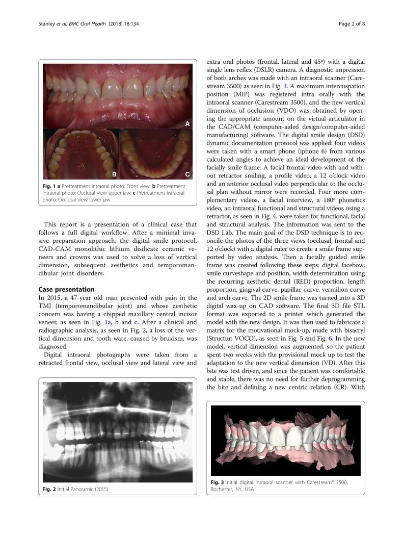
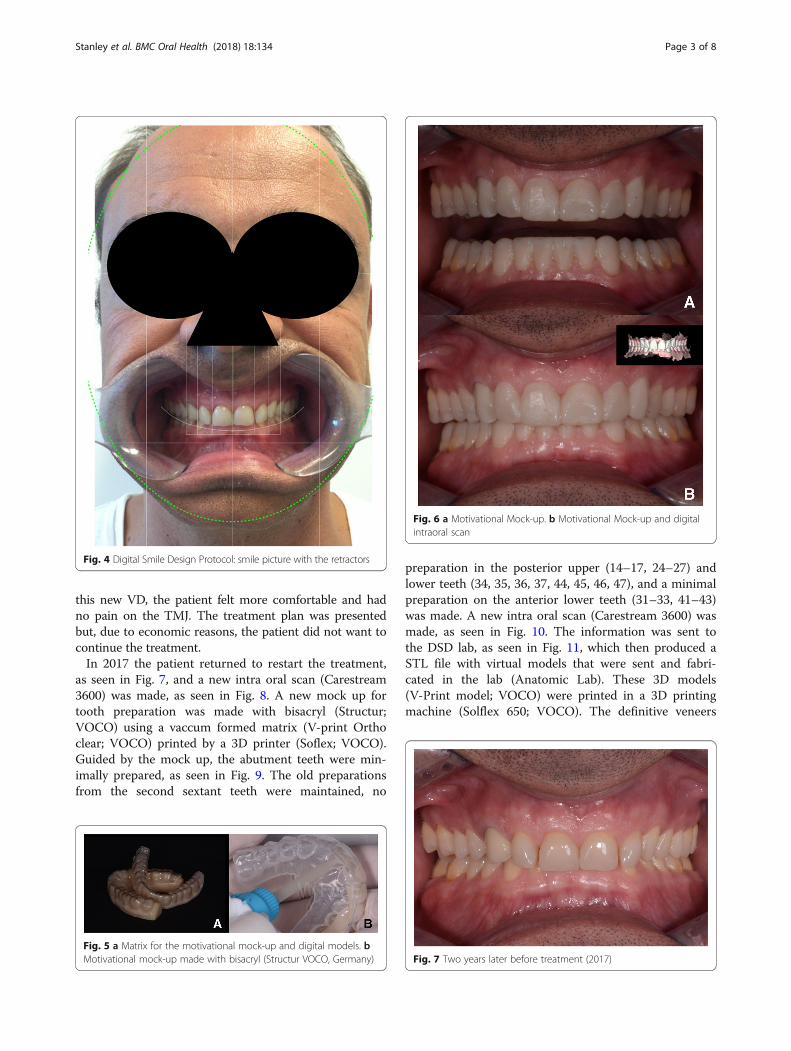
extra oral photos (frontal, lateral and 45º) with a digitalsingle lens reflex (DSLR) camera. A diagnostic impressionof both arches was made with an intraoral scanner (Care-stream 3500) as seen in Fig. 3. A maximum intercuspationposition (MIP) was registered intra orally with theintraoral scanner (Carestream 3500), and the new verticaldimension of occlusion (VDO) was obtained by open-ing the appropriate amount on the virtual articulator inthe CAD/CAM (computer-aided design/computer-aidedmanufacturing) software. The digital smile design (DSD)dynamic documentation protocol was applied: four videoswere taken with a smart phone (iphone 6) from variouscalculated angles to achieve an ideal development of thefacially smile frame. A facial frontal video with and with-out retractor smiling, a profile video, a 12 o’clock videoand an anterior occlusal video perpendicular to the occlu-sal plan without mirror were recorded. Four more com-plementary videos, a facial interview, a 180º phoneticsvideo, an intraoral functional and structural videos using aretractor, as seen in Fig. 4, were taken for functional, facialand structural analysis. The information was sent to theDSD Lab. The main goal of the DSD technique is to rec-oncile the photos of the three views (occlusal, frontal and12 o’clock) with a digital ruler to create a smile frame sup-ported by video analysis. Then a facially guided smileframe was created following these steps: digital facebow,smile curveshape and position, width determination usingthe recurring aesthetic dental (RED) proportion, lengthproportion, gingival curve, papillae curve, vermilion curveand arch curve. The 2D smile frame was turned into a 3Ddigital wax-up on CAD software. The final 3D file STLformat was exported to a printer which generated themodel with the new design. It was then used to fabricate amatrix for the motivational mock-up, made with bisacryl(Structur; VOCO), as seen in Fig. 5 and Fig. 6. In the newmodel, vertical dimension was augmented, so the patientspent two weeks with the provisional mock up to test theadaptation to the new vertical dimension (VD). After thisbite was test driven, and since the patient was comfortableand stable, there was no need for further deprogrammingthe bite and defining a new centric relation (CR). With
Fig. 1 a Pretreatment intraoral photo: Front view. b Pretreatmentintraoral photo: Occlusal view upper jaw. c Pretreatment intraoralphoto: Occlusal view lower jaw
Fig. 2 Initial Panoramic (2015)Fig. 3 Initial digital intraoral scanner with Carestream® 3500;Rochester, NY, USA
Stanley et al. BMC Oral Health (2018) 18:134 Page 2 of 8
this new VD, the patient felt more comfortable and hadno pain on the TMJ. The treatment plan was presentedbut, due to economic reasons, the patient did not want tocontinue the treatment.In 2017 the patient returned to restart the treatment,
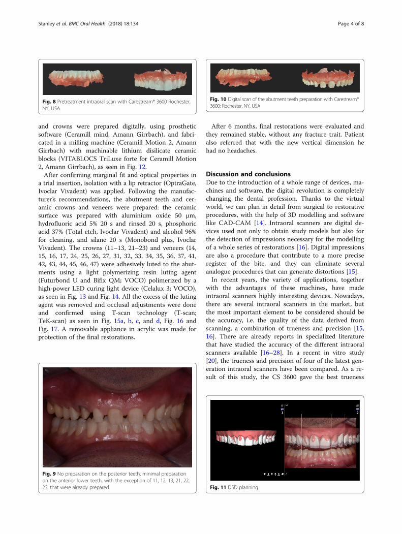
as seen in Fig. 7, and a new intra oral scan (Carestream3600) was made, as seen in Fig. 8. A new mock up fortooth preparation was made with bisacryl (Structur;VOCO) using a vaccum formed matrix (V-print Orthoclear; VOCO) printed by a 3D printer (Soflex; VOCO).Guided by the mock up, the abutment teeth were min-imally prepared, as seen in Fig. 9. The old preparationsfrom the second sextant teeth were maintained, no
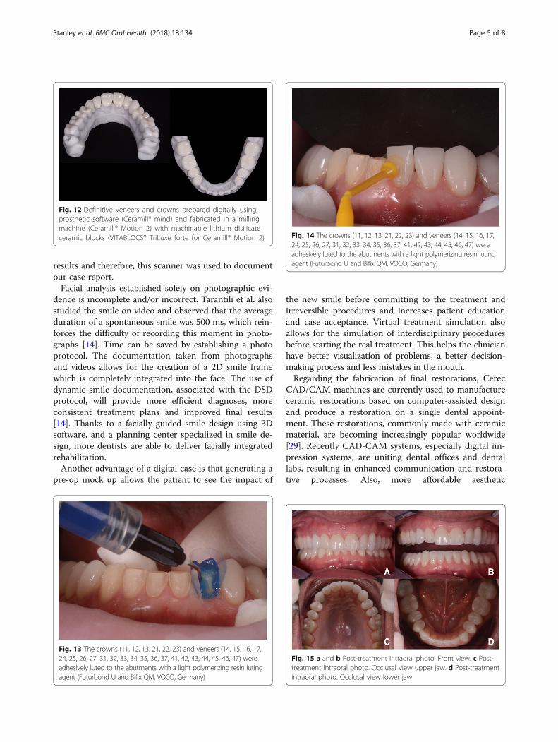
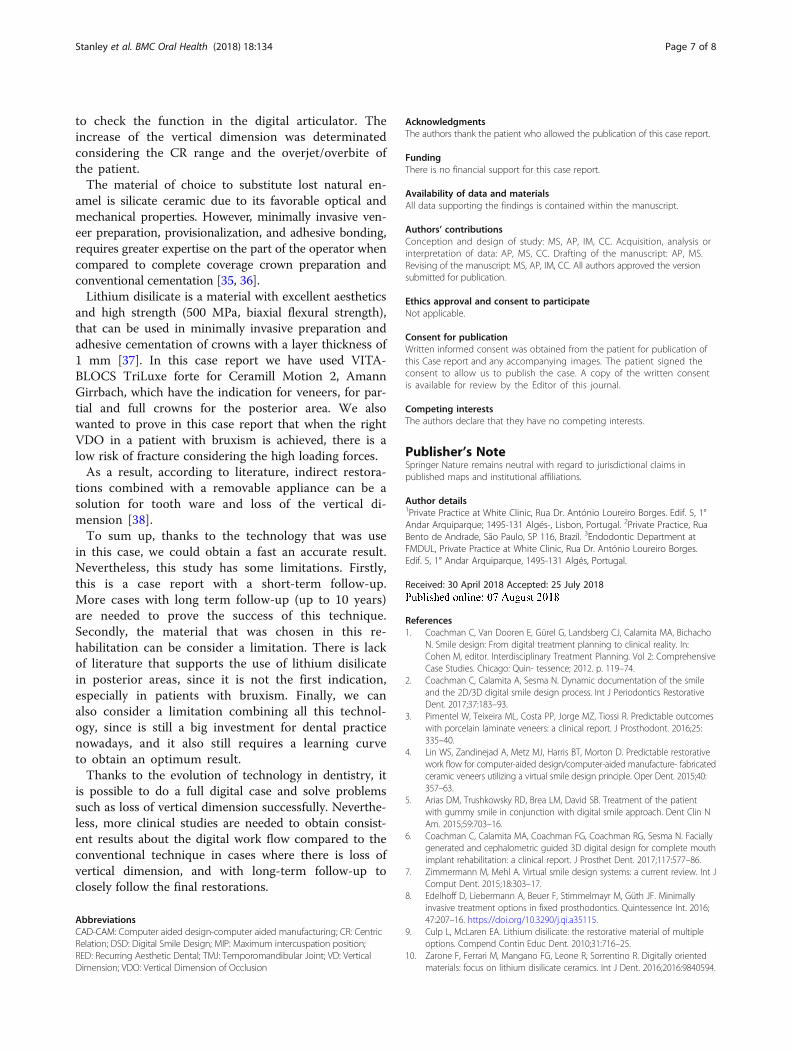
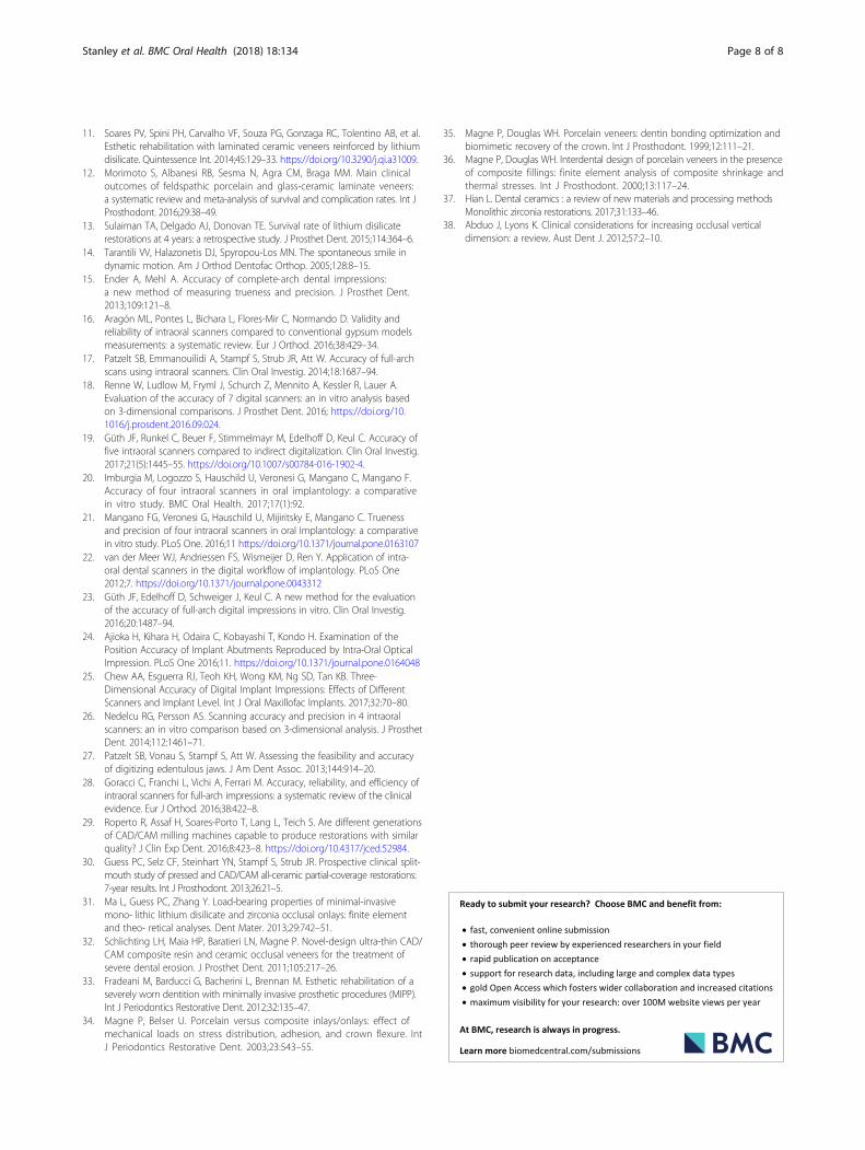
preparation in the posterior upper (14–17, 24–27) andlower teeth (34, 35, 36, 37, 44, 45, 46, 47), and a minimalpreparation on the anterior lower teeth (31–33, 41–43)was made. A new intra oral scan (Carestream 3600) wasmade, as seen in Fig. 10. The information was sent tothe DSD lab, as seen in Fig. 11, which then produced aSTL file with virtual models that were sent and fabri-cated in the lab (Anatomic Lab). These 3D models(V-Print model; VOCO) were printed in a 3D printingmachine (Solflex 650; VOCO). The definitive veneers
Fig. 4 Digital Smile Design Protocol: smile picture with the retractors
Fig. 5 a Matrix for the motivational mock-up and digital models. bMotivational mock-up made with bisacryl (Structur VOCO, Germany)
Fig. 6 a Motivational Mock-up. b Motivational Mock-up and digitalintraoral scan
Fig. 7 Two years later before treatment (2017)
Stanley et al. BMC Oral Health (2018) 18:134 Page 3 of 8
and crowns were prepared digitally, using prostheticsoftware (Ceramill mind, Amann Girrbach), and fabri-cated in a milling machine (Ceramill Motion 2, AmannGirrbach) with machinable lithium disilicate ceramicblocks (VITABLOCS TriLuxe forte for Ceramill Motion2, Amann Girrbach), as seen in Fig. 12.After confirming marginal fit and optical properties in
a trial insertion, isolation with a lip retractor (OptraGate,Ivoclar Vivadent) was applied. Following the manufac-turer’s recommendations, the abutment teeth and cer-amic crowns and veneers were prepared: the ceramicsurface was prepared with aluminium oxide 50 μm,hydrofluoric acid 5% 20 s and rinsed 20 s, phosphoricacid 37% (Total etch, Ivoclar Vivadent) and alcohol 96%for cleaning, and silane 20 s (Monobond plus, IvoclarVivadent). The crowns (11–13, 21–23) and veneers (14,15, 16, 17, 24, 25, 26, 27, 31, 32, 33, 34, 35, 36, 37, 41,42, 43, 44, 45, 46, 47) were adhesively luted to the abut-ments using a light polymerizing resin luting agent(Futurbond U and Bifix QM; VOCO) polimerized by ahigh-power LED curing light device (Celalux 3; VOCO),as seen in Fig. 13 and Fig. 14. All the excess of the lutingagent was removed and occlusal adjustments were doneand confirmed using T-scan technology (T-scan;TeK-scan) as seen in Fig. 15a, b, c, and d, Fig. 16 andFig. 17. A removable appliance in acrylic was made forprotection of the final restorations.
After 6 months, final restorations were evaluated andthey remained stable, without any fracture trait. Patientalso referred that with the new vertical dimension hehad no headaches.
Discussion and conclusionsDue to the introduction of a whole range of devices, ma-chines and software, the digital revolution is completelychanging the dental profession. Thanks to the virtualworld, we can plan in detail from surgical to restorativeprocedures, with the help of 3D modelling and softwarelike CAD-CAM [14]. Intraoral scanners are digital de-vices used not only to obtain study models but also forthe detection of impressions necessary for the modellingof a whole series of restorations [16]. Digital impressionsare also a procedure that contribute to a more preciseregister of the bite, and they can eliminate severalanalogue procedures that can generate distortions [15].In recent years, the variety of applications, together
with the advantages of these machines, have madeintraoral scanners highly interesting devices. Nowadays,there are several intraoral scanners in the market, butthe most important element to be considered should bethe accuracy, i.e. the quality of the data derived fromscanning, a combination of trueness and precision [15,16]. There are already reports in specialized literaturethat have studied the accuracy of the different intraoralscanners available [16–28]. In a recent in vitro study[20], the trueness and precision of four of the latest gen-eration intraoral scanners have been compared. As a re-sult of this study, the CS 3600 gave the best trueness
Fig. 8 Pretreatment intraoral scan with Carestream® 3600 Rochester,NY, USA
Fig. 9 No preparation on the posterior teeth, minimal preparationon the anterior lower teeth, with the exception of 11, 12, 13, 21, 22,23, that were already prepared
Fig. 10 Digital scan of the abutment teeth preparation with Carestream®3600; Rochester, NY, USA
Fig. 11 DSD planning
Stanley et al. BMC Oral Health (2018) 18:134 Page 4 of 8
results and therefore, this scanner was used to documentour case report.Facial analysis established solely on photographic evi-
dence is incomplete and/or incorrect. Tarantili et al. alsostudied the smile on video and observed that the averageduration of a spontaneous smile was 500 ms, which rein-forces the difficulty of recording this moment in photo-graphs [14]. Time can be saved by establishing a photoprotocol. The documentation taken from photographsand videos allows for the creation of a 2D smile framewhich is completely integrated into the face. The use ofdynamic smile documentation, associated with the DSDprotocol, will provide more efficient diagnoses, moreconsistent treatment plans and improved final results[14]. Thanks to a facially guided smile design using 3Dsoftware, and a planning center specialized in smile de-sign, more dentists are able to deliver facially integratedrehabilitation.Another advantage of a digital case is that generating a
pre-op mock up allows the patient to see the impact of
the new smile before committing to the treatment andirreversible procedures and increases patient educationand case acceptance. Virtual treatment simulation alsoallows for the simulation of interdisciplinary proceduresbefore starting the real treatment. This helps the clinicianhave better visualization of problems, a better decision-making process and less mistakes in the mouth.Regarding the fabrication of final restorations, Cerec
CAD/CAM machines are currently used to manufactureceramic restorations based on computer-assisted designand produce a restoration on a single dental appoint-ment. These restorations, commonly made with ceramicmaterial, are becoming increasingly popular worldwide[29]. Recently CAD-CAM systems, especially digital im-pression systems, are uniting dental offices and dentallabs, resulting in enhanced communication and restora-tive processes. Also, more affordable aesthetic
Fig. 12 Definitive veneers and crowns prepared digitally usingprosthetic software (Ceramill® mind) and fabricated in a millingmachine (Ceramill® Motion 2) with machinable lithium disilicateceramic blocks (VITABLOCS® TriLuxe forte for Ceramill® Motion 2)
Fig. 13 The crowns (11, 12, 13, 21, 22, 23) and veneers (14, 15, 16, 17,24, 25, 26, 27, 31, 32, 33, 34, 35, 36, 37, 41, 42, 43, 44, 45, 46, 47) wereadhesively luted to the abutments with a light polymerizing resin lutingagent (Futurbond U and Bifix QM, VOCO, Germany)
Fig. 14 The crowns (11, 12, 13, 21, 22, 23) and veneers (14, 15, 16, 17,24, 25, 26, 27, 31, 32, 33, 34, 35, 36, 37, 41, 42, 43, 44, 45, 46, 47) wereadhesively luted to the abutments with a light polymerizing resin lutingagent (Futurbond U and Bifix QM, VOCO, Germany)
Fig. 15 a and b Post-treatment intraoral photo. Front view. c Post-treatment intraoral photo. Occlusal view upper jaw. d Post-treatmentintraoral photo. Occlusal view lower jaw
Stanley et al. BMC Oral Health (2018) 18:134 Page 5 of 8

restorations are produced by using CAD/CAM and nat-ural shapes libraries.Adhesive all-ceramic partial coverage restorations are
also recognized as being a reliable treatment option forthe posterior region. In this context, one should keep inmind that the majority of clinical long-term studies arebased on leucite-reinforced glass-ceramics, whereastoday, considerably stronger ceramic materials based onlithium disilicate are available [30–32]. The use ofmonolithic lithium disilicate material for restorations,reduces restorative failures as it eliminates layering andinterfaces, which is usually the weak link between mate-rials [31]. According to this case report, we chose to re-habilitate this patient using monolithic lithium disilicatematerial for restorations, since this case demonstratesthat this material successfully works in a patient withbruxism when rehabilitated with the right VD.
All-ceramic onlays offer a sensible treatment option,since they permit a defect-oriented preparation methodand eliminate the need for a retentive preparation de-sign, thus bypassing conventional invasive treatmentmethods [33, 34]. In addition to eliminating the abra-sion- and biocorrosion-inducing causes, restoring theaesthetic and functional properties and reconstructingthe biomechanical properties of the affected teeth, theyare considered the main treatment objectives. Further-more, any restorative measures should be aimed at pre-venting any further pathologic wear in the long run. Inthis case report, minimal preparation of the posteriorteeth was done to augment the VD and re-establish theaesthetics, and transitional restorations were made totest drive function and aesthetics of the patient and alsoallow for a minimally invasive treatment.When establishing the new VD, by means of an anter-
ior jig, the bite is deprogrammed and the centric rela-tionship position is registered as a reliable starting pointto achieve a comfortable and healthy inter maxillary pos-ition. Since the centric relation (CR) is a range and notone specific position, the OVD (occlusal vertical dimen-sion) can then be fine-tuned, for more or less opening,in the Digital Articulator inside the CAD/CAM software(Exocad) based on the restorative and functional con-venience, as described in this case report. Opening thebite inside the CR range will usually create clearance fora more conservative and simple restorative approach.But the limitation of opening will usually be determinedby the anterior upper/lower tooth relationship sincereasonable overbite/overjet is also a goal. In this case re-port, the facially guided smile design project (NemoDSD3D) was exported into a CAD/CAM software (Exocad)
Fig. 16 Final Panoramic
Fig. 17 Occlusion confirmed using T-scan technology
Stanley et al. BMC Oral Health (2018) 18:134 Page 6 of 8
to check the function in the digital articulator. Theincrease of the vertical dimension was determinatedconsidering the CR range and the overjet/overbite ofthe patient.The material of choice to substitute lost natural en-
amel is silicate ceramic due to its favorable optical andmechanical properties. However, minimally invasive ven-eer preparation, provisionalization, and adhesive bonding,requires greater expertise on the part of the operator whencompared to complete coverage crown preparation andconventional cementation [35, 36].Lithium disilicate is a material with excellent aesthetics
and high strength (500 MPa, biaxial flexural strength),that can be used in minimally invasive preparation andadhesive cementation of crowns with a layer thickness of1 mm [37]. In this case report we have used VITA-BLOCS TriLuxe forte for Ceramill Motion 2, AmannGirrbach, which have the indication for veneers, for par-tial and full crowns for the posterior area. We alsowanted to prove in this case report that when the rightVDO in a patient with bruxism is achieved, there is alow risk of fracture considering the high loading forces.As a result, according to literature, indirect restora-
tions combined with a removable appliance can be asolution for tooth ware and loss of the vertical di-mension [38].To sum up, thanks to the technology that was use
in this case, we could obtain a fast an accurate result.Nevertheless, this study has some limitations. Firstly,this is a case report with a short-term follow-up.More cases with long term follow-up (up to 10 years)are needed to prove the success of this technique.Secondly, the material that was chosen in this re-habilitation can be consider a limitation. There is lackof literature that supports the use of lithium disilicatein posterior areas, since it is not the first indication,especially in patients with bruxism. Finally, we canalso consider a limitation combining all this technol-ogy, since is still a big investment for dental practicenowadays, and it also still requires a learning curveto obtain an optimum result.Thanks to the evolution of technology in dentistry, it
is possible to do a full digital case and solve problemssuch as loss of vertical dimension successfully. Neverthe-less, more clinical studies are needed to obtain consist-ent results about the digital work flow compared to theconventional technique in cases where there is loss ofvertical dimension, and with long-term follow-up toclosely follow the final restorations.
AbbreviationsCAD-CAM: Computer aided design-computer aided manufacturing; CR: CentricRelation; DSD: Digital Smile Design; MIP: Maximum intercuspation position;RED: Recurring Aesthetic Dental; TMJ: Temporomandibular Joint; VD: VerticalDimension; VDO: Vertical Dimension of Occlusion
AcknowledgmentsThe authors thank the patient who allowed the publication of this case report.
FundingThere is no financial support for this case report.
Availability of data and materialsAll data supporting the findings is contained within the manuscript.
Authors’ contributionsConception and design of study: MS, AP, IM, CC. Acquisition, analysis orinterpretation of data: AP, MS, CC. Drafting of the manuscript: AP, MS.Revising of the manuscript: MS, AP, IM, CC. All authors approved the versionsubmitted for publication.
Ethics approval and consent to participateNot applicable.
Consent for publicationWritten informed consent was obtained from the patient for publication ofthis Case report and any accompanying images. The patient signed theconsent to allow us to publish the case. A copy of the written consentis available for review by the Editor of this journal.
Competing interestsThe authors declare that they have no competing interests.
Publisher’s NoteSpringer Nature remains neutral with regard to jurisdictional claims inpublished maps and institutional affiliations.
Author details1Private Practice at White Clinic, Rua Dr. António Loureiro Borges. Edif. 5, 1°Andar Arquiparque; 1495-131 Algés-, Lisbon, Portugal. 2Private Practice, RuaBento de Andrade, São Paulo, SP 116, Brazil. 3Endodontic Department atFMDUL, Private Practice at White Clinic, Rua Dr. António Loureiro Borges.Edif. 5, 1° Andar Arquiparque, 1495-131 Algés, Portugal.
Received: 30 April 2018 Accepted: 25 July 2018
References1. Coachman C, Van Dooren E, Gürel G, Landsberg CJ, Calamita MA, Bichacho
N. Smile design: From digital treatment planning to clinical reality. In:Cohen M, editor. Interdisciplinary Treatment Planning. Vol 2: ComprehensiveCase Studies. Chicago: Quin- tessence; 2012. p. 119–74.
2. Coachman C, Calamita A, Sesma N. Dynamic documentation of the smileand the 2D/3D digital smile design process. Int J Periodontics RestorativeDent. 2017;37:183–93.
3. Pimentel W, Teixeira ML, Costa PP, Jorge MZ, Tiossi R. Predictable outcomeswith porcelain laminate veneers: a clinical report. J Prosthodont. 2016;25:335–40.
4. Lin WS, Zandinejad A, Metz MJ, Harris BT, Morton D. Predictable restorativework flow for computer-aided design/computer-aided manufacture- fabricatedceramic veneers utilizing a virtual smile design principle. Oper Dent. 2015;40:357–63.
5. Arias DM, Trushkowsky RD, Brea LM, David SB. Treatment of the patientwith gummy smile in conjunction with digital smile approach. Dent Clin NAm. 2015;59:703–16.
6. Coachman C, Calamita MA, Coachman FG, Coachman RG, Sesma N. Faciallygenerated and cephalometric guided 3D digital design for complete mouthimplant rehabilitation: a clinical report. J Prosthet Dent. 2017;117:577–86.
7. Zimmermann M, Mehl A. Virtual smile design systems: a current review. Int JComput Dent. 2015;18:303–17.
8. Edelhoff D, Liebermann A, Beuer F, Stimmelmayr M, Güth JF. Minimallyinvasive treatment options in fixed prosthodontics. Quintessence Int. 2016;47:207–16. https://doi.org/10.3290/j.qi.a35115.
9. Culp L, McLaren EA. Lithium disilicate: the restorative material of multipleoptions. Compend Contin Educ Dent. 2010;31:716–25.
10. Zarone F, Ferrari M, Mangano FG, Leone R, Sorrentino R. Digitally orientedmaterials: focus on lithium disilicate ceramics. Int J Dent. 2016;2016:9840594.
Stanley et al. BMC Oral Health (2018) 18:134 Page 7 of 8
11. Soares PV, Spini PH, Carvalho VF, Souza PG, Gonzaga RC, Tolentino AB, et al.Esthetic rehabilitation with laminated ceramic veneers reinforced by lithiumdisilicate. Quintessence Int. 2014;45:129–33. https://doi.org/10.3290/j.qi.a31009.
12. Morimoto S, Albanesi RB, Sesma N, Agra CM, Braga MM. Main clinicaloutcomes of feldspathic porcelain and glass-ceramic laminate veneers:a systematic review and meta-analysis of survival and complication rates. Int JProsthodont. 2016;29:38–49.
13. Sulaiman TA, Delgado AJ, Donovan TE. Survival rate of lithium disilicaterestorations at 4 years: a retrospective study. J Prosthet Dent. 2015;114:364–6.
14. Tarantili VV, Halazonetis DJ, Spyropou-Los MN. The spontaneous smile indynamic motion. Am J Orthod Dentofac Orthop. 2005;128:8–15.
15. Ender A, Mehl A. Accuracy of complete-arch dental impressions:a new method of measuring trueness and precision. J Prosthet Dent.2013;109:121–8.
16. Aragón ML, Pontes L, Bichara L, Flores-Mir C, Normando D. Validity andreliability of intraoral scanners compared to conventional gypsum modelsmeasurements: a systematic review. Eur J Orthod. 2016;38:429–34.
17. Patzelt SB, Emmanouilidi A, Stampf S, Strub JR, Att W. Accuracy of full-archscans using intraoral scanners. Clin Oral Investig. 2014;18:1687–94.
18. Renne W, Ludlow M, Fryml J, Schurch Z, Mennito A, Kessler R, Lauer A.Evaluation of the accuracy of 7 digital scanners: an in vitro analysis basedon 3-dimensional comparisons. J Prosthet Dent. 2016; https://doi.org/10.1016/j.prosdent.2016.09.024.
19. Güth JF, Runkel C, Beuer F, Stimmelmayr M, Edelhoff D, Keul C. Accuracy offive intraoral scanners compared to indirect digitalization. Clin Oral Investig.2017;21(5):1445–55. https://doi.org/10.1007/s00784-016-1902-4.
20. Imburgia M, Logozzo S, Hauschild U, Veronesi G, Mangano C, Mangano F.Accuracy of four intraoral scanners in oral implantology: a comparativein vitro study. BMC Oral Health. 2017;17(1):92.
21. Mangano FG, Veronesi G, Hauschild U, Mijiritsky E, Mangano C. Truenessand precision of four intraoral scanners in oral Implantology: a comparativein vitro study. PLoS One. 2016;11 https://doi.org/10.1371/journal.pone.0163107
22. van der Meer WJ, Andriessen FS, Wismeijer D, Ren Y. Application of intra-oral dental scanners in the digital workflow of implantology. PLoS One2012;7. https://doi.org/10.1371/journal.pone.0043312
23. Güth JF, Edelhoff D, Schweiger J, Keul C. A new method for the evaluationof the accuracy of full-arch digital impressions in vitro. Clin Oral Investig.2016;20:1487–94.
24. Ajioka H, Kihara H, Odaira C, Kobayashi T, Kondo H. Examination of thePosition Accuracy of Implant Abutments Reproduced by Intra-Oral OpticalImpression. PLoS One 2016;11. https://doi.org/10.1371/journal.pone.0164048
25. Chew AA, Esguerra RJ, Teoh KH, Wong KM, Ng SD, Tan KB. Three-Dimensional Accuracy of Digital Implant Impressions: Effects of DifferentScanners and Implant Level. Int J Oral Maxillofac Implants. 2017;32:70–80.
26. Nedelcu RG, Persson AS. Scanning accuracy and precision in 4 intraoralscanners: an in vitro comparison based on 3-dimensional analysis. J ProsthetDent. 2014;112:1461–71.
27. Patzelt SB, Vonau S, Stampf S, Att W. Assessing the feasibility and accuracyof digitizing edentulous jaws. J Am Dent Assoc. 2013;144:914–20.
28. Goracci C, Franchi L, Vichi A, Ferrari M. Accuracy, reliability, and efficiency ofintraoral scanners for full-arch impressions: a systematic review of the clinicalevidence. Eur J Orthod. 2016;38:422–8.
29. Roperto R, Assaf H, Soares-Porto T, Lang L, Teich S. Are different generationsof CAD/CAM milling machines capable to produce restorations with similarquality? J Clin Exp Dent. 2016;8:423–8. https://doi.org/10.4317/jced.52984.
30. Guess PC, Selz CF, Steinhart YN, Stampf S, Strub JR. Prospective clinical split-mouth study of pressed and CAD/CAM all-ceramic partial-coverage restorations:7-year results. Int J Prosthodont. 2013;26:21–5.
31. Ma L, Guess PC, Zhang Y. Load-bearing properties of minimal-invasivemono- lithic lithium disilicate and zirconia occlusal onlays: finite elementand theo- retical analyses. Dent Mater. 2013;29:742–51.
32. Schlichting LH, Maia HP, Baratieri LN, Magne P. Novel-design ultra-thin CAD/CAM composite resin and ceramic occlusal veneers for the treatment ofsevere dental erosion. J Prosthet Dent. 2011;105:217–26.
33. Fradeani M, Barducci G, Bacherini L, Brennan M. Esthetic rehabilitation of aseverely worn dentition with minimally invasive prosthetic procedures (MIPP).Int J Periodontics Restorative Dent. 2012;32:135–47.
34. Magne P, Belser U. Porcelain versus composite inlays/onlays: effect ofmechanical loads on stress distribution, adhesion, and crown flexure. IntJ Periodontics Restorative Dent. 2003;23:543–55.
35. Magne P, Douglas WH. Porcelain veneers: dentin bonding optimization andbiomimetic recovery of the crown. Int J Prosthodont. 1999;12:111–21.
36. Magne P, Douglas WH. Interdental design of porcelain veneers in the presenceof composite fillings: finite element analysis of composite shrinkage andthermal stresses. Int J Prosthodont. 2000;13:117–24.
37. Hian L. Dental ceramics : a review of new materials and processing methodsMonolithic zirconia restorations. 2017;31:133–46.
38. Abduo J, Lyons K. Clinical considerations for increasing occlusal verticaldimension: a review. Aust Dent J. 2012;57:2–10.
Stanley et al. BMC Oral Health (2018) 18:134 Page 8 of 8