Embed Size (px)
Citation preview
1522-2942/$-see fronthttp://dx.doi.org/10.10
Department of ThoracInstitute, Cleveland
Dr. Roselli reports recand Edwards. Hehas no commercial
Address reprint requesCardiovascular SurCleveland, OH 441
Frozen Elephant Trunk ProcedureEric E. Roselli, MD, and Monica A. Isabella, BA
Hybrid procedures combining conventional open ap-proaches with endovascular techniques have emerged
as a safe and effective surgical option for high-risk patientswith extensive aortic disease. One of the more common ofthese hybrid aortic repair procedures is the frozen elephanttrunk (FET) arch repair that has been used for multifocaldegenerative thoracic aneurysms, chronic dissections withaneurysm, and acute extensive (DeBakey I) dissections. Inthis single-stage procedure, patients undergo a mediansternotomy for conventional proximal aortic replacementcombined with antegrade delivery of a stentgraft through thetransected arch during circulatory arrest with selectiveantegrade brain perfusion. The stentgraft is sutured into thearch on its proximal end and fixed endovascularly, or “frozen,”in the descending aorta on its distal end (Figs. 1-14).FET was developed as a one-stage alternative to the
conventional two-stage elephant trunk strategy for patientswith extensive thoracic aortic disease.1 The FET eliminatesthe need for patients to undergo an early second-stageoperation, thereby reducing the risk of mortality betweenstages. This operation is also attractive to many patients whoare reluctant to undergo another major surgery aftersurviving the first, but it is important to choose patientswisely.2-4
Indications and Patient SelectionPatients with more extensive aneurysmal disease requiringcoverage of the distal descending thoracic or thoracoabdo-minal aorta are at a higher risk for spinal cord injury. Whentreatment requires extension of the stent graft beyond thelevel of about T6-T8, we have chosen to perform a two-staged repair usually with endovascular second-stageelephant trunk completion.5 Another important anatomicalconsideration relates to the proximal point of fixation. Tooptimize device fixation and seal, it is important to choose aportion of the arch where the stentgraft can be sutured to theaortic adventitia. If the middle or distal arch is extremelydilated, the device can be positioned and sutured more
matter r 2013 Elsevier Inc. All rights reserved.53/j.optechstcvs.2013.09.002
ic and Cardiovascular Surgery, Heart and VascularClinic, Cleveland, OH.
eiving consulting fees from Medtronic, Direct Flow,also receives lecture fees from Terumo. Ms. Isabellainterests to disclosets to Eric E. Roselli, MD, Department of Thoracic andgery, Cleveland Clinic, 9500 Euclid Ave, Desk J4-1,95-5108. E-mail: [email protected]
proximally into a less dilated segment of the aorta. Withsuch a technique, the arch branch vessels are best handledwith separate bypasses and oversewing of the origins.For patients with chronic aortic dissection, the proximal
portion of the operation is similar to those with degenerativeaneurysm, but it is important to place the device within thetrue lumen of the aorta. This can be facilitated with the useof fluoroscopy. The FET approach is particularly helpful inpatients with chronic dissections, where the true lumen is ina compressed configuration. That kind of anatomy can leadto kinking of the graft, if such a patient were to undergo themore traditional elephant trunk repair.For patients with an acute DeBakey type 1 aortic
dissection, the use of a FET strategy not only saves thepatient from the life-threatening immediate sequelae ofdissection in the proximal aorta, but also optimizes true-lumen perfusion and promotes distal aortic remodeling.6
The ability of the aorta to heal after false-lumen thrombosisis best achieved if the repair is extended during the acutephase of injury. This is facilitated by FET that treats theaortic isthmus, the aortic segment that is most vulnerable toaneurysmal degeneration and late complications.7 Because ofthe high-risk nature of repair at the time of acute aorticdissection, we have utilized a modified FET repair technique,which simplifies the repair with a single anastomosis in thearch. The modified FET operation has been describedelsewhere and is not discussed in this article.6
Perioperative ConsiderationsPreoperative computed tomography (CT) of the chest,abdomen, and pelvis is needed to assess the extent ofdisease, determine landing zones, and for sizing of thestentgraft. The stentgraft diameter is chosen based on the sitewhere proximal suturing is planned and the diameter of thedistal landing zone. Usually this is at the level between theleft carotid and subclavian origins. The device is usually notoversized at the proximal end as the seal and fixation isachieved with direct suturing, but the rules of endovascularoversizing by 10%-20% are maintained for the distallanding zone.The compounded insults of the inflammatory response
associated with a large operation and coverage of a longsegment of the aorta can put patients at risk for spinal cordinjury, even if the operation is limited to the proximalportion of the descending aorta. For patients at increasedrisk, especially those with a history of abdominal aortic
87

E.E. Roselli and M.A. Isabella88
repair, we place a spinal drain the day before the operation.Postoperatively, mean arterial pressures are kept between 80and 100 mm Hg and hourly neurologic checks are donewhile in the intensive care unit. After confirmation that thepatient is neurologically intact, the drain may be removed,typically after about 48 hours. A CT of the chest, abdomen,and pelvis with arterial and delayed venous phase contrast isobtained and analyzed with 3-dimensional reconstructionsoftware before patient’s discharge.
Figure 1 A hybrid operating room is the ideal setting to perform FET proccardiac surgery, the fluoroscopic equipment for safe wire delivery, and coof the patient. The C-arm is docked in the corner and brought in and outhen a portable C-arm may be used. CPB = cardiopulmonary bypass; TE
Postoperative follow-up is crucial to monitor the deviceand aortic remodeling. Outpatient office visits are typicallyscheduled at 3 months postoperatively and then annuallyfrom the date of operation. At these follow-ups, bloodpressure is monitored, and CT scans are reviewed to assessfor repair stability, device integrity, endoleaks, and false-lumen thrombosis.In this article, an elective FET operation for the indication
of chronic aneurysm is discussed.
edure as it is equipped with all that is needed for performing openmpletion angiography. The primary surgeon works on the right sidet of the field as needed. If a hybrid operating room is not available,E = transesophageal echocardiography.

Figure 2 The right axillary artery is the preferred arterial cannulation site as selective antegrade brain perfusion will be used during circulatoryarrest. After dissecting the artery, 2 snares are used for gentle traction away from the brachial plexus. The patient is given 5-10,000 U heparin,then clamps are applied (Satinsky proximally and profunda distally). An 8-mm Dacron side graft with a three-eights-in. � one-quarter-in.tubing connector within the end of it is sewn end-to-side to the axillary artery with running 5-0 prolene suture. Snares are left in place and theincision packed with a sponge. The cannula may be sutured to the skin. Patients who are prone to extensive aortic disease often have friableaxillary arteries so a side-biting clamp should be avoided because an iatrogenic dissection may occur. A median sternotomy is then performed.The pericardium is opened, and the patient is fully heparinized. The superior vena cava and right atrium are cannulated, and cardiopulmonarybypass is established.
Frozen elephant trunk procedure 89

Figure 3 The patient is cooled to about 201C before circulatoryarrest. During this time and before the heart fibrillates, percuta-neous access is obtained into the common femoral artery. TheC-arm is brought into the operating field to guide delivery. Theaccess needle with a starter wire is exchanged for a 5-Fr sheath anda floppy hydrophilic guidewire is driven into the ascending aorta. A100-cm catheter is then delivered over the floppy wire and parkedin the ascending aorta. Fluoroscopy is used to deliver and confirmwire placement in the ascending aorta. Transesophageal echocar-diographic guidance may be used in combination with fluoroscopy.The C-arm is docked in the corner and attention is returned to theopen chest.
Figure 4 Before circulatory arrest, the graft for arch debranching canbe created by suturing 8-10-mm limbs to the side of a larger 12-18-mm Dacron graft. The limbs are beveled so that they originate at a451 angle about 1 cm apart from each other. The anastomosis isperformed with 5-0 prolene suture and reinforced with strips ofbovine pericardium. Alternatively, prefabricated grafts are commer-cially available with similar configurations.
E.E. Roselli and M.A. Isabella90

Figure 5 The supra-aortic vessels are dissected out with a combination of electrocautery and sharp dissection. Care should be taken around thebrachiocephalic bifurcation and just distal to the left subclavian artery to avoid the right and left recurrent laryngeal nerves, respectively. Oncethe patient achieves adequate cooling as evidenced by electroencephalographic silence, the brachiocephalic, left common carotid, and leftsubclavian arteries are clamped. The circulation is arrested and selective antegrade brain perfusion is established via the axillary cannulationsite. Arterial flow is decreased to 750-1000 mL/min, or about 10 mL/kg, to maintain a left arm pressure between 40 and 60 mm Hg. Bags of iceare placed around the head. Particular attention should be paid to both the electroencephalogram and infrared cerebral oximetry to ensureadequate protection and bilateral flow. On a rare occasion when cervical or intracranial collateral circulation is incomplete, antegrade cerebralperfusion will be obtained with additional direct cannulation of the left common carotid artery. A = artery.
Frozen elephant trunk procedure 91
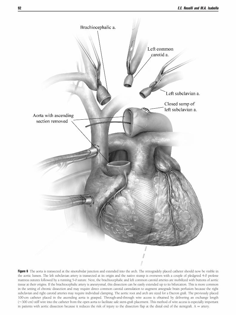
Figure 6 The aorta is transected at the sinotubular junction and extended into the arch. The retrogradely placed catheter should now be visible inthe aortic lumen. The left subclavian artery is transected at its origin and the native stump is oversewn with a couple of pledgeted 4-0 prolenemattress sutures followed by a running 5-0 suture. Next, the brachiocephalic and left common carotid arteries are mobilized with buttons of aortictissue at their origins. If the brachiocephalic artery is aneurysmal, this dissection can be easily extended up to its bifurcation. This is more commonin the setting of chronic dissection and may require direct common carotid cannulation to augment antegrade brain perfusion because the rightsubclavian and right carotid arteries may require individual clamping. The aortic root and arch are sized for a Dacron graft. The previously placed100-cm catheter placed in the ascending aorta is grasped. Through-and-through wire access is obtained by delivering an exchange length(�300 cm) stiff wire into the catheter from the open aorta to facilitate safe stent-graft placement. This method of wire access is especially importantin patients with aortic dissection because it reduces the risk of injury to the dissection flap at the distal end of the stentgraft. A ¼ artery.
E.E. Roselli and M.A. Isabella92
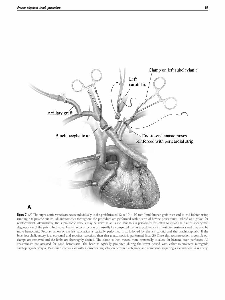
Figure 7 (A) The supra-aortic vessels are sewn individually to the prefabricated 12� 10� 10-mm3 multibranch graft in an end-to-end fashion usingrunning 5-0 prolene suture. All anastomoses throughout the procedure are performed with a strip of bovine pericardium utilized as a gasket forreinforcement. Alternatively, the supra-aortic vessels may be sewn as an island, but this is performed less often to avoid the risk of aneurysmaldegeneration of the patch. Individual branch reconstruction can usually be completed just as expeditiously in most circumstances and may also bemore hemostatic. Reconstruction of the left subclavian is typically performed first, followed by the left carotid and the brachiocephalic. If thebrachiocephalic artery is aneurysmal and requires resection, then that anastomosis is performed first. (B) Once this reconstruction is completed,clamps are removed and the limbs are thoroughly deaired. The clamp is then moved more proximally to allow for bilateral brain perfusion. Allanastomoses are assessed for good hemostasis. The heart is typically protected during the arrest period with either intermittent retrogradecardioplegia delivery at 15-minute intervals, or with a longer-acting solution delivered antegrade and commonly requiring a second dose. A = artery.
Frozen elephant trunk procedure 93
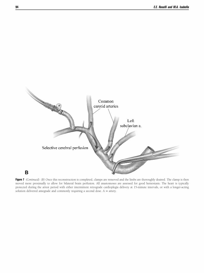
Figure 7 (Continued). (B) Once this reconstruction is completed, clamps are removed and the limbs are thoroughly deaired. The clamp is thenmoved more proximally to allow for bilateral brain perfusion. All anastomoses are assessed for good hemostasis. The heart is typicallyprotected during the arrest period with either intermittent retrograde cardioplegia delivery at 15-minute intervals, or with a longer-actingsolution delivered antegrade and commonly requiring a second dose. A = artery.
E.E. Roselli and M.A. Isabella94
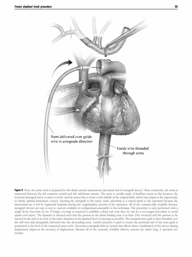
Figure 8 Next, the aortic arch is prepared for the distal sutured anastomosis (proximal end of stentgraft device). Most commonly, the aorta istransected between the left common carotid and left subclavian arteries. The aorta is usually made of healthier tissue in this location, therecurrent laryngeal nerve is easier to avoid, and the suture line is closer to the middle of the surgical field, which may improve the opportunityto obtain optimal hemostatic control. Suturing the stentgraft to the native aortic adventitia is a critical detail to the operation because theaneurysmal sac is fed by segmental branches during the coagulopathic periods of the operation. All of the commercially available thoracicstentgraft devices are easy to sew to, and are available in configurations amenable to this technique. The procedure is only performed with asingle device less than 22 cm. If longer coverage is required to establish a distal seal zone then we opt for a two-staged procedure to avoidspinal cord injury. The diameter is selected such that the portion in the distal landing zone is at least 10% oversized and the portion to besutured in the arch is as close to the native diameter at the planned level of suturing as possible. The prepared stent graft is then threaded overthe stiff wire and antegradely delivered into the descending aorta. Careful attention is paid to ensure the proximal end of the stent graft ispositioned at the level of the transected aortic arch. Choosing a stentgraft delivery system that allows direct visualization of the device duringdeployment improves the accuracy of deployment. Because all of the currently available delivery systems are rather long, 2 operators areneeded.
Frozen elephant trunk procedure 95

Figure 9 After the stent graft is fully deployed, the delivery system is retrieved back through the device and off the field. A long 5-Frangiography catheter is advanced forward again from the groin over the stiff wire. The stiff wire is then removed. The catheter is left in thestent graft for completion of the aortogram at the end of the case.
E.E. Roselli and M.A. Isabella96

Figure 10 The aortic graft is beveled and placed adjacent to the stent graft. The proximal end of the stent graft is included in the anastomosis.The surgical graft, stent graft, and the patient’s native aorta are all included in the distal aortic anastomosis using a running 4-0 prolene suture.Suturing the stentgraft to the native aortic adventitia is a critical detail to the operation because the aneurysmal sac is fed by segmentalbranches initially, and later during the coagulopathic periods of the operation.
Frozen elephant trunk procedure 97
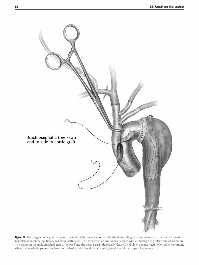
Figure 11 The surgical arch graft is opened near the right greater curve of the distal ascending neoaorta to serve as the site for proximalreimplantation of the multibranched supra-aortic graft. This is sewn in an end-to-side fashion with a running 5-0 prolene-reinforced suture.The clamp on the multibranched graft is removed and the head is again thoroughly deaired. Full flow is reinstituted, followed by rewarmingwhen the metabolic parameters have normalized on the blood gas analysis (typically within a couple of minutes).
E.E. Roselli and M.A. Isabella98
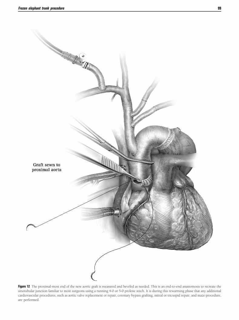
Figure 12 The proximal-most end of the new aortic graft is measured and beveled as needed. This is an end-to-end anastomosis to recreate thesinotubular junction familiar to most surgeons using a running 4-0 or 5-0 prolene stitch. It is during this rewarming phase that any additionalcardiovascular procedures, such as aortic valve replacement or repair, coronary bypass grafting, mitral or tricuspid repair, and maze procedure,are performed.
Frozen elephant trunk procedure 99

Figure 13 After the anastomosis is completed, the aortic cross clampis removed. Hemostasis is assessed and repair stitches placed asnecessary. The heart is allowed to reperfuse for 8 to 15 minutesdepending upon the length of cardiac arrest and severity ofmyocardial remodeling due to left ventricular hypertrophy.
Figure 14 While the heart is reperfusing, the long angiographypigtail catheter is positioned into the ascending aortic graft. TheC-arm is brought back into the operating field for a completionaortogram typically using an injection rate of 15 mL per second for25 mL of 50:50 dilute contrast. To obtain the best view, the C-armis positioned left anterior oblique as steep as is reasonable. Theaortogram is assessed for flow through a patent fully expandedstentgraft and patent branch vessels. The images are also reviewedfor evidence of type 1 or 3 endoleaks, which may require the use ofa conforming balloon that can be delivered through a separatepuncture of the graft and the use of a 12-Fr sheath. After asatisfactory aortogram, the C-arm is docked. The patient is weanedoff cardiopulmonary bypass, and closed in the usual fashion aftersternotomy.
E.E. Roselli and M.A. Isabella100
References1. Lima B, Roselli EE, Soltesz EG, et al: Modified and “reverse” frozen
elephant trunk repairs for extensive disease and complications after stentgrafting. Ann Thorac Surg 93:103–109, 2012
2. Svensson LG, Kim KH, Blackstone EH, et al: Elephant trunk procedure:Newer indications and uses. Ann Thorac Surg 78:109–116, 2004
3. Karck M, Kamiya H: Progress of the treatment for extended aorticaneurysms: Is the frozen elephant trunk technique the next standard inthe treatment of complex aortic disease including the arch? Eur JCardiothorac Surg 33:1007–1013, 2008
4. Safi HJ, Miller 3rd CC, Estrera AL, et al: Optimization of aortic archreplacement: Two-stage approach. Ann Thorac Surg 83:S815–S818, 2007
5. Roselli EE, Subramanian S, Sun Z, et al: Endovascular versus openelephant trunk completion for extensive aortic disease. J ThoracCardiovasc Surg, in press. 2013, doi:10.1016/j.jtcvs.2013.07.070.
6. Roselli EE, Rafael A, Soltesz EG, et al: Simplified frozen elephant trunkrepair for acute DeBakey type I dissection. J Thorac Cardiovasc Surg 145:S197–S201. doi: 10.1016/j.jtcvs.2013.07.070, 2013
7. Tsai TT, Trimarchi S, Nienaber CA: Acute aortic dissection: Perspectivesfrom the International Registry of Acute Aortic Dissection (IRAD). Eur JVasc Endovasc Surg 37:149–159, 2009


![The Frozen Elephant Trunk Technique: European Association ......proach, the elephant trunk (ET) technique described in 1983 by Borst et al. [1]. Over time, this technique evolved [2,3],](https://img.dokumen.tips/doc/110x75/5f0c10807e708231d43391b2/the-frozen-elephant-trunk-technique-european-association-proach-the-elephant.jpg)