Embed Size (px)
Citation preview
BioMed CentralFrontiers in Zoology
ss
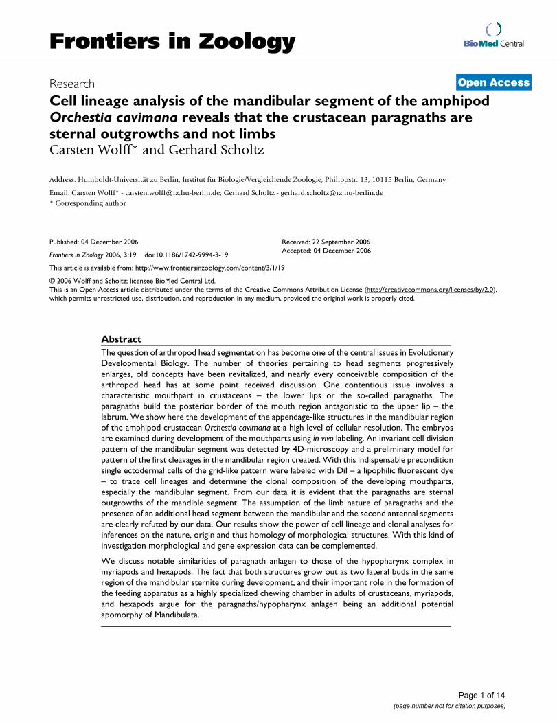
Open AcceResearchCell lineage analysis of the mandibular segment of the amphipod Orchestia cavimana reveals that the crustacean paragnaths are sternal outgrowths and not limbsCarsten Wolff* and Gerhard ScholtzAddress: Humboldt-Universität zu Berlin, Institut für Biologie/Vergleichende Zoologie, Philippstr. 13, 10115 Berlin, Germany
Email: Carsten Wolff* - [email protected]; Gerhard Scholtz - [email protected]
* Corresponding author
AbstractThe question of arthropod head segmentation has become one of the central issues in EvolutionaryDevelopmental Biology. The number of theories pertaining to head segments progressivelyenlarges, old concepts have been revitalized, and nearly every conceivable composition of thearthropod head has at some point received discussion. One contentious issue involves acharacteristic mouthpart in crustaceans – the lower lips or the so-called paragnaths. Theparagnaths build the posterior border of the mouth region antagonistic to the upper lip – thelabrum. We show here the development of the appendage-like structures in the mandibular regionof the amphipod crustacean Orchestia cavimana at a high level of cellular resolution. The embryosare examined during development of the mouthparts using in vivo labeling. An invariant cell divisionpattern of the mandibular segment was detected by 4D-microscopy and a preliminary model forpattern of the first cleavages in the mandibular region created. With this indispensable preconditionsingle ectodermal cells of the grid-like pattern were labeled with DiI – a lipophilic fluorescent dye– to trace cell lineages and determine the clonal composition of the developing mouthparts,especially the mandibular segment. From our data it is evident that the paragnaths are sternaloutgrowths of the mandible segment. The assumption of the limb nature of paragnaths and thepresence of an additional head segment between the mandibular and the second antennal segmentsare clearly refuted by our data. Our results show the power of cell lineage and clonal analyses forinferences on the nature, origin and thus homology of morphological structures. With this kind ofinvestigation morphological and gene expression data can be complemented.
We discuss notable similarities of paragnath anlagen to those of the hypopharynx complex inmyriapods and hexapods. The fact that both structures grow out as two lateral buds in the sameregion of the mandibular sternite during development, and their important role in the formation ofthe feeding apparatus as a highly specialized chewing chamber in adults of crustaceans, myriapods,and hexapods argue for the paragnaths/hypopharynx anlagen being an additional potentialapomorphy of Mandibulata.
Published: 04 December 2006
Frontiers in Zoology 2006, 3:19 doi:10.1186/1742-9994-3-19
Received: 22 September 2006Accepted: 04 December 2006
This article is available from: http://www.frontiersinzoology.com/content/3/1/19
© 2006 Wolff and Scholtz; licensee BioMed Central Ltd. This is an Open Access article distributed under the terms of the Creative Commons Attribution License (http://creativecommons.org/licenses/by/2.0), which permits unrestricted use, distribution, and reproduction in any medium, provided the original work is properly cited.
Page 1 of 14(page number not for citation purposes)
Frontiers in Zoology 2006, 3:19 http://www.frontiersinzoology.com/content/3/1/19
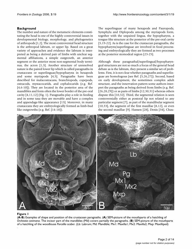
BackgroundThe number and nature of the metameric elements consti-tuting the head is one of the highly controversial issues indevelopmental biology, morphology, and phylogeneticsof arthropods [1,2]. The most controversial head structureis the arthropod labrum, or upper lip. Based on a greatvariety of approaches and evidence the labrum is inter-preted as being a derived pair of limbs with unclear seg-mental affiliations, a simple outgrowth, an anteriorsegment or the anterior most non-segmental body termi-nus, the acron [1,3]. Another structure of unresolvednature is the paired lower lip which is called paragnaths incrustaceans or superlinguae/hypopharynx in hexapodsand some myriapods [4,5]. Paragnaths have beendescribed for malacostracans, branchiopods, copepods,ostracods, mystacocarids, and cephalocarids (e.g. Ref.[4,6-10]). They are located in the posterior area of themandibles and form often the lower border of the pre-oralcavity [4,11,12] (Fig. 1). Paragnaths play a role in feedingand in some taxa they are movable and have a complexand appendage-like appearance [13]. Moreover, in manycrustaceans they are embryologically formed as limb-budlike outgrowths (e.g. Ref. [14-18]).
The superlinguae of many hexapods and Pauropoda,Symphyla and Diplopoda among the myriapods form,together with the unpaired lingua, the hypopharynx, atongue-like structure at the posterior of the pre-oral cavity[5,19-22]. As is the case for the crustacean paragnaths, thehypopharynx/superlinguae are involved in food process-ing and embryologically they are formed as two processesat the posterior stomodeal region [23-25].
Although these paragnathal/superlingual/hypopharyn-geal structures are not so much a focus of the general headdebate as is the labrum, they present a similar set of prob-lems. First, it is not clear whether paragnaths and superlin-guae are homologous (see Ref. [5,26,27]). Second, basedon early development, the sometimes complex adultstructure, and the innervation pattern some authors inter-pret the paragnaths as being derived from limbs (e.g. Ref.[26,28,29]) or as parts of limbs [12,30,31] whereas othersdispute this [10,32]. Third, the segmental relation is seencontroversially either as postoral lip not related to anyparticular segment [7], as part of the mandibular segment[10,33], the segment of the first maxillae [8,12], or eventhe second maxillae [9]. Hansen [28], Denis [34], Chau-
(A-B) Examples of shape and position of the crustacean paragnathsFigure 1(A-B) Examples of shape and position of the crustacean paragnaths. (A) SEM-picture of the mouthparts of a hatchling of Orchestia cavimana. The incisor part of the mandibles (Md) covers partially the paragnaths. (B) SEM-picture of the mouthparts of a hatchling of the woodlouse Porcellio scaber. (Lb: Labrum; Md: Mandible; Mx1: Maxilla1; Mx2: Maxilla2; Mxp: Maxilliped)
Page 2 of 14(page number not for citation purposes)
Frontiers in Zoology 2006, 3:19 http://www.frontiersinzoology.com/content/3/1/19
donneret[26], Laverack [35], and Casanova [29] suggestthat the paragnaths indicate an additional segment eitherbetween mandibular and maxillary segments [28] or thetritocerebral and mandibular segments [26,29,34,35],concluding that the arthropod or mandibulate head com-prises one more segment than generally thought. Lauter-bach [32] hypothesized the origin of the paragnaths insternal folds ("sternale Falten") of ancestral arthropods.According to Lauterbach the paragnaths are the result ofprogressive bulging and fusion of sternal elements of thefirst post-oral head segments in the Mandibulata, thoughonly in some crustacean taxa do these folded sternal for-mations ("Faltenbildungen") have an appendage-likeappearance.
Here we address the problem of the nature and origin ofcrustacean paragnaths with a cell lineage approach. Toresolve the segmental affinities of the paragnaths we studythe cell lineage in the area around the mandibular seg-ment of the freshwater beach-hopper, the amphipod crus-tacean Orchestia cavimana. This animal is well suited forthis kind of investigation because it forms a pair of largebuds of paragnaths during embryonic development [17].Furthermore, as an amphipod representative its stereo-typic cell division pattern in the post-naupliar region (seg-ments of the first maxillae to the terminal segment of thepleon) is known (for this see Fig. 2D) and has beendescribed up to the formation of morphological structuressuch as limbs, segments, and ganglia (Fig. 2E) [36]. Incontrast to the well studied post-naupliar region, the proc-esses of cell division and morphogenesis of the segmentsof the first and second antennae and the mandible (i.e.the naupliar region) are less known. This is due to themore irregular mode of cell division which, apart fromsome indications in the posterior mandibular region,does not show an obvious stereotypic pattern [36,37].
We combine the methods of 4D-microscopy [38] and thein-vivo labeling of single cells with the fluorescent dye DiI[39] to resolve the cell division pattern in the posteriornaupliar region to trace the origin and formation of theparagnaths and other mouthparts by analyzing the clonalcomposition of the mandibular segment and adjacentareas.
It can be shown that the posterior region of the mandibu-lar segment shows an unexpected degree of cell divisiondetermination with a reproducible cell lineage. The clear-cut results of our study shed new light on the segmenta-tion pattern of crustacean heads by dismissing some olderhypotheses on the origin and nature of paragnaths. Thecomparison and discussion of putative homologous struc-tures in other arthropod taxa offer new perspectives onarthropod heads in general.
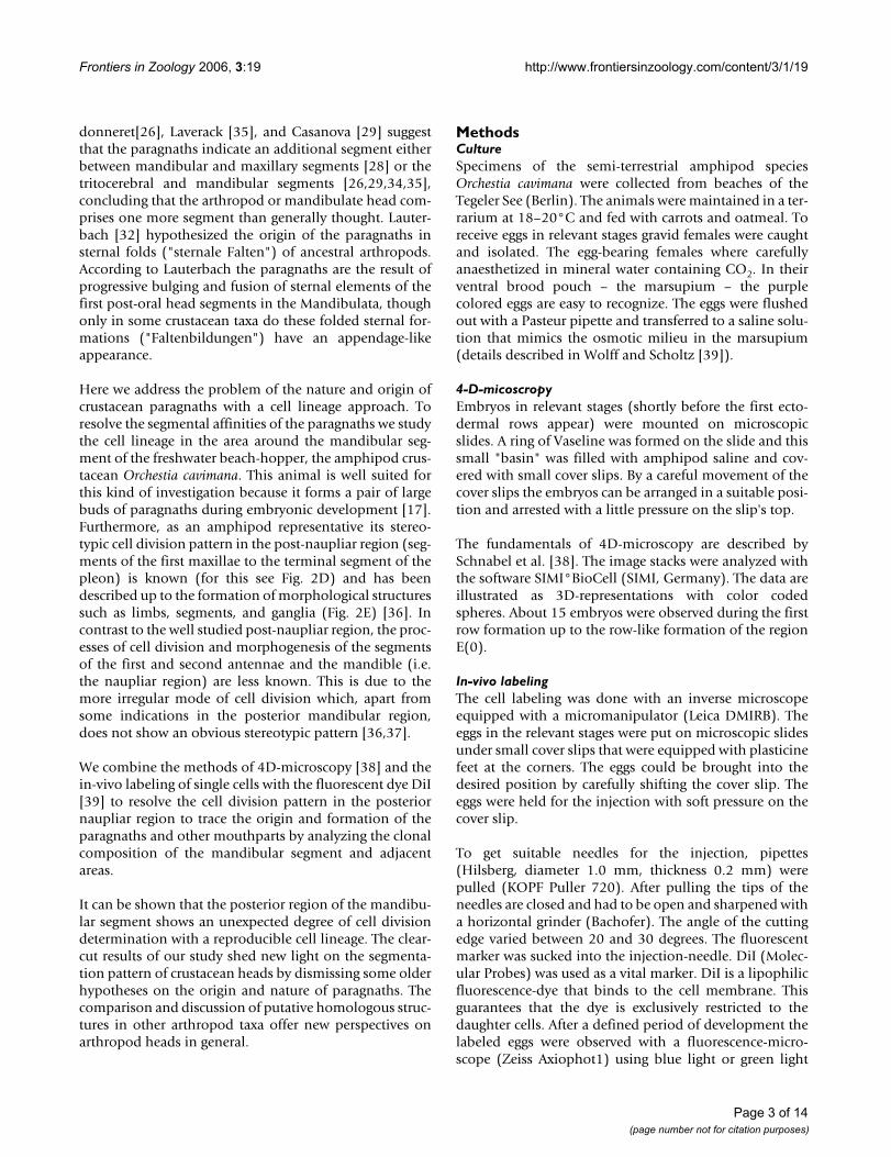
MethodsCultureSpecimens of the semi-terrestrial amphipod speciesOrchestia cavimana were collected from beaches of theTegeler See (Berlin). The animals were maintained in a ter-rarium at 18–20°C and fed with carrots and oatmeal. Toreceive eggs in relevant stages gravid females were caughtand isolated. The egg-bearing females where carefullyanaesthetized in mineral water containing CO2. In theirventral brood pouch – the marsupium – the purplecolored eggs are easy to recognize. The eggs were flushedout with a Pasteur pipette and transferred to a saline solu-tion that mimics the osmotic milieu in the marsupium(details described in Wolff and Scholtz [39]).
4-D-micoscropyEmbryos in relevant stages (shortly before the first ecto-dermal rows appear) were mounted on microscopicslides. A ring of Vaseline was formed on the slide and thissmall "basin" was filled with amphipod saline and cov-ered with small cover slips. By a careful movement of thecover slips the embryos can be arranged in a suitable posi-tion and arrested with a little pressure on the slip's top.
The fundamentals of 4D-microscopy are described bySchnabel et al. [38]. The image stacks were analyzed withthe software SIMI°BioCell (SIMI, Germany). The data areillustrated as 3D-representations with color codedspheres. About 15 embryos were observed during the firstrow formation up to the row-like formation of the regionE(0).
In-vivo labelingThe cell labeling was done with an inverse microscopeequipped with a micromanipulator (Leica DMIRB). Theeggs in the relevant stages were put on microscopic slidesunder small cover slips that were equipped with plasticinefeet at the corners. The eggs could be brought into thedesired position by carefully shifting the cover slip. Theeggs were held for the injection with soft pressure on thecover slip.
To get suitable needles for the injection, pipettes(Hilsberg, diameter 1.0 mm, thickness 0.2 mm) werepulled (KOPF Puller 720). After pulling the tips of theneedles are closed and had to be open and sharpened witha horizontal grinder (Bachofer). The angle of the cuttingedge varied between 20 and 30 degrees. The fluorescentmarker was sucked into the injection-needle. DiI (Molec-ular Probes) was used as a vital marker. DiI is a lipophilicfluorescence-dye that binds to the cell membrane. Thisguarantees that the dye is exclusively restricted to thedaughter cells. After a defined period of development thelabeled eggs were observed with a fluorescence-micro-scope (Zeiss Axiophot1) using blue light or green light
Page 3 of 14(page number not for citation purposes)
Frontiers in Zoology 2006, 3:19 http://www.frontiersinzoology.com/content/3/1/19
Page 4 of 14(page number not for citation purposes)
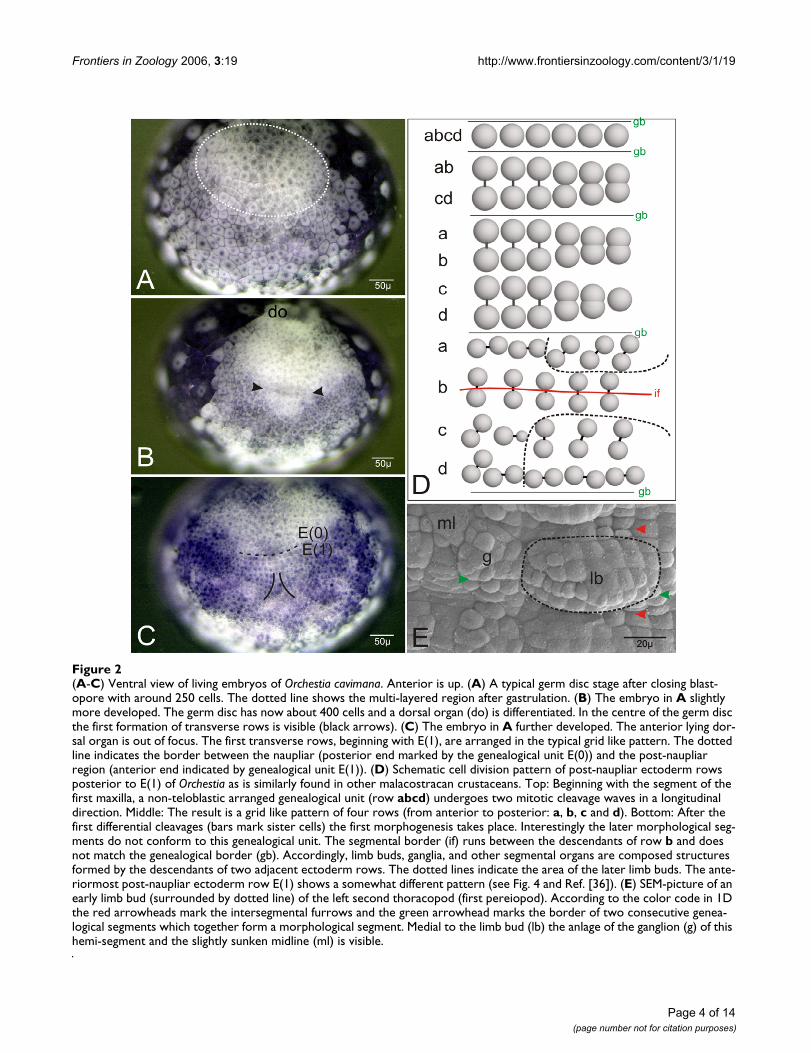
(A-C) Ventral view of living embryos of Orchestia cavimanaFigure 2(A-C) Ventral view of living embryos of Orchestia cavimana. Anterior is up. (A) A typical germ disc stage after closing blast-opore with around 250 cells. The dotted line shows the multi-layered region after gastrulation. (B) The embryo in A slightly more developed. The germ disc has now about 400 cells and a dorsal organ (do) is differentiated. In the centre of the germ disc the first formation of transverse rows is visible (black arrows). (C) The embryo in A further developed. The anterior lying dor-sal organ is out of focus. The first transverse rows, beginning with E(1), are arranged in the typical grid like pattern. The dotted line indicates the border between the naupliar (posterior end marked by the genealogical unit E(0)) and the post-naupliar region (anterior end indicated by genealogical unit E(1)). (D) Schematic cell division pattern of post-naupliar ectoderm rows posterior to E(1) of Orchestia as is similarly found in other malacostracan crustaceans. Top: Beginning with the segment of the first maxilla, a non-teloblastic arranged genealogical unit (row abcd) undergoes two mitotic cleavage waves in a longitudinal direction. Middle: The result is a grid like pattern of four rows (from anterior to posterior: a, b, c and d). Bottom: After the first differential cleavages (bars mark sister cells) the first morphogenesis takes place. Interestingly the later morphological seg-ments do not conform to this genealogical unit. The segmental border (if) runs between the descendants of row b and does not match the genealogical border (gb). Accordingly, limb buds, ganglia, and other segmental organs are composed structures formed by the descendants of two adjacent ectoderm rows. The dotted lines indicate the area of the later limb buds. The ante-riormost post-naupliar ectoderm row E(1) shows a somewhat different pattern (see Fig. 4 and Ref. [36]). (E) SEM-picture of an early limb bud (surrounded by dotted line) of the left second thoracopod (first pereiopod). According to the color code in 1D the red arrowheads mark the intersegmental furrows and the green arrowhead marks the border of two consecutive genea-logical segments which together form a morphological segment. Medial to the limb bud (lb) the anlage of the ganglion (g) of this hemi-segment and the slightly sunken midline (ml) is visible.
Frontiers in Zoology 2006, 3:19 http://www.frontiersinzoology.com/content/3/1/19
(strongest stimulation of DiI), and the results were docu-mented with a digital camera (Nikon D1).
CLSM and 3D-reconstructionFor fixation and documentation on the laser scanningmicroscope (Leica SP2) the embryos were dissected inPBS-buffered 4% formaldehyde-solution, counterstainedwith nuclear staining dye (Hoechst) and mounted inDABCO-Glycerol (25 mg DABCO (1,4 diazabicyclol-2,2,2-octane, Merck) in 1 ml PBS to 9 ml glycerol), whichis an anti-bleaching-detergent. The image stacks producedby the laser scanning microscope were analyzed with thesoftware Imaris 5.0.1 (Bitplane AG). The 3D-reconstruc-tion of the counter staining (Hoechst) and the clones ofthe in-vivo labeled cell have the advantage of very highresolution with respect to morphological data. The feature"Volume" in the program module "Surpass" created athree dimensional object, which can be magnified andmoved in all directions. For better visualization of theobjects, movies (AVI-files) were created in the programmodule "Animation".
NomenclatureFor the following description we adopt the commonnomenclature for malacostracan crustacean cell lineageswhich was modified for amphipods by Scholtz [36]. Inaddition we introduce a nomenclature for the ectodermrow E(0). The anterior row of E(0) is named E(0)a and theposterior row E(0)p. If after the subsequent divisions acell lies anterior it is again labeled with an "a". The poste-rior sister cell is labeled correspondingly with "p". As inmore posterior rows the cells in positions to the midline(these cells and their early descendants are designated ascolumns) are numbered consecutively from the middletowards lateral (e.g. E(0)p1).
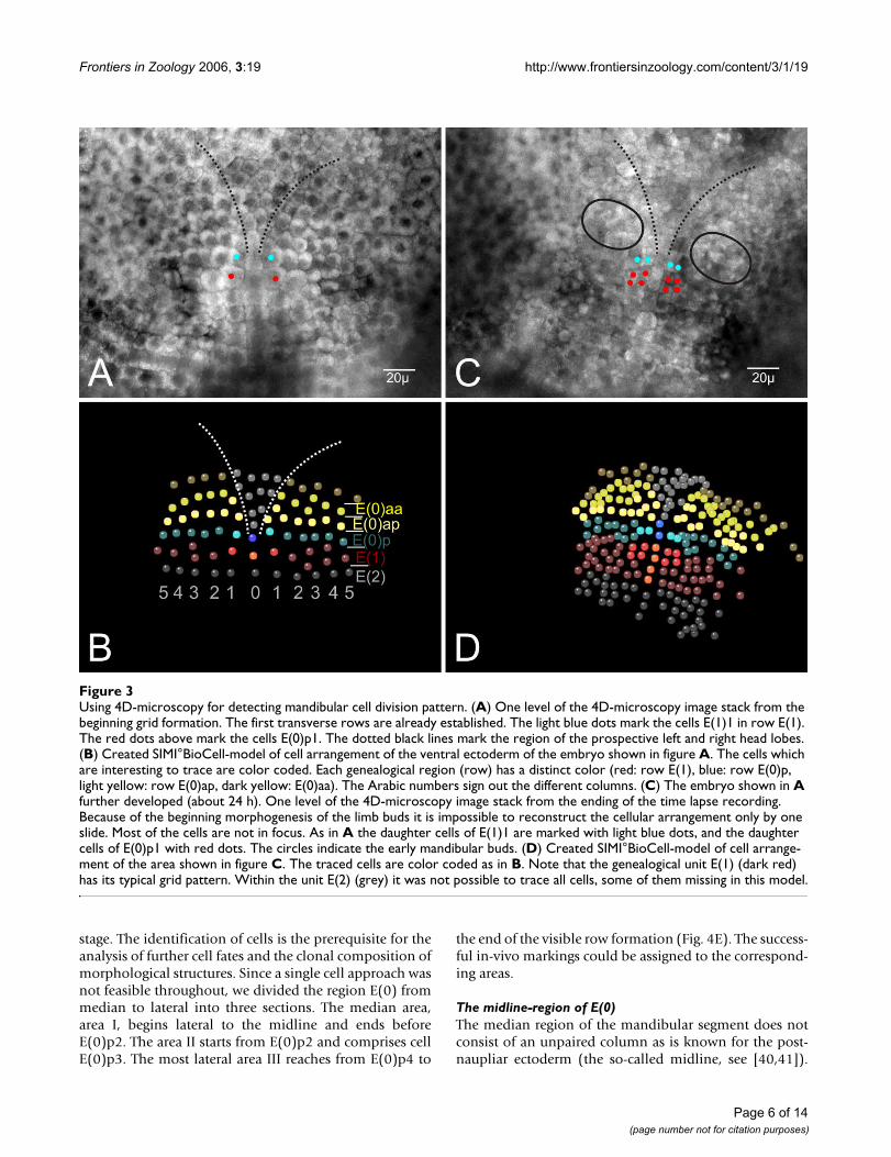
ResultsThe early cell lineage of the mandibular region (E(0))After gastrulation, the blastopore is closed. The germ discconsists of about 250 cells (Fig. 2A). The descendants ofthe macromeres B (left) and D (right) form the anteriorparts of the germ disc by migration towards the middle(for details see Wolff and Scholtz [39]). At this time thereis no regular cellular pattern in the ventral ectoderm. Thefirst recognizable transverse ectoderm row occurs in agerm disc with about 400 cells (Fig. 2B). This row marksthe border between the naupliar (head lobes, the seg-ments of the first and second antennae and of the mandi-bles) and the post-naupliar (the segment of the firstmaxillae up to the telson anlage) regions of the develop-ing embryo. This first recognizable cell row forms thegenealogical unit E(1) which provides most of the mate-rial for the segment of the first maxilla (Fig. 2C). The cellsanterior to E(1) show a typical spatial configuration.According to Scholtz [36] we named this region E(0),
which forms part of the prospective mandibular segment(Fig. 2C). The cells of E(0) are somewhat smaller androunder than the more posterior cells. They are arrangedin two rows of at least 6 cells on either body half (Fig. 2C,3A–B). The anterior row is designated as E(0)a and theposterior row as E(0)p (Fig. 3A,B). It is not clear whetherthese two rows originate from one transverse ectodermrow by longitudinally oriented cell divisions. A distinctunpaired column of midline cells as is typical for the post-naupliar region (Fig. 2E) does not exist in the naupliarpart of the germ band. In the median area of the posteriornaupliar region E(0) we found about 10–15 smaller cellsin a V-shaped arrangement (Fig. 3A,B).
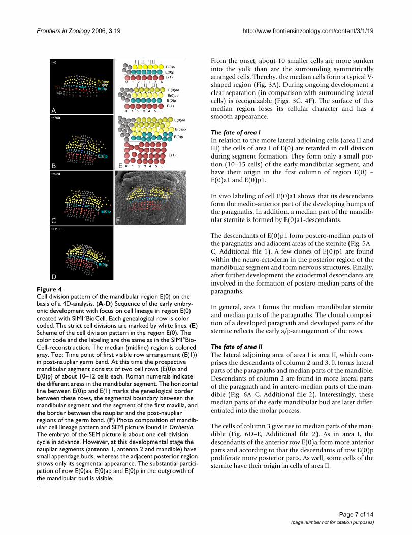
At about this stage, cells of the anterior row E(0)a dividemore or less synchronously (mitotic wave) in a longitudi-nal direction. The products are the anterior row E(0)aaand the posterior row E(0)ap (Fig. 3A,B). The followingdivision pattern of cells in row E(0)aa is not clear in detailbut the cells divide symmetrically in each body half (seesupplemental material). In row E(0)ap a detailed patterncould not completely be described but some features ofthe more medially lying cells of this row could be traced.The cells E(0)ap1 und E(0)ap2 are the first to divide in alongitudinal direction whereas E(0)ap3 divides a bit laterin a horizontal direction (Fig. 4E). The cell E(0)ap4 showsa delayed division. In relation to the division cycles in rowE(0)a, the cells of E(0)p are delayed in their division.
During the second mitotic wave in row E(0)aa, E(0)p2 isthe first cell that divides in row E(0)p. It divides in a lon-gitudinal direction. More or less at the same time the mid-line cell E(0)p0 divides also in a longitudinal direction.Subsequently the cells E(0)p3, E(0)p5, and E(0)p6 dividelikewise in a longitudinal direction E(0)p2 (Fig. 4B,C). Atthe same time the relatively large cell E(0)p1 divides in ahorizontal direction (Fig. 4C). At last, the cell E(0)p4undergoes a division, again in a horizontal direction (Fig.4D). In this developmental stage small buds of the nau-pliar appendages (first and second antennae, mandible)become visible (Fig. 4F). This invariant cleavage pattern inthe prospective mandibular region eventually produces areproducible arrangement of cells in the posterior regionof E(0)(Fig. 4E).
This is an important pre-condition for our detailed lineagestudy of the mandibular segment. Unfortunately, wecould not reconstruct the complete lineage pattern of themore anterior lying region E(0)a.
The clonal composition of the mandibular segment and its appendagesIdentified cells of the transverse ectoderm rows aroundthe boundary between the naupliar and post-naupliarregions were labeled in-vivo during the early germ band
Page 5 of 14(page number not for citation purposes)
Frontiers in Zoology 2006, 3:19 http://www.frontiersinzoology.com/content/3/1/19
stage. The identification of cells is the prerequisite for theanalysis of further cell fates and the clonal composition ofmorphological structures. Since a single cell approach wasnot feasible throughout, we divided the region E(0) frommedian to lateral into three sections. The median area,area I, begins lateral to the midline and ends beforeE(0)p2. The area II starts from E(0)p2 and comprises cellE(0)p3. The most lateral area III reaches from E(0)p4 to
the end of the visible row formation (Fig. 4E). The success-ful in-vivo markings could be assigned to the correspond-ing areas.
The midline-region of E(0)The median region of the mandibular segment does notconsist of an unpaired column as is known for the post-naupliar ectoderm (the so-called midline, see [40,41]).
Using 4D-microscopy for detecting mandibular cell division patternFigure 3Using 4D-microscopy for detecting mandibular cell division pattern. (A) One level of the 4D-microscopy image stack from the beginning grid formation. The first transverse rows are already established. The light blue dots mark the cells E(1)1 in row E(1). The red dots above mark the cells E(0)p1. The dotted black lines mark the region of the prospective left and right head lobes. (B) Created SIMI°BioCell-model of cell arrangement of the ventral ectoderm of the embryo shown in figure A. The cells which are interesting to trace are color coded. Each genealogical region (row) has a distinct color (red: row E(1), blue: row E(0)p, light yellow: row E(0)ap, dark yellow: E(0)aa). The Arabic numbers sign out the different columns. (C) The embryo shown in A further developed (about 24 h). One level of the 4D-microscopy image stack from the ending of the time lapse recording. Because of the beginning morphogenesis of the limb buds it is impossible to reconstruct the cellular arrangement only by one slide. Most of the cells are not in focus. As in A the daughter cells of E(1)1 are marked with light blue dots, and the daughter cells of E(0)p1 with red dots. The circles indicate the early mandibular buds. (D) Created SIMI°BioCell-model of cell arrange-ment of the area shown in figure C. The traced cells are color coded as in B. Note that the genealogical unit E(1) (dark red) has its typical grid pattern. Within the unit E(2) (grey) it was not possible to trace all cells, some of them missing in this model.
Page 6 of 14(page number not for citation purposes)
Frontiers in Zoology 2006, 3:19 http://www.frontiersinzoology.com/content/3/1/19
From the onset, about 10 smaller cells are more sunkeninto the yolk than are the surrounding symmetricallyarranged cells. Thereby, the median cells form a typical V-shaped region (Fig. 3A). During ongoing development aclear separation (in comparison with surrounding lateralcells) is recognizable (Figs. 3C, 4F). The surface of thismedian region loses its cellular character and has asmooth appearance.
The fate of area IIn relation to the more lateral adjoining cells (area II andIII) the cells of area I of E(0) are retarded in cell divisionduring segment formation. They form only a small por-tion (10–15 cells) of the early mandibular segment, andhave their origin in the first column of region E(0) –E(0)a1 and E(0)p1.
In vivo labeling of cell E(0)a1 shows that its descendantsform the medio-anterior part of the developing humps ofthe paragnaths. In addition, a median part of the mandib-ular sternite is formed by E(0)a1-descendants.
The descendants of E(0)p1 form postero-median parts ofthe paragnaths and adjacent areas of the sternite (Fig. 5A–C, Additional file 1). A few clones of E(0)p1 are foundwithin the neuro-ectoderm in the posterior region of themandibular segment and form nervous structures. Finally,after further development the ectodermal descendants areinvolved in the formation of postero-median parts of theparagnaths.
In general, area I forms the median mandibular sterniteand median parts of the paragnaths. The clonal composi-tion of a developed paragnath and developed parts of thesternite reflects the early a/p-arrangement of the rows.
The fate of area IIThe lateral adjoining area of area I is area II, which com-prises the descendants of column 2 and 3. It forms lateralparts of the paragnaths and median parts of the mandible.Descendants of column 2 are found in more lateral partsof the paragnath and in antero-median parts of the man-dible (Fig. 6A–C, Additional file 2). Interestingly, thesemedian parts of the early mandibular bud are later differ-entiated into the molar process.
The cells of column 3 give rise to median parts of the man-dible (Fig. 6D–E, Additional file 2). As in area I, thedescendants of the anterior row E(0)a form more anteriorparts and according to that the descendants of row E(0)pproliferate more posterior parts. As well, some cells of thesternite have their origin in cells of area II.
Cell division pattern of the mandibular region E(0) on the basis of a 4D-analysisFigure 4Cell division pattern of the mandibular region E(0) on the basis of a 4D-analysis. (A-D) Sequence of the early embry-onic development with focus on cell lineage in region E(0) created with SIMI°BioCell. Each genealogical row is color coded. The strict cell divisions are marked by white lines. (E) Scheme of the cell division pattern in the region E(0). The color code and the labeling are the same as in the SIMI°Bio-Cell-reconstruction. The median (midline) region is colored gray. Top: Time point of first visible row arrangement (E(1)) in post-naupliar germ band. At this time the prospective mandibular segment consists of two cell rows (E(0)a and E(0)p) of about 10–12 cells each. Roman numerals indicate the different areas in the mandibular segment. The horizontal line between E(0)p and E(1) marks the genealogical border between these rows, the segmental boundary between the mandibular segment and the segment of the first maxilla, and the border between the naupliar and the post-naupliar regions of the germ band. (F) Photo composition of mandib-ular cell lineage pattern and SEM picture found in Orchestia. The embryo of the SEM picture is about one cell division cycle in advance. However, at this developmental stage the naupliar segments (antenna 1, antenna 2 and mandible) have small appendage buds, whereas the adjacent posterior region shows only its segmental appearance. The substantial partici-pation of row E(0)aa, E(0)ap and E(0)p in the outgrowth of the mandibular bud is visible.
Page 7 of 14(page number not for citation purposes)

Frontiers in Zoology 2006, 3:19 http://www.frontiersinzoology.com/content/3/1/19
In general, area II is responsible for the formation of lat-eral parts of the paragnaths and more median parts of themandibles.
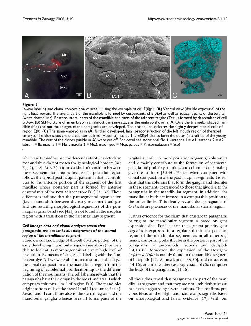
The fate of area IIIArea III lies laterally adjacent to area II and comprises col-umns 4 and 5 of region E(0). They form lateral parts of themandible and neighboring parts of the tergite. That is whythe cell E(0)p4 produces cell material for the outer (pos-tero-lateral) part of the mandible and lateral borderingtergite (Fig. 7A–C, Additional file 3). In contrast, descend-ants of row E(0)a in area III form more anterior parts ofthe outer (antero-lateral) mandible and parts of the adja-cent tergite. Remarkably, the lateral portion of the mandi-bles differentiates by cell proliferation into theprospective pars incisiva.
Summarizing for area III, column 4 forms lateral parts ofthe mandibles and adjacent tergites and column 5 is notinvolved in the development of mandibles or paragnaths.
Surrounding areas of E(0)anteriorThe fate of the cells anterior to the region of E(0) has notyet been resolved in detail. Furthermore, it is not clear ifthere is a stereotypic cell division pattern at all formingthe anterior embryonic head. Some labeling reveals thatmedian cells of E(0) form part of the stomodaeum andmore lateral cells form part of the second antennae.
posteriorBeginning with row E(1) the typical grid-like pattern ofthe post-naupliar germ band of Orchestia is established.
The cell division pattern of E(1) differs somewhat fromthat of the more posterior rows (compare Figs. 2D and 4E)(for details see [36]). The 4-D analysis in row E(1) showsthat its descendant cells are not involved in the formationof the mandibular segment (Fig. 4E) Hence, in contrast tothe post-naupliar segments, in the mandibular segmentthe posterior genealogical boundary corresponds with thesegment border.
Discussion and conclusionA stereotyped cell division pattern is found at the posterior border of the mandibular segmentLike all malacostracan crustaceans studied in this respect(for a recent review see Dohle et al. [42]), amphipodsshow a stereotyped cell divisions pattern in the post-nau-pliar region during growth, differentiation and segmenta-tion of the germ band [36,43,44]. In contrast to this, thenaupliar region does not exhibit an obvious stereotypedcell division pattern [37,42]. Only Scholtz [36] suggestedthat there might be a certain regularity in the divisions andarrangements of the posterior cells of the developingmandibular segment in the amphipod Gammarus pulexbut with the methods then at hand the details were notresolvable With the technique of 4D-microscopy we havebeen able to provide the first evidence for an invariant celldivision pattern in the naupliar region of another amphi-pod species, the freshwater beach hopper Orchestia cavi-mana. At least in the posterior part of the region of E(0) werecognized a relatively strict cell division pattern. This pat-tern has only superficial similarities to the post-naupliarcell division pattern of malacostracan crustaceans but it isnot as elaborated in terms of timing of mitoses, cell size,and the spatial arrangement of the resulting cells. In E(0)
In-vivo labeling of area I in region E(0)Figure 5In-vivo labeling of area I in region E(0). (A) Double exposure of an in-vivo labeled embryo. The ventral ectodermal rows are marked with white lines and labeled with nomenclature. The fluorescent dye is completely diffused in the membrane of cell E(0)p1(of the left body half). (B) Same embryo as in A further developed. Ventral view of head region. The descendants of the marked cell form postero-median parts of the sternite (blurry because not in focus) and of the mandible (Md). (C) Embryo as in B further developed. Ventral view of the left mandibular segment. The mandible consists of two lobes, the inner pars molaris (Pm) and the outer pars incisivus (Pi). Few E(0)p1-clones form postero-median parts of the paragnath hump (Pg). The sternal clones seen in B are out of focus.
Page 8 of 14(page number not for citation purposes)

Frontiers in Zoology 2006, 3:19 http://www.frontiersinzoology.com/content/3/1/19
the sequence of the individual divisions is not as strict andthe direction of the mitotic spindles is more or less longi-tudinally oriented. It appears that the posterior border ofthe mandibular segment forms some kind of transitionbetween the more irregular divisions and cell arrange-ments of the anterior naupliar region and the highly com-plex stereotyped post-naupliar patterns.
It is not clear whether our findings of a regular pattern inthe posterior part of the mandibular segment holds truefor other malacostracans as well, although some datafrom the isopod Porcellio scaber hint to that possibility[45]. However, the cellular events in the corresponding
region in Porcellio are much more irregular when com-pared with those in Orchestia. It has been even shown forPorcellio that some cells of the most anterior row of thepost-naupliar segments can migrate into the posterior areaof the mandible segment [45], a phenomenon that doesnot occur in Orchestia.
Interestingly enough, our results reveal that the posteriorsegmental boundary of the mandibular segment corre-sponds to the genealogical border between rows E(0) andE(1), i.e. E(1) does not contribute to the posterior part ofthe mandibular segment. This stands in contrast to allmore posterior post-naupliar segmental boundaries
In-vivo labeling of area II in region E(0)Figure 6In-vivo labeling of area II in region E(0). The images are in ventral view and double exposure. (A-C) Labeling of cell E(0)a2 of the left body half. (A)Anterior embryonic region which at this time lacks visible paragnath buds. The descendants of E(0)a2 form anterior parts of the mandible bud (Md) as well as parts of the developing sternite. (B) Left gnathal region of the embryo as in A further developed. The small palp (P) of the maxilla 1 (Mx1) is now formed. There are two distinct clone domains. One forms anterior part of the small paragnath bud (Pg), the other forms anterior parts of the developing mandible (Md). (C) Left mandibular region of the embryo as in B after further development. The paragnaths grow out and the mandible differentiates into two lobes. The clonal composition of the descendants of E(0)a2 is more or less the same as in (E). (D-E) Labeling of cell E(0)p3 of the left body half. (D) Ventral view of the mouth region. The descendants of the marked cell form posterior parts of the mandibular bud (Md), but they do not form parts of the anlage of the paragnath (Pg). The arrow head shows the oil drop of the marking. (E) Same embryo as in D slightly further developed. Imaris-reconstruction of the left gnathal region. The blue spots represent the counter-stained (Hoechst) nuclei. The descendants of E(0)p3 are involved in the formation of posterior parts of the mandible, which now consists of two well developed lobes. For detail see Additional file 2.
Page 9 of 14(page number not for citation purposes)
Frontiers in Zoology 2006, 3:19 http://www.frontiersinzoology.com/content/3/1/19
which are formed within the descendants of one ectodermrow and thus do not match the genealogical borders (seeFig. 2), [42]. Row E(1) forms a kind of transition betweenthese segmentation modes because its posterior regionfollows the typical post-naupliar pattern in that it contrib-utes to the anterior portion of the segment of the firstmaxillae whose posterior part is formed by anteriordescendants of the next adjacent row E(2) [36,37]. Thesedifferences indicate that the parasegmental organization(i.e. a frame-shift between the early metameric anlagenand the resulting morphological segments) of the post-naupliar germ band (see [42]) is not found in the naupliarregion with a transition in the first maxillary segment.
Cell lineage data and clonal analyses reveal that paragnaths are not limbs but outgrowths of the sternal region of the mandibular segmentBased on our knowledge of the cell division pattern of theearly developing mandibular region (see above) we wereable to look at its morphogenesis at a very high level ofresolution. By means of single cell labeling with the fluo-rescent dye DiI we were able to reconstruct and analyzethe clonal composition of the mandibular region from thebeginning of ectodermal proliferation up to the differen-tiation of the mouthparts. The cell labeling reveals that theparagnaths have their origin in the area I and area II whichcomprises columns 1 to 3 of region E(0). The mandiblesoriginate from cells of the areas II and III (columns 2 to 4).Areas I and II contribute also to the sternal region and themandibular ganglia whereas area III forms parts of the
tergites as well. In more posterior segments, columns 1and 2 mainly contribute to the formation of segmentalganglia and probably sternites, and columns 3 to 5 mainlygive rise to limbs [36,46]. Hence, when compared withclonal composition of the post-naupliar segments it is evi-dent that the columns that form the ganglia and sternitesin these segments correspond to those that give rise to theparagnaths in the mandibular segment. In addition, themandibular buds are formed in a comparable position tothe other limbs. This clearly reveals that paragnaths ofOrchestia are processes of the mandibular sternal region.
Further evidence for the claim that crustacean paragnathsbelong to the mandibular segment is based on geneexpression data. For instance, the segment polarity geneengrailed is expressed in a regular stripe in the posteriorregion of the mandibular segment, as in all other seg-ments, comprising cells that form the posterior part of theparagnaths in amphipods, isopods and decapods[14,18,37]. Moreover, the expression of the Hox-geneDeformed (Dfd) is mainly found in the mandible segmentof hexapods [47,48], myriapods [49,50], and crustaceans[14,16], and in the latter case expression of Dfd comprisesthe buds of the paragnaths [14,16].
All these data reveal that paragnaths are part of the man-dibular segment and that they are not limb derivatives ashas been suggested by several authors. This confirms pre-vious ideas on the origin and nature of paragnaths basedon embryological and larval evidence [17]. With our
In-vivo labeling and clonal composition of area III using the example of cell E(0)p4Figure 7In-vivo labeling and clonal composition of area III using the example of cell E(0)p4. (A) Ventral view (double exposure) of the right head region. The lateral part of the mandible is formed by descendants of E(0)p4 as well as adjacent parts of the tergite (white dotted line). Postero-lateral parts of the mandible and parts of the adjacent tergite (Ter) is formed by descendant of cell E(0)p4. (B) SEM-picture of an embryo in an almost the same stage as the embryo shown in A. Only the triangular shaped man-dible (Md) and not the anlagen of the paragnaths are developed. The dotted line indicates the slightly deeper medial cells of region E(0). (C) The same embryo as in (A) further developed. Imaris-reconstruction of the left mouth region of the fixed embryo. The blue spots are the counter-stained (Hoechst) nuclei. The E(0)p4-clones form the outer (lateral) tip of the young mandible. The rest of the clones (visible in A) were cut off. For detail see Additional file 3. (antenna 1 = A1; antenna 2 = A2; labrum = lb; maxilla 1 = Mx1; maxilla 2 = Mx2; maxilliped = Mxp; palpus = P; stomodaeum = Sto)
Page 10 of 14(page number not for citation purposes)

Frontiers in Zoology 2006, 3:19 http://www.frontiersinzoology.com/content/3/1/19
clonal analysis we can definitely rule out the possibilitythat paragnaths indicate the existence of an additionalhead segment as has been claimed for example by Casa-nova [29], Chaudonneret [26], Denis [34], and Hansen[28]. Furthermore, the limb-bud like early appearance ofthe paragnaths in Orchestia and other crustaceans is noindication for a limb-related nature of these structures butis only a superficial similarity that does not represent agenealogical relation. Our results show the power of celllineage and clonal analyses for inferences on the nature,origin and thus homology of morphological structures.With this kind of investigation morphological and geneexpression data can be complemented.
In many cases the origin of the paragnaths during crusta-cean embryonic and larval development has either notbeen specified (e.g. Ref. [51-53]) or it has been suggestedthat paragnaths develop from mandibular and/or maxil-lary segments (e.g. Ref. [54]). A look at the correspondingfigures in these articles with our results in mind revealsthat it is possible in almost all examples to relate the par-agnathal structures to the mandibular segment (e.g. Man-ton [51], plate 24, fig. 27; Manton [52], plate 25, fig. 23;Moeller [53], figs. 2, 7). Even the median lobe of the so-called lower lip of the raptorial cladoceran Leptodorakindtii might represent fused paragnaths [55]. Accord-ingly, we tentatively conclude that the paragnaths lobesare homologous throughout Crustacea. Stein et al. [56]suggest that a pair of paragnaths humps is an apomorphyfor a crustacean subgroup comprising Eucrustacea andPhosphatocopina (Labrophora). However, these authorsdid not consider similar structures in myriapods andhexapods which indicate a much more widespread occur-rence among euarthropods. (see next chapter).
Are the crustacean paragnaths homologous to the superlinguae in hexapods and myriapods?There are several reports of post-oral paired bud-like anla-gen in myriapods (e.g. Chilopoda: Heymons [57], Progo-neata: Tiegs [19]) and Hexapoda (e.g. Ref. [24,25,58-60]).In dicondylian hexapods the situation is somewhatambiguous. Larink [61] and Scholl [62] report for Lepismaand Carausius an undivided early hypopharynx anlagewhereas Rohrschneider [24] for Periplaneta and Ibrahim[63] for Tachycines describe two separated buds. Whetherthese are factual differences is not clear. However, thepresence of paired buds in a number of pterygotes as wellas collembolans, diplurans, and archaeognathans allowsthe conclusion that these paired buds were present in thehexapod stem species. Nevertheless, all these buds have incommon that they originate from the sternum of the man-dible segment directly ventral to the mandibular ganglionanlagen and between the mandibular limb buds, afterthese have formed. This very much resembles the earlystages of crustacean paragnaths (see above,
[14,17,18,51,52]). These sternal buds of the mandibularsegment give either rise to the superlinguae, paired laterallobes of the hypopharynx, in Symphyla [19], Collembola[58,59], Diplura, Archaeognatha [25], or to (another partof) the hypopharynx [22,24,57] when superlinguae arenot present as for instance is the case in many Pterygotaand all Chilopoda [22,64]. However, the contribution ofthese mandibular sternal buds to the hypopharynx body(linguae) is interpreted controversially. The hypopharynxof Hexapoda and Progoneata is thought to be a compositestructure formed by protrusions of the sternites of differ-ent numbers of gnathal segments (e.g. Ref.[19,22,25,61,62]). In contrast to this, Haget [65] andWada [66] suggest, based on their experimental teratolog-ical studies that the entire hypopharynx originates fromthe mandibular sternites. According to Heymons [57],this is also the case in Chilopoda. The situation is furthercomplicated by the fact that buds appearing in the inter-calary segment of hexapods are called "hypopharyngeallobes" (Hypopharynxhöcker) and have been suggested toform (part of) the hypopharynx, an obvious misinterpre-tation (see Ref. [23,63,67]). Reading the numerous arti-cles dealing with this problem it is evident thatinvestigations focusing on the differentiation of thehypopharynx in myriapods and hexapods using modernapproaches are badly needed to solve this issue.
Authors such as Crampton [27], Snodgrass [68], andBitsch and Bitsch [5] suggest that the paragnaths and thesuperlinguae are homologous. However, based on ourdata and what we find in the above discussed literatureabout the development of the hexapod and myriapodhypopharyngeal complex, we think that the conclusion ofthese authors is simplifying the matters because parag-naths cannot be homologous to superlinguae alone if themajor part of the hypopharynx of myriapods and hexa-pods originates from the mandibular sternites as well.Accordingly, we modify the homology statement concern-ing crustacean paragnaths and the myriapod/hexapodsuperlinguae/hypopharynx by suggesting that the earlyanlagen of these structures are homologous, taking intoaccount that the homology of early developmental stagesdoes not necessarily mean that more advanced stages arealso homologous [69]. Since a comparable structure isabsent in the corresponding segment of Chelicerata andOnychophora (first walking leg, see Ref. [1,2,70-72]) it islikely that a pair of sternal buds in the mandibular seg-ment is a shared apomorphy of Crustacea, Myriapoda,and Hexapoda. Accordingly these structures provide fur-ther support for the Mandibulata hypothesis (see also Ref.[2,73-75]) which has been disputed based on moleculardata (e.g. Ref. [76-78]).
Page 11 of 14(page number not for citation purposes)
Frontiers in Zoology 2006, 3:19 http://www.frontiersinzoology.com/content/3/1/19
Additional material
AcknowledgementsWe thank Greg Edgecombe for invaluable comments on the manuscript and for improving the English. The support by the Deutsche Forschungsge-meinschaft (DFG) (Scho 442/5-3) is gratefully acknowledged.
References1. Scholtz G, Edgecombe GD: The evolution of arthropod heads:
reconciling morphological, developmental and palaeonto-logical evidence. Dev Genes Evol 2006, 216:395-415.
2. Scholtz G, Edgecombe GD: Heads, Hox and the phylogeneticposition of trilobites. In Crustacea and arthropod relationships Editedby: Koenemann S, Jenner RA. Boca Raton , CRC; Taylor & Francis;2005:139-165.
3. Kimm MA, Prpic NM: Formation of the arthropod labrum byfusion of paired and rotated limb-bud-like primordia. Zoomor-phology 2005, 125:147-155.
4. Gruner HE: 1. Klasse Crustacea. In Lehrbuch der speziellen ZoologieVolume Band 1: Wirbellose Tiere, 4. Teil: Arthropoda (ohne Insecta). Editedby: Gruner HE. Jena , Gustav Fischer Verlag; 1993.
5. Bitsch C, Bitsch J: The phylogenetic interrelationships of thehigher taxa of apterygote hexapods. Zool Scr 2000,29(1):131-156.
6. Cannon HG, Leak FMC: On the mouth parts of the Branchiop-oda. Proc R Soc London B 1933, 222:340-352.
7. Giesbrecht W: II. Klasse: Crustacea. In Handbuch der Morphologieder wirbellosen Tiere Volume 4. Edited by: Lang A. Jena , Gustav Fischer;1913.
8. Olesen J: External morphology and larval development ofDerocheilocaris remanei Delamare-Deboutteville & Chap-puis, 1951 (Crustacea, Mystacocarida), with a comparison ofcrustacean segmentation and tagmosis pattern. Biol Skr, KongDan Videnskab Selskab 2001, 53:1-59.
9. Sanders HL: The Cephalocarida. Functional morphology, lar-val development, comparative external morphology. MemConnet Acad Art Sci 1963, 15:1-80.
10. Walossek D: The Upper Cambrian Rehbachiella and the phyl-ogeny of Branchiopoda and Crustacea. Foss Strat 1993,32:1-202.
11. Schram FR: Crustacea. New York, Oxford, Oxford Press;1986:1-606.
12. Claus C: Neue Beiträge zur Morphologie der Crustaceen. ArbZool Inst Wien 1886, 6:1-108 plus plates.
13. Richter S: The mouthparts of two lophogastrids, Chalaraspi-dum alatum and Pseudochalaraspidum hanseni (Lophogastr-ida, Peracarida, Malacostraca), including some remarks onthe monophyly of the Lophogastrida. J Nat Hist 2003,37:2773-2786.
14. Abzhanov A, Kaufman TC: Homeotic genes and the arthropodhead: Expression patterns of the labial, proboscipedia, andDeformed genes in crustaceans and insects. Proc Natl Acad SciUSA 1999, 96(18):10224-10229.
15. Walossek D: Cambrian 'Orsten'-type arthropods and the phy-logeny of Crustacea. In The new panorama of animal evolution Pro-ceedings of the 18th International Congress of Zoology: 2003; Athen Editedby: Legakis A. Pensoft Publishers; 2003:67-84.
16. Abzhanov A, Kaufman TC: Hox genes and tagmatization of thehigher Crustacea (Malacostraca). In Evolutionary DevelopmentalBiology of Crustacea Edited by: Scholtz G. Lisse , A.A.Balkema;2004:43-71.
17. Ungerer P, Wolff C: External morphology of limb developmentin the amphipod Orchestia cavimana (Crustacea, Malacost-raca, Peracarida). Zoomorphology 2005, 124:89-99.
18. Alwes F, Scholtz G: Stages and other aspects of the embryol-ogy of the parthenogenetic Marmorkrebs (Decapoda,Reptantia, Astacida). Dev Genes Evol 2006, 216:169-184.
19. Tiegs OW: The embryology and affinities of the Symphylabased on a study of Hanseniella agilis. Quart J Microsc Sci 1940,82(1):3-225 plus plates.
20. Kraus O, Kraus M: Phylogenetic system of the Tracheata(Mandibulata): on "Myriapoda" - Insecta interrelationships,phylogenetic age and primary ecological niches. Verh naturwissVer Hamburg (NF) 1994, 34:5-31.
21. Dohle W: Sind die Myriapoden ein monophyletische Gruppe?Abh naturwiss Ver Hamburg 1980, 23 (NF):45-104.
22. Matsuda R: Morphology and evolution of the insect head. InMemoirs of the American Entomological Institute Volume 4. Ann Arbor,Michigan ; 1965:1-334.
23. Rogers BT, Kaufman TC: Structure of the insect head in ontog-eny and phylogeny: a view from Drosophila. Int Rev Cyt 1997,174:1-84.
24. Rohrschneider I: Beiträge zur Entwicklung des Vorderkopfesund der Mundregion von Periplaneta americana. Zool Jb Anat1968, 85:537-578.
25. Larink O: Zur Entwicklungsgeschichte von Petrobius brevistylis(Thysanura, Insecta). Helgol Mar Res 1969, 19:111-155.
26. Chaudonneret J: Le système nerveux de la région gnathale del´écerevisse Cambarus affinis (Say). Ann des Sc Nat, Zool 1956,11:33-61.
27. Crampton GC: The origin and homologies of the so-called"superlinguae" or "paraglossae" (paragnaths) of insects andrelated arthropods. Psyche 1921, 28(3):84-92.
28. Hansen HJ: Zur Morphologie der Gliedmaßen und Mundtheilebei Crustaceen und Insecten. Zool Anz 1893, 16:193-212.
29. Casanova JP: Gnathophausia childressi, new species, a mysidfrom deep near-bottom waters off california, with remarkson the mouthparts of the genus Gnathophausia. J Crust Biol1996, 16(1):192-200.
30. Claus C: Zur Kenntniss des Baues und der Entwicklung vonBranchipus stagnalis und Apus cancriformes. Abhandl Königl GesWiss Göttingen 1873, 18:1-48 plus plates.
Additional file 1Example of the clonal composition of area I on hand cell E(0)p1.Detail of the left mouth region (see Fig. 5). For better orientation the mandible and labrum are marked. The descendants of E(0)p1 form postero-median parts of the outgrowing paragnath. Additionally adjacent parts of the ster-num are formed by these cells. You have to go with the mouse cursor into the frame. By pressing the left mouse button you can move the reconstruc-tion in any position.Click here for file[http://www.biomedcentral.com/content/supplementary/1742-9994-3-19-S1.mov]
Additional file 2Example of the clonal composition of area II on hand cell E(0)p3. Detail of the left mouth region (see Fig. 6). For better orientation the mandible and labrum are marked. The descendants of the marked cell form postero-median parts of the mandible. Some clones are outside the mandible. You have to go with the mouse cursor into the frame. By pressing the left mouse button you can move the reconstruction in any position.Click here for file[http://www.biomedcentral.com/content/supplementary/1742-9994-3-19-S2.mov]
Additional file 3Example of the clonal composition of area III on hand cell E(0)p4. Detail of the right mouth region (see Fig. 7). For better orientation the mandible and labrum are marked. The clones of the marked cell form lateral parts of the mandible and adjacent parts of the tergite. These cells were cut off during dissection, check the Fig. 7A. You have to go with the mouse cursor into the frame. By pressing the left mouse button you can move the recon-struction in any position.Click here for file[http://www.biomedcentral.com/content/supplementary/1742-9994-3-19-S3.mov]
Page 12 of 14(page number not for citation purposes)
Frontiers in Zoology 2006, 3:19 http://www.frontiersinzoology.com/content/3/1/19
31. Stoll E: Über den Bau des Zentralnervensystems von Astacusfluviatilis (Potamobius astacus L.). Z wiss Zool 1925, 126:145-179.
32. Lauterbach KE: Zum Grundplan der Crustacea. Verh naturwissVer Hamburg 1986, 28:27-63.
33. Mc Murrich JP: Embryology of the isopod Crustacea. J Morph1895, 11:63-154 plus pates.
34. Denis JR: Etudes sur l'anatomie de la tête des quelques Colle-mboles suivies de considérations sur la morphologie de latête des Insectes. Arch Zool Exp Gén 1928, 58:1-291.
35. Laverack MS: The nervous system of the Crustacea, with spe-cial reference of the sensory system. In Nervous Systems in Inver-tebrates Volume Series A: Life Sciences, Volume141. Edited by: Ali MA.NATO Advanced Study Institute; 1987:323-351.
36. Scholtz G: The formation, differentiation and segmentation ofthe post-naupliar germ band of the amphipod Gammaruspulex (L.) (Crustacea, Malacostraca, Peracarida). Proc R SocLondon B 1990, 239:163-211.
37. Scholtz G, Patel NH, Dohle W: Serially homologous engrailedstripes are generated via different cell lineages in the germband of amphipod crustaceans (Malacostraca, Peracarida).Int J Dev Biol 1994, 38:471-478.
38. Schnabel R, Hutter H, Moerman D, Schnabel H: Assessing normalembryogenesis in Caenorhabditis elegans using a 4D micro-scope: Variability of development and regional specification.Dev Biol 1997, 184(2):234-265.
39. Wolff C, Scholtz G: Cell lineage, axis formation, and the originof germ layers in the amphipod crustacean Orchestia cavi-mana. Dev Biol 2002, 250:44-58.
40. Gerberding M, Scholtz G: Cell lineage of the midline cells in theamphipod crustacean Orchestia cavimana (Crustacea, Mala-costraca) during formation and separation of the germ band.Dev Genes Evol 1999, 209(2):91-102.
41. Gerberding M, Scholtz G: Neurons and glia in the midline of thehigher crustacean Orchestia cavimana are generated via aninvariant cell lineage that comprises a median neuroblastand glial progenitors. Dev Biol 2001, 235:397-409.
42. Dohle W, Gerberding M, Hejnol A, Scholtz G: Cell lineage, seg-ment differentiation, and gene expression in Crustaceans. InEvolutionary Developmental Biology of Crustacea Edited by: Scholtz G.Lisse , A.A.Balkema; 2004:95-133.
43. Dohle W, Scholtz G: Clonal analysis of the crustacean segment:the discordance between genealogical and segmental bor-ders. Development 1988, Suppl. 104:147-160.
44. Scholtz G: Evolution of the nauplius stage in malacostracancrustaceans. J Zool Syst Evol Res 2000, 38:175-187.
45. Hejnol A, Schnabel R, Scholtz G: A 4D-microscopic analysis ofthe germ band in the isopod crustacean Porcellio scaber (Per-acarida, Malacostraca) - developmental and phylogeneticimplications. Dev Genes Evol 2006, 216:755-767.
46. Hejnol A, Scholtz G: Clonal analysis of Distal-less and engrailedexpression patterns during early morphogenesis ofuniramous and biramous crustacean limbs. Dev Genes Evol2004, 214(10):473-485.
47. Popadic A, Abzhanov A, Rusch D, Kaufman TC: Understanding thegenetic basis of morphological evolution: the role of home-otic genes in the diversification of the arthropod bauplan. IntJ Dev Biol 1998, 42:453-461.
48. Rogers BT, Peterson MD, Kaufman TC: The development andevolution of insect mouthparts as revealed by the expressionpatterns of gnathocephalic genes. Evol Dev 2002, 4(2):96–110.
49. Hughes CL, Kaufman TC: Exploring myriapod body plan:Expression patterns of the Hox genes in a centipede. Devel-opment 2002, 129:1125-1238.
50. Janssen R, Damen WGM: The ten Hox genes of the millipedeGlomeris marginata. Dev Genes Evol 2006, 216:451-465.
51. Manton SM: On the embryology of a mysid crustacean, Hem-imysis lamornae. Phil Trans R Soc London 1928, 216:363-463.
52. Manton SM: On the embryology of the crustacean Nebaliabipes. Phil Trans R Soc London B 1934, 498(223):163-238.
53. Moeller OS, Olesen J, Hoeg JT: On the larval development ofEubranchipus grubii (Crustacea, Branchiopoda, Anostraca),with notes on the basal phylogeny of the Branchiopoda.Zoomorphology 2004, 123:107-123.
54. Olesen J, Walossek D: Limb ontogeny and trunk segmentationin Nebalia bipes (Crustacea, Malacostraca, Leptostraca).Zoomorphology 2000, 120:47-64.
55. Olesen J, Richter S, Scholtz G: On the ontogeny of Leptodorakindtii (Crustacea, Branchiopoda, Cladocera), with notes onthe phylogeny of the Cladocera. J Morph 2003, 256:235-259.
56. Stein M, Waloszek D, Maas A: Oelandocaris oelandica and thestem lineage of Crustacea. In Crustacea and arthropod relationshipsEdited by: Koenemann S, Jenner RA. Boca Raton , CRC; Taylor &Francis; 2005:55-71.
57. Heymons R: Die Entwicklungsgeschichte der Scolopender.Zoologica 1901, 33:1-244.
58. Bruckmoser P: Embryologische Untersuchungen über denKopfbau der Collembole Orchesella villosa L. Zool Jb Anat 1965,82:299-364.
59. Philiptschenko J: Beiträge zur Kenntnis der Apterygoten. III.Die Embryonalentwicklung von Isostoma cinerea Nic. Z wissZool 1912, 53:519-660 plus Tafeln X-XIV.
60. Silvestri F: Sulle appendici del capo degli "Japygidae" (Thysa-nura Entrophaga) e rispettivo confronto con quelle dei Chi-lopodi, dei Diplopodi e dei Crostacei.In 5th Congress ofInternational Entomology: 1933; Paris; 1933:329-343.
61. Larink O: Die Kopfentwicklung von Lepisma saccharina L.(Insecta, Thysanura). Z Morph Tiere 1970, 67:1-15.
62. Scholl G: Die Embryonalentwicklung des Kopfes und Protho-rax von Carausius morosus Br. (Insecta, Phasmida). Z MorphTiere 1969, 65:1-142.
63. Ibrahim MM: Grundzüge der Organbildung im Embryo vonTachycines (Insecta, Saltatoria). Zool Jb Anat 1958, 76:541-594.
64. Koch M, Edgecombe GD: Peristomatic structures in Scutigero-morpha (Chilopoda): a comparative study, with new charac-ters for higher-level systematics. Zoomorphology 2006,125:187-207.
65. Haget A: Recherches expérimentales sur l'origine embryon-naire du crâne d'un Coléoptère: le Doryphore (Leptinotarsadecemlineata Say). Bull Soc Zool France 1957, 82:269-295.
66. Wada S: Analyse der Kopf-Hals-Region von Tachycines (Salta-toria) in morphogenetische Einheiten. 2. Mitteilung: Experi-mentell-teratologischer Befund am Kopfskelett mitBerücksichtigung des zentralen Nervensystems. Zool Jb Anat1966, 83:235-326.
67. Jürgens G, Hartenstein V: The terminal regions of the body pat-tern. In The Development of Drosphila melanogaster Cold Spring Har-bor Laboratory Press; 1993.
68. Snodgrass RE: Evolution of arthropod mechanism. SmithsonMisc Coll 1958, 138(2):1-77.
69. Scholtz G: Homology and ontogeny: Pattern and process incomparative developmental biology. Theor Biosci 2005,124:121-143.
70. Damen WGM, Hausdorf M, Seyfarth EA, Tautz D: A conservedmode of head segmentation in arthropods revealed by theexpression pattern of Hox genes in a spider. Proc Natl Acad SciUSA 1998, 95:10665-10670.
71. Mittmann B, Scholtz G: Development of the nervous system inthe "head" of Limulus polyphemus (Chelicerata: Xiphosura):morphological evidence for a correspondence between thesegments of the chelicerae and and of the (first) antennae ofMandibulata. Dev Genes Evol 2003, 1(213):9-17.
72. Telford MJ, Thomas RH: Expression of homeobox genes showschelicerate arthropods retain their deutocerebral segment.Proc Natl Acad Sci USA 1998, 95:10671–10675.
73. Edgecombe GD, Richter S, Wilson GDF: The mandibular gnathaledges: Homologous structures throughout Mandibulata. AfrInvert 2003, 44:115-135.
74. Harzsch S, Müller CHG, Wolf H: From variable to constant cellnumbers: cellular characteristics of the arthropod nervoussystem argue against a sister-group relationship of Chelicer-ata and “Myriapoda” but favour the Mandibulata concept.Dev Genes Evol 2005, 215:53-68.
75. Giribet G, Richter S, Edgecombe GD, Wheeler WC: The positionof crustaceans within Arthropoda - Evidence from nine lociand morphology. In Crustacea and arthropod relationships Edited by:Koenemann S, Jenner RA. Boca Raton , CRC; Taylor & Francis; 2005.
76. Cook CE, Smith ML, Telford MJ, Bastianello A, Akam M: Hox genesand the phylogeny of the arthropods. Curr Biol 2001,11:759-763.
77. Hwang UW, Friedrich M, Tautz D, Park CJ, Kim W: Mitochondrialprotein phylogeny joins myriapods with chelicerates. Nature2001, 413:154-157.
Page 13 of 14(page number not for citation purposes)
Frontiers in Zoology 2006, 3:19 http://www.frontiersinzoology.com/content/3/1/19
Publish with BioMed Central and every scientist can read your work free of charge
"BioMed Central will be the most significant development for disseminating the results of biomedical research in our lifetime."
Sir Paul Nurse, Cancer Research UK
Your research papers will be:
available free of charge to the entire biomedical community
peer reviewed and published immediately upon acceptance
cited in PubMed and archived on PubMed Central
yours — you keep the copyright
Submit your manuscript here:http://www.biomedcentral.com/info/publishing_adv.asp
BioMedcentral
78. Mallatt JM, Garey JR, Shultz JW: Ecdysozoan phylogeny andBayesian inference: First use of nearly complete 28S and 18SrRNA to classify the arthropods and their kin. Mol PhylogenetEvol 2004, 31:178-191.
Page 14 of 14(page number not for citation purposes)


![RESEARCH Open Access - Frontiers in Zoology · 2017. 8. 24. · Polychaete relationships represent one of the most intrac-table problems of phylogenetic research [6] and the his-tory](https://img.dokumen.tips/doc/110x75/60ec93cf80d9b8679b362a3a/research-open-access-frontiers-in-zoology-2017-8-24-polychaete-relationships.jpg)


![Frontiers in Zoology BioMed Central - Home - Springer · 2017. 8. 29. · lar pattern recognition, and is currently being employed by the ECOCEAN whale shark database [20]. This system](https://img.dokumen.tips/doc/110x75/6076c7b49114e776ae38bb25/frontiers-in-zoology-biomed-central-home-springer-2017-8-29-lar-pattern.jpg)