Embed Size (px)
Citation preview

Center for Biofilm Engineering
www.biofilm.montana.edu
Annual Report
2012fromseeingtosolving. . .imaging sparksthe imaginationJ Connolly, PhD student, environmental engineering
M Tigges, PhD student, chemistry/biochemistry
M Penic, UG student, microbiology
A Ag
ostin
ho
K Gorham, MSU News Services

Could we customize microbial communities to clean up contaminated soils?Using the new confocal microscope with its expanded capabilities to view stains of interest, graduate student Kristen Brileya (pictured on front cover) captured this image of a dual-species anaerobic biofilm. Blue areas indicate presence of cellular material; red areas indicate cells that are metabolically active.
In contrast with biofilms that thrive on oxygen, Brileya’s series of images revealed that this anaerobic biofilm contains active cells throughout the biofilm, not just on a thin top layer.
Brileya is interested in exploring the differences in cell activity and loca-tion when anaerobic species form biofilms. The results will contribute to research on subsurface bioremediation, where ex-posure to oxygen is limited.
See what Brileya has to say about her work, biofilms, and the CBE’s new microscopes. http://youtu.be/cRAefQbFNrg
Research 3
Education 7
Industry 11
Outreach 14
Programinfo 16
See the 2012 Appendix online for additional details
about CBE activity during the past year.
2
Confocal Scanning Laser Microscope (CSLM) image by Kristen Brileya,
PhD student, microbiology
bioremediation
fromthedirectorPhilStewart
T he step from imaging to imagining is a short one. Microscopic visualization sparks breakthroughs in the biofilm field because
it reveals astonishing spatial organization, heterogene-ity, and behaviors. The complexity of these phenomena defies our imaginations when we cast microorganisms as primitive single-celled creatures. In this year’s report we celebrate biofilm imaging by shar-ing with you some of the stunning images and movies we are collecting on our two newly installed confocal scan-ning laser microscopes (CSLM).
We are taking advantage of an electronically delivered an-nual report to bring you brief video stories and examples of our compiled microscope data. See students sampling in Yellowstone National Park, hear about the new single tube disinfectant efficacy test method, or study a computer model simulation of biofilm in motion. Our capability for microscopic time-lapse imaging is state-of-the-art, and you can see for yourself the penetra-tion of mouthwash or chlorine into biofilms. I hope you find the video links an enhancement to our report-ing and invite your feedback on this format.
Like everything else we do at the CBE, imaging influences not only our research but also education and technology transfer. All of our microscope users—including undergraduate and graduate students—receive individual training and collect their own images. Users have unrestricted access after paying a modest one-time fee. I believe this gives our microscope facility an exceptional and unusual educational com-ponent. Microscopy also contributes to the numerous testing and research projects we run for industrial sponsors each year. In addition to providing important quantitative data, microscope images and video are often powerful communication and marketing tools.
Enjoy the images in the pages that follow and prepare to activate your imagination!
Credits: Biofilm images in this report were
collected using Leica confocal microscope systems purchased with
funding from the NSF Major Research Instrumentation Program and the
M.J. Murdock Charitable Trust. Special thanks to:
Betsey Pitts, CBE microscope facilities manager, for microscope user training and
imaging assistance; and to the following CBEers who have generously assisted
with the microscope facilities: Alessandra Agostinho, Hans Bernstein, Kristen Brileya, James Connolly, Steve Fisher, Rob Gardner,
Ellen Lauchnor, Egan Lohman, Lindsey Lorenz, Karen Moll, and Liz Sandvik.

Research
G rants awarded this year speak to the di-versity in research at the CBE. This year’s new funding comes from the Department
of Energy, National Institutes of Health, National Science Foundation, and other sources. Three of the awards involve collaboration with Native American groups in Montana. Researchers will be cultivating algae for biofuels, using nuclear magnetic resonance technologies to monitor subsurface processes, devel-oping new compounds for treating biofilms in chronic wounds, and characterizing microbial diversity in en-vironmental samples as part of these new projects. CBE researchers published 49 peer-reviewed papers during the past year based on their investigations. The CBE’s partnership with industry and investment in applied research and testing continued apace this year: industry sponsored project work expanded almost 50% in 2012, with a total of 48 projects sponsored by 37 companies—approximately half of which were CBE indus-trial associates—representing a total of over $900,000 in revenue for the CBE.
The biofilm imaged above, grown from saliva, has undergone anti-microbial treatment: red indicates dead cells, and green cells are alive and active. Agostinho has observed similar patterns of selective killing in other studies and is intrigued that she often sees the same morpho-types survive.
She hopes to identify what organ-isms are surviving and why. At the same time she wants to determine what is easily killed and perhaps change the dynamics of oral biofilm growth by suppressing the growth of early colonizers.
The image at bottom right was collected during a study of denture surfaces modified to reduce yeast colonization.
3
Two Confocal Scanning Laser Microscope (CSLM) images of oral biofilms by Alessandra Agostinho, DDS, CBE staff researcher
Could we effectively treat a range of specific oral microorganisms to improve dental health?
oralbiofilms
A Agostinho, CBE

CBE garners two more covers
About the cover: Reconstructed image slices through clusters of bacteria growing around calcium carbonate precipitates inside a glass capillary, by Logan Schultz, MS student, Chem & Biol Eng, and B Pitts. Image width = 230 mm.
Schultz L, Pitts B, Mitchell AC, Cun-ningham AB, Gerlach R, “Imaging biologically induced mineralization in fully hydrated flow systems,” Microscopy Today, Sep 2011; (19)5: 12–15.
About the cover: CSLM image of Staphylococcus epidermidis by Betsey Pitts, MSU Center for Biofilm Engineering.
Brindle ER, Miller DA, Stewart PS, “Hydrodynamic deformation and removal of Staphylococcus epider-midis biofilms treated with urea, chlorhexidine, iron chloride, or Dis-persinB,” Biotechnol Bioeng, 2011; 108(12):2968–2977.
4
geneexpressionresearch
M ichael Franklin, professor of microbiology, (above) along with graduate student Aileen Pérez-Osorio and staff scientist Kerry Wil-
liamson (right) have been leading the development of laser microdissection for analyzing genetic material in biofilms at the microscale. This technique allows experimenters to excise a small amount of material from a frozen slice (cryosection) of a biofilm specimen and purify RNA or DNA for analysis. In the image above, three sampled regions are false-colored red, the biofilm is false-colored blue, and the region of expression of a green fluorescent protein appears green. This technique gives us access to local, micro-scale patterns of gene expression without having to construct special reporter strains. The Franklin group recently achieved a remarkable technical feat by combining microdissec-tion with microarrays to compare the global pattern of gene expression (more than 5,000 different genes) between the top and bottom of the same biofilm. Cells at the top of the bio-film express genes associated with oxygen limitation and cells at the bottom of the biofilm hold onto transcripts for hibernation factors, which are important for survival of the cell in periods of prolonged starvation.
Overlay of transmitted and epifluorescent images of a Pseudomonas aeruginosa biofilm with sections (red) re-
moved by microdissection, by K Williamson, B Pitts, and M Franklin. Blue and red false-coloring for clarity.
Williamson KS, Richards LA, Perez-Osorio AC, Pitts B, McInnerney K, Stewart PS, Franklin MJ, “Heterogeneity in Pseudomonas aeruginosa biofilms includes expression of ribosome hibernation factors in the antibiotic-tolerant subpopulation and hypoxia-induced stress response in the metabolically active population,” J Bacteriol 2012;194:2062–2073.
What do localgene expression patterns tell usabout biofilms?

5
research
Research projects based on use of the new confocal microscopes in the last year:
Visualizing penetration and action of cationic antimicrobial peptides on Pseudomonas biofilms
Time lapse imaging of mouth rinse action on oral biofilms
Development of a new flow cell to enable top-down, time lapse, three dimensional imaging
Visualizing microbes from cold tem-perature environments to determine spatial arrangement on sediment particles
Testing of liquid treatments using the Treatment Flow Cell to supplement viable plate count data for development of standard methods
Analysis of targeting efficacy of oral biofilm drugs
Investigation of interactions between microbial biofilms and reactive car-bonate minerals in three dimensions over time
The study of structure-function in a dual-species anaerobic biofilm by measuring biovolume and spatial arrangement of each species via fluorescence in situ hybridization (FISH)
Measuring swimming speed and dis-tance of a methanogenic archaeon toward hydrogen in a glass capillary
Identification of polysaccharides pro-duced by three Pseudomonas strains via targeted fluorescent staining
Using fluorescent reporter genes to monitor bacterial attachment and biofilm development on wetland plant roots
Identification and distribution of novel archaea from acidic ferric iron mats in Yellowstone National Park
The CBE’s Standardized Biofilm Methods Lab and the Medical Bio-films Lab use the new confocals fre-quently in testing projects sponsored by industry (including CBE Industrial Associates) and regulatory agencies.
D irect microscopic observation of antimicrobial agents acting on biofilms shows that in many instances the disinfecting agent fails to remove biofilm even when many bacte-ria are killed. One of the exceptions is chlorine, which can, in concert with fluid flow,
erode biofilm (see the video on page 12 for example). Inspired by such microscopic image sequences, mathematician Tianyu Zhang set out to simulate the complex interaction of antimicrobial, biofilm, and hydrodynamics. His work shows that antimicrobial agents can remove biofilm if they alter the mechani-cal properties of the biofilm in a way that reduces cohesion. In the video linked on this page, a flowing chlorine solution (flow is from left to right) simultaneously kills and weakens the biofilm. A flow recir-culation on the downstream edge of the cell cluster causes pinching off and release of the liquefied biomass.
modeling
Zhang T, “Modeling of biocide action against biofilms,” Bull Math Biol, 2012; 74:1427–1447.
Can biofilms be removed by an antimicrobial treatment?
See Zhang’s biofilm model in action at:
http://youtu.be/Etx_RK1NoM8
See a video made by Montana State University student Alan Franks about the use of micro-bial cement to sequester carbon dioxide deep underground: https://vimeo.com/42147696 6
research biomineralization
CBE researchers are investigating the ability of bacteria to produce a kind of microbial cement that might be applied to a variety of applications, from dust suppression to
groundwater remediation and subsurface barriers.
Geologic sequestration of carbon dioxide (CO2) is an application of particular interest, proposed to handle the carbon dioxide output from large commercial sources. Under the direction of the CBE fac-ulty members Al Cunningham and Robin Gerlach, a team is investigating the effectiveness of sealing potential leakage sites in deep carbon dioxide reservoirs with a combination of microbial biofilm and microbially induced calcium carbonate precipitation. Geologic sequestration involves the injection of CO2 into underground formations including oil beds, deep un-minable coal seams, and deep saline aquifers under temperature and pressure conditions that make it likely that CO2 will be in the super-critical (fluid) state (scCO2). The CBE’s concept for enhancing geologic sequestration is based on the use of engineered microbial biofilms capable of biomineralization. The engineered biomineralization process produces biofilm and mineral deposits that reduce the permeability of geologic formations.
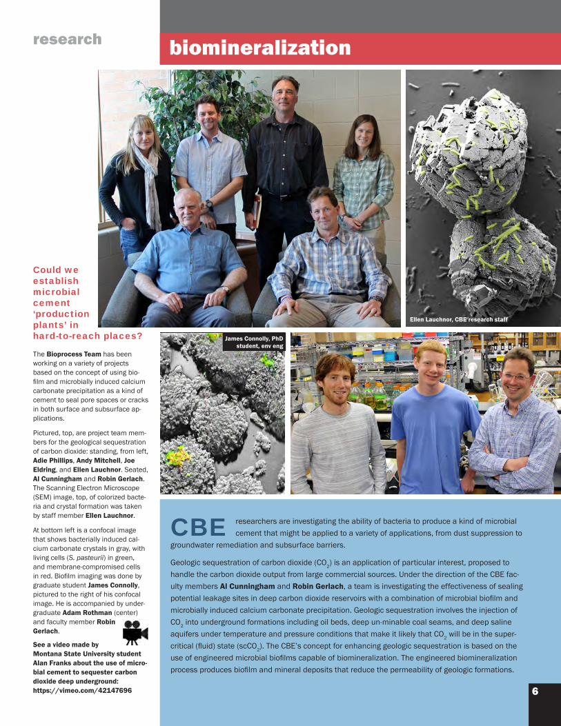
Ellen Lauchnor, CBE research staff
James Connolly, PhD student, env eng
Could we establish microbial cement ‘production plants’ in hard-to-reach places? The Bioprocess Team has been working on a variety of projects based on the concept of using bio-film and microbially induced calcium carbonate precipitation as a kind of cement to seal pore spaces or cracks in both surface and subsurface ap-plications.
Pictured, top, are project team mem-bers for the geological sequestration of carbon dioxide: standing, from left, Adie Phillips, Andy Mitchell, Joe Eldring, and Ellen Lauchnor. Seated, Al Cunningham and Robin Gerlach. The Scanning Electron Microscope (SEM) image, top, of colorized bacte-ria and crystal formation was taken by staff member Ellen Lauchnor.
At bottom left is a confocal image that shows bacterially induced cal-cium carbonate crystals in gray, with living cells (S. pasteurii) in green, and membrane-compromised cells in red. Biofilm imaging was done by graduate student James Connolly, pictured to the right of his confocal image. He is accompanied by under-graduate Adam Rothman (center) and faculty member Robin Gerlach.

7
Education
A record 95 students partici-pated in biofilm
projects during the past year. The number and variety of stu-dents inspired us to represent them as multi-species biofilm clusters! CBE students work on interdis-ciplinary teams to do research relevant to chronic infections, remediation of contaminated soil, mitigation of fouling and corrosion in industry, and development of constructed wetlands to treat wastewater.
grads
undergradsSummary of undergraduate students 2011–1249 undergraduate students28 female/21malerepresenting 10 departments:
Cell Biology & Neuroscience Chemical & Biological Engineering Chemistry & Biochemistry Civil Engineering Ecology Health & Human Development Land Resources & Environmental Sciences Mechanical & Industrial Engineering Microbiology Nursing (Bridges)
Summary of graduate students 2011–1246 graduate students24 female/22 male 35 PhD/11 MS representing 9 departments:
Cell Biology & Neuroscience Chemical & Biological Engineering Chemistry & Biochemistry Civil/Environmental Engineering Health & Human Development Land Resources & Environmental Sciences Mathematical Sciences Mechanical & Industrial Engineering Microbiology
CBE students, from left, Chris Allen, Rachel Van Kempen-Fryling, Natasha Mallette, and James Connolly were among many who joined CBE microscope facilities manager Betsey Pitts for a
confocal open house introduction to the capabilities of the new scopes.

8
education
A mber Schmit, center, an undergraduate
in chemical and biological engineering, used the confocal microscope with Department of Land Resources & Envi-ronmental Sciences (LRES) faculty Christine Foreman, left, and LRES graduate student Heidi Smith, right, to capture these rare portraits of microbial life within Antarctic ice samples. A nucleic acid stain renders bacterial colonies visible as green areas colonizing the gray landscape of a cryoconite granule. Cryoconites are composed of windblown sediment embed-ded in the Antarctic ice. A close-up of this grain of dust clearly shows a string of autofluorescing cyanobacteria as red and yellow. These pictures are testament to the ubiquity of microbial life, even in the harshest environments. Since active microbial communities produce gases such as oxygen, carbon dioxide, and methane, their presence may affect ice-core gas records.
environmentalecology
How might microbial metabolism within glacial samples from Antarctica affect the ice-core gas records that we use to understand the earth’s climate history?
Glacial microbes imaged in situ on Antarctic sediment particles by Amber Schmit, UG stu-dent, chem & biol eng, using a combination of reflection and fluorescent confocal imaging

9
education
Could we use the principles of microbial species interactions found in nature to engineer applications in biotechnology?
With a team of geobiochemical researchers, graduate student Hans Bernstein has been exploring waters in the backcountry of Yellowstone National Park, where biofilms grow in pristine, extremely hot conditions, pictured at left. Microbial communi-ties such as these are a source of inspiration for engineering biofilms that could be designed to address biofuel production, environmental remediation, and sustainable food processing.
Back in the laboratory, Bernstein and undergraduate Alissa Bleem have grown dual-species model biofilms in order to better understand basic inter-species microbial interactions.
The CSLM image above shows one of the model biofilms composed of Cyanobacteria synechococcus (red) and Escherichia coli (blue). The pho-toautotrophic cyanobacteria convert light, water, and carbon dioxide into products that can support the E. coli. The cyanobacteria benefit from the exchange through an oxygen removal mechanism. Both organisms are used extensively in biotechnology applications.
The project is part of the microbial consortia engineering collabora-tion between the CBE, NSF-IGERT program in geobiological systems and Pacific Northwest National Laboratories.
Visit the research site at Yellow-stone National Park, where Hans Bernstein talks about the microbial communities that inspire his labora-tory work. http://youtu.be/UdSrYHWLHZE
biotechnology
CSLM biofilm image, right, and Yellowstone National Park hot spring biofilm image, bottom, by Hans
Bernstein, PhD student, chem & biol eng
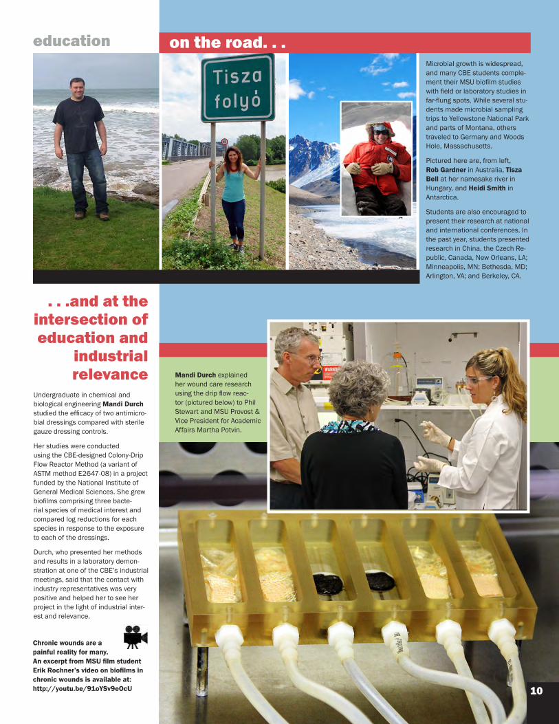
Microbial growth is widespread, and many CBE students comple-ment their MSU biofilm studies with field or laboratory studies in far-flung spots. While several stu-dents made microbial sampling trips to Yellowstone National Park and parts of Montana, others traveled to Germany and Woods Hole, Massachusetts.
Pictured here are, from left, Rob Gardner in Australia, Tisza Bell at her namesake river in Hungary, and Heidi Smith in Antarctica.
Students are also encouraged to present their research at national and international conferences. In the past year, students presented research in China, the Czech Re-public, Canada, New Orleans, LA; Minneapolis, MN; Bethesda, MD; Arlington, VA; and Berkeley, CA.
10
education
...andattheintersectionofeducationand
industrialrelevance
Undergraduate in chemical and biological engineering Mandi Durch studied the efficacy of two antimicro-bial dressings compared with sterile gauze dressing controls.
Her studies were conducted using the CBE-designed Colony-Drip Flow Reactor Method (a variant of ASTM method E2647-08) in a project funded by the National Institute of General Medical Sciences. She grew biofilms comprising three bacte-rial species of medical interest and compared log reductions for each species in response to the exposure to each of the dressings.
Durch, who presented her methods and results in a laboratory demon-stration at one of the CBE’s industrial meetings, said that the contact with industry representatives was very positive and helped her to see her project in the light of industrial inter-est and relevance.
Mandi Durch explained her wound care research using the drip flow reac-tor (pictured below) to Phil Stewart and MSU Provost & Vice President for Academic Affairs Martha Potvin.
Chronic wounds are a painful reality for many. An excerpt from MSU film student Erik Rochner’s video on biofilms in chronic wounds is available at:http://youtu.be/91oYSv9eOcU
ontheroad...

11
Industry
industryhighlights
The CBE’s Medical Biofilms Laboratory hosted the 2012 sum-mer workshop on oral biofilms; CBE researcher Alessandra Agostinho is pictured above, center, with two workshop participants.
Alex Rickard, (not pictured) assistant professor of biological sciences, Uni-versity of Michigan, participated as a guest instructor for the workshop and presented a talk on interbacterial communication in chronic wounds at the CBE conference following.
Agostinho captured the confocal im-age, left, of biofilm (clusters of small green dots) in wound tissue; the con-nective tissue shows up as red and the nuclei as large green spots.
woundbiofilms
I n 2012 the CBE’s Industrial Associates program participation expanded to 35 subscribing members (26 full members and 9 small business members, see list on page 13). Healthcare-related companies continue to make up approximately half of
our membership base, with strong support from consumer products and specialty chemi-cal companies as well. The Standardized Biofilm Methods Laboratory at the CBE was very active in moving a new biofilm method (the Single Tube Method, E2871-12; see page 12) to ASTM Standard Method status. Additionally, method E2799-12 (MBEC™ standard method) was modified to include results from an inter-laboratory study (ILS). The Montana Biofilm Science & Technology Meetings continue to be the major venue for industrial interaction. The meetings included diverse session topics, from medical and oral biofilms to thermal biofilms and green control strategies—a reflection of the diversity of our industrial appeal and relevance. CBE biofilm meetings also included participation by regula-tory authorities from FDA and EPA, providing a valuable link between industry and govern-ment. The CBE seeks to expand the value of membership to our partner companies through our semi-annual meetings (February and July of each year), our bio-films methods workshops, visits to companies, and regulatory outreach.
A Agostinho, CBE
industry
See a movie of the effect of a biofilm treatment in the new flow cell at: http://youtu.be/OEzNExgcLw8
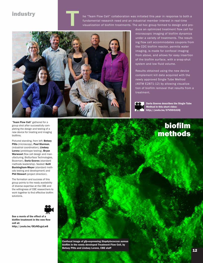
“Team Flow Cell” gathered for a group shot after successfully com-pleting the design and testing of a new device for treating and imaging biofilms.
Pictured standing, from left: Betsey Pitts (microscopy), Paul Sturman, (industrial coordination), Lindsey Lorenz (prototoype testing), Bryan Warwood (flow cell design and man-ufacturing, BioSurface Technologies, Bozeman), Darla Goeres (standard methods leadership). Seated: Kelli Buckingham-Meyer (standard meth-ods testing and development) and Phil Stewart (project direction).
The formation and success of this group points to the ready availability of diverse expertise at the CBE and the willingness of CBE researchers to work together to find effective biofilm solutions.
Confocal image of gfp-expressing Staphylococcus aureus biofilm in the newly developed Treatment Flow Cell, by Betsey Pitts and Lindsey Lorenz, CBE staff
T he “Team Flow Cell” collaboration was initiated this year in response to both a fundamental research need and an industrial member interest in real-time visualization of biofilm treatments. The ad hoc group formed to design and pro-
duce an optimized treatment flow cell for microscopic imaging of biofilm dynamics under a variety of treatments. The result-ing flow cell accommodates coupons from the CDC biofilm reactor, permits water imaging, is made for confocal imaging from above, and allows for easy insertion of the biofilm surface, with a snap-shut system and low fluid volume. Results obtained using the new device complement kill data acquired with the newly approved Single Tube Method (ASTM E2871-12) by allowing visualiza-tion of biofilm removal that results from a treatment.
biofilmmethods
12
Darla Goeres describes the Single Tube Method in this short video: http://youtu.be/37VDD3JiJiQ

industrialassociatesNew associates in bold 3M
Agile Sciences
BardAccessSystems
BASF
Bausch & Lomb
Baxter Healthcare
BCGSolutions
BendResearch
Bridge Preclinical Testing Services
CareFusion
Church & Dwight
Colgate-Palmolive
Covidien
DowCorning
Dow Microbial Control
Embro Corporation
ExxonMobil
ICU Medical
Johnson & Johnson Consumer and Personal Products
Kane Biotech
KCI
Kimberly-Clark
Masco Corporation
NASA
NCHCorporation
Novozymes A/S
Procter & Gamble
Reckitt Benckiser
Sample6Technologies
Semprus BioSciences
STERIS
The Sherwin-Williams Company
Unilever
W.L. Gore & Associates
WuXi AppTec
sponsoredprojects
industry
oral care……mining……drinking water……methods development……cooling water……wound care……soap dispensers……food safety……pools and spas…… well biofouling……medical devices……oil and gas……industrial process water……food safety…… toilet bowlsgroundwater contamination……antimicrobials……sinusitis……pharmaceutical water……
biofuelsAscocoryne sarcoides (red), a filamentous fungus growing on
cellulose (blue/green) in liquid culture, imaged with the CBE’s new confocal microscopes. A. sarcoides produces gasoline and diesel
fuel-related compounds on cellulose, the most abundant and afford-able source for producing biofuels. Imaged by Natasha Mallette, PhD
student in chemical and biological engineering.
Expertise in diverse biofilm issues and applications is evident in the topics of the past year’s industry spon-sored projects, below.
The interdisciplinary team-building possible at the CBE ensures that projects are developed with the input of relevant disciplines. Contact Industrial Coordinator Paul Sturman with inquiries: [email protected] (406) 994-2102
13

Adrianna Collazo Ortiz Visiting AIRO-Bridges student,
University of Puerto Rico, San Juan
Mijeong Jang Visiting postdoctoral researcher,
University of Seoul, Korea
Irina Khilyas Visiting graduate student,
Kazan State University, Russia
Trond Møretrø Visiting research scientist,
Nofima Food, Ås, Norway
Carole Nagant Visiting graduate student,
Université Libre de Bruxelles, Belgium
Lucy Qi, MD Visiting clinical researcher,
Chongqing Medical University and Children’s Hospital,
Chongqing, China
Jeremy Richey Visiting AIRO-Bridges student,
Fort Belknap College, Harlem, MT
Shoji Takenaka, DDS Visiting faculty,
Niigata University, Japan
Kendra Teague Visiting AIRO-Bridges student, Fort Peck Community College,
Wolf Point, MT
Vincent Wang, MD Visiting clinical researcher,
Mackay Memorial Hospital in Taipei, Taiwan
VisitingresearchersfromLittleBighornCollege,CrowAgency,MT:
Zachary Cummins Visiting undergraduate student
Dayle “Candy” Felicia Visiting undergraduate student
Jonah Morsette Staff member
Amanda Not Afraid Visiting undergraduate student
Miranda Rowland Visiting undergraduate student
Elaine Stone Visiting undergraduate student
outreach
visitingresearchers
Shoji Takenaka, DDS, a return visitor (2007) from Niigata University, Japan, was eager to learn how to use the new microscopes and to spend as much time as possible collecting images of oral biofilms during his stay. His time-lapse movie (still shot above) of commercially available mouthwash penetration into biofilm was featured on the Cell.com web site in their Cell Picture Show. You can also see it on the CBE’s YouTube channel at: http://youtu.be/ldaUx_pU--w
14
CSLM imaging of treated oral biofilmby Shoji Takenaka

Outreach
15
Threeundergraduatestudents worked at the CBE in the summer of 2011 as part of the American Indian Research Opportunities (AIRO) BRIDGES program. The program’s objective is to build a seam-less educational experience between reservation-based colleges and Montana State University and, in the process, to increase the number of underrepresented Native American students successfully transferring from the two-year tribal colleges to MSU and pursuing academic studies in the biomedical and other health-related sciences. The students worked at the CBE for eight weeks and then traveled to Old Greenwich, Connecticut, to present posters of their work at the Leadership Alliance National Symposium.
Adrianna Collazo Ortiz, undergraduate, University of Puerto Rico San Juan, studied pathogens and constructed wetlands with CBE faculty Mark Burr, research assistant professor, land resources and environmental sciences.
Jeremy Richey participated in a dentistry project titled “Testing of denture base materials incorpo-rated with silver nanoparticles to prevent Candida colonization” under the supervision of CBE research scientist Alessandra Agostinho. Jeremy is an undergraduate student from Fort Belknap College in Harlem, Montana.
Kendra Teague worked with Heidi Smith, CBE PhD student, land resources and environmental sci-ences, on a project that characterizes Antarctic isolates based on different carbon source utilization. Kendra is an undergraduate student from Fort Peck Community College in Wolf Point, Montana.
TheMSUAlgalBiofuelsGroup worked with five Native American students and staff from Little Bighorn College (LBHC) during the ten-week project: “Cultivation and characterization of oil-producing algae.” The project was funded by a collabora-tion of commercial, state, federal, and tribal entities focusing on the development of “Green coal to transportation fuel technology.” This project with LBHC involved training students in the use of algae as a means to capture carbon dioxide and produce bio-fuels. The specific objectives included collection of algae from natural field sites, isolation of pure cultures, building photo-bioreactor systems, characterizing algal growth in these systems, and measuring oil productivity. The participants from LBHC, located in Crow Agency, Montana, were: Jonah Morsette, staff member, and undergraduates: Zachary Cum-mins, Amanda Not Afraid, Miranda Rowland, and Elaine Stone. CBE mentors on the project were Brent Peyton, profes-sor, and Rich Macur, research assistant professor, both of MSU’s Department of Chemical and Biological Engineering.
Photo courtesy B Peyton, R Macur

Montana State University is an equal opportunity/affirmative action institu-tion. Copyright © 2012, MSU Center for Biofilm Engineering.
More than 200 master’s and doctoral students have earned their degrees in the CBE’s graduate research program since the CBE was founded in 1990. CBE graduate students acquire valuable
experience by designing and performing research that crosses traditional academic discipline boundar-ies and has direct impact on current environmental, industrial, and medical issues. In addition, the CBE’s Industrial Associates program brings students into working relationships with potential employers. CBE graduate students are encouraged to develop their communication and lead-ership skills by presenting at research conferences, mentoring undergraduate students, organizing the CBE’s seminar series, and assisting with outreach efforts. The CBE’s standing in the international re-search community attracts visiting students and faculty from all parts of the world, providing a culturally diverse and stimulating academic environment. Graduate students pursue their degree in a discipline offered through one of the science, agriculture, or engineering departments at Montana State University while conducting research in CBE laboratories.
For more information, go to: www.biofilm.montana.edu/cbe-graduate-education.html
Over 600 undergraduate students have participated
in CBE research since 1990. Undergraduate students are highly valued team members in the MSU Center for Biofilm Engi-neering and are fully integrated into the research process. Our undergraduates learn to design and implement experiments that will provide results relevant to industry and the science commu-nity—and they develop the skills that will broaden their career opportunities and make them more valuable to prospective employers. For undergraduates who decide to pursue graduate degrees, their CBE research experience is often cited as a key component in being selected by their program of choice.For more information, go to:www.biofilm.montana.edu/cbe-undergraduate-education.html
Undergraduate program
Graduate program
Center for Biofilm Engineering366 EPS BuildingMontana State UniversityBozeman, Montana 59717-3980USAPhone: 406-994-4770Fax: 406-994-6098
www.biofilm.montana.eduCarole Nagant, visiting researcher, Belgium, took this CSLM image of Pseudomonas biofilm, side view, in studying action of cationic antimicrobial peptides.
Combination of reflection and fluorescent confocal imaging of anaerobic bacteria on activated carbon by Betsey Pitts, microscope facilities manager, in collaboration with Birthe Kjellerup, (visiting researcher, 2002) assistant professor, biological sciences, Goucher College, Baltimore, Maryland. Project ER-2135 funded by SERDP.
Karen Moll, MS, microbiology