Embed Size (px)
Citation preview

Reviewhttps://doi.org/10.9758/cpn.2018.16.1.18 pISSN 1738-1088 / eISSN 2093-4327Clinical Psychopharmacology and Neuroscience 2018;16(1):18-31 Copyrightⓒ 2018, Korean College of Neuropsychopharmacology
18
Received: July 24, 2017 / Revised: November 22, 2017Accepted: December 14, 2017Address for correspondence: Seung-Hwan Lee, MD, PhDDepartment of Psychiatry, Inje University Ilsan Paik Hospital, 170 Juhwa-ro, Ilsanseo-gu, Goyang 10380, Korea Tel: +82-31-910-7260, Fax: +82-31-910-7268E-mail: [email protected]
This is an Open-Access article distributed under the terms of the Creative Commons Attribution Non-Commercial License (http://creativecommons.org/licenses/by-nc/4.0) which permits unrestricted non-commercial use, distribution, and reproduction in any medium, provided the original work is properly cited.
From Neurons to Social Beings: Short Review of the Mirror Neuron System Research and Its Socio-Psychological and Psychiatric ImplicationsHyeonjin Jeon1, Seung-Hwan Lee1,2
1Clinical Emotion and Cognition Research Laboratory, 2Department of Psychiatry, Inje University Ilsan Paik Hospital, Goyang, Korea
The mirror neuron system (MNS) is a brain network activated when we move our body parts and when we observe the actions of other agent. Since the mirror neuron’s discovery in research on monkeys, several studies have examined its network and properties in both animals and humans. This review discusses MNS studies of animals and human MNS studies related to high-order social cognitions such as emotion and empathy, as well as relations between MNS dysfunction and mental disorders. Finally, these evidences are understood from an evolutionary perspective.
KEY WORDS: Mirror neurons; Social cognition; Mental disorders.
INTRODUCTION
The mirror neuron system (MNS) is a network of neuron groups that discharge when an individual performs an ac-tion and/or observes an action of another agent. The MNS is divided into two principal hubs; the premotor area in the frontal lobe and the inferior parietal lobule (IPL).1,2) Additionally, the superior temporal sulcus (STS) is consid-ered to be a key area of the MNS.3) Mirror neurons were first discovered in area F5 of the ventral premotor cortex (PMv) in macaques.4-6) They fire when monkeys observe other individuals grasp toward objects and when the monkeys execute the grasping motion themselves. Some neurons in the F5 posterior (F5p) area that respond to the presentation of objects as the monkey grasps an object are called “canonical neurons.”7,8) The F5p is also known as a hand-related area that encodes goal-directed actions. Both motor and mirror neurons are located in the F5 con-vexity (F5c) area and they fire during both observation and execution of specific goal-directed actions involving
the hand and mouth.9)
In macaques, the frontal mirror area of the brain is div-ided into the lateral surface area 4 (primary motor cortex; M1), caudal part of area 6 (also a part of M1), medial sur-face area 6 (supplementary motor area; SMA) and pre-motor area. The premotor area is divided into caudal part (PMc)—with a direct connection to subcortical areas—and rostral part (PMr) that indirectly affects motor genera-tion. The F5 area is a part of the parietal-frontal and pre-frontal-frontal networks.10) Kraskov et al.11) have revealed that pyramidal tract neurons (PTN) in F5 suppress firing during observation but activate firing during movement. They interpreted the results as an inhibition effect sup-pressing motor generation during observation. Contra-stingly, Vigneswaran et al.12) found that some PTN fired more during action observation when they recorded a sig-nal from PTN in the area F1. Early mirror neuron studies suggested that mirror neurons are only engaged in action execution and in understanding the intention and trans-formation of visual perception into action execution. They suggest that the PMv has a connection with the cau-dal part of the inferior frontal gyrus (IFG). The IFG is also known as Broca’s area, a special motor area for human language generation.13,14)
Mirror aspects were also discovered in the IPL of macaque brains.15) Mirror neurons in the parietal area PF/PFG of the

Short Review of the Mirror Neuron System Research 19
Fig. 1. Key regions (red, large sphere) and subregions (blue, small sphere) of the mirror neuron system. The image on the left is lateral view of left hemisphere and that on the right is top view of both hemispheres.S, superior; I, inferior; A, anterior; P, posterior; IFG, inferior frontal gyrus; PMv, ventral premotor area; PMd, dorsal premotor area; SMA, supplementary motor area; STS; superior temporal sulcus; M1, primary motor cortex; S1, primary somatosensory cortex; pMTG, posterior middle temporal gyrus; FFA, fusiform face area; IPL, inferior parietal lobule; MT/V5, middle temporal area.
IPL also discharge during both the observation and execution of complex actions such as grasping an object to place it at a target locus or to eat it. Moreover, activity is modulated by the final goal of the action.16) The IPL receives visual in-formation from eyes and somatosensory information from mouth, hands, and arms.17) This suggests that the visuo-motor organization of the IPL may build the neural basis of the abil-ity to understand the intention of others’ action.18)
Research shows that the STS also has a mirror property. The STS is a brain area which responds to biological motions.19-21) It is not generally considered a part of the MNS because it does not have motor properties.2) The STS provides an audiovisual-to-motor link, that is, integration between seeing, hearing, and doing.22) Via the IPL, the area F5 is connected to higher-order visual areas of the STS.2) The first stream originates from a sector in the upper bank of the STS (STPm), reaches the parietal area PFG, and terminates in the area F5c. The second stream arises in the lower bank of the STS, reaches the anterior intra-parietal area (AIP), and then the area F5a.23,24) Thus, re-cent literature suggests that the STS be considered a part of temporo-parieto-premotor pathways and the MNS.25,26)
Experiments comparing execution, imitation, observa-tion, and imagination conditions using various neuro-imaging techniques has revealed a network of the MNS.26-29) We present key regions and subregions of the hu-man MNS in Figure 1. The key areas consist of the SMA, IPL, and STS. Subregions in the frontal lobe are the dorsal and PMv, IFG (including Broca’s area), and M1, which are re-
lated to motor function. The subregions in the parietal lobe include the primary somatosensory cortex (S1). The tempo-ral and occipital subregions include the posterior middle temporal gyrus (pMTG) and middle temporal (MT/V5) area. Interestingly, the fusiform face area (FFA) is related with spe-cific conditions such as facial expressions.30,31)
In this review, we first introduce various properties of mirror neurons based on animal electrophysiology. We then discuss the relationship between human MNS and higher-order cognitive abilities such as empathy and so-cial cognition and conclude with an exploration of clin-ical connotations related to MNS dysfunction and mental disorders.
MIRROR NEURON SYSTEM RESEARCH
MNS Research in Animals
Multiple properties of mirror neurons from primate studies
Multiple properties of mirror neurons have been re-vealed in studies on primates’ premotor mirror neuron. Rizzolatti et al.32) reported that some F5 neurons fire dur-ing grasping, but not holding, while others fire during holding, but not grasping. Umiltà et al.33) showed that more than half of the F5 mirror neurons discharged even in the invisible motor-action condition. Some neurons re-spond to observations of goal-directed actions (experi-menter’s hand or mouth actions related to food items) but
20 H. Jeon and S.H. Lee
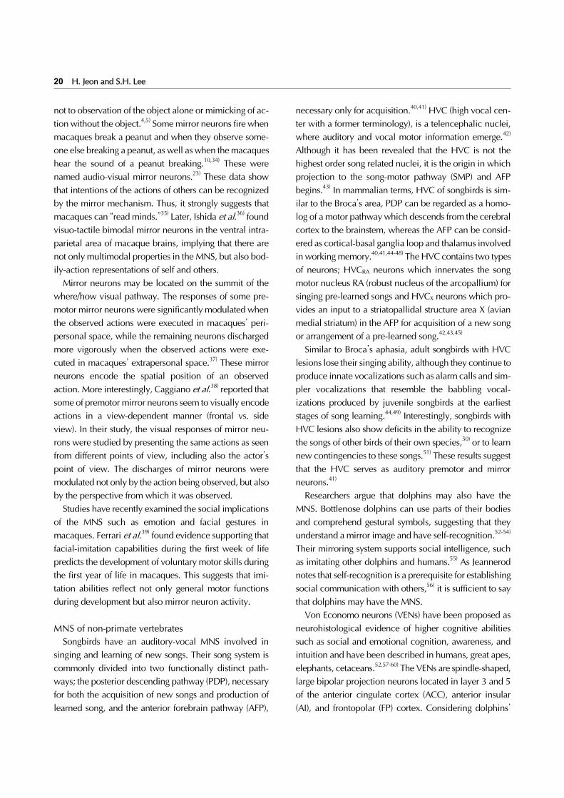
not to observation of the object alone or mimicking of ac-tion without the object.4,5) Some mirror neurons fire when macaques break a peanut and when they observe some-one else breaking a peanut, as well as when the macaques hear the sound of a peanut breaking.10,34) These were named audio-visual mirror neurons.23) These data show that intentions of the actions of others can be recognized by the mirror mechanism. Thus, it strongly suggests that macaques can “read minds.”35) Later, Ishida et al.36) found visuo-tactile bimodal mirror neurons in the ventral intra-parietal area of macaque brains, implying that there are not only multimodal properties in the MNS, but also bod-ily-action representations of self and others.
Mirror neurons may be located on the summit of the where/how visual pathway. The responses of some pre-motor mirror neurons were significantly modulated when the observed actions were executed in macaques’ peri-personal space, while the remaining neurons discharged more vigorously when the observed actions were exe-cuted in macaques’ extrapersonal space.37) These mirror neurons encode the spatial position of an observed action. More interestingly, Caggiano et al.38) reported that some of premotor mirror neurons seem to visually encode actions in a view-dependent manner (frontal vs. side view). In their study, the visual responses of mirror neu-rons were studied by presenting the same actions as seen from different points of view, including also the actor’s point of view. The discharges of mirror neurons were modulated not only by the action being observed, but also by the perspective from which it was observed.
Studies have recently examined the social implications of the MNS such as emotion and facial gestures in macaques. Ferrari et al.39) found evidence supporting that facial-imitation capabilities during the first week of life predicts the development of voluntary motor skills during the first year of life in macaques. This suggests that imi-tation abilities reflect not only general motor functions during development but also mirror neuron activity.
MNS of non-primate vertebratesSongbirds have an auditory-vocal MNS involved in
singing and learning of new songs. Their song system is commonly divided into two functionally distinct path-ways; the posterior descending pathway (PDP), necessary for both the acquisition of new songs and production of learned song, and the anterior forebrain pathway (AFP),
necessary only for acquisition.40,41) HVC (high vocal cen-ter with a former terminology), is a telencephalic nuclei, where auditory and vocal motor information emerge.42) Although it has been revealed that the HVC is not the highest order song related nuclei, it is the origin in which projection to the song-motor pathway (SMP) and AFP begins.43) In mammalian terms, HVC of songbirds is sim-ilar to the Broca’s area, PDP can be regarded as a homo-log of a motor pathway which descends from the cerebral cortex to the brainstem, whereas the AFP can be consid-ered as cortical-basal ganglia loop and thalamus involved in working memory.40,41,44-48) The HVC contains two types of neurons; HVCRA neurons which innervates the song motor nucleus RA (robust nucleus of the arcopallium) for singing pre-learned songs and HVCX neurons which pro-vides an input to a striatopallidal structure area X (avian medial striatum) in the AFP for acquisition of a new song or arrangement of a pre-learned song.42,43,45)
Similar to Broca’s aphasia, adult songbirds with HVC lesions lose their singing ability, although they continue to produce innate vocalizations such as alarm calls and sim-pler vocalizations that resemble the babbling vocal-izations produced by juvenile songbirds at the earliest stages of song learning.44,49) Interestingly, songbirds with HVC lesions also show deficits in the ability to recognize the songs of other birds of their own species,50) or to learn new contingencies to these songs.51) These results suggest that the HVC serves as auditory premotor and mirror neurons.41)
Researchers argue that dolphins may also have the MNS. Bottlenose dolphins can use parts of their bodies and comprehend gestural symbols, suggesting that they understand a mirror image and have self-recognition.52-54) Their mirroring system supports social intelligence, such as imitating other dolphins and humans.55) As Jeannerod notes that self-recognition is a prerequisite for establishing social communication with others,56) it is sufficient to say that dolphins may have the MNS.
Von Economo neurons (VENs) have been proposed as neurohistological evidence of higher cognitive abilities such as social and emotional cognition, awareness, and intuition and have been described in humans, great apes, elephants, cetaceans.52,57-60) The VENs are spindle-shaped, large bipolar projection neurons located in layer 3 and 5 of the anterior cingulate cortex (ACC), anterior insular (AI), and frontopolar (FP) cortex. Considering dolphins’

Short Review of the Mirror Neuron System Research 21
social intelligence and the existence of VENs, the supposi-tion that dolphins may have the MNS is justified.
Expansion of MNS Research in Humans
Neural correlates of MNS activity in humansElectroencephalography (EEG) methods have mainly fo-
cused on mu rhythms. Mu rhythms refer to sensorimotor ac-tivity in alpha bands (8-13 Hz in adults, 6-9 Hz in children) recorded over central scalp locations C3, Cz, and C4; it is al-so referred to as “rolandic alpha.”22,61) Mu power decreases during both execution and observation of movements, and even when imagining movements.22,62) It is therefore called mu suppression or mu power desynchronization. Some studies have reported infant’s mu rhythm in central regions of the brain.61,63) A recent EEG study indicated greater mu suppression (putatively reflecting mirror neuron activation) in infants and adults when observing transitive movements compared to intransitive movements.64)
Hari et al.’s source localization study65) of mu rhythm activity based on magnetoencephalography (MEG) identi-fied sensorimotor regions. Concurrent functional mag-netic resonance imaging (fMRI) studies suggest that mu suppression may represent activity in brain areas of the MNS.66-69) On the other hand, some researchers suggest that mu suppression is not a perfect index of MNS activity. Aleksandrov and Tugin70) compared mu suppression from various types of motor activity and found that mu sup-pression was not significantly lower than it was in con-ditions where participants viewed human movement. They argued that mu suppression instead depends on the attention demands of the task, which decreases over time. Perry and Bentin71) found a similar pattern of changes in EEG power at the occipital and central electrodes. They argued that the significant differences could be attributed to differences in attentional demands between conditions, rather than differences in mirror neuron activity.
Studies using EEG and EMG have also included beta- and theta-frequency band activity. Beta band is usually defined as 13 to 35 Hz, with a typical peak frequency of ∼20 Hz. Researchers have also suggested that changes in beta activity index mirror neuron activity.72-74) Beta sup-pression is more related to motor processing and the pri-mary motor cortex, while mu rhythm is linked predom-inantly to the somatosensory system.75,76) Theta rhythm (4-8 Hz) is also related to movement and activity in
non-mu rhythm frequency bands by voluntary movement from the somatosensory area have been considered as an alternative index of the MNS function.77,78)
A number of transcranial magnetic stimulation (TMS) studies provide another putative measure of the human MNS. For example, Heiser et al.79) stimulated the Broca’s area with repetitive TMS (rTMS) while participants perform a key-press experiment. In their study, participants imitated a key-pressing movement done by another individual in experimental condition. In the control condition, partic-ipants pressed the keys in response to a red dot indicating the key to be pressed. rTMS lowered the participants’ per-formance during imitation but not during the control task. That is, a transient lesion by rTMS over left or right IFG in-duced a selective impairment of action imitation.
fMRI has been a popular research method in MNS studies. Iacoboni et al.80) conducted the first fMRI study with imitation. Participants were asked to observe, ex-ecute, or imitate the hands action on the screen. The IFG pars opercularis (IFGpo), posterior parietal cortex, and pa-rietal operculum showed higher activation during imi-tation than during execution, and the former two areas were activated during observation but less than they were during execution. Buccino et al.’s fMRI study81) found bi-lateral premotor and parietal activations during object-re-lated movements, and only premotor activation during non-object-related movements.
Lui et al.82) reported activations in key regions of the MNS when human participants observed mimed and meaningless movements. In addition, fMRI studies have consistently shown activation of MNS areas when partic-ipants perform, observe, and imagine a movement; when participants look at point-light biological animations or non-biological motions likely to evoke associations with biological action; and when participants hear object-dire-cted action sounds.2,83-86) Caspers et al.’s meta-analysis28) on human MNS studies with fMRI and positron emission tomography confirmed basic key regions such as SMA, IPL, and STS activation in observation, imitation, and execution conditions and presented multiple associative areas. Molenberghs et al.87) also conducted a similar meta-analy-sis study on human MNS and consolidated MNS regions.
Emotion, empathy, theory-of-mind, and MNS Some researchers argue that the MNS is engaged during
the experience of emotion. For example, disgust induced
22 H. Jeon and S.H. Lee
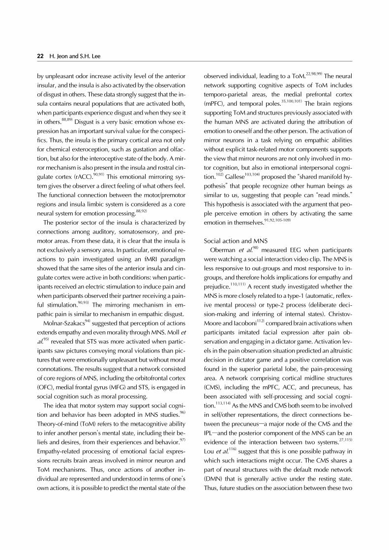
by unpleasant odor increase activity level of the anterior insular, and the insula is also activated by the observation of disgust in others. These data strongly suggest that the in-sula contains neural populations that are activated both, when participants experience disgust and when they see it in others.88,89) Disgust is a very basic emotion whose ex-pression has an important survival value for the conspeci-fics. Thus, the insula is the primary cortical area not only for chemical exteroception, such as gustation and olfac-tion, but also for the interoceptive state of the body. A mir-ror mechanism is also present in the insula and rostral cin-gulate cortex (rACC).90,91) This emotional mirroring sys-tem gives the observer a direct feeling of what others feel. The functional connection between the motor/premotor regions and insula limbic system is considered as a core neural system for emotion processing.88,92)
The posterior sector of the insula is characterized by connections among auditory, somatosensory, and pre-motor areas. From these data, it is clear that the insula is not exclusively a sensory area. In particular, emotional re-actions to pain investigated using an fMRI paradigm showed that the same sites of the anterior insula and cin-gulate cortex were active in both conditions: when partic-ipants received an electric stimulation to induce pain and when participants observed their partner receiving a pain-ful stimulation.90,93) The mirroring mechanism in em-pathic pain is similar to mechanism in empathic disgust.
Molnar-Szakacs94) suggested that perception of actions extends empathy and even morality through MNS. Moll et al.95) revealed that STS was more activated when partic-ipants saw pictures conveying moral violations than pic-tures that were emotionally unpleasant but without moral connotations. The results suggest that a network consisted of core regions of MNS, including the orbitofrontal cortex (OFC), medial frontal gyrus (MFG) and STS, is engaged in social cognition such as moral processing.
The idea that motor system may support social cogni-tion and behavior has been adopted in MNS studies.96) Theory-of-mind (ToM) refers to the metacognitive ability to infer another person’s mental state, including their be-liefs and desires, from their experiences and behavior.97) Empathy-related processing of emotional facial expres-sions recruits brain areas involved in mirror neuron and ToM mechanisms. Thus, once actions of another in-dividual are represented and understood in terms of one’s own actions, it is possible to predict the mental state of the
observed individual, leading to a ToM.22,98,99) The neural network supporting cognitive aspects of ToM includes temporo-parietal areas, the medial prefrontal cortex (mPFC), and temporal poles.35,100,101) The brain regions supporting ToM and structures previously associated with the human MNS are activated during the attribution of emotion to oneself and the other person. The activation of mirror neurons in a task relying on empathic abilities without explicit task-related motor components supports the view that mirror neurons are not only involved in mo-tor cognition, but also in emotional interpersonal cogni-tion.102) Gallese103,104) proposed the “shared manifold hy-pothesis” that people recognize other human beings as similar to us, suggesting that people can “read minds.” This hypothesis is associated with the argument that peo-ple perceive emotion in others by activating the same emotion in themselves.91,92,105-109)
Social action and MNSOberman et al.98) measured EEG when participants
were watching a social interaction video clip. The MNS is less responsive to out-groups and most responsive to in- groups, and therefore holds implications for empathy and prejudice.110,111) A recent study investigated whether the MNS is more closely related to a type-1 (automatic, reflex-ive mental process) or type-2 process (deliberate deci-sion-making and inferring of internal states). Christov- Moore and Iacoboni112) compared brain activations when participants imitated facial expression after pain ob-servation and engaging in a dictator game. Activation lev-els in the pain observation situation predicted an altruistic decision in dictator game and a positive correlation was found in the superior parietal lobe, the pain-processing area. A network comprising cortical midline structures (CMS), including the mPFC, ACC, and precuneus, has been associated with self-processing and social cogni-tion.113,114) As the MNS and CMS both seem to be involved in self/other representations, the direct connections be-tween the precuneus—a major node of the CMS and the IPL—and the posterior component of the MNS can be an evidence of the interaction between two systems.27,115) Lou et al.116) suggest that this is one possible pathway in which such interactions might occur. The CMS shares a part of neural structures with the default mode network (DMN) that is generally active under the resting state. Thus, future studies on the association between these two

Short Review of the Mirror Neuron System Research 23
networks can help understand the link between human cerebral processes and social abilities.
Clinical implications of MNS researchA reduction in mirror neuron activity may be involved
in the pathophysiology of psychiatric conditions that are characterized by social cognitive deficits. Prime examples are autism, schizophrenia, and psychopathy.98,117-119)
Autism spectrum disorder and MNSIt is well established that children with autism spectrum
disorders (ASD) have deficits in imitation.120,121) Because the MNS is implicated in imitation learning, recent studies have sought to examine the development of the MNS in children with ASD. In a MEG study, individuals with Asperger’s syndrome showed delayed activation of the MNS in the inferior frontal lobe during an imitation task.122) Oberman et al.123) compared mu suppression in high-functioning individuals with ASD and age- and gen-der-matched control subjects while watching videos of a moving hand, a bouncing ball, and virtual noise, and while moving their own hand. Control subjects showed significant mu suppression in both, self and observed hand movements, whereas the ASD group showed sig-nificant mu suppression only in self-performed hand movements, but not in observed hand movements.
In a follow-up study, Oberman et al.124) on mu sup-pression in high-functioning children with ASD and age- and gender-matched control subjects, mu suppression was measured while the ASD children watched videos of a stranger opening and closing their right hand, watched their guardian or sibling performing the same hand mo-tion, watched their own hand performing the same action, and watched a video of two bouncing balls moving verti-cally toward and away from each other. The results re-vealed that children with ASD and typically developing children showed greater mu suppression for actions per-formed by familiar individuals (such as their guard-ians/parents) compared to those of strangers. These data showed a lack of mu suppression in the autistic children during observation, implying a possible dysfunction in the MNS. This was later developed into the “broken mirror” hypothesis.
Dapretto et al.’s fMRI study117) specifically investigated the neural correlates of the capacity of imitating facial ex-pressions of basic emotions in high-functioning ASD
individuals. Their results showed that during observation and imitation, autistic individuals did not show activation of the MNS in the IFGpo. Importantly, activity in this area was inversely related with symptom severity in the social domain. The authors concluded that “a dysfunctional MNS may underlie the social deficits observed in autism.”125)
Other mental disorders and MNSLimited studies have examined the association be-
tween other mental disorders and problems in the MNS. They suggest that schizophrenia and psychopathy may be related to a dysfunction in the MNS. According to McCormick et al.,126) participants with active schizo-phrenia-spectrum and psychotic disorders showed sig-nificantly greater mu suppression in the sensorimotor cor-tex of the left hemisphere than they did in their residual phase and when compared to healthy individuals. Those in the residual phase and healthy individuals showed sim-ilar levels of mu suppression. That is, greater left-sided mu suppression was positively correlated with psychotic symptoms, suggesting that abnormal mirror neuron activ-ity may exist among patients with schizophrenia during the active phase of the illness.
Negative symptoms of schizophrenia include anhedo-nia, affective flattening and impaired socio-emotional processing.127-131) Empirical research has argued an asso-ciation between negative symptoms and anomalous MNS activity.80,132-134) Using fMRI, Derntl et al.135) identified emphatic deficit in schizophrenia patients. They reported decreased brain activities in schizophrenia patients com-pared to healthy participants during three tasks related to empathy. Specific brain regions included those related to the MNS, such as IFG and MTG, and those associated to social cognition and emotion, such as the precuneus, cin-gulate, and amygdala. Using diffusion tensor imaging (DTI), Clark et al.136) have revealed abnormality in the temporal part of the superior longitudinal fasciculus (tSLF) in patients with schizophrenia. Considering that the SLF connects the frontal, parietal, temporal, and occipital lobes and it terminates the SMA, PMv and IFG in the fron-tal lobe, their results suggest that damages in neural tracts connected to frontal subregions of MNS might be asso-ciated with schizophrenia. Voineskos et al.137) have dis-covered abnormality in neural tracts of patients with defi-cit schizophrenia compared to patients with non-deficit

24 H. Jeon and S.H. Lee
Table 1. List of brain areas constitute the mirror neuron system
Area Brodmann area Related function
Supplementary motor area (SMA), secondary motor cortex (M2)
BA6 Postural stabilizationControl of sequences of movements (hypothetical)
Premotor cortex (PMC) BA6 Planning movement, spatial guidance of movement, sensory guidance of movement, understanding the actions of other, complex movement in hand and the mouth
Primary motor cortex (M1) BA4(task-relevant)
Force in a muscle
Pars opercularis of the inferior frontal gyrus (IFG), part of Broca’s area
BA44(task-relevant)
Language production and phonological processing, music perceptionhand movementsImpulse control (risk aversion)
Pars triangularis of the IFG, part of Broca’s area BA45 Semantic processingArea PF/PFt of the inferior parietal lobule (IPL) Supramarginal gyrus (SMG)
BA40 (rostral) Reading both as regards meaning and phonology
Intraparietal sulcus (IPS) of the human intraparietal area 3 (hIP3)
- Symbolic numerical information processing visuospatial working memory, interpreting the intent of others
Primary somatosensory cortex (S1) BA2 Tactile perceptionVisual area 5 (V5) in the extrastriate areas
=middle temporal (MT) visual areaBA19 (partial) Perception of motion, integration of local motion signals into global
percepts, guidance of some eye movementsFusiform face area (FFA)Fusiform body area (FBA)
BA37 (partial) Face and body recognitionRecognition for familiar stimuli
Posterior middle temporal gyrus (pMTG) BA21 (caudal) Contemplating distance, recognition of known faces accessing word meaning while reading
Insula BA13 (anterior) Olfactory perception, motor control, self-awareness interpersonal experience (social emotion)
schizophrenia and healthy participants. Their study sug-gests a significant relationships between negative symp-toms of schizophrenia and abnormality of MNS, namely the arcuate fasciculus (AF) which connects two key re-gions of MNS, the inferior frontal and temporo-parietal areas.
Mehta et al.138) have systematically reviewed neuro-imaging studies on mirror neuron activity in schizo-phrenia or patients with psychosis. Most of the studies in-cluded in their review reported decreased activity of MNS in the patients; only a few reported increased activity of MNS. In addition, most studies described premotor and motor cortices as the specific brain regions for mirror activity. The IFG, IPL, and posterior superior temporal sul-cus (pSTS) were also identified as parts of MNS that ex-hibited abnormal activity in patients.
Psychopathy appears to be the inverse of autism. In psy-chopathy, emotional empathy was impaired with ToM and motor empathy intact; however, in autism, emotional empathy was fully functional and ToM and motor em-pathy showed deficits.119) The study of psychopathy sug-gested that distancing should not be pushed too far. In both cases, the diseases were understood through a re-figuring of the two differences in the MNS.139)
CONCLUSION
Since its incidental discovery in macaques, the defi-nition of mirror neurons has been extended to include a huge cerebral network. The MNS is located on the top of visual dorsal stream. This system contains supplementary- motor and temporo-parietal association areas that process visual, auditory, and somatosensory information. Table 1 includes the names and main functions of each region that constitute the MNS. Table 2 lists researches that ex-amined specific functions of the brain in relations to spe-cific brain regions and subregions of the MNS. Table 3 presents researches on specific psychological/psychiatric disorders related with certain areas of the brain and the subregions of MNS. The MNS is considered to be related not only with motor imitation and reading intention of other agents, but also with concepts in social interaction, such as emotion, empathy, and ToM. Hence, it is specu-lated that the cause of psychiatric disorders such as ASD may be strongly associated with dysfunctions of the MNS.
Indeed, MNS is important in language, learning and de-velopment itself. For example, a linkage between action and perception is thought to be important in language ac-quisition and utilization.140) Subtle evidences include

Short Review of the Mirror Neuron System Research 25
Table 3. List of studies concerned with the mental disorders and human mirror neuron system (MNS)
Study Topics Related brain regions Related MNS regions
Dapretto et al. (2006)117) Autism (observation & imitation) Precentral gyrusACCIFGpoInsulaAmygdala
M1IFGInsula
Nishitani et al. (2004)122) Autism (imitation) M1 M1Oberman et al. (2005)123) Autism (observation) Sensorymotor cortex M1
S1Oberman et al. (2008)124) Autism (observation) Sensorymotor cortex M1
S1McCormick et al. (2012)126) Schizophrenia (empathy) Sensorymotor cortex M1Derntl et al. (2012)135) Schizophrenia (empathy) IFG
MFGMTGACCThalamusPrecuneus
IFGpMTG
Clark et al. (2011)136) Schizophrenia SLFILFIFO
SLF (fronto-parietal link)
Voineskos et al. (2013)137) Schizophrenia Right AFRight ILFLeft UF
AF (fronto-temporal link)
M1, primary motor cortex; ACC, anterior cingulate cortex; IFG, inferior frontal gyrus; IFGpo, IFG pars opercularis; S1, primary somatosensory cortex; MFG, middle frontal gyrus; MTG, middle temporal gyrus; pMTG, posterior MTG; SLF, superior longitudinal fasciculus; ILF, inferior longitudinal fasciculus; IFO, inferior fronto-occipital fasciculus; AF, arcuate fasciculus; UF, uncinate fasciculus.
how the IFG shows strong activation when participants imitate the actions of an experimenter.141) More direct evi-dences include how language acquisition by imitation of speech sounds have been supported by the acoustic mir-ror neurons in Broca’s area.2,34) In their study, Tettamanti et al.142) measured motor evoked potentials (MEP) after a single pulse TMS and simultaneously presented action-re-lated sentences and found that the premotor mirror sys-tem modulates verbal language processing.
Regardless of the empirical support, there are skeptical opinions and controversies surrounding the existence and the role of the human MNS. Hickok143) made a critical opinion that strongly opposed motor simulation in the Broca’s area. Moreover, he argued that sensory systems are essential to recognize the actions of others. Heyes144) also has remarked that human MNS does not work as a dominant network specialized in understanding the ac-tions of others and has hypothesized that the mirror neu-rons are a byproduct of associative learning. Critiques against the human MNS, have commonly mentioned that the relation between primates’ mirror neurons and the hu-man MNS is either non-parallel or undetermined.143-145)
For such reason, Hickok143) has argued that propositions suggesting a significant relation between the human MNS and higher social cognitive functions are mostly based on assumptions with little empirical evidence. However, these critiques do not deny the existence of mirror neu-rons in primates, and the existence of human mirror neu-rons based on a single neuron recording study by Mukamel et al.146) These opposing arguments on the hu-man MNS are worth considering when interpreting its re-lations to human behaviors.
The MNS is hypothesized to be an evolutionary pre-cursor and launching point for much of higher cognition and complex social behavior unique and fundamental to humans.78) Plotnik et al.60) suggested that elephants’ and dolphins’ mirror self-recognition could be linked to a con-vergent evolution related to complex sociality and cooperation. Reiss and Marino52) also suggested different neurological substrates of mirror self-recognition for dol-phins and primates, as their brain physiology have di-verged about 65 million to 70 million years ago.
Research on MNS across human, primates and other non-primate animals emphasize the idea of an ancient

26 H. Jeon and S.H. Lee
Table 2. List of studies concerned with the social cognitive function and human mirror neuron system (MNS)
Study Topics Related brain regions Related MNS regions
Wicker et al. (2003)88) Emotion AmygdalaAnterior insulaIFGRight ACC
InsulaIFG
Jabbi et al. (2007)89) Emotion, empathy IFO InsulaCarr et al. (2003)92) Empathy Precentral sulcus
M1IFGSTSInsulaRight amygdala
PMCM1IFGSTSInsula
Singer et al. (2004)93) Emotion, empathy PFCInsulaACCThalamusCerebellum
Insula
Moll et al. (2002)95) Morality Right medial frontal gyrusRight medial OFCAmygdalaPrecuneusAnterior STSPosterior MTG
STSMTG
Oberman et al. (2007)98) Social action Sensorymotor cortex M1S1
Vogeley et al. (2001)101) Theory-of-mind Right ACCRight motor cortexRight PMCRight TPJ
M1PMCTemporoparietal area
Baird et al. (2011)102) Empathy, theory-of-mind Temporoparietal areamPFCTemporal pole
Temporoparietal area
Cheng et al. (2008)110) Empathy Postcentral gyrus S1Gutsell and Inzlicht (2010)111) Prejudice Left sensorymotor cortex M1
S1Christov-Moore and Iacoboni (2016)112) Empathy, altruism dlPFC
SPLPrecentral gyrusAmygdala
Parietal areaM1
Lou et al. (2004)116) Concept of mental self Left IFCLeft medial frontal cortexIPCPCCPrecuneus
IFGIPL
IFG, inferior frontal gyrus; ACC, anterior cingulate cortex; IFO, anterior insula and adjacent frontal operculum; PMC, premotor cortex; M1, primary motor cortex; STS, superior temporal sulcus; PFC, prefrontal cortex; MTG, middle temporal gyrus; OFC, orbitofrontal cortex; S1, primary somatosensory cortex; TPJ, temoprotaietal junction; mPFC, middle PFC; dlPFC, dorsolateral PFC; SPL, superior parietal lobule; IFC, inferior frontal cortex; IPL, inferior parietal lobule; IPC, inferior parietal cortex; PCC, posterior cingulate cortex.
phylogenetic origin of the mirror mechanism and propose the function of MNS in the course of evolution.147) A more recent epigenetic perspective can provide new insights to understand the development of mirror neurons and the differences of mirror neurons between monkeys and humans.148) The epigenetic hypothesis proposes that the
development of the MNS, which functions to encode goals and understands actions, is not simply a result of as-sociative learning or natural selection, but is influenced by complicated epigenetic regulations that underlie neu-ronal plasticity, satisfying neuronal, cognitive and envi-ronmental “niches.”149) For example, research by Matsunaga

Short Review of the Mirror Neuron System Research 27
et al.150) suggests that the demethylation of the Gadd45 gene family may be one of the major epigenetic regu-lation mechanisms responsible for the growth and differ-entiation of neurons in the parietal cortex of mice and marmosets.
Although there are a many studies on the MNS, several questions remain unanswered. First, the relationship be-tween the MNS and DMN needs to be clarified, as they appear to share some brain areas related to cognition re-garding self. Further, some research suggests that ASD is a dysfunction of the DMN.151) Finally, the relationship be-tween psychiatric disorders and MNS dysfunction needs in-depth understanding, requiring more robust evidence beyond functional connectivity and neural correlates.
This work was supported by the Brain Research Program through the National Research Foundation of Korea (NRF) funded by the Ministry of Science, ICT & Future Planning (NRF-2015M3C7A1028252), and a grant from the Korea Science and Engineering Foundation (KOSEF), funded by the Korean government (NRF-2015 R1A2A2A01003564). The authors declare that there is no conflict of interest.
REFERENCES1. Hecht EE, Parr LA. The chimpanzee mirror system and the
evolution of frontoparietal circuits for action observation and social learning. In: Ferrari PF, Rizzolatti G, editors. New fron-tiers in mirror neurons research. Oxford:Oxford University Press;2015. p.153-181.
2. Rizzolatti G, Craighero L. The mirror-neuron system. Annu Rev Neurosci 2004;27:169-192.
3. Molenberghs P, Brander C, Mattingley JB, Cunnington R. The role of the superior temporal sulcus and the mirror neuron system in imitation. Hum Brain Mapp 2010;31:1316- 1326.
4. di Pellegrino G, Fadiga L, Fogassi L, Gallese V, Rizzolatti G. Understanding motor events: a neurophysiological study. Exp Brain Res 1992;91:176-180.
5. Gallese V, Fadiga L, Fogassi L, Rizzolatti G. Action recog-nition in the premotor cortex. Brain 1996;119:593-609.
6. Rizzolatti G, Fadiga L, Gallese V, Fogassi L. Premotor cortex and the recognition of motor actions. Brain Res Cogn Brain Res 1996;3:131-141.
7. Murata A, Fadiga L, Fogassi L, Gallese V, Raos V, Rizzolatti G. Object representation in the ventral premotor cortex (area F5) of the monkey. J Neurophysiol 1997;78:2226-2230.
8. Raos V, Umiltá MA, Murata A, Fogassi L, Gallese V.
Functional properties of grasping-related neurons in the ven-tral premotor area F5 of the macaque monkey. J Neurophysiol 2006;95:709-729.
9. Orban GA. The mirror system in human and nonhuman pri-mates: Comparative functional imaging studies suggest mul-tiple systems. In: Ferrari PF, Rizzolatti G, editors. New fron-tiers in mirror neurons research. Oxford:Oxford University Press;2015. p.116-137.
10. Rizzolatti G, Luppino G. The cortical motor system. Neuron 2001;31:889-901.
11. Kraskov A, Dancause N, Quallo MM, Shepherd S, Lemon RN. Corticospinal neurons in macaque ventral premotor cor-tex with mirror properties: a potential mechanism for action suppression? Neuron 2009;64:922-930.
12. Vigneswaran G, Philipp R, Lemon RN, Kraskov A. M1 corti-cospinal mirror neurons and their role in movement sup-pression during action observation. Curr Biol 2013;23:236- 243.
13. Bahlmann J, Schubotz RI, Friederici AD. Hierarchical artifi-cial grammar processing engages Broca’s area. Neuroimage 2008;42:525-534.
14. Fazio P, Cantagallo A, Craighero L, D’Ausilio A, Roy AC, Pozzo T, et al. Encoding of human action in Broca’s area. Brain 2009;132:1980-1988.
15. Rizzolatti G, Fadiga L, Fogassi L, Gallese V. From mirror neu-rons to imitation: facts and speculations. In: Meltzoff AN, Prinz W, editors. The imitative mind: development, evolu-tion, and brain bases. Cambridge:Cambridge University Press;2002. p.247-266.
16. Bonini L, Rozzi S, Serventi FU, Simone L, Ferrari PF, Fogassi L. Ventral premotor and inferior parietal cortices make dis-tinct contribution to action organization and intention understanding. Cereb Cortex 2010;20:1372-1385.
17. Rozzi S, Ferrari PF, Bonini L, Rizzolatti G, Fogassi L. Functional organization of inferior parietal lobule convexity in the macaque monkey: electrophysiological character-ization of motor, sensory and mirror responses and their cor-relation with cytoarchitectonic areas. Eur J Neurosci 2008; 28:1569-1588.
18. Fogassi L, Ferrari PF, Gesierich B, Rozzi S, Chersi F, Rizzolatti G. Parietal lobe: from action organization to intention understanding. Science 2005;308:662-667.
19. Puce A, Perrett D. Electrophysiology and brain imaging of bi-ological motion. Philos Trans R Soc Lond B Biol Sci 2003;358:435-445.
20. Perrett DI, Harries MH, Bevan R, Thomas S, Benson PJ, Mistlin AJ, et al. Frameworks of analysis for the neural repre-sentation of animate objects and actions. J Exp Biol 1989; 146:87-113.
21. Allison T, Puce A, McCarthy G. Social perception from visual cues: role of the STS region. Trends Cogn Sci 2000;4:267- 278.
22. Pineda JA. The functional significance of mu rhythms: trans-
■ Acknowledgments

28 H. Jeon and S.H. Lee
lating “seeing” and “hearing” into “doing”. Brain Res Brain Res Rev 2005;50:57-68.
23. Fabbri-Destro M, Rizzolatti G. Mirror neurons and mirror sys-tems in monkeys and humans. Physiology (Bethesda) 2008;23:171-179.
24. Borra E, Belmalih A, Calzavara R, Gerbella M, Murata A, Rozzi S, et al. Cortical connections of the macaque anterior intraparietal (AIP) area. Cereb Cortex 2008;18:1094-1111.
25. Pineda JA. Sensorimotor cortex as a critical component of an ‘extended’ mirror neuron system: Does it solve the develop-ment, correspondence, and control problems in mirroring? Behav Brain Funct 2008;4:47.
26. Rozzi S. The neuroanatomy of the mirror neuron system. In: Ferrari PF, Rizzolatti G, editors. New frontiers in mirror neu-rons research. Oxford:Oxford University Press;2015. p.3-22.
27. Uddin LQ, Iacoboni M, Lange C, Keenan JP. The self and so-cial cognition: the role of cortical midline structures and mir-ror neurons. Trends Cogn Sci 2007;11:153-157.
28. Caspers S, Zilles K, Laird AR, Eickhoff SB. ALE meta-analysis of action observation and imitation in the human brain. Neuroimage 2010;50:1148-1167.
29. Gerbella M, Belmalih A, Borra E, Rozzi S, Luppino G. Cortical connections of the macaque caudal ventrolateral prefrontal areas 45A and 45B. Cereb Cortex 2010;20:141- 168.
30. Hadjikhani N, Joseph RM, Snyder J, Tager-Flusberg H. Abnormal activation of the social brain during face percep-tion in autism. Hum Brain Mapp 2007;28:441-449.
31. Montgomery KJ, Haxby JV. Mirror neuron system differ-entially activated by facial expressions and social hand ges-tures: a functional magnetic resonance imaging study. J Cogn Neurosci 2008;20:1866-1877.
32. Rizzolatti G, Camarda R, Fogassi L, Gentilucci M, Luppino G, Matelli M. Functional organization of inferior area 6 in the macaque monkey. II. Area F5 and the control of distal movements. Exp Brain Res 1988;71:491-507.
33. Umiltà MA, Kohler E, Gallese V, Fogassi L, Fadiga L, Keysers C, et al. I know what you are doing. A neurophysiological study. Neuron 2001;31:155-165.
34. Kohler E, Keysers C, Umiltà MA, Fogassi L, Gallese V, Rizzolatti G. Hearing sounds, understanding actions: action representation in mirror neurons. Science 2002;297:846- 848.
35. Frith CD, Frith U. Social cognition in humans. Curr Biol 2007;17:R724-R732.
36. Ishida H, Nakajima K, Inase M, Murata A. Shared mapping of own and others’ bodies in visuotactile bimodal area of mon-key parietal cortex. J Cogn Neurosci 2010;22:83-96.
37. Caggiano V, Fogassi L, Rizzolatti G, Thier P, Casile A. Mirror neurons differentially encode the peripersonal and ex-trapersonal space of monkeys. Science 2009;324:403-406.
38. Caggiano V, Fogassi L, Rizzolatti G, Pomper JK, Thier P, Giese MA, et al. View-based encoding of actions in mirror
neurons of area f5 in macaque premotor cortex. Curr Biol 2011;21:144-148.
39. Ferrari PF, Paukner A, Ruggiero A, Darcey L, Unbehagen S, Suomi SJ. Interindividual differences in neonatal imitation and the development of action chains in rhesus macaques. Child Dev 2009;80:1057-1068.
40. Nottebohm F. The neural basis of birdsong. PLoS Biol 2005; 3:e164.
41. Mooney R. Auditory-vocal mirroring in songbirds. Philos Trans R Soc Lond B Biol Sci 2014;369:20130179.
42. Reiner A, Perkel DJ, Mello CV, Jarvis ED. Songbirds and the revised avian brain nomenclature. Ann N Y Acad Sci 2004;1016:77-108.
43. Mooney R. Neurobiology of song learning. Curr Opin Neurobiol 2009;19:654-660.
44. Nottebohm F, Stokes TM, Leonard CM. Central control of song in the canary, Serinus canarius. J Comp Neurol 1976; 165:457-486.
45. Prather JF, Mooney R. Mirror neurons in the songbird brain: a neural interface for learned vocal communication. In: Ferrari PF, Rizzolatti G, editors. New frontiers in mirror neurons research. Oxford:Oxford University Press;2015. p.182-197.
46. Vates GE, Vicario DS, Nottebohm F. Reafferent thalamo- “cortical” loops in the song system of oscine songbirds. J Comp Neurol 1997;380:275-290.
47. Bottjer SW, Johnson F. Circuits, hormones, and learning: vo-cal behavior in songbirds. J Neurobiol 1997;33:602-618.
48. Luo M, Perkel DJ. A GABAergic, strongly inhibitory projec-tion to a thalamic nucleus in the zebra finch song system. J Neurosci 1999;19:6700-6711.
49. Aronov D, Andalman AS, Fee MS. A specialized forebrain cir-cuit for vocal babbling in the juvenile songbird. Science 2008;320:630-634.
50. Brenowitz EA. Altered perception of species-specific song by female birds after lesions of a forebrain nucleus. Science 1991;251:303-305.
51. Gentner TQ, Hulse SH, Bentley GE, Ball GF. Individual vocal recognition and the effect of partial lesions to HVc on dis-crimination, learning, and categorization of conspecific song in adult songbirds. J Neurobiol 2000;42:117-133.
52. Reiss D, Marino L. Mirror self-recognition in the bottlenose dolphin: a case of cognitive convergence. Proc Natl Acad Sci U S A 2001;98:5937-5942.
53. Herman LM, Matus DS, Herman EYK, Ivancic M, Pack AA. The bottlenosed dolphin’s (Tursiops truncatus) under-standing of gestures as symbolic representations of its body parts. Anim Learn Behav 2001;29:250-264.
54. Marten K, Psarakos S. Evidence for self-awareness in the bot-tlenose dolphin (Tursiops truncates). In: Parker ST, Mitchell RW, Boccia ML, editors. Self-awareness in animals and hu-mans: developmental perspectives. Cambridge:Cambridge University Press;1994. p.361-379.
55. Herman LM, Morrel-Samuels P, Pack AA. Bottlenosed dol-

Short Review of the Mirror Neuron System Research 29
phin and human recognition of veridical and degraded video displays of an artificial gestural language. J Exp Psychol Gen 1990;119:215-230.
56. Jeannerod M. The mechanism of self-recognition in humans. Behav Brain Res 2003;142:1-15.
57. Allman JM, Watson KK, Tetreault NA, Hakeem AY. Intuition and autism: a possible role for Von Economo neurons. Trends Cogn Sci 2005;9:367-373.
58. Hakeem AY, Sherwood CC, Bonar CJ, Butti C, Hof PR, Allman JM. Von Economo neurons in the elephant brain. Anat Rec (Hoboken) 2009;292:242-248.
59. Butti C, Sherwood CC, Hakeem AY, Allman JM, Hof PR. Total number and volume of Von Economo neurons in the cerebral cortex of cetaceans. J Comp Neurol 2009;515:243-259.
60. Plotnik JM, de Waal FB, Reiss D. Self-recognition in an Asian elephant. Proc Natl Acad Sci U S A 2006;103:17053-17057.
61. Marshall PJ, Bar-Haim Y, Fox NA. Development of the EEG from 5 months to 4 years of age. Clin Neurophysiol 2002; 113:1199-1208.
62. Muthukumaraswamy SD, Johnson BW, McNair NA. Mu rhythm modulation during observation of an object-directed grasp. Brain Res Cogn Brain Res 2004;19:195-201.
63. Saby JN, Marshall PJ, Meltzoff AN. Neural correlates of being imitated: an EEG study in preverbal infants. Soc Neurosci 2012;7:650-661.
64. Nyström P. The infant mirror neuron system studied with high density EEG. Soc Neurosci 2008;3:334-347.
65. Hari R, Forss N, Avikainen S, Kirveskari E, Salenius S, Rizzolatti G. Activation of human primary motor cortex dur-ing action observation: a neuromagnetic study. Proc Natl Acad Sci U S A 1998;95:15061-15065.
66. Arnstein D, Cui F, Keysers C, Maurits NM, Gazzola V. -sup-pression during action observation and execution correlates with BOLD in dorsal premotor, inferior parietal, and SI cortices. J Neurosci 2011;31:14243-14249.
67. Braadbaart L, Williams JH, Waiter GD. Do mirror neuron areas mediate mu rhythm suppression during imitation and action observation? Int J Psychophysiol 2013;89:99-105.
68. Mizuhara H. Cortical dynamics of human scalp EEG origins in a visually guided motor execution. Neuroimage 2012;62: 1884-1895.
69. Perry A, Bentin S. Mirror activity in the human brain while ob-serving hand movements: a comparison between EEG de-synchronization in the mu-range and previous fMRI results. Brain Res 2009;1282:126-132.
70. Aleksandrov AA, Tugin SM. Changes in the Mu rhythm in dif-ferent types of motor activity and on observation of movements. Neurosci Behav Physiol 2012;42:302-307.
71. Perry A, Bentin S. Does focusing on hand-grasping intentions modulate electroencephalogram and α suppressions? Neuroreport 2010;21:1050-1054.
72. Muthukumaraswamy SD, Singh KD. Modulation of the hu-man mirror neuron system during cognitive activity.
Psychophysiology 2008;45:896-905. 73. Rossi S, Tecchio F, Pasqualetti P, Ulivelli M, Pizzella V,
Romani GL, et al. Somatosensory processing during move-ment observation in humans. Clin Neurophysiol 2002;113: 16-24.
74. Hobson HM, Bishop DVM. Mu suppression - A good measure of the human mirror neuron system? Cortex 2016;82:290- 310.
75. Hari R, Salmelin R. Human cortical oscillations: a neuro-magnetic view through the skull. Trends Neurosci 1997;20: 44-49.
76. Ritter P, Moosmann M, Villringer A. Rolandic alpha and beta EEG rhythms’ strengths are inversely related to fMRI-BOLD signal in primary somatosensory and motor cortex. Hum Brain Mapp 2009;30:1168-1187.
77. Frenkel-Toledo S, Bentin S, Perry A, Liebermann DG, Soroker N. Dynamics of the EEG power in the frequency and spatial domains during observation and execution of manual movements. Brain Res 2013;1509:43-57.
78. Yoo KH, Fox NA. The mirror neuron system and the mu rhythm. In: Ferrari PF, Rizzolatti G, editors. New frontiers in mirror neurons research. Oxford:Oxford University Press; 2015. p.256-273.
79. Heiser M, Iacoboni M, Maeda F, Marcus J, Mazziotta JC. The essential role of Broca’s area in imitation. Eur J Neurosci 2003;17:1123-1128.
80. Iacoboni M, Woods RP, Brass M, Bekkering H, Mazziotta JC, Rizzolatti G. Cortical mechanisms of human imitation. Science 1999;286:2526-2528.
81. Buccino G, Binkofski F, Fink GR, Fadiga L, Fogassi L, Gallese V, et al. Action observation activates premotor and parietal areas in a somatotopic manner: an fMRI study. Eur J Neurosci 2001;13:400-404.
82. Lui F, Buccino G, Duzzi D, Benuzzi F, Crisi G, Baraldi P, et al. Neural substrates for observing and imagining non-ob-ject-directed actions. Soc Neurosci 2008;3:261-275.
83. Saygin AP, Wilson SM, Hagler DJ Jr, Bates E, Sereno MI. Point-light biological motion perception activates human premotor cortex. J Neurosci 2004;24:6181-6188.
84. Tai YF, Scherfler C, Brooks DJ, Sawamoto N, Castiello U. The human premotor cortex is ‘mirror’ only for biological actions. Curr Biol 2004;14:117-120.
85. Gazzola V, Aziz-Zadeh L, Keysers C. Empathy and the soma-totopic auditory mirror system in humans. Curr Biol 2006;16:1824-1829.
86. Engel A, Burke M, Fiehler K, Bien S, Rosler F. How moving objects become animated: the human mirror neuron system assimilates non-biological movement patterns. Soc Neurosci 2008;3:368-387.
87. Molenberghs P, Cunnington R, Mattingley JB. Brain regions with mirror properties: a meta-analysis of 125 human fMRI studies. Neurosci Biobehav Rev 2012;36:341-349.
88. Wicker B, Keysers C, Plailly J, Royet JP, Gallese V, Rizzolatti

30 H. Jeon and S.H. Lee
G. Both of us disgusted in My insula: the common neural ba-sis of seeing and feeling disgust. Neuron 2003;40:655-664.
89. Jabbi M, Swart M, Keysers C. Empathy for positive and neg-ative emotions in the gustatory cortex. Neuroimage 2007; 34:1744-1753.
90. de Vignemont F, Singer T. The empathic brain: how, when and why? Trends Cogn Sci 2006;10:435-441.
91. Gallese V, Keysers C, Rizzolatti G. A unifying view of the ba-sis of social cognition. Trends Cogn Sci 2004;8:396-403.
92. Carr L, Iacoboni M, Dubeau MC, Mazziotta JC, Lenzi GL. Neural mechanisms of empathy in humans: a relay from neu-ral systems for imitation to limbic areas. Proc Natl Acad Sci U S A 2003;100:5497-5502.
93. Singer T, Seymour B, O’Doherty J, Kaube H, Dolan RJ, Frith CD. Empathy for pain involves the affective but not sensory components of pain. Science 2004;303:1157-1162.
94. Molnar-Szakacs I. From actions to empathy and morality-A neural perspective. J Econ Behav Organ 2011;77:76-85.
95. Moll J, de Oliveira-Souza R, Eslinger PJ, Bramati IE, Mourão- Miranda J, Andreiuolo PA, et al. The neural correlates of mo-ral sensitivity: a functional magnetic resonance imaging in-vestigation of basic and moral emotions. J Neurosci 2002; 22:2730-2736.
96. Woodward AL, Gerson SA. Mirroring and the development of action understanding. Philos Trans R Soc Lond B Biol Sci 2014;369:20130181.
97. Schulte-Rüther M, Markowitsch HJ, Fink GR, Piefke M. Mirror neuron and theory of mind mechanisms involved in face-to-face interactions: a functional magnetic resonance imaging approach to empathy. J Cogn Neurosci 2007;19: 1354-1372.
98. Oberman LM, Pineda JA, Ramachandran VS. The human mir-ror neuron system: a link between action observation and so-cial skills. Soc Cogn Affect Neurosci 2007;2:62-66.
99. Rizzolatti G, Fogassi L, Gallese V. Neurophysiological mech-anisms underlying the understanding and imitation of action. Nat Rev Neurosci 2001;2:661-670.
100. Vogeley K, Fink GR. Neural correlates of the first-per-son-perspective. Trends Cogn Sci 2003;7:38-42.
101. Vogeley K, Bussfeld P, Newen A, Herrmann S, Happé F, Falkai P, et al. Mind reading: neural mechanisms of theory of mind and self-perspective. Neuroimage 2001;14:170-181.
102. Baird AD, Scheffer IE, Wilson SJ. Mirror neuron system in-volvement in empathy: a critical look at the evidence. Soc Neurosci 2011;6:327-335.
103. Gallese V. The ‘shared manifold’ hypothesis. From mirror neurons to empathy. J Conscious Stud 2001;8:33-50.
104. Gallese V. The roots of empathy: the shared manifold hypoth-esis and the neural basis of intersubjectivity. Psychopathology 2003;36:171-180.
105. Rizzolatti G, Craighero L. Mirror neuron: a neurological ap-proach to empathy. In: Changeux JP, Damasio AR, Singer W, Christen Y, editors. Neurobiology of human values. Berlin:
Springer-Verlag Berlin Heidelberg;2005. p.107-123.106. Phillips ML, Young AW, Senior C, Brammer M, Andrew C,
Calder AJ, et al. A specific neural substrate for perceiving fa-cial expressions of disgust. Nature 1997;389:495-498.
107. Calder AJ, Keane J, Manes F, Antoun N, Young AW. Impaired recognition and experience of disgust following brain injury. Nat Neurosci 2000;3:1077-1078.
108. Adolphs R, Tranel D, Damasio AR. Dissociable neural sys-tems for recognizing emotions. Brain Cogn 2003;52:61-69.
109. Goldman AI, Sripada CS. Simulationist models of face-based emotion recognition. Cognition 2005;94:193-213.
110. Cheng Y, Yang CY, Lin CP, Lee PL, Decety J. The perception of pain in others suppresses somatosensory oscillations: a magnetoencephalography study. Neuroimage 2008;40: 1833-1840.
111. Gutsell JN, Inzlicht M. Empathy constrained: Prejudice pre-dicts reduced mental simulation of actions during ob-servation of outgroups. J Exp Soc Psychol 2010;46:841-845.
112. Christov-Moore L, Iacoboni M. Self-other resonance, its con-trol and prosocial inclinations: Brain-behavior relationships. Hum Brain Mapp 2016;37:1544-1558.
113. Northoff G, Bermpohl F. Cortical midline structures and the self. Trends Cogn Sci 2004;8:102-107.
114. Schilbach L, Wohlschlaeger AM, Kraemer NC, Newen A, Shah NJ, Fink GR, et al. Being with virtual others: Neural cor-relates of social interaction. Neuropsychologia 2006;44: 718-730.
115. Goldman AI. Mirroring, simulating and mindreading. Mind Lang 2009;24:235-252.
116. Lou HC, Luber B, Crupain M, Keenan JP, Nowak M, Kjaer TW, et al. Parietal cortex and representation of the mental Self. Proc Natl Acad Sci U S A 2004;101:6827-6832.
117. Dapretto M, Davies MS, Pfeifer JH, Scott AA, Sigman M, Bookheimer SY, et al. Understanding emotions in others: mir-ror neuron dysfunction in children with autism spectrum disorders. Nat Neurosci 2006;9:28-30.
118. Enticott PG, Hoy KE, Herring SE, Johnston PJ, Daskalakis ZJ, Fitzgerald PB. Reduced motor facilitation during action ob-servation in schizophrenia: a mirror neuron deficit? Schizophr Res 2008;102:116-121.
119. Fecteau S, Pascual-Leone A, Théoret H. Psychopathy and the mirror neuron system: preliminary findings from a non-psy-chiatric sample. Psychiatry Res 2008;160:137-144.
120. Williams JH, Whiten A, Singh T. A systematic review of ac-tion imitation in autistic spectrum disorder. J Autism Dev Disord 2004;34:285-299.
121. Oh DH, Kim IB, Kim SH, Ahn DH. Predicting autism spec-trum disorder using blood-based gene expression signatures and machine learning. Clin Psychopharmacol Neurosci 2017;15:47-52.
122. Nishitani N, Avikainen S, Hari R. Abnormal imitation-re-lated cortical activation sequences in Asperger’s syndrome. Ann Neurol 2004;55:558-562.

Short Review of the Mirror Neuron System Research 31
123. Oberman LM, Hubbard EM, McCleery JP, Altschuler EL, Ramachandran VS, Pineda JA. EEG evidence for mirror neu-ron dysfunction in autism spectrum disorders. Brain Res Cogn Brain Res 2005;24:190-198.
124. Oberman LM, Ramachandran VS, Pineda JA. Modulation of mu suppression in children with autism spectrum disorders in response to familiar or unfamiliar stimuli: the mirror neuron hypothesis. Neuropsychologia 2008;46:1558-1565.
125. Gallese V. Intentional attunement: a neurophysiological per-spective on social cognition and its disruption in autism. Brain Res 2006;1079:15-24.
126. McCormick LM, Brumm MC, Beadle JN, Paradiso S, Yamada T, Andreasen N. Mirror neuron function, psychosis, and em-pathy in schizophrenia. Psychiatry Res 2012;201:233-239.
127. Kirkpatrick B, Buchanan RW, Ross DE, Carpenter WT Jr. A separate disease within the syndrome of schizophrenia. Arch Gen Psychiatry 2001;58:165-171.
128. Lee KH, Farrow TF, Spence SA, Woodruff PW. Social cogni-tion, brain networks and schizophrenia. Psychol Med 2004; 34:391-400.
129. Mölle M, Marshall L, Fehm HL, Born J. EEG theta synchroni-zation conjoined with alpha desynchronization indicate in-tentional encoding. Eur J Neurosci 2002;15:923-928.
130. Kim SJ, Shim JC, Kong BG, Kang JW, Moon JJ, Jeon DW, et al. The relationship between language ability and cognitive function in patients with schizophrenia. Clin Psychopharma-col Neurosci 2015;13:288-295.
131. Kim SW, Lee JY, Kang HJ, Kim SY, Bae KY, Kim JM, et al. Gender-specific associations of the brain-derived neuro-trophic factor Val66met polymorphism with neurocognitive and clinical features in schizophrenia. Clin Psychopharma-col Neurosci 2016;14:270-278.
132. Iacoboni M. Neural mechanisms of imitation. Curr Opin Neurobiol 2005;15:632-637.
133. Iacoboni M, Molnar-Szakacs I, Gallese V, Buccino G, Mazziotta JC, Rizzolatti G. Grasping the intentions of others with one’s own mirror neuron system. PLoS Biol 2005;3:e79.
134. Gur RE, Kohler CG, Ragland JD, Siegel SJ, Lesko K, Bilker WB, et al. Flat affect in schizophrenia: relation to emotion process-ing and neurocognitive measures. Schizophr Bull 2006;32: 279-287.
135. Derntl B, Finkelmeyer A, Voss B, Eickhoff SB, Kellermann T, Schneider F, et al. Neural correlates of the core facets of em-pathy in schizophrenia. Schizophr Res 2012;136:70-81.
136. Clark KA, Nuechterlein KH, Asarnow RF, Hamilton LS, Phillips OR, Hageman NS, et al. Mean diffusivity and frac-tional anisotropy as indicators of disease and genetic liability to schizophrenia. J Psychiatr Res 2011;45:980-988.
137. Voineskos AN, Foussias G, Lerch J, Felsky D, Remington G, Rajji TK, et al. Neuroimaging evidence for the deficit subtype of schizophrenia. JAMA Psychiatry 2013;70:472-480.
138. Mehta UM, Thirthalli J, Aneelraj D, Jadhav P, Gangadhar BN, Keshavan MS. Mirror neuron dysfunction in schizophrenia and its functional implications: a systematic review. Schizophr Res 2014;160:9-19.
139. Guenther K. Imperfect reflections: norms, pathology, and dif-ference in mirror neuron research. In: Bates D, Bassiri N, editors. Plasticity and pathology: on the formation of the neu-ral subject. New York:Fordham University Press;2016. p.268-308.
140. Nishitani N, Schürmann M, Amunts K, Hari R. Broca’s region: from action to language. Physiology (Bethesda) 2005; 20:60-69.
141. Grafton ST, Arbib MA, Fadiga L, Rizzolatti G. Localization of grasp representations in humans by positron emission tomography. 2. Observation compared with imagination. Exp Brain Res 1996;112:103-111.
142. Tettamanti M, Buccino G, Saccuman MC, Gallese V, Danna M, Scifo P, et al. Listening to action-related sentences acti-vates fronto-parietal motor circuits. J Cogn Neurosci 2005; 17:273-281.
143. Hickok G. Eight problems for the mirror neuron theory of ac-tion understanding in monkeys and humans. J Cogn Neurosci 2009;21:1229-1243.
144. Heyes C. Where do mirror neurons come from? Neurosci Biobehav Rev 2010;34:575-583.
145. Dinstein I, Thomas C, Behrmann M, Heeger DJ. A mirror up to nature. Curr Biol 2008;18:R13-R18.
146. Mukamel R, Ekstrom AD, Kaplan J, Iacoboni M, Fried I. Single-neuron responses in humans during execution and ob-servation of actions. Curr Biol 2010;20:750-756.
147. Bonini L, Ferrari PF. Evolution of mirror systems: a simple mechanism for complex cognitive functions. Ann N Y Acad Sci 2011;1225:166-175.
148. Ferrari PF, Tramacere A, Simpson EA, Iriki A. Mirror neurons through the lens of epigenetics. Trends Cogn Sci 2013;17: 450-457.
149. Tramacere A, Ferrari PF, Iriki A. Epigenetic regulation of mir-ror neuron development, and related evolutionary hypo-theses. In: Ferrari PF, Rizzolatti G, editors. New frontiers in mirror neurons research. Oxford:Oxford University Press;2015. p.222-244.
150. Matsunaga E, Nambu S, Oka M, Iriki A. Comparative analysis of developmentally regulated expressions of Gadd45a, Gadd45b, and Gadd45g in the mouse and marmoset cerebral cortex. Neuroscience 2015;284:566-580.
151. Yerys BE, Gordon EM, Abrams DN, Satterthwaite TD, Weinblatt R, Jankowski KF, et al. Default mode network seg-regation and social deficits in autism spectrum disorder: Evidence from non-medicated children. Neuroimage Clin 2015;9:223-232.