Embed Size (px)
Citation preview

REVIEW
From Gene to Behavior: L-Type Calcium Channel MechanismsUnderlying Neuropsychiatric Symptoms
Zeeba D. Kabir1,2 & Arlene Martínez-Rivera1,2 & Anjali M. Rajadhyaksha1,2,3
Published online: 11 May 2017# The American Society for Experimental NeuroTherapeutics, Inc. 2017
Abstract The L-type calcium channels (LTCCs) Cav1.2 andCav1.3, encoded by the CACNA1C and CACNA1D genes, re-spectively, are important regulators of calcium influx into cellsand are critical for normal brain development and plasticity. Inhumans, CACNA1C has emerged as one of the most widelyreproduced and prominent candidate risk genes for a range ofneuropsychiatric disorders, including bipolar disorder (BD),schizophrenia (SCZ), major depressive disorder, autism spec-trum disorder, and attention deficit hyperactivity disorder.Separately, CACNA1D has been found to be associated withBD and autism spectrum disorder, as well as cocaine depen-dence, a comorbid feature associated with psychiatric disorders.Despite growing evidence of a significant link betweenCACNA1C andCACNA1D and psychiatric disorders, our under-standing of the biological mechanisms by which these LTCCsmediate neuropsychiatric-associated endophenotypes, many ofwhich are shared across the different disorders, remains rudimen-tary. Clinical studies with LTCC blockers testing their efficacy toalleviate symptoms associated with BD, SCZ, and drug depen-dence have provided mixed results, underscoring the importanceof further exploring the neurobiological consequences of dysreg-ulated Cav1.2 and Cav1.3. Here, we provide a review of clinicalstudies that have evaluated LTCC blockers for BD, SCZ, and
drug dependence-associated symptoms, as well as rodent studiesthat have identified Cav1.2- and Cav1.3-specific molecular andcellular cascades that underlie mood (anxiety, depression), socialbehavior, cognition, and addiction.
Keywords Cav1.2 . Cav1.3 .CACNA1C .CACNA1D .
Mood . Social . Addiction
Introduction
Over the last 2 decades, work from multiple laboratories hasestablished that Cav1.2 and Cav1.3 L-type calcium channels(LTCCs) are critical mediators of experience-dependent plas-ticity in the brain. More recently these channels have beenidentified to be key for various neuronal processes that areessential for normal brain development, connectivity, andfunction [1–3]. This is further underscored by the discoveryof neuropsychiatric risk genetic variants in CACNA1C, whichcodes for the Cav1.2 α1 subunit of LTCCs [4, 5], andCACNA1D, which codes for the Cav1.3 α1 subunit [6, 7].These variants can alter levels and function of the channelswith consequences on neural processing and connectivity asrevealed by human imaging studies [1, 8, 9].
The LTCCs belong to the family of voltage-gated Ca2+
channels, with Cav1.2 and Cav1.3 being the primary LTCCsubunits expressed in the brain [10]. Although there is consid-erable overlap in their expression pattern, as revealed by in situhybridization (Table 1), Fos expression, a measure of neuronalactivity [21], and studies with genetic mutant mice ([1, 2] andas discussed below) have revealed differential contributions ofthese isoforms to neuronal function and behavior. These LTCCisoforms are present as heteromeric complexes with Cav1encoding the α1 pore-forming subunit that determines thephysiological and pharmacological properties of these channels
Z.D. Kabir and A. Martínez-Rivera contributed equally to this paper.
* Anjali M. [email protected]
1 Pediatric Neurology, Pediatrics, Weill Cornell Medicine, NewYork, NY, USA
2 Weill Cornell Autism Research Program, Weill Cornell Medicine,New York, NY, USA
3 Feil Family Brain and Mind Research Institute, Weill CornellMedicine, New York, NY, USA
Neurotherapeutics (2017) 14:588–613DOI 10.1007/s13311-017-0532-0

[2, 22]. The Cav1.2 and Cav1.3 subunits share a high degree ofsequence and structural similarity resulting in lack of selectivityof LTCC pharmacological activators and blockers [2].However, as we now know, Cav1.2 and Cav1.3 have differentphysiological characteristics [23–25] and associate with differ-ent proteins to form unique subunit-specific signaling com-plexes at the neuronal membrane [26–28], resulting in differ-ential contributions to neuronal function and neuropathologyunderlying disease.
In this review we will first provide an overview ofCACNA1C andCACNA1D genetic risk variants linked to neu-ropsychiatric disorders. As recent genetic findings have raisedgreat interest in targeting LTCCs as a potential strategy for thetreatment of neuropsychiatric disorders and drug dependence,as well as repurposing current clinically used LTCC medica-tions [2, 6, 29, 30], we will next review clinical studies per-formed to date with LTCC blockers. We will then provide anoverview of our current knowledge of the brain-region-specific contribution of Cav1.2 and Cav1.3 channels to neuraland molecular mechanisms underlying the pathophysiologyof neuropsychiatric and neurodevelopmental-associated be-havioral endophenotypes, obtained using preclinical animalmodels (Fig. 1). Given the complex nature of neuropsychiatricdisorders, we believe that understanding biological pheno-types in the context of behavioral endophenotypes, will great-ly help both in better understanding neuropathology, as well asprovide a framework for exploring new therapeutic targets forCACNA1C- and CACNA1D-associated disorders.
Functional Impact of CACNA1C and CACNA1DGenetic Risk Variants
As recently reviewed in detail in Heyes et al. [5], genome-wideassociation studies have identified multiple single nucleotidepolymorphisms (SNPs) in CACNA1C to be significantly asso-ciated with bipolar disorder (BD) [31] and schizophrenia (SCZ)[32]. Additionally, risk SNPs withinCACNA1C have also beenlinked to major depressive disorder (MDD) [33], autism
Table 1 Cav1.2 and Cav1.3 mRNA expression within mesocorticolimbicbrain regions in rodents
Brain region Cav1.2 mRNA Cav1.3 mRNA Reference
Hippocampus [11]CA1 * **
CA2 *** **
CA3 *** **
Dentate gyrus *** ***
Hippocampus [12]Dentate gyrus ** **
Amygdala * *
Hippocampus [13]CA1 NR **
CA3 NR **
Dentate gyrus NR ***
Hippocampus [14]Dentate gyrus *** NR
Amygdala ** NR
VTA * *** [15]NAc
Core * **
Shell ** ***
Hippocampus [16]CA *** ***
Dentate gyrus *** ***
VTA * *
PFC ** **
Hippocampus [17]CA1 ** *
CA3 *** *
Dentate gyrus *** *
Amygdala
BLA ** *
Hippocampus [18]CA1 * **
CA3 *** **
Dentate gyrus *** ***
Hippocampus [19]Hippocampal formation *** NR
Hippocampus [20]CA1 ** **
CA3 ** **
Dentate gyrus ** ***
Amygdala
CeA * **
MeA * **
BLA * **
NAc
Core * *
Shell ** **
PFC
Cing * **
PreL * **
Table 1 (continued)
Brain region Cav1.2 mRNA Cav1.3 mRNA Reference
IL * **
OFC * **
Comparison of Cav1.2 and Cav1.3 mRNA levels are made within andacross brain regions for each study and not across different studies
NR = not reported; VTA = ventral tegmental area; NAc = nucleus accum-bens; CA = hippocampal comus ammon regions CA1, CA2, and CA3;PFC = prefrontal cortex; BLA = basolateral amygdala; CeA = centralamygdala; MeA = medial amygdala; Cing = cingulate cortex; PreL =prelimbic cortex; IL = infralimbic cortex; OFC = orbitofrontal cortex
*Low expression, **moderate, or ***strong expression
Cav1.2 and Cav1.3 in Psychiatric Disorders 589

spectrum disorder (ASD) [34], and attention deficit hyperactiv-ity disorder (ADHD) [35]. Furthermore, a meta-analysis studyhas linked disease-associated CACNA1C SNPs to all theabovementioned 5 disorders [35]. The majority of these SNPsare present in the intronic, 5' or 3' untranslated regions ofCACNA1C [4, 5], in accordance with the growing body ofliterature establishing that large numbers of SNPs associatedwith complex diseases such as neuropsychiatric disorders, arepresent within noncoding regions [36]. Most of the CACNA1Crisk SNPs associated with SCZ and BP, particularly SNPrs1006737 and those in linkage disequilibrium, are present ina large intron between exons 3 and 4 [5]. Functional studies toevaluate the impact of risk SNPs on gene expression are begin-ning to establish that these SNPs lie within regions that areunder tight transcriptional control, with risk SNPs being ableto alter gene expression by differentially binding nuclear pro-teins and also altering long-range intronic enhancer and pro-moter interactions within CACNA1C [37, 38]. Biological stud-ies to measure levels of CACNA1C have found both increased[38–40] and decreased [37, 38, 40, 41] expression ofCACNA1C, depending on the brain region and cellular systemexamined. This suggests that transcriptional control ofCACNA1C is highly complex and most likely, differentiallycontrolled at the level of brain region and cell type.Nevertheless, these studies demonstrate that intronicCACNA1C risk SNPs can alter levels of CACNA1C and thatloss or gain of Cav1.2 can contribute to disease symptoms.
In addition to the noncoding variants, 2 truncating mutationsinCACNA1C have been identified in awhole-exome sequencing
study of SCZ that are predicted to cause loss of function [42],though this remains to be confirmed. As discussed inHeyes et al.[5], these truncation mutations in CACNA1C could exert a dom-inant negative effect through the interaction of truncated mutantproteins with the Cav1.2 channel, as has been reported for othervoltage-gated subunits [43–45].
A codingCACNA1C variant has also been linked to ASD. Again-of-function mutation in Cav1.2 causes Timothy syndrome(TS), an autosomal dominant developmental disorder [46].This syndrome is a multiorgan disorder associated withmalformations, cardiac symptoms (long QT), and neurologicaldevelopmental defects [46–48], including manifestation ofneuropsychiatric phenotypes [48]. The mutation in Cav1.2 thatcauses TS is a sporadic, dominant glycine-to-arginine mutationthat is located at position 406 (G406R) in a mutually exclusive,alternatively spliced exon 8 or 8A of CACNA1C [46]. G406Rin exon 8a causes TS1 and G406R or G402S in exon 8 causes arare variant of TS, namely TS2 [46, 48]. TS1 has been associ-ated with ASD [46, 48], whereas patients with TS2 additionallymanifest with neuropsychiatric conditions [41, 48]. One patientwith TS2 was reported to exhibit obsessive compulsive disor-der and depression [48], while another patient with TS2 whocarried the G402S mutation developed BD in adulthood [41].
Similar to CACNA1C, genetic variants in CACNA1D havealso been identified in neuropsychiatric disorders. A SNP inCACNA1D (rs893363), which codes for Cav1.3, has been as-sociated in the 5-disorder gene analysis, including BD, SCZ,ADHD, MDD, and ASD [35]. Separately noncoding variants[31] and 2 coding variants (A1751P and R1771W) have been
PFC
HPC
NAcVTA
Cav1.2
Cav1.2
protein synthesis
P-eIF2
social behavior
Cav1.3
ERK2
CP-AMPAR
AMPAR
cocaine-related behavior
adultneurogenesis
anxiety
learning & memory
depressive-like behavior
REDD1
CaMKII
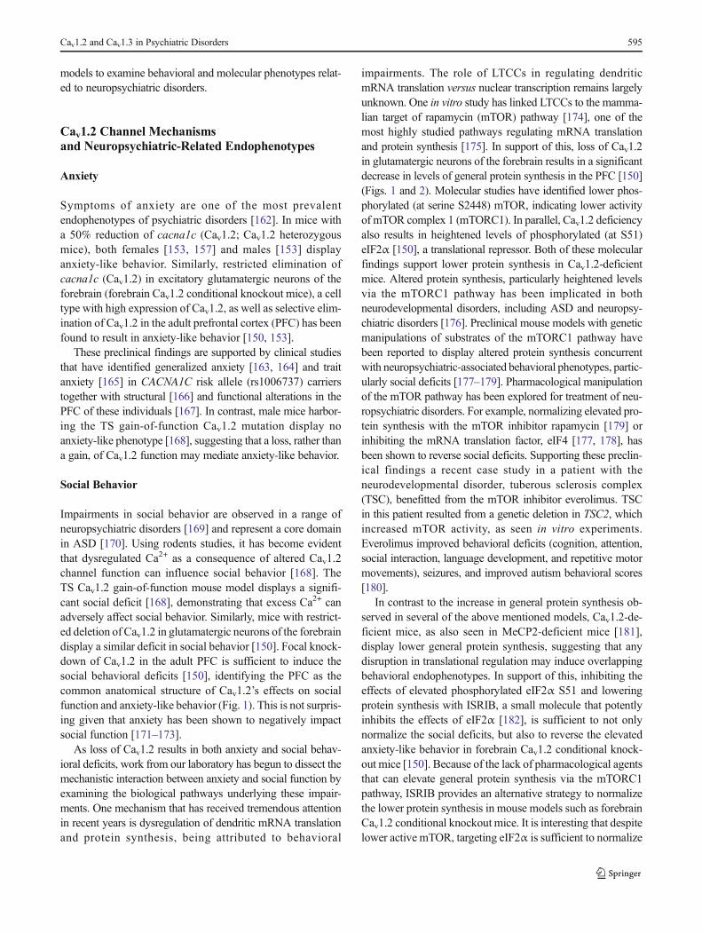
Fig. 1 Cav1.2- and Cav1.3-mediated anatomical andmolecular pathways underlyingthe endophenotypes associatedwith neuropsychiatric disorders.Solid lines indicate pathways thathave been identified for therespective behavioralendophenotypes and dotted linesindicate potential pathways thatmay be recruited AMPAR = α-amino-3-hydroxy-5-methyl-4-isoxazolepropionic acidreceptors; PFC = prefrontalcortex; HPC = hippocampus;NAc = nucleus accumbens; VTA= ventral tegmental area; REDD1= regulated in development andDNA damage response 1; CP-AMPAR = Ca2+-permeableAMPAR; ERK2 = extracellularregulated kinase 2; CaMKII =CaM-dependent protein kinase II
590 Kabir et al.

linked to BD [49]. However, a study in the Han Chinese pop-ulation found no significant SNPs in CACNA1D associatedwith SCZ [50]. SNPs in CACNA1D have also been associatedwith cocaine dependence [51]. Furthermore, whole-exome se-quencing studies have identified 2 de novo mutations(p.A749G and p.G407R) in CACNA1D in patients with spo-radic autism and intellectual disability [52, 53]. Functionalstudies have identified these mutations as gain-of-function ofCav1.3 LTCCs [7, 54].
Thus, based on the above findings on the impact ofCACNA1C and CACNA1D disease-associated genetic vari-ants on gene expression and channel function, the availabledata suggest that higher or lower levels of Ca2+ influx in neu-rons can be detrimental. In addition, LTCCs can also regulateneuronal firing. For example, LTCCs can directly provide adepolarizing stimulus; this can stabilize upstates/plateau po-tentials (e.g. [55]), thus affecting neuronal firing. LTCCs canalso couple to Ca2+-activated K channels [56, 57], thus mod-erating neuronal firing. Therefore, decreased LTCC activitycould actually increase firing in some neurons, which maytrigger Ca2+ influx through other sources (other voltage-gated Ca2+ channels subtypes or glutamateN-methyl-D-aspar-tate receptors). Similarly, increase in LTCC activity as a resultof gain of LTCC function could silence neurons. Thus, alteredCav1.2 and Cav1.3 LTCC levels or activity, in a cell-type-specific manner, can affect neuronal function in multiple waysthat could negatively affect brain function and contribute toneuropsychiatric symptoms in disorders associated withCACNA1C and CACNA1D.
Targeting L-Type Calcium Channelsin the Treatment of Neuropsychiatric Disordersand Drug Dependence
LTCC blockers have been used clinically for several decades inthe treatment of cardiac conditions such as hypertension, arrhyth-mias, and cardiac ischemia [2]. Recently, with growing evidenceof the association between LTCC genes and neuropsychiatricdisorders [5], there has been increased interest in repurposingthese drugs for the treatment of neuropsychiatric conditionsand drug dependence [6, 29]. Since the 1980s several clinicalstudies have examined the efficacy of LTCC blockers using the 3classes of LTCC pharmacological blockers—dihydropyridines(DHPs; nifedipine, amlodipine, felodipine, isradipine,nicardipine, nisoldipine, and nimodipine); phenylalkylamines(verapamil); and benzothiazepines (diltiazem)—to alleviatesymptoms in patients presenting with neuropsychiatric condi-tions and drug dependence. Below, we review the findings ofthese studies (see Table 2). Of note,many of these clinical studieshave utilized high systemic doses of LTCC blockers, particularlythe DHPs and thus could have significant confounding effects onbehavior, as demonstrated by rodent studies [105, 106].
Additionally, several of these studies have used thephenylalkylamine blocker, verapamil (Table 2). In addition toverapamil’s nonspecific effects such as blocking other Ca2+
channels [107], potassium channels [108–110], and α-adrenergic receptors [111], studies examining how well verapa-mil enters the brain in humans have been sparse. Verapamil is asubstrate for P-glycoprotein (P-gp) substrate, an efflux transport-er found in several organs, including the blood–brain barrier,where it plays an important role for the entry of drugs, includingverapamil, into the brain [112]. Verapamil can function as astimulator of the P-gp activity at low concentrations, preventingbrain entry, whereas at high concentrations it acts as an inhibitorof P-gp, allowing brain entry [113, 114]. However, verapamil athigh concentrations, although capable of penetrating the blood–brain barrier, can have toxics effects [115].
BD
The efficacy of the LTCC blocker verapamil in alleviatingacute mania observed in BD has been explored in several stud-ies [116]. While some studies report a significant decrease inthe severity of mania in patients with BD or manic patients [58,59, 62–66, 68, 69], several others report no antimanic effectswhen verapamil was administered as a monotherapy [60, 61,70–72]. However, when verapamil was administered in com-bination with the mood stabilizer lithium to patients with BDwho were unresponsive to lithium [70] or in conjunction withthe antipsychotic medication chlorpromazine [60] there wassignificant improvement in manic symptoms. This suggeststhat verapamil could have some potential for maintenance ther-apy of mania but only when administered in combination withother drugs. In addition to these findings, verapamil was foundto improve significantly major psychotic depressive symptomsin 1 patient [67] and improve depressive symptom scores inpatients prescribed verapamil for hypertension [73].
Although there are only a handful of studies with nimodipineand diltiazem, the findings are more consistent, though not asthoroughly investigated as verapamil. Bipolar manic patientstreated with nimodipine either as a monotherapy [74–76] or incombination with lithium [77] or the antiseizure medication car-bamazepine [74] showed significant improvements. Similar im-provements in mood were reported when an adolescent withrefractory, ultradian rapid cycling BD was treated withnimodipine [78]. Likewise, both bipolar manic patients [79]and treatment-resistant bipolar patients [80] treated with diltia-zem had significantly decreased manic symptoms. However, inpatients with no prior psychiatric history, the use of nifedipine totreat angina was associated with the onset of depression [81].
SCZ
Verapamil has also been tested in patients with SCZ. In pa-tients with acute SCZ, verapamil treatment when administered
Cav1.2 and Cav1.3 in Psychiatric Disorders 591

as a monotherapy resulted in a significant decrease in psychot-ic symptoms [82]. These findings were confirmed in a sepa-rate study that reported a similar small, but significant,
attenuation of psychotic symptoms with verapamil [83].Similarly, patients with chronic SCZ treated with verapamilfor 28 days displayed significant improvements in positive
Table 2 Type of clinical study using L-type calcium channel blockers in neuropsychiatric disease
Neuropsychiatric diseasetreated
L-type calciumchannelblocker used
Type of clinical study performed Reference(s)
Bipolar disorder Verapamil Nonrandomized [58–61]
Randomized; patients received either verapamil or lithium and efficacycompared
[62]
Randomized: crossover study; patients received either verapamil or lithium andthen treatment switched
[63]
Case report [64–67]
Randomized placebo-controlled within subject [68]
Nonrandomized placebo within subjects [69]
Randomized: phase I—lithium, phase II—nonresponders (verapamil orlithium);
phase III—nonresponders (combined verapamil/lithium)
[70]
Randomized placebo [71, 72]
Randomized; patients received verapamil or the antihypertensive drug atenololandefficacy compared
[73]
Nimodipine Randomized placebo-controlled within-subjects [74]
Nonrandomized placebo within-subject [75]
Nonrandomized [76]
Case report [77, 78]
Diltiazem Nonrandomized [79, 80]
Nifedipine Case report [81]
Schizophrenia Verapamil Case report [82]
Randomized placebo [83, 84]
Randomized placebo-controlled within subject [85]
Nonrandomized [86]
Nonrandomized placebo within subject [87, 88]
Nifedipine Nonrandomized [89]
Randomized placebo [90]
Cocaine dependence Nimodipine Randomized placebo-controlled within subject [91]
Isradipine Randomized placebo-controlled within subject [92, 93]
Nimodipine Randomized placebo [94]
Nifedipine Randomized placebo-controlled within subject [95]
Amlodipine Nonrandomized [96]
Isradipine Randomized placebo-controlled within subject [97]
Morphine dependence Verapamil Randomized placebo [98]
Diltiazem, verapamil,nimodipine
Randomized placebo-controlled within subject [99]
Nifedipine Randomized; patients received nifedipine or the α-adrenoceptor blocker, clo-nidine;only nifedipine effects reported
[100]
Alcohol dependence Verapamil,nimodipine
Randomized placebo [101]
Isradipine Quasirandom placebo within subject; patients received 9 different combinationsofethanol or isradipine at different doses; only the first and second treatmentwas not randomly given
[102]
Verapamil, nifedipine Randomized placebo-controlled within subject [103]
Nimodipine Nonrandomized [104]
592 Kabir et al.

and negative symptoms, as well as in anxiety and depression[86]. In contrast to these findings, other studies have reportedan increase in paranoia in verapamil-treated patients withchronic schizophrenia who were recently withdrawn fromneuroleptics [87], or no effect on the psychological state [88]and negative symptoms [84]. Similar to the results with ve-rapamil, the effects of nifedipine on alleviating the psychoticsymptoms are conflicting. While some studies showed thatpatients with chronic schizophrenia receiving nifedipineshowed an improvement on the Dementia Scale [85] and theBrief Psychiatric Rating Scale [89], another showed no im-provements based on the Psychiatric Symptom AssessmentScale ratings [90].
Cocaine Dependence
Evidence from rodent studies has identified an important roleof LTCCs in multiple aspects of cocaine addiction, makingthese channels a potential target for the treatment of addiction.Over the last few decades, clinical studies have tested theefficacy of LTCC blockers on the subjective effects of co-caine, though the results have beenmixed.While some studiesreport no effect with isradipine and nimodipine [91, 92],others have reported a reduction in the subjective responseto cocaine with nifedipine [95]. In contrast, a recent studyreported enhanced subjective effects with isradipine [93].The effects of LTCCs have also been tested on cocaine crav-ing, the primary cause of relapse to cocaine use, withnimodipine [94], amlodipine [96], and isradipine [97] reduc-ing cocaine craving.
Morphine Dependence
The impact of LTCC blockers on the subjective effects ofmorphine are mixed. While 1 study reported a reduction inthe subjective effect of morphine with verapamil [98], anotherstudy showed no influence of nimodipine, verapamil, and dil-tiazem on counteracting morphine’s subjective effects inhealthy humans [99]. Separately, it has been reported thatnifedipine treatment caused confusion in individuals that werein morphine withdrawal [100]. These studies suggest thatLTCC blockers may show selective effects in morphine-dependent individuals, having beneficial effects in some butdetrimental effects in others.
Alcohol Dependence
With ethanol, LTCC blockers nimodipine and verapamil havebeen reported to have no effect on the subjective and psycho-motor effects of the drug [101]. Similarly, isradipine had noeffect on ethanol’s acute effects on poor psychomotor perfor-mance [102], and failed to antagonize ethanol intoxication
[103]. However, nimodipine has been found to be effectivefor the treatment of ethanol’s withdrawal symptoms [104].
The discrepancies in the effects of LTCC blockers in thetreatment of psychiatric disorder-associated symptoms and onthe symptoms associated with drug dependence, as reviewedabove, may be a consequence of a myriad of factors, includingthe high doses used; however, it highlights the complexity ofneuropsychiatric disorders, as well as how dysregulatedLTCCs may influence different psychiatric symptoms. Givenour current knowledge of the varying impact of disease-riskvariants to CACNA1C and CACNA1D levels (increase or de-crease) and Cav1.2 and Cav1.3 function (gain or loss), it is notsurprising that LTCC blockers may be efficacious in someindividuals but not all and for some symptoms and not allthose seen in CACNA1C and CACNA1D-linked neuropsychi-atric conditions. Thus, understanding how Cav1.2 and Cav1.3channel mechanisms contribute to neuropsychiatric-relatedsymptoms can be greatly helpful. As progress on the develop-ment of new therapeutics for psychiatric disorders has beenslow, understanding biological pathways in the context of dis-ease symtoms (e.g., anxiety, social, depression, cognition) arekey in identifying new targets for developing medications.Webelieve that this is particularly important given the complexnature of neuropsychiatric disorders versus the idea that theyare sole entities as previously considered.
L-Type Calcium Channel Signaling in Neuronsand Relevance to Neuropsychiatric Disorders
Several lines of evidence from both human and animal studieshave unequivocally established a critical contribution of Ca2+
signaling pathways to the pathophysiology of both neuropsy-chiatric and neurodevelopmental disorder [1, 6, 117]. Cav1.2and Cav1.3 channels are key mediators of Ca2+ signaling inneurons [26]. In vitro studies have established thatdepolarization-induced increase in local Ca2+ activates thecalcium sensor calmodulin (CaM) at the synapse that subse-quently activates a series of Ca2+/CaM-dependent protein ki-nases (CaMKs) [118–122] and also the Ras/mitogen-activatedprotein kinase (MAPK) pathway [123–125], both of whichtransduce molecular cascades to the nucleus, activating geneexpression via the extensively studied transcriptional factorcAMP response element-binding protein (CREB) (Fig. 2).CREB-activated genes are critically involved in synaptic, neu-ronal, and behavioral plasticity [126–130]. LTCC-induced ki-nase pathways are also recruited for phosphorylation of sig-naling molecules that activate other transcription regulators,including myocyte enhancer factor-2 [131–134] and MeCP2(Fig. 2), key factors involved in neuronal development, be-havioral alterations, and neurodevelopmental disorders. In ad-dition to activation of the kinase pathways, LTCCs can alsoactivate the phosphatase pathway that likewise modulates
Cav1.2 and Cav1.3 in Psychiatric Disorders 593

transcription factor function. This includes the nuclear factorof activated T cells (NFAT) family of transcription factors thatis regulated by the LTCC-activated Ca2+–calmodulin-depen-dent phosphatase calcineurin that dephosphorylates NFAT cy-toplasmic 3 [135] and NFATcytoplasmic 4 [136], inducing itstranslocation from the cytoplasm to the nucleus to regulategene expression. Alteration of myocyte enhancer factor-2and NFAT, as well as CREB signaling networks, has beenlinked to dendritic retraction as a consequence of elevatedCa2+ in induced pluripotent cells from patients with TS [132].
The significance of these LTCC-activated pathways to neu-ropsychiatric disease pathology is underscored by severalpathway network analyses that have repeatedly identified sig-nificant association of the calcium signaling pathway to BD[137, 138], SCZ [138, 139], MDD [138], and ASD [140],highlighting the calcium pathway as a common dysregulatedmechanism underlying the etiology of these disorders. Thishas also been observed using proteomic approaches with post-synaptic density fractions from the cortex of patients withSCZ [141] and BD [142] that have identified altered levelsof proteins that are mediators of calcium signaling.Additionally, pathway analyses have identified significant en-richment of the MAPK/extracellular regulated kinase (ERK)pathway in ASD [140, 143], SCZ [141], and depression [144],and whole-exome sequencing has found rare and likelyprotein-damaging mutations in members of the MAPK/ERK and CREB-regulated intracellular signaling pathways
in patients with BD [145]. Another pathway enriched in theproteomics-based pathway analysis from the postsynapticdensity of patients with BDwas the eukaryotic initiation factor2α (eIF2α) signaling pathway [142], which is involved inmRNA translation [146] and a pathway we review below asa new candidate pathway linked to behavioral deficits inCav1.2-deficient mice. In light of these findings, it is evidentthat dysregulation in LTCC and Ca2+ signaling can result inneuronal alterations that contribute to the pathogenesis ofneuropsychiatric-related cellular [51, 147–150], physiologi-cal, synaptic [150, 151], and behavioral phenotypes [51,150, 152, 153], all of which we discuss below.
L-Type Ca2+ Channelsand Neuropsychiatric-Related Phenotypes:Preclinical Animal Studies
Rodents have proved to be a useful tool to study humandisease-related behavioral symptoms. Given the predominanceof noncoding variants in CACNA1C that are predicted to affecttranscriptional control and result in lower or higher levels ofCACNA1C, studying gene knockout and overexpressing micecan be highly useful. Similarly, mice harboring coding muta-tions that cause either loss or gain of function can be informa-tive. The Cav1.2 and Cav1.3 LTCC isoforms have structuralsimilarities making them both equally sensitive to LTCC phar-macological agents [154], posing a challenge to study isoform-specific brain and behavioral alterations. To overcome this,several different laboratories have developed genetic mutantmouse models that have altered expression or function of eitherCav1.2 or Cav1.3. The most common models used to studyCav1.2-specific mechanisms have been the heterozygous con-stitutive knockout mouse model (homozygous is embryoniclethal [155]), and temporal, spatial, and cell-type-specific con-ditional mouse models with restricted knockdown of cacna1c(encoding Cav1.2) in the brain using Cre-recombinase specificdrivers (mouse lines and viral vectors). Constitutive Cav1.2heterozygous knockout mice develop a cardiac phenotype, par-ticularly following stress [156]; however, it is unlikely that thiscardiac phenotype affects brain phenotypes at baseline [153,157] or following stress [158]. For Cav1.3 studies, there existsthe Cav1.3 knockout mouse, though because of the high ex-pression of Cav1.3 in the hair cells of the ear are deaf and limittheir use for behavioral studies. To overcome this and the lackof subunit-specific pharmacological agents, the Striessnig lab-oratory developed a Cav1.2 DHP-insensitive (Cav1.2DHP
–/–)mouse that harbors a single point mutation in the Cav1.2α1subunit at the DHP binding site, rendering Cav1.2 channelsinsensitive to DHPs [159], allowing the specific pharmacolog-ical manipulation of Cav1.3 channels [21, 51, 159–161]. Belowwe review studies that have utilized these preclinical mouse
P-MAPKP-CaMK
Cav1.2
P-CREB
gene expression
P-mTORC1
P-eIF2
protein synthesis
CaN
NFAT
REDD1
P-FoxO3a
P-Akt
Cav1.3
MEF2 MeCP2
Fig. 2 Cav1.2 and Cav1.3 signaling mechanisms. Solid lines indicatepathways that have been directly linked to Cav1.2 or Cav1.3 channelsand dotted line indicates potential pathway that may be recruited. Blackarrows indicate Cav1.2-specific pathways; red arrows indicate Cav1.3-specific pathways. mTORC1 = mammalian target of rapamycincomplex 1; REDD1 = regulated in development and DNA damageresponse 1; P-MAPK = phosphorylatedmitogen-activated protein kinase;P-CaMK = phosphorylated CAM-dependent protein kinase; P-eIF2α =phosphorylated eukaryotic initiation factor 2 alpha; CaN = calcineurin; P-Akt = phosphorylated protein kinase B;MEF2 =myocyte enhancer factor2; NFAT = nuclear factor of activated T cells; P-CREB = phosphorylatedcAMP response element-binding protein
594 Kabir et al.
models to examine behavioral and molecular phenotypes relat-ed to neuropsychiatric disorders.
Cav1.2 Channel Mechanismsand Neuropsychiatric-Related Endophenotypes
Anxiety
Symptoms of anxiety are one of the most prevalentendophenotypes of psychiatric disorders [162]. In mice witha 50% reduction of cacna1c (Cav1.2; Cav1.2 heterozygousmice), both females [153, 157] and males [153] displayanxiety-like behavior. Similarly, restricted elimination ofcacna1c (Cav1.2) in excitatory glutamatergic neurons of theforebrain (forebrain Cav1.2 conditional knockout mice), a celltype with high expression of Cav1.2, as well as selective elim-ination of Cav1.2 in the adult prefrontal cortex (PFC) has beenfound to result in anxiety-like behavior [150, 153].
These preclinical findings are supported by clinical studiesthat have identified generalized anxiety [163, 164] and traitanxiety [165] in CACNA1C risk allele (rs1006737) carrierstogether with structural [166] and functional alterations in thePFC of these individuals [167]. In contrast, male mice harbor-ing the TS gain-of-function Cav1.2 mutation display noanxiety-like phenotype [168], suggesting that a loss, rather thana gain, of Cav1.2 function may mediate anxiety-like behavior.
Social Behavior
Impairments in social behavior are observed in a range ofneuropsychiatric disorders [169] and represent a core domainin ASD [170]. Using rodents studies, it has become evidentthat dysregulated Ca2+ as a consequence of altered Cav1.2channel function can influence social behavior [168]. TheTS Cav1.2 gain-of-function mouse model displays a signifi-cant social deficit [168], demonstrating that excess Ca2+ canadversely affect social behavior. Similarly, mice with restrict-ed deletion of Cav1.2 in glutamatergic neurons of the forebraindisplay a similar deficit in social behavior [150]. Focal knock-down of Cav1.2 in the adult PFC is sufficient to induce thesocial behavioral deficits [150], identifying the PFC as thecommon anatomical structure of Cav1.2’s effects on socialfunction and anxiety-like behavior (Fig. 1). This is not surpris-ing given that anxiety has been shown to negatively impactsocial function [171–173].
As loss of Cav1.2 results in both anxiety and social behav-ioral deficits, work from our laboratory has begun to dissect themechanistic interaction between anxiety and social function byexamining the biological pathways underlying these impair-ments. One mechanism that has received tremendous attentionin recent years is dysregulation of dendritic mRNA translationand protein synthesis, being attributed to behavioral
impairments. The role of LTCCs in regulating dendriticmRNA translation versus nuclear transcription remains largelyunknown. One in vitro study has linked LTCCs to the mamma-lian target of rapamycin (mTOR) pathway [174], one of themost highly studied pathways regulating mRNA translationand protein synthesis [175]. In support of this, loss of Cav1.2in glutamatergic neurons of the forebrain results in a significantdecrease in levels of general protein synthesis in the PFC [150](Figs. 1 and 2). Molecular studies have identified lower phos-phorylated (at serine S2448) mTOR, indicating lower activityof mTOR complex 1 (mTORC1). In parallel, Cav1.2 deficiencyalso results in heightened levels of phosphorylated (at S51)eIF2α [150], a translational repressor. Both of these molecularfindings support lower protein synthesis in Cav1.2-deficientmice. Altered protein synthesis, particularly heightened levelsvia the mTORC1 pathway has been implicated in bothneurodevelopmental disorders, including ASD and neuropsy-chiatric disorders [176]. Preclinical mouse models with geneticmanipulations of substrates of the mTORC1 pathway havebeen reported to display altered protein synthesis concurrentwith neuropsychiatric-associated behavioral phenotypes, partic-ularly social deficits [177–179]. Pharmacological manipulationof the mTOR pathway has been explored for treatment of neu-ropsychiatric disorders. For example, normalizing elevated pro-tein synthesis with the mTOR inhibitor rapamycin [179] orinhibiting the mRNA translation factor, eIF4 [177, 178], hasbeen shown to reverse social deficits. Supporting these preclin-ical findings a recent case study in a patient with theneurodevelopmental disorder, tuberous sclerosis complex(TSC), benefitted from the mTOR inhibitor everolimus. TSCin this patient resulted from a genetic deletion in TSC2, whichincreased mTOR activity, as seen in vitro experiments.Everolimus improved behavioral deficits (cognition, attention,social interaction, language development, and repetitive motormovements), seizures, and improved autism behavioral scores[180].
In contrast to the increase in general protein synthesis ob-served in several of the above mentioned models, Cav1.2-de-ficient mice, as also seen in MeCP2-deficient mice [181],display lower general protein synthesis, suggesting that anydisruption in translational regulation may induce overlappingbehavioral endophenotypes. In support of this, inhibiting theeffects of elevated phosphorylated eIF2α S51 and loweringprotein synthesis with ISRIB, a small molecule that potentlyinhibits the effects of eIF2α [182], is sufficient to not onlynormalize the social deficits, but also to reverse the elevatedanxiety-like behavior in forebrain Cav1.2 conditional knock-out mice [150]. Because of the lack of pharmacological agentsthat can elevate general protein synthesis via the mTORC1pathway, ISRIB provides an alternative strategy to normalizethe lower protein synthesis in mouse models such as forebrainCav1.2 conditional knockout mice. It is interesting that despitelower active mTOR, targeting eIF2α is sufficient to normalize
Cav1.2 and Cav1.3 in Psychiatric Disorders 595

behavioral deficits, suggesting cross-talk between the mTORand eIF2α pathways [183].
These findings provide a unique role of Cav1.2 in proteinsynthesis via the eIF2α pathway and potentially the mTORpathway (Fig. 2), and also identify a common Cav1.2-mediat-ed molecular mechanism underlying social and anxiety-likebehaviors (Fig. 1). Furthermore, it identifies a novel target in aCav1.2-deficient mouse model that can be manipulated in theadult brain to rescue behavioral deficits. The contribution ofeIF2α and its pathway to psychiatric and neurodevelopmentaldisease-associated symptoms remains unexplored but war-rants further investigation. Increases in phosphorylation ofeIF2α at S51 can not only decrease translation of mostmRNAs [184], but can also induce translation of a subset ofmRNAs containing short upstream open reading frames in anactivating transcription factor 4-dependent manner [146], amember of the CREB family of transcription factors [185].Thus, further studies to explore the specific proteins targetedby elevated phosphorylated eIF2α in the PFC of Cav1.2-defi-cient mice that are modulating both anxiety and social func-tion will be informative.
Depressive-Like Behavior
In addition to the above mentioned endophenotypes,depression-related symptoms are also commonly observed inBD,MDD, and SCZ [162]. In the late 1980s, a role for LTCCsin regulating depression-related behavior was first realizedusing LTCC pharmacological agents demonstrating that theDHP LTCC blocker nifedipine has an antidepressant-like ef-fect in rats [186]. This was further expanded in a later studythat showed that, in addition to nifedipine, other DHPblockers, including nicardipine, nitrendipine, isradipine,felodipine, and nimodipine but not amlodipine, had a similarantidepressant-like effect [187]. More recently, in support ofthe pharmacological antidepressant-like effect, the use of ge-netic mutant mice has revealed that Cav1.2 heterozygous miceexhibit antidepressant-like behavior [152, 157]. Furthermore,focal knockdown of cacna1c (Cav1.2) in the adult PFC wassufficient to induce a similar antidepressant-like effect [152],consistent with antidepressants exerting their effects throughcellular changes in the PFC [188].
In the context of CACNA1C SNPs the above findings sug-gest that gain of Cav1.2 function mutations would contribute todepression-related symptoms. In support of this, it has beenshown that pharmacological activation of LTCCs with theDHP activator BayK8644 induces a depressive-like phenotype[189]. Although depression-related behavior has not been test-ed in the Cav1.2 gain-of-function TS mouse, there are casereports identifying 1 patient with TS with depression [48] andanother patient with TS who developed BD in adulthood [41].Together, these findings support the theory that gain of Cav1.2function results in depression-related symptoms.
It is intriguing that loss of Cav1.2 in the PFC results inanxiety-like behavior but has an antidepressant-like effect. Adeeper understanding of this differential effect of Cav1.2 defi-ciency has come from molecular studies that have identifiedseparate mechanisms influencing anxiety and depression-related behaviors. In contrast to dysregulation of the mRNAtranslation pathway underlying anxiety in Cav1.2-deficient mice(described above), the regulated in development and DNA dam-age response 1 (REDD1; also known at DDIT4 or RTP801)pathway modulates depression-related behaviors (Fig. 1;[152]). Cav1.2 heterozygous mice that exhibit anantidepressant-like phenotype have lower levels of thedepression-related protein REDD1 in the PFC, and increasinglevels of REDD1 in this anatomical region of adult Cav1.2 het-erozygous mice is sufficient to reverse the antidepressant-likephenotype [152]. This is consistent with present findings ofhigher REDD1 levels in the PFC of depressed patients [190].
Downstream of REDD1, the protein FoxO3a has beenidentified to play a role in modulating the antidepressant-likeeffect observed in Cav1.2 heterozygous mice (Fig. 2; [152]).FoxO3a belongs to the FoxO family of transcription factorswith SNPs within the FoxO3a gene linked to BD [191]. Inrodents FoxO3a has been identified as a modulator ofdepression-related behavior [192]. FoxO3a knockout mice ex-hibit an antidepressant-like phenotype, and the antidepressantimipramine [193] and the mood stabilizer lithium [194] havebeen found to decrease levels of nuclear FoxO3a, suggestingthat higher levels of nuclear FoxO3a may be associated withdepressive behavior. In support of this, elevated levels of nu-clear FoxO3a in the PFC of Cav1.2 heterozygous mice, fol-lowing REDD1 overexpression, has been associated withdampening the antidepressant-like phenotype seen in thesemice [152]. These findings provide the first evidence of a roleof the REDD1/FoxO3a signaling pathway in the PFC in reg-ulating depression-related behavior in Cav1.2 heterozygousmice (Figs. 1 and 2), and a new framework to study Cav1.2-associated depressive behavior.
Cognitive Function
Although not as broadly recognized as a symptom in neuro-psychiatric disorders as changes in mood and emotion, cogni-tive impairments are a prominent feature of the CACNA1C-associated psychiatric disorders, as well as ASD [195]. In par-ticular, deficits within different aspects of learning and mem-ory have been observed in BD, SCZ, MDD, ASD, and, to alesser extent, ADHD [195]. Using rodent models, there isevidence that loss of Cav1.2 influences discrete forms of learn-ing and memory. In the hippocampal-dependent Morris watermaze (MWM) spatial memory task, Cav1.2 conditional knock-out mice display normal acquisition and consolidation of theplatform location with similar performance to the controls dur-ing the short- and long-term (24 hour) probe tests [150,
596 Kabir et al.

196], whereas during the remote 30-day probe trial Cav1.2knockout mice display a significant deficit [196]. Recently, ithas been demonstrated that by increasing the difficulty of theMWM task by decreasing the number of available spatial cues,Cav1.2 conditional knockout mice exhibit a delay in the ac-quisition of the platform location and a significant deficit in thelong-term memory probe test [197]. A similar hippocampal-dependent deficit has been observed in the fear-associatedcontext discrimination task [197]. This supports previous databy Moosmang et al. [198] demonstrating that Cav1.2 channelsin the hippocampus (using hippocampus-specific conditionalknockout mice) are necessary for a hippocampal-dependentdiscriminatory water-maze task. This is supported by a roleof Cav1.2 in hippocampal long-term potentiation [198], a cel-lular model of learning and memory [199], as well as a role inadult hippocampal neurogenesis [149, 197], a cellular mecha-nism linked to learning/memory processes ([200, 201];discussed below).
Similar to Cav1.2-deficient mice, the Cav1.2 gain-of-function TS mice display no deficit in learning and memoryin the MWM spatial memory task or the Y-maze task [168].However, when the hidden escape platform is moved to adifferent location to test reversal learning in both tasks, micedisplay a significant delay in determining the new platformlocation [168]. Because of this mild persistence in continuingto seek the original location of the platform in the MWM andrepeatedly attempting to enter the arm of the Y-maze with theoriginal platform, despite the presence of a physical obstruc-tion, the authors interpret these observations as evidence ofrepetitive, restrictive, and perseverative behavior [168], andpossibly a sign of lack of cognitive flexibility, seen in disor-ders such as SCZ [202]. In contrast to these findings, loss ofCav1.2 in the glutamatergic neurons of the forebrain appearsto negatively impact learning and memory in the Y-maze hid-den-platform task [150], an observation that begs further ex-ploration on the anatomical and cell-type specificity of loss ofCav1.2 signaling.
Separately from studies in rodents, it is clear that theLTCCs also play an important role in fear-associated memo-ries, the most common behavioral paradigm utilized to studythe biological basis of emotion [203–205]. This is not surpris-ing given that altered emotional processing has been reportedin patients with SCZ, BD, and ADHD [206]. In rodents, it hasbeen observed that systemic inhibition of LTCCs with nifed-ipine had no impact on the acquisition or long-term expressionof cue-associated fear memories [207]. However, focal deliv-ery of verapamil in the basolateral amygdala, a brain regioninvolved in fear, of adult rats immediately prior to fearconditioning blocked cue-associated long-term fear memorybut not the short-term memory [208]. Similarly, focal deliveryof the LTCC blockers verapamil or nifedipine into thebasolateral amygdala of adult rats immediately before cueextinction training impaired the long-term memory of fear
extinction [209]. This is consistent with a previous study thatshowed impaired cue extinction with systemic administrationof the LTCC blockers nifedipine and nimodipine [207]. Thesestudies suggest that differential LTCC-mediated mechanismsare being recruited for acquisition versus extinction of cue-associated fear memories.
Using genetic mutant mice, recent studies have focused ondissociating the differential contribution of the Cav1.2 andCav1.3 isoforms in fear processes. In agreement with pharma-cological studies, brain-specific Cav1.2 knockout mice haveno deficit in the consolidation and recall of a cue-associatedfear memory [210]. However, selective conditional knockoutof Cav1.2 in glutamatergic neurons of the forebrain displayenhanced freezing during the long-term cue-associated fearmemory test [150]. This discrepancy is most likely a resultof compensatory upregulation of Ca2+-permeable glutamateα-amino-3-hydroxy-5-methyl-4-isoxazolepropionic acid re-ceptors (AMPARs) in the amygdala [210]. Interestingly inthe Cav1.2 gain-of-function TS mouse, an increase in freezingduring the long-term cue-associated fear memory test has beenreported [168], suggesting that too much or too little Cav1.2signaling can have similar behavioral phenotypes in certaintasks. Interestingly, in contextual fear conditioning, loss ofCav1.2 in genetic mutant mice [197, 211] did not impact freez-ing during the context-associated fear memory test. In con-trast, the Cav1.2 gain-of-function TS mice exhibited enhancedfreezing during the context-associated fear memory test [168],identifying that loss and gain of function can have differentialeffects of certain brain region-specific tasks. In addition to theconventional fear-conditioning paradigms, LTCC inhibitorshave been shown to block latent inhibition of conditioned fear[212] and fear-potentiated startle [213], suggesting a role ofthese channels in other forms of emotional learning andplasticity.
The mechanism underlying altered fear memories de-scribed above are not known. Late-phase long-term potentia-tion (LTP) at thalamic inputs to the lateral amygdala, a mech-anism that requires LTCCs [198, 208, 214, 215], has beenassociated with cue fear conditioning [216]. One moleculethat mediates LTP in the thalamoamygdala pathway is brain-derived neurotrophic factor (BDNF) [217], a downstream tar-get of LTCCs [149, 218]. Induction of BDNF in the amygdalais required for consolidation of fear memories [219].Supporting these findings, administration of the LTCC inhib-itor verapamil blocks the induction of BDNF after cue-associated fear conditioning [220]. The findings from thisstudy suggest that this occurs as a result of lack of activationof CaMKIV [220], a CREB kinase and a downstream target ofLTCCs [221], resulting in decreased binding of phosphorylat-ed CREB at the promoter regions of BDNF [220]. This isconsistent with previous studies that have identified LTCCsas critical regulators of BDNF expression both in vitro [222]and in vivo [149]. Currently, the specific LTCC isoform
Cav1.2 and Cav1.3 in Psychiatric Disorders 597

mediating this fear conditioning-induced LTP in thethalamoamygdalar pathway is unknown.
Cav1.2 Channels, Adult Hippocampal Neurogenesis,and Neuropsychiatric-Related Phenotypes
Adult hippocampal neurogenesis that involves addition ofnewborn neurons (granule cells) to the dentate gyrus of thehippocampus throughout life has been implicated in the pa-thology underlying SCZ, based on rodent models of the dis-order [223–230], BD based on genes associated with the dis-order [137, 231], and depression [232, 233], autism[234–237], and ADHD, based on rodent disease models[229, 235, 238]. Rodent behavioral studies suggest a role ofadult hippocampal neurogenesis in many of the phenotypesrelated to these disorders that are also observed in Cav1.2-deficient mice described above, including memory formation[200, 201], context discrimination [239], modulation of anxi-ety and depressive-like behavior [240], as well as mediatingthe effects of antidepressants. In support of this, 2 independentstudies have identified a deficit in adult hippocampalneurogenesis in mice with loss of Cav1.2 restricted to gluta-matergic neurons of the forebrain [149] and in neurons of theentire brain [197]. This is consistent with a previous in vitrostudy identifying a role of LTCCs in activity-dependent regu-lation of adult-derived neural precursor cells [241].
The deficit in adult hippocampal neurogenesis in Cav1.2conditional knockout mice is specific to survival and not pro-liferation of neural precursor cells (NPCs) [149]. This findingis supported by an in vitro study that has identified a role ofLTCCs during the later neurogenic stages of survival and mat-uration of NPCs derived from adult rat hippocampus [242]. Asadult hippocampal neurogenesis is a highly regulated processand results from a balance of proliferation of NPCs and thesurvival of young newborn neurons into which NPCs differ-entiate [243], the discovery of Cav1.2 channels in supportingthe survival of newborn neurons suggests that this stage ofadult neurogenesis may be important for certain aspects ofneuropsychiatric disease.
The precise mechanism of the deficit in survival of new-born neurons remains unknown. Given that Cav1.2 expressionis restricted to mature young hippocampal neurons in adultmice [244], one potential mechanism is via the neurotrophicfactor, BDNF. LTCCs mediate BDNF production in gluta-matergic neurons of the hippocampus [149], release of whichacts on both the secreting neuron and neighboring neurons[245]. LTCCs serve as a primary Ca2+ source for Bdnf tran-scriptional regulation, particularly at the promoter of Bdnfexon IV, a splice variant critically involved in experience-dependent neuronal plasticity [246–249]. Multiple LTCC-activated transcription regulators, including CREB, Ca2+ re-sponse factor, and MeCP2, which are also involved in
regulating adult hippocampal neurogenesis [127, 237, 250,251], control Bdnf expression by binding to Bdnf exon IVpromoter in hippocampal neurons [222, 252–255]. Thus, itis plausible that lack of activation of these factors in the hip-pocampus results in lower BDNF and thus lowers survival ofnewborn neurons. This mechanism may also contribute to theLTP deficit observed in hippocampal-specific Cav1.2 knock-out mice [198] as BDNF signaling and adult newborn neuronsare required for hippocampal-dependent learning and memoryprocesses [256]. Additionally, BDNF is a key player in regu-lating mood-related phenotypes [257]. Thus, collectively, it isplausible that the lower survival of adult born neurons as aresult of Cav1.2/BDNF deficiency could contribute to theneuropsychiatric-related phenotypes observed in Cav1.2-defi-cient mice, a mechanism to be confirmed in future studies.
Another key question is whether restoring reduced survivalof newborn neurons is sufficient to rescue phenotypes ob-served in Cav1.2-deficient mice. Using the neuroprotectiveaminopropyl carbazole P7C3-A20, a small molecule thatblocks neuronal cell death [258–264] and thus increases cellsurvival of hippocampal newborn neurons [243, 265, 266], ithas been found that this compound is capable of restoringhippocampal neurogenesis to normal levels in forebrainCav1.2 conditional knockout mice [149]. This therapeutic ef-fect occurred despite a lack in the normalization of BDNFlevels. Given that BDNF-enhancing agents have not provedto be effective therapeutically to date, P7C3-A20 offers analternative therapeutic mechanistic route to restore impairedadult neurogenesis in Cav1.2-deficient mice that circumventslower BDNF signaling. If P7C3-A20 is able to rescue behav-ioral deficits, this work may provide new treatment opportu-nities for patients suffering from CACNA1C-associated neu-ropsychiatric symptoms. Additionally the identification of apreviously unidentified role for Cav1.2 channels in neuronalcell survival may provide novel insight and approaches totreating neuropsychiatric disease, particularly in situations ofdecreased Cav1.2 or loss of Cav1.2 function.
Cav1.2, Excitatory/Inhibitory Imbalanceand Neuropsychiatric Phenotypes
One emerging hypothesis for the pathophysiological mecha-nisms underlying the behavioral impairments in neuropsychi-atric disorders is altered synaptic excitation (E) to inhibition(I) balance [151, 267], a cellular perturbation reported in mul-tiple mouse models exhibiting anxiety-like behavior, alteredsocial behavior, and impaired cognitive function [150, 177,178, 267–269]. However, the impact of loss or gain ofLTCC function on synaptic E/I balance and its impact onbehavior remains largely unknown. In vitro pharmacologicalstudies have provided evidence that LTCC-mediated mecha-nisms modulate synaptic plasticity in a homeostatic fashion
598 Kabir et al.

[270]. Cortical neurons treated for 24 hours with the LTCCblocker nifedipine have been shown to increase both frequen-cy and amplitude of miniature excitatory postsynaptic currents[271]. Furthermore, in hippocampal neurons, 24-hour block-ade of LTCCs with nifedipine has been found to decrease theexpression of synaptic γ-aminobutryic acid A receptors [272],which mediate inhibitory neurotransmission, suggesting thatLTCCs can modulate E/I balance. This is supported by anin vivo study, which found that loss of Cav1.2 in glutamatergicneurons of the forebrain results in higher frequency and am-plitude of miniature excitatory postsynaptic currents in layer-5neurons of the PFC, suggesting an increase in the overall E/Ibalance in this region [150], supporting other mouse modelsof neuropsychiatric disorders with higher E/I associated withneuropsychiatric-related behaviors.
These in vivo and in vitro studies suggest that chronic lossof LTCC signaling can have long-term consequences on syn-aptic scaling and, subsequently, function. The precise mecha-nism underlying this synaptic plasticity is not known. Thereare, however, 2 possible mechanisms that may be involved.First, impaired LTCC signaling may impact on nuclear tran-scriptional processes that can subsequently affect dendriticsynaptic protein changes. This possibility is supported by anin vitro study that found that 24-hour inhibition of LTCCsresults in increased (as opposed to the expected decrease)CREB-dependent transcription of the GluA1 subunit of theexcitatory AMPARs [271]. Second, loss of LTCC signalingcan negatively affect the mRNA translation machinery withinspines and alter the composition of synaptic proteins [150].These findings add to the growing literature that neuropsychi-atric disorders are disorders of the synapse and that alteredCav1.2 signaling, even though shown not to impact spineand dendritic architecture [198, 210] as opposed to Cav1.3channels [28, 273], can impact synaptic function via second-ary homeostatic effects.
Cav1.3 Channels and Neuropsychiatric-RelatedPhenotypes
Anxiety-Like, Depressive-Like, and Social Behavior
Using rodent preclinical models, Cav1.3 channels, althoughless studied than Cav1.2 channels, also modulateneuropsychiatric-related endophenotypes. Cav1.3-deficientmice demonstrate an anxiolytic-like phenotype [274], al-though this effect may be attributed to the congenital deafnessobserved in these mice [275]. This is supported by recentfindings in Cav1.2DHP
–/– mice wherein treatment with theLTCC activator BayK8644, which selectively activatesCav1.3 channels, does not induce anxiety-like behavior [51].In contrast to this, Cav1.3 regulates depressive-like behaviorwith Cav1.3 deficiency resulting in an antidepressant-like
phenotype [274], whereas systemic activation of the Cav1.3channels induces a depressive-like phenotype [159]. Cav1.3channel activation with BayK8644 has also been found toinduce a deficit in social behavior [51], in support of Cav1.3gain-of-function mutations associated with ASD [7, 54]. Thesystemic effect of Cav1.3 channel activation on depression-related and social behaviors has been attributed to its role inthe ventral tegmental area (VTA) [51], supporting the role ofdopaminergic neurotransmission in both depressive and socialbehavior (discussed below).
Cognitive Function
Rodent studies have identified a role of Cav1.3 in certainforms of learning and memory. Although Cav1.3-deficientmice displayed no deficit in the MWM spatial memory task[276], these mice have significantly impaired object locationmemory in a discrimination test [244], suggesting that Cav1.3channels may be recruited in specific spatial memory tasks.However, given the findings of Temme et al. [197] that Cav1.2is recruited when the difficulty of the MWM is increased, itwould be interesting to test Cav1.3-deficient mice in a similartask, particularly given the deficit in adult hippocampalneurogenesis observed in these mice ([244]; reviewed below).In the contextual fear-conditioning test, Cav1.3 channels arenot required during acquisition or extinction of the condi-tioned memory [106, 276], but play an important role in theconsolidation of the context-associated fear memory [276].This has been attributed to reduced LTP in the basolateralamygdala [277].
Adult Hippocampal Neurogenesis
In contrast to the high expression of Cav1.2 LTCCs in thehippocampus, Cav1.3 channels are expressed at much lowerlevels [278]. Despite this, loss of Cav1.3 channels has a pro-found effect on adult hippocampal neurogenesis [244]. Cav1.3knockout mice display a deficit in both proliferation of neuralprogenitor cells and survival of newborn hippocampal neu-rons [244], an effect not observed following deletion ofCav1.2 channels that only impacts survival [149]. These dif-ferential roles of Cav1.2 and Cav1.3 may be a consequence ofthe differential expression of Cav1.2 and Cav1.3 channels inthe adult neurogenic regions [244]. While Cav1.2 is expressedexclusively in mature young hippocampal neurons, Cav1.3 isexpressed in both newly formed immature NPCs, as well asmature young hippocampal neurons [244]. The contributionof reduced adult hippocampal neurogenesis to mood andlearning/memory behaviors as a result of deficient Cav1.3 re-mains currently unknown, but it is plausible that it could con-tribute to the mood and memory deficits associated with dys-regulated Cav1.3 Ca2+ signaling.
Cav1.2 and Cav1.3 in Psychiatric Disorders 599

L-Type Ca2+ Channels and Drug Dependence
LTCCs have been demonstrated to play a role in mediating theeffects of multiple drugs of abuse, including psychostimulants(cocaine, amphetamine), opioids (morphine), alcohol, and nic-otine. To date, no genetic studies have been reported linkingCACNA1C to drug dependence; however, CACNA1C-riskSNP carriers have altered reward processing [279], and a re-cent study has weakly linked CACNA1C to food addiction[280]. In contrast, recent work has identified a significant as-sociation between CACNA1D and cocaine dependence [51].Below we review both LTCC pharmacological studies(summarized in Table 3) and LTCC isoform-specific contri-bution to drug dependence-specific phenotypes in rodentmodels.
Cocaine
In rodents, pharmacological studies have established an im-portant role of LTCCs in various aspects of cocaine’s effects.LTCC blockers, nimodipine, nifedipine, and diltiazem havebeen shown to attenuate the development and expression ofcocaine behavioral sensitization [160, 283, 284], a model ofdrug-induced plasticity [320–322]. Using cocaine-conditioned place preference (CPP), a model used to studythe rewarding effects of drugs [281], isradipine [282, 285]and nifedipine [51, 286, 287] diminish the rewarding effectsof cocaine. Additionally, using cocaine self-administration, amodel of the reinforcing effects of drugs [323], isradipine andnimodipine attenuate the reinforcing effects of cocaine [288].
LTCC blockers have also demonstrated efficacy in rodentmodels of relapse to cocaine-seeking behavior, one of the centralclinical problems in treating cocaine addiction. In a self-administration model of relapse following extinction ofcocaine-seeking behavior, diltiazem treatment in the nucleus ac-cumbens (NAc) has been shown to block the effects of cocaine-primed seeking behavior [289]. Similarly, blocking LTCCs withisradipine in the VTA has also shown efficacy in attenuatingcocaine-seeking behavior following exposure to drug-associated cues in drug-abstinent rats [324], another model ofrelapse. Of clinical significance, this study found that isradipinehad no effect on sucrose-seeking behavior, suggesting thatisradipine could be directly targeted in cocaine-dependent indi-viduals without affecting their natural reward processing.
Studies addressing the specific role of the individual LTCCisoforms have identified a critical role of the Cav1.3 LTCCs indopaminergic neurons of the VTA in the development of co-caine behavioral sensitization [325] and the acquisition of co-caine CPP [51]. Given that Cav1.3 channels are the primary L-type subunit in VTA dopamine neurons [15], the effects ofisradipine in the VTA on attenuation of cocaine CPP [282]and cocaine-seeking behavior [324] are most likely due to itseffects on Cav1.3 channels. However, VTA Cav1.2 channels
may also play a role as theymediate acute responses [326] andVTA physiology [327]. In contrast, Cav1.2 channels play arole in mediating the long-term effects of cocaine via its ef-fects in the NAc [160, 161].
Molecular studies find that Cav1.3 channels activate theCamKII/ERK pathway in the VTA and Cav1.2 channels inthe NAc activate CamKII (Fig. 2), which increases GluA1phosphorylation and elevates surface expression of GluA1[161]. Recently, these findings have been extended to demon-strate that Ca2+-permeable AMPARs (CP-AMPARs) in theNAc mediate the long-term effects of cocaine (Fig. 1) [51],and add to the growing body of evidence that long-lastingaddiction-related behaviors are mediated by an increase inNAc CP-AMPAR neurotransmission [328].
Morphine
LTCCs have also been shown to play a role in the rewardingeffects of morphine. The LTCC blockers nimodipine, nifedi-pine, and verapamil attenuate the development of morphinesensitization [290]. Similarly, isradipine and nifedipine atten-uate the rewarding effects of morphine using CPP [286, 287,291], and isradipine and nimodipine suppress the reinforcingeffects of morphine using self-administration [288].
A major aspect of morphine and opioids, in general, is themanifestation of physical withdrawal symptoms [329]. Thiscan be modeled in rodents by precipitating withdrawal symp-toms with the use of the compound naloxone [330, 331].Multiple studies have also shown that LTCC blockers whenadministered prior to (nifedipine, nimodipine, verapamil[292]), along with (diltiazem, nimodipine, nifedipine [293,294, 298–300]) or after morphine treatment (verapamil,nimodipine, diltiazem, nicardipine [295–297]) can alleviatephysical withdrawal symptoms, suggesting that LTCCblockers may be helpful in easing withdrawal symptoms fol-lowing onset.
The molecular mechanisms by which LTCCs mediate theeffects of morphine remain unknown. However, there is evi-dence of increased Cav1.2 and Cav1.3 protein levels in thefrontal cortex and limbic forebrain regions of mice exposedto morphine [287, 332]. Separately, it has also been reportedthat chronic morphine treatment results in a decrease in Cav1.3but not Cav1.2 protein levels in midbrain regions (pons, mid-brain, and medulla [333]). Together, these findings suggestthat morphine may regulate Cav1.2 and Cav1.3 in a brainregion-specific manner.
Ethanol
Pharmacological studies provide evidence that LTCCs are im-portant mediators of the effects of ethanol. Verapamil,isradipine, nifedipine, felodipine, nimodipine, nicardipine,nitrendipine, diltiazem, and verapamil reduce ethanol
600 Kabir et al.

Table 3 L-Type calcium-channel pharmacological studies and its contribution to drug dependence-specific phenotypes in rodent models
Drug Behavioral paradigm LTCC blocker Behavioral outcome Reference
Cocaine Behavioral sensitization Nimodipine Suppressed development [157, 281]and expression [157, 282] ofsensitization
[283]
Nifedipine [160]
Diltiazem [284]
Conditioned place preference Isradipine Decreased conditioned place preference [282, 285]
Nifedipine and verapamil [286]
Nifedipine [51, 287]
Self-administration Isradipine and nimodipine Decreased self administration [288]
Self-administration:cocaine-primed reinstatement
Diltiazem Decreased reinstatement of cocaineseeking following extinction
[289]
Morphine Behavioral sensitization Nimodipine, nifedipine, and verapamil Suppressed development of sensitization [290]
Conditioned place preference Nifedipine, verapamil Decreased conditioned place preference [286]
Nifedipine [287]
Isradipine [291]
Self-administration Isradipine and nimodipine Decreased self-administration [288]
Naloxone-inducedwithdrawal
Nifedipine, nimodipine and verapamil(prior to morphine)
Attenuated the withdrawal effects [292]
Diltiazem (along with morphine) Attenuated the withdrawal effects [293, 294]
Verapamil (after morphine but beforenaloxone)
[295]
Verapamil, nimodipine (after morphinebut before naloxone)
[296]
Verapamil, nicardipine and diltiazem(after morphine but before naloxone)
[297]
Withdrawal after chronicmorphine
Nimodipine (along with morphine) Attenuated some of the withdrawaleffects
[298, 299]
Nifedipine (along with morphine) [300]
Ethanol Consumption Nifedipine Decreased ethanol intake [301]
Verapamil [302]
Nifedipine, verapamil and isradipine [303]
Nifedipine, felodipine, nimodipine,isradipine, nicardipine, nitredipineanddiltiazem
[304]
Isradipine and nifedipine No effect [305]
Nimodipine [306]
Locomotor activity Nifedipine Decreased locomotor activity inducedby low dose of ethanol (2.5 g/kg)exposure
[301, 307, 308]Verapamil and diltiazem
Nifedipine and verapamil No effect after chronic treatment of theblockers
[309]
Self-administration Verapamil Prevented cue-primed reinstatementfollowing 21 days of abstinence
[20]
Withdrawal following chronicethanol consumption
Nitrendipine Reduced withdrawal symptoms [310, 311]
Withdrawal following chronicethanol inhalation
Nitrendipine Reduced withdrawal symptoms [310, 311]
Nimodipine, nitrendipine andisradipine
Reduced convulsive behaviorduring withdrawal
[310]
Withdrawal following bingedrinking
Nimodipine Reduced withdrawal symptomsfollowing binge drinking
[312]
Chronic alcohol-inducedseizures
Nifedipine Reduced chronic alcohol-inducedseizures
[313]
Nifedipine and nimodipine [314]
Nicotine Locomotor activity Nimodipine Reduced acute nicotine inducedlocomotor activity
[315]
Behavioral sensitization;Locomotor activity
Nimodipine, verapamil anddiltiazem
Suppressed development and expressionof behavioral sensitization
[316]
Nifedipine [16]
Cav1.2 and Cav1.3 in Psychiatric Disorders 601

consumption [301–304], and nifedipine, verapamil, and dilti-azem decrease the heightened locomotor activity induced bylow doses of ethanol [301, 307, 308]. In contrast, a studyreported that nifedipine and verapamil had no impact on theethanol-induced increase in locomotor activity [309], nor didisradipine, nifedipine or nimodipine on ethanol consumption[305, 306]. This discrepancy in findings may rely mainly onthe different doses of LTCC blockers used. Verapamil has alsobeen shown to block alcohol-seeking behavior in response toalcohol-associated cues following abstinence using self-administration [20], and mice deficient in Cav1.2 in forebrainglutamatergic neurons show a deficit in alcohol-seeking be-havior [20]. These behavioral findings are consistent with theprevious report showing that protracted abstinence from alco-hol increases Cav1.2 but not Cav1.3 in the amygdala and hip-pocampus [20], 2 brain regions involved in mediating theeffects of alcohol [334–336].
Separately, nitrendipine has been shown to reduce with-drawal symptoms when administered during chronic ethanolexposure [310, 311]. Excessive ethanol consumption in ashort period of time (also referred to as Bbinge drinking^)can be mimicked in rodents, with nimodipine reducing thewithdrawal effects resulting from binge drinking [312].Furthermore, nifedipine and nimodipine have been found toreduce seizures that occur during withdrawal from chronicalcohol [313, 314], a symptom suggested to be driven, in part,by increased LTCC currents [337]. Similarly, nimodipine,nitrendipine, and isradipine can also decrease the convulsivebehavior associated with chronic ethanol withdrawal [338].
Nicotine
As with other drugs of abuse, the continuous use of nicotineresults in dependency and adversewithdrawal symptomswhilein abstinence [339]. Acute nicotine treatment in mice increasesforebrain Cav1.3 mRNA levels 24 hours after exposure, whilechronic nicotine treatment alters Cav1.2 mRNA levels [16].Additionally, cortical neurons exposed to long-term nicotine,enhances Cav1.2 and Cav1.3 protein levels [340]. This waslater confirmed by chronic nicotine treatment for 7 days inmice that led to an increase in Cav1.2 and Cav1.3 protein
levels in the cortex [341]. Behaviorally, LTCC blockers havebeen shown to attenuate acute nicotine-induced locomotor ac-tivity (nimodipine [315]), as well as decrease the development(nimodipine, verapamil, diltiazem [316]) and expression(nimodipine, verapamil, diltiazem, nifedipine [16, 316]) of nic-otine behavioral sensitization. Similarly, treatment withnimodipine, diltiazem, and verapamil reduced the rewardingeffects of nicotine using CPP [316]. Moreover, nimodipinewas capable of attenuating nicotine-induced drug seekingusing the self-administration paradigm [318].
Nicotine dependent individuals undergo physical with-drawal that can be modeled in rodents using the compoundmecamylamine [342]. Nimodipine, verapamil, and diltiazemhave been shown to attenuate the mecamylamine-inducedwithdrawal symptoms [317]. Anxiety is one of the most com-mon features observed in nicotine-dependent individualswhen they abstain from smoking [343]. Nimodipine, verapa-mil, and diltiazem reduced the anxiogenic effects during with-drawal resulting from acute nicotine treatment [318].However, one study found that nimodipine and verapamiladministered during withdrawal from nicotine had no impacton the anxiogenic effect of chronic nicotine treatment [319].These studies suggest that LTCCs can modulate nicotine-induced anxiety only if the blocker is administered beforenicotine dependency.
Model of Comorbid Mood and SubstanceUse Disorders
Genetic factors significantly influence susceptibility to mooddisorders and substance abuse that are often comorbid, partic-ularly as seen for BD and cocaine dependence [344], condi-tions linked to CACNA1D [31, 49, 51]. Overlapping neuralcircuitry and convergent cellular and molecular mechanismshave been suggested to underlie such comorbidity [345–347].As reviewed above, emerging data on the impact ofCACNA1D mutations on Cav1.3 physiology [7, 54], togetherwith animal studies, suggest that enhanced Cav1.3 activity(resulting from gain-of-function mutations or increased geneexpression from noncoding variants) may contribute to co-
Table 3 (continued)
Drug Behavioral paradigm LTCC blocker Behavioral outcome Reference
Suppressed expression of behavioralsensitization
Mecamylamine-induced withdrawal Nimodipine, verapamil anddiltiazem
Reduced withdrawal symptoms [317]
Withdrawal from acute nicotine Nimodipine, verapamil anddiltiazem
Attenuated withdrawal- induced anxiety [315, 318]
Withdrawal from chronicnicotine treatment
Nimodipine and verapamil No effect on withdrawal-induced anxiety [319]
602 Kabir et al.

occurring mood and drug-dependence phenotypes. Recent ro-dent studies have found that common molecular mechanismscan regulate depressive-like behavior, deficits in social behav-ior, and cocaine-related behaviors [348–352]. In support of thehuman genetic findings, work from our laboratory has identi-fied that repeated activation of Cav1.3 channels in the VTA inCav1.2DHP
–/– mice, with high Cav1.3 expression [15], is suf-ficient to induce depressive-like behavior, social deficits, andcocaine-related behaviors [51]. A potential mechanism couldbe via Cav1.3 channels increasing burst firing of VTA dopa-mine neurons [327], a neuronal property known to mediatedepressive-like behavior [353–355], social behavior [356],and reward-related behavior [357].
The NAc is another key brain reward region that mediatesthe effects of all 3 behaviors, and molecular adaptations withinthis region drive long-term behavioral changes [347, 358,359], a crucial problem in substance abuse disorders and pos-sibly depressive behavior and social impairments, particularlyfollowing stressful insults. In fact, it has been shown that the
NAc mediates the effects of Cav1.3 channel activation in theVTA [51]. This is not surprising as the VTA–NAcmesolimbicpathway has a central role in mediating the effects ofdepressive-like behavior [353, 354], social behavior [356],and cocaine [360], with glutamatergic signaling in the NAcdriving many of these behaviors [328]. In support of this,depressive-like and cocaine behaviors resulting from VTACav1.3 activation are mediated by increased CP-AMPARs inthe NAc shell, whereas social deficits are mediated by in-creased GluA1/GluA2 AMPARs in the NAc core [51], 2 sub-regions demonstrated to play distinct roles within the brain’sreward pathway [361]. This is consistent with studies thathave identified CP-AMPARs as a key synaptic mechanismunderlying cocaine-behaviors [328] and heightened AMPARactivity in the NAc as a mediator of depressive [362–364] andsocial behaviors [365]. Together, these findings provide evi-dence of a useful, disease relevant model to study mechanismsof co-occurring mood- and cocaine-dependence-related be-havioral phenotypes.
BDNF
DA
VTA
CaMKII
P-CREB
ERK
BDNF
BayK
Cav1.3
NAc
D2R
TrkB
D2 Receptors
D1 Receptors
CP-AMPARs
AMPARs
GluA1/GluA2
D1R
Depressive
Behavior
Cocaine
Behavior
Fig. 3 Proposed mechanistic model for ventral tegmental area (VTA)Cav1.3 activation, which leads to depressive-like and cocaine-relatedbehaviors. VTA Cav1.3 channel activation by BayK 8644 promotesCAM-dependent protein kinase (CaMK)II/extracellular regulated kinase(ERK)/ cAMP response element-binding protein (CREB) signaling,which results in the production of the neurotrophic factor, brain-derivedneurotrophic factor (BDNF). Subsequently, BDNF gets transported from
the VTA to the nucleus accumbens (NAc), which may mediate bothdepressive-like and cocaine behaviors, via increase in Ca2+-permeableα-amino-3-hydroxy-5-methyl-4-isoxazolepropionic acid receptors (CP-AMPARs). We propose that depressive like-behavior is mediated by aBDNF/tropomyosin receptor kinase B (TrkB) mechanism in NAcdopamine (DA) D1 receptor-expressing cells [350] and cocaine-relatedbehaviors in NAc DA D2 receptor-expressing cells [351]
Cav1.2 and Cav1.3 in Psychiatric Disorders 603

How VTA Cav1.3 channel activation can simultaneouslypromote depression- and cocaine-related behaviors remains anunanswered question. Given that Cav1.3 activation is expectedto increase VTA dopamine burst firing [327] and thus increasedopamine release in the NAc that is expected to increase co-caine behaviors but decrease depressive symptoms, oppositeof what is seen in patients and animal models, dopamine aloneis not sufficient to explain the emergence of both behaviors. Apotential candidate that could be mediating both these behav-iors is BDNF. It has been found that BDNF via its receptor,tropomyosin receptor kinase B (TrkB) in the NAc mediatesboth depressive behavior [366] and cocaine behaviors [367];however, it exerts its effects on the 2 behaviors in a cell-type-specific manner. The NAc is composed of 2 primary popula-tions: the dopamine D1 receptor-containing and dopamine D2receptor-containing cells [368, 369]. BDNF/TrkB in D1 re-ceptor cells have been shown to mediate depressive behavior[366], whereas BDNF/TrkB in D2 receptor cells mediate co-caine behaviors [367]. As BDNF is a downstream target ofLTCCs it is plausible that BDNF is generated followingCav1.3 activation in the VTA, most likely via activation ofthe CaMKII/ERK/CREB pathway [325] that is transportedto the NAc (Fig. 3).
Conclusions
From these studies, it is clear that dysregulation of both brain-specific LTCC isoforms Cav1.2 and Cav1.3 can contribute toneuropsychiatric-associated endophenotypes and that LTCCblockers have the potential for alleviating some, if not all,psychiatric symptoms resulting from CACNA1C andCACNA1D dysfunction. Targeted anatomical approachesand molecular studies in preclinical animal models provideevidence that dysfunction of Cav1.2 and Cav1.3 channels alterdistinct signaling cascades in separate anatomical structuresthat influence behavioral outcomes. This underscores thecomplexity of neuropsychiatric disorders and the cell-type-and brain-region-specific influence of CACNA1C and possi-bly CACNA1D risk SNPs on disease symptoms. Another fac-tor to consider as studies continue to examine potential mech-anisms of CACNA1C- and CACNA1D-associated disorders,as well as treatment options, is the impact of secondary effectsas a result of persistent dysregulated Ca2+ in neurons on neu-ropsychiatric symptoms. We now know that dysregulatedCa2+-mediated molecular and transcriptional gene networks[370], as well as the inability to maintain neuronal homeosta-sis [371], can disrupt normal development of neuronal circuitsand lead to brain disease. Thus, by identifying biological path-ways underlying specific symptoms and discovering novelsubstrates that are altered as a consequence of dysregulatedCav1.2 and Cav1.3 may provide better targets for therapeutics.
Required Author Forms Disclosure forms provided by the authors areavailable with the online version of this article.
References
1. Kabir ZD, Lee AS, Rajadhyaksha AM. L-type Ca2+ channels inmood, cognition and addiction: integrating human and rodentstudies with a focus on behavioural endophenotypes. J Physiol594(20), 5823-5837 (2016).
2. Zamponi GW, Striessnig J, Koschak A, Dolphin AC. The physi-ology, pathology, and pharmacology of voltage-gated calciumchannels and their future therapeutic potential. Pharmacol Rev67(4), 821-870 (2015).
3. Ebert DH, GreenbergME. Activity-dependent neuronal signallingand autism spectrum disorder. Nature 493(7432), 327-337 (2013).
4. Bhat S, Dao DT, Terrillion CE, et al. CACNA1C (Cav1.2) in thepathophysiology of psychiatric disease. Prog Neurobiol 99(1), 1-14 (2012).
5. Heyes S, Pratt WS, Rees E, et al. Genetic disruption of voltage-gated calcium channels in psychiatric and neurological disorders.Prog Neurobiol 2015; 134:35-54.
6. Ortner NJ, Striessnig J. L-type calcium channels as drug targets inCNS disorders. Channels (Austin) 10(1), 7-13 (2016).
7. Pinggera A, Lieb A, Benedetti B, et al. CACNA1D de novo mu-tations in autism spectrum disorders activate Cav1.3 L-type calci-um channels. Biol Psychiatry 77(9), 816-822 (2015).
8. Romme IA, de Reus MA, Ophoff RA, Kahn RS, van den HeuvelMP. Connectome disconnectivity and cortical gene expression inpatients with schizophrenia. Biol Psychiatry 81(6), 495-502 (2017).
9. Gurung R, Prata DP. What is the impact of genome-wide support-ed risk variants for schizophrenia and bipolar disorder on brainstructure and function? A systematic review. Psychol Med 45(12),2461-2480 (2015).
10. Striessnig J, Pinggera A, Kaur G, Bock G, Tuluc P. L-type Ca(2+)channels in heart and brain. Wiley Interdiscip Rev Membr TranspSignal 3(2), 15-38 (2014).
11. Tanaka O, Sakagami H, Kondo H. Localization of mRNAs ofvoltage-dependent Ca(2+)-channels: four subtypes of alpha 1-and beta-subunits in developing and mature rat brain. Brain ResMol Brain Res 30(1), 1-16 (1995).
12. Ludwig A, Flockerzi V, Hofmann F. Regional expression andcellular localization of the alpha1 and beta subunit of highvoltage-activated calcium channels in rat brain. J Neurosci17(4), 1339-1349 (1997).
13. Herman JP, Chen KC, Booze R, Landfield PW. Up-regulation ofalpha1D Ca2+ channel subunit mRNA expression in the hippo-campus of aged F344 rats. Neurobiol Aging 19(6), 581-587(1998).
14. Clark NC, Nagano N, Kuenzi FM, et al. Neurological phenotypeand synaptic function in mice lacking the CaV1.3 alpha subunit ofneuronal L-type voltage-dependent Ca2+ channels. Neuroscience120(2), 435-442 (2003).
15. Rajadhyaksha A, Husson I, Satpute SS, et al. L-type Ca2+ chan-nels mediate adaptation of extracellular signal-regulated kinase 1/2 phosphorylation in the ventral tegmental area after chronic am-phetamine treatment. J Neurosci 24(34), 7464-7476 (2004).
16. Bernardi RE, Uhrig S, Spanagel R, Hansson AC. Transcriptionalregulation of L-type calcium channel subtypes Cav1.2 and Cav1.3by nicotine and their potential role in nicotine sensitization.Nicotine Tob Res 16(6), 774-785 (2014).
17. Liebmann L, Karst H, Sidiropoulou K, et al.Differential effects ofcorticosterone on the slow afterhyperpolarization in the basolateral
604 Kabir et al.

amygdala and CA1 region: possible role of calcium channel sub-units. J Neurophysiol 99(2), 958-968 (2008).
18. Brewer LD, Dowling AL, Curran-Rauhut MA, Landfield PW,Porter NM, Blalock EM. Estradiol reverses a calcium-related bio-marker of brain aging in female rats. J Neurosci 29(19), 6058-6067 (2009).
19. Daschil N, Kniewallner KM, Obermair GJ, et al. L-type calciumchannel blockers and substance P induce angiogenesis of corticalvessels associated with beta-amyloid plaques in an Alzheimermouse model. Neurobiol Aging 36(3), 1333-1341 (2015).
20. Uhrig S, Vandael D, Marcantoni A, et al. Differential roles for L-type calcium channel subtypes in alcohol dependence.Neuropsychopharmacology 2017;42:1058-1069.
21. Hetzenauer A, Sinnegger-Brauns MJ, Striessnig J, Singewald N.Brain activation pattern induced by stimulation of L-type Ca2+-channels: contribution of Ca(V)1.3 and Ca(V)1.2 isoforms.Neuroscience 139(3), 1005-1015 (2006).
22. Catterall WA. Structure and regulation of voltage-gated Ca2+
channels. Annu Rev Cell Dev Biol 16, 521-555 (2000).23. XuW, Lipscombe D. Neuronal Ca(V)1.3alpha(1) L-type channels
activate at relatively hyperpolarized membrane potentials and areincompletely inhibited by dihydropyridines. J Neurosci 21(16),5944-5951 (2001).
24. Lipscombe D. L-type calcium channels: highs and new lows. CircRes 90(9), 933-935 (2002).
25. Koschak A, Reimer D, Huber I, et al. alpha 1D (Cav1.3) subunitscan form l-type Ca2+ channels activating at negative voltages. JBiol Chem, 276(25), 22100-22106 (2001).
26. Simms BA, Zamponi GW. Neuronal voltage-gated calcium chan-nels: structure, function, and dysfunction. Neuron, 82(1), 24-45(2014).
27. Calin-Jageman I, Lee A. Ca(v)1 L-type Ca2+ channel signalingcomplexes in neurons. J Neurochem, 105(3), 573-583 (2008).
28. Stanika R, Campiglio M, Pinggera A, et al. Splice variants of theCaV1.3 L-type calcium channel regulate dendritic spine morphol-ogy. Sci Rep 6, 34528 (2016).
29. Zamponi GW. Targeting voltage-gated calcium channels in neu-rological and psychiatric diseases.Nat RevDrugDiscov 15(1), 19-34 (2016).
30. Striessnig J, Ortner NJ, Pinggera A. Pharmacology of L-type cal-cium channels: novel drugs for old targets? Curr Mol Pharmacol8(2), 110-122 (2015).
31. Ament SA, Szelinger S, Glusman G, et al. Rare variants in neu-ronal excitability genes influence risk for bipolar disorder. ProcNatl Acad Sci U S A 112(11), 3576-3581 (2015).
32. Nyegaard M, Demontis D, Foldager L, et al. CACNA1C(rs1006737) is associated with schizophrenia. Mol Psychiatry15(2), 119-121 (2010).
33. Rao S, Yao Y, Zheng C, et al. Common variants in CACNA1Cand MDD susceptibility: a comprehensive meta-analysis. Am JMed Genet B Neuropsychiatr Genet 171(6), 896-903 (2016).
34. Li J, Zhao L, You Y, et al. Schizophrenia related variants inCACNA1C also confer risk of autism. PLOS ONE 10(7),e0133247 (2015).
35. Cross-Disorder Group of the Psychiatric Genomics Consortium.Identification of risk loci with shared effects on five major psychi-atric disorders: a genome-wide analysis. Lancet 381(9875), 1371-1379 (2013).
36. Zhang F, Lupski JR. Non-coding genetic variants in human dis-ease. Hum Mol Genet 24(R1), R102-110 (2015).
37. Roussos P, Mitchell AC, Voloudakis G, et al.A role for noncodingvariation in schizophrenia. Cell Rep 9(4), 1417-1429 (2014).
38. Eckart N, Song Q, Yang R, et al. Functional characterization ofschizophrenia-associated variation in CACNA1C. PLOS ONE11(6), e0157086 (2016).
39. Bigos KL, Mattay VS, Callicott JH, et al. Genetic variation inCACNA1C affects brain circuitries related to mental illness.Arch Gen Psychiatry 67(9), 939-945 (2010).
40. Yoshimizu T, Pan JQ, Mungenast AE, et al. Functional implica-tions of a psychiatric risk variant within CACNA1C in inducedhuman neurons.Mol Psychiatry 20(2), 162-169 (2015).
41. Gershon ES, Grennan K, Busnello J, et al. A rare mutation ofCACNA1C in a patient with bipolar disorder, and decreased geneexpression associated with a bipolar-associated common SNP ofCACNA1C in brain.Mol Psychiatry 19(8), 890-894 (2014).
42. International Schizophrenia C, Purcell SM, Wray NR, et al.Common polygenic variation contributes to risk of schizophreniaand bipolar disorder. Nature 460(7256), 748-752 (2009).
43. Page KM, Heblich F, Margas W, et al. N terminus is key to thedominant negative suppression of Ca(V)2 calcium channels: im-plications for episodic ataxia type 2. J Biol Chem 285(2), 835-844(2010).
44. Page KM, Heblich F, Davies A, et al. Dominant-negative calciumchannel suppression by truncated constructs involves a kinaseimplicated in the unfolded protein response. J Neurosci 24(23),5400-5409 (2004).
45. Mezghrani A, Monteil A, Watschinger K, et al. A destructiveinteraction mechanism accounts for dominant-negative effects ofmisfolded mutants of voltage-gated calcium channels. J Neurosci28(17), 4501-4511 (2008).
46. Splawski I, Timothy KW, Sharpe LM, et al. Ca(V)1.2 calciumchannel dysfunction causes a multisystem disorder including ar-rhythmia and autism. Cell 119(1), 19-31 (2004).
47. Gillis J, Burashnikov E, Antzelevitch C, et al. Long QT, syndac-tyly, joint contractures, stroke and novel CACNA1C mutation:expanding the spectrum of Timothy syndrome. Am J Med GenetA 158A(1), 182-187 (2012).
48. Splawski I, Timothy KW, Decher N, et al. Severe arrhythmiadisorder caused by cardiac L-type calcium channel mutations.Proc Natl Acad Sci U S A 102(23), 8089-8096 (2005).
49. Ross J, Gedvilaite E, Badner JA, et al. A rare variant inCACNA1D segregates with 7 bipolar I disorder cases in a largepedigree. Mol Neuropsychiatry 2(3), 145-150 (2016).
50. Guan F, Li L, Qiao C, et al. Evaluation of genetic susceptibility ofcommon variants in CACNA1D with schizophrenia in HanChinese. Sci Rep 5, 12935 (2015).
51. Martinez-Rivera A, Hao J, Tropea TF, et al. Enhancing VTACav1.3 L-type Ca2+ channel activity promotes cocaine andmood-related behaviors via overlapping AMPA receptor mecha-nisms in the nucleus accumbens. Mol Psychiatry 2017 Feb 14[Epub ahead of print].
52. Iossifov I, O'Roak BJ, Sanders SJ, et al. The contribution of denovo coding mutations to autism spectrum disorder. Nature515(7526), 216-221 (2014).
53. O'Roak BJ, Vives L, Girirajan S, et al. Sporadic autism exomesreveal a highly interconnected protein network of de novo muta-tions. Nature 485(7397), 246-250 (2012).
54. Limpitikul WB, Dick IE, Ben-Johny M, Yue DT. An autism-associated mutation in CaV1.3 channels has opposing effects onvoltage- and Ca(2+)-dependent regulation. Sci Rep 6, 27235(2016).
55. Olson PA, Tkatch T, Hernandez-Lopez S, et al.G-protein-coupledreceptor modulation of striatal CaV1.3 L-type Ca2+ channels isdependent on a Shank-binding domain. J Neurosci 25(5), 1050-1062 (2005).
56. Berkefeld H, Fakler B. Repolarizing responses of BKCa-Cavcomplexes are distinctly shaped by their Cav subunits. JNeurosci 28(33), 8238-8245 (2008).
57. Berkefeld H, Sailer CA, Bildl W, et al. BKCa-Cav channel com-plexes mediate rapid and localized Ca2+-activated K+ signaling.Science 314(5799), 615-620 (2006).
Cav1.2 and Cav1.3 in Psychiatric Disorders 605

58. Goodnick PJ. Treatment of mania: relationship between responseto verapamil and changes in plasma calcium and magnesiumlevels. South Med J 89(2), 225-226 (1996).
59. Wisner KL, Peindl KS, Perel JM, Hanusa BH, Piontek CM, BaabS. Verapamil treatment for women with bipolar disorder. BiolPsychiatry 51(9), 745-752 (2002).
60. Lenzi A, Marazziti D, Raffaelli S, Cassano GB. Effectiveness ofthe combination verapamil and chlorpromazine in the treatment ofsevere manic or mixed patients. Prog NeuropsychopharmacolBiol Psychiatry 19(3), 519-528 (1995).
61. Barton BM, Gitlin MJ. Verapamil in treatment-resistant mania: anopen trial. J Clin Psychopharmacol 7(2), 101-103 (1987).
62. Garza-Trevino ES, Overall JE, Hollister LE. Verapamil versuslithium in acute mania. Am J Psychiatry 149(1), 121-122 (1992).
63. Giannini AJ, Taraszewski R, Loiselle RH. Verapamil and lithiumin maintenance therapy of manic patients. J Clin Pharmacol27(12), 980-982 (1987).
64. Dubovsky SL, Franks RD, Allen S. Verapamil: a new antimanicdrug with potential interactions with lithium. J Clin Psychiatry48(9), 371-372 (1987).
65. Solomon L, Williamson P. Verapamil in bipolar illness. Can JPsychiatry 31(5), 442-444 (1986).
66. Gitlin MJ, Weiss J. Verapamil as maintenance treatment in bipolarillness: a case report. J Clin Psychopharmacol 4(6), 341-343(1984).
67. Jacques RM, Cox SJ. Verapamil in major (psychotic) depression.Br J Psychiatry 158, 124-125 (1991).
68. Dubovsky SL, Franks RD, Allen S, Murphy J. Calcium antago-nists in mania: a double-blind study of verapamil. Psychiatry Res18(4), 309-320 (1986).
69. Giannini AJ, Houser WL, Jr., Loiselle RH, Giannini MC, PriceWA. Antimanic effects of verapamil. Am J Psychiatry 141(12),1602-1603 (1984).
70. Mallinger AG, Thase ME, Haskett R, et al. Verapamil augmenta-tion of lithium treatment improves outcome inmania unresponsiveto lithium alone: preliminary findings and a discussion of thera-peutic mechanisms. Bipolar Disord 10(8), 856-866 (2008).
71. Janicak PG, Sharma RP, Pandey G, Davis JM. Verapamil for thetreatment of acute mania: a double-blind, placebo-controlled trial.Am J Psychiatry 155(7), 972-973 (1998).
72. Hoschl C, Kozeny J. Verapamil in affective disorders: a controlled,double-blind study. Biol Psychiatry 25(2), 128-140 (1989).
73. Ried LD, Tueth MJ, Handberg E, Kupfer S, Pepine CJ; InvestStudy Group. A Study of Antihypertensive Drugs andDepressive Symptoms (SADD-Sx) in patients treated with a cal-cium antagonist versus an atenolol hypertension TreatmentStrategy in the International Verapamil SR-Trandolapril Study(INVEST). Psychosom Med 67(3), 398-406 (2005).
74. Pazzaglia PJ, Post RM, Ketter TA, et al.Nimodipine monotherapyand carbamazepine augmentation in patients with refractory recur-rent affective illness. J Clin Psychopharmacol 18(5), 404-413(1998).
75. Pazzaglia PJ, Post RM, Ketter TA, George MS, Marangell LB.Preliminary controlled trial of nimodipine in ultra-rapid cyclingaffective dysregulation. Psychiatry Res 49(3), 257-272 (1993).
76. Brunet G, Cerlich B, Robert P, Dumas S, Souetre E, Darcourt G.Open trial of a calcium antagonist, nimodipine, in acute mania.Clin Neuropharmacol 13(3), 224-228 (1990).
77. Grunze H,Walden J, Wolf R, Berger M. Combined treatment withlithium and nimodipine in a bipolar I manic syndrome. ProgNeuropsychopharmacol Biol Psychiatry 20(3), 419-426 (1996).
78. Davanzo PA, Krah N, Kleiner J, McCracken J. Nimodipine treat-ment of an adolescent with ultradian cycling bipolar affective ill-ness. J Child Adolesc Psychopharmacol 9(1), 51-61 (1999).
79. Caillard V. Treatment of mania using a calcium antagonist—pre-liminary trial. Neuropsychobiology 14(1), 23-26 (1985).
80. Silverstone PH, Birkett L. Diltiazem as augmentation therapy inpatients with treatment-resistant bipolar disorder: a retrospectivestudy. J Psychiatry Neurosci 25(3), 276-280 (2000).
81. Hullett FJ, Potkin SG, Levy AB, Ciasca R. Depression associatedwith nifedipine-induced calcium channel blockade. Am JPsychiatry 145(10), 1277-1279 (1988).
82. Price WA, Heil D. Treatment of the negative symptoms of schizo-phrenia with verapamil. Jefferson Journal of Psychiatry 5(1)(1987).
83. Price WA. Antipsychotic effects of verapamil in schizophrenia.Hillside J Clin Psychiatry 9(2), 225-230 (1987).
84. Uhr SB, Jackson K, Berger PA, Csernansky JG. Effects of verap-amil administration on negative symptoms of chronic schizophre-nia. Psychiatry Res 23(3), 351-352 (1988).
85. Schwartz BL, Fay-McCarthy M, Kendrick K, Rosse RB, DeutschSI. Effects of nifedipine, a calcium channel antagonist, on cogni-tive function in schizophrenic patients with tardive dyskinesia.Clin Neuropharmacol 20(4), 364-370 (1997).
86. Bartko G, Horvath S, Zador G, Frecska E. Effects of adjunctiveverapamil administration in chronic schizophrenic patients. ProgNeuropsychopharmacol Biol Psychiatry 15(3), 343-349 (1991).
87. Pickar D, Wolkowitz OM, Doran AR, et al. Clinical and biochem-ical effects of verapamil administration to schizophrenic patients.Arch Gen Psychiatry 44(2), 113-118 (1987).
88. Grebb JA, Shelton RC, Taylor EH, Bigelow LB. A negative, dou-ble-blind, placebo-controlled, clinical trial of verapamil in chronicschizophrenia. Biol Psychiatry 21(7), 691-694 (1986).
89. Stedman TJ, Whiteford HA, Eyles D, Welham JL, Pond SM.Effects of nifedipine on psychosis and tardive dyskinesia inschizophrenic patients. J Clin Psychopharmacol 11(1), 43-47(1991).
90. Suddath RL, StrawGM, FreedWJ, Bigelow LB, KirchDG,WyattRJ. A clinical trial of nifedipine in schizophrenia and tardive dys-kinesia. Pharmacol Biochem Behav 39(3), 743-745 (1991).
91. Kosten TR, Woods SW, Rosen MI, Pearsall HR. Interactions ofcocaine with nimodipine: a brief report. Am J Addict 8(1), 77-81(1999).
92. Sofuoglu M, Singha A, Kosten TR, McCance-Katz FE, Petrakis I,Oliveto A. Effects of naltrexone and isradipine, alone or in com-bination, on cocaine responses in humans. Pharmacol BiochemBehav 75(4), 801-808 (2003).
93. Roache JD, Johnson BA, Ait-Daoud N, et al. Effects of repeated-dose isradipine on the abuse liability of cocaine. Exp ClinPsychopharmacol 13(4), 319-326 (2005).
94. Rosse RB, Alim TN, Fay-McCarthy M, et al. Nimodipinepharmacotherapeutic adjuvant therapy for inpatient treatment ofcocaine dependence. Clin Neuropharmacol 17(4), 348-358(1994).
95. Muntaner C, Kumor KM, Nagoshi C, Jaffe JH. Effects of nifedi-pine pretreatment on subjective and cardiovascular responses tointravenous cocaine in humans. Psychopharmacology (Berl)105(1), 37-41 (1991).
96. Malcolm R, Brady KT, Moore J, Kajdasz D. Amlodipine treat-ment of cocaine dependence. J Psychoactive Drugs 31(2), 117-120 (1999).
97. Johnson BA, Roache JD, Ait-Daoud N, Wells LT, Mauldin JB.Effects of isradipine on cocaine-induced subjective mood. J ClinPsychopharmacol 24(2), 180-191 (2004).
98. Vaupel DB, Lange WR, London ED. Effects of verapamil onmorphine-induced euphoria, analgesia and respiratory depressionin humans. J Pharmacol Exp Ther 267(3), 1386-1394 (1993).
99. Hasegawa AE, Zacny JP. The influence of three L-type calciumchannel blockers on morphine effects in healthy volunteers.Anesth Analg 85(3), 633-638 (1997).
100. Silverstone PH, Attenburrow MJ, Robson P. The calcium channelantagonist nifedipine causes confusion when used to treat opiate
606 Kabir et al.

withdrawal in morphine-dependent patients. Int ClinPsychopharmacol 7(2), 87-90 (1992).
101. Zacny JP, Yajnik S. Effects of calcium channel inhibitors on eth-anol effects and pharmacokinetics in healthy volunteers. Alcohol10(6), 505-509 (1993).
102. Rush CR, Pazzaglia PJ. Pretreatment with isradipine, a calcium-channel blocker, does not attenuate the acute behavioral effects ofethanol in humans. Alcohol Clin Exp Res 22(2), 539-547 (1998).
103. Perez-Reyes M, White WR, Hicks RE. Interaction between etha-nol and calcium channel blockers in humans. Alcohol Clin ExpRes 16(4), 769-775 (1992).
104. Altamura AC, Regazzetti MG, Porta M. Nimodipine in humanalcohol wi thdrawal syndrome–an open s tudy. EurNeuropsychopharmacol 1(1), 37-40 (1990).
105. Waltereit R, Mannhardt S, Nescholta S, Maser-Gluth C, BartschD. Selective and protracted effect of nifedipine on fear memoryextinction correlates with induced stress response. Learn Mem15(5), 348-356 (2008).
106. Busquet P, Hetzenauer A, Sinnegger-Brauns MJ, Striessnig J,Singewald N. Role of L-type Ca2+ channel isoforms in the extinc-tion of conditioned fear. Learn Mem 15(5), 378-386 (2008).
107. Bergson P, Lipkind G, Lee SP, Duban ME, Hanck DA. Verapamilblock of T-type calcium channels.Mol Pharmacol 79(3), 411-419(2011).
108. Zhang S, Zhou Z, Gong Q,Makielski JC, January CT.Mechanismof block and identification of the verapamil binding domain toHERG potassium channels. Circ Res 84(9), 989-998 (1999).
109. Catacuzzeno L, Trequattrini C, Petris A, Franciolini F. Mechanismof verapamil block of a neuronal delayed rectifier K channel:active form of the blocker and location of its binding domain. BrJ Pharmacol 126(8), 1699-1706 (1999).
110. Harper AA, Catacuzzeno L, Trequattrini C, Petris A, Franciolini F.Verapamil block of large-conductance Ca-activated K channels inrat aortic myocytes. J Membr Biol 179(2), 103-111 (2001).
111. Motulsky HJ, Snavely MD, Hughes RJ, Insel PA. Interaction ofverapamil and other calcium channel blockers with alpha 1- andalpha 2-adrenergic receptors. Circ Res 52(2), 226-231 (1983).
112. Marzolini C, Paus E, Buclin T, KimRB. Polymorphisms in humanMDR1 (P-glycoprotein): recent advances and clinical relevance.Clin Pharmacol Ther 75(1), 13-33 (2004).
113. Pauli-Magnus C, von Richter O, Burk O, et al. Characterization ofthe major metabolites of verapamil as substrates and inhibitors ofP-glycoprotein. J Pharmacol Exp Ther 293(2), 376-382 (2000).
114. Loscher W, Luna-Tortos C, Romermann K, Fedrowitz M. DoATP-binding cassette transporters cause pharmacoresistance inepilepsy? Problems and approaches in determining which antiep-ileptic drugs are affected. Curr Pharm Des 17(26), 2808-2828(2011).
115. Raderer M, Scheithauer W. Clinical trials of agents that reversemultidrug resistance. A literature review. Cancer 72(12), 3553-3563 (1993).
116. Cipriani A, Saunders K, Attenburrow MJ, et al. A systematicreview of calcium channel antagonists in bipolar disorder andsome considerations for their future development. MolPsychiatry 21(10), 1324-1332 (2016).
117. Casamassima F, Hay AC, Benedetti A, Lattanzi L, Cassano GB,Perlis RH. L-type calcium channels and psychiatric disorders: abrief review. Am J Med Genet B Neuropsychiatr Genet 153B(8),1373-1390 (2010).
118. Deisseroth K, Heist EK, Tsien RW. Translocation of calmodulin tothe nucleus supports CREB phosphorylation in hippocampal neu-rons. Nature 392(6672), 198-202 (1998).
119. Ma H, Groth RD, Cohen SM, et al. gammaCaMKII shuttlesCa(2)(+)/CaM to the nucleus to trigger CREB phosphorylationand gene expression. Cell 159(2), 281-294 (2014).
120. Wheeler DG, Barrett CF, Groth RD, Safa P, Tsien RW. CaMKIIlocally encodes L-type channel activity to signal to nuclear CREBin excitation-transcription coupling. J Cell Biol 183(5), 849-863(2008).
121. Cohen SM, Li B, Tsien RW, Ma H. Evolutionary and functionalperspectives on signaling from neuronal surface to nucleus.Biochem Biophys Res Commun 460(1), 88-99 (2015).
122. Ma H, Li B, Tsien RW. Distinct roles of multiple isoforms ofCaMKII in signaling to the nucleus. Biochim Biophys Acta1853(9), 1953-1957 (2015).
123. Wu GY, Deisseroth K, Tsien RW. Activity-dependent CREBphosphorylation: convergence of a fast, sensitive calmodulin ki-nase pathway and a slow, less sensitive mitogen-activated proteinkinase pathway. Proc Natl Acad Sci U S A 98(5), 2808-2813(2001).
124. Wu GY, Deisseroth K, Tsien RW. Spaced stimuli stabilize MAPKpathway activation and its effects on dendritic morphology. NatNeurosci 4(2), 151-158 (2001).
125. Dolmetsch RE, Pajvani U, Fife K, Spotts JM, Greenberg ME.Signaling to the nucleus by an L-type calcium channel-calmodulin complex through the MAP kinase pathway. Science294(5541), 333-339 (2001).
126. Hofmann HA. Functional genomics of neural and behavioral plas-ticity. J Neurobiol 54(1), 272-282 (2003).
127. Ortega-Martinez S. A new perspective on the role of the CREBfamily of transcription factors in memory consolidation via adulthippocampal neurogenesis. Front Mol Neurosci 8, 46 (2015).
128. Nestler EJ. Cellular basis of memory for addiction.Dialogues ClinNeurosci 15(4), 431-443 (2013).
129. Harrington AJ, Raissi A, Rajkovich K, et al. MEF2C regulatescortical inhibitory and excitatory synapses and behaviors relevantto neurodevelopmental disorders. Elife 2016;5:e20059.
130. Lombardi LM, Baker SA, Zoghbi HY.MECP2 disorders: from theclinic to mice and back. J Clin Invest 125(8), 2914-2923 (2015).
131. Mao Z, Bonni A, Xia F, Nadal-Vicens M, Greenberg ME.Neuronal activity-dependent cell survival mediated by transcrip-tion factor MEF2. Science 286(5440), 785-790 (1999).
132. Tian Y, Voineagu I, Pasca SP, et al. Alteration in basal and depo-larization induced transcriptional network in iPSC derived neu-rons from Timothy syndrome. Genome Med 6(10), 75 (2014).
133. Chen WG, Chang Q, Lin Y, et al. Derepression of BDNF tran-scription involves calcium-dependent phosphorylation of MeCP2.Science 302(5646), 885-889 (2003).
134. Pfeiffer BE, Zang T, Wilkerson JR, et al. Fragile X mental retar-dation protein is required for synapse elimination by the activity-dependent transcription factor MEF2. Neuron 66(2), 191-197(2010).
135. Murphy JG, Sanderson JL, Gorski JA, et al. AKAP-anchoredPKA maintains neuronal L-type calcium channel activity andNFAT transcriptional signaling. Cell Rep 7(5), 1577-1588 (2014).
136. Graef IA, Mermelstein PG, Stankunas K, et al. L-type calciumchannels and GSK-3 regulate the activity of NF-ATc4 in hippo-campal neurons. Nature 401(6754), 703-708 (1999).
137. Nurnberger JI, Jr., Koller DL, Jung J, et al. Identification of path-ways for bipolar disorder: a meta-analysis. JAMA Psychiatry71(6), 657-664 (2014).
138. Darby MM, Yolken RH, Sabunciyan S. Consistently altered ex-pression of gene sets in postmortem brains of individuals withmajor psychiatric disorders. Transl Psychiatry 6(9), e890 (2016).
139. Hertzberg L, Katsel P, Roussos P, Haroutunian V, Domany E.Integration of gene expression and GWAS results supports in-volvement of calcium signaling in Schizophrenia. SchizophrRes, 164(1-3), 92-99 (2015).
140. Wen Y, Alshikho MJ, Herbert MR. Pathway network analyses forautism reveal multisystem involvement, major overlaps with other
Cav1.2 and Cav1.3 in Psychiatric Disorders 607

diseases and convergence upon MAPK and calcium signaling.PLOS ONE 11(4), e0153329 (2016).
141. Focking M, Lopez LM, English JA, et al. Proteomic and genomicevidence implicates the postsynaptic density in schizophrenia.MolPsychiatry 20(4), 424-432 (2015).
142. Focking M, Dicker P, Lopez LM, et al. Proteomic analysis of thepostsynaptic density implicates synaptic function and energy path-ways in bipolar disorder. Transl Psychiatry 6(11), e959 (2016).
143. Pinto D, Delaby E, Merico D, et al. Convergence of genes andcellular pathways dysregulated in autism spectrum disorders. Am JHum Genet 94(5), 677-694 (2014).
144. Nho K, Ramanan VK, Horgusluoglu E, et al. Comprehensivegene- and pathway-based analysis of depressive symptoms inolder adults. J Alzheimers Dis 45(4), 1197-1206 (2015).
145. Kerner B, Rao AR, Christensen B, Dandekar S, Yourshaw M,Nelson SF. Rare genomic variants link bipolar disorder with anx-iety disorders to CREB-regulated intracellular signaling pathways.Front Psychiatry 4, 154 (2013).
146. Wek RC, Cavener DR. Translational control and the unfoldedprotein response. Antioxid Redox Signal 9(12), 2357-2371 (2007).
147. Krey JF, Paşca SP, Shcheglovitov A, et al. Timothy syndrome isassociated with activity-dependent dendritic retraction in rodentand human neurons. Nat Neurosci 16(2), 201-209 (2013).
148. Pasca SP, Portmann T, Voineagu I, et al. Using iPSC-derived neu-rons to uncover cellular phenotypes associated with Timothy syn-drome. Nat Med 17(12), 1657-1662 (2011).
149. Lee AS, De Jesus-Cortes H, Kabir ZD, et al. The neuropsychiatricdisease-associated gene cacna1c mediates survival of young hip-pocampal neurons. eNeuro 3(2) (2016).
150. Kabir ZD, Che A, D F et al. Rescue of impaired sociability andanxietylike behavior in adult cacna1cdeficient mice by pharmaco-logically targeting eIF2α. Molecular Psychiatry (2017, in press).
151. Mullins C, Fishell G, Tsien RW. Unifying views of autism spec-trum disorders: a consideration of autoregulatory feedback loops.Neuron 89(6), 1131-1156 (2016).
152. Kabir ZD, Lee AS, Burgdorf CE, et al. Cacna1c in the prefrontalcortex regulates depression-related behaviors via REDD1.Neuropsychopharmacology 2017 Jan 4 [Epub ahead of print].
153. Lee AS, Ra S, Rajadhyaksha AM, et al. Forebrain elimination ofcacna1c mediates anxiety-like behavior in mice. Mol Psychiatry17(11), 1054-1055 (2012).
154. Hess P, Lansman JB, Tsien RW. Different modes of Ca channelgating behaviour favoured by dihydropyridine Ca agonists andantagonists. Nature 311(5986), 538-544 (1984).
155. Seisenberger C, Specht V, Welling A et al. Functional embryoniccardiomyocytes after disruption of the L-type alpha1C (Cav1.2)calcium channel gene in the mouse. J Biol Chem 275(50), 39193-39199 (2000).
156. Goonasekera SA, Hammer K, Auger-Messier M, et al.Decreased cardiac L-type Ca(2)(+) channel activity induceshypertrophy and heart failure in mice. J Clin Invest 122(1),280-290 (2012).
157. Dao DT, Mahon PB, Cai X, et al. Mood disorder susceptibilitygene CACNA1C modifies mood-related behaviors in mice andinteracts with sex to influence behavior in mice and diagnosis inhumans. Biol Psychiatry 68(9), 801-810 (2010).
158. Bavley CC, Fischer DK, Rizzo BK, Rajadhyaksha AM. Cav1.2channels mediate persistent chronic stress-induced behavioral def-icits that are associated with prefrontal cortex activation of thep25/Cdk5-glucocorticoid receptor pathway. Neurobiol Stress 7,27-37 (2017).
159. Sinnegger-Brauns MJ, Hetzenauer A, Huber IG, et al. Isoform-specific regulation of mood behavior and pancreatic beta cell andcardiovascular function by L-type Ca 2+ channels. J Clin Invest113(10), 1430-1439 (2004).
160. Giordano TP, Tropea TF, Satpute SS, et al.Molecular switch fromL-type Ca v 1.3 to Ca v 1.2 Ca2+ channel signaling underlies long-term psychostimulant-induced behavioral and molecular plastici-ty. J Neurosci 30(50), 17051-17062 (2010).
161. Schierberl K, Hao J, Tropea TF, et al. Cav1.2 L-type Ca(2)(+)channels mediate cocaine-induced GluA1 trafficking in the nucle-us accumbens, a long-term adaptation dependent on ventral teg-mental area Ca(v)1.3 channels. J Neurosci 31(38), 13562-13575(2011).
162. Kessler RC, Aguilar-Gaxiola S, Alonso J, et al. The global burdenof mental disorders: an update from the WHO World MentalHealth (WMH) surveys. Epidemiol Psichiatr Soc 18(1), 23-33(2009).
163. Erk S, Meyer-Lindenberg A, Linden DE, et al. Replication ofbrain function effects of a genome-wide supported psychiatric riskvariant in the CACNA1C gene and new multi-locus effects.Neuroimage 94, 147-154 (2014).
164. Erk S, Meyer-Lindenberg A, Schnell K, et al. Brain function incarriers of a genome-wide supported bipolar disorder variant. ArchGen Psychiatry 67(8), 803-811 (2010).
165. Roussos P, Giakoumaki SG, Georgakopoulos A, Robakis NK,Bitsios P. The CACNA1C and ANK3 risk alleles impact on affec-tive personality traits and startle reactivity but not on cognition orgating in healthy males. Bipolar Disord 13(3), 250-259 (2011).
166. Wang F, McIntosh AM, He Y, Gelernter J, Blumberg HP. Theassociation of genetic variation in CACNA1C with structure andfunction of a frontotemporal system. Bipolar Disord 13(7-8), 696-700 (2011).
167. Dima D, Jogia J, Collier D, Vassos E, Burdick KE, Frangou S.Independent modulation of engagement and connectivity of thefacial network during affect processing by CACNA1C and ANK3risk genes for bipolar disorder. JAMA Psychiatry 70(12), 1303-1311 (2013).
168. Bader PL, Faizi M, Kim LH, et al. Mouse model of Timothysyndrome recapitulates triad of autistic traits. Proc Natl Acad SciU S A 108(37), 15432-15437 (2011).
169. Kennedy DP, Adolphs R. The social brain in psychiatric and neu-rological disorders. Trends Cogn Sci 16(11), 559-572 (2012).
170. American Psychiatric Association. Diagnostic and statistical man-ual of mental disorders (5th ed.), (2013). Arlington, VA: AmericanPsychiatric Association.
171. Grant BF, Hasin DS, Blanco C, et al. The epidemiology of socialanxiety disorder in the United States: results from the NationalEpidemiologic Survey on Alcohol and Related Conditions. JClin Psychiatry 66(11), 1351-1361 (2005).
172. Hidalgo RB, Barnett SD, Davidson JR. Social anxiety disorder inreview: two decades of progress. Int J Neuropsychopharmacol4(3), 279-298 (2001).
173. Allsop SA, VanderWeele CM,WichmannR, TyeKM.Optogeneticinsights on the relationship between anxiety-related behaviors andsocial deficits. Front Behav Neurosci 8, 241 (2014).
174. Workman ER, Niere F, Raab-Graham KF. mTORC1-dependentprotein synthesis underlying rapid antidepressant effect requiresGABABR signaling. Neuropharmacology 73, 192-203 (2013).
175. Nandagopal N, Roux PP. Regulation of global and specific mRNAtranslation by the mTOR signaling pathway. Translation 3(1),e983402 (2015).
176. Costa-Mattioli M, Monteggia LM. mTOR complexes inneurodevelopmental and neuropsychiatric disorders. NatNeurosci 16(11), 1537-1543 (2013).
177. Gkogkas CG, Khoutorsky A, Ran I, et al. Autism-related deficitsvia dysregulated eIF4E-dependent translational control. Nature493(7432), 371-377 (2013).
178. Santini E, Huynh TN, MacAskill AF, et al. Exaggerated transla-tion causes synaptic and behavioural aberrations associated withautism. Nature 493(7432), 411-415 (2013).
608 Kabir et al.

179. Sato A, Kasai S, Kobayashi T, et al. Rapamycin reverses impairedsocial interaction in mouse models of tuberous sclerosis complex.Nat Commun 3, 1292 (2012).
180. Hwang SK, Lee JH, Yang JE, et al. Everolimus improves neuro-psychiatric symptoms in a patient with tuberous sclerosis carryinga novel TSC2 mutation. Mol Brain 9(1), 56 (2016).
181. Ricciardi S, Boggio EM, Grosso S, et al. Reduced AKT/mTORsignaling and protein synthesis dysregulation in a Rett syndromeanimal model. Hum Mol Genet 20(6), 1182-1196 (2011).
182. Sidrauski C, McGeachy AM, Ingolia NT, Walter P. The smallmolecule ISRIB reverses the effects of eIF2alpha phosphorylationon translation and stress granule assembly. Elife 4 (2015).
183. Zimmerman HR, Beckelman B, Yang W, Ma T. Interactions be-tween the eIF2a and mTORC1 signaling pathways. Program No.126.02. 2016 Neuroscience Meeting Planner, San Diego, Societyfor Neuroscience, Online (2016).
184. Sidrauski C, Acosta-Alvear D, Khoutorsky A, et al.Pharmacological brake-release of mRNA translation enhancescognitive memory. Elife 2, e00498 (2013).
185. Hai T, Hartman MG. The molecular biology and nomenclature ofthe activating transcription factor/cAMP responsive element bind-ing family of transcription factors: activating transcription factorproteins and homeostasis. Gene 273(1), 1-11 (2001).
186. Mogilnicka E, Czyrak A, Maj J. Dihydropyridine calcium channelantagonists reduce immobility in the mouse behavioral despairtest; antidepressants facilitate nifedipine action. Eur J Pharmacol138(3), 413-416 (1987).
187. Cohen C, Perrault G, Sanger DJ. Assessment of the antidepressant-like effects of L-type voltage-dependent channel modulators. BehavPharmacol 8(6-7), 629-638 (1997).
188. Li N, Lee B, Liu RJ, et al. mTOR-dependent synapse formationunderlies the rapid antidepressant effects of NMDA antagonists.Science 329(5994), 959-964 (2010).
189. Mogilnicka E, Czyrak A, Maj J. BAY K 8644 enhances immobil-ity in the mouse behavioral despair test, an effect blocked bynifedipine. Eur J Pharmacol 151(2), 307-311 (1988).
190. Ota KT, Liu RJ, Voleti B, et al. REDD1 is essential for stress-induced synaptic loss and depressive behavior. Nat Med 20(5),531-535 (2014).
191. Magno LA, Santana CV, Sacramento EK, et al.Genetic variationsin FOXO3A are associated with Bipolar Disorder withoutconfering vulnerability for suicidal behavior. J Affect Disord133(3), 633-637 (2011).
192. Wang H, Quirion R, Little PJ, et al. Forkhead box O transcriptionfactors as possible mediators in the development of major depres-sion. Neuropharmacology 99, 527-537 (2015).
193. Polter A, Yang S, Zmijewska AA et al. Forkhead box, class Otranscription factors in brain: regulation and behavioral manifes-tation. Biol Psychiatry 65(2), 150-159 (2009).
194. Mao Z, Liu L, Zhang R, Li X. Lithium reduces FoxO3a transcrip-tional activity by decreasing its intracellular content. BiolPsychiatry 62(12), 1423-1430 (2007).
195. Millan MJ, Agid Y, Brune M, et al. Cognitive dysfunction inpsychiatric disorders: characteristics, causes and the quest for im-proved therapy. Nat Rev Drug Discov 11(2), 141-168 (2012).
196. White JA, McKinney BC, John MC, Powers PA, Kamp TJ,Murphy GG. Conditional forebrain deletion of the L-type calciumchannel Ca V 1.2 disrupts remote spatial memories in mice. LearnMem 15(1), 1-5 (2008).
197. Temme SJ, Bell RZ, Fisher GL, Murphy GG. Deletion of themouse homolog of CACNA1C disrupts discrete forms ofhippocampal-dependent memory and neurogenesis within thedentate gyrus. eNeuro, 3(6) (2016).
198. Moosmang S, Haider N, Klugbauer N, et al. Role of hippocampalCav1.2 Ca2+ channels in NMDA receptor-independent synaptic
plasticity and spatial memory. J Neurosci 25(43), 9883-9892(2005).
199. Nicoll RA. A brief history of long-term potentiation. Neuron93(2), 281-290 (2017).
200. Yau SY, Li A, So KF. Involvement of adult hippocampalneurogenesis in learning and forgetting. Neural Plast 2015,717958 (2015).
201. DengW, Aimone JB, Gage FH. New neurons and new memories:how does adult hippocampal neurogenesis affect learning andmemory? Nat Rev Neurosci 11(5), 339-350 (2010).
202. Waltz JA. The neural underpinnings of cognitive flexibility andtheir disruption in psychotic illness. Neuroscience 345, 203-217(2017).
203. LeDoux JE. Emotion circuits in the brain. Annu Rev Neurosci 23,155-184 (2000).
204. Pezze MA, Feldon J. Mesolimbic dopaminergic pathways in fearconditioning. Prog Neurobiol 74(5), 301-320 (2004).
205. Singewald N, Schmuckermair C, Whittle N, Holmes A, ResslerKJ. Pharmacology of cognitive enhancers for exposure-based ther-apy of fear, anxiety and trauma-related disorders. Pharmacol Ther149, 150-190 (2015).
206. Phillips ML, Drevets WC, Rauch SL, Lane R. Neurobiology ofemotion perception I: The neural basis of normal emotion percep-tion. Biol Psychiatry 54(5), 504-514 (2003).
207. Cain CK, Blouin AM, Barad M. L-type voltage-gated calciumchannels are required for extinction, but not for acquisition orexpression, of conditional fear in mice. J Neurosci 22(20), 9113-9121 (2002).
208. Bauer EP, Schafe GE, LeDoux JE. NMDA receptors and L-typevoltage-gated calcium channels contribute to long-term potentia-tion and different components of fear memory formation in thelateral amygdala. J Neurosci 22(12), 5239-5249 (2002).
209. Davis SE, Bauer EP. L-type voltage-gated calcium channels in thebasolateral amygdala are necessary for fear extinction. J Neurosci32(39), 13582-13586 (2012).
210. Langwieser N, Christel CJ, Kleppisch T, Hofmann F, Wotjak CT,Moosmang S. Homeostatic switch in hebbian plasticity and fearlearning after sustained loss of Cav1.2 calcium channels. JNeurosci 30(25), 8367-8375 (2010).
211. McKinney BC, Sze W, White JA, Murphy GG. L-type voltage-gated calcium channels in conditioned fear: a genetic and pharma-cological analysis. Learn Mem 15(5), 326-334 (2008).
212. Barad M, Blouin AM, Cain CK. Like extinction, latent inhibitionof conditioned fear in mice is blocked by systemic inhibition of L-type voltage-gated calcium channels. Learn Mem 11(5), 536-539(2004).
213. Shinnick-Gallagher P, McKernan MG, Xie J, Zinebi F. L-typevoltage-gated calcium channels are involved in the in vivo andin vitro expression of fear conditioning. Ann N Y Acad Sci 985,135-149 (2003).
214. Weisskopf MG, Bauer EP, LeDoux JE. L-type voltage-gated cal-cium channels mediate NMDA-independent associative long-termpotentiation at thalamic input synapses to the amygdala. JNeurosci 19(23), 10512-10519 (1999).
215. Lee O, Lee CJ, Choi S. Induction mechanisms for L-LTP at tha-lamic input synapses to the lateral amygdala: requirement ofmGluR5 activation. Neuroreport 13(5), 685-691 (2002).
216. Pape HC, Pare D. Plastic synaptic networks of the amygdala forthe acquisition, expression, and extinction of conditioned fear.Physiol Rev 90(2), 419-463 (2010).
217. Meis S, Endres T, Lessmann V. Postsynaptic BDNF signallingregulates long-term potentiation at thalamo-amygdala afferents. JPhysiol 590(1), 193-208 (2012).
218. Ghosh A, Carnahan J, Greenberg ME. Requirement for BDNF inactivity-dependent survival of cortical neurons. Science263(5153), 1618-1623 (1994).
Cav1.2 and Cav1.3 in Psychiatric Disorders 609

219. Rattiner LM, Davis M, French CT, Ressler KJ. Brain-derived neu-rotrophic factor and tyrosine kinase receptor B involvement inamygdala-dependent fear conditioning. J Neurosci 24(20), 4796-4806 (2004).
220. Ou LC, Gean PW. Transcriptional regulation of brain-derived neu-rotrophic factor in the amygdala during consolidation of fear mem-ory. Mol Pharmacol 72(2), 350-358 (2007).
221. See V, Boutillier AL, Bito H, Loeffler JP. Calcium/calmodulin-dependent protein kinase type IV (CaMKIV) inhibits apoptosisinduced by potassium deprivation in cerebellar granule neurons.FASEB J 15(1), 134-144 (2001).
222. Tao X, Finkbeiner S, Arnold DB, Shaywitz AJ, Greenberg ME.Ca2+ influx regulates BDNF transcription by a CREB family tran-scription factor-dependent mechanism. Neuron 20(4), 709-726(1998).
223. Pieper AA, Wu X, Han TW, et al. The neuronal PAS domainprotein 3 transcription factor controls FGF-mediated adult hippo-campal neurogenesis in mice. Proc Natl Acad Sci U S A 102(39),14052-14057 (2005).
224. Pickard BS, Pieper AA, Porteous DJ, Blackwood DH, Muir WJ.The NPAS3 gene—emerging evidence for a role in psychiatricillness. Ann Med 38(6), 439-448 (2006).
225. Reif A, Schmitt A, Fritzen S, Lesch KP. Neurogenesis and schizo-phrenia: dividing neurons in a divided mind? Eur Arch PsychiatryClin Neurosci 257(5), 290-299 (2007).
226. Pickard B. Progress in defining the biological causes of schizo-phrenia. Exp Rev Mol Med 13, e25 (2011).
227. Wu Q, Li Y, Xiao B. DISC1-related signaling pathways in adultneurogenesis of the hippocampus. Gene 518(2), 223-230 (2013).
228. Schreiber R, Newman-Tancredi A. Improving cognition in schizo-phrenia with antipsychotics that elicit neurogenesis through 5-HT(1A) receptor activation. Neurobiol Learn Mem 110, 72-80(2014).
229. Ohira K, Kobayashi K, Toyama K, et al. Synaptosomal-associatedprotein 25 mutation induces immaturity of the dentate granulecells of adult mice. Mol Brain 6, 12 (2013).
230. Le Strat Y, Ramoz N, Gorwood P. The role of genes involved inneuroplasticity and neurogenesis in the observation of a gene-environment interaction (GxE) in schizophrenia. Curr Mol Med9(4), 506-518 (2009).
231. Knight HM, Walker R, James R, et al. GRIK4/KA1 protein ex-pression in human brain and correlation with bipolar disorder riskvariant status. Am J Med Genet B Neuropsychiatr Genet 159B(1),21-29 (2012).
232. Serafini G, Hayley S, Pompili M, et al. Hippocampalneurogenesis, neurotrophic factors and depression: possible ther-apeutic targets? CNS Neurol Disord Drug Targets 13(10), 1708-1721 (2014).
233. Walker AK, Rivera PD, Wang Q, et al. The P7C3 class of neuro-protective compounds exerts antidepressant efficacy in mice byincreasing hippocampal neurogenesis. Mol Psychiatry 20(4),500-508 (2015).
234. Amiri A, ChoW, Zhou J, et al. Pten deletion in adult hippocampalneural stem/progenitor cells causes cellular abnormalities and al-ters neurogenesis. J Neurosci 32(17), 5880-5890 (2012).
235. Jolly LA, HomanCC, Jacob R, Barry S, Gecz J. The UPF3B gene,implicated in intellectual disability, autism, ADHD and childhoodonset schizophrenia regulates neural progenitor cell behaviour andneuronal outgrowth. Hum Mol Genet 22(23), 4673-4687 (2013).
236. Cope EC, Briones BA, Brockett AT, et al. Immature neurons andradial glia, but not astrocytes or microglia, are altered in adultCntnap2 and Shank3mice, models of autism. eNeuro, 3(5) (2016).
237. Chen Z, Li X, Zhou J, et al. Accumulated quiescent neural stemcells in adult hippocampus of the mouse model for the MECP2duplication syndrome. Sci Rep 7, 41701 (2017).
238. Dabe EC, Majdak P, Bhattacharya TK, Miller DS, Rhodes JS.Chronic D-amphetamine administered from childhood to adult-hood dose-dependently increases the survival of new neurons inthe hippocampus of male C57BL/6J mice. Neuroscience 231,125-135 (2013).
239. Sahay A, Scobie KN, Hill AS, et al. Increasing adult hippocampalneurogenesis is sufficient to improve pattern separation. Nature472(7344), 466-470 (2011).
240. Hill AS, Sahay A, Hen R. Increasing adult hippocampalneurogenesis is sufficient to reduce anxiety and depression-likebehaviors. Neuropsychopharmacology 40(10), 2368-2378(2015).
241. Deisseroth K, Singla S, Toda H, Monje M, Palmer TD, MalenkaRC. Excitation-neurogenesis coupling in adult neural stem/progenitor cells. Neuron 42(4), 535-552 (2004).
242. Teh DB, Ishizuka T, Yawo H. Regulation of later neurogenicstages of adult-derived neural stem/progenitor cells by L-typeCa2+ channels. Dev Growth Differ 56(8), 583-594 (2014).
243. Pieper AA, Xie S, Capota E, et al. Discovery of a proneurogenic,neuroprotective chemical. Cell 142(1), 39-51 (2010).
244. Marschallinger J, Sah A, Schmuckermair C, et al. The L-typecalcium channel Cav1.3 is required for proper hippocampalneurogenesis and cognitive functions. Cell Calcium 58(6), 606-616 (2015).
245. Duman RS, Monteggia LM. A neurotrophic model for stress-related mood disorders. Biol Psychiatry 59(12), 1116-1127(2006).
246. Hill JL, Martinowich K. Activity-dependent signaling: influenceon plasticity in circuits controlling fear-related behavior. CurrOpin Neurobiol 36, 59-65 (2016).
247. Martinowich K, Manji H, Lu B. New insights into BDNF functionin depression and anxiety. Nat Neurosci 10(9), 1089-1093 (2007).
248. Lu B, Martinowich K. Cell biology of BDNF and its relevance toschizophrenia. Novartis Foundation symposium, 289, 119-129;discussion 129-135, 193-115 (2008).
249. Zheng F, Zhou X, Luo Y, Xiao H, Wayman G, Wang H.Regulation of brain-derived neurotrophic factor exon IV tran-scription through calcium responsive elements in cortical neurons.PLOS ONE 6(12), e28441 (2011).
250. Smrt RD, Eaves-Egenes J, Barkho BZ, et al. Mecp2 deficiencyleads to delayed maturation and altered gene expression in hippo-campal neurons. Neurobiol Dis 27(1), 77-89 (2007).
251. Li H, Zhong X, Chau KF, et al. Cell cycle-linked MeCP2 phos-phorylation modulates adult neurogenesis involving the Notchsignalling pathway. Nat Commun 5, 5601 (2014).
252. Chen WG, West AE, Tao X, et al. Upstream stimulatory factorsare mediators of Ca2+-responsive transcription in neurons. JNeurosci 23(7), 2572-2581 (2003).
253. Tao J, Hu K, Chang Q, et al. Phosphorylation of MeCP2 at Serine80 regulates its chromatin association and neurological function.Proc Natl Acad Sci U S A 106(12), 4882-4887 (2009).
254. Tao X,West AE, ChenWG, Corfas G, GreenbergME. A calcium-responsive transcription factor, CaRF, that regulates neuronalactivity-dependent expression of BDNF. Neuron 33(3), 383-395(2002).
255. Chao HT, Zoghbi HY. The yin and yang of MeCP2 phosphoryla-tion. Proc Natl Acad Sci U S A 106(12), 4577-4578 (2009).
256. Cunha C, Brambilla R, Thomas KL. A simple role for BDNF inlearning and memory? Front Mol Neurosci 3, 1 (2010).
257. Castren E, Kojima M. Brain-derived neurotrophic factor in mooddisorders and antidepressant treatments. Neurobiol Dis 97(Pt B),119-126 (2017).
258. Kemp SW, Szynkaruk M, Stanoulis KN, et al. Pharmacologicrescue of motor and sensory function by the neuroprotective com-pound P7C3 following neonatal nerve injury. Neuroscience 284,202-216 (2015).
610 Kabir et al.

259. Tesla R, Wolf HP, Xu P, et al. Neuroprotective efficacy ofaminopropyl carbazoles in a mouse model of amyotrophic lateralsclerosis. ProcNatl Acad Sci U S A 109(42), 17016-17021 (2012).
260. De Jesus-Cortes H, Xu P, Drawbridge J et al. Neuroprotectiveefficacy of aminopropyl carbazoles in a mouse model ofParkinson disease. Proc Natl Acad Sci U S A 109(42), 17010-17015 (2012).
261. Blaya MO, Bramlett HM, Naidoo J, Pieper AA, Dietrich WD.Neuroprotective efficacy of a proneurogenic compound after trau-matic brain injury. J Neurotrauma 31(5), 476-486 (2014).
262. Yin TC, Britt JK, De Jesus-Cortes H, et al. P7C3 neuroprotectivechemicals block axonal degeneration and preserve function aftertraumatic brain injury. Cell Rep 8(6), 1731-1740 (2014).
263. Dutca LM, Stasheff SF, Hedberg-Buenz A, et al. Early detectionof subclinical visual damage after blast-mediated TBI enables pre-vention of chronic visual deficit by treatment with P7C3-S243.Invest Ophthalmol Vis Sci 55(12), 8330-8341 (2014).
264. De Jesus-Cortes H, Miller AD, Britt JK, et al. Protective efficacyof P7C3-S243 in the 6-hydroxydopamine model of Parkinson'sdisease. NPJ Parkinsons Dis 1 (2015).
265. Naidoo J, De Jesus-Cortes H, Huntington P, et al. Discovery of aneuroprotective chemical, (S)-N-(3-(3,6-dibromo-9H-carbazol-9-yl)-2-fluoropropyl)-6-methoxypyridin-2-amine [(-)-P7C3-S243],with improved druglike properties. J Med Chem 57(9), 3746-3754 (2014).
266. Pieper AA, McKnight SL, Ready JM. P7C3 and an unbiasedapproach to drug discovery for neurodegenerative diseases.Chem Soc Rev 43(19), 6716-6726 (2014).
267. Gao R, Penzes P. Common mechanisms of excitatory and inhibi-tory imbalance in schizophrenia and autism spectrum disorders.Curr Mol Med 15(2), 146-167 (2015).
268. Bicks LK, Koike H, Akbarian S, Morishita H. Prefrontal cortexand social cognition in mouse and man. Front Psychol 6, 1805(2015).
269. Nelson SB, Valakh V. Excitatory/inhibitory balance and circuithomeostasis in autism spectrum disorders. Neuron 87(4), 684-698 (2015).
270. Frank CA. How voltage-gated calcium channels gate forms ofhomeostatic synaptic plasticity. Front Cell Neurosci 8, 40 (2014).
271. Gong B, Wang H, Gu S, Heximer SP, Zhuo M. Genetic evidencefor the requirement of adenylyl cyclase 1 in synaptic scaling offorebrain cortical neurons. Eur J Neurosci 26(2), 275-288 (2007).
272. Saliba RS, Gu Z, Yan Z, Moss SJ. Blocking L-type voltage-gatedCa2+ channels with dihydropyridines reduces gamma-aminobutyric acid type A receptor expression and synaptic inhibi-tion. J Biol Chem 284(47), 32544-32550 (2009).
273. Hirtz JJ, Braun N, Griesemer D, et al. Synaptic refinement of aninhibitory topographic map in the auditory brainstem requiresfunctional Cav1.3 calcium channels. J Neurosci 32(42), 14602-14616 (2012).
274. Busquet P, Nguyen NK, Schmid E, et al. CaV1.3 L-type Ca2+channels modulate depression-like behaviour in mice independentof deaf phenotype. Int J Neuropsychopharmacol 13(4), 499-513(2010).
275. Platzer J, Engel J, Schrott-Fischer A, et al. Congenital deafnessand sinoatrial node dysfunction in mice lacking class D L-typeCa2+ channels. Cell 102(1), 89-97 (2000).
276. McKinney BC, Murphy GG. The L-Type voltage-gated calciumchannel Cav1.3 mediates consolidation, but not extinction, of con-textually conditioned fear in mice. Learn Mem 13(5), 584-589(2006).
277. McKinney BC, Sze W, Lee B, Murphy GG. Impaired long-termpotentiation and enhanced neuronal excitability in the amygdala ofCa(V)1.3 knockout mice. Neurobiol Learn Mem 92(4), 519-528(2009).
278. Hell JW, Westenbroek RE, Warner C, et al. Identification anddifferential subcellular localization of the neuronal class C andclass D L-type calcium channel alpha 1 subunits. J Cell Biol123(4), 949-962 (1993).
279. Lancaster TM, Heerey EA, Mantripragada K, Linden DE.CACNA1C risk variant affects reward responsiveness in healthyindividuals. Transl Psychiatry 4, e461 (2014).
280. Pedram P, Zhai G, Gulliver W, Zhang H, Sun G. Two novel can-didate genes identified in adults from the Newfoundland popula-tion with addictive tendencies towards food. Appetite 2017 Jan 20[Epub ahead of print].
281. BardoMT, Bevins RA. Conditioned place preference: what does itadd to our preclinical understanding of drug reward?Psychopharmacology (Berl) 153(1), 31-43 (2000).
282. Degoulet M, Stelly CE, Ahn KC, Morikawa H. L-type Ca(2)(+)channel blockade with antihypertensive medication disrupts VTAsynaptic plasticity and drug-associated contextual memory. MolPsychiatry 21(3), 394-402 (2016).
283. Reimer AR, Martin-Iverson MT. Nimodipine and haloperidol at-tenuate behavioural sensitization to cocaine but only nimodipineblocks the establishment of conditioned locomotion induced bycocaine. Psychopharmacology (Berl) 113(3-4), 404-410 (1994).
284. Pierce RC, Quick EA, Reeder DC, Morgan ZR, Kalivas PW.Calcium-mediated second messengers modulate the expressionof behavioral sensitization to cocaine. J Pharmacol Exp Ther286(3), 1171-1176 (1998).
285. Pani L, Kuzmin A, Martellotta MC, Gessa GL, Fratta W. Thecalcium antagonist PN 200-110 inhibits the reinforcing propertiesof cocaine. Brain Res Bull 26(3), 445-447 (1991).
286. Biala G, Langwinski R. Effects of calcium channel antagonists onthe reinforcing properties of morphine, ethanol and cocaine asmeasured by place conditioning. J Physiol Pharmacol 47(3),497-502 (1996).
287. Shibasaki M, Kurokawa K, Ohkuma S. Upregulation of L-typeCa(v)1 channels in the development of psychological dependence.Synapse 64(6), 440-444 (2010).
288. Kuzmin A, Zvartau E, Gessa GL, Martellotta MC, Fratta W.Calcium antagonists isradipine and nimodipine suppress cocaineand morphine intravenous self-administration in drug-naive mice.Pharmacol Biochem Behav 41(3), 497-500 (1992).
289. Anderson SM, Famous KR, Sadri-Vakili G, et al. CaMKII: a bio-chemical bridge linking accumbens dopamine and glutamate sys-tems in cocaine seeking. Nat Neurosci 11(3), 344-353 (2008).
290. Zhang Q, Li J-X, Zheng J-W, Liu R-K, Liang J-H. L-type Ca2+
channel blockers inhibit the development but not the expression ofsensitization to morphine in mice. Eur J Pharmacol 467(1-3),145-150 (2003).
291. Kuzmin A, Patkina N, PchelintsevM, Zvartau E. Isradipine is ableto separate morphine-induced analgesia and place conditioning.Brain Res 593(2), 221-225 (1992).
292. Michaluk J, Karolewicz B, Antkiewicz-Michaluk L, Vetulani J.Effects of various Ca2+ channel antagonists on morphine analge-sia, tolerance and dependence, and on blood pressure in the rat.Eur J Pharmacol, 352(2-3), 189-197 (1998).
293. Tokuyama S, Feng Y, Wakabayashi H, Ho IK. Ca2+ channelblocker, diltiazem, prevents physical dependence and the enhance-ment of protein kinase C activity by opioid infusion in rats. Eur JPharmacol 279(1), 93-98 (1995).
294. Tokuyama S, Ho IK. Inhibitory effects of diltiazem, an L-typeCa2+ channel blocker, on naloxone-increased glutamate levels inthe locus coeruleus of opioid-dependent rats. Brain Res 722(1-2),212-216 (1996).
295. Baeyens JM, Esposito E, Ossowska G, Samanin R. Effects ofperipheral and central administration of calcium channel blockersin the naloxone-precipitated abstinence syndrome in morphine-dependent rats. Eur J Pharmacol 137(1), 9-13 (1987).
Cav1.2 and Cav1.3 in Psychiatric Disorders 611

296. Bongianni F, Carla V, Moroni F, Pellegrini-Giampietro DE.Calcium channel inhibitors suppress the morphine-withdrawalsyndrome in rats. Br J Pharmacol 88(3), 561-567 (1986).
297. BarriosM,Baeyens JM.Differential effects of L-type calciumchannelblockers and stimulants on naloxone-precipitated withdrawal in miceacutely dependent on morphine. Psychopharmacology (Berl) 104(3),397-403 (1991).
298. Zharkovsky A, Totterman AM, Moisio J, Ahtee L. Concurrentnimodipine attenuates the withdrawal signs and the increase ofcerebral dihydropyridine binding after chronic morphine treatmentin rats. Naunyn Schmiedebergs Arch Pharmacol 347(5), 483-486(1993).
299. Vaseghi G, Rabbani M, Hajhashemi V. The effect of nimodipineon memory impairment during spontaneous morphine withdrawalin mice: Corticosterone interaction. Eur J Pharmacol 695(1-3),83-87 (2012).
300. Vitcheva V, Mitcheva M. Effects of nifedipine on behavioral andbiochemical parameters in rats after multiple morphine adminis-tration.Methods Find Exp Clin Pharmacol 26(8), 631-634 (2004).
301. Engel JA, Fahlke C, Hulthe P, et al. Biochemical and behavioralevidence for an interaction between ethanol and calcium channelantagonists. J Neural Transm 74(3), 181-193 (1988).
302. Rezvani AH, Janowsky DS. Decreased alcohol consumption byverapamil in alcohol preferring rats. Prog NeuropsychopharmacolBiol Psychiatry 14(4), 623-631 (1990).
303. Fadda F, Garau B, Colombo G, Gessa GL. Isradipine and othercalcium channel antagonists attenuate ethanol consumption inethanol-preferring rats. Alcohol Clin Exp Res 16(3), 449-452(1992).
304. De Beun R, Schneider R, Klein A, Lohmann A, De Vry J. Effectsof nimodipine and other calcium channel antagonists in alcohol-preferring AA rats. Alcohol 13(3), 263-271 (1996).
305. Green-Jordan K, Grant KA. Modulation of the ethanol-like dis-criminative stimulus effects of diazepam and phencyclidine by L-type voltage-gated calcium-channel l igands in rats.Psychopharmacology (Berl) 149(1), 84-92 (2000).
306. Holt JD, Watson WP, Little HJ. Studies on a model of long termalcohol drinking. Behav Brain Res 123(2), 193-200 (2001).
307. Balino P, Pastor R, Aragon CM. Participation of L-type calciumchannels in ethanol-induced behavioral stimulation and motor in-coordination: effects of diltiazem and verapamil. Behav Brain Res209(2), 196-204 (2010).
308. White JM, Smith AM. Modification of the behavioural effects ofethanol by nifedipine. Alcohol Alcohol 27(2), 137-141 (1992).
309. Czarnecka E, Kubik-Bogucka E. Effects of calcium antagonists oncentral actions of ethanol: comparative studies with nifedipine, ve-rapamil and cinnarizine. Alcohol Alcohol 28(6), 649-655 (1993).
310. WhittingtonMA, Dolin SJ, Patch TL, Siarey RJ, Butterworth AR,Little HJ. Chronic dihydropyridine treatment can reverse the be-havioural consequences of and prevent adaptations to, chronicethanol treatment. Br J Pharmacol 103(3), 1669-1676 (1991).
311. Gatch MB. Nitrendipine blocks the nociceptive effects of chroni-cally administered ethanol. Alcohol Clin Exp Res 26(8), 1181-1187 (2002).
312. Rossetti ZL, Isola D, De Vry J, Fadda F. Effects of nimodipine onextracellular dopamine levels in the rat nucleus accumbens in eth-anol withdrawal. Neuropharmacology 38(9), 1361-1369 (1999).
313. Veatch LM, Gonzalez LP. Nifedipine alleviates alterations in hip-pocampal kindling after repeated ethanol withdrawal.Alcohol ClinExp Res 24(4), 484-491 (2000).
314. N'Gouemo P. Altered voltage-gated calcium channels in rat infe-rior colliculus neurons contribute to alcohol withdrawal seizures.Eur Neuropsychopharmacol 25(8), 1342-1352 (2015).
315. Hart C, Kisro NA, Robinson SL, Ksir C. Effects of the calciumchannel blocker nimodipine on nicotine-induced locomotion inrats. Psychopharmacology (Berl) 128(4), 359-361 (1996).
316. Biala G. Calcium channel antagonists suppress nicotine-inducedplace preference and locomotor sensitization in rodents. Pol JPharmacol 55(3), 327-335 (2003).
317. Biala G,Weglinska B. Blockade of the expression of mecamylamine-precipitated nicotine withdrawal by calcium channel antagonists.Pharmacol Res 51(5), 483-488 (2005).
318. Biala G, Budzynska B. Reinstatement of nicotine-conditionedplace preference by drug priming: effects of calcium channel an-tagonists. Eur J Pharmacol 537(1-3), 85-93 (2006).
319. Jackson KJ, Damaj MI. L-type calcium channels and calcium/calmodulin-dependent kinase II differentially mediate behaviorsassociated with nicotine withdrawal in mice. J Pharmacol ExpTher 330(1), 152-161 (2009).
320. Robinson TE, Berridge KC. The neural basis of drug craving: anincentive-sensitization theory of addiction. Brain Res Brain ResRev 18(3), 247-291 (1993).
321. Pierce RC, Kalivas PW. A circuitry model of the expression ofbehavioral sensitization to amphetamine-like psychostimulants.Brain Res Brain Res Rev 25(2), 192-216 (1997).
322. Vanderschuren LJ, Pierce RC. Sensitization processes in drug ad-diction. Curr Top Behav Neurosci 3, 179-195 (2010).
323. Stewart J. Review. Psychological and neural mechanisms of re-lapse. Philos Trans R Soc Lond B Biol Sci 363(1507), 3147-3158(2008).
324. Nunes EJ, Hughley SM, Small KM, Rajadhyaksha AM, AddyNA. Ventral tegmental area L-type calcium channels mediatecue-induced cocaine seeking and dopamine release during earlywithdrawal. Program No. 351.05/FFF18, San Diego, CA(Societyfor Neuroscience), Online (2016).
325. Schierberl K, Hao J, Tropea TF, et al. Cav1.2 L-type Ca2+ chan-nels mediate cocaine-induced GluA1 trafficking in the nucleusaccumbens, a long-term adaptation dependent on ventral tegmen-tal area Ca(v)1.3 channels. J Neurosci 31(38), 13562-13575(2011).
326. Terrillion CE, Dao DT, Cachope R, et al. Reduced levels ofCacna1c attenuate mesolimbic dopamine system function. GenesBrain Behav 2017 Feb 10 [Epub ahead of print].
327. Liu Y, Harding M, Pittman A, et al. Cav1.2 and Cav1.3 L-typecalcium channels regulate dopaminergic firing activity in themouse ventral tegmental area. J Neurophysiol 112(5), 1119-1130(2014).
328. Wolf ME. Synaptic mechanisms underlying persistent cocainecraving. Nat Rev Neurosci 17(6), 351-365 (2016).
329. Enck RE. Understanding tolerance, physical dependence and ad-diction in the use of opioid analgesics. Am J Hosp Palliat Care8(1), 9-11 (1991).
330. Blasig J, Herz A, Reinhold K, Zieglgansberger S. Development ofphysical dependence on morphine in respect to time and dosageand quantification of the precipitated withdrawal syndrome in rats.Psychopharmacologia 33(1), 19-38 (1973).
331. Wei E, Loh HH, Way EL. Quantitative aspects of precipitatedabstinence in morphine-dependent rats. J Pharmacol Exp Ther184(2), 398-403 (1973).
332. Shibasaki M, Kurokawa K, Mizuno K, Ohkuma S. Up-regulationof Ca(v)1.2 subunit via facilitating trafficking induced by Vps34on morphine-induced place preference in mice. Eur J Pharmacol651(1-3), 137-145 (2011).
333. Haller VL, Bernstein MA,Welch SP. Chronic morphine treatmentdecreases the Cav1.3 subunit of the L-type calcium channel. Eur JPharmacol 578(2-3), 101-107 (2008).
334. Roberto M, Gilpin NW, Siggins GR. The central amygdala andalcohol: role of gamma-aminobutyric acid, glutamate, and neuro-peptides. Cold Spring Harb Perspect Med 2(12), a012195 (2012).
335. Silberman Y, Winder DG. Ethanol and corticotropin releasing fac-tor receptor modulation of central amygdala neurocircuitry: anupdate and future directions. Alcohol 49(3), 179-184 (2015).
612 Kabir et al.
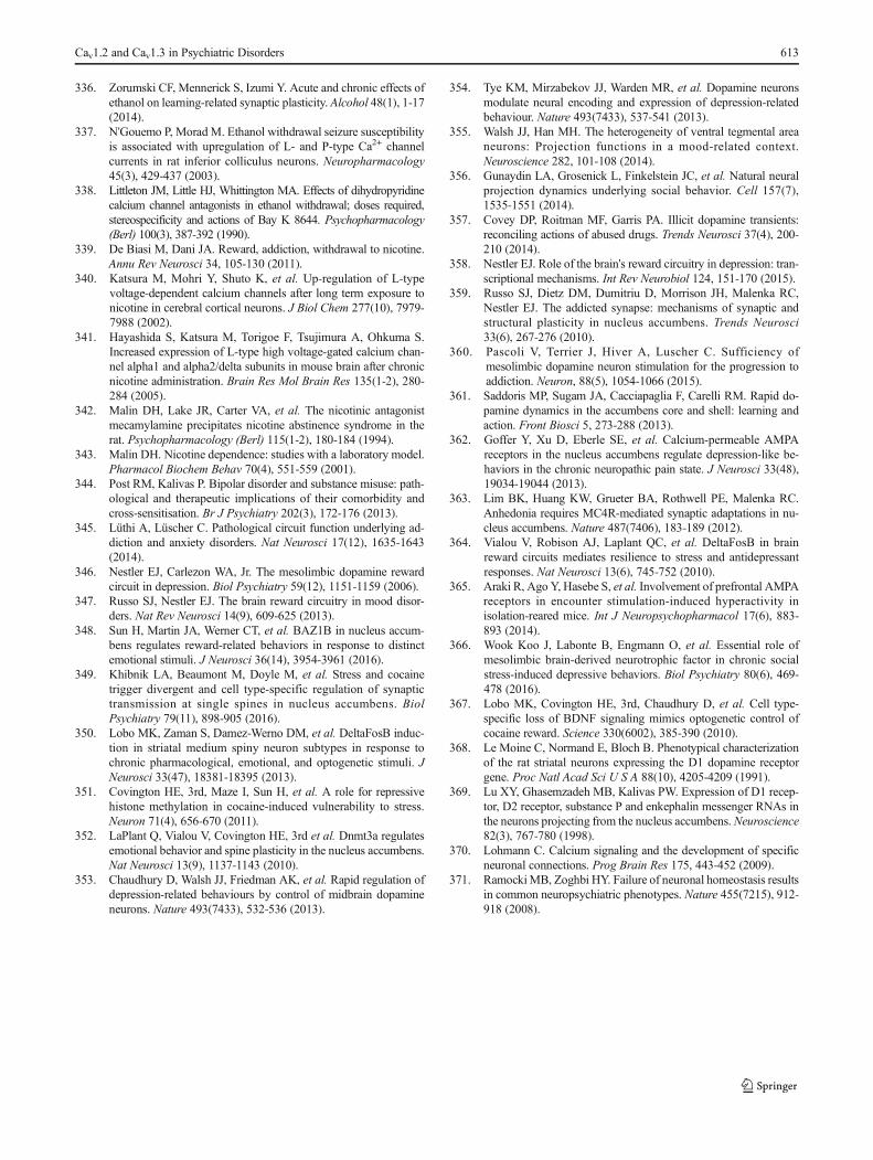
336. Zorumski CF, Mennerick S, Izumi Y. Acute and chronic effects ofethanol on learning-related synaptic plasticity. Alcohol 48(1), 1-17(2014).
337. N'Gouemo P, Morad M. Ethanol withdrawal seizure susceptibilityis associated with upregulation of L- and P-type Ca2+ channelcurrents in rat inferior colliculus neurons. Neuropharmacology45(3), 429-437 (2003).
338. Littleton JM, Little HJ, Whittington MA. Effects of dihydropyridinecalcium channel antagonists in ethanol withdrawal; doses required,stereospecificity and actions of Bay K 8644. Psychopharmacology(Berl) 100(3), 387-392 (1990).
339. De Biasi M, Dani JA. Reward, addiction, withdrawal to nicotine.Annu Rev Neurosci 34, 105-130 (2011).
340. Katsura M, Mohri Y, Shuto K, et al. Up-regulation of L-typevoltage-dependent calcium channels after long term exposure tonicotine in cerebral cortical neurons. J Biol Chem 277(10), 7979-7988 (2002).
341. Hayashida S, Katsura M, Torigoe F, Tsujimura A, Ohkuma S.Increased expression of L-type high voltage-gated calcium chan-nel alpha1 and alpha2/delta subunits in mouse brain after chronicnicotine administration. Brain Res Mol Brain Res 135(1-2), 280-284 (2005).
342. Malin DH, Lake JR, Carter VA, et al. The nicotinic antagonistmecamylamine precipitates nicotine abstinence syndrome in therat. Psychopharmacology (Berl) 115(1-2), 180-184 (1994).
343. Malin DH. Nicotine dependence: studies with a laboratory model.Pharmacol Biochem Behav 70(4), 551-559 (2001).
344. Post RM, Kalivas P. Bipolar disorder and substance misuse: path-ological and therapeutic implications of their comorbidity andcross-sensitisation. Br J Psychiatry 202(3), 172-176 (2013).
345. Lüthi A, Lüscher C. Pathological circuit function underlying ad-diction and anxiety disorders. Nat Neurosci 17(12), 1635-1643(2014).
346. Nestler EJ, Carlezon WA, Jr. The mesolimbic dopamine rewardcircuit in depression. Biol Psychiatry 59(12), 1151-1159 (2006).
347. Russo SJ, Nestler EJ. The brain reward circuitry in mood disor-ders. Nat Rev Neurosci 14(9), 609-625 (2013).
348. Sun H, Martin JA, Werner CT, et al. BAZ1B in nucleus accum-bens regulates reward-related behaviors in response to distinctemotional stimuli. J Neurosci 36(14), 3954-3961 (2016).
349. Khibnik LA, Beaumont M, Doyle M, et al. Stress and cocainetrigger divergent and cell type-specific regulation of synaptictransmission at single spines in nucleus accumbens. BiolPsychiatry 79(11), 898-905 (2016).
350. Lobo MK, Zaman S, Damez-Werno DM, et al. DeltaFosB induc-tion in striatal medium spiny neuron subtypes in response tochronic pharmacological, emotional, and optogenetic stimuli. JNeurosci 33(47), 18381-18395 (2013).
351. Covington HE, 3rd, Maze I, Sun H, et al. A role for repressivehistone methylation in cocaine-induced vulnerability to stress.Neuron 71(4), 656-670 (2011).
352. LaPlant Q, Vialou V, Covington HE, 3rd et al. Dnmt3a regulatesemotional behavior and spine plasticity in the nucleus accumbens.Nat Neurosci 13(9), 1137-1143 (2010).
353. Chaudhury D, Walsh JJ, Friedman AK, et al. Rapid regulation ofdepression-related behaviours by control of midbrain dopamineneurons. Nature 493(7433), 532-536 (2013).
354. Tye KM, Mirzabekov JJ, Warden MR, et al. Dopamine neuronsmodulate neural encoding and expression of depression-relatedbehaviour. Nature 493(7433), 537-541 (2013).
355. Walsh JJ, Han MH. The heterogeneity of ventral tegmental areaneurons: Projection functions in a mood-related context.Neuroscience 282, 101-108 (2014).
356. Gunaydin LA, Grosenick L, Finkelstein JC, et al. Natural neuralprojection dynamics underlying social behavior. Cell 157(7),1535-1551 (2014).
357. Covey DP, Roitman MF, Garris PA. Illicit dopamine transients:reconciling actions of abused drugs. Trends Neurosci 37(4), 200-210 (2014).
358. Nestler EJ. Role of the brain's reward circuitry in depression: tran-scriptional mechanisms. Int Rev Neurobiol 124, 151-170 (2015).
359. Russo SJ, Dietz DM, Dumitriu D, Morrison JH, Malenka RC,Nestler EJ. The addicted synapse: mechanisms of synaptic andstructural plasticity in nucleus accumbens. Trends Neurosci33(6), 267-276 (2010).
360. Pascoli V, Terrier J, Hiver A, Luscher C. Sufficiency ofmesolimbic dopamine neuron stimulation for the progression toaddiction. Neuron, 88(5), 1054-1066 (2015).
361. Saddoris MP, Sugam JA, Cacciapaglia F, Carelli RM. Rapid do-pamine dynamics in the accumbens core and shell: learning andaction. Front Biosci 5, 273-288 (2013).
362. Goffer Y, Xu D, Eberle SE, et al. Calcium-permeable AMPAreceptors in the nucleus accumbens regulate depression-like be-haviors in the chronic neuropathic pain state. J Neurosci 33(48),19034-19044 (2013).
363. Lim BK, Huang KW, Grueter BA, Rothwell PE, Malenka RC.Anhedonia requires MC4R-mediated synaptic adaptations in nu-cleus accumbens. Nature 487(7406), 183-189 (2012).
364. Vialou V, Robison AJ, Laplant QC, et al. DeltaFosB in brainreward circuits mediates resilience to stress and antidepressantresponses. Nat Neurosci 13(6), 745-752 (2010).
365. Araki R, Ago Y, Hasebe S, et al. Involvement of prefrontal AMPAreceptors in encounter stimulation-induced hyperactivity inisolation-reared mice. Int J Neuropsychopharmacol 17(6), 883-893 (2014).
366. Wook Koo J, Labonte B, Engmann O, et al. Essential role ofmesolimbic brain-derived neurotrophic factor in chronic socialstress-induced depressive behaviors. Biol Psychiatry 80(6), 469-478 (2016).
367. Lobo MK, Covington HE, 3rd, Chaudhury D, et al. Cell type-specific loss of BDNF signaling mimics optogenetic control ofcocaine reward. Science 330(6002), 385-390 (2010).
368. Le Moine C, Normand E, Bloch B. Phenotypical characterizationof the rat striatal neurons expressing the D1 dopamine receptorgene. Proc Natl Acad Sci U S A 88(10), 4205-4209 (1991).
369. Lu XY, Ghasemzadeh MB, Kalivas PW. Expression of D1 recep-tor, D2 receptor, substance P and enkephalin messenger RNAs inthe neurons projecting from the nucleus accumbens. Neuroscience82(3), 767-780 (1998).
370. Lohmann C. Calcium signaling and the development of specificneuronal connections. Prog Brain Res 175, 443-452 (2009).
371. RamockiMB, Zoghbi HY. Failure of neuronal homeostasis resultsin common neuropsychiatric phenotypes. Nature 455(7215), 912-918 (2008).
Cav1.2 and Cav1.3 in Psychiatric Disorders 613