Embed Size (px)
Citation preview

BioSystems 86 (2006) 46–52
From electric patterns to drugs:Perspectives of computational neuroscience in drug design
Tamas Kissa,b, Peter Erdia,b,∗a Department of Biophysics, KFKI Research Institute for Particle and Nuclear Physics of the Hungarian Academy of Sciences,
Budapest, Hungaryb Center for Complex Systems Studies, Kalamazoo College, Kalamazoo, MI, USA
Received 21 December 2005; received in revised form 13 February 2006; accepted 15 February 2006
Abstract
A novel way of computational modeling by integrating compartmental neural techniques and detailed kinetic description ofpharmacological modulation of transmitter–receptor interaction is offered as a method to test the electro-physiological and behavioraleffects of potential drugs. Even more, an inverse method is suggested as a method for controlling a neural system to realize a prescribedtemporal pattern. Generation and pharamcological modulation of theta rhythm in area CA1 of the hippocampus related to anxiety
is analyzed. Integrative modeling might help to find positive allosteric modulators of α1 subunits as potential candidates for beingselective anxyolitics.© 2006 Elsevier Ireland Ltd. All rights reserved.g; Reac
Keywords: Hippocampus; Theta oscillation; Compartmental modelin1. Introduction
The hippocampal formation is known to be involvedin cognitive processes (the two most extensively studiedare navigation and memory formation – for reviews seee.g. (Lengyel et al., 2005; Buzsaki, 2005)) and being partof the Papez circuit (Papez, 1937) it might play a pos-sible role in mood regulation (Gray and McNaughton,2000; Freund, 2003). To properly perform its functionsthe hippocampus has to maintain both its structural anddynamical properties. When an impairment occurs ei-ther in its architecture or dynamics it is reflected at thebehavioral and neural levels. Alzheimer’s disease and is-
chemia are associated with learning and memory impair-ment, and are accompanied by selective neuronal death∗ Corresponding author.
E-mail address: [email protected] (Peter Erdi).
0303-2647/$ – see front matter © 2006 Elsevier Ireland Ltd. All rights reservdoi:10.1016/j.biosystems.2006.02.016
tion kinetics; Systems description
or characteristic changes in the hippocampal neural cir-cuitry. Epilepsy, on the other hand, is often regarded as adynamic disease (Milton and Jung, 2003) where the neu-ral structure, cells and connections among them are in-tact, only some change in the parameters (i.e. propertiesof neurons or synapses) occur leading to a pathologicalbehavior. Recent studies have also indicated that the hip-pocampal formation is affected in human depression aswell as in animal models of depression (Sapolsky, 2000;Nestler et al., 2002) and anxiety (Gray and McNaughton,2000).
On the neural level a class of neurological disordersare accompanied by a characteristic change in brain ac-tivity. In the healthy hippocampus of the rat, for exam-ple, two main, globally occurring brain states are known:
the rhythmic slow activity, called the theta rhythm withthe associated gamma oscillation, and the irregular sharpwaves with the associated high frequency (ripple) oscil-lation (Erdi and Szalisznyo, 2002). These rhythms areed.

T. Kiss, P. Erdi / BioSystems 86 (2006) 46–52 47
esis of c
cmasesn
dciafpmMpodMtlnaai(atto
ltlr
Fig. 1. Schematic representation of the working hypoth
orrelated with the behavior or more generally with theental state of the animal and are influenced by sever
l factors (e.g. ascending aminergic activation, internaltate of the hippocampus, inputs from associated regions,tc.) However, there exists an other, pathological braintate characterized by large amplitude, highly synchro-ized oscillations associated with epileptic seizures.
Similar to other brain disorders, anxiety a psychiatricisorder of increasing interest can be described by itslinical appearance at the behavioral level or physiolog-cally at the neural-system level. Besides the amygdala,neural center classically thought to be responsible for
ear generation and associated emotions, the hippocam-us is also regarded as a key participant in the develop-ent of anxiety (Rosen and Schulkin, 1998; Gray andcNaughton, 2000). Physiologically, a potential, though
ossibly ambiguous marker of anxiety level is the powerf hippocampal field theta oscillation (Green and Ar-uini, 1954; Fontani and Carli, 1997; Yamamoto, 1998;cNaughton and Gray, 2000). It is ambiguous because
he presence and power of theta oscillation also corre-ates with the performance of animals in memory andavigation tasks (e.g. see Klimesch, 1999; Hasselmo etl., 2002; Jones and Wilson, 2005), probably not related –t least not in a direct way – to fear and anxiety. However,t is known that theta oscillations of the hippocampusBuzsaki, 2002) have common but dissimilar propertiess well. These properties depend on the specific form ofhe ongoing activity, which fact allows for the hypothesishat the specific form of theta related to the generationf anxiety can be identified.
In this paper we argue for the use of computer simu-
ations in the search of putative drugs, specifically selec-ive anti-anxiolytics. In general, the argument is the fol-owing: we presume that some central nervous system-elated illnesses have identifiable electro-physiologicalomputational neuropharmacology (see text for details).
correlates (patterns of population activity); identify ma-jor mechanisms and key components underlying patterngeneration; give a mathematical model of the systemincorporating key receptor and network level elements;modify parameters to produce a desired pattern or mod-ify a certain pattern in a desired way (Fig. 1). In par-ticular, in the followings we consider anxiety and thehippocampal theta rhythm as the psychiatric disorderand its physiological correlate. The mathematical modelbuilds on the detailed description of hippocampal CA1neuron populations presumed to play a key role in thetageneration. Parameters of the model were the maximalsynaptic conductances of GABAA synapses, which weremodified to enhance or suppress the population theta os-cillation in the model. As synapses are regarded as targetelements of pharmacological manipulations ways of in-corporating different synaptic description into the modelframework will be analyzed.
2. Models of theta rhythm generation in thehippocampal CA1
Hippocampal theta generation has been extensivelystudied in different paradigms. This paper does not aimat reviewing this large body literature, the reader is kindlyreferred to the paper of Buzsaki (2002) for a comprehen-sive review of theta generation and its functional signif-icance and to Lengyel et al. (2005) for a review of thetheoretical considerations of the function of theta. Herewe confine ourselves to briefly introduce a few relevantcomputer models, which propose putative mechanismsto explain theta generation.
When models of brain disorders are considered themodeler has to face the question of setting up themodel of an intricately interconnected, multi-level sys-tem. However, the model can account for a very lim-

oSystem
48 T. Kiss, P. Erdi / Biited sub-system (relative to the whole) and a restrictedamount of detail only. For example, as in the pharma-cological research – and much more in the practice –systemic administration of drugs is a widely used andconventional method effects of a drug in different areashas to be considered. Methodologically, one way to ac-count for these effects is to identify external inputs of thesystem and deal with them in a simplified, abstract way,while keeping detailedness of the analyzed system at thedesired high level. In the paper of Orban et al. (2001)synchronization properties of a CA3 interneuron net-work was studied. Authors set up a detailed conductance-based model of the system of interest and accountedfor extrinsic innervation by taking into account differ-ent types of modulatory inputs. Results showed that agamma-frequency input with dispersed phase among in-terneurons could modulate gamma-frequency synchro-nization of interneurons in the theta frequency band,i.e. a 4–12 Hz modulation of population synchronizationemerged due to phase dispersion resulting from axonal
delays and cellular heterogeneity.The paper of Wang (2002) argues for the septal pace-maker hypothesis. In the model medial septal GABAer-gic cells and hippocampo-septal GABAergic cells form a
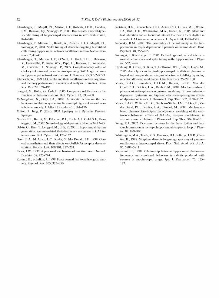
Fig. 2. Proposed mechanism of theta oscillation generation via the propaga(Left) the hippocampal part of the model by Hajos et al. (2004) consisted of twresided at the alveus/oriens border (O-LM cells) – i(o/a)) and pyramidal cellsevoke action potentials in the O-LM cell population via NMDA and in baskcell activity inhibits pyramidal cell firing and hyperpolarizes the pyramidal cthis activity successfully inhibits basket cells and disinhibits pyramidal cells.of cells were binned into 10 ms bins and plotted normalized to the population
s 86 (2006) 46–52
feed-back loop and generate the population theta rhythm.Here the external influence is accounted for by the inclu-sion of an I injected current parameter. The author identi-fies this parameter with the excitatory drive by choliner-gic neurons in the medial septum, but it can also be usedto mimic the effect of other tonic brain-state-dependentactivation (e.g. by the aminergic or serotonergic system).Here external phasic input or some form of an informa-tional input was not included.
In an interesting paper Borisyuk and Hoppenstadt(1999) describes the hippocampus by an abstract, highlysimplified model and studies spatial resonance propertiesof this system with two external oscillators representingthe entorhinal cortex and the septum in the frequencyand time domains. This model takes the approach to ac-count for extrinsic inputs of the hippocampus at a levelcomparable to the detailedness of the intrahippocampalmodel. It is important to study, however, mainly inter-nal mechanism responsible for oscillation generation inthe hippocampus. Rotstein et al. (2005) proposed that a
mechanism of hippocampal CA1 theta generation relieson two populations of hippocampal interneurons, wherefast-firing neurons innervated slow-firing neurons. Thismodel gives a detailed, entirely intrahippocampal modeltion of a “synchronized firing state” in sub-networks of the system.o interneuron populations (basket cells – i(b) and cells whose somata
(pyr). In a theta cycle the gradually recruited firing of pyramidal cellset cells via AMPA receptor-mediated synapses. The resulting basketell membrane. When the population activity of O-LM cells build up(Right) population activity of different cell populations. Firing timessize vs. time.

oSystem
oipTmat(g
(s(teo22ctpams→brbdIst(po“ElobchcboG
3r
p
T. Kiss, P. Erdi / Bi
f theta generation showing that synchronized behav-or might emerge in a pure CA1 slice preparation ex-laining experimental results of Gillies et al. (2002).his experimental measurement, however, required thatetabotropic glutamate receptors were activated – prob-
bly mimicking a state-dependent tonic background ac-ivation – which was achieved by bath application ofS)-3,5-dihydroxyphenylglycine a group I metabotropiclutamate receptor agonist.
In a joint pharmacological and computational workHajos et al., 2004) a highly detailed model of theeptum and of area CA1 of the hippocampus was set upfor a detailed description and simulation data see alsohe online supplementary material at http://geza.kzoo.du/theta/theta.html). The model builds upon the seriesf seminal experiments by Klausberger et al. (2003,004, 2005) (for a review see Somogyi and Klausberger,005), which enabled the design of a highly detailedomputer model incorporating different interneuronypes, which – along with principal pyramidal cells –lay different but complementary roles in rhythm gener-tion and synchronization. According to this model theechanism of theta oscillation generation includes: (i)
ignal propagation in the pyramidal cell → O-LM cellbasket cell → pyramidal cell → feed-forward/feed-
ack loop, (ii) synchronization of neural activity via theecurrent, inhibitory GABAA connections within theasket cell network and (iii) synchronization of pyrami-al cell firing due to rebound action potential generation.t also worth noting that the propagation of a singleignal throughout this trisynaptic loop would not requirehe amount of time characteristic to the theta oscillation0.2–0.25 s), thus authors propose that the measurableopulation oscillation is created not by the propagationf single signals but rather by the propagation of asynchronized firing state” in the network (Fig. 2).xtra-hippocampal inputs were considered at different
evels of detailedness: the phasic medial septal inputriginating from GABAergic neurons was simulatedy modeling presynaptic neurons and synapses, septalholinergic input and ascending activation, on the otherand, was taken into account as a constant depolarizingurrent only. This model framework was used – as wille shown later in the next section – to study the effectf positive and negative allosteric modulators of theABAA receptors, diazepam and FG-7142, respectively.
. Modeling the pharmacological modulation of
hythmsWhittington et al. (1998) studied the effect of mor-hine and -endorphin both by electro-physiological
s 86 (2006) 46–52 49
techniques in transversal hippocampal slice preparationsand in accompanying computer simulations. They pro-pose that the clinically observed effects of these drugsmight be in connection with the loss of spatial long-range synchrony of the EEG gamma band (>25 Hz) os-cillation caused by bath application of the drugs. An ef-fect of morphine in the hippocampus is the decrease ofpresynaptic GABA release resulting in the diminution ofGABAA receptor-mediated inhibitory postsynaptic po-tentials both in inhibitory and in excitatory cells. Thisresults in an increase in firing frequency of inhibitoryoscillations and burst generation in interneurons. Thecomputer model proposed to describe phenomena ob-served in the slice preparation consisted of pyramidalcells and interneurons interconnected by AMPA andGABAA receptor-mediated synapses.
Key component of this model, from our point of view,is the description of the synaptic coupling. Both synap-tic conductances were described phenomenologically bysingle-exponential (alpha) functions of the form
g(t) = gmax × t × exp(−t/τ) (1)
g(t) being the value of the synaptic conductance at time t,gmax the maximal synaptic conductance, and τ is the timeconstant of the synapse. The complex effect of morphine,which is known to involve the activation of -opioidreceptors that in turn decrease presynaptic GABA releaseby a G protein-mediated mechanism (Capogna et al.,1993) was taken into account simply by the decrease ofthe maximal synaptic conductance by 75 or 80%.
In the paper by Hajos et al. (2004) instead of the sim-pler but computationally more efficient phenomenolog-ical description synaptic transmission in this paper wasdescribed by gating variables similarly to the Hodgkin–Huxley (Hodgkin and Huxley, 1952) formalism:
Isyn = gsyns(V − Esyn) (2a)
ds
dt= αF (Vpre)(1 − s) − βs (2b)
F (Vpre) = 1
1 + exp(−Vpre−Θsyn
K
) (2c)
with Isyn being the synaptic current, gsyn the maximalsynaptic conductance, s the gating variable of the synap-tic channel, Esyn the synaptic reversal potential, F (·) anactivation function, α and β the rate functions describingopening and closing of the gate of the synaptic channel,
and Θsyn is a threshold. This way synaptic shunting andsaturation – both saturation of the postsynaptic poten-tial and of the postsynaptic conductance – effects canbe described, which can significantly modulate cellular
oSystem
50 T. Kiss, P. Erdi / Biand network behavior under synchronized oscillationsi.e. when several synaptic inputs are received simultane-ously by a neuron.
Similarly to (Whittington et al., 1998) authors sim-ulated the effects of the positive and negative allostericmodulators of the GABAA receptors by modifying gsynthe maximal synaptic conductance. Moreover, as twointrahippocampal interneuron types were distinguished(Fig. 2) selective modulation of synapses were possible.Changing the maximal synaptic conductance parameterone at a time authors found that the modification of the re-current connections within the basket cell network exertsthe most pronounced effect on theta power. Identificationof such sensitive targets are important from the respectof designing selective still efficient drugs. Clearly, whenreceptors can be distinguished based on a characteris-tic property like the subunit composition in the case ofGABAA receptors, there is a chance to produce a selec-tive drug acting at the targets identified by the computermodel (Ujfalussy et al., 2005).
4. Detailed modeling of pharmacological actionat synapses
Computer simulations should not finish at identifyingthe most sensitive targets in the network. In this sectionwe propose the use of a composite modeling techniquethat integrates the microscopic description of receptorsinto the more general framework of computational neu-roscience yielding a tool for pharmacologists to enhancethe drug design and screening process.
In the previous section the effect of drugs weredescribed by a phenomenological way, the change ofa parameter representing the maximal strength of thesynapse. However, drugs – besides certainly influencingthe amplitude of unitary conductance changes – mightmodulate other characteristics of signal generation (e.g.binding efficacy of the transmitter, dissociation time con-stant, or creating new reaction pathways of transmitterbinding, etc.). Additionally to a change in the maxi-mal conductance these changes might result in differ-ent transfer properties, filtering and saturation effects,etc. To establish a sound connection between drug ef-fects and the observed network behavior (e.g. local fieldpotential or the EEG) a detailed model of transmitter–drug–receptor interaction should be studied.
Transmitter–receptor interaction and the generationof signals might be studied by different approaches. In
a series of papers Visser et al. (2002, 2003) suggesteda theoretical framework based on pharmacokinetics–pharmacodynamics to model the effects of GABA mod-ulators on the EEG. In their mechanism-based models 86 (2006) 46–52
the authors describe the generation of the synaptic re-sponse by separately representing the diffusion and dis-position of the drug in the plasma and its equilibra-tion in the biophase (i.e. the effect-site of the drug)(pharmacokinetics) and the drug–receptor interactionand signal-transduction (pharmacodynamics). The inputof the first part of the model is an infusion rate of thedrug and its output is a concentration at the effect-site.The second part of the model calculates a stimulus fromthis concentration and uses it to evaluate the response,which in the present case is the -band amplitude ofthe EEG. A key component of the second part of themodel is the stimulus–response function. This initiallyunknown function represents the connection between thestimulus and the observed pharmacological effect: the“initial stimulus is then propagated into the observedpharmacological effect through a chain of postreceptorevents, which is characterized by an unknown functionf ” (Visser et al., 2002). This function is experimentallydetermined and intentionally neglects the architecture ofthe system under investigation.
A more detailed but less general model is given byBaker et al. (2002). In their approach postsynaptic events(i.e. the interaction of the GABA transmitter with theGABAA receptor) are described in high detail using aspecific kinetic model modified from the original modelof Jones and Westbrook (1995). In this description eachchemical reaction taking place between the transmit-ter and the receptor is accounted for by a linear ordi-nary differential equation containing rate constants rep-resenting binding and dissociation rates of different lig-ands and products, respectively. Moreover, the modelcontains an interneuron model based on the Hodgkin–Huxley equations. Using this model framework authorshave the possibility to study the modulation of synchro-nization within a small system (the maximal networkwas made up of two interneurons) due to the change ofrate constants. Quantitative connection between the rateconstants and the studied drugs (propofol and midazo-lam) was made by experimental studies (Bai et al., 1999;Orser et al., 1998; Ghansah and Weiss, 1999).
The two papers presented in this section demonstratetwo possible ways to connect the molecular level to thesystems level. The papers of Visser et al. (2002, 2003)use a detailed approach on the microscopic- and a phe-nomenological approach at the macroscopic scale, whileBaker et al. (2002) increases detailedness of the micro-scopic level and analyzes a relatively small system only.
In the last section of our paper we propose that build-ing on the above reviewed ideas and incorporating theminto a mesoscopic-scale description of neural networksa useful tool can be produced.
oSystem
5
gttc(b2sbetttr
mobdlpcmobmeoe(skRatomammds
A
ed
T. Kiss, P. Erdi / Bi
. Conclusions and propositions
The aim of the present paper is to offer an inte-rated conceptual framework to study the effect of po-ential drugs on the electrical activity of neural cen-ers. In Section 1 we argued for the hypothesis thatharacteristic EEG and local field potential patternsrhythmic brain activities) can be used as potentialiomarkers of associated brain disorders. In Sectiona brief review was given on different approaches totudy the generation of rhythmic population activityy computer models. In Section 3 some existing mod-ls of pharmacological modification of oscillatory ac-ivity were surveyed with an emphasis on the descrip-ion of drug action at the synapse. Finally, in Section 4wo ways of describing drug–synapse interaction wereeviewed.
To be able to give advice to drug designers a computerodel shall be able to account for the basic mechanisms
f the generation of the activity pattern chosen as aiomarker of the targeted illness and has to have enoughetails to pinpoint a very specific location of action. Thisatter might mean a brain region, a sub-region, a cellopulation, a synapse type or even a reaction within theell or synapse. Specifically, we propose to understandechanisms of pattern generation – hippocampal theta
scillation – and to identify target locations – recurrentasket cell connections – in a detailed compartmentalodeling framework (Hajos et al., 2004; Ujfalussy
t al., 2005). To further specify the characteristicsf a desired drug a more detailed description at theffect-site – i.e. the postsynaptic receptor – is requiredErdi and Toth, 2005). To achieve this goal a detailedynapse model shall be analyzed in detail based on theinetic description of the receptor–ligand interaction.esults of this analysis than can be incorporated intoless computation-demanding formulation (e.g. a
ransfer function or a response function similar to thatf Visser et al. (2002)) and fed-back into the networkodel yielding a mapping between electrical activities
nd kinetic parameters. This mapping could be theathematical basis of a new computational neurophar-acology leading to a quantitative understanding of
rug actions and a more efficient drug design andcreening.
cknowledgements
Authors thank Mihaly Hajos and the Pfizer Inc. forlectro-physiological data and helpful comments andiscussions.
s 86 (2006) 46–52 51
References
Bai, D., Pennefather, P.S., MacDonald, J.F., Orser, B.A., 1999.The general anesthetic propofol slows deactivation and de-sensitization of GABA(A) receptors. J. Neurosci. 19, 10635–10646.
Baker, P.M., Pennefather, P.S., Orser, B.A., Skinner, F.K., 2002. Dis-ruption of coherent oscillations in inhibitory networks with anes-thetics: role of GABAA receptor desensitization. J. Neurophysiol.88, 2821–2833.
Borisyuk, R., Hoppenstadt, F., 1999. Oscillatory models of the hip-pocampus: a study of spatio-temporal patterns of neural activity.Biol. Cybern. 81, 359–371.
Buzsaki, Gy., 2002. Theta oscillations in the hippocampus. Neuron 33,325–340.
Buzsaki, Gy., 2005. Theta rhythm of navigation: link between path in-tegration and landmark navigation, episodic and semantic memory.Hippocampus 15, 827–840.
Capogna, M., Gahwiler, B.H., Thompson, S.M., 1993. Mechanismof mu-opioid receptor-mediated presynaptic inhibition in the rathippocampus in vitro. J. Physiol. 470, 539–558.
Erdi, P., Szalisznyo, K., Hippocampal rhythm generation. In: Arbib,M.A. (Ed.), 2002. The Handbook of Brain Theory and Neural Net-works. 2nd ed.. The MIT Press.
Erdi, P., Toth, J., 2005. Towards a dynamic neuropharmacology: inte-grating network and receptor levels. In: De Gregorio, M., Di Maio,V., Frucci, M., Musio, C. (Eds.), Brain, Vision and Artifical In-telligence volume 3704 of Lecture Notes in Computer Science.Springer, Berlin, Heidelberg 1–14.
Fontani, G., Carli, G., 1997. Hippocampal electrical activity and be-havior in the rabbit. Arch. Ital. Biol. 135, 49–71.
Freund, T.F., 2003. Interneuron diversity series: rhythm andmood in perisomatic inhibition. Trends Neurosci. 28, 489–495.
Ghansah, E., Weiss, D.S., 1999. Benzodiazepines do not modulatedesensitization of recombinant α1β2γ2 GABAA receptors. Neu-roreport 10, 817–821.
Gillies, M.J., Traub, R.D., LeBeau, F.E.N., Davies, C.H., Gloveli, T.,Buhl, E.H., Whittington, M.A., 2002. A model of atropine-resistanttheta oscillations in rat hippocampal area CA1. J. Physiol. 543,779–793.
Gray, J.A., McNaughton, N., 2000. The Neuropsychology of Anxiety:An Enquiry into the Functions of the Septo-hippocampal System.2nd ed.. Oxford University Press, Oxford.
Green, J.D., Arduini, A.A., 1954. Hippocampal electrical activity inarousal. J. Physiol. 17, 533–557.
Hajos, M., Hoffmann, W.E., Orban, G., Kiss, T., Erdi, P., 2004. Modu-lation of septo-hippocampal θ activity by GABAA receptors: an ex-perimental and computational approach. Neuroscience 126, 599–610.
Hasselmo, M.E., Hay, J., Ilyn, M., Gorchetchnikov, A., 2002. Neuro-modulation, theta rhythm and rat spatial navigation. Neural Net-works 15, 689–707.
Hodgkin, A.L., Huxley, A.F., 1952. A quantitative description of mem-brane current and its application to conduction and excitation innerve. J. Physiol. (London) 117, 500–544.
Jones, B., Westbrook, G.K., 1995. Desensitized states prolong GABAA
channel responses to bried agonist pulses. Neuron 15, 181–191.
Jones, M.W., Wilson, M.A., 2005. Theta rhythms coordinatehippocampal-prefrontal interactions in a spatial memory task.PLoS Biol. 3, e402.
oSystem
52 T. Kiss, P. Erdi / BiKlausberger, T., Magill, P.J., Marton, L.F., Roberts, J.D.B., Cobden,P.M., Buzsaki, Gy., Somogyi, P., 2003. Brain-state- and cell-type-specific firing of hippocampal interneurons in vivo. Nature 421,844–848.
Klausberger, T., Marton, L., Baude, A., Roberts, J.D.B., Magill, P.J.,Somogyi, P., 2004. Spike timing of dendrite-targeting bistratifiedcells during hippocampal network oscillations in vivo. Nature Neu-rosci. 7, 41–47.
Klausberger, T., Marton, L.F., O’Neill, J., Huck, J.H.J., Dalezios,Y., Fuentealba, P., Suen, W.Y., Papp, E., Kaneko, T., Watanabe,M., Csicsvari, J., Somogyi, P., 2005. Complementary roles ofcholecystokinin- and parvalbumin expressing GABAergic neuronsin hippocampal network oscillations. J. Neurosci. 25, 9782–9793.
Klimesch, W., 1999. EEG alpha and theta oscillations reflect cognitiveand memory performance: a review and analysis. Brain Res. BrainRes. Rev. 29, 169–195.
Lengyel, M., Huhn, Zs., Erdi, P., 2005. Computational theories on thefunction of theta oscillations. Biol. Cybern. 92, 393–408.
McNaughton, N., Gray, J.A., 2000. Anxiolytic action on the be-havioural inhibition system implies multiple types of arousal con-tribute to anxiety. J. Affect. Disorders 61, 161–176.
Milton, J., Jung, P. (Eds.), 2003. Epilepsy as a Dynamic Disease.Springer.
Nestler, E.J., Barrot, M., DiLeone, R.J., Eisch, A.J., Gold, S.J., Mon-teggia, L.M., 2002. Neurobiology of depression. Neuron 34, 13–25.
Orban, G., Kiss, T., Lengyel, M., Erdi, P., 2001. Hippocampal rhythmgeneration: gamma-related theta-frequency resonance in CA3 in-terneurons. Biol. Cybern. 84, 123–132.
Orser, B.A., McAdam, L.C., Roder, S., MacDonald, J.F., 1998. Gen-eral anaesthetics and their effects on GABA(A) receptor desensi-
tization. Toxicol. Lett. 100/101, 217–224.Papez, J.W., 1937. A proposed mechanism of emotion. Arch. Neurol.Psychiat. 38, 725–744.
Rosen, J.B., Schulkin, J., 1998. From normal fear to pathological anx-iety. Psychol. Rev. 105, 325–350.
s 86 (2006) 46–52
Rotstein, H.G., Pervouchine, D.D., Acker, C.D., Gillies, M.J., White,J.A., Buhl, E.H., Whittington, M.A., Kopell, N., 2005. Slow andfast inhibition and an h-current interact to create a theta rhythm ina model CA1 interneuron network. J. Physiol. 94, 1509–1518.
Sapolsky, R.M., 2000. The possibility of neurotoxicity in the hip-pocampus in major depression: a premier on neuron death. Biol.Psychiat. 48, 755–765.
Somogyi, P., Klausberger, T., 2005. Defined types of cortical interneu-rone structure space and spike timing in the hippocampus. J. Phys-iol. 562, 9–26.
Ujfalussy, B., Orban, G., Kiss, T., Hoffmann, W.E., Erdi, P., Hajos, M.,2005. Anxiolytics and septo-hippocamapal oscillation: pharmaco-logical and computational analysis of action of GABAA α1 and α2
receptor allosteric modulators. Clin. Neurosci. 25–29, 100.Visser, S.A.G., Smulders, C.J.G.M., Reijers, B.P.R., Van der
Graaf, P.H., Peletier, L.A., Danhof, M., 2002. Mechanism-basedpharmacokinetic–pharmacodynamic modeling of concentration-dependent hysteresis and biphasic electroencephalogram effectsof alphaxalone in rats. J. Pharmacol. Exp. Ther. 302, 1158–1167.
Visser, S.A.G., Wolters, F.L.C., Gubbens-Stibbe, J.M., Tukker, E., Vander Graaf, P.H., Peletier, L.A., Danhof, M., 2003. Mechanism-based pharmacokinetic/pharmacodynamic modeling of the elec-troencephalogram effects of GABAA receptor modulators: invitro–in vivo correlations. J. Pharmacol. Exp. Ther. 304, 88–101.
Wang, X.J., 2002. Pacemaker neurons for the theta rhythm and theirsynchronization in the septohippocampal reciprocal loop. J. Phys-iol. 87, 889–900.
Whittington, M.A., Traub, R.D., Faulkner, H.J., Jefferys, J.G.R., Chet-tiar, K., 1998. Morphine disrupts long-range syncrony of gamma-oscillations in hippocampal slices. Proc. Natl. Acad. Sci. U.S.A.95, 5807–5811.
Yamamoto, J., 1998. Relationship between hippocampal theta-wavefrequency and emotional behaviors in rabbits produced withstresses or psychotropic drugs. Jpn. J. Pharmacol. 76, 125–127.