Embed Size (px)
Citation preview

From Coronary Care Unit to Acute Cardiac Care Unit – the evolving role of
specialist cardiac care
October 2011 Recommendations of the British Cardiovascular Society Working Group on Acute Cardiac Care

2
Contents
Foreword ............................................................................................................................... 4
Executive Summary .............................................................................................................. 6
Introduction ........................................................................................................................... 7
The importance of Acute Cardiac Care ................................................................................. 8
1. Ischaemic Heart Disease/Acute Coronary Syndromes (ACS) ........................................... 9
1.1 Patient population ....................................................................................................... 9
1.2 STEMI patients .......................................................................................................... 10
1.3 NSTEACS patients – NSTEMI and Unstable Angina ................................................. 11
2. Acute Heart Failure ......................................................................................................... 14
3. Acute Arrhythmias ........................................................................................................... 17
3.1 Atrial Fibrillation (AF) ................................................................................................. 17
3.2 Regular Tachycardia (narrow and broad complex) .................................................... 17
3.3 Bradycardia ............................................................................................................... 18
4. Other conditions requiring admission to an Acute Cardiac Care Unit .............................. 19
4.1 Pulmonary embolism ................................................................................................. 19
4.2 Infective endocarditis ................................................................................................. 19
4.3 Pericardial effusion .................................................................................................... 20
4.4 Acute Thoracic Aortic Emergencies ........................................................................... 20
4.5 Other conditions which may require acute cardiac care ............................................. 20
5. Requirements for an Acute Cardiac Care Unit ................................................................. 21
5.1 Size and Provision of Acute Cardiac Care Units ........................................................ 21
5.2 Medical Staffing ......................................................................................................... 22
5.3 Nursing and Workforce .............................................................................................. 22
5.4 Staff Training and Skill mix ........................................................................................ 23
5.5 Diagnostic requirements for Acute Cardiac Care (2011-2020) ................................... 24
5.6 ECG based diagnostics ............................................................................................. 24
5.6.1 Electrocardiography ............................................................................................ 24
5.7 Imaging ..................................................................................................................... 24
5.7.1 X-ray Fluoroscopy .............................................................................................. 24
5.7.2 Coronary Angiography Suite ............................................................................... 25
5.7.3 Trans-thoracic Echocardiography (TTE) ............................................................. 25
5.7.4 Trans-oesophageal Echocardiography (TOE) ..................................................... 26
5.8 Functional Cardiac Assessment or Advanced Anatomical Imaging ........................... 26
5.9 Cardiac Device Management .................................................................................... 27
5.10 Electrophysiological studies and RF ablation ........................................................... 27

3
5.11 Laboratory based diagnostics .................................................................................. 27
Conclusions: Acute Cardiac Care Unit rather than Coronary Care Unit ............................... 29
Appendix 1: Key features of the Acute Cardiac Care Unit ................................................... 30
References ......................................................................................................................... 32

4
Working Group Members
DM Walker (Chair of Working Group, British Cardiovascular Society)
NEJ West (Deputy Chair of Working Group, British Cardiovascular Society)
SG Ray (Vice-President Clinical Standards, British Cardiovascular Society)
S Bridge (CEO, Papworth Hospital)
SS Furniss (Heart Rhythm UK)
J Keenan (British Association for Nursing in Cardiovascular Care)
M Knapton (British Heart Foundation)
C Knight (British Cardiovascular Intervention Society)
GW Lloyd (British Society of Echocardiography)
C Marley (NHS Improvement: Heart)
TA McDonagh (British Society for Heart Failure)
TJ Quinn (MINAP)
D Richley (Society for Cardiological Science and Technology)
K Timmis (Heart Care Partnership)
K Wilmer (Royal College of Physicians)

5
Foreword
The management of heart attack has changed beyond all recognition over the last thirty
years so a review of the Cardiac Care Unit and its functions is timely.
When I was a house physician, CCUs were a novelty and certainly not uniformly available
across the country. The only treatments available were the defibrillator (in hospitals not
ambulances) and diamorphine.
It was not until the 1980s that thrombolysis began to take hold and not until the advent of the
National Service Framework for Coronary Heart Disease in 2000 did this treatment became
universally and rapidly available which heralded the widespread and long overdue
development of cardiology expertise in district hospitals.
Later, when it became clear that primary angioplasty offered better outcomes, fewer
complications and shorter lengths of stay, the centralisation of heart attack care in Heart
Attack Centres challenged this devolution requiring collaboration between hospitals.
The concept of the Heart Attack Centre is a trifle misleading since most deal primarily with
ST elevation heart attacks whereas the majority of attacks are non ST elevation cases often
managed in less than ideal fashion in Medical Assessment Units. These so-called minor
heart attacks are in fact anything but. Data from the Myocardial Ischaemia National Audit
Project (MINAP) shows that at 30 days and beyond, their outlook is now less good than for
ST elevation cases.
We know from a range of data that these cases together with other acute cardiac cases are
better managed by cardiology staff and it is time that systems are developed to achieve this.
So this review of the role of the CCU is extremely timely and welcome. Some years ago,
Douglas Chamberlain berated us for calling the units Coronary Care Units and insisted on
the term Cardiac Care Unit.
This report suggests that he was right.
Professor Sir Roger Boyle CBE
September 2011

6
Executive Summary
The changing demographics of the UK population have led to increased admission of elderly
and more complex cardiac patients. This combined with the reorganisation of care required
to provide primary PCI across cardiac networks, has caused a significant change in the
acute cardiology workload for all acute hospitals.
In this document we present evidence that patients presenting with acute cardiac conditions
who are managed in specialised cardiac wards have demonstrably better outcomes.
However, a significant proportion of such patients are not currently managed within a cardiac
service, leading to greater morbidity and mortality, and increased costs to the NHS.
The British Cardiovascular Society recommends that patients presenting with acute cardiac
conditions should be managed by a specialist, multi-disciplinary cardiac team and have
access to key cardiac investigations and interventions, at all times. All hospitals admitting
unselected acute medical patients should have an appropriately sized, staffed and equipped
Acute Cardiac Care Unit, where high risk patients with a primary cardiac diagnosis should be
managed. Access to these Acute Cardiac Care Units should be open to all high risk cardiac
patients and in particular, should not be restricted to patients with ACS.

7
Introduction
The development of coronary care units (CCUs) in the mid 20th century was a major
advance in cardiology practice as it allowed the concentration of patients with ST elevation
myocardial infarction (STEMI) in an area with specialist monitoring, nursing and medical
care. This became particularly important as the medical management of STEMI became
more aggressive and specialised. The development of primary angioplasty (PPCI) programs
for STEMI following Roger Boyle’s report ‘Mending hearts and Brains’ in 2006[1] has led to a
further shift in the role of the CCU. Some units no longer admit STEMI patients, while in
PPCI centres the concentrated influx of patients previously treated across a network has
placed CCU beds and staff under considerable pressure.
However, other factors are also at work in changing and increasing the workload of acute
cardiology and the development of PPCI cannot be considered in isolation. Important drivers
for change include:
1. The changing demographics of the population with an increasing proportion of
elderly patients presenting acutely with complex cardiac problems In particular,
the increasing detection of non ST elevation MI through the use of high sensitivity
troponin and the evidence that this impacts on outcomes;[2]
2. The unmet needs of patients with acute heart failure, atrial fibrillation, and other
conditions that cause major haemodynamic compromise;
3. The availability of new valvular interventions suitable for the elderly and for those
with significant co-morbidities;[3]
4. The reduction in the incidence of ST elevation Myocardial Infarction (STEMI).[4]
The net result is that CCUs remain busy but that the nature of the workload is changing with
admission of older, sicker and more complex patients. In practical terms, units are no longer
CCUs but are better described as Acute Cardiac Care Units.
The mode of delivery of acute cardiac care is important. There is good evidence that when
acute cardiac care is delivered by cardiologists on a cardiology ward, outcomes are
improved.[5-7] It follows that all hospitals accepting acute medical admissions should have
access to an Acute Cardiac Care Unit with appropriate staffing, medical and nursing
expertise, where unstable patients in the acute phase of their ischaemic syndrome, heart
failure, arrhythmia or other major haemodynamic disturbance can be managed.[5,6] To deal
with all these patient groups effectively cardiology services must be well organised,

8
appropriately resourced and efficient, and this presents a significant challenge for NHS
Trusts.
The importance of Acute Cardiac Care
Roughly 30% of the acute medical take is comprised of patients with a primary cardiac
problem.[8] The majority of these acute cardiac patients are admitted to District General
Hospitals, under the initial care of acute or general physicians. The advent of PPCI centres
has had little impact on this as STEMI patients comprise a limited and decreasing proportion
of the acute cardiac workload.[4]
The organisation and provision of care for all acute cardiac conditions has been considered
by the BCS Working Group including staffing, physical location, equipment and the role of
specialist nurses and cardiac physiologists.

9
1. Ischaemic Heart Disease/Acute Coronary Syndromes (ACS)
1.1 Patient population
In 2009-10, there were 31,412 STEMI and an estimated 100,000 NSTEMI in England and
Wales.[5] Overall ACS mortality is falling, but 30-day mortality for NSTEMI remains similar to
that of STEMI patients (Figure 1, data from England and Wales);[5]The incidence of STEMI
in the UK is also falling, but in contrast the numbers of NSTEMI recorded are rising, despite
many not being captured in the MINAP dataset.[5,9] Of those that are recorded, less than
50% are admitted to a dedicated cardiac facility (CCU/specialist cardiac ward).[9] This
implies that many high risk NSTEMI patients are not being managed in an appropriate
specialist environment.
.
Figure 1. 30 day mortality from STEMI & NSTEMI in England &Wales 2003-2010. Figure modified
from MINAP Ninth Public Report 2010. (Note: data for 2009-10 reflect first 9 months only and may be
revised).
Despite this, the majority of patients currently admitted to acute cardiac care units for
monitoring and treatment are suffering from ACS. The proportion of patients admitted with
STEMI and NSTEMI will vary depending on the local arrangements for provision of PPCI
services, although many units not providing on-site PPCI will continue to receive such
patients transferred from their local PPCI centre within 12 hours of admission. The

10
expanding range of patients requiring acute cardiac care will inevitably place additional
pressure on the already limited beds traditionally used for the management of ACS.
1.2 STEMI patients
In 2009-10, 63% of eligible STEMI patients received PPCI in England and Wales, an
increase from 44% in 2008-9 and from only 22% in 2007-8.[5,9] In 2011, PPCI networks
cover approximately 95% of the population of England, as predicted in the final report of the
National Infarct Audit Programme (NIAP).[10] Thrombolysis will continue to be utilised in
some more remote areas, where geography impacts on transit times to local PPCI centres
(Figure 2).
Figure 2. Use of reperfusion treatment for patients with a final diagnosis of STEMI in England & Wales
2003-2010. Figure modified from MINAP Ninth Public Report 2010. (Note: data for 2009-10 reflect
first 9 months only and may be revised).
Optimal management of patients with STEMI is summarised in ESC/EACTS guidance [11]
and is the subject of a NICE clinical guideline currently in development.
Key features include:
Direct paramedic transfer to PPCI centre or timely inter-hospital transfer
PPCI with first medical contact-to-balloon time <120 minutes
Monitoring on CCU (including for repatriated patients)
Early initiation of secondary prevention and cardiac rehabilitation
Some patients will require prolonged CCU stay for:

11
Invasive pressure monitoring
Circulatory or respiratory support
Clinical instability (arrhythmias etc.)
Pending further revascularisation
The majority of patients undergoing uncomplicated PPCI may be safely discharged home
within 48-72 hours,[12] without need for further prolonged in-hospital stay, and it is likely that
only the first 12-24 hours of this will need to be spent on the CCU.
STEMI patients receiving thrombolysis also require monitoring, availability of investigations
and access to cardiological expertise, including cardiac rehabilitation. 30% of thrombolysed
STEMIs will fail to reperfuse, and are best managed by immediate rescue PCI.[13] The
remainder should undergo coronary angiography with a view to revascularisation as
recommended current ESC/EACTS guidance [11] and therefore may need transfer to a PCI
centre.
1.3 NSTEACS patients – NSTEMI and Unstable Angina
The NSTEACS population comprises a heterogeneous patient group, varying from high risk
cases with elevated troponin, dynamic ECG changes and multiple risk factors to those with
chest pain with none of these features. Those proven to have NSTEMI have a comparable
mortality to STEMI patients and warrant Acute Cardiac Care Unit admission. The established
role of chest pain specialist/ACS nurses is critical to improvements in care for NSTEMI
patients, by bringing them to the early attention of cardiologists and thereby facilitating
access to specialist services and shortening inpatient stay.[14]
Analysis of MINAP data demonstrates important differences in outcomes of NSTEMI
patients when initially admitted to specialist cardiac units (CCU or dedicated cardiology
wards) compared with non-specialised facilities including general medical wards (Tables
1,2). Therefore the recommendation for routine use of a dedicated acute cardiac care unit for
all NSTEMI admissions can be based on economic grounds (shorter length of hospital stay),
better access to care (higher angiography rates and use of appropriate secondary
prevention pre-discharge) and most importantly, outcome (both crude and adjusted 30-day
mortality favours patients admitted to a specialised unit).[9]

12
Admitted to CCU?
Secondary prevention at discharge No Yes
Aspirin 94.2 96.7
Clopidogrel/thienopyridine 90.4 92.1
Betablocker 80.6 84.9
ACE inhibitor/ARB 85.1 88.8
Statin 96.1 96.9
Table 1. Differences in medications for secondary prevention prescribed at discharge in England &
Wales 2008-2009 (from MINAP data). Patients divided by admission to CCU. Values quoted as %
receiving specified medication. Data derived from reference [9].
Quartile
1 2 3 4
Patients admitted to CCU ≤ 27% 28-47% 47-72% > 72%
Length of stay (days) 7.5 (4.9-13.0) 6.8 (4.6-11.8) 6.5 (4.3-11.2) 5.6 (3.6-9.6)
Angiography performed 56.2% 52.1% 66.9% 66.9%
30-day mortality 7.9% 7.8% 7.7% 5.6%
Table 2. Differences in NSTEMI outcomes in England & Wales 2008-2009 (from MINAP data).
Hospitals divided into quartiles by proportion of NSTEMI cases admitted to CCU. Length of stay
reported as median (25th-75
th percentile). After correction for confounding variables, length of stay
less than median (6.5 days) fell by quartile: Q2 relative risk 1.21 (95% CI 1.08-1.25), Q3 RR 1.32
(1.28-1.35) and Q4 RR 1.62 (1.58-1.67); p<0.001 for all comparisons. Compared with admission to
general medical facility, and after adjustment for age, gender, risk factors and admission medication,
relative risk of 30-day mortality for NSTEMI patients admitted to CCU compared with general medical
facility was 0.88 (0.82-0.94); p<0.001. Cox regression analysis to adjust for confounding factors
demonstrated significantly reduced 30-day mortality for those in Q4 compared with other quartiles RR
0.78 (0.76-0.81); p<0.001. Data derived from reference [9].

13
Optimal care for NSTEACS is detailed in NICE guidance.[15] Many NSTEMI patients will
require some or all of the facilities necessary for the routine care of STEMI patients,
including access to early-phase cardiac rehabilitation, invasive/non-invasive monitoring and
echocardiography.
Optimal care includes:
Early risk stratification: GRACE*[16] or TIMI risk score.[17]
*The GRACE score may be easily downloaded to handheld devices for bedside use
(http://www.outcomes-umassmed.org/grace/acs_risk.aspx), and has been recently
validated in unselected admission cohorts.[18]
Monitoring on CCU.[19]
Triage to early coronary angiography (followed by revascularisation) within 24 hours
for high-risk patients (6-month mortality >3.0%, identified by risk scoring
systems),[11] others should undergo angiography within 72-96 hours of hospital
admission.[11,15]

14
2. Acute Heart Failure
Heart Failure is increasing in prevalence as the demography of Western populations
changes to include more elderly people. It is a common condition with a prevalence in the
general population of 1%, rising to >10% in the over 75s, and an incidence overall of 1 per
1000 population per annum rising to 25 per 1000 per annum in the over 75s.[19] In 2009-10
Hospital Episode Statistics show that there were 74,796 admissions to hospital with a
primary diagnosis of heart failure in England alone, with an additional 125,935 patients in
whom heart failure contributed significantly to the admission (ie coded as second or third
diagnosis).[21]
Epidemiological data confirm that heart failure has a high mortality, but until recently, robust
outcome data for patients hospitalised with heart failure in the UK has been lacking. In the
last year, the National Heart Failure Audit for England and Wales reported a 30% one year
mortality rate for patients admitted to hospital with heart failure in 2009-10 with an in-patient
case fatality rate of 10%.[6] In a parallel Scottish audit during the same period, in-hospital
mortality was 18%.[22] These results are considerably worse than contemporary US or
European data, where in-patient mortality rates are between 4 and 6.8%.[23] The Heart
Failure audit mortality statistics also contrast with data from UK centres where there are well
developed heart failure services, with greater access to heart failure specialists for in-
patients. These centres have lower current mortality and show a marked improvement over
the last ten years.[24]
In the UK the poor outcomes for heart failure, particularly after hospital admission, now
contrast markedly with that of its main precursor myocardial infarction where one year
mortality rates after discharge have fallen by over 30% in the last six years (Figure 1).
However, the revolution in MI care started with the development of CCUs. Although initially
designed to reduce arrhythmic death post-MI through monitoring and prompt defibrillation,
the CCU concentrated patients with acute MI in one place, allowing their care to be overseen
by cardiologists and delivered by specialist nursing staff. This then opened the door for rapid
implementation of new treatments, such as thrombolysis, intervention and device therapies.
Management of heart failure with a multi-professional approach improves outcomes for
patients, and has been adopted into the latest NICE guidance and quality standards.[25] In
particular, the role of specialist heart failure nurses in chronic disease management has
been emphasised.[26] However, it is also the case that the in-patient management of
patients with acute heart failure by cardiologists leads to lower mortality rates. Data from the

15
2009-10 heart failure audit in England and Wales confirm this.[6] Admission under a general
medical team was associated with a 30% higher one year mortality, even after adjusting for
known confounding variables. Access to in-patient cardiology care was associated with an
in-hospital absolute mortality rate of 6% as compared with 12% for non cardiology care, with
cardiology care being an independent predictor of a better outcome after adjusting for age
and known confounders. The improved outcomes persisted in this group out to one year of
follow up - 16.2% versus 32% mortality for general medical care. Patients admitted under
cardiology had more disease modifying treatment prescribed and were more likely to have
heart failure specialist nursing follow up.[6,27]
0.0
00.2
50.5
00.7
51.0
0
Cu
mula
tive s
urv
ival
0 90 180 270 360Survival time (days)
Cardiology (n=2739) General Medicine (n=2790)
Other (n=599)
Figure 3 One year mortality by place of care. Data from the National heart Failure Audit 2008-9 [27]
In the UK therefore, the key to optimal heart failure care for those admitted with
decompensated or acute heart failure is being seen by a cardiology team while in hospital.
Many are admitted from acute assessment units to general or geriatric medicine and never
have the opportunity to be managed by specialists in their primary condition.
Reconfiguring CCUs as acute cardiac care units is an obvious way to bring heart failure
patients under the care of Cardiologists and specialist cardiac nursing. Current acute cardiac
care unit capacity in the UK could not deal with all heart failure admissions, but it is
reasonable to propose that all those with acutely decompensated heart failure are managed
in a high dependency setting. Patients with NSTEACS often use the cardiac care unit for
part of their stay, combined with a spell on the general cardiology ward, and a similar model
could be adopted for heart failure with immediate benefit to length of stay and mortality.

16
Such an initiative would have to be combined with hospitals having robust acute heart failure
pathways to channel those who would benefit most from cardiological care and to make sure
that any patients remaining in the Acute Medicine or Care of the Elderly sector have input
from Cardiology during their admission and have subsequent access to multi-professional
follow up post discharge.

17
3. Acute Arrhythmias
There are many patients with acute cardiac rhythm problems that benefit from acute cardiac
care. Such patients fall into 3 main arrhythmia groups:
1) Acute Atrial Fibrillation & atrial flutter
2) Regular tachycardia – both narrow & broad QRS
3) Symptomatic Bradycardia
3.1 Atrial Fibrillation (AF)
AF is the commonest arrhythmia worldwide and is increasingly recognised as a major cause
of both morbidity and mortality, particularly from stroke which is increased 5-fold.[28,29] AF
(or atrial flutter) was the primary cause of admission for 92,258 patients in England in 2009-
10, with an average age of 70 and mean length of stay of 4 days. 53,971 patients were over
75 years of age.[21]
Given the age preponderance and the large numbers of admissions with AF, most patients
in acute hospitals are cared for by general physicians and may not be admitted to an acute
cardiac care unit, or indeed to any cardiology ward, even if there is haemodynamic
compromise. Patients presenting with uncontrolled AF are best managed by specialists,[30]
with resulting greater adherence to guideline based therapy. The diagnostic and therapeutic
services required to manage these patients are also best accessed through admission to an
acute cardiac unit, where patients come under the care of a cardiologist. Arrhythmia
specialist nurses can provide an efficient link to cardiology services, directing patients from
acute assessment units or A&E to cardiac wards.
3.2 Regular Tachycardia (narrow and broad complex)
Admission with these arrhythmias usually causes symptoms and is a medical emergency.
There were 12,996 admissions with supraventricular tachycardia in England in 2009-10, with
a lower mean age than AF patients (55 years) and shorter length of stay (mean 2.4
days).[21] Broad complex tachycardias, thought to be ventricular in origin (VT), were less
frequent (4899 in 2009-10), but patients were older (mean age 66 years) and required a
longer stay in hospital (mean 6.9 days). All these patients require admission to an acute
cardiac care unit where monitoring and full resuscitation facilities are available.
Patients with VT often have significant structural heart disease and require further
investigation & treatment on the same admission. This may include coronary angiography,

18
echocardiography, and device implantation. Patients may require urgent transfer to a more
specialist unit when definitive treatment is not available locally, but will require monitoring
prior to transfer.
The increasing number of patients in the community with Implantable Cardioverter
Defibrillators in-situ increases the complexity of patients who present to hospital. Patients
may now be admitted having survived a VT storm, after multiple shocks from the ICD, which
will then require careful interrogation and reprogramming. Local arrangements must be in
place to ensure that there is availability of a cardiac technician with experience in
programming.
3.3 Bradycardia
In 2009-10, 8383 patients were admitted to hospital in England with a high degree of heart
block.[21] An additional 2272 patients were admitted with sinus node disease. Both of these
groups tend to be elderly (mean age 76 – 77 years) and thus significant comorbidity is
common. Symptomatic bradycardia requires admission to an acute cardiac care unit for
monitoring and urgent rhythm management. Many patients will require temporary trans-
venous pacing and subsequent permanent pacemaker insertion.
The reduced need for temporary pacing post acute MI has led to a reduction in exposure
and subsequent deskilling of general medical trainees.[31] Consultant physicians are usually
also unhappy to perform temporary pacing. Although relatively infrequently required, these
cases often present as an emergency and local arrangements need to be in place to ensure
appropriately skilled staff are available. Where District General Hospitals do not have an on-
call cardiology rota (registrar or consultant), it will be necessary for there to be a network
protocol to provide an emergency pacing service, either by specialists cross-covering across
hospitals or by transfer to another unit. Whatever the solution, a formal arrangement must be
in place. Management of all patients with a temporary pacemaker should be in a monitored
bed on an acute cardiac care unit.

19
4. Other conditions requiring admission to an Acute Cardiac Care
Unit
4.1 Pulmonary embolism
Pulmonary embolism (PE) is a relatively common cardiovascular emergency, occurring in
0.4% of hospital admissions in the USA.[32] In 2009-10 there were nearly 20,000
admissions for PE in England.[21] High risk PE presenting with shock or hypotension has a
PE related mortality in excess of 15%, and the treatment recommended includes circulatory
support with inotropes, thrombolysis, catheter fragmentation of proximal thrombus and in
some cases surgical embolectomy. Patients with high risk PE are therefore best managed in
an acute cardiac care unit, with easy access to investigations such as CT pulmonary
angiography and echocardiography as required - and should initially be monitored there
even if transfer to a surgical unit is contemplated.[32]
4.2 Infective endocarditis
In 2009-10, there were 1863 admissions with infective endocarditis in England, occupying
over 50,000 bed days with a mean length of stay of around 28 days.[21] Mortality within 90
days of admission is 16.7%.[21]
Deterioration of valvular function in infective endocarditis may be a slow gradual process,
and assessing the need for surgical intervention is not always straightforward. Although
many patients with infective endocarditis are treated in hospitals without onsite cardiac
surgery, the pivotal role of surgery and the complexity of timing procedures necessitates that
cardiac surgeons and microbiologists are involved in the management from the outset.
All patients with endocarditis should therefore be managed from diagnosis to discharge
under the care of a cardiologist as part of a team approach with microbiology and
cardiothoracic surgical input, with access to essential investigations whenever they are
required e.g. trans-thoracic and/or trans-oesophageal echocardiography. This approach
leads to significant reduction in mortality.[33] Patients with acute complications of
endocarditis such as decompensated heart failure or uncontrolled infection should be
managed in high dependency beds i.e. in an acute cardiac care unit, pending transfer for
surgery.
20
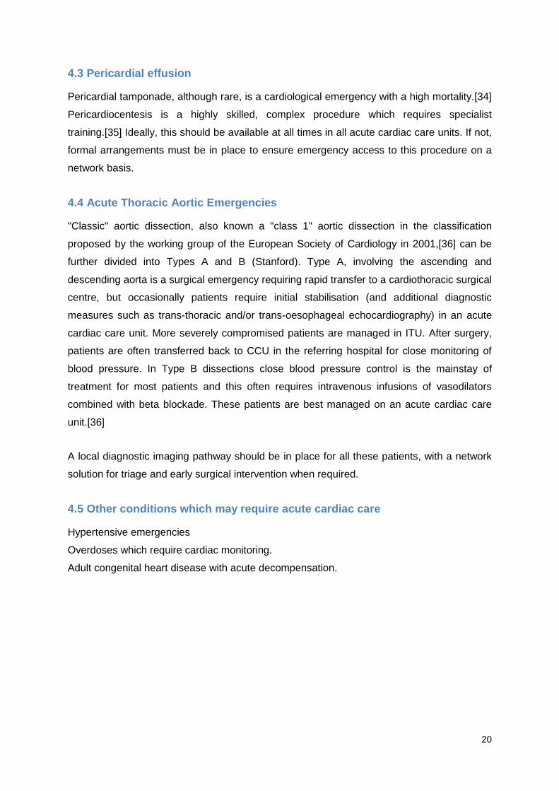
4.3 Pericardial effusion
Pericardial tamponade, although rare, is a cardiological emergency with a high mortality.[34]
Pericardiocentesis is a highly skilled, complex procedure which requires specialist
training.[35] Ideally, this should be available at all times in all acute cardiac care units. If not,
formal arrangements must be in place to ensure emergency access to this procedure on a
network basis.
4.4 Acute Thoracic Aortic Emergencies
"Classic" aortic dissection, also known a "class 1" aortic dissection in the classification
proposed by the working group of the European Society of Cardiology in 2001,[36] can be
further divided into Types A and B (Stanford). Type A, involving the ascending and
descending aorta is a surgical emergency requiring rapid transfer to a cardiothoracic surgical
centre, but occasionally patients require initial stabilisation (and additional diagnostic
measures such as trans-thoracic and/or trans-oesophageal echocardiography) in an acute
cardiac care unit. More severely compromised patients are managed in ITU. After surgery,
patients are often transferred back to CCU in the referring hospital for close monitoring of
blood pressure. In Type B dissections close blood pressure control is the mainstay of
treatment for most patients and this often requires intravenous infusions of vasodilators
combined with beta blockade. These patients are best managed on an acute cardiac care
unit.[36]
A local diagnostic imaging pathway should be in place for all these patients, with a network
solution for triage and early surgical intervention when required.
4.5 Other conditions which may require acute cardiac care
Hypertensive emergencies
Overdoses which require cardiac monitoring.
Adult congenital heart disease with acute decompensation.

21
5. Requirements for an Acute Cardiac Care Unit
The successful operation of an Acute Cardiac Care Unit as envisaged by the British
Cardiovascular Society relies on appropriate provision of beds, monitoring facilities, staffing
levels and access to expertise (medical, nursing and technical) and diagnostics. Precise
requirements for staffing and organisation of Acute Cardiac Care Units were outlined by the
Working Group on Acute Cardiac Care of the European Society of Cardiology. Suggested
bed numbers for CCUs in the UK were also included in the publication by the British Cardiac
Society and Royal College of Physicians in 2005.[37] The size of the unit not only depends
on the catchment area of the hospital in question but also the availability of other high-
dependency units within the hospital. Appropriate facilities for invasive and non-invasive
monitoring are mandatory, as well as 24/7 availability of diagnostic tests and this is covered
in detail below. Close liaison with ITU outreach or ALERT teams is appropriate for more
unstable patients requiring circulatory and/or respiratory support in a critical care level 2-3
facility.
5.1 Size and Provision of Acute Cardiac Care Units
Although the majority of UK hospitals have an acute cardiac care unit in some form (208/221
in 2006 with median 6 beds), most are ‘stand-alone’ units not associated with a larger
cardiac facility. With current rates of ACS, many hospitals will be unable to provide CCU
care for all such patients, even if STEMI patients are triaged to neighbouring centres for
PPCI. Therefore, in order to adequately care for ACS patients alone, most hospitals would
require larger units. Centres undertaking PPCI face the additional problem of having to
accommodate patients referred in from around a network, whether they are managed on a
treat and return basis or treat and keep. The need to transfer such patients rapidly to and
from the catheter labs produces a particular strain on CCU beds and staff.
The proposed shift in emphasis to the provision of all acute cardiological care in a single
area, with improved access to monitoring, investigation and specialist expertise, will require
considerable expansion of existing CCUs. Such a goal may, in part, be achieved by the
reorganisation of existing cardiac beds, but if patients currently managed on general medical
wards are to receive specialist cardiac care it is likely that further reorganisation of bed stock
will be required in many cases. Increasing the level of care available for these beds or ward
areas may not be seen as an attractive option in the straitened circumstances of the current
NHS, however based on the measurable differences in outcomes highlighted between

22
‘specialised’ and ‘non-specialised’ cardiac care in the MINAP dataset and the National Heart
Failure Audit, there are clear benefits to be achieved.
It should also be noted that the acute cardiac care unit is not the appropriate place to
manage high dependency general medical patients from other specialities with primarily non-
cardiac complaints, for instance sepsis or impending hypovolaemic shock, for which a
separate medical HDU facility is required. All Acute Cardiac Care Units should have
protocols in place to call on additional specialist expertise for procedures such as
haemofiltration and invasive ventilation that may require transfer to an intensive care unit.
5.2 Medical Staffing
The shift towards more patients with acute cardiac conditions being managed by
cardiologists will inevitably require an increase in the consultant workforce. In 2002, the Fifth
Report on the provision of services for patients with heart disease stated that in order to
provide a full cardiology on call rota, all District General Hospitals should have at least 5
cardiologists and Tertiary Centres double this figure.[37] It suggested a minimum of one
cardiologist per 50,000 population, which in 2002 equated to 1194 cardiologists in the UK,
but also noted that this number would need to rise to 1500 cardiologists by 2010 due to the
increasing workload, complexity of practice and patient expectation. Over recent years the
consultant workforce has been expanding by about 5% per year and there are currently
about 1200 cardiologists in the UK. This expansion will need to continue if consultant-led
specialist care is to be delivered to patients with acute cardiac problems. Numbers of
specialist trainees are currently relatively static, and the Centre for Workforce Intelligence
believes that the number of trainees is broadly in balance with expected availability of
consultant posts.
5.3 Nursing and Workforce
In tertiary centres and those providing a PPCI service there is increasing focus on the need
for skilled personnel to manage people presenting with STEMI. At one time the remit of the
coronary care unit team alone, these patients are now managed across the pathway by a
multi-professional group which will include cardiac physiologists, nurses, and radiographers
as well as medical teams (cardiac and critical care). For this reason close working and
integration of cardiac catheter laboratory and coronary care teams is a model that appears to
serve exceptionally well, with protocols in place for rapid access to critical care when
required. Additional skills are required by the nursing staff on acute cardiac care units
dealing with PPCI (and PCI in general which has also expanded rapidly into DGHs), for

23
example to recognise cardiac tamponade after wire exit, retroperitoneal haemorrhage or
acute stent thrombosis. Many DGHs which are not designated PPCI Centres will continue to
provide rapid access to PCI for the larger NSTEACS population.
5.4 Staff Training and Skill mix
Increasingly complex patients in need of increasingly complex intervention require a
healthcare team with a highly developed and developing skill base to provide immediate
care, to facilitate patients' future self management and to refer appropriately to other
specialist groups both within and outside the hospital environment to support long term care.
Training in techniques such as IABP counterpulsation, CPAP, temporary pacing and
invasive monitoring are required, as is full Advanced Life Support Training. Strong clinical
leadership for the nursing team is essential, as is a local programme of continuing
professional development to supplement any specialist professional qualifications.
In 2010 the British Association for Critical Care Nursing (BACCN) reviewed the evidence that
suggests that a rich nursing skill mix reduces adverse events [38] and there is also evidence
to suggest a link between the number of nurses, patient mortality and length of stay.[39] The
BACCN standard for nursing in a critical care environment such as an Acute Cardiac Care
Unit is that the nurse patient ratio should not fall below one nurse to two patients.[38] This
view is echoed by the ESC [19] who recommend that a cardiac care unit employ only
registered nurses, with at least 75% having training in cardiac care.
The current context of acute cardiac care in the UK is such that there is no diurnal variation
in caseload; a patient in crisis is in need of intervention and specialist support 24 hours a
day. There is evidence that patients admitted overnight have a higher risk of mortality [40,
41] and again there is an association between staffing levels and patient outcomes. In one
study patients in an environment with higher levels of registered nurse staffing were less
likely to die whilst in hospital [42]. In addition where there is a need to staff other areas (e.g.
an angiography suite) or where there is a ‘cardiac outreach’ service to support the
management of ACS, STEMI or acutely ill cardiac patients in other areas, staffing levels
should be organised to reflect the additional demands on staffing. The BCS do not therefore
support any variation in staffing levels between day and night in an acute cardiac care unit.
Where ACS or Heart Failure patients or people with complex arrhythmias and devices are
admitted via medical ‘take’ there should be a clear and rapid route of referral to the
cardiology service. The role of the specialist nurse in supporting early referral and expediting
intervention or management for these patients is fundamental to this process.

24
5.5 Diagnostic requirements for Acute Cardiac Care (2011-2020)
The increasing role of front end diagnostics in the rapid triage of cardiovascular patients
presenting acutely, must form part of the strategy for forward planning of acute cardiac care.
With the increase in focus on rapid hospital turnaround, traditional models of diagnostic
provision (e.g. in hours only services) are no longer acceptable. As cardiac units further
develop their role in the treatment of a broader range of acute cardiology presentations, the
need for access to the appropriate range of diagnostic tests at all times will increase.
Recognising that this development is evolutionary, some of the projections are aspirational
while others represent basic core requirements. In considering each technology, not just the
physical machinery but also the service requirements underpinning the provision of any
specific diagnostic test must be considered.
5.6 ECG based diagnostics
5.6.1 Electrocardiography
Electrocardiography is a basic tool in cardiovascular medicine; it should be available
immediately on every unit, performed by someone with formal training in ECG lead
placement and read by someone of suitable experience in interpretation. Likewise
continuous ECG monitoring is required for patients judged to be at risk of cardiac
arrhythmias after an acute cardiac presentation. The facility for central monitoring is an
important component of every CCU. The provision of telemetry elsewhere in the hospital
allows extended expert remote monitoring for patients whose primary problem may not be
cardiac but in whom cardiac complications are possible.
5.7 Imaging
5.7.1 X-ray Fluoroscopy
All acute cardiac units should ideally have access to emergency fluoroscopy for temporary
trans-venous pacing and positioning of IABPs at all times. The C-arm should be operated by
a radiographer experienced in its use and with the requirements for temporary pacing.
Where not available at present, a formal network solution must be in place to offer these
services.

25
5.7.2 Coronary Angiography Suite
Not all hospitals offering acute cardiac care services run a coronary angiography suite. ACS
patients managed in such units must be transferred to centres possessing the capability of
coronary angiography and PCI.
Some hospitals have access to coronary angiography facilities either in a dedicated cardiac
laboratory or a shared vascular cardiac facility, but without the capability of performing PCI.
Other hospitals may perform PCI during normal working hours only, covering out of hours for
complications. Some centres (or occasionally linked hospitals) provide a full PPCI service.
All hospitals should be part of a network protocol, whereby, where geographically
appropriate, patients with STEMI are rapidly diagnosed and transferred for PPCI. It should
be emphasised that where a patient has been admitted to a non-PPCI centre it is essential
for the ambulance service to provide an emergency transfer of the patient to the PPCI
centre. NSTEACS will continue to be initially managed in all acute hospitals, ideally on their
Acute Cardiac Care Unit, with transfer to an angioplasty centre where required for timely PCI
according to guidelines.[11,15]
5.7.3 Trans-thoracic Echocardiography (TTE)
The provision of cardiac ultrasound is fundamental to the diagnosis of many heart diseases
but in particular acute heart failure, suspected cardiac tamponade, complications post
myocardial infarction, acute valvular heart disease including endocarditis and acute disease
of the ascending aorta. [15,33,36,43]
TTE of suitable quality should be available to all patients who require it. Where this is not
currently achievable and in particular where patients may require urgent or emergent out of
hours scanning a formal network protocol for emergency echocardiography must be in place.
Out of hours TTE should not be undertaken by junior members of the medical team unless
they have British Society of Echocardiography (BSE) accreditation, have undergone a
documented locally approved competency assessment or can have the images rapidly
reviewed by someone with the appropriate expertise.
TTE should always be performed to an acceptable standard using a high quality ultrasound
machine and within a departmental structure that maintains quality control. BSE personal
and departmental accreditation is a benchmark of quality which encompasses all these
factors.[44] An acute cardiac care unit should only operate where there is an
26
echocardiography service that is either BSE accredited or meets the principle standards of
BSE / EAE[45] or similar accreditation.
Where full TTE cannot be performed, in patients with acute circulatory collapse, there is
evolving interest in limited echocardiograms called “FOCUS” (Focused Cardiac Ultrasound
in Emergency Setting).[46] Whilst the BSE provides a clear lead in the importance of
performing a full echocardiographic assessment, a number of groups have developed
programs which promote a variety of approaches to performing more limited studies. There
are strict limitations to the scope of such studies which are only suitable for identification of
severe LV dysfunction, large pericardial or pleural effusion and right ventricular dilatation
(PE). Where FOCUS studies are undertaken they should always be incorporated into the
quality assurance program of the echocardiography department.
FOCUS should not replace standard echocardiography and should be seen as a means of
facilitating fast decision making in the acute setting. Patients undergoing this form of
echocardiography should generally undergo standard trans-thoracic echocardiography as
soon as practical.
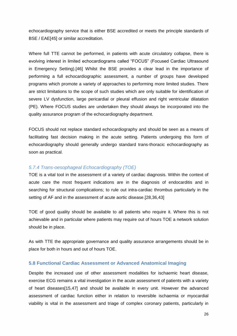
5.7.4 Trans-oesophageal Echocardiography (TOE)
TOE is a vital tool in the assessment of a variety of cardiac diagnosis. Within the context of
acute care the most frequent indications are in the diagnosis of endocarditis and in
searching for structural complications; to rule out intra-cardiac thrombus particularly in the
setting of AF and in the assessment of acute aortic disease.[28,36,43]
TOE of good quality should be available to all patients who require it. Where this is not
achievable and in particular where patients may require out of hours TOE a network solution
should be in place.
As with TTE the appropriate governance and quality assurance arrangements should be in
place for both in hours and out of hours TOE.
5.8 Functional Cardiac Assessment or Advanced Anatomical Imaging
Despite the increased use of other assessment modalities for ischaemic heart disease,
exercise ECG remains a vital investigation in the acute assessment of patients with a variety
of heart diseases[15,47] and should be available in every unit. However the advanced
assessment of cardiac function either in relation to reversible ischaemia or myocardial
viability is vital in the assessment and triage of complex coronary patients, particularly in

27
those with complex presentations. Local expertise varies between hospitals but a modern
cardiac assessment unit would be expected to have access to stress echocardiography,
nuclear perfusion imaging or cardiac MRI and preferably more than one of these modalities.
Cardiac gated CT scanning is an emerging technology. Its main use is currently in the
diagnosis of chronic chest pain and particularly in the exclusion of CAD in low risk
individuals.[47] There is also an increasing data set supporting the use of CT as an acute
triage tool but this has not yet entered guidelines in the UK.[48]
5.9 Cardiac Device Management
With the increasing use and complexity of cardiac devices it is important that all units are
able to manage device complications out of hours and program and interrogate devices in
hours. Where an out of hours service is not available a formal network solution should be in
place.
1) Brady-pacing. To check pacing and sensing (thresholds, under and over-sensing).
Detect arrhythmic events from pacemaker recordings.
2) AICD. Override the device in the appropriate clinical context (e.g. VT storm)
Diagnose inappropriate shocks, to check pacing and sensing (both thresholds, under and
over-sensing), deactivate the device in the case of terminal care or dying patients. To
deliver an emergency shock via the device
3) Biventricular devices. To check pacing and sensing (both thresholds under and
over-sensing). To deactivate Coronary Sinus lead in the case of significant diaphragmatic
pacing
4) Implantable Loop Recorders. Device interrogation
5.10 Electrophysiological studies and RF ablation
Electrophysiological study and ablation are becoming a major component of the
management of acute arrhythmias. At present they remain specialist services and as such
fall outside the expectations of a general acute cardiac care unit. Protocols should be in
place for the emergency transfer of patients to a unit providing EP/RF.
5.11 Laboratory based diagnostics
The use of biomarkers is now a vital component in the diagnosis of NSTEACS[15] and
increasingly heart failure.[25] All acute cardiac care units should have access to urgent
troponin assessment, whether this be troponin I, troponin T or high sensitivity Troponin

28
T.[49] These biomarkers should be provided by a locally agreed protocol and with an
acceptable margin of error.
Likewise serum natriuretic peptides (BNP or NTpro-terminal BNP) are increasingly
important, and there is good evidence for their use in triage for patients admitted with
suspected heart failure.[25,50]
Where near patient testing is integrated into care, rather than laboratory tests, it should be
with appropriate evidence of accuracy and regular calibration.
29
Conclusions: Acute Cardiac Care Unit rather than Coronary Care Unit
Patients presenting with cardiac conditions managed in specialised cardiac
wards have demonstrably better outcomes.
A significant proportion of these patients are not currently managed within a
cardiac service, leading to a greater morbidity and mortality, and cost to the
NHS.
Patients presenting with acute cardiac conditions should be managed by a
specialist, multi-disciplinary cardiac team and have access to key cardiac
investigations and interventions, at all times.
All hospitals admitting unselected acute medical patients should have an
appropriately sized, staffed and equipped Acute Cardiac Care Unit, where
high risk patients with a primary cardiac diagnosis should be managed.
All high risk cardiac patients must have access to Acute Cardiac Care Units,
and access should not be restricted to patients with ACS.
30
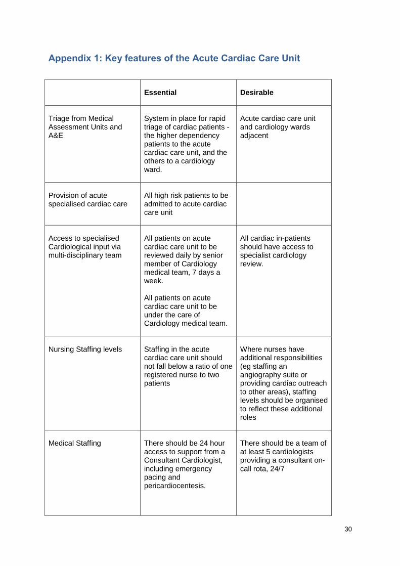
Appendix 1: Key features of the Acute Cardiac Care Unit
Essential
Desirable
Triage from Medical Assessment Units and A&E
System in place for rapid triage of cardiac patients - the higher dependency patients to the acute cardiac care unit, and the others to a cardiology ward.
Acute cardiac care unit and cardiology wards adjacent
Provision of acute specialised cardiac care
All high risk patients to be admitted to acute cardiac care unit
Access to specialised Cardiological input via multi-disciplinary team
All patients on acute cardiac care unit to be reviewed daily by senior member of Cardiology medical team, 7 days a week. All patients on acute cardiac care unit to be under the care of Cardiology medical team.
All cardiac in-patients should have access to specialist cardiology review.
Nursing Staffing levels
Staffing in the acute cardiac care unit should not fall below a ratio of one registered nurse to two patients
Where nurses have additional responsibilities (eg staffing an angiography suite or providing cardiac outreach to other areas), staffing levels should be organised to reflect these additional roles
Medical Staffing
There should be 24 hour access to support from a Consultant Cardiologist, including emergency pacing and pericardiocentesis.
There should be a team of at least 5 cardiologists providing a consultant on-call rota, 24/7

31
Essential
Desirable
Education
Trusts must provide an ongoing programme of training incorporating medical, nursing and cardiac physiology teams.
Diagnostics
24/7 access to CXR, trans-thoracic echocardiography and CT/TOE
Audit
Participation in mandatory National Audits
Research
Contributing to the activities of a research network

32
References
1 Mending Hearts and Brains. Clinical case for change. A report by Professor Roger Boyle, National Director for Heart Disease and Stroke http://www.dh.gov.uk/prod_consum_dh/groups/dh_digitalassets/documents/digitalasset/dh_072493.pdf 2 Mills NL, Churchhouse AM, Lee KK, Anand A, Gamble D, Shah AS, Paterson E, MacLeod M, Graham C, Walker S, Denvir MA, Fox KA, Newby DE. Implementation of a sensitive troponin I assay and risk of recurrent myocardial infarction and death in patients with suspected acute coronary syndrome. JAMA 2011;305(12):1210-6.
3. Smith CR, Leon MB, Mack MJ, Miller DC, Moses JW, Svensson LG, Tuzcu EM, Webb JG, Fontana GP, Makkar RR, Williams M, Dewey T, Kapadia S, Babaliaros V, Thourani VH, Corso P, Pichard AD, Bavaria JE, Herrmann HC, Akin JJ, Anderson WN, Wang D, Pocock SJ; PARTNER Trial Investigators. Transcatheter versus surgical aortic-valve replacement in high-risk patients. N Engl J Med. 2011 ;364(23):2187-98.
4. Hardoon SL, Whincup PH, Lennon LT, Wannamethee SG, Capewell S, Morris RW. How much of the recent decline in the incidence of myocardial infarction in British men can be explained by changes in cardiovascular risk factors? Evidence from a prospective population-based study. Circulation 2008;117(5):598-604.
5. MINAP Steering Group. Myocardial Ischaemia National Audit Project (MINAP) – How the NHS cares for patients with heart attack. Ninth Public Report 2010. http://www.rcplondon.ac.uk/sites/default/files/minap-public-report-sept-2010.pdf
6. The National Heart Failure Audit, April 2009 – March 2010. British Society for Heart Failure. http://www.ic.nhs.uk/webfiles/publications/002_Audits/NHS_IC_National_Heart_Failure_Audit_2010_04-01-11.pdf
7. Birkhead JS, Weston C, Lowe D. Impact of specialty of admitting physician and type of hospital on care and outcome for myocardial infarction in England and Wales during 2004-5: observational study. BMJ. 2006;332(7553):1306-11.
8. McMullan R, Silke B, Bennett K, Callachand S. Resource utilisation, length of hospital stay, and pattern of investigation during acute medical hospital admission. Postgrad Med J. 2004;80 (939):23-6.
9. Birkhead JS. From coronary care unit to acute cardiac care unit – the evolving role of specialist cardiac care. Personal communication based on MINAP data 2008-9. 10. Department of Health Vascular Programme Team. Treatment of Heart Attack: National Guidance. Final report of the National Infarct Angioplasty Project (NIAP) 2008. http://www.dh.gov.uk/en/Publicationsandstatistics/Publications/PublicationsPolicyAndGuidance/DH_083061 11. The Task Force on Myocardial Revascularization of the European Society of Cardiology (ESC) and the European Association for Cardio-Thoracic Surgery (EACTS). Guidelines on myocardial revascularization. Eur Heart J 2010; 31: 2501-55. 12. De Luca G, Suryapranata H, van 't Hof AW et al. Prognostic assessment of patients with acute myocardial infarction treated with primary angioplasty: implications for early discharge. Circulation 2004; 109: 2737-43. 13. Gershlick AH, Stephens-Lloyd A, Hughes S et al. Rescue angioplasty after failed thrombolytic therapy for acute myocardial infarction. N Engl J Med 2005; 353: 2758-68.

33
14. Motherwell DW, Rogers J, Kellagher M, Craig D, O’Reilly DS, Cobbe SM. The introduction of a chest pain nurse and fast-track troponin service reduces the length of stay of patients presenting with chest pain. Scott Med J 2007; 52: 6-9. 15. NICE Guideline CG94: Unstable angina and NSTEMI. The early management of unstable angina and non-ST-elevation myocardial infarction. 2010. www.nice.org.uk/guidance/CG94 16. Pieper KS, Gore JM, FitzGerald G, Granger CB, Goldberg RJ, Steg G, Eagle KA, Anderson FA, Budaj A, Fox KA; Global Registry of Acute Coronary Events (GRACE) Investigators. Validity of a risk-prediction tool for hospital mortality: the Global Registry of Acute Coronary Events. Am Heart J. 2009;157:1097-105. 17. Aragam KG, Tamhane UU, Kline-Rogers E, Li J, Fox KA, Goodman SG, Eagle KA, Gurm HS. Does simplicity compromise accuracy in ACS risk prediction? A retrospective analysis of the TIMI and GRACE risk scores. PLoS One. 2009;4:e7947. 18. Fox KAA, McLean S. Nice guidance on the investigation of chest pain. Heart 2010; 96: 903-6. 19. Hasin Y Danchin N Filippatos G et al (2005) Recommendations for the structure organisation and operation of intensive cardiac care units. Europ Heart J Vol. 26 pp.1676-1682 20. Cowie MR, Wood DA, Coats AJ, Thompson SG, Poole-Wilson PA, Suresh V, Sutton GC. Incidence and aetiology of heart failure; a population-based study. Eur Heart J. 1999 Mar;20(6):421-8 21. Data from Hospital Episode Statistics Online www.hesonline.nhs.uk [accessed 13/6/2011] 22. Dargie HJ et al. British Society for Heart Failure Annual Scientific Sessions 2009. 23. Nieminen MS, Brutsaert D, Dickstein K, Drexler H, Follath F, Harjola VP, Hochadel M, Komajda M, Lassus J, Lopez-Sendon JL, Ponikowski P, Tavazzi L. EuroHeart Failure Survey II: a survey on hospitalized acute heart failure patients: description of population. Eur Heart J. 2006 Nov;27(22):2725-36. 24. Mehta PA, Dubrey SW, McIntyre HF, Walker DM, Hardman SM, Sutton GC, McDonagh TA, Cowie MR. (2009) Improving survival in the 6 months after diagnosis of heart failure in the past decade: population-based data from the UK. Heart 95: 1851-1856 25. NICE Guideline CG108 Chronic Heart Failure: management of chronic heart failure in adults in primary and secondary care. www.nice.org.uk/guidance/CG108 26. McAlister FA, Lawson FM, Teo KK, Armstrong PW. A systematic review of randomized trials of disease management programs in heart failure. Am J Med (2001); 110: 378-384 27. Cleland JGF, McDonagh T, Rigby AS, Yassin A, Whittaker T, Dargie HJ. The national heart failure audit for England and Wales 2008-2009. Heart 2011 (11): 876-886 28. Camm AJ, Kirchhof P, Lip GYH, Schotten U, Savelieva I, Ernst S, Van Gelder IC, Al-Attar N, Hindricks G, Prendergast B, Heidbuchel H, Alfieri O, Angelini A, Atar D, Colonna P, De Catarina R, De Sutter J, Goette A, Gorenek B, Heldal M, Hohloser SH, Kolh P, Le Heuzey J-Y, Ponikowski P, Rutten FH. Guidelines for the management of atrial fibrillation. Europ Heart J (2010); 31: 2369-2429 29. Stewart S, Hart CL, Hole DJ, McMurray JJ. A population based study of the long-term risks associated with atrial fibrillation: 20-year follow up of the Renfrew/Paisley study. Am J Med 2002; 113: 359-364 30. Kirchhof P, Nabauer M, Gerth A, Limbourg T, Lewalter T, Goette A, Wegscheider K, Trezl A, Meinertz T, Oeff M, Ravens U, Breithardt G, Steinbeck G Impact of the type of centre on management of AF patients: Surprising evidence for differences in antithrombotic decisions. Thromb. Haemost. 2011; 105: [Epub ahead of print]

34
31. Murphy JJ. Problems with temporary pacing. Expecting trainees in medicine to perform transvenous pacing is no longer acceptable.BMJ 2001; 323: 527 32. Torbicki, A, Perrier A, Konstantinides S , Agnelli G, Galie N, Pruszczyk P, Bengel F, Brady AJB, Ferreira D, Janssens U, Klepetko W, Mayer E, Remy-Jardin M, Bassand J-P. Guidelines on the diagnosis and management of acute pulmonary embolism. Europ. Heart J (2008) 29, 2276–2315 33. Botelho-Nevers E, Thuny F, Casalta JP, Richet H, Gouriet F, Collart F, Riberi A, Habib G, Raoult D. Dramatic reduction in infective endocarditis-related mortality with a management-based approach. Arch Intern Med. 2009;169(14):1290-8. 34. Chong HH, Plotnick GD. Pericardial effusion and tamponade: evaluation, imaging modalities, and management. Compr. Ther.1995;21:378–85. 35. Maisch B, Seferovic P, Ristic AD, Erbel R, Rienmüller R, Adler Y, Tomkowski WZ, Thiene G, Yacoub MH Guidelines on the Diagnosis and Management of Pericardial Diseases. Europ. Heart J 2004;25:587-610 36. Erbel R, Alfonso F, Boileau C, Dirsch O, Eber B, Haverich A, Rakowski H, Struyven J, Radegran K, Sechtem U, Taylor J, Zollikofer C. Diagnosis and management of aortic dissection. Eur Heart J (2001) 22, 1642–1681
37. Fifth report on the provision of services for patients with heart disease. Heart 2002;88:Supp iii1-56
38. British Association for Critical Care Nursing (BACCN). Standards for nurse staffing in critical care 2010 http://www.baccn.org.uk/downloads/BACCN_Staffing_Standards.pdf 39. Needleman J, Buerhaus P, Pankratz VS, Leibson CL, Stevens SR, Harris M. Nurse staffing and inpatient hospital mortality. N Engl J Med. 2011;364(11):1037-45.
40. Hilson S, Bryan D, Rich E and Luxenberg M (1992) Call nights and patient care: effects on inpatients at one teaching hospital Journal of Internal Medicine Vol. 7 pp. 405-410 41. Deakin C, Nolan J, Soar J, Sunde K, Koster R, Smith G and Perkins G (2010) European Resuscitation Guidelines for Resuscitation Section 4: Adult Advanced Life Support Resuscitation Vol. 81 pp. 1305-1352 42. Person S; Allison J, Weaver M, Williams O, Centor R and Weissman N (2004) Nurse staffing and mortality for medicare patients with acute myocardial infarction Medical Care Vol. 2 pp.4-12 43. Habib G, Hoen B, Tornos P, Thuny F, Prendergast B, Vilacosta I, Moreillon P, de Jesus Antunes M, Thilen U, Lekakis J, Lengyel M, Müller L, Naber CK, Nihoyannopoulos P, Moritz A, Zamorano JL; ESC Committee for Practice Guidelines. Guidelines on the prevention, diagnosis, and treatment of infective endocarditis (new version 2009): the Task Force on the Prevention, Diagnosis, and Treatment of Infective Endocarditis of the European Society of Cardiology (ESC. Eur Heart J. 2009 Oct;30(19):2369-413. 44. British Society of Cardiology. Personal and Departmental Accreditation in echocardiography. www.bsecho.org 45. European Society of Cardiology, Echocardiography Association. Personal and Departmental Accreditation. http://www.escardio.org/communities/EAE/accreditation/Pages/welcome.aspx
46. Labovitz AJ, Noble VE, Bierig M, Goldstein SA, Jones R, Kort S, Porter TR, Spencer KT, Tayal VS, Wei K. Focused cardiac ultrasound in the emergent setting: a consensus statement of the American Society of Echocardiography and American College of Emergency Physicians. J Am Soc Echocardiogr. 2010 Dec;23(12):1225-30.
47. NICE Guideline CG 95.2010 Chest Pain of recent onset. http://guidance.nice.org.uk/CG95

35
48. Hoffmann U, Bamberg F, Chae CU, Nichols JH, Rogers IS, Seneviratne SK, Truong QA, Cury RC, Abbara S, Shapiro MD, Moloo J, Butler J, Ferencik M, Lee H, Jang IK, Parry BA, Brown DF, Udelson JE, Achenbach S, Brady TJ, Nagurney JT. Coronary computed tomography angiography for early triage of patients with acute chest pain: the ROMICAT (Rule Out Myocardial Infarction using Computer Assisted Tomography) trial. J Am Coll Cardiol. 2009 May 5;53(18):1642-50.
49. Jaffe AS. The 10 commandments of troponin, with special reference to high sensitivity assays.
Heart. 2011 Jun;97(11):940-6.
50. Dickstein K, Cohen-Solal A, Filippatos G, McMurray JJ, Ponikowski P, Poole-Wilson PA, Strömberg A, van Veldhuisen DJ, Atar D, Hoes AW, Keren A, Mebazaa A, Nieminen M, Priori SG, Swedberg K; ESC Committee for Practice Guidelines, Vahanian A, Camm J, De Caterina R, Dean V, Dickstein K, Filippatos G, Funck-Brentano C, Hellemans I, Kristensen SD, McGregor K, Sechtem U, Silber S, Tendera M, Widimsky P, Zamorano JL. ESC Guidelines for the diagnosis and treatment of acute and chronic heart failure 2008. Eur Heart J. 2008 Oct;29(19):2388-442.